Embed Size (px)
Citation preview

How To Interpret Breast MRIKathryn Zamora, MD, MPH
University of Alabama Birmingham
Rebecca Leddy, MDMedical University of South Carolina

Indications for MRI
• Screening• High‐risk• Intermediate risk (may be considered)• Occult contralateral breast malignancy in the setting of newly diagnosed breast cancer (3‐5%)• Breast augmentation
• Extent of Disease• Multifocality and multicentricity• Invasion of deep fascia• Lumpectomy with positive margins• Neoadjuvant therapy
• Additional evaluation of clinical or imaging findings• Recurrence of breast cancer
• Including patients with reconstruction
• Metastasis of Unknown primary • Lesions characterization

Organized and Systematic Approach
•Prior to interpretation review the following•Clinical History•MRI Technique•Previous Exams
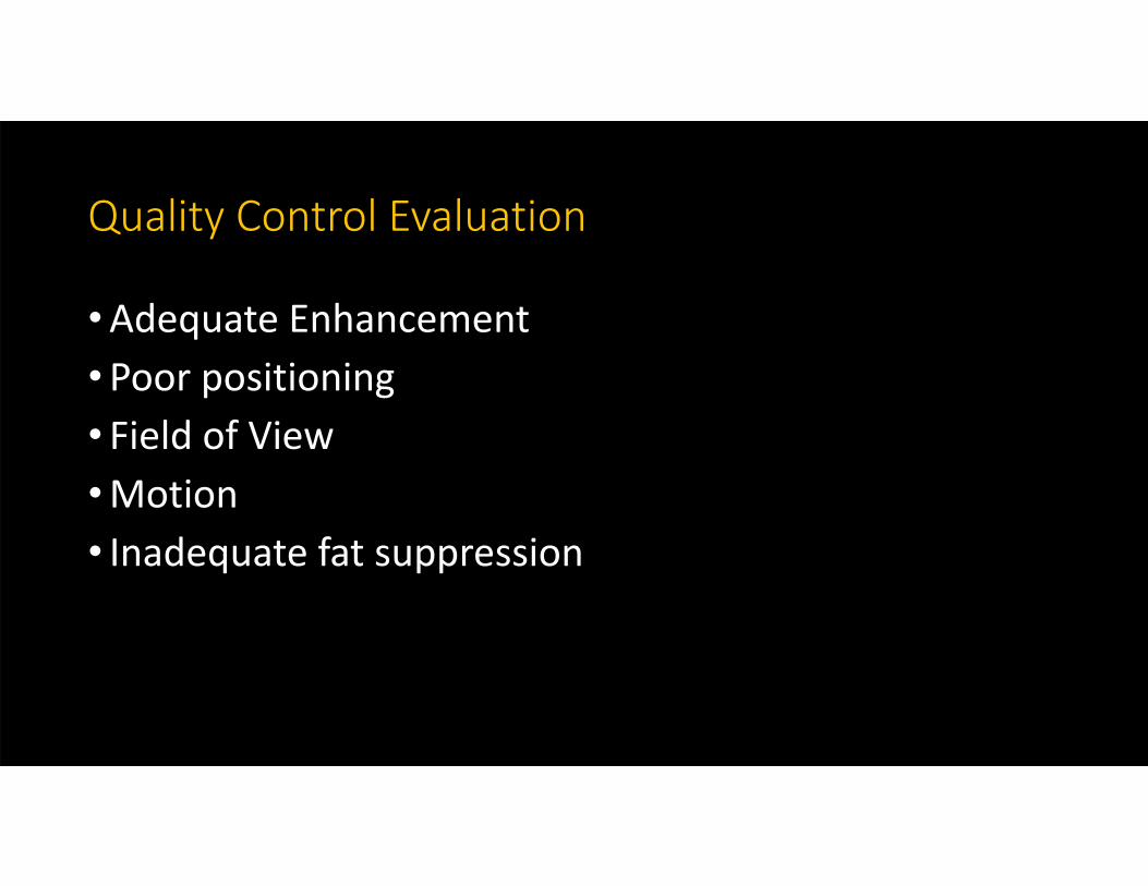
Quality Control Evaluation
•Adequate Enhancement•Poor positioning• Field of View•Motion• Inadequate fat suppression
T1 SUBTRACTION
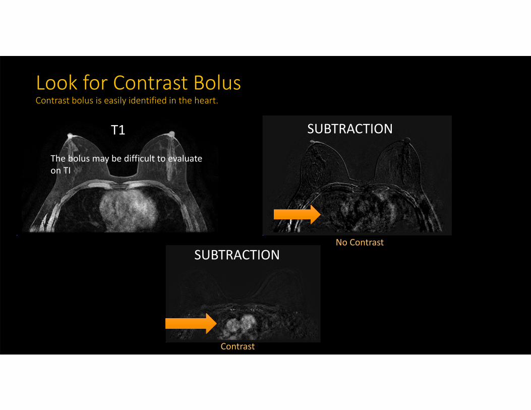
Look for Contrast BolusContrast bolus is easily identified in the heart.
No Contrast
Contrast
SUBTRACTION
The bolus may be difficult to evaluate on TI

Evaluate the Field of View
More superior images do not include the Axilla
Left upper outer quadrant IDC
Ultrasound demonstrates an abnormal level 1 axillary lymph node not imaged on the MRI
FOV is too small
?
?

Asses for MotionMotion may limit evaluation of subtle non mass enhancement and may cause mis‐registration artifact on subtraction images.
Mis‐registration artifact from T1 hyperintense material in the ducts may appear as non mass enhancement on the MIP and subtraction images.
Use T1 pre and post to confirm if enhancement is real
MIPT1 Pre Contrast FS Subtraction

Evaluate for Adequate Fat Saturation
Inadequate Fat Sat limits evaluation of the region.

Evaluate Background Parenchymal Enhancement (BPE)
• NOT related to breast density• Evaluate Maximum Intensity Projection (MIP) and 1st post contrast sequence
MINIMAL MODERATE MARKEDMILD

Image Interpretation
Sequences:• T2 weight imaging• T1 non fat sat images• Post Contrast • Maximum Intensity Projection (MIP)• Source images (not subtracted)• Subtracted
• axial and sagittal
• Kinetics• Correlate with mammogram and ultrasounds
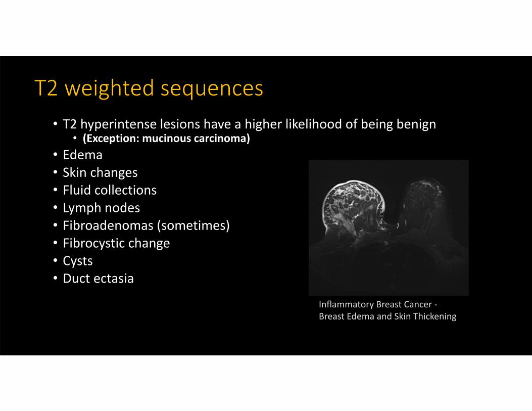
T2 weighted sequences• T2 hyperintense lesions have a higher likelihood of being benign
• (Exception: mucinous carcinoma)• Edema• Skin changes• Fluid collections • Lymph nodes• Fibroadenomas (sometimes)• Fibrocystic change• Cysts• Duct ectasia
Inflammatory Breast Cancer ‐Breast Edema and Skin Thickening

Dark Internal Septations
Fibroadenoma
T2 Subtraction
T2 Hyperintense mass

T2 Hyperintense Lesions
Cyst
Lymph Node
Post Operative Seroma
Mucinous Carcinomamay be T2 hyperintense
CAUTION
T2 T1 Postcontrast

T1 Non Fat Sat
• Asses for fat• Fat containing masses• Fat necrosis• Lymph nodes
• Evaluate margin and mass effect• Demonstrate susceptibility artifact (blood, clips)
T1 post contrast fat sat
Benign lymph node
T1 hyperintense fat
T1 non fat sat

T1 Non Fat Sat
Fat Necrosis
T1 post contrast fat sat T1 non fat satSubtraction
Hamartoma
T1 non fat satSubtraction

Susceptibility Artifact
clip
T1 non fat sat

Post Contrast Imaging• Review peak subtraction images
• Identify areas of enhancement• Determine if it is real (exclude artifact)
• Interrogate pre and post imaging• Location (quadrant, clock)• Characterize the type of enhancement• Associated features: Involvement skin, nipple, chest wall

Characterize Types of EnhancementMRI BIRADS Terminology: Types of enhancement (Mass versus Non‐mass enhancement)

Non‐mass enhancement
• Distribution• SEGMENTAL
• Internal Enhancement pattern• CLUMPED
MIP Subtraction

Non‐mass enhancement
• Distribution• FOCAL
• Internal Enhancement pattern• CLUSTERED RING
Axial SubtractionSagittal Subtraction

Mass
Sagittal Subtraction Sagittal MIP
• Shape• IRREGULAR
• Margins• NOT CIRCUMSCRIBED• IRREGULAR
• Internal Enhancement pattern• HOMOGENOUS

Multiple additional satellite massesTotal Extent of Disease
Maximum Intensity Projection MIP • Shape
• IRREGULAR
• Margins
• NOT CIRCUMSCRIBED
• IRREGULAR
• Internal Enhancement pattern
• HOMOGENOUS
Axial Subtraction

Further characterization of enhancement
• Analyze region of enhancement on other sequences• T1 non Fat Sat• T2 weighted images• T1 post contrast non subtracted (source)• Sagittal post delayed
• Size, distribution, position, shape
• Evaluate the delayed imaging• Especially important in neoadjuvant response to therapy examination• Source images (not subtracted):
• Evaluate musculature, axilla, skin, findings outside breast, nipple areolar complex• Pectoralis muscle involvement is not considered chest wall invasion
• Correlate with mammogram and ultrasounds
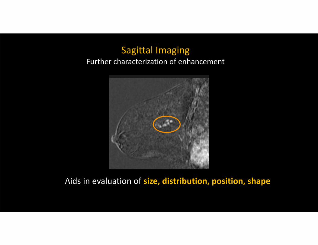
Sagittal ImagingFurther characterization of enhancement
Aids in evaluation of size, distribution, position, shape

Source (Non subtracted) Imaging
Pectoralis Muscle
• Evaluate involvement chest wall, musculature, skin, nipple areolar complex
• Axilla • Evaluate findings
outside breast
Chest wall
Pectoralis Muscle Invasion Chest Wall Invasion

Source (Non subtracted) Imaging
• Evaluate involvement chest wall, musculature, skin, nipple areolar complex
• Axilla • Evaluate findings
outside breast
Nipple areolar complex
T1 post contrast fat sat

Delayed Post Contrast ImagingResponse to Neoadjuvant Systemic Therapy
MIP early post contrast
Subtraction early post contrast
MIP delayed post contrast
Subtraction delayed post contrast
Delayed post contrast may assist in the detection of residual disease in patient undergoing neoadjuvantsystemic therapy

Evaluation of AxillaryLymph Nodes • Level I ‐ lateral to the lateral border of the pectoralis minor muscle• Level II ‐ between the medial and lateral borders of the pectoralis minor muscle and also include the interpectoral (Rotter’s) lymph nodes. • Level III ‐medial to the medial margin of the pectoralis minor muscle and inferior to the clavicle. LEVEL 3
LEVEL 2
LEVEL 1
Pectoralis Minor

Evaluation of Lymph Nodes
• Internal mammary chain lymph nodes• Changes the radiation field• 5mm or greater
Axial T1 post contrast fat sat
Sagittal T1 post contrast fat sat
Enlarged internal mammary chain lymph node

Evaluation of Areas Outside of Breast• Non breast finding• Review localizer images• T2• Source images (non subtracted)
Lung Cancer incidentally seen breast MRI
LOCALIZERT1 POST FSPhyllodes pulmonary metastasis

KINETIC Evaluation
• Evaluate LAST• May assist in final management• If suspicious finding identified on kinetics, then review• Possibly a benign lymph node• Area of fat necrosis

Kinetic Curves
• Type 1• Rapid rise and persistent• 6% change of malignancy
• Type 2• Rapid rise and plateau
• Type 3• Rapid rise and washout• 29‐77% chance of malignancy

Summary
• Apply an Organized and Systematic Approach• Review indication, history and prior exams• Evaluate Exam Quality• Determine Background Parenchymal Enhancement• Identify imaging findings and evaluate thoroughly on all sequences • Utilize kinetics • Remember to look outside of breast for any abnormalities