Embed Size (px)
Citation preview

Host restriction of murine gammaherpesvirus 68 replication by humanAPOBEC3 cytidine deaminases but not murine APOBEC3
Nana Minkah a, Kevin Chavez a, Parth Shah b, Thomas MacCarthy b, Hui Chen c,d,1,Nathaniel Landau c,d, Laurie T. Krug a,n
a Department of Molecular Genetics and Microbiology, Stony Brook University, Stony Brook, NY 11794, USAb Department of Applied Mathematics and Statistics, Stony Brook University, Stony Brook, NY 11794, USAc Department of Microbiology, NYU Langone Medical Center, New York, NY 10016, USAd Infectious Disease Laboratory, Salk Institute, La Jolla, CA 92037, USA
a r t i c l e i n f o
Article history:Received 31 October 2013Returned to author for revisions27 November 2013Accepted 20 February 2014Available online 13 March 2014
Keywords:HerpesvirusMurine gammaherpesvirusPathogenesisReplicationLatencyHost restrictionAPOBEC3Cytidine deaminase
a b s t r a c t
Humans encode seven APOBEC3 (A3A-A3H) cytidine deaminase proteins that differ in their expressionprofiles, preferred nucleotide recognition sequence and capacity for restriction of RNA and DNA viruses.We identified APOBEC3 hotspots in numerous herpesvirus genomes. To determine the impact of hostAPOBEC3 on herpesvirus biology in vivo, we examined whether murine APOBEC3 (mA3) restricts murinegammaherpesvirus 68 (MHV68). Viral replication was impaired by several human APOBEC3 proteins, butnot mA3, upon transfection of the viral genome. The restriction was abrogated upon mutation of the A3Aand A3B active sites. Interestingly, virus restriction by A3A, A3B, A3C, and A3DE was lost if the infectiousDNA was delivered by the virion. MHV68 pathogenesis, including lung replication and splenic latency,was not altered in mice lacking mA3. We infer that mA3 does not restrict wild type MHV68 andrestriction by human A3s may be limited in the herpesvirus replication process.
& 2014 Elsevier Inc. All rights reserved.
Introduction
Apolipoprotein B mRNA editing enzyme catalytic polypeptide-like editing complex 3 (APOBEC3) proteins belong to a family ofhost cytidine deaminases with a wide array of biological functions,subcellular localization and tissue expression profiles (Bransteitteret al., 2009; Smith et al., 2012). Eleven members of this enzymefamily have been discovered in humans; APOBEC1, APOBEC2,APOBEC3, APOBEC4 and activation induced deaminase (AID).APOBEC1 (A1), the founding member of this family, is an mRNAediting enzyme that deaminates Apolipoprotein B mRNA toproduce two isoforms of apolipoproteins that are required forlow-density lipid formation(Driscoll and Zhang, 1994; Mehta andDriscoll, 1998). APOBEC2 (A2) is a more recently discoveredmember detected predominantly in the heart and skeletal muscle(Mikl et al., 2005). Mice transgenic for human A2 develop lung andliver tumors (Okuyama et al., 2012). APOBEC4 (A4) was discovered
by computational searches for genes homologous to A1 and, likeA2, has no ascribed function. The most widely studied membersare AID, which is critical for generating antibody diversity throughssDNA deamination of the immunoglobulin locus, and APOBEC3(A3) proteins, which act as innate immune barriers to the replica-tion of viruses and endogenous retroelements (Cullen, 2006;Refsland and Harris, 2013).
APOBEC3 genes have undergone duplication events in thecourse of mammalian evolution. Murid rodents encode the singleAPOBEC3, mA3, while felines encode four APOBEC3s (Munk et al.,2008) and the equine genome encodes six (Bogerd et al., 2008).Primates have an expanded repertoire of seven APOBEC3 proteins;A3A, A3B, A3C, A3DE, A3F, A3G and A3H with each having slightlydifferent target sequences. A3s have been widely studied since theidentification of human A3G (hA3G) as a host restriction factor ofviral infectivity factor (Vif)-deficient HIV (Sheehy et al., 2002). InVif-deficient HIV infections, hA3G is packaged into viral coresthrough interactions with both the HIV nucleopcapsid protein andthe viral genomic RNA. A3G deaminates cytosine residues touracils in the newly synthesized strand. Vif enhances HIV replica-tion by binding to hA3G and either targeting it for degradation orpreventing packaging into the HIV core (Goila-Gaur and Strebel,2008; Zhang et al., 2003).
Contents lists available at ScienceDirect
journal homepage: www.elsevier.com/locate/yviro
Virology
http://dx.doi.org/10.1016/j.virol.2014.02.0220042-6822 & 2014 Elsevier Inc. All rights reserved.
n Correspondence to: Stony Brook University, Life Sciences Building, Room 130,Stony Brook, NY 11794-5222, USA.
E-mail address: [email protected] (L.T. Krug).1 Current address: Cornell Center for Technology Enterprise and Commercia-
lization, Cornell University, Ithaca, NY 14850, USA.
Virology 454-455 (2014) 215–226

Retroviruses, retroelements and more recently DNA viruses haveemerged as targets of hA3 proteins (Bishop et al., 2004; Delebecqueet al., 2006; Mahieux et al., 2005; Mangeat et al., 2003; Narvaizaet al., 2009; Stenglein et al., 2010; Suspene et al., 2005a, 2005b, 2004;Vartanian et al., 2008). Human A3A and A3C have been reported torestrict herpes simplex virus type 1 (HSV-1) replication in transientexpression studies (Suspene et al., 2011). Additionally, Suspene et al.(2011) reported the detection of virus variants consistent withcytidine deamination in clinical specimens positive for either HSV-1 or Epstein-Barr virus (EBV). However, in most cases the impact ofhA3 activity on the in vivo pathogenesis of a given virus in the hosthas not been extensively studied. The murine APOBEC3 exhibitsantiviral activity against multiple murine and human retroviral
targets (Langlois et al., 2009; Low et al., 2009; Okeoma et al., 2007;Takeda et al., 2008) implying that mA3 retains the retroviraltargeting function of the human A3. However, the antiviral functionof mA3 against herpesviruses is not known. In this report, weexamined the ability of mA3 to restrict murine gammaherpesvirus68 (MHV68), a natural pathogen of murid rodents that has stronggenetic colinearity with, and biological similarities to, the humangammaherpesviruses, Kaposi's sarcoma-associated herpesvirus(KSHV/HHV-8) and Epstein Barr Virus (EBV). We determined thedistribution of A3 hotspots in the MHV68 genome, queried the effectof A3 overexpression on viral replication and then ascertained theimpact of loss of A3 on in vivo pathogenesis. Although we observedrestriction of MHV68 replication with co-expression of several hA3s,
Fig. 1. APOBEC3 hotspots in herpesvirus genomes. (A) Percentage of 1 kb windows neutral, overrepresented or underrepresented for human and mouse APOBEC3 hotspotsin the indicated viral genome. (B) Heatmap of A3 hotspots in herpesvirus genomes. Scale indicates the percentage of 1 kb windows of the viral genome withoverrepresentation or underrepresentation of hotspots as described in (A). (C) Distribution of 1 kb regions of the MHV68 genome overrepresented (red bars) orunderrepresented (blue bars) for the indicated A3 hotspots. The boundary of each bar corresponds to the midpoint of the first and last 1 kb window represented within thebar. The gray trace represents the distribution of the hotspot occurrence in a region compared to the occurrence in 1000 randomly shuffled 1 kb genomic regions. The scale ofthe trace is from �0.5 to 0.5, the numbers on the y-axis indicate the distance from the mean of each distribution (y¼0). Genomic regions are significantly overrepresented(red bars) or underrepresented (blue bars) if they are above 0.45 or below �0.45 respectively.
N. Minkah et al. / Virology 454-455 (2014) 215–226216

the murine APOBEC3 did not alter MHV68 replication andpathogenesis.
Results
APOBEC3 hotspots are prevalent in herpesvirus genomes
In a single report outlining the impact of APOBEC3 cytidinedeaminase on herpesviruses, HSV-1 replication was reduced 4–10fold upon transient expression of hA3C. In addition, hypereditedHSV-1 and EBV sequences consistent with hA3 deamination weredetected in patient samples using a sensitive, but non-quantitativePCR detection method (Suspene et al., 2011). However, the naturaloccurrence of APOBEC3-mediated cytidine deamination and theconsequence of this selective pressure are not known. We utilizedan in silico approach to predict the distribution of APOBEC3 recogni-tion sites in herpesvirus genomes. The ‘hotspot’ or consensusrecognition sequence is TC (where the underlined C represents thetargeted cytidine) for hA3A, hA3B, hA3F and hA3H (Hultquist et al.,2011; Liddament et al., 2004; Senavirathne et al., 2012; Yu et al.,2004; Zheng et al., 2004) TTC for hA3C (Taylor et al., 2013), CCC forhA3G (Jern et al., 2009), and TCC for mA3 (MacMillan et al., 2013). Todetermine whether particular A3 hotspot motifs were over- orunder-represented throughout the viral genomes we measured thefrequency of each A3 hotspot motif (TC, TTC, CCC and TCC) within a1 kb sliding window across each genome at intervals of 100 nt. Theobserved frequencies in each window were then compared to a nullmodel based on randomly shuffled versions of that genomesequence, allowing us to statistically evaluate overrepresentationand underrepresentation.
Viruses that are susceptible to various A3 enzymes would likelyhave evolved to limit hotspot motifs in their genomes. Viruses thatdo not encounter certain A3 proteins or those with a viralcountermeasure might have an overrepresentation of motifs intheir genomes, especially if they have evolved under pressures toavoid a different A3 protein. For example, under-representation ofTTC (the motif for hA3C) will increase the relative frequency ofmotifs such as CCC (the motif for hA3G) assuming that there are noadditional selective pressures against the other motifs. We eval-uated the predictive ability of our model by examining theAPOBEC3 hotspot distribution in human papilloma virus 16(HPV16). HPV genomes are targets for deamination upon over-expression of hA3A, hA3C, and hA3H, and mutation signatures thatare consistent with deamination at TC hotspots are detected by asensitive PCR assay in clinical wart specimens (Vartanian et al., 2008).Human A3A, hA3B and hA3H are expressed in dermal epithelial cells,the preferred site of infection and viral replication of HPV (Madsenet al., 1999; OhAinle et al., 2006; Quinlan et al., 1984; Trivedi et al.,2006). Taken together, HPV is subjected to the selective pressures ofhA3s with TC hotspots (Vartanian et al., 2008). In agreement, ourcomputational analysis determined that TC hotspots were largelyunderrepresented in the HPV genome (Fig. 1A). This depletion of TChotspots in the genome is consistent with evasion from the antiviralpressures of hA3A, hA3B, hA3F or hA3H (A3A/A3B/A3F/A3H). More-over, the HPV genome had an underrepresentation of TTC hotspotsfavored by the ubiquitously expressed hA3C. In contrast, the HPVgenome was largely neutral with some enrichment for CCC hotspotsrecognized by hA3G, which is expressed predominantly in lympho-cytes, a cell type not known as a reservoir for HPV. Finally, the HPVgenome was predominately neutral with some occurrences of mA3hotspot-enriched regions. This is consistent with a human pathogenthat would not have been under selective pressure to limit mA3 sitesin the genome.
We next applied this analysis to multiple human herpesvirusesand MHV68. As depicted in Fig. 1A, we found that the genomes of
the alphaherpesviruses HSV-1 and VZV were enriched for A3Ghotspots. These genomes were largely neutral or even under-represented for A3A/A3B/A3F/A3H hotspots. Depletion of hotspotsfor A3A/A3B/A3H expressed at high levels in epithelial cells isconsistent with selective pressure to limit recognition of virus thatreplicates in the dermal epithelium, yet a finding of enrichment forA3G in the VZV genome was unexpected given its T lymphocyte-associated viremia (Zerboni et al., 2014). Within the betaherpes-viruses, the HCMV genome had regions overrepresented with A3A/A3B/A3F/A3H and A3C hotspots, but was largely underrepresentedfor A3G. The HHV-6A genome was also enriched for A3A/A3B/A3F/A3H and A3C hotspots. While the betaherpesviruses were largelydepleted or neutral for A3G, the genomes of the gammaherpes-viruses that are also lymphotropic were enriched for A3G hotspots.The EBV and KSHV gammaherpesvirus genomes were underrepre-sented for A3A/A3B/A3F/A3H hotspots. The MHV68 genome waslargely neutral for APOBEC3 hotspots with a higher representation ofhA3G and mA3 hotspots. With the exception of HCMV, mA3 siteswere overrepresented in each human herpesvirus genome analyzed.In Fig. 1B, the heatmap representation of the percentage of over-represented regions (left panel) and underrepresented regions (rightpanel) in the genomes of each human herpesvirus and MHV68illustrates similar trends in the profile of human andmouse APOBEC3hotspots among members within the subfamilies. Lastly, we exam-ined the distribution of hotspots in the MHV68 genome. With theexception of the internal repeats at nt positions 26,778–28,191 and98,981–101,170, the regions of overrepresented hotspots were evenlydistributed along the MHV68 genome (Fig. 1C). Taken together, ouranalysis predicts that some herpesvirus genomes are targets ofAPOBEC3 deamination.
Human APOBEC3 restriction of MHV68 replication
Given the identification of A3 hotspots in the MHV68 genome, wenext examined the impact of A3 expression on lytic replication. Weco-transfected A3 expression constructs with infectious MHV68þBAC DNA that encodes the YFP marker to monitor viral replication byfluorescent microscopy. Expression of hA3A limited the infection tosingle YFPþ cells (Fig. 2, top panel). In contrast, co-transfection of theempty vector or the A3A E72A active site mutant resulted in foci of10–20 YFPþ cells, indicating no impairment of virus spread. Expres-sion of wild type mA3 did not impair the formation of YFPþ foci inthe infection (Fig. 2, bottom panel).
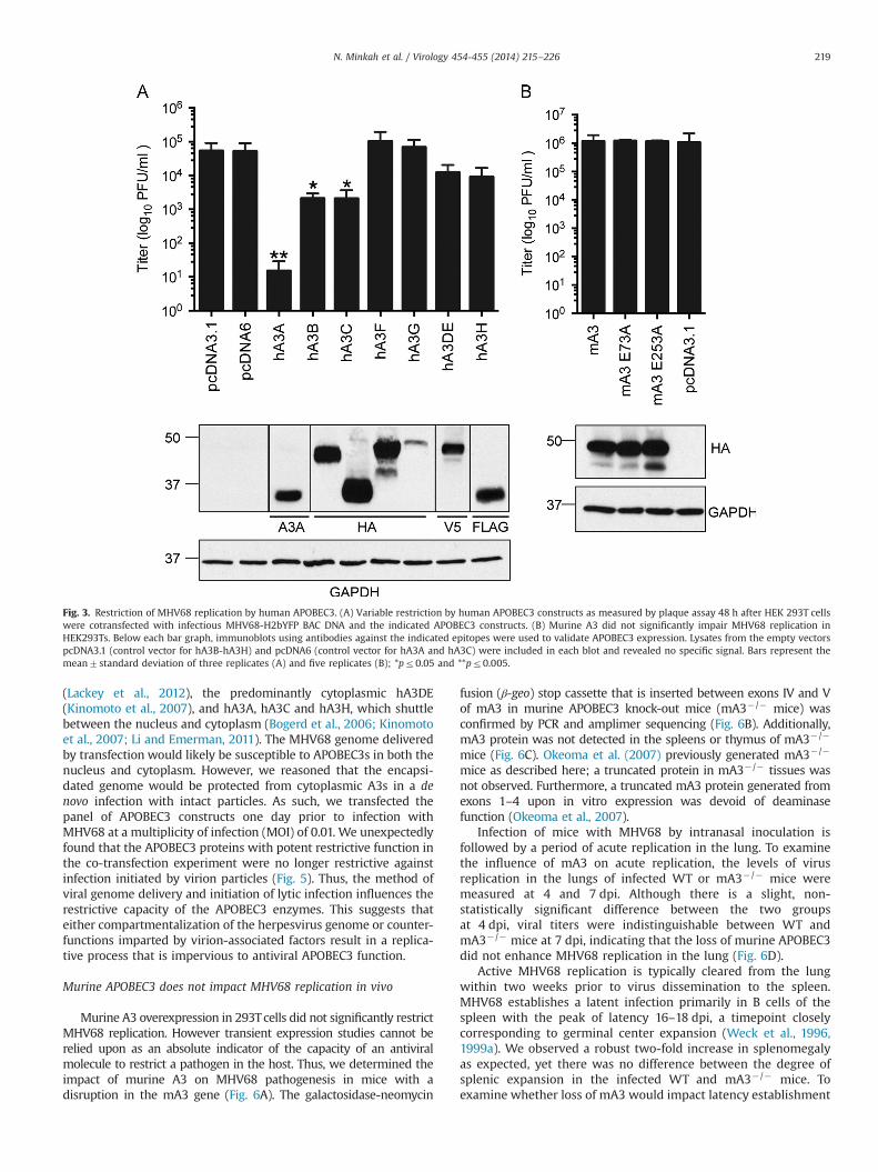
These observations of virus restriction by hA3 but not mA3were confirmed and extended to other human APOBEC3s uponquantitation of virus yield by plaque assay (Fig. 3A). Human A3Ainhibited viral replication by nearly 4 logs while co-transfectionwith hA3B and hA3C proteins resulted in a 2.5 log-fold reductionin replication. Co-transfection of either hA3DE or hA3H resulted ina lesser 5-fold reduction in virus output while hA3F and hA3G didnot impair MHV68 replication. In agreement with our previousobservation that mA3 overexpression did not impact virus spreadin the culture, co-transfection with mA3 did not impact MHV68yield (Fig. 3B). Expression of the human and mouse APOBEC3proteins was verified by immunoblot (Fig. 3A and B, respectively).We conclude that hA3A, hA3B and hA3C are restrictive againstMHV68 while mA3 is not.
Human APOBEC3 restriction of MHV68 replication is dependenton A3 active sites
Given the observation that several human APOBEC3 proteinsimpair MHV68 replication, we next examined if APOBEC3 enzymaticactivity is required by comparing restriction of MHV68 by the WTand active site mutant forms of hA3A and hA3B. Similar to otherAPOBEC proteins, A3 enzymes have retained a conserved active site
N. Minkah et al. / Virology 454-455 (2014) 215–226 217

sequence consisting of His-X-Glu-X23–28-Pro-Cys-X2–4-Cys. The histi-dine and cysteine residues coordinate a zinc ion while the glutamicacid serves as a proton shuttle in catalysis (Cullen, 2006). Mutation ofeither the cysteine or the glutamic acid abolishes the cytidinedeaminase function of A3 enzymes (Hakata and Landau, 2006;Narvaiza et al., 2009). The hA3A E72A active site mutant did notrestrict MHV68 yield compared to WT hA3A (Fig. 4A). Restriction byhA3B was lost with the N-terminal E68A single active site mutantand the E68A/E255A double mutant. However, the C-terminal singlehA3B E255A mutant retained restrictive function by reducing virusyield by 4.5-fold, albeit 56% less than the WT hA3B (Fig. 4A). Thechanges in restrictionwere not attributed to differences in expressionlevels of the WT and active site hA3 mutants (Fig. 4A, lower panel).Since hA3A reduced MHV68 DNA copy number by greater than 50-fold compared to the empty vector and A3A E72A mutant controls(Fig. 4B), we examined two regions of the MHV68 genome for C to Tor G to A transition mutations. In samples derived from A3A and A3Bcotransfections, the incidence of mutation was extremely lowin 18 and 11 respective clones of a GC-rich region of the genome
(bp 69,873–70,761) and 24 and 23 respective clones of a secondregion of the genome that is enriched for TC hotspots (bp 88,884–89,613) (Table 1). While three transition mutations were observed forregion two in A3A-restricted samples, these led to silent or con-servative aa changes. We also found a similar number of transitionmutations in the viral DNA of samples derived from the emptycontrol vector cotransfections. The mean mutation frequency forMHV68-infected samples restricted by A3A (0.15%) and A3B (0.06%)contrasts sharply with the mutation frequencies reported for HSV-1and EBV (6–20% by Suspene et al. 2011) and hepatitis B virus (25–50%by Vartanian et al. 2008). Taken together, the active sites of hA3A andhA3B are required for MHV68 restriction, but we found no strongevidence for hA3A or hA3B deamination of the genome.
Human APOBEC3 restriction of MHV68 replication is lost upon denovo infection
Several human APOBEC3 proteins were identified as potentinhibitors of MHV68 replication, the nuclear localized hA3B
Fig. 2. Restriction of MHV68 spread by human APOBEC3A, but not murine APOBEC3. HEK 293T cells were cotransfected with MHV68-H2bYFP BAC and the indicated human(hA3) and murine (mA3) APOBEC3 constructs and virus spread was visualized by fluorescence microscopy (upper panels) and bright field microscopy (lower panels) 48 hlater. HA3A E72A is an active site mutant of human A3A. MA3 E73A and mA3 E253A are active site mutants of mA3. Images shown are cropped fields of pictures taken at20� magnification. The images of MHV68-H2bYFPþ foci were false-colored green.
N. Minkah et al. / Virology 454-455 (2014) 215–226218
(Lackey et al., 2012), the predominantly cytoplasmic hA3DE(Kinomoto et al., 2007), and hA3A, hA3C and hA3H, which shuttlebetween the nucleus and cytoplasm (Bogerd et al., 2006; Kinomotoet al., 2007; Li and Emerman, 2011). The MHV68 genome deliveredby transfection would likely be susceptible to APOBEC3s in both thenucleus and cytoplasm. However, we reasoned that the encapsi-dated genome would be protected from cytoplasmic A3s in a denovo infection with intact particles. As such, we transfected thepanel of APOBEC3 constructs one day prior to infection withMHV68 at a multiplicity of infection (MOI) of 0.01. We unexpectedlyfound that the APOBEC3 proteins with potent restrictive function inthe co-transfection experiment were no longer restrictive againstinfection initiated by virion particles (Fig. 5). Thus, the method ofviral genome delivery and initiation of lytic infection influences therestrictive capacity of the APOBEC3 enzymes. This suggests thateither compartmentalization of the herpesvirus genome or counter-functions imparted by virion-associated factors result in a replica-tive process that is impervious to antiviral APOBEC3 function.
Murine APOBEC3 does not impact MHV68 replication in vivo
Murine A3 overexpression in 293Tcells did not significantly restrictMHV68 replication. However transient expression studies cannot berelied upon as an absolute indicator of the capacity of an antiviralmolecule to restrict a pathogen in the host. Thus, we determined theimpact of murine A3 on MHV68 pathogenesis in mice with adisruption in the mA3 gene (Fig. 6A). The galactosidase-neomycin
fusion (β-geo) stop cassette that is inserted between exons IV and Vof mA3 in murine APOBEC3 knock-out mice (mA3� /� mice) wasconfirmed by PCR and amplimer sequencing (Fig. 6B). Additionally,mA3 protein was not detected in the spleens or thymus of mA3� /�
mice (Fig. 6C). Okeoma et al. (2007) previously generated mA3� /�
mice as described here; a truncated protein in mA3� /� tissues wasnot observed. Furthermore, a truncated mA3 protein generated fromexons 1–4 upon in vitro expression was devoid of deaminasefunction (Okeoma et al., 2007).
Infection of mice with MHV68 by intranasal inoculation isfollowed by a period of acute replication in the lung. To examinethe influence of mA3 on acute replication, the levels of virusreplication in the lungs of infected WT or mA3� /� mice weremeasured at 4 and 7 dpi. Although there is a slight, non-statistically significant difference between the two groupsat 4 dpi, viral titers were indistinguishable between WT andmA3� /� mice at 7 dpi, indicating that the loss of murine APOBEC3did not enhance MHV68 replication in the lung (Fig. 6D).
Active MHV68 replication is typically cleared from the lungwithin two weeks prior to virus dissemination to the spleen.MHV68 establishes a latent infection primarily in B cells of thespleen with the peak of latency 16–18 dpi, a timepoint closelycorresponding to germinal center expansion (Weck et al., 1996,1999a). We observed a robust two-fold increase in splenomegalyas expected, yet there was no difference between the degree ofsplenic expansion in the infected WT and mA3�/� mice. Toexamine whether loss of mA3 would impact latency establishment
Fig. 3. Restriction of MHV68 replication by human APOBEC3. (A) Variable restriction by human APOBEC3 constructs as measured by plaque assay 48 h after HEK 293T cellswere cotransfected with infectious MHV68-H2bYFP BAC DNA and the indicated APOBEC3 constructs. (B) Murine A3 did not significantly impair MHV68 replication inHEK293Ts. Below each bar graph, immunoblots using antibodies against the indicated epitopes were used to validate APOBEC3 expression. Lysates from the empty vectorspcDNA3.1 (control vector for hA3B-hA3H) and pcDNA6 (control vector for hA3A and hA3C) were included in each blot and revealed no specific signal. Bars represent themean7standard deviation of three replicates (A) and five replicates (B); *pr0.05 and **pr0.005.
N. Minkah et al. / Virology 454-455 (2014) 215–226 219

in the spleen 16 dpi, intact splenocytes were analyzed by a limitingdilution PCR assay to determine the frequency of viral genomepositive cells. MHV68 established equivalent levels of latency inthe spleens of WT (1/156) and mA3� /� mice (1/128) (Fig. 6F). Thefrequency of intact splenocytes that spontaneously reactivatedwas examined by the observation of CPE upon explant andcoculture with MEFs in a limiting dilution reactivation assay. Lossof mA3 had an insignificant two-fold impact on MHV68 reactiva-tion, with splenocytes from infected WT mice and infectedmA3�/� mice reactivating at frequencies of 1/19,074 and1/10,966 cells, respectively (Fig. 6G). Moreover, this slight differ-ence in reactivation at 16 dpi had no effect on the maintenance ofthe frequency of genome positive cells later during chronicinfection at 42 dpi (data not shown). Taken together, the absenceof mA3 did not impact any aspect of MHV68 pathogenesis up to sixweeks following an intranasal infection.
Discussion
Targets of human APOBEC3 deaminase activity have been recentlyexpanded to include plasmid DNA (Stenglein et al., 2010), small DNA
viruses (Chen et al., 2006) and the large human herpesviruses(Suspene et al., 2011). In this study, we set out to determine if thesingle APOBEC3 protein, mA3, encoded by the mouse had the abilityto restrict the rodent herpesvirus pathogen MHV68. Our in silicoanalysis determined that A3 sites were abundant in MHV68, implyingthat this genome would be susceptible to A3 mediated deamination.Overexpression of mA3 together with infectious herpesvirus DNA didnot significantly impact MHV68 titers compared to a more substantialrestriction observed with hA3s. Surprisingly, the hA3 restriction waslost upon de novo infection. WT and mA3�/� C57BL6 mice wereindistinguishable in the levels of acute MHV68 replication in the lung,latency establishment in the spleen and reactivation from the spleen.Thus, loss of mA3 had no discernable effect on MHV68 replicationin vivo.
In this study we identified several human APOBEC3s as potentrestrictors of MHV68 replication. Additionally, we found that therestriction by hA3A and hA3B is dependent on their previouslyidentified active sites (Hakata and Landau, 2006; Stenglein et al.,2010). Interestingly hA3B restriction of MHV68 was absolutelydependent on the N-terminal amino acid, E68 and less dependenton the C-terminal E255 residue, possibly indicating a separation offunction between the E68 and E255 active sites. The requirement
Fig. 4. APOBEC3A and APOBEC3B active sites are required for restriction of viral replication and viral DNA synthesis. (A) Loss of restriction by mutant human APOBEC3A(hA3) and APOBEC3B (hA3B) constructs as measured by plaque assay and quantitative PCR (hA3B) 48 h after HEK 293T cells were cotransfected with infectious MHV68-H2bYFP BAC DNA and the indicated APOBEC3 constructs. Below each bar graph, immunoblots using antibodies against the indicated epitopes were used to validate APOBEC3expression. Lysates from the empty vectors pcDNA6 (control vector for WT and mutant hA3A) and pcDNA3.1 (control vector for WT and mutant hA3B) were included in eachblot and revealed no specific signal. Bars represent the mean7standard deviation of three replicates; *pr0.05 and **pr0.005. (B) Quantitation of MHV68 genomes by qPCRanalysis of DpnI-digested DNA. DNA levels were normalized to levels upon cotransfection with the empty vector, pCDNA6; *pr0.05 and **pr0.005.
N. Minkah et al. / Virology 454-455 (2014) 215–226220

for these hA3 active sites to restrict MHV68 growth and DNAreplication is consistent with deamination of the genome as thebasis of restriction. However, we have no direct evidence tosupport this hypothesis since DNA from A3A or A3B cotransfec-tions contained few C to T or G to A transitions. Although twotransitions were present in A3A cotransfected samples, weobserved a similar frequency of transitions in DNA from controltransfections. The lack of a high mutation frequency in the A3Asamples might be attributed to heavily deaminated DNA thatis unable to serve as a template for replication or rare eventsthat are below the limit of detection. Alternatively, A3A mightimpair MHV68 replication using a deamination-independentmechanism that is dependent on the active site. For example,A3A mutants that were deaminase defective but retained otherknown functions such as DNA binding and localization to thenucleus were found to restrict parvoviruses (Chen et al., 2006;Narvaiza et al., 2009).
We hypothesized that in silico analysis would reveal a hotspotprofile for a herpesvirus that reflects tropism for cell types thatexpress high levels of particular host APOBEC3s. Namely, virusgenomes under selective pressure by one or more APOBEC3s would
be underrepresented or depleted of that particular APOBEC hotspot.The HPV genome is depleted of A3A/A3B/A3H hotspots and neutralor enriched for hA3G, consistent with its epithelial cell tropism.However, the association of cell tropism and hotspot profiles was notas straightforward for the herpesviruses. Human herpesviruses suchas VZV, EBV, and KSHV that reside in lymphocytes during their lifecycle were not depleted for A3G hotspots. The VZV genome wasdepleted for A3A/A3B/A3H hotspots, yet HSV-1, another virus thatreplicates in the epithelium was predominately neutral with over-representation in some regions. The overrepresentation of TC andTTC hotspots in the HSV-1 genome is consistent with the restrictionof HSV-1 replication by the ectopic expression of hA3A and hA3Creported by Suspene et al. (2011). Interestingly, this study reported ahigh incidence of edited HSV-1 genomes upon transient transfectionof hA3C and a striking frequency of hypereditied HSV-1 and EBVgenomes in clinical specimens. These findings relied on the detectionof rare deamination events by PCR based amplification at lowdenaturing temperatures (Suspene et al., 2011). A non-biased quan-titative determination of the incidence of herpesvirus deamination inhuman infections and the impact on pathogenesis warrants furtherinvestigation.
Table 1Analysis of mutations arising in the viral genome upon cotransfection with human APOBEC3A or APOBEC3B.
Experimenta Number of clonesb Mutation frequencyc SNPd, genomic coordinatee
Region 1 (bp 69,873–70,761)APOBEC3A 18 0.008%, 1/12,492 T-C 70,265APOBEC3B 11 0%; 0/5,269Empty vector 11 0%; 0/7,634
Region 2 (bp 88,884–89,613)APOBEC3A replicate 1 6 0.14%; 6/4,086 A-G 88,939
T-C 89,050, G-A 89,230A-G 89,191A-G 89,283, A-G 89,286
APOBEC3A replicate 2 9 0.16%; 9/5,526 C-T 89,033f, A-G 89,178G-A 89,083f
T-C 89,248, T-C 89,270T-C 89,293A-G 89,308T-C 89,407A-T 89,450
APOBEC3A replicate 3 9 0.16%; 8/5,148 A-G 88,985T-C 89,197A-G 89,207, T-C 89,417A-C 89,320A-G 89,352A-G 89,357A-G 89,434
APOBEC3B replicate 1 7 0.09%; 4/4,417 A-G 89,103, T-C 89,236A-G 89,495, T-C 89,507
APOBEC3B replicate 2 7 0%; 0/4,921APOBEC3B replicate 3 8 0.09%; 5/5,416 T-C 89,100, A-G 89,106
T-C 89,234A-G 89,265T-C 89,286
Empty vector replicate1 7 0.14%; 7/4893 T-C 89,050A-G 89,083, T-C 89,419T-C 89,432, T-C 89,499, C-T 89,587f
T-C 89,500Empty vector replicate 2 11 0.10%; 7/6699 T-A 88,908
G-A 88,926f, A-G 89,936, A-G 89,506G-A 89,083f
A-G 89,481T-C 89,498
Empty vector replicate 3 6 0.08%; 3/3774 A-G 89,127, A-G 89,199, A-G 89,287
a Cotransfection of MHV68 genomic BAC DNA with the identified construct.b Number of independent clones sequenced for mutation analysis.c Mutation frequency is calculated as follows: (number of changes/total number of nucleotides sequenced per experiment)�100.d SNP – single nucelotide polymorphism.e Position of SNP in the MHV68 genome.f SNPs that reflect transition mutations (G to A or C to T changes).
N. Minkah et al. / Virology 454-455 (2014) 215–226 221

At this juncture, the APOBEC3 hotspot analysis has some pre-dictive value and correlates with some aspects of herpesvirusbiology, but an interpretation of hotspot frequencies is more complexsince a multitude of factors apply evolutionary pressures. Theoccurrence of a particular dinucleotide or trinucleotide sequence isinfluenced by codon bias, a wide expanse of protein–DNA interac-tions, and restrictive pressures by one APOBEC3 deaminase that mayin turn bias the incidence of another hotspot. These pressures arefurther compounded by the fact that each herpesvirus engagesmultiple cell types in its life cycle and, as discussed below, theseviruses may have evolved countermeasures.
Strikingly, hA3 restriction of MHV68 replication was observedupon transfection of nonencapsidated infectious DNA but was lostupon de novo infection with virion particles. MHV68 virions maydeliver a factor that counteracts the hA3 proteins. Tegument proteinsfrom several herpesviruses families function in either the cytoplasmor the nucleus to limit the antiviral response from the host. KSHVORF45 inhibits type I interferon signaling by impairing the phosphor-ylation and nuclear translocation of IRF-7 (Sathish and Yuan, 2011;Zhu et al., 2002). HSV1 ICPO, HCMV pp71, HVS ORF3, EBV BNRF1 andMHV68 ORF75C translocate to the nucleus upon virus infection todegrade or relocalize components of the antiviral nuclear domain 10structures (ND10) (Boutell and Everett, 2013; Full et al., 2012; Linget al., 2008; Penkert and Kalejta, 2012; Sewatanon and Ling, 2013; Tsaiet al., 2011). We did not find significant homology of MHV68 virioncomponents including glycoproteins, tegument and capsid proteins toknown A3 antagonists. However, we cannot rule out the existence of anovel mechanism by which the virion structural components or evenRNA molecules mediates resistance to APOBEC3 proteins.
The loss of MHV68 restriction might also be explained by thecompartmental separation of A3 proteins from the MHV68 gen-ome in a natural course of infection. Several of the hA3 proteinsthat exhibit restriction of MHV68, namely hA3A, hA3B, hA3C andhA3H are targeted to the nucleus (Bogerd et al., 2006; Kinomotoet al., 2007). We propose that the hA3 proteins might be separatedfrom viral replication centers in the nucleus via either the directaction of a viral factor or indirectly due to viral remodeling of thenucleus. Herpesvirus replication, transcription and the formationof viral capsids transpires in intranuclear structures known asreplication centers that are defined by the presence of key viralreplication proteins as well as host proteins (de Bruyn Kops andKnipe, 1994; Quinlan et al., 1984; Weller, 2010). Nuclear substruc-tures such as the ND10, as well as nuclear protein localization andabundance are tightly regulated by herpesviruses to maintainoptimal viral gene expression and replication (Saffert andKalejta, 2008). The subcellular localization of A3 proteins andherpesvirus replication compartments could provide mechanisticinsight for viral evasion of hA3 restriction.
Several human APOBEC3 proteins exhibit restriction of MHV68replication, but mA3 exhibited no restrictive capacity in thecotransfection experiments. This lack of restriction with mA3overexpression is not likely due to the absence of a co-factor inthe HEK 293T cells. Murine APOBEC3 partially restricts Moloneymurine leukemia virus replication and restricts HIV-1 lacking Vifto similar levels as hA3G in 293T cells (Doehle et al., 2005;Sanchez-Martinez et al., 2012). The absence of restriction withoverexpressed mA3 was supported by a lack of enhancement inMHV68 pathogenesis in mA3 knockout mice. Thus, MHV68 mightencode a mA3 antagonist that significantly limits antiviral APO-BEC3 activity such as HIV Vif (Malim, 2009; Malim and Emerman,2008; Mariani et al., 2003) or Murine Leukemia Virus (MLV) glyco-gag (Stavrou et al., 2013). Additionally, a recent report identified asignificant role for host uracil DNA glycosylase (UNG) in reducingthe effect of hA3G-induced hyperediting of duck hepatitis B virus(Kitamura et al., 2013). MHV68, like other herpesviruses encodes auracil DNA glycosylase (UNG/ORF46). However, we did not observean increased restriction of MHV68 replication by hA3A, hA3B ormA3 in cells infected with a recombinant MHV68 harboring a stopcodon in ORF46 compared to the control repair virus (Minkah,Chavez, and Krug, unpublished results).
Infection of mice lacking mA3 did not lead to an enhancement ofMHV68 pathogenesis. This may be attributed to a viral counter-measure as described above, a lack of mA3 antiviral function in thecontext of a herpesvirus infection or a yet undiscovered role ina particular aspect of chronic infection in this pathogenesis model.The intranasal route of inoculation is characterized by high levels ofproductive replication in the lungs, a tissue with lower mA3 expres-sion levels (Mikl et al., 2005). MA3 protein is expressed at high levelsin the spleens, but its absence did not influence latency establish-ment in the spleen or reactivation from latency. Another route ofinfection, such as intraperitoneal inoculation might reveal theimportance of MHV68 replication in peritoneal exudate cells whereinthe primary latency reservoir is macrophages (Weck et al., 1999b).Indeed, the influence of some host proteins such as promyelocyticleukemia nuclear bodies (PML) (Sewatanon et al., 2013) or viralproteins such as M1 and M2 on MHV68 pathogenesis is dependenton the route of infection (Clambey et al., 2000; Jacoby et al., 2002).Finally, it is possible that another APOBEC protein compensates forthe loss of mA3 to limit MHV68 replication. A hyperediting signaturesimilar to murine APOBEC1 deamination was reported in a mousemodel of hepatitis B virus infection (Petit et al., 2009; Renard et al.,2010). Furthermore, in a rat model of HSV-1-induced encephalitis,APOBEC1 levels were increased upon HSV-1 infection and over-expression of APOBEC1 both enhanced hyperediting of HSV-1genomes and reduced viral titers (Gee et al., 2011).
Fig. 5. Loss of APOBEC3 restriction in the context of de novo infection with virusparticles. HEK 293T cells were transfected with the indicated APOBEC3 constructs24 h prior to de novo infection with MHV68-H2bYFP virus. MHV68 replication wasmeasured by plaque assay 48 hpi. Below each bar graph, immunoblots usingantibodies against the indicated epitopes were used to validate APOBEC3 expres-sion. Lysates from the empty vectors pcDNA3.1 (control vector for hA3B, hA3DE-hA3H) and pcDNA6 (control vector for hA3A and hA3C) were included in each blotand revealed no specific signal.
N. Minkah et al. / Virology 454-455 (2014) 215–226222

MHV68 infection of mice provides a tractable model pathogen-esis system to dissect the contribution of virus and host factorsthat influence chronic gammaherpesvirus infection. Primates havediversified their repertoire of A3 cytidine deaminases under theselective pressure of retroelements and viral pathogens. Wesought to examine whether the evolutionary distinct yet ‘primor-dial’ APOBEC3 encoded by mice shared the restrictive property ofthe expanded human APOBEC3 molecules against MHV68, withthe aim of utilizing mA3� /� mice as a platform for pathogenesisand mechanistic investigations. However, mA3 did not restrictMHV68 replication in cell culture or pathogenesis in mice. Theunexpected loss of the hA3 restrictive phenotype when replicationwas initiated upon de novo infection with virus particles leads usto posit that herpesviruses are largely protected from hA3 restric-tion. Possible evasion mechanisms to explore include a virion-associated countermeasure such as a tegument protein or a blockin APOBEC3 access to the viral genome either due to encapsidationof the viral genome during cytoplasmic transport to the nucleus orcompartmentalization of the genome in nuclear replicationcenters.
Materials and methods
Viruses, cells and mice
The recombinant marking virus, MHV68-H2bYFP was usedthe for cell-culture experiments. This virus encodes a histone 2b
(H2b)-enhanced yellow fluorescent protein (eYFP) fusion proteinthat tethers the YFP protein to nucleosomes and eliminates thepassive diffusion of the fluorescent YFP signal out of the nucleus toenhance infected cell imaging by microscopy (Collins and Speck,2012). Mice were infected with MHV68 WUMS (ATCC VR1465) asthe wild-type virus. Virus passage and titer determination wereperformed as previously described (Weck et al., 1996, 1999a).
Human epithelial kidney cells (HEK 293T) were maintained inDulbecco's modified Eagle's medium (DMEM) supplemented with10% fetal bovine serum, 1% penicillin, and streptomycin at 37 1C in5% CO2. Murine fibroblast cells (NIH 3T12) were maintained inDulbecco's modified Eagle's medium (DMEM) supplemented with8% fetal calf serum, 100 U of penicillin per ml and 100 mg ofstreptomycin per ml at 37 1C in 5% CO2
Murine APOBEC3 knock-out (mA3� /�) mice were generated byblastocyst injection of 129P2/OlaHsd embryonic stem cell cloneXN450 (Baygenomics, San Francisco) that contained an 8 kbgalactosidase-neomycin fusion (β-geo) gene trap insertion retro-viral vector between exons 4 and 5 of mA3. The mice were crossedfor several generations onto a C57/Bl6 background and genotypedby PCR using primers spanning the insertion site. The WTgenotype was amplified with primer 21 (50 CTGTAACCTGG-TATCTCCCGTC 30) and primer 22 (50 GGAAAAACTGCTTGCCAGGCTC30). The mA3�/� genotype was amplified with primer 21 (50
CTGTAACCTGGTATCTCCCGTC 30) and primer 23 (50 CACAAGGTT-CATATGGTGCCGT 30). The mA3� /� mice and their WT C57BL6counterparts were maintained at the Stony Brook UniversityDivision of Laboratory Animal Research (DLAR) facility in
Fig. 6. Absence of murine APOBEC3 does not alter MHV68 pathogenesis. (A) Schematic depicting the disruption of murine APOBEC3 (mA3) by the insertion of a β-geocassette to generate mA3 knockout mice (mA3� /�). (B) Genotype PCR analysis of DNA harvested from the splenocytes of infected WT C57BL/6 or mA3�/� mice. The locationsof primers for either the WT or mutant mA3 gene are shown in panel A. (C) Immunoblot demonstrating expression of mA3 in WT but not mA3�/� mice. (D) Acute replicationin the lungs of WT or mA3� /� lungs 4 and 7 days post-intranasal infection with 1000 PFU of WT MHV68. (E) Spleen weights from naïve WT, infected mA3� /� mice orinfected WT mice 16 dpi. (F) Latency is determined as the frequency of viral genome positive splenocytes determined by limiting dilution PCR. (G) Frequency of reactivationfrom intact splenocytes determined by a limiting dilution explant reactivation assay 16 dpi. Open symbols represent preformed infectious virus from mechanically disruptedcells plated in parallel. The data shown represent three independent experiments with splenocytes pooled from three to five mice per experimental group.
N. Minkah et al. / Virology 454-455 (2014) 215–226 223

accordance with protocols approved by the Institutional Animal Careand Use Committee of Stony Brook. Eight- to twelve-week old WTand mA3�/� mice were infected intranasally with 1000 PFU ofMHV68 under isoflurane anesthesia. At the indicated times post-infection, organs were harvested and processed as described below.
Transfections
For transient co-transfections, 4�105 HEK 293T cells wereseeded per well of a 12-well tissue culture plate one day prior totransfection with 2.5 μg of APOBEC3 expression plasmids and1.55 μg of MHV68 BAC DNA. All transient co-transfections wereperformed using TransIT-LT1 Transfection Reagent according tothe manufacturer0s instructions (Mirus, Madison WI). Forty-eighthours later, the cells were subjected to multiple freeze–thawcycles to release infectious virus that was then titered by plaqueassay on a NIH 3T12 cell monolayer.
For the transfection followed by infection experiments, HEK293T cells were transfected with the desired human and mouseAPOBEC3 proteins as outlined above. Twenty-four hours post-transfection, cells were infected with MHV68-H2bYFP virus at anMOI of 0.01. Forty-eight hours after the infection virus output wasquantified by a plaque assay.
Fluorescence microscopy
Virus infection with MHV68-H2bYFP was visualized by fluor-escence microscopy. Imaging was performed with a Zeiss AxiovertS100 inverted microscope (Carl Zeiss Microscopy GmbH, JenaGermany) equipped with a Luminera INFINITY 3-1UR 1.4 mega-pixel low light CCD digital camera (Lumenera, Ottawa ON Canada).Images were analyzed using Axiovision Software (Axiovision LERel.4.3, Carl Zeiss Microscopy GmbH, Jena Germany).
Plaque assay
1.8�105 NIH 3T12 cells were seeded per well in a 6-well tissueculture plate one day prior to infection. The next day, the NIH 3T12cells were infected with serial dilutions of cell homogenate andoverlayed with 1.5% methylcellulose in DMEM containing 5% FBS.One week later, the methylcellulose was removed and cells werewashed twice with PBS prior to methanol fixation and stainingwith a 0.1% crystal violet solution in 10% methanol.
Immunoblot
Total protein lysate was harvested in lysis buffer (150 mMsodium chloride, 1.0% IGEPAL CA-630, 0.5% sodium deoxycholate,0.1% sodium dodecyl sulfate, 50 mM Tris pH 8.0) supplementedwith a protease inhibitor cocktail (Sigma, St. Louis MO). Fiftymicrograms of each lysate were separated in a 12% SDS PAGE geland transferred onto polyvinylidene fluoride (PVDF) membrane.APOBEC3 proteins were detected using a mouse monoclonalantibody specific for the HA-epitope (Cell signaling, Danvers MA)on mA3, A3B, A3C, A3F, and A3G constructs, a V5 epitope (SantaCruz biotechnology, Dallas Texas) for A3DE and a rabbit mono-clonal specific for human A3A (Hakata and Landau, 2006). A rabbitmonoclonal antibody to human glyceraldehyde 3-phosphate dehy-drogenase (GAPDH) was used as a loading control. HRP-conjugatedsecondary antibodies were detected using an enhanced chemilu-minescence reagent (ECL, Thermo Scientific, Waltham MA).
Quantitative PCR
Total cell DNA from the co-transfection experiments wascolumn-purified (Qiagen, Limburg, Netherlands) and digested
with 20 units of Dpn1 (New England Biolabs, Ipswich MA) for12 h at 37 1C to remove input MHV68 BAC DNA. One ng of DpnI-digested DNA was input into a quantitative PCR reaction (QuantaBiosceinces- Perfecta SYBR GREEN, Gaithersburg MD) using pri-mers specific to a region of MHV68 ORF12 (forward primer,50 GTCTACAACAGGATCTGCATTT 30, reverse primer 50 AAAACTC-TACCGTGACTGTGAA 30) and primers for human GAPDH (forwardprimer, 50 GTATGACTGGGGGTGTTGGG 30, reverse primer 50
GCGCCCAATAGGACCAAATC 30). The relative level of viral genomecopy number was determined by ΔΔCt.
Sequencing
Total cell DNA from the co-transfection experiments wasextracted as outlined above. The DNA was digested with 20 unitsof Dpn1 (New England Biolabs, Ipswich MA) for 12 h at 37 1C toremove input MHV68 BAC DNA. Two regions in the MHV68genome (bp 69,873–70,761 and bp 88,884 to 89,613) were ampli-fied with Dynazyme II DNA polymerase (Thermo Scientific,Waltham MA), cloned into the TOPO TA cloning vector (LifeTechnologies, Carlsbad, CA) and sequenced to identify mutationsthat arose during infection.
Pathogenesis
For acute titers, the right and left lungs of mice were removed4 and 9 days post-infection and disrupted with a Mini-Beadbeater(Biospec, Bartlesville OK). The homogenates were titered byplaque assay. Spleens were harvested to determine viral latencyestablishment and reactivation from latency 16 dpi.
To determine the frequency of cells harboring the viral genome,a single cell suspension of splenocytes was prepared and analyzedby limiting dilution PCR. Six, three-fold serial dilutions of spleno-cytes were plated in a 96-well PCR plate in a background of NIH3T12 cells and lysed overnight at 56 1C with Proteinase K. The platewas then subjected to an 80-cycle nested PCR with primersspecific for MHV68 ORF50. Twelve replicates were analyzed ateach serial dilution and plasmid DNA at 0.1, 1 and 10 copies wasincluded to verify the sensitivity of the assay.
To determine the frequency of cells harboring latent viruscapable of reactivation upon explant, single cell splenocytes wereprepared from mice 16 dpi, resuspended in DMEM containing 10%fetal bovine serum and plated in twelve serial two-fold dilutionsonto a monolayer of mouse embryonic fibroblast (MEF) cellsprepared from C57BL6J mice in 96-well tissue culture plates.Twenty-four replicates were plated per serial dilution. The wellswere scored for cytopathic effect (CPE) two to three weeks afterplating. To differentiate between pre-formed infectious virus andvirus spontaneously reactivating upon cell explant, parallel sam-ples were mechanically disrupted using a Mini-Beadbeater prior toplating on the monolayer of MEFs to release preformed virus thatis scored as CPE.
Bioinformatic analysis
To measure under- and over-representation of APOBEC3 hot-spots, we compared the observed frequency of each APOBECconsensus motif (TC for hA3A, hA3B, hA3F and hA3H, TTC forhA3C, CCC for hA3G and TCC for mA3) to a random expectationnull model. To assess the effects throughout the viral genomes weused sliding 1 kilobase windows to scan each genome at intervalsof 100 nucleotides (i.e. first window from position 1 to 1000,second window from 101 to 1100, and so on). The null model wasgenerated using 1000 randomly shuffled versions of each genome.The number of occurrences of the motif (TC, TTC, etc.) in eachrandomly shuffled sequence was used to build a null model
N. Minkah et al. / Virology 454-455 (2014) 215–226224

distribution for that window. If the true frequency that wasobserved in the unshuffled sequence was within the lowest 5thpercentile of this distribution, we labeled that window as beingunderrepresented. Conversely, if the true frequency was in thehighest 5th percentile, the window was labeled as overrepre-sented for that motif. The NCBI accession numbers for thegenomes analyzed are: Human papilloma virus type 16(NC_001526), Herpes simplex virus 1(NC_001806), Herpes simplexvirus 2(NC_001798), Varicella-zoster virus (NC_001348), Epstein-Barr virus (NC_007605), Human cytomegalovirus (NC_006273),Human herpesvirus 6A (NC_001664), Human herpesvirus 6B(NC_000898), Human herpesvirus 7(NC_001716), Kaposi'ssarcoma-associated herpesvirus (NC_009333) and Murine gam-maherpesvirus 68 (NC_001826).
Statistical analyses
Data was analyzed using Graphpad Prism Software (Prism 5,La Jolla CA). The statistical significance of differences betweengroups was tested using a non-paired two-tailed t test. UnderPoisson distribution analysis, the frequencies of latency establish-ment and reactivation from latency were determined by theintersection of nonlinear regression curves with the line at 63.2%.
Acknowledgments
Kevin Chavez was supported by an NIH-MARC fellowship grant(Grant #5T34GM008655). Parth Shah was partly supported by theStony Brook University Simons Summer Research Program (2013)and Thomas MacCarthy was supported by Stony Brook Universitystartup funds. Nathaniel Landau was supported by NIH grants(AI058864 and AI074967). Laurie Krug was supported by anAmerican Cancer Society research scholar grant, RSG-1-160-01-MPC and NIH AI097875. Special thanks to Steven Reddy fortechnical support and members of the Krug laboratory for helpfuldiscussions.
References
Bishop, K.N., Holmes, R.K., Sheehy, A.M., Malim, M.H., 2004. APOBEC-mediatedediting of viral RNA. Science 305, 645.
Bogerd, H.P., Tallmadge, R.L., Oaks, J.L., Carpenter, S., Cullen, B.R., 2008. Equineinfectious anemia virus resists the antiretroviral activity of equine APOBEC3proteins through a packaging-independent mechanism. J. Virol. 82,11889–11901.
Bogerd, H.P., Wiegand, H.L., Hulme, A.E., Garcia-Perez, J.L., O’Shea, K.S., Moran, J.V.,Cullen, B.R., 2006. Cellular inhibitors of long interspersed element 1 and Aluretrotransposition. Proc. Natl. Acad. Sci. USA 103, 8780–8785.
Boutell, C., Everett, R.D., 2013. Regulation of alphaherpesvirus infections by the ICP0family of proteins. J. Gen. Virol. 94, 465–481.
Bransteitter, R., Prochnow, C., Chen, X.S., 2009. The current structural andfunctional understanding of APOBEC deaminases. Cell. Mol. Life Sci. 66,3137–3147.
Chen, H., Lilley, C.E., Yu, Q., Lee, D.V., Chou, J., Narvaiza, I., Landau, N.R., Weitzman,M.D., 2006. APOBEC3A is a potent inhibitor of adeno-associated virus andretrotransposons. Curr. Biol. 16, 480–485.
Clambey, E.T., Virgin, H.W.t., Speck, S.H., 2000. Disruption of the murine gamma-herpesvirus 68 M1 open reading frame leads to enhanced reactivation fromlatency. J. Virol. 74, 1973–1984.
Collins, C.M., Speck, S.H., 2012. Tracking murine gammaherpesvirus 68 infection ofgerminal center B cells in vivo. PloS One 7, e33230.
Cullen, B.R., 2006. Role and mechanism of action of the APOBEC3 family ofantiretroviral resistance factors. J. Virol. 80, 1067–1076.
de Bruyn Kops, A., Knipe, D.M., 1994. Preexisting nuclear architecture defines theintranuclear location of herpesvirus DNA replication structures. J. Virol. 68,3512–3526.
Delebecque, F., Suspene, R., Calattini, S., Casartelli, N., Saib, A., Froment, A., Wain-Hobson, S., Gessain, A., Vartanian, J.P., Schwartz, O., 2006. Restriction of foamyviruses by APOBEC cytidine deaminases. J. Virol. 80, 605–614.
Doehle, B.P., Schafer, A., Wiegand, H.L., Bogerd, H.P., Cullen, B.R., 2005. Differentialsensitivity of murine leukemia virus to APOBEC3-mediated inhibition isgoverned by virion exclusion. J. Virol. 79, 8201–8207.
Driscoll, D.M., Zhang, Q., 1994. Expression and characterization of p27, the catalyticsubunit of the apolipoprotein B mRNA editing enzyme. J. Biol. Chem. 269,19843–19847.
Full, F., Reuter, N., Zielke, K., Stamminger, T., Ensser, A., 2012. Herpesvirus saimiriantagonizes nuclear domain 10-instituted intrinsic immunity via an ORF3-mediated selective degradation of cellular protein Sp100. J. Virol. 86,3541–3553.
Gee, P., Ando, Y., Kitayama, H., Yamamoto, S.P., Kanemura, Y., Ebina, H., Kawaguchi,Y., Koyanagi, Y., 2011. APOBEC1-mediated editing and attenuation of herpessimplex virus 1 DNA indicate that neurons have an antiviral role during herpessimplex encephalitis. J. Virol. 85, 9726–9736.
Goila-Gaur, R., Strebel, K., 2008. HIV-1 Vif, APOBEC, and intrinsic immunity.Retrovirology 5, 51.
Hakata, Y., Landau, N.R., 2006. Reversed functional organization of mouse andhuman APOBEC3 cytidine deaminase domains. J. Biol. Chem. 281,36624–36631.
Hultquist, J.F., Lengyel, J.A., Refsland, E.W., LaRue, R.S., Lackey, L., Brown, W.L.,Harris, R.S., 2011. Human and rhesus APOBEC3D, APOBEC3F, APOBEC3G, andAPOBEC3H demonstrate a conserved capacity to restrict Vif-deficient HIV-1. J.Virol. 85, 11220–11234.
Jacoby, M.A., Virgin, H.W.t., Speck, S.H., 2002. Disruption of the M2 gene of murinegammaherpesvirus 68 alters splenic latency following intranasal, but notintraperitoneal, inoculation. J. Virol. 76, 1790–1801.
Jern, P., Russell, R.A., Pathak, V.K., Coffin, J.M., 2009. Likely role of APOBEC3G-mediated G-to-A mutations in HIV-1 evolution and drug resistance. PLoSPathog. 5, e1000367.
Kinomoto, M., Kanno, T., Shimura, M., Ishizaka, Y., Kojima, A., Kurata, T., Sata, T.,Tokunaga, K., 2007. All APOBEC3 family proteins differentially inhibit LINE-1retrotransposition. Nucl. Acids Res. 35, 2955–2964.
Kitamura, K., Wang, Z., Chowdhury, S., Simadu, M., Koura, M., Muramatsu, M., 2013.Uracil DNA glycosylase counteracts APOBEC3G-induced hypermutation ofhepatitis B viral genomes: excision repair of covalently closed circular DNA.PLoS Pathog. 9, e1003361.
Lackey, L., Demorest, Z.L., Land, A.M., Hultquist, J.F., Brown, W.L., Harris, R.S., 2012.APOBEC3B and AID have similar nuclear import mechanisms. J. Mol. Biol.419,301–314.
Langlois, M.A., Kemmerich, K., Rada, C., Neuberger, M.S., 2009. The AKV murineleukemia virus is restricted and hypermutated by mouse APOBEC3. J. Virol. 83,11550–11559.
Li, M.M., Emerman, M., 2011. Polymorphism in human APOBEC3H affects aphenotype dominant for subcellular localization and antiviral activity. J. Virol.85, 8197–8207.
Liddament, M.T., Brown, W.L., Schumacher, A.J., Harris, R.S., 2004. APOBEC3Fproperties and hypermutation preferences indicate activity against HIV-1in vivo. Curr. Biol. 14, 1385–1391.
Ling, P.D., Tan, J., Sewatanon, J., Peng, R., 2008. Murine gammaherpesvirus 68 openreading frame 75c tegument protein induces the degradation of PML and isessential for production of infectious virus. J. Virol. 82, 8000–8012.
Low, A., Okeoma, C.M., Lovsin, N., de las Heras, M., Taylor, T.H., Peterlin, B.M., Ross,S.R., Fan, H., 2009. Enhanced replication and pathogenesis of Moloney murineleukemia virus in mice defective in the murine APOBEC3 gene. Virology 385,455–463.
MacMillan, A.L., Kohli, R.M., Ross, S.R., 2013. APOBEC3 inhibition of mousemammary tumor virus infection: the role of cytidine deamination versusinhibition of reverse transcription. J. Virol. 87, 4808–4817.
Madsen, P., Anant, S., Rasmussen, H.H., Gromov, P., Vorum, H., Dumanski, J.P.,Tommerup, N., Collins, J.E., Wright, C.L., Dunham, I., MacGinnitie, A.J., Davidson,N.O., Celis, J.E., 1999. Psoriasis upregulated phorbolin-1 shares structural butnot functional similarity to the mRNA-editing protein apobec-1. J Invest.Dermatol113, 162–169.
Mahieux, R., Suspene, R., Delebecque, F., Henry, M., Schwartz, O., Wain-Hobson, S.,Vartanian, J.P., 2005. Extensive editing of a small fraction of human T-cellleukemia virus type 1 genomes by four APOBEC3 cytidine deaminases. J. Gen.Virol. 86, 2489–2494.
Malim, M.H., 2009. APOBEC proteins and intrinsic resistance to HIV-1 infection.Philos. Trans. R. Soc. Lond. Ser. B: Biol. Sci. 364, 675–687.
Malim, M.H., Emerman, M., 2008. HIV-1 accessory proteins–ensuring viral survivalin a hostile environment. Cell Host Microbe3, 388–398.
Mangeat, B., Turelli, P., Caron, G., Friedli, M., Perrin, L., Trono, D., 2003. Broadantiretroviral defence by human APOBEC3G through lethal editing of nascentreverse transcripts. Nature 424, 99–103.
Mariani, R., Chen, D., Schrofelbauer, B., Navarro, F., Konig, R., Bollman, B., Munk, C.,Nymark-McMahon, H., Landau, N.R., 2003. Species-specific exclusion of APO-BEC3G from HIV-1 virions by Vif. Cell 114, 21–31.
Mehta, A., Driscoll, D.M., 1998. A sequence-specific RNA-binding proteincomplements apobec-1 To edit apolipoprotein B mRNA. Mol. Cell. Biol.18,4426–4432.
Mikl, M.C., Watt, I.N., Lu, M., Reik, W., Davies, S.L., Neuberger, M.S., Rada, C., 2005.Mice deficient in APOBEC2 and APOBEC3. Mol. Cell. Biol. 25, 7270–7277.
Munk, C., Beck, T., Zielonka, J., Hotz-Wagenblatt, A., Chareza, S., Battenberg, M.,Thielebein, J., Cichutek, K., Bravo, I.G., O’Brien, S.J., Lochelt, M., Yuhki, N., 2008.Functions, structure, and read-through alternative splicing of feline APOBEC3genes. Genome Biol. 9, R48.
Narvaiza, I., Linfesty, D.C., Greener, B.N., Hakata, Y., Pintel, D.J., Logue, E., Landau, N.R., Weitzman, M.D., 2009. Deaminase-independent inhibition of parvovirusesby the APOBEC3A cytidine deaminase. PLoS Pathog. 5, e1000439.
N. Minkah et al. / Virology 454-455 (2014) 215–226 225

OhAinle, M., Kerns, J.A., Malik, H.S., Emerman, M., 2006. Adaptive evolution andantiviral activity of the conserved mammalian cytidine deaminase APOBEC3H.J. Virol. 80, 3853–3862.
Okeoma, C.M., Lovsin, N., Peterlin, B.M., Ross, S.R., 2007. APOBEC3 inhibits mousemammary tumour virus replication in vivo. Nature 445, 927–930.
Okuyama, S., Marusawa, H., Matsumoto, T., Ueda, Y., Matsumoto, Y., Endo, Y., Takai,A., Chiba, T., 2012. Excessive activity of apolipoprotein B mRNA editing enzymecatalytic polypeptide 2 (APOBEC2) contributes to liver and lung tumorigenesis.J. Int. Cancer 130, 1294–1301.
Penkert, R.R., Kalejta, R.F., 2012. Tale of a tegument transactivator: the past, presentand future of human CMV pp71. Future virol. 7, 855–869.
Petit, V., Guetard, D., Renard, M., Keriel, A., Sitbon, M., Wain-Hobson, S., Vartanian, J.P., 2009. Murine APOBEC1 is a powerful mutator of retroviral and cellular RNAin vitro and in vivo. J. Mol. Biol. 385, 65–78.
Quinlan, M.P., Chen, L.B., Knipe, D.M., 1984. The intranuclear location of a herpessimplex virus DNA-binding protein is determined by the status of viral DNAreplication. Cell 36, 857–868.
Refsland, E.W., Harris, R.S., 2013. The APOBEC3 family of retroelement restrictionfactors. Curr. Top. Microbiol. Immunol. 371, 1–27.
Renard, M., Henry, M., Guetard, D., Vartanian, J.P., Wain-Hobson, S., 2010. APOBEC1and APOBEC3 cytidine deaminases as restriction factors for hepadnaviralgenomes in non-humans in vivo. J. Mol. Biol. 400, 323–334.
Saffert, R.T., Kalejta, R.F., 2008. Promyelocytic leukemia-nuclear body proteins:herpesvirus enemies, accomplices, or both? Future Virol. 3, 265–277.
Sanchez-Martinez, S., Aloia, A.L., Harvin, D., Mirro, J., Gorelick, R.J., Jern, P., Coffin, J.M., Rein, A., 2012. Studies on the restriction of murine leukemia viruses bymouse APOBEC3. PloS One 7, e38190.
Sathish, N., Yuan, Y., 2011. Evasion and subversion of interferon-mediated antiviralimmunity by Kaposi's sarcoma-associated herpesvirus: an overview. J. Virol. 85,10934–10944.
Senavirathne, G., Jaszczur, M., Auerbach, P.A., Upton, T.G., Chelico, L., Goodman, M.F.,Rueda, D., 2012. Single-stranded DNA scanning and deamination by APOBEC3Gcytidine deaminase at single molecule resolution. J. Biol. Chem. 287,15826–15835.
Sewatanon, J., Ling, P.D., 2013. Murine gammaherpesvirus 68 ORF75c containsubiquitin E3 ligase activity and requires PML SUMOylation but not other knowncellular PML regulators, CK2 and E6AP, to mediate PML degradation. Virology440, 140–149.
Sewatanon, J., Liu, H., Ling, P.D., 2013. PML protein modulates establishment andmaintenance of latent gamma-herpesvirus infection in peritoneal cells. J. Virol.
Sheehy, A.M., Gaddis, N.C., Choi, J.D., Malim, M.H., 2002. Isolation of a human genethat inhibits HIV-1 infection and is suppressed by the viral Vif protein. Nature418, 646–650.
Smith, H.C., Bennett, R.P., Kizilyer, A., McDougall, W.M., Prohaska, K.M., 2012.Functions and regulation of the APOBEC family of proteins. Semin. Cell. Dev.Biol. 23, 258–268.
Stavrou, S., Nitta, T., Kotla, S., Ha, D., Nagashima, K., Rein, A.R., Fan, H., Ross, S.R.,2013. Murine leukemia virus glycosylated Gag blocks apolipoprotein B editingcomplex 3 and cytosolic sensor access to the reverse transcription complex.Proc. Natl. Acad. Sci. USA 110, 9078–9083.
Stenglein, M.D., Burns, M.B., Li, M., Lengyel, J., Harris, R.S., 2010. APOBEC3 proteinsmediate the clearance of foreign DNA from human cells. Nat. Struct. Mol. Biol.17, 222–229.
Suspene, R., Aynaud, M.M., Koch, S., Pasdeloup, D., Labetoulle, M., Gaertner, B.,Vartanian, J.P., Meyerhans, A., Wain-Hobson, S., 2011. Genetic editing of herpes
simplex virus 1 and Epstein-Barr herpesvirus genomes by human APOBEC3cytidine deaminases in culture and in vivo. J. Virol. 85, 7594–7602.
Suspene, R., Guetard, D., Henry, M., Sommer, P., Wain-Hobson, S., Vartanian, J.P.,2005a. Extensive editing of both hepatitis B virus DNA strands by APOBEC3cytidine deaminases in vitro and in vivo. Proc. Natl. Acad. Sci. USA 102,8321–8326.
Suspene, R., Henry, M., Guillot, S., Wain-Hobson, S., Vartanian, J.P., 2005b. Recoveryof APOBEC3-edited human immunodeficiency virus G-4A hypermutants bydifferential DNA denaturation PCR. J. Gen. Virol. 86, 125–129.
Suspene, R., Sommer, P., Henry, M., Ferris, S., Guetard, D., Pochet, S., Chester, A.,Navaratnam, N., Wain-Hobson, S., Vartanian, J.P., 2004. APOBEC3G is a single-stranded DNA cytidine deaminase and functions independently of HIV reversetranscriptase. Nucl. Acids Res. 32, 2421–2429.
Takeda, E., Tsuji-Kawahara, S., Sakamoto, M., Langlois, M.A., Neuberger, M.S., Rada,C., Miyazawa, M., 2008. Mouse APOBEC3 restricts friend leukemia virusinfection and pathogenesis in vivo. J. Virol. 82, 10998–11008.
Taylor, B.J., Nik-Zainal, S., Wu, Y.L., Stebbings, L.A., Raine, K., Campbell, P.J., Rada, C.,Stratton, M.R., Neuberger, M.S., 2013. DNA deaminases induce break-associatedmutation showers with implication of APOBEC3B and 3A in breast cancerkataegis. eLife 2, e00534.
Trivedi, N.R., Gilliland, K.L., Zhao, W., Liu, W., Thiboutot, D.M., 2006. Gene arrayexpression profiling in acne lesions reveals marked upregulation of genesinvolved in inflammation and matrix remodeling. J. Invest. Dermatol. 126,1071–1079.
Tsai, K., Thikmyanova, N., Wojcechowskyj, J.A., Delecluse, H.J., Lieberman, P.M.,2011. EBV tegument protein BNRF1 disrupts DAXX-ATRX to activate viral earlygene transcription. PLoS Pathog. 7, e1002376.
Vartanian, J.P., Guetard, D., Henry, M., Wain-Hobson, S., 2008. Evidence for editingof human papillomavirus DNA by APOBEC3 in benign and precancerous lesions.Science 320, 230–233.
Weck, K.E., Barkon, M.L., Yoo, L.I., Speck, S.H., Virgin, H.I., 1996. Mature B cells arerequired for acute splenic infection, but not for establishment of latency, bymurine gammaherpesvirus 68. J. Virol. 70, 6775–6780.
Weck, K.E., Kim, S.S., Virgin, H.I., Speck, S.H., 1999a. B cells regulate murinegammaherpesvirus 68 latency. J. Virol. 73, 4651–4661.
Weck, K.E., Kim, S.S., Virgin, H.I., Speck, S.H., 1999b. Macrophages are the majorreservoir of latent murine gammaherpesvirus 68 in peritoneal cells. J. Virol. 73,3273–3283.
Weller, S.K., 2010. Herpes simplex virus reorganizes the cellular DNA repair andprotein quality control machinery. PLoS Pathog. 6, e1001105.
Yu, Q., Chen, D., Konig, R., Mariani, R., Unutmaz, D., Landau, N.R., 2004. APOBEC3Band APOBEC3C are potent inhibitors of simian immunodeficiency virus replica-tion. J. Biol. Chem. 279, 53379–53386.
Zerboni, L., Sen, N., Oliver, S.J., Arvin, A.M., 2014. Molecular mechanisms of varicellazoster virus pathogenesis. Nat. Rev. Microbiol. 12, 197–210.
Zhang, H., Yang, B., Pomerantz, R.J., Zhang, C., Arunachalam, S.C., Gao, L., 2003. Thecytidine deaminase CEM15 induces hypermutation in newly synthesized HIV-1DNA. Nature 424, 94–98.
Zheng, Y.H., Irwin, D., Kurosu, T., Tokunaga, K., Sata, T., Peterlin, B.M., 2004. HumanAPOBEC3F is another host factor that blocks human immunodeficiency virustype 1 replication. J. Virol. 78, 6073–6076.
Zhu, F.X., King, S.M., Smith, E.J., Levy, D.E., Yuan, Y., 2002. A Kaposi's sarcoma-associated herpesviral protein inhibits virus-mediated induction of type Iinterferon by blocking IRF-7 phosphorylation and nuclear accumulation. Proc.Natl. Acad. Sci. USA. 99, 5573–5578.
N. Minkah et al. / Virology 454-455 (2014) 215–226226