Embed Size (px)
Citation preview
1
Malaysian J Pathol 2010; 32(1) : 1 – 11
Histopathological study of the hepatic and renal toxicity associated with the co-administration of Imatinib and Acetaminophen in a preclinical mouse model
Inthisham NASSAR BMedSci, Thanikachalam PASUPATI MBBS, John Paul JUDSON* MBBS, MS, Ignacio SEGARRA** PhD
Departments of Pathology, *Human Biology and **Pharmaceutical Technology, International Medical University, Kuala Lumpur, Malaysia
Abstract
Imatinib, a selective tyrosine kinase inhibitor, is the fi rst line treatment against chronic myelogenous leukaemia (CML) and gastrointestinal stromal tumors (GIST). Several fatal cases have been associated with imatinib hepatotoxicity. Acetaminophen, an over-the-counter analgesic, anti-pyretic drug, which can cause hepatotoxicity, is commonly used in cancer pain management. We assessed renal and hepatic toxicity after imatinib and acetaminophen co-administration in a preclinical model. Four groups of male ICR mice (30-35 g) were fasted overnight and administered either saline solution orally (baseline control), imatinib 100 mg/kg orally (control), acetaminophen 700 mg/kg intraperitoneally (positive control) or co-administered imatinib 100 mg/kg orally and acetaminophen 700 mg/kg intraperitoneally (study group), and sacrifi ced at 15 min, 30 min, 1 h, 2 h, 4 h and 6 h post-administration (n=4 per time point). The liver and kidneys were harvested for histopathology assessment. The liver showed reversible cell damage like feathery degeneration, microvesicular fatty change, sinusoidal congestion and pyknosis, when imatinib or acetaminophen were administered separately. The damage increased gradually with time, peaked at 2 h but resolved by 4 h. When both drugs were administered concurrently, the liver showed irreversible damage (cytolysis, karyolysis and karyorrhexis) which did not resolve by 6 h. Very minor renal changes were observed. Acetaminophen and imatinib co-administration increased hepatoxicity which become irreversible, probably due to shared P450 biotransformation pathways and transporters in the liver.
Keywords: Imatinib, acetaminophen, histopathology, drug-drug interaction, hepatotoxicity, renal toxicity
Address for correspondence and reprint requests: Ignacio Segarra, Ph.D., No 126, Jalan 19/155B, Bukit Jalil, 57000 Kuala Lumpur, Malaysia. Tel:+603-27317237; Fax:+603-86567229; E-mail: [email protected]
ORIGINAL ARTICLE
INTRODUCTION
Imatinib (Gleevec® or Glivec® Novartis, NJ), is a specifically rationally designed drug for the treatment of chronic myeloid leukaemia (CML). Imatinib selectively inhibits BCR-ABL gene1 which has been identified as the cause of CML and was approved by the USA FDA as a first line treatment for CML.1,2 Currently it is the treatment of choice for the management of the chronic and accelerated phases of CML with an overall survival rate of 89% after 5 years.3, 4
Besides the BCR-ABL kinase, imatinib also inhibits the expression of c-Kit tyrosine kinase receptor in the gastrointestinal tract5 which is involved in the pathogenesis of gastrointestinal
stromal tumours (GIST).6 Imatinib has been also approved for the treatment of unresectable GIST.7 I n addition, imatinib inhibits the platelet-derived growth factor (PDGF) receptor5 which may allow further therapeutic applications including dermatofibrosarcoma protuberans,8 glioblastoma,9 and non-cancer related pathologies like rheumatoid arthritis10 and atherosclerosis.11
In humans, imatinib presents complete absorption12 but large patient AUC variability.13 It is highly bound to α-1-acid glycoprotein,14 has an elimination half-life around 13 hours2 and its tissue distribution is limited by the P-glycoprotein efflux pump.15 Imatinib undergoes P450 mediated metabolism mainly via CYP3A4 and CYP3A5, and CYP1A2, CYP2D6, CYP2C9 and CYP2C19

Malaysian J Pathol June 2010
2
which play a minor role.16 The main metabolite, CGP 74588 presents equivalent activity to that of the parent drug and longer elimination half-life (40 h). Imatinib and metabolites are excreted in the bile17 and only around 5% is excreted unchanged in urine.2
The main adverse effects include severe neutropenia and thrombocytopenia, oedema, fluid retention, nausea, mild diarrhoea, skin rashes, arthralgia, myalgia, bone pain, acute renal failure18,19 and hepatotoxicity.20 Hepatotoxicity has been observed in 5% of CML patients which show elevated levels of AST, ALT and bilirubin21 as well as hepatic necrosis,22 acute hepatitis, cytolytic hepatitis including spotty and piecemeal necrosis.23,24
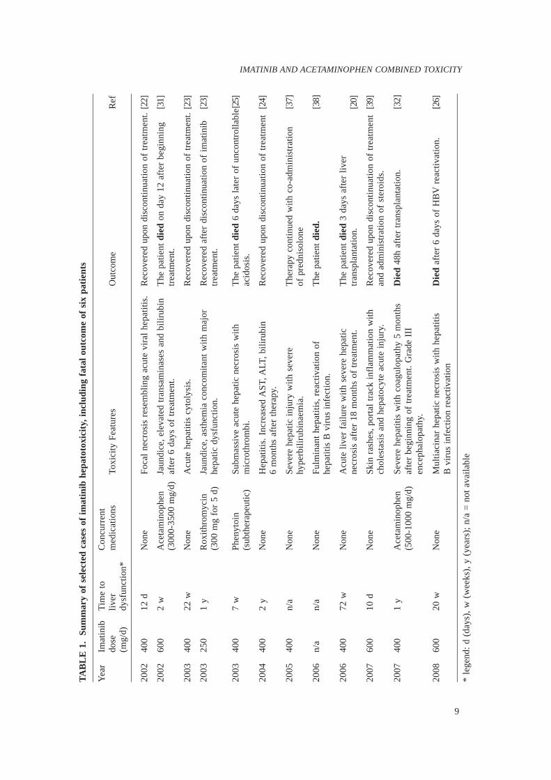
Although in most cases, hepatotoxicity (Table 1) resolved after imatinib discontinuation and steroids administration,21 in some patients, the hepatic condition further deteriorated leading to fatal liver failure.20 Histopathological analyses revealed severe necrosis, cellular canalicular cholestasis, submassive acute hepatic necrosis25 and multiacinar hepatocellular necrosis.26 Research on the pathogenic mechanisms of imatinib-induced hepatotoxicity or renal toxicity suggests that toxicity may be related to the P450 mediated metabolic pathway or idiosyncratic reactions in susceptible individuals.4
Acetaminophen is a commonly used over-the-counter analgesic and antipyretic drug that the WHO recommends as a first-line, non-opioid co-adjuvant analgesic for the relief of persistent pain affecting cancer patients,27 even with an established opioid regime.28 Acetaminophen presents drug-drug interactions with a variety of drugs including imatinib29 and may also cause hepatotoxicity at high doses.30 Severe hepatotoxicity associated with acetaminophen coadministration has been suggested previously in CML patients who developed fatal liver failure.31,32
The cur ren t s tudy eva lua tes the histopathological changes that occur in the liver and kidneys when imatinib and acetaminophen are co-administered and whether a drug-drug interaction exists in a preclinical mouse model.
MATERIALS AND METHODS
Experimental animals and procedures
Prior to initiation of the study, the Institutional Animal Use Ethics Committee reviewed and approved all the experimental protocols. Male
ICR mice, 12–14 weeks, ~30 g weight (Institute for Medical Research, Kuala Lumpur, Malaysia) were housed in the animal holding facility at 20 ± 2ºC and a 12-hour light/dark cycle, let to acclimatize for a week before any experiment was performed and provided feed and water ad libitum. The mice were randomly assigned to the experimental groups and fasted overnight. Group 1 did not receive any drug and was designated as the control for the histopathological analysis. Group 2 was dosed 100 mg/kg imatinib orally (in acidifi ed saline solution) using a 24G feeding needle and a 1 ml syringe (Terumo Corporation, Philippines). Group 3 received 700 mg/kg acetaminophen intraperitoneally (water) using a 26G needle with a 1 ml syringe. Lastly, Group 4 was dosed 100 mg/kg imatinib PO and 700 mg/kg acetaminophen IP. Imatinib was given fi rst immediately followed by acetaminophen. Mice (n=4 per time point) were euthanized at 15 min, 30 min, 1h, 2h, 4h and 6h by cervical dislocation and exsanguination performed via cardiac puncture with a 23G needle and a 1 mL syringe containing EDTA (Merck, Malaysia). The liver and the kidneys were harvested, rinsed twice in 0.9% saline solution, a piece cut and stored in 10% neutral buffered formalin solution (Richard-Allan Scientifi c, Malaysia) at room temperature. The remaining tissue was stored at -30ºC.
Preparation of histological sections
The tissue samples were grossed and transferred into Histosette® cassettes, placed in an automatic tissue processor (Leica, Japan) and processed as follows: (1) 10% neutral buffered formalin – two consecutive changes of 1 h and 1.5 h; (2) 85% alcohol – 1.5 h; (3) 95% alcohol – 1.5 h; (4) 100% Alcohol – three consecutive changes of 1.5 h each; (5) xylene (Fisher Scientifi c, Malaysia) – three consecutive changes of 1.5 h each; and (6) paraffi n (Richard-Allan Scientifi c) bath at 55ºC – two changes of 1.5 h each. Upon completion, the tissues were placed in 1.5x2 cm moulds lined with molten paraffi n wax at 65ºC. The mould was transferred to a cold plate at -5ºC, the tissue adjusted to the desired orientation and the cassette base placed on top of the mould, fi lled with molten wax and let to solidify for 1 h, removed and stored at -20ºC until sectioning. The frozen embedded wax blocks were sectioned at 3 μm thickness, placed on frosted glass slides and dried overnight at 37°C. Prior

3
IMATINIB AND ACETAMINOPHEN COMBINED TOXICITY
to modified Harris Haematoxylin and Eosin (Richard & Allan Scientific) staining, the samples were washed in xylene twice (3 min each), hydrated in five sequential changes of alcohol 100%, 100%, 95%, 80% and 70% for 3 min each, rinsed with water for 3 min and stained. Finally, the stained slides were dehydrated in three sequential 1-min changes of alcohol 70%, 80% and 95% and two changes of 100% alcohol for three minutes each. The sections were then dried and mounted onto the slide for analysis.
Histopathological analysis
Two stained slides for each mouse tissue samples were observed at 100x and 400x magnifi cation using a Nikon’s Brightfi eld Compound Nikon microscope (model YS100). Changes in lobular architecture, fatty changes, nuclear alterations and congestion of the sinusoids were evaluated in liver samples. Similarly, changes in cyto-architecture of the glomeruli, proximal and distal convoluted tubules and interstitium were assessed in the kidney samples. A semi-quantitative analysis was done to assess the extent of the histopathological changes. Photomicrographs of representative changes were also taken using a Nikon (Japan) camera attached to the microscope.
RESULTS
Histopathology in liver tissue
Histological examination of the liver tissues showed varied damage when compared with the baseline-control group (Figure 1). Experimental groups 2 and 3 showed signs of reversible damage, which were different from the results in
group 4 which showed irreversible damage. Mice dosed 100 mg/kg imatinib orally (group 2) showed feathery degeneration, microvesicular fatty change and very mild sinusoidal congestion (Figure 2). Toxicokinetic assessment revealed no effect at the earliest time point: the hepatocytes were normal with intact cell margins and normal nucleus. The lobular architecture and sinusoids appeared normal and no congestion of sinusoids was seen (Figure 2). The fi rst signs of toxicity appeared at 30 min when zones 1 and 2 showed feathery degeneration and loss of cellular outlines. By 1 h, the feathery degeneration extended to zone 3 and fatty change became diffuse at microvesicular level. This condition remained at 2 h but the histological signs of toxicity resolved by 4 h when all cells appeared normal, as was observed at 6 h. On the other hand, 700 mg/kg IP acetaminophen administration (group 3) caused reversible toxicity: sinusoidal congestion and centrilobular cell damage (Figure 3). Initial toxicity was observed 15 min after IP administration, when sinusoidal prominence and mild congestion was noted. Toxicity progressed to focal damage around the central vein and congestion at 30 min, which evolved to diffuse damage, poorly defi ned cellular outlines, loss of lobular architecture and congestion of the sinusoids at 1 h after administration. Nuclear clumping (pyknosis), further loss of lobular architecture and congestion of the sinusoids were clearly visible at 2 h post administration. These signs remained at 4 h with some microvesicular and macrovesicular fatty change, congestion and pyknosis. The toxicity was resolved at 6 h when no cellular damage and very minimal congestion were observed, and the cells were fully normal looking.
FIG. 1: Photomicrographs of liver and kidney after administration of 0.9 % saline solution (400x). (A) normal hepatocytic outlines and (B) normal glomerulus (black arrow) and proximal and distal convoluted tubules (white arrow).

Malaysian J Pathol June 2010
4
Mice that were treated with 100 mg/kg imatinib PO and 700 mg/kg acetaminophen IP (group 4) experienced the greatest damage which was mainly irreversible and included nuclear changes (pyknosis, karyolysis and karyorrhexis), cytolysis and massive congestion (Figure 4). At 30 min there were clear signs of toxicity: the hepatocytes appeared swollen, their nuclei enlarged and there was microvesicular
fatty change although minimal congestion. Progression of toxicity was clear at 1 h when macrovesicular fatty change, loss of cellular outline and lobular architecture and nuclear damage (pyknosis) were observed. Toxicity increased at 2 h with greater loss of lobular architecture, cellular outlines became completely indistinct and macrovesicular fatty change visible at zones 1 and 2. Nuclear damage progressed
FIG. 2: Photomicrographs of liver after 100 mg/kg imatinib PO administration (group 2). Feathery degeneration (blue arrow), lost of cellular outlines (red arrow) at zones 1 and 2 and normal centrilobular zone (black arrow) is seen at 30 min. These changes extended to zone 3 (circled) at 1 h. By 4 h the cell damage was resolved and the lobular architecture restored.

5
IMATINIB AND ACETAMINOPHEN COMBINED TOXICITY
FIG. 3: Photomicrographs of the liver after administration of 700 mg/kg IP acetaminophen. Mild sinusoidal congestion (black arrows), focal damage around the central vein (circled), feathery degeneration, loss of lobular architecture with damaged cellular outlines and congestion are seen at 1 h. By 2 h, nuclei have become condensed and pyknotic (blue arrow) which intensifi ed at 4 h with microvesicular fatty change (white arrows). By 6 h the damage has been reverted.

Malaysian J Pathol June 2010
6
FIG. 4: Photomicrographs of liver after administration of 700 mg/kg IP acetaminophen and 100 mg/kg PO imatinib. Mild sinusoidal congestion (black arrows), swollen hepatocytes and nuclei was seen within 30 min, by feathery degeneration (encircled), macrovesicular fatty change (black arrow) and pyknosis. The nuclear damage progressed with pyknosis (black arrow) and karyolysis (blue arrow) present at 6 h.

7
IMATINIB AND ACETAMINOPHEN COMBINED TOXICITY
to pyknosis and karyolysis and unlike the other two groups that recovered by 4 h, the combined administration of imatinib and acetaminophen led to further toxicity at 6 h: uniform macrovesicular fatty change, massive congestion and nuclear pyknosis, karyolysis and karyorrhexis. Moreover, cytolysis and loss of lobular architecture were still apparent.
Histopathology in renal tissue
Qualitative assessment of tissue sections of the kidney showed no damage in the experimental groups that received either imatinib or acetaminophen. This was consistent with other studies where no histopathological changes were observed in the kidney after an overdose of acetaminophen.33 Group 4 (combination treatment) revealed mild cloudy degeneration of the kidney tubules at 6 h time point, which is a reversible change (Figure 5).
DISCUSSION
Although imatinib is well tolerated it may lead to hepatotoxicity. Mice treated with 100 mg/
kg imatinib orally showed mild liver damage with the peak cell damage at 1 hour but with clear signs of restoration at 4 h and complete restoration by 6 h. This level of toxicity is in agreement with the overall findings in other preclinical studies or in clinical trials. Studies in dogs showed hepatotoxicity 2 weeks after imatinib administration. The histological assessment revealed mild focal hepatocellular necrosis, single cell bile duct necrosis and bile duct hyperplasia (associated with peribiliary fibrosis) and elevation of liver enzymes.3 In humans, signs of liver dysfunction were found in CML patients with grade 3 AST abnormalities in 2%, AST in 3% and 2% in bilirubin as well as hepatobiliary disorders (Budd-Chiari syndrome and hepatotoxicity).27 Similar signs of hepatoxicity were found in other human studies involving CML patients where grade 3 or 4 increased ALT and AST levels in ~5% of them.34 Discontinuation of treatment and initiation of steroids usually stopped and reverted toxicity progression.20 However in several cases, fatal liver failure occurred24,26 (Table 1). Our histological findings in mice show lesser
FIG. 5: Photomicrographs (400x) of kidney 6 h after administration of (A) vehicle control; (B) 100 mg/kg oral imatinib; (C) 700 mg/kg IP acetaminophen and (D) combination treatment of 100 mg/kg PO imatinib and 700 mg/kg IP acetaminophen.

Malaysian J Pathol June 2010
8
degree of toxicity which may be anticipated since we used a single oral dose of imatinib (100 mg/kg); while the greater toxicity observed in the reported cases may be contributed by imatinib accumulation due to the multiple dosage administration.3
Acetaminophen toxicity at high doses is well known.30 Our dose of 700 mg/kg IP was established after initial studies showed dose dependent hepatotoxicity. After acetaminophen IP administration, we observed congestion of the sinusoids and centrilobular necrosis (Figure 3) which extended around the central vein which are hallmark signs of acetaminophen toxicity.30 Pyknotic nuclear change and microvesicular fatty change observed are consistent with findings from other studies as well as the reversible nature of the toxicity.35
Coadministration of acetaminophen with imatinib changed the hepatoxicity from reversible to irreversible and increased its intensity and extent (Figure 4). After co-administration, we
observed initial mild congestion of sinusoids progressing to loss of lobular architecture, macrovesicular fatty change and pyknosis was not restored at 6 hours (Figure 4). In fact, pyknosis and karyolysis in the nuclei together with cytolysis mark irreversible toxicity suggesting synergistic toxicity associated with both drugs coadministration. As shown in the toxicokinetic comparative time profile in Figure 6, imatinib seems to prevent hepatocyte recovery from the damage caused, probably via the acetaminophen metabolite, NAPQI. While there are clear signs of restoration at 4 h in the control groups, the toxicity signs in the combination treatment further intensified and progressed without recovery by 6 hours. This toxicokinetic profile suggests that the presence of imatinib leads to a ‘perpetuation’ of acetaminophen toxicity. This appears similar to cases of acetaminophen coadministration with imatinib in cancer patients leading to irreversible fatal liver failure.27,28
Sinusoidal Congestion
Architecture & Cell outline
Fatty Change
Nuclear Change
0 2 4 6
Time (h)ImatinibAcetaminophenAcetaminophen + Imatinib
Sinusoidal Congestion
Architecture & Cell outline
Fatty Change
Nuclear Change
0 2 4 6
Time (h)ImatinibAcetaminophenAcetaminophen + Imatinib
ImatinibImatinibAcetaminophenAcetaminophenAcetaminophen + ImatinibAcetaminophen + Imatinib
FIG. 6: Comparative toxicokinetic assessment of main histopathological fi ndings in liver for control and combination groups.
9
IMATINIB AND ACETAMINOPHEN COMBINED TOXICITY
TA
BL
E 1
. Su
mm
ary
of s
elec
ted
case
s of
im
atin
ib h
epat
otox
icit
y, i
nclu
ding
fat
al o
utco
me
of s
ix p
atie
nts
Yea
r Im
atin
ib
Tim
e to
C
oncu
rren
t
dose
li
ver
med
icat
ions
To
xici
ty F
eatu
res
Out
com
e R
ef
(m
g/d)
dy
sfun
ctio
n*
2002
40
0 12
d
Non
e Fo
cal
necr
osis
res
embl
ing
acut
e vi
ral
hepa
titi
s.
Rec
over
ed u
pon
disc
onti
nuat
ion
of t
reat
men
t. [2
2]
2002
60
0 2
w
Ace
tam
inop
hen
Jaun
dice
, ele
vate
d tr
ansa
min
ases
and
bil
irub
in
The
pat
ient
die
d on
day
12
afte
r be
ginn
ing
[31]
(3
000-
3500
mg/
d)
afte
r 6
days
of
trea
tmen
t. tr
eatm
ent.
2003
40
0 22
w
Non
e A
cute
hep
atit
is c
ytol
ysis
. R
ecov
ered
upo
n di
scon
tinu
atio
n of
tre
atm
ent.
[23]
2003
25
0 1
y R
oxit
hrom
ycin
Ja
undi
ce, a
sthe
mia
con
com
itan
t w
ith
maj
or
Rec
over
ed a
fter
dis
cont
inua
tion
of
imat
inib
[2
3]
(300
mg
for
5 d)
he
pati
c dy
sfun
ctio
n.
trea
tmen
t.
2003
40
0 7
w
Phen
ytoi
n Su
bmas
sive
acu
te h
epat
ic n
ecro
sis
wit
h T
he p
atie
nt d
ied
6 da
ys l
ater
of
unco
ntro
llab
le [2
5]
(sub
ther
apeu
tic)
m
icro
thro
mbi
. ac
idos
is.
2004
40
0 2
y N
one
Hep
atit
is. I
ncre
ased
AST
, ALT
, bil
irub
in
Rec
over
ed u
pon
disc
onti
nuat
ion
of t
reat
men
t [2
4]
6
mon
ths
afte
r th
erap
y.
2005
40
0 n/
a N
one
Seve
re h
epat
ic i
njur
y w
ith
seve
re
The
rapy
con
tinu
ed w
ith
co-a
dmin
istr
atio
n [3
7]
hy
perb
ilir
ubin
aem
ia.
of p
redn
isol
one
2006
n/
a n/
a N
one
Fulm
inan
t he
pati
tis,
rea
ctiv
atio
n of
T
he p
atie
nt d
ied.
[3
8]
he
pati
tis
B v
irus
inf
ecti
on.
2006
40
0 72
w
Non
e A
cute
liv
er f
ailu
re w
ith
seve
re h
epat
ic
The
pat
ient
die
d 3
days
aft
er l
iver
necr
osis
aft
er 1
8 m
onth
s of
tre
atm
ent.
tran
spla
ntat
ion.
[2
0]
2007
60
0 10
d
Non
e Sk
in r
ashe
s, p
orta
l tr
ack
infl
amm
atio
n w
ith
Rec
over
ed u
pon
disc
onti
nuat
ion
of t
reat
men
t [3
9]
chol
esta
sis
and
hepa
tocy
te a
cute
inj
ury.
an
d ad
min
istr
atio
n of
ste
roid
s.
2007
40
0 1
y A
ceta
min
ophe
n Se
vere
hep
atit
is w
ith
coag
ulop
athy
5 m
onth
s D
ied
48h
afte
r tr
ansp
lant
atio
n.
[32]
(5
00-1
000
mg/
d)
afte
r be
ginn
ing
of t
reat
men
t. G
rade
III
en
ceph
alop
athy
.
2008
60
0 20
w
Non
e M
ulti
acin
ar h
epat
ic n
ecro
sis
wit
h he
pati
tis
Die
d af
ter
6 da
ys o
f H
BV
rea
ctiv
atio
n.
[26]
B
vir
us i
nfec
tion
rea
ctiv
atio
n
* le
gend
: d
(day
s),
w (
wee
ks),
y (
year
s);
n/a
= n
ot a
vail
able

Malaysian J Pathol June 2010
10
Working hypothesis
The pathogenic mechanisms of imatinib-induced hepatotoxicity are unknown. How imatinib may interfere in the pharmacokinetics of acetaminophen remains to be clarified.29 Imatinib presents numerous drug-drug interactions which involve P450 mediated metabolism and efflux transporters (e.g. P-glycoprotein).2 The change from reversible to irreversible toxicity suggests that the interaction may be synergistic in nature. The extensive and sustained damage to hepatocytes in group 4 compared to groups 2 and 3 may reflect the inability of hepatocytes to eliminate acetaminophen and its metabolites. Imatinib may interfere with transporter-mediated removal mechanisms of acetaminophen or its metabolites. In vitro evaluation of the role of P-glycoprotein in acetaminophen toxicity showed that the presence of verapamil (a P-glycoprotein and CYP3A4 substrate) significantly increased acetaminophen induced cell damage, even at non toxic doses of acetaminophen.36 The results of our study in vivo are consistent with that finding. It is plausible that imatinib, which shares the same transporters and cytochrome P450 may be affecting the ability of the efflux pumps to reduce acetaminophen-induced hepatotoxicity.
Conclusion
Liver histopathological changes observed after coadministration of imatinib and acetaminophen show an increase of acetaminophen toxicity and a change from reversible to irreversible. This fi nding may have deep consequences in the clinical management of cancer related pain. The fact that several deaths associated with acetaminophen-imatinib coadministration have occurred, clearly points to the need for close monitoring of liver function in cancer patients receiving imatinib treatment and acetaminophen for pain management. No signifi cant renal toxicity was observed in the study.
ACKNOWLEDGEMENTS
The authors thank Ms Kanakeswary Karisnan and Mr Kuzairy Abu Bakar for their technical assistance and the International Medical University (IMU) for fi nancial support: grant # BMS I-02/2007(09).
REFERENCES
1. Deininger M, Buchdunger E, Druker BJ. The development of imatinib as a therapeutic agent
for chronic myeloid leukemia. Blood. 2005; 105: 2640-53.
2. Peng B, Lloyd P, Schran H. Clinical pharmacokinetics of imatinib. Clin Pharmacokinet. 2005; 44: 879-94.
3. Cohen MH, Williams G, Johnson JR, et al. Approval summary for imatinib mesylate capsules in the treatment of chronic myelogenous leukemia. Clin Can Res. 2002; 8: 935-42.
4. Druker BJ, Guilhot F, O’Brien SG, et al. Five year follow-up of patients receiving imatinib for chronic myeloid leukemia. N Engl J Med. 2007; 356: 2408-17.
5. Buchdunger E, Cioffi CL, Law N, Stover D, Ohno-Jones S, Druker BJ, Lydon NB. Abl protein-tyrosine kinase inhibitor ST1571 inhibits in vitro signal transduction mediated by c-kit and platelet derived growth factor receptors. J Pharmacol Exp Ther. 2000; 295: 139-45.
6. Braconi C, Bracci R, Cellerino R. Molecular targets in Gastrointestinal Stromal Tumors (GIST) therapy. Curr Cancer Drug Targets. 2008; 8:359-66.
7. Cassier PA, Dufresne A, Arifi S, et al. Imatinib mesilate for the treatment of gastrointestinal stromal tumour. Expert Opin Pharmacother. 2008; 9: 1211-22.
8. Handolias D, McArthur GA. Imatinib as effective therapy for dermatofi brosarcoma protuberans: proof of concept of the autocrine hypothesis for cancer. Future Oncol. 2008; 4: 211-7.
9. Haberler C, Gelpi E, Marosi C, Rössler K, Birner P, Budka H, Hainfellner JA. Immunohistochemical analysis of platelet-derived growth factor receptor -alpha, -beta, c-kit, c-abl, and arg proteins in glioblastoma: possible implications for patient selection for imatinib mesylate therapy. J Neurooncol. 2006; 76: 105-9.
10. Leder C, Ortler S, Seggewiss R, Einsele H, Wiendl H. Modulation of T-effector function by imatinib at the level of cytokine secretion. Exp Hematol. 2007; 35: 1266-71.
11. Lassila M, Allen TJ, Cao Z, Thallas V, Jandeleit-Dahm KA, Candido R, Cooper ME. Imatinib attenuates diabetes-associated atherosclerosis. Arterioscler Thromb Vasc Biol. 2004; 24: 935-942.
12. Peng B, Dutreix C, Mehring G, et al. Absolute bioavailability of imatinib (Glivec) orally versus intravenous infusion. J Clin Pharmacol. 2004; 44: 158-62.
13. Nikolova Z, Peng B, Hubert M, et al. Bioequivalence, safety, and tolerability of imatinib tablets compared with capsules. Cancer Chemother Pharmacol. 2004; 53: 433-8.
14. Kretz O, Weiss HM, Schumacher MM, Gross G. In vitro blood distribution and plasma protein binding of the tyrosine kinase inhibitor imatinib and its active metabolite, CGP74588, in rat, mouse, dog, monkey, healthy humans and patients with acute lymphatic leukemia. Br J Clin Pharmacol. 2004; 58: 212-6.
15. Dai H, Marbach P, Lemaire M, Hayes M, Elmquist WF. Distribution of STI-571 to the Brain is Limited by P-Glycoprotein-Mediated Effl ux. J Pharmacol Exp Ther. 2003; 304: 1085-92.
16. Gschwind HP, Pfaar U, Waldmeier F, et al. Metabolism and disposition of imatinib in healthy volunteers. Drug Metab Dispos. 2005; 33: 1503-12.

11
IMATINIB AND ACETAMINOPHEN COMBINED TOXICITY
17. Ramalingam S, Lagattuta TF, Egorin MJ, et al. Biliary excretion of imatinib mesylate and its metabolite CGP 74588 in humans. Pharmacotherapy. 2004; 24: 1232-5.
18. Pou M, Saval N, Vera M, Saurina A, Solé M, Cervantes F, Botey A. Acute renal failure secondary to imatinib mesylate treatment in chronic myeloid leukemia. Leuk Lymphoma. 2003; 44: 1239-41.
19. Foringer JR, Verami RR, Tjia VM, et al. Acute renal failure to imatinib mesylate treatment in prostate cancer. Ann Pharmacother. 2005; 39: 2136-8.
20. Cross TJ, Bagot C, Portmann B, Wendon J, Gillett D. Imatinib mesylate as a cause of acute liver failure. Am J Hematol. 2006; 81: 189-92.
21. Ferrero D, Pogliani EM, Rege-Cambrin G, et al. Corticosteriods can reverse severe imantinb-induded hepatotoxicity. Haematologica. 2006; 91: e78-e80.
22. Ohyashiki K, Kuriyama Y, Nakajima A, et al. Imatinib mesylate-induced hepato-toxicity in chronic myeloid leukemia demonstrated focal necrosis resembling acute viral hepatitis. Leukemia 2002; 16: 2160-61.
23. James C, Trouette H, Marit G, Cony-Makhoul P, Mahon F-X. Histological features of acute hepatitis after imatinib mesylate treatment. Leukemia. 2003; 17: 978-9.
24. Kikuchi S, Muroi K, Takahashi S, et al. Severe hepatitis and complete molecular response caused by imatinib mesylate: possible association of its serum concentration with clinical outcomes. Leuk Lymphoma. 2004; 45: 2349-51.
25. Lin NU, Sarantopoulos S, Stone JR, Galinsky I, Stone RM, Deangelo DJ, Soiffer RJ. Fatal hepatic necrosis following imatinib mesylate therapy. Blood. 2003; 102: 3455-6.
26. Thia TJK, Tan HH, Chuah THC, Chow WC, Lui HF. Imatinib mesylate-related fatal acute hepatic failure in a patient with chronic myeloid leukaemia and chronic hepatitis B infection. Singapore Med J. 2008; 49: e86-e89.
27. Christo PJ, Mazloomdoost D. Cancer pain and an-algesia. Ann N Y Acad Sci. 2008; 1138: 278-98.
28. Stockler M, Vardy J, Pillai V, Warr D. Acetami-nophen (Paracetamol) improves pain and well-being in people with advanced cancer already receiving a strong opioid regimen: A randomized, double-blind, placebo-controlled cross-over trial. J Clin Oncol. 2004; 22: 3389-94.
29. Nassar I, Pasupati T, Judson JP, Segarra I. Reduced exposure of imatinib after coadministration with acetaminophen to mice. Ind J Pharmacol. 2009; 41: 167-72.
30. Larson AM. Acetaminophen hepatotoxicity. Clin Liver Dis 2007; 11: 525-48.
31. Talpaz M, Silver RT, Druker BJ, et al. Imatinib induces durable hematologic and cytogenetic re-sponses in patients with accelerated phase chronic myeloid leukemia: results of a phase 2 study. Blood. 2002; 99: 1928-37.
32. Ridruejo E, Cacchione R, Villamil AG, Marciano S, Gadano AC, Mando OG. Imatinib-induced fatal acute liver failure. World J Gastroenterol. 2007;13: 6608-11.
33. Mazer M, Perrone J. Acetaminophen-induced nephrotoxicity: pathophysiology, clinical manifesta-
tions, and management. J Med Toxicol 2008; 4: 2-6. 34. O’Brien SG, Guilhot F, Larson RA, et al. Imatinib
compared with interferon and low dose cytarabine for newly diagnosed chronic phase chronic myeloid leukemia. N Engl J Med 2003; 384: 994-1004.
35. Ghosh A, Parames C. Anti-oxidative effect of a protein from cajanus indicus L against acetaminophen-induced hepato-nephro toxicity. J Biochem Mol Bio 2007; 40: 1039-49.
36. Manov I, Bashenko Y, Hirsh M, Iancu TC. Involvement of the multidrug resistance P-glycoprotein in acetaminophen-induced toxicity in hepatoma-derived HepG2 and Hep3B cells. Basic Clin Pharmacol Toxicol 2006; 99: 213-24.
37. Ikuta K, Torimoto Y, Jimbo J, et al. Severe hepatic injury caused by imatinib mesylate administered for the treatment of chronic myeloid leukemia and the effi cacy of prednisolone for its management. Int J Hematol. 2005; 82: 343-6.
38. Ikeda K, Shiga Y, Takahashi A, et al. Fatal hepatitis B virus reactivation in a chronic myeloid leukemia patient during imatinib mesylate treatment. Leuk Lymphoma. 2006; 47: 155-7.
39. Kong JH, Yoo SH, Lee KE, et al. Early imatinib-mesylate-induced hepatotoxicity in chronic my-elogenous leukaemia. Acta Haematol. 2007; 118: 205-8.