Embed Size (px)
Citation preview

Romanian Journal of Morphology and Embryology 2009, 50(3):327–339
OORRIIGGIINNAALL PPAAPPEERR
Histopathological changes in acute ischemic stroke
OTILIA MĂRGĂRITESCU1), L. MOGOANTĂ2), IONICA PIRICI3), D. PIRICI2), DANIELA CERNEA4), CL. MĂRGĂRITESCU5)
1)Department of Neurosurgery, Emergency County Hospital, Craiova
2)Department of Histology 3)Department of Neurology
4)Department of Anesthesia and Intensive Care 5)Department of Pathology
University of Medicine and Pharmacy of Craiova
Abstract We study here the histopathological changes in twenty-two cases of acute ischemic stroke. The average age of the patients was 62-year-old, and the interval from the onset of the disease to the death varied from 6 hours to 15 years. The brain lesions after acute stroke were observed in all regions. Their evolution allowed us to classify them in fourth stages. Phase one changes (1–2 days after onset) (n=2 patients) included red hypoxic and “ghost” neurons and other acute neuronal injury and spongiosis. The second phase (n=14 patients) was subdivided into: (a) a phase of acute inflammation (3–37 days after onset) (n=5 patients), where we observed especially features of acute inflammation together with coagulative necrosis, and (b) phase of chronic inflammation (10 days–53 years after the onset) (n=9 patients), in which prevail mononuclear and macrophage infiltrate, astrogliosis, spongiosis and neo-vascularization. In the third phase (26 days–23 years after the onset), we included six cases characterized by the absence of an inflammatory reaction, presence of cavitation, astrogliosis and macrophages. Our study describes the heterogeneity of brain injury after acute ischemic stroke with the participation of all brain components, and the chronology in which these lesions develop and evolve. Keywords: brain, infarct, histopathology, stroke.
Introduction
Stroke, previously called cerebrovascular accident (CVA) is characterized by the sudden loss of blood circulation in an area of the brain, resulting in a corresponding loss of neurological function.
In USA, incidence for first-time stroke is more than 700.000 per year, of which 20% of the patients will die within the first year after the onset of the stroke. Keeping the current trends, this number is projected to rise up to 1 million per year by the year 2050 [1]. Worldwide, in 1990, cerebrovascular disease was the second leading cause of death, killing more than 4.3 million people [2], and also was the fifth leading cause of occupational impairment [3]. Although stroke often is considered a disease of the elderly, one third of strokes occur in persons younger than 65 years, men being at higher risk for stroke than women [1].
Strokes are broadly classified as either hemorrhagic or ischemic. In the last years, a great deal of efforts was directed toward understanding the pathophysiology and histopathology of cerebral infarcts, in both humans and experimental animal models [1, 4–21].
The aim of our study was to investigate the morphological changes in cerebral parenchyma after acute ischemic stroke, attempting to establish the cellular sequential steps that define this pathology.
Material and Methods
Twenty-two cases of acute cerebral ischemic stroke were selected from the archive of Pathology Department from Emergency County Hospital, Craiova, Romania, over a period of 20 years (1987–2006).
Medical records were reviewed to retrieve informa-tion regarding patient age at the time of death or surgery, gender, signs and symptoms at presentation, radiological characteristics of the lesions, duration of survival from the date of admission until death or surgery, and the cause of death. Information obtained from the autopsy protocol included lesion location, size, distribution within a particular arterial territory, macroscopic characteristics, status of intracranial arteries and general autopsy findings.
The histological specimens were collected during necropsy and were process in routine histopathological techniques (10% buffered neutral formalin fixation, paraffin embedding, 3–5 µm thick-section cutting), and stained by Hematoxylin–Eosin, Cresyl Violet, Mallory’s trichrome, and Luxol Fast Blue–Periodic Acid-Schiff–Toluidine Blue stains.
Macrophages, astrocytes and lymphocytes involved in cerebral infarction were studied by immunohisto-chemistry using “ABC”-peroxidase technique (with VECTASTAIN Elite ABC Kit, Vector Laboratories,
Otilia Mărgăritescu et al.
328
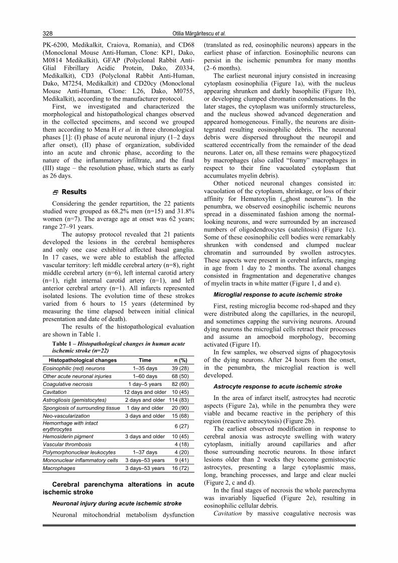
PK-6200, Medikalkit, Craiova, Romania), and CD68 (Monoclonal Mouse Anti-Human, Clone: KP1, Dako, M0814 Medikalkit), GFAP (Polyclonal Rabbit Anti-Glial Fibrillary Acidic Protein, Dako, Z0334, Medikalkit), CD3 (Polyclonal Rabbit Anti-Human, Dako, M7254, Medikalkit) and CD20cy (Monoclonal Mouse Anti-Human, Clone: L26, Dako, M0755, Medikalkit), according to the manufacturer protocol.
First, we investigated and characterized the morphological and histopathological changes observed in the collected specimens, and second we grouped them according to Mena H et al. in three chronological phases [1]: (I) phase of acute neuronal injury (1–2 days after onset), (II) phase of organization, subdivided into an acute and chronic phase, according to the nature of the inflammatory infiltrate, and the final (III) stage – the resolution phase, which starts as early as 26 days.
Results
Considering the gender repartition, the 22 patients studied were grouped as 68.2% men (n=15) and 31.8% women (n=7). The average age at onset was 62 years; range 27–91 years.
The autopsy protocol revealed that 21 patients developed the lesions in the cerebral hemispheres and only one case exhibited affected basal ganglia. In 17 cases, we were able to establish the affected vascular territory: left middle cerebral artery (n=8), right middle cerebral artery (n=6), left internal carotid artery (n=1), right internal carotid artery (n=1), and left anterior cerebral artery (n=1). All infarcts represented isolated lesions. The evolution time of these strokes varied from 6 hours to 15 years (determined by measuring the time elapsed between initial clinical presentation and date of death).
The results of the histopathological evaluation are shown in Table 1.
Table 1 – Histopathological changes in human acute ischemic stroke (n=22)
Histopathological changes Time n (%) Eosinophilic (red) neurons 1–35 days 39 (28) Other acute neuronal injuries 1–60 days 68 (50) Coagulative necrosis 1 day–5 years 82 (60) Cavitation 12 days and older 10 (45) Astrogliosis (gemistocytes) 2 days and older 114 (83) Spongiosis of surrounding tissue 1 day and older 20 (90) Neo-vascularization 3 days and older 15 (68) Hemorrhage with intact erythrocytes 6 (27)
Hemosiderin pigment 3 days and older 10 (45) Vascular thrombosis 4 (18) Polymorphonuclear leukocytes 1–37 days 4 (20) Mononuclear inflammatory cells 3 days–53 years 9 (41) Macrophages 3 days–53 years 16 (72)
Cerebral parenchyma alterations in acute ischemic stroke
Neuronal injury during acute ischemic stroke
Neuronal mitochondrial metabolism dysfunction
(translated as red, eosinophilic neurons) appears in the earliest phase of infarction. Eosinophilic neurons can persist in the ischemic penumbra for many months (2–6 months).
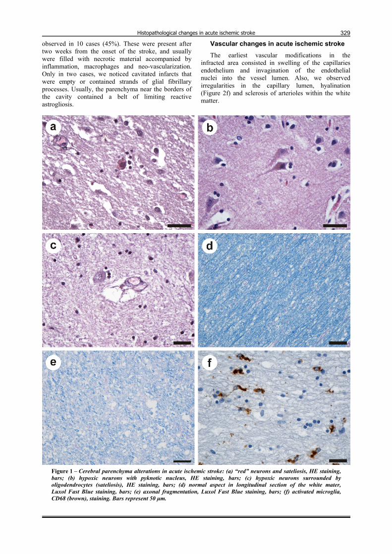
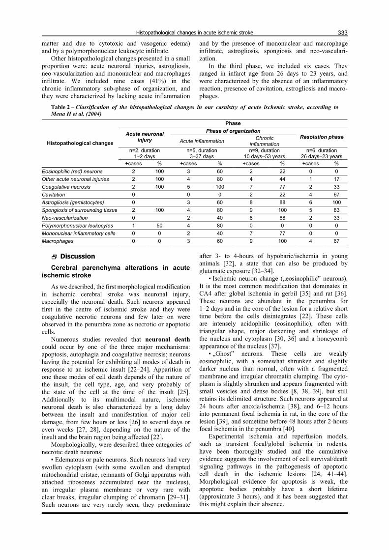
The earliest neuronal injury consisted in increasing cytoplasm eosinophilia (Figure 1a), with the nucleus appearing shrunken and darkly basophilic (Figure 1b), or developing clumped chromatin condensations. In the later stages, the cytoplasm was uniformly structureless, and the nucleus showed advanced degeneration and appeared homogeneous. Finally, the neurons are disin-tegrated resulting eosinophilic debris. The neuronal debris were dispersed throughout the neuropil and scattered eccentrically from the remainder of the dead neurons. Later on, all these remains were phagocytized by macrophages (also called “foamy” macrophages in respect to their fine vacuolated cytoplasm that accumulates myelin debris).
Other noticed neuronal changes consisted in: vacuolation of the cytoplasm, shrinkage, or loss of their affinity for Hematoxylin („ghost neurons”). In the penumbra, we observed eosinophilic ischemic neurons spread in a disseminated fashion among the normal-looking neurons, and were surrounded by an increased numbers of oligodendrocytes (satelitosis) (Figure 1c). Some of these eosinophilic cell bodies were remarkably shrunken with condensed and clumped nuclear chromatin and surrounded by swollen astrocytes. These aspects were present in cerebral infarcts, ranging in age from 1 day to 2 months. The axonal changes consisted in fragmentation and degenerative changes of myelin tracts in white matter (Figure 1, d and e).
Microglial response to acute ischemic stroke
First, resting microglia become rod-shaped and they were distributed along the capillaries, in the neuropil, and sometimes capping the surviving neurons. Around dying neurons the microglial cells retract their processes and assume an amoeboid morphology, becoming activated (Figure 1f).
In few samples, we observed signs of phagocytosis of the dying neurons. After 24 hours from the onset, in the penumbra, the microglial reaction is well developed.
Astrocyte response to acute ischemic stroke
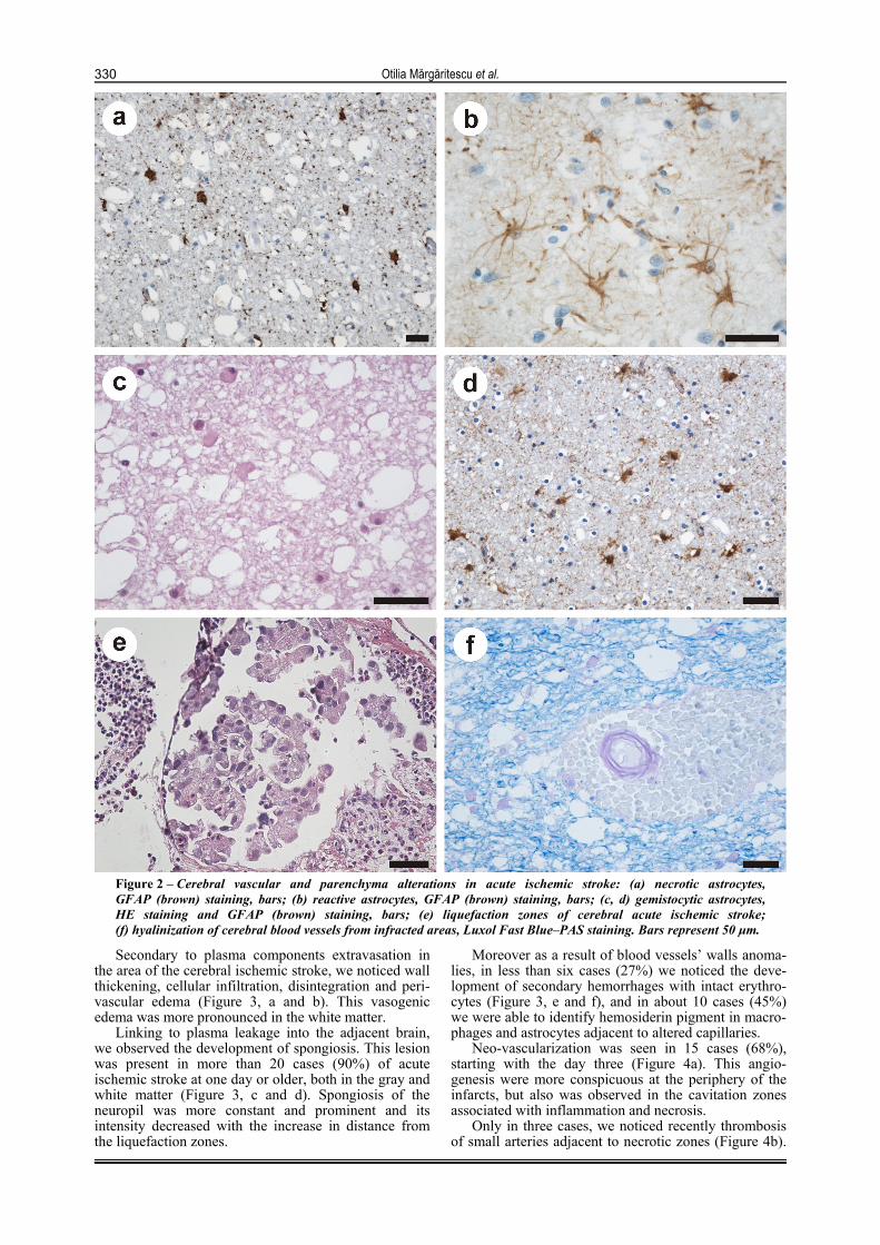
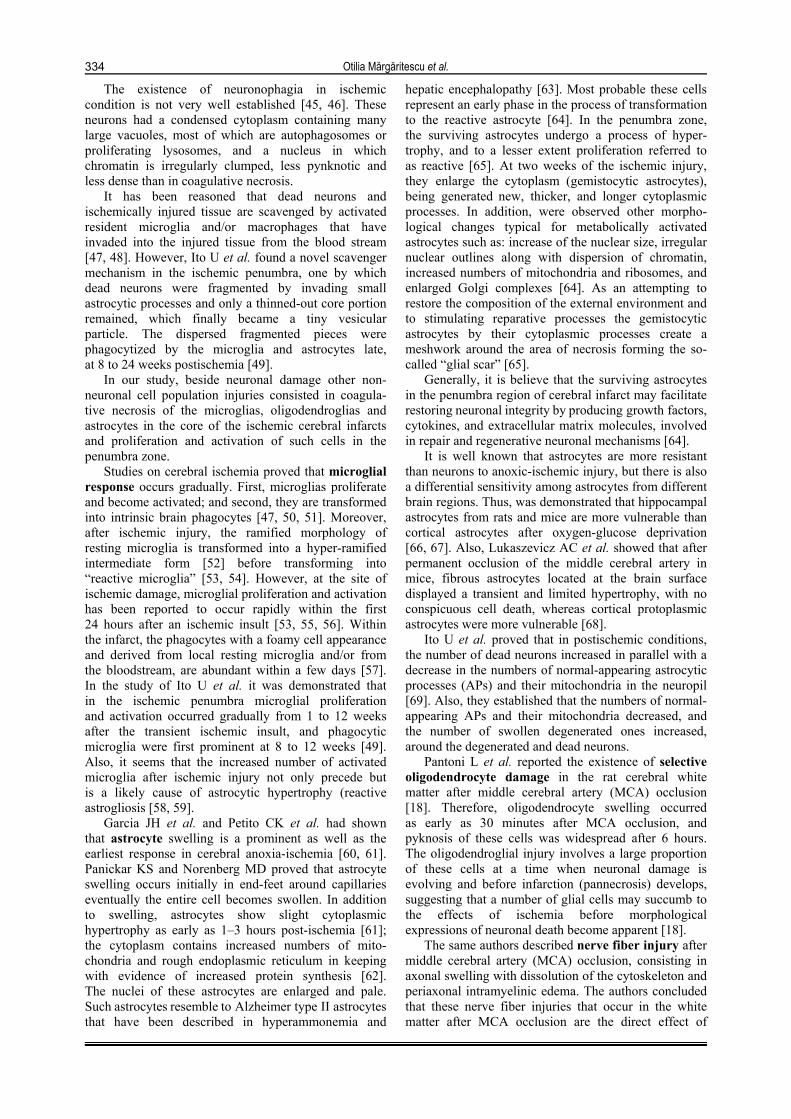
In the area of infarct itself, astrocytes had necrotic aspects (Figure 2a), while in the penumbra they were viable and became reactive in the periphery of this region (reactive astrocytosis) (Figure 2b).
The earliest observed modification in response to cerebral anoxia was astrocyte swelling with watery cytoplasm, initially around capillaries and after those surrounding necrotic neurons. In those infarct lesions older than 2 weeks they become gemistocytic astrocytes, presenting a large cytoplasmic mass, long, branching processes, and large and clear nuclei (Figure 2, c and d).
In the final stages of necrosis the whole parenchyma was invariably liquefied (Figure 2e), resulting in eosinophilic cellular debris.
Cavitation by massive coagulative necrosis was
Histopathological changes in acute ischemic stroke
329
observed in 10 cases (45%). These were present after two weeks from the onset of the stroke, and usually were filled with necrotic material accompanied by inflammation, macrophages and neo-vascularization. Only in two cases, we noticed cavitated infarcts that were empty or contained strands of glial fibrillary processes. Usually, the parenchyma near the borders of the cavity contained a belt of limiting reactive astrogliosis.
Vascular changes in acute ischemic stroke The earliest vascular modifications in the
infracted area consisted in swelling of the capillaries endothelium and invagination of the endothelial nuclei into the vessel lumen. Also, we observed irregularities in the capillary lumen, hyalination (Figure 2f) and sclerosis of arterioles within the white matter.
Figure 1 – Cerebral parenchyma alterations in acute ischemic stroke: (a) “red” neurons and sateliosis, HE staining, bars; (b) hypoxic neurons with pyknotic nucleus, HE staining, bars; (c) hypoxic neurons surrounded by oligodendrocytes (sateliosis), HE staining, bars; (d) normal aspect in longitudinal section of the white mater, Luxol Fast Blue staining, bars; (e) axonal fragmentation, Luxol Fast Blue staining, bars; (f) activated microglia, CD68 (brown), staining. Bars represent 50 µm.
Otilia Mărgăritescu et al.
330
Figure 2 – Cerebral vascular and parenchyma alterations in acute ischemic stroke: (a) necrotic astrocytes, GFAP (brown) staining, bars; (b) reactive astrocytes, GFAP (brown) staining, bars; (c, d) gemistocytic astrocytes, HE staining and GFAP (brown) staining, bars; (e) liquefaction zones of cerebral acute ischemic stroke; (f) hyalinization of cerebral blood vessels from infracted areas, Luxol Fast Blue–PAS staining. Bars represent 50 µm.
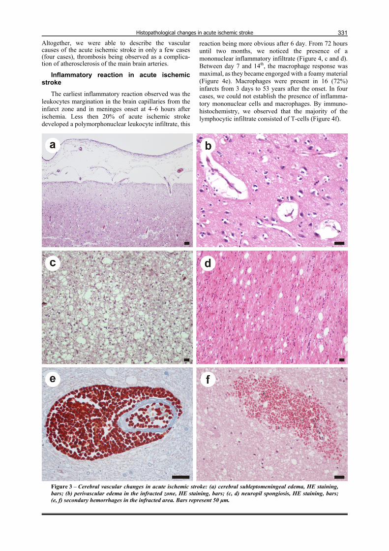
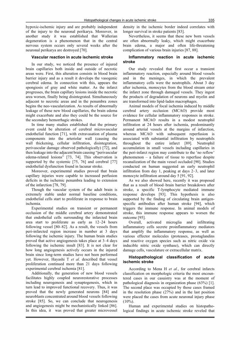
Secondary to plasma components extravasation in the area of the cerebral ischemic stroke, we noticed wall thickening, cellular infiltration, disintegration and peri-vascular edema (Figure 3, a and b). This vasogenic edema was more pronounced in the white matter.
Linking to plasma leakage into the adjacent brain, we observed the development of spongiosis. This lesion was present in more than 20 cases (90%) of acute ischemic stroke at one day or older, both in the gray and white matter (Figure 3, c and d). Spongiosis of the neuropil was more constant and prominent and its intensity decreased with the increase in distance from the liquefaction zones.
Moreover as a result of blood vessels’ walls anoma-lies, in less than six cases (27%) we noticed the deve-lopment of secondary hemorrhages with intact erythro-cytes (Figure 3, e and f), and in about 10 cases (45%) we were able to identify hemosiderin pigment in macro-phages and astrocytes adjacent to altered capillaries.
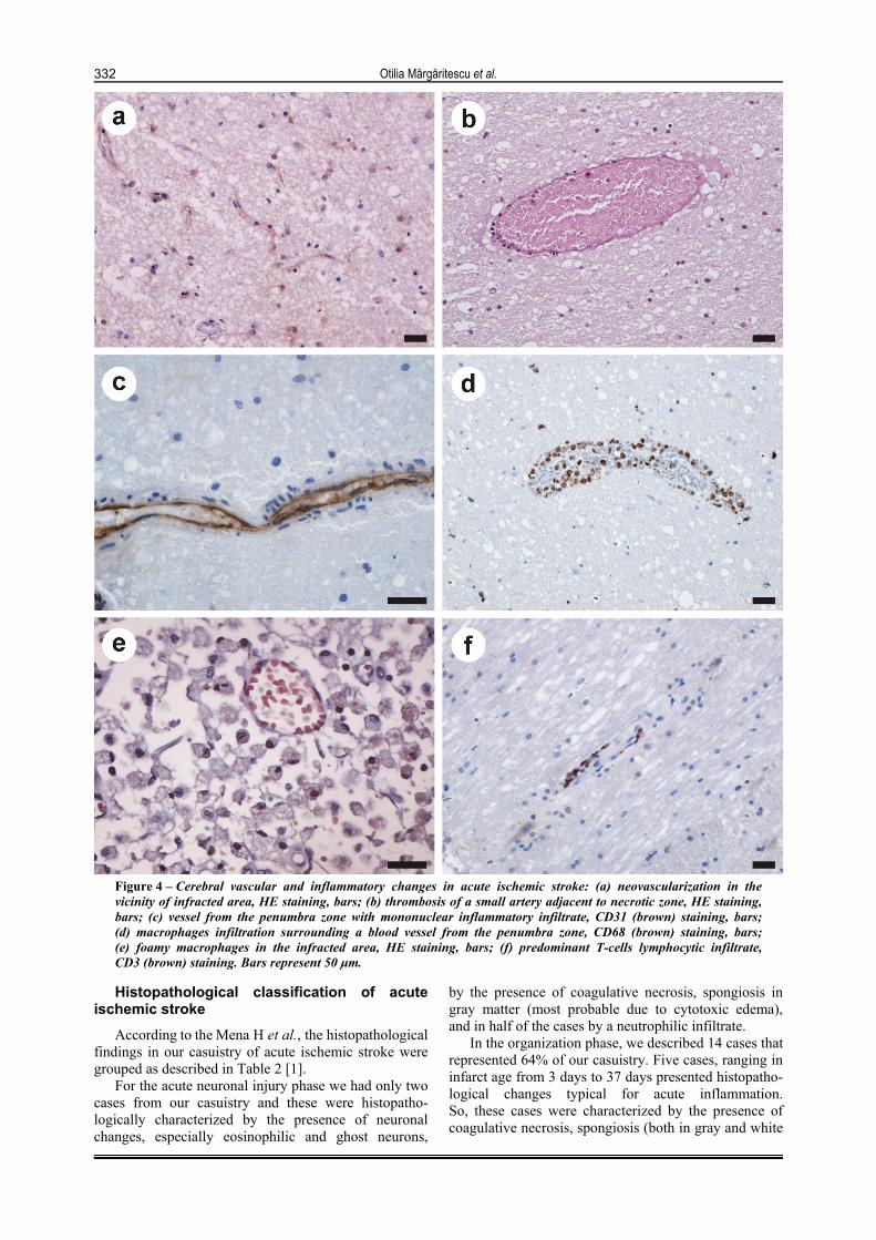
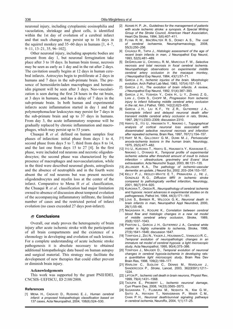
Neo-vascularization was seen in 15 cases (68%), starting with the day three (Figure 4a). This angio-genesis were more conspicuous at the periphery of the infarcts, but also was observed in the cavitation zones associated with inflammation and necrosis.
Only in three cases, we noticed recently thrombosis of small arteries adjacent to necrotic zones (Figure 4b).
Histopathological changes in acute ischemic stroke
331
Altogether, we were able to describe the vascular causes of the acute ischemic stroke in only a few cases (four cases), thrombosis being observed as a complica-tion of atherosclerosis of the main brain arteries.
Inflammatory reaction in acute ischemic stroke
The earliest inflammatory reaction observed was the leukocytes margination in the brain capillaries from the infarct zone and in meninges onset at 4–6 hours after ischemia. Less then 20% of acute ischemic stroke developed a polymorphonuclear leukocyte infiltrate, this
reaction being more obvious after 6 day. From 72 hours until two months, we noticed the presence of a mononuclear inflammatory infiltrate (Figure 4, c and d). Between day 7 and 14th, the macrophage response was maximal, as they became engorged with a foamy material (Figure 4e). Macrophages were present in 16 (72%) infarcts from 3 days to 53 years after the onset. In four cases, we could not establish the presence of inflamma-tory mononuclear cells and macrophages. By immuno-histochemistry, we observed that the majority of the lymphocytic infiltrate consisted of T-cells (Figure 4f).
Figure 3 – Cerebral vascular changes in acute ischemic stroke: (a) cerebral subleptomeningeal edema, HE staining, bars; (b) perivascular edema in the infracted zone, HE staining, bars; (c, d) neuropil spongiosis, HE staining, bars; (e, f) secondary hemorrhages in the infracted area. Bars represent 50 µm.
Otilia Mărgăritescu et al.
332
Figure 4 – Cerebral vascular and inflammatory changes in acute ischemic stroke: (a) neovascularization in the vicinity of infracted area, HE staining, bars; (b) thrombosis of a small artery adjacent to necrotic zone, HE staining, bars; (c) vessel from the penumbra zone with mononuclear inflammatory infiltrate, CD31 (brown) staining, bars; (d) macrophages infiltration surrounding a blood vessel from the penumbra zone, CD68 (brown) staining, bars; (e) foamy macrophages in the infracted area, HE staining, bars; (f) predominant T-cells lymphocytic infiltrate, CD3 (brown) staining. Bars represent 50 µm.
Histopathological classification of acute
ischemic stroke According to the Mena H et al., the histopathological
findings in our casuistry of acute ischemic stroke were grouped as described in Table 2 [1].
For the acute neuronal injury phase we had only two cases from our casuistry and these were histopatho-logically characterized by the presence of neuronal changes, especially eosinophilic and ghost neurons,
by the presence of coagulative necrosis, spongiosis in gray matter (most probable due to cytotoxic edema), and in half of the cases by a neutrophilic infiltrate.
In the organization phase, we described 14 cases that represented 64% of our casuistry. Five cases, ranging in infarct age from 3 days to 37 days presented histopatho-logical changes typical for acute inflammation. So, these cases were characterized by the presence of coagulative necrosis, spongiosis (both in gray and white
Histopathological changes in acute ischemic stroke
333
matter and due to cytotoxic and vasogenic edema) and by a polymorphonuclear leukocyte infiltrate.
Other histopathological changes presented in a small proportion were: acute neuronal injuries, astrogliosis, neo-vascularization and mononuclear and macrophages infiltrate. We included nine cases (41%) in the chronic inflammatory sub-phase of organization, and they were characterized by lacking acute inflammation
and by the presence of mononuclear and macrophage infiltrate, astrogliosis, spongiosis and neo-vasculari-zation.
In the third phase, we included six cases. They ranged in infarct age from 26 days to 23 years, and were characterized by the absence of an inflammatory reaction, presence of cavitation, astrogliosis and macro-phages.
Table 2 – Classification of the histopathological changes in our casuistry of acute ischemic stroke, according to Mena H et al. (2004)
Phase Phase of organization Acute neuronal
injury Acute inflammation Chronic inflammation
Resolution phase
n=2, duration 1–2 days
n=5, duration 3–37 days
n=9, duration 10 days–53 years
n=6, duration 26 days–23 years
Histopathological changes
+cases % +cases % +cases % +cases % Eosinophilic (red) neurons 2 100 3 60 2 22 0 0 Other acute neuronal injuries 2 100 4 80 4 44 1 17 Coagulative necrosis 2 100 5 100 7 77 2 33 Cavitation 0 0 0 2 22 4 67 Astrogliosis (gemistocytes) 0 3 60 8 88 6 100 Spongiosis of surrounding tissue 2 100 4 80 9 100 5 83 Neo-vascularization 0 2 40 8 88 2 33 Polymorphonuclear leukocytes 1 50 4 80 0 0 0 0 Mononuclear inflammatory cells 0 0 2 40 7 77 0 0 Macrophages 0 0 3 60 9 100 4 67
Discussion
Cerebral parenchyma alterations in acute ischemic stroke
As we described, the first morphological modification in ischemic cerebral stroke was neuronal injury, especially the neuronal death. Such neurons appeared first in the centre of ischemic stroke and they were coagulative necrotic neurons and few later on were observed in the penumbra zone as necrotic or apoptotic cells.
Numerous studies revealed that neuronal death could occur by one of the three major mechanisms: apoptosis, autophagia and coagulative necrosis; neurons having the potential for exhibiting all modes of death in response to an ischemic insult [22–24]. Apparition of one these modes of cell death depends of the nature of the insult, the cell type, age, and very probably of the state of the cell at the time of the insult [25]. Additionally to its multimodal nature, ischemic neuronal death is also characterized by a long delay between the insult and manifestation of major cell damage, from few hours or less [26] to several days or even weeks [27, 28], depending on the nature of the insult and the brain region being affected [22].
Morphologically, were described three categories of necrotic death neurons:
▪ Edematous or pale neurons. Such neurons had very swollen cytoplasm (with some swollen and disrupted mitochondrial cristae, remnants of Golgi apparatus with attached ribosomes accumulated near the nucleus), an irregular plasma membrane or very rare with clear breaks, irregular clumping of chromatin [29–31]. Such neurons are very rarely seen, they predominate
after 3- to 4-hours of hypobaric/ischemia in young animals [32], a state that can also be produced by glutamate exposure [32–34].
▪ Ischemic neuron change („eosinophilic” neurons). It is the most common modification that dominates in CA4 after global ischemia in gerbil [35] and rat [36]. These neurons are abundant in the penumbra for 1–2 days and in the core of the lesion for a relative short time before the cells disintegrates [22]. These cells are intensely acidophilic (eosinophilic), often with triangular shape, major darkening and shrinkage of the nucleus and cytoplasm [30, 36] and a honeycomb appearance of the nucleus [37].
▪ „Ghost” neurons. These cells are weakly eosinophilic, with a somewhat shrunken and slightly darker nucleus than normal, often with a fragmented membrane and irregular chromatin clumping. The cyto-plasm is slightly shrunken and appears fragmented with small vesicles and dense bodies [8, 38, 39], but still retains its delimited structure. Such neurons appeared at 24 hours after anoxia/ischemia [38], and 6–12 hours into permanent focal ischemia in rat, in the core of the lesion [39], and sometime before 48 hours after 2-hours focal ischemia in the penumbra [40].
Experimental ischemia and reperfusion models, such as transient focal/global ischemia in rodents, have been thoroughly studied and the cumulative evidence suggests the involvement of cell survival/death signaling pathways in the pathogenesis of apoptotic cell death in the ischemic lesions [24, 41–44]. Morphological evidence for apoptosis is weak, the apoptotic bodies probably have a short lifetime (approximate 3 hours), and it has been suggested that this might explain their absence.
Otilia Mărgăritescu et al.
334
The existence of neuronophagia in ischemic condition is not very well established [45, 46]. These neurons had a condensed cytoplasm containing many large vacuoles, most of which are autophagosomes or proliferating lysosomes, and a nucleus in which chromatin is irregularly clumped, less pynknotic and less dense than in coagulative necrosis.
It has been reasoned that dead neurons and ischemically injured tissue are scavenged by activated resident microglia and/or macrophages that have invaded into the injured tissue from the blood stream [47, 48]. However, Ito U et al. found a novel scavenger mechanism in the ischemic penumbra, one by which dead neurons were fragmented by invading small astrocytic processes and only a thinned-out core portion remained, which finally became a tiny vesicular particle. The dispersed fragmented pieces were phagocytized by the microglia and astrocytes late, at 8 to 24 weeks postischemia [49].
In our study, beside neuronal damage other non-neuronal cell population injuries consisted in coagula-tive necrosis of the microglias, oligodendroglias and astrocytes in the core of the ischemic cerebral infarcts and proliferation and activation of such cells in the penumbra zone.
Studies on cerebral ischemia proved that microglial response occurs gradually. First, microglias proliferate and become activated; and second, they are transformed into intrinsic brain phagocytes [47, 50, 51]. Moreover, after ischemic injury, the ramified morphology of resting microglia is transformed into a hyper-ramified intermediate form [52] before transforming into “reactive microglia” [53, 54]. However, at the site of ischemic damage, microglial proliferation and activation has been reported to occur rapidly within the first 24 hours after an ischemic insult [53, 55, 56]. Within the infarct, the phagocytes with a foamy cell appearance and derived from local resting microglia and/or from the bloodstream, are abundant within a few days [57]. In the study of Ito U et al. it was demonstrated that in the ischemic penumbra microglial proliferation and activation occurred gradually from 1 to 12 weeks after the transient ischemic insult, and phagocytic microglia were first prominent at 8 to 12 weeks [49]. Also, it seems that the increased number of activated microglia after ischemic injury not only precede but is a likely cause of astrocytic hypertrophy (reactive astrogliosis [58, 59].
Garcia JH et al. and Petito CK et al. had shown that astrocyte swelling is a prominent as well as the earliest response in cerebral anoxia-ischemia [60, 61]. Panickar KS and Norenberg MD proved that astrocyte swelling occurs initially in end-feet around capillaries eventually the entire cell becomes swollen. In addition to swelling, astrocytes show slight cytoplasmic hypertrophy as early as 1–3 hours post-ischemia [61]; the cytoplasm contains increased numbers of mito-chondria and rough endoplasmic reticulum in keeping with evidence of increased protein synthesis [62]. The nuclei of these astrocytes are enlarged and pale. Such astrocytes resemble to Alzheimer type II astrocytes that have been described in hyperammonemia and
hepatic encephalopathy [63]. Most probable these cells represent an early phase in the process of transformation to the reactive astrocyte [64]. In the penumbra zone, the surviving astrocytes undergo a process of hyper-trophy, and to a lesser extent proliferation referred to as reactive [65]. At two weeks of the ischemic injury, they enlarge the cytoplasm (gemistocytic astrocytes), being generated new, thicker, and longer cytoplasmic processes. In addition, were observed other morpho-logical changes typical for metabolically activated astrocytes such as: increase of the nuclear size, irregular nuclear outlines along with dispersion of chromatin, increased numbers of mitochondria and ribosomes, and enlarged Golgi complexes [64]. As an attempting to restore the composition of the external environment and to stimulating reparative processes the gemistocytic astrocytes by their cytoplasmic processes create a meshwork around the area of necrosis forming the so-called “glial scar” [65].
Generally, it is believe that the surviving astrocytes in the penumbra region of cerebral infarct may facilitate restoring neuronal integrity by producing growth factors, cytokines, and extracellular matrix molecules, involved in repair and regenerative neuronal mechanisms [64].
It is well known that astrocytes are more resistant than neurons to anoxic-ischemic injury, but there is also a differential sensitivity among astrocytes from different brain regions. Thus, was demonstrated that hippocampal astrocytes from rats and mice are more vulnerable than cortical astrocytes after oxygen-glucose deprivation [66, 67]. Also, Lukaszevicz AC et al. showed that after permanent occlusion of the middle cerebral artery in mice, fibrous astrocytes located at the brain surface displayed a transient and limited hypertrophy, with no conspicuous cell death, whereas cortical protoplasmic astrocytes were more vulnerable [68].
Ito U et al. proved that in postischemic conditions, the number of dead neurons increased in parallel with a decrease in the numbers of normal-appearing astrocytic processes (APs) and their mitochondria in the neuropil [69]. Also, they established that the numbers of normal-appearing APs and their mitochondria decreased, and the number of swollen degenerated ones increased, around the degenerated and dead neurons.
Pantoni L et al. reported the existence of selective oligodendrocyte damage in the rat cerebral white matter after middle cerebral artery (MCA) occlusion [18]. Therefore, oligodendrocyte swelling occurred as early as 30 minutes after MCA occlusion, and pyknosis of these cells was widespread after 6 hours. The oligodendroglial injury involves a large proportion of these cells at a time when neuronal damage is evolving and before infarction (pannecrosis) develops, suggesting that a number of glial cells may succumb to the effects of ischemia before morphological expressions of neuronal death become apparent [18].
The same authors described nerve fiber injury after middle cerebral artery (MCA) occlusion, consisting in axonal swelling with dissolution of the cytoskeleton and periaxonal intramyelinic edema. The authors concluded that these nerve fiber injuries that occur in the white matter after MCA occlusion are the direct effect of
Histopathological changes in acute ischemic stroke
335
hypoxic-ischemic injury and are probably independent of the injury to the neuronal perikarya. Moreover, in another study it was established that Wallerian degeneration is a phenomenon that in the central nervous system occurs only several weeks after the neuronal perikarya are destroyed [70].
Vascular reaction in acute ischemic stroke In our study, we noticed the presence of injured
brain capillaries both inside and outside of necrotic areas were. First, this alteration consists in blood brain barrier injury and as a result it develops the vasogenic cerebral edema. In connection with this, appears the spongiosis of gray and white matter. As the infarct progresses, the brain capillary lesions inside the necrotic area worsen, finally being disintegrated. Simultaneously adjacent to necrotic areas and in the penumbra zones begins the neo-vascularization. As results of abnormally leakage of these new blood capillaries, the brain edema might exacerbate and also they could be the source for the secondary hemorrhagic strokes.
In time many studies established that the primary event could be alteration of cerebral microvascular endothelial function [71], with extravasation of plasma components into the arteriolar wall (causing the wall thickening, cellular infiltration, disintegration, perivascular damage observed pathologically) [72], and then leakage into the adjacent brain causing “perivascular edema-related lesions” [73, 74]. This observation is supported by the systemic [75, 76] and cerebral [77] endothelial dysfunction found in lacunar stroke.
Moreover, experimental studies proved that brain capillary injuries were capable to increased perfusion deficits in the ischemic penumbra leading to extension of the infarction [78, 79].
Though the vascular system of the adult brain is extremely stable under normal baseline conditions, endothelial cells start to proliferate in response to brain ischemia.
Experimental studies on transient or permanent occlusion of the middle cerebral artery demonstrated that endothelial cells surrounding the infarcted brain area start to proliferate as early as 12–24 hours following vessel [80–82]. As a result, the vessels from peri-infarcted region increase in number at 3 days following the ischemic injury. The human brain studies proved that active angiogenesis takes place at 3–4 days following the ischemic insult [83]. It is not clear for how long angiogenesis actively occurs in the injured brain since long-term studies have not been performed yet. However, Hayashi T et al. described that vessel proliferation continued more than 21 days following experimental cerebral ischemia [81].
Additionally, the generation of new blood vessels facilitates highly coupled neurorestorative processes including neurogenesis and synaptogenesis, which in turn lead to improved functional recovery. Thus, it was proved that the newly generated neurons [84] and neuroblasts concentrated around blood vessels following stroke [85]. So, we can conclude that neurogenesis and angiogenesis might be mechanistically linked [86]. In this idea, it was proved that greater microvessel
density in the ischemic border indeed correlates with longer survival in stroke patients [83].
Nevertheless, it seems that these new born vessels are often abnormally leaky, which might exacerbate brain edema, a major and often life-threatening complication of various brain injuries [87, 88].
Inflammatory reaction in acute ischemic stroke
Our study revealed that first occur a transient inflammatory reaction, especially around blood vessels and in the meninges, in which the prevalent inflammatory cells were the neutrophils. About 3 day after ischemia, monocytes from the blood stream enter the infarct zone through damaged vessels. They ingest the products of degradation of neurons and myelin and are transformed into lipid-laden macrophages.
Animal models of focal ischemia induced by middle cerebral artery occlusion (MCAO) provide most evidence for cellular inflammatory responses in stroke. Permanent MCAO results in a modest neutrophil infiltration at 24 hours after ischemia, predominantly around arterial vessels at the margins of infarction, whereas MCAO with subsequent reperfusion is associated with substantial infiltration by neutrophils throughout the entire infarct [89]. Neutrophil accumulation in small vessels including capillaries in the peri-infarct region may contribute to the ‘no reflow’ phenomenon – a failure of tissue to reperfuse despite recanalization of the main vessel occluded [90]. Studies conducted on human suggested an early neutrophil infiltration from day 1, peaking at days 2–3, and later monocyte infiltration around day 5 [91, 92].
As we also showed here, recently it was proposed that as a result of blood–brain barrier breakdown after stroke, a specific T-lymphocyte mediated immune response develops [93]. This hypothesis is also supported by the finding of circulating brain antigen-specific antibodies after human stroke [94], which triggers the immune response. In animal models of stroke, this immune response appears to worsen the outcome [95].
Overall, activated microglia and infiltrating inflammatory cells secrete proinflammatory mediators that amplify the inflammatory response, as well as various effector molecules (proteases, prostaglandins and reactive oxygen species such as nitric oxide via inducible nitric oxide synthase), which can directly damage cells, vasculature or extracellular matrix.
Histopathological classification of acute ischemic stroke
According to Mena H et al., for cerebral infarcts classification on morphologic criteria the most encoun-tered cases in our casuistry was at the moment of pathological diagnosis in organization phase (63%) [1]. The second place was occupied by those cases framed in the resolution phase (27%) and in the last position were placed the cases from acute neuronal injury phase (10%).
Human and experimental studies on histopatho-logical findings in acute ischemic stroke reveled that
Otilia Mărgăritescu et al.
336
neuronal injury, including cytoplasmic eosinophilia and vacuolation, shrinkage and ghost cells, is identified within the 1st day of evolution of a cerebral infarct and that such changes may persist up to 16 days in the squirrel monkey and 35–60 days in humans [1, 4–7, 9–11, 13–21, 35, 96–102].
Other neuronal injury, including apoptotic bodies are present from day 1, but neuronal ferrugination take place after 3 to 10 days. In human brain tissue, necrosis may be seen as early as 1 day and in the rat after 2 days. The cavitation usually begins at 12 days in human cere-bral infarcts. Astrocytes begin to proliferate at 2 days in humans and 7 days in the sub-primate brain. The pre-sence of hemosiderin-laden macrophages and hemato-idin pigment will be seen after 3 days. Neo-vasculari-zation is seen during the first 24 hours in the rat brain, at 3 days in humans, and has a delay of 7 days in the sub-primate brain. In both human and experimental infarcts acute inflammation started in day 1 and the polymorphonuclear leukocytes may persist for 7 days in the sub-primate brain and up to 37 days in humans. From day 3, the acute inflammatory response will be gradually replaced by chronic inflammation and macro-phages, which may persist up to 53 years.
Chuaqui R et al. defined on human samples four phases of infarction: initial phase from days 1 to 4, second phase from days 5 to 7, third from days 8 to 14, and the last one from days 15 to 27 [4]. In the first phase, were included red neurons and necrotic oligoden-drocytes; the second phase was characterized by the presence of macrophages and neovascularization, while in the third were described neuronal ghosts, astrogliosis and the absence of neutrophils and in the fourth were absent the of red neurons but was present necrotic oligodendrocytes and myelin loss at the center of the infarct. Comparative to Mena H et al. classification, the Chuaqui R et al. classification had major limitation owned to absence of discussion about temporal evolution of the accompanying inflammatory infiltrate, the limited number of cases and the restricted period of infarct evolution (no cases exceeded 27 days post-infarct).
Conclusions
Overall, our study proves the heterogeneity of brain injury after acute ischemic stroke with the participation of all brain compartments and the existence of a chronology in developing and evolution of such lesions. For a complete understanding of acute ischemic stroke pathogenesis it is absolute necessary to obtained additional histopathologic data based on human autopsy and surgical material. This strategy may facilitate the development of new therapies that could either prevent or diminish brain injury.
Acknowledgements This work was supported by the grant PNII/IDEI,
CNCSIS–UEFISCU, ID 2188/2008.
References [1] MENA H., CADAVID D., RUSHING E. J., Human cerebral
infarct: a proposed histopathologic classification based on 137 cases, Acta Neuropathol, 2004, 108(6):524–530.
[2] ADAMS H. P. JR., Guidelines for the management of patients with acute ischemic stroke: a synopsis. A Special Writing Group of the Stroke Council, American Heart Association, Heart Dis Stroke, 1994, 3(6):407–411.
[3] FLYNN R. W., MACWALTER R. S., DONEY A. S., The cost of cerebral ischaemia, Neuropharmacology, 2008, 55(3):250–256.
[4] CHUAQUI R., TAPIA J., Histologic assessment of the age of recent brain infarcts in man, J Neuropathol Exp Neurol, 1993, 52(5):481–489.
[5] DEGIROLAMI U., CROWELL R. M., MARCOUX F. W., Selective necrosis and total necrosis in focal cerebral ischemia. Neuropathologic observations on experimental middle cerebral artery occlusion in the macaque monkey, J Neuropathol Exp Neurol, 1984, 43(1):57–71.
[6] GARCIA J. H., Ischemic injuries of the brain. Morphologic evolution, Arch Pathol Lab Med, 1983, 107(4):157–161.
[7] GARCIA J. H., The evolution of brain infarcts. A review, J Neuropathol Exp Neurol, 1992, 51(4):387–393.
[8] GARCIA J. H., YOSHIDA Y., CHEN H., LI Y., ZHANG Z. G., LIAN J., CHEN S., CHOPP M., Progression from ischemic injury to infarct following middle cerebral artery occlusion in the rat, Am J Pathol, 1993, 142(2):623–635.
[9] GARCIA J. H., LIU K. F., YE Z. R., GUTIERREZ J. A., Incomplete infarct and delayed neuronal death after transient middle cerebral artery occlusion in rats, Stroke, 1997, 28(11):2303–2309; discussion 2310.
[10] HANYU S., ITO U., HAKAMATA Y., NAKANO I., Topographical analysis of cortical neuronal loss associated with disseminated selective neuronal necrosis and infarction after repeated ischemia, Brain Res, 1997, 767(1):154–157.
[11] HART M. N., GALLOWAY G. M., DUNN M. J., Perivascular anoxia-ischemia lesions in the human brain, Neurology, 1975, 25(5):477–482.
[12] ITO U., KUROIWA T., HANYU S., HAKAMATA Y., KAWAKAMI E., NAKANO I., OYANAGI K., Temporal profile of experimental ischemic edema after threshold amount of insult to induce infarction – ultrastructure, gravimetry and Evans’ blue extravasation, Acta Neurochir Suppl, 2003, 86:131–135.
[13] JELLINGER K. A., The pathology of ischemic-vascular dementia: an update, J Neurol Sci, 2002, 203–204:153–157.
[14] KELLY P. J., HEDLEY-WHYTE E. T., PRIMAVERA J., HE J., GONZALEZ R. G., Diffusion MRI in ischemic stroke compared to pathologically verified infarction, Neurology, 2001, 56(7):914–920.
[15] KUROIWA T., OKEDA R., Neuropathology of cerebral ischemia and hypoxia: recent advances in experimental studies on its pathogenesis, Pathol Int, 1994, 44(3):171–181.
[16] LOVE S., BARBER R., WILCOCK G. K., Neuronal death in brain infarcts in man, Neuropathol Appl Neurobiol, 2000, 26(1):55–66.
[17] NAGASAWA H., KOGURE K., Correlation between cerebral blood flow and histologic changes in a new rat model of middle cerebral artery occlusion, Stroke, 1989, 20(8):1037–1043.
[18] PANTONI L., GARCIA J. H., GUTIERREZ J. A., Cerebral white matter is highly vulnerable to ischemia, Stroke, 1996, 27(9):1641–1646; discussion 1647.
[19] TOWFIGHI J., ZEC N., YAGER J., HOUSMAN C., VANNUCCI R. C., Temporal evolution of neuropathologic changes in an immature rat model of cerebral hypoxia: a light microscopic study, Acta Neuropathol, 1995, 90(4):375–386.
[20] TOWFIGHI J., MAUGER D., Temporal evolution of neuronal changes in cerebral hypoxia-ischemia in developing rats: a quantitative light microscopic study, Brain Res Dev Brain Res, 1998, 109(2):169–177.
[21] WARLOW C., SUDLOW C., DENNIS M., WARDLAW J., SANDERCOCK P., Stroke, Lancet, 2003, 362(9391):1211–1224.
[22] LIPTON P., Ischemic cell death in brain neurons, Physiol Rev, 1999, 79(4):1431–1568.
[23] TAOUFIK E., PROBERT L., Ischemic neuronal damage, Curr Pharm Des, 2008, 14(33):3565–3573.
[24] SUGAWARA T., FUJIMURA M., NOSHITA N., KIM G. W., SAITO A., HAYASHI T., NARASIMHAN P., MAIER C. M., CHAN P. H., Neuronal death/survival signaling pathways in cerebral ischemia, NeuroRx, 2004, 1(1):17–25.
Histopathological changes in acute ischemic stroke
337[25] MARTIN L. J., AL-ABDULLA N. A., BRAMBRINK A. M.,
KIRSCH J. R., SIEBER F. E., PORTERA-CAILLIAU C., Neuro-degeneration in excitotoxicity, global cerebral ischemia, and target deprivation: A perspective on the contributions of apoptosis and necrosis, Brain Res Bull, 1998, 46(4):281–309.
[26] MCGEE-RUSSELL S. M., BROWN A. W., BRIERLEY J. B., A combined light and electron microscope study of early anoxic-ischaemic cell change in rat brain, Brain Res, 1970, 20(2):193–200.
[27] DU C., HU R., CSERNANSKY C. A., HSU C. Y., CHOI D. W., Very delayed infarction after mild focal cerebral ischemia: a role for apoptosis?, J Cereb Blood Flow Metab, 1996, 16(2):195–201.
[28] KIRINO T., TAMURA A., SANO K., Delayed neuronal death in the rat hippocampus following transient forebrain ischemia, Acta Neuropathol, 1984, 64(2):139–147.
[29] KALIMO H., GARCIA J. H., KAMIJYO Y., TANAKA J., TRUMP B. F., The ultrastructure of “brain death”. II. Electron microscopy of feline cortex after complete ischemia, Virchows Arch B Cell Pathol, 1977, 25(3):207–220.
[30] KALIMO H., OLSSON Y., PALJÄRVI L., SÖDERFELDT B., Structural changes in brain tissue under hypoxic- ischemic conditions, J Cereb Blood Flow Metab, 1982, 2 Suppl 1:S19–S22.
[31] VAN REEMPTS J., The hypoxic brain: histological and ultra-structural aspects, Behav Brain Res, 1984, 14(2):99–108.
[32] IKONOMIDOU C., PRICE M. T., MOSINGER J. L., FRIERDICH G., LABRUYERE J., SALLES K. S., OLNEY J. W., Hypobaric-ischemic conditions produce glutamate-like cytopathology in infant rat brain, J Neurosci, 1989, 9(5):1693–1700.
[33] OLNEY J. W., Glutamate-induced neuronal necrosis in the infant mouse hypothalamus. An electron microscopic study, J Neuropathol Exp Neurol, 1971, 30(1):75–90.
[34] PREHN J. H., KRIEGLSTEIN J., Platelet-activating factor antagonists reduce excitotoxic damage in cultured neurons from embryonic chick telencephalon and protect the rat hippocampus and neocortex from ischemic injury in vivo, J Neurosci Res, 1993, 34(2):179–188.
[35] KIRINO T., SANO K., Selective vulnerability in the gerbil hippocampus following transient ischemia, Acta Neuropathol,
1984, 62(3):201–208. [36] PETITO C. K., PULSINELLI W. A., Sequential development
of reversible and irreversible neuronal damage following cerebral ischemia, J Neuropathol Exp Neurol, 1984, 43(2):141–153.
[37] FERNANDO D. A., LAU J. K., An electron microscope study of the effects of acute ischaemia in the brain, Acta Anat (Basel), 1978, 100(2):241–249.
[38] BROWN A. W., Structural abnormalities in neurones. J Clin Pathol Suppl (R Coll Pathol), 1977, 11:155–169.
[39] GARCIA J. H., LIU K. F., HO K. L., Neuronal necrosis after middle cerebral artery occlusion in Wistar rats progresses at different time intervals in the caudoputamen and the cortex, Stroke, 1995, 26(4):636–642; discussion 643.
[40] LI Y., CHOPP M., JIANG N., YAO F., ZALOGA C., Temporal profile of in situ DNA fragmentation after transient middle cerebral artery occlusion in the rat, J Cereb Blood Flow Metab, 1995, 15(3):389–397.
[41] FUJIMURA M., MORITA-FUJIMURA Y., NOSHITA N., SUGAWARA T., KAWASE M., CHAN P. H., The cytosolic antioxidant copper/zinc-superoxide dismutase prevents the early release of mitochondrial cytochrome c in ischemic brain after transient focal cerebral ischemia in mice, J Neurosci, 2000, 20(8):2817–2824.
[42] NOSHITA N., SUGAWARA T., HAYASHI T., LEWÉN A., OMAR G., CHAN P. H., Copper/zinc superoxide dismutase attenuates neuronal cell death by preventing extracellular signal-regulated kinase activation after transient focal cerebral ischemia in mice, J Neurosci, 2002, 22(18):7923–7930.
[43] SUGAWARA T., NOSHITA N., LEWÉN A., GASCHE Y., FERRAND-DRAKE M., FUJIMURA M., MORITA-FUJIMURA Y., CHAN P. H., Overexpression of copper/zinc superoxide dismutase in transgenic rats protects vulnerable neurons against ischemic damage by blocking the mitochondrial pathway of caspase activation, J Neurosci, 2002, 22(1):209–17.
[44] SAITO A., HAYASHI T., OKUNO S., FERRAND-DRAKE M., CHAN P. H., Overexpression of copper/zinc superoxide dismutase in transgenic mice protects against neuronal cell death after transient focal ischemia by blocking activation of the Bad cell death signaling pathway, J Neurosci, 2003, 23(5):1710–1718.
[45] CHARRIAUT-MARLANGUE C., MARGAILL I., REPRESA A., POPOVICI T., PLOTKINE M., BEN-ARI Y., Apoptosis and necrosis after reversible focal ischemia: an in situ DNA fragmentation analysis, J Cereb Blood Flow Metab, 1996, 16(2):186–194.
[46] CLARKE P. G., Developmental cell death: morphological diversity and multiple mechanisms, Anat Embryol (Berl), 1990, 181(3):195–213.
[47] GRABER M., BLAKEMORE W., KREUTZBERG G., Cellular pathology of the central nervous system. In: GRAHAM D. I., LANTOS P. L. (eds), Greenfield’s Neuropathology, vol. 1, 7th edition, Arnold, London, 2002, 158–171.
[48] KALIMO H., KASTE M., HALTIA M., Vascular disease. In: GRAHAM D. I., LANTOS P. L. (eds), Greenfield’s Neuropathology, vol. 1, 7th edition, Arnold, London, 2002, 321–332.
[49] ITO U., NAGASAO J., KAWAKAMI E., OYANAGI K., Fate of disseminated dead neurons in the cortical ischemic penumbra: ultrastructure indicating a novel scavenger mechanism of microglia and astrocytes, Stroke, 2007, 38(9):2577–2583.
[50] KREUTZBERG G. W., Microglia: a sensor for pathological events in the CNS, Trends Neurosci, 1996, 19(8):312–318.
[51] STREIT W. J., GRAEBER M. B., KREUTZBERG G. W., Functional plasticity of microglia: a review, Glia, 1988, 1(5):301–307.
[52] WILSON M. A., MOLLIVER M. E., Microglial response to degeneration of serotonergic axon terminals, Glia, 1994, 11(1):18–34.
[53] MORIOKA T., KALEHUA A. N., STREIT W. J., Characterization of microglial reaction after middle cerebral artery occlusion in rat brain, J Comp Neurol, 1993, 327(1):123–132.
[54] STREIT W. J., WALTER S. A., PENNELL N. A., Reactive microgliosis, Prog Neurobiol, 1999, 57(6):563–581.
[55] GEHRMANN J., BONNEKOH P., MIYAZAWA T., HOSSMANN K. A., KREUTZBERG G. W., Immunocytochemical study of an early microglial activation in ischemia, J Cereb Blood Flow Metab, 1992, 12(2):257–269.
[56] KATO H., KOGURE K., LIU X. H., ARAKI T., ITOYAMA Y., Progressive expression of immunomolecules on activated microglia and invading leukocytes following focal cerebral ischemia in the rat, Brain Res, 1996, 734(1–2):203–212.
[57] SCHILLING M., BESSELMANN M., MÜLLER M., STRECKER J. K., RINGELSTEIN E. B., KIEFER R., Predominant phagocytic activity of resident microglia over hematogenous macro-phages following transient focal cerebral ischemia: an investigation using green fluorescent protein transgenic bone marrow chimeric mice, Exp Neurol, 2005, 196(2):290–297.
[58] JENSEN M. B., GONZÁLEZ B., CASTELLANO B., ZIMMER J., Microglial and astroglial reactions to anterograde axonal degeneration: a histochemical and immunocytochemical study of the adult rat fascia dentata after entorhinal perfo-rant path lesions, Exp Brain Res, 1994, 98(2):245–260.
[59] OHNO M., AOTANI H., SHIMADA M., Glial responses to hypoxic/ischemic encephalopathy in neonatal rat cerebrum, Brain Res Dev Brain Res, 1995, 84(2):294–298.
[60] GARCIA J. H., KALIMO H., KAMIJYO Y., TRUMP B. F., Cellular events during partial cerebral ischemia. I. Electron microscopy of feline cerebral cortex after middle-cerebral-artery occlusion, Virchows Arch B Cell Pathol, 1977, 25(3):191–206.
[61] PETITO C. K., BABIAK T., Early proliferative changes in astrocytes in postischemic noninfarcted rat brain, Ann Neurol, 1982, 11(5):510–518.
[62] HORI O., MATSUMOTO M., MAEDA Y., UEDA H., OHTSUKI T., STERN D. M., KINOSHITA T., OGAWA S., KAMADA T., Metabolic and biosynthetic alterations in cultured astrocytes exposed to hypoxia/reoxygenation, J Neurochem, 1994, 62(4):1489–1495.
Otilia Mărgăritescu et al.
338
[63] NORENBERG M. D., The role of astrocytes in hepatic encephalopathy, Neurochem Pathol, 1987, 6(1–2):13–33.
[64] PANICKAR K. S., NORENBERG M. D., Astrocytes in cerebral ischemic injury: morphological and general considerations, Glia, 2005, 50(4):287–298.
[65] NORENBERG M. D., The reactive astrocyte. In: ASCHNER M., COSTA L. G. (eds), The role of glia in neurotoxicity, CRC Press, Boca Raton, Florida, 2005, 73–92.
[66] ZHAO G., FLAVIN M. P., Differential sensitivity of rat hippocampal and cortical astrocytes to oxygen-glucose deprivation injury, Neurosci Lett, 2000, 285(3):177–180.
[67] XU L., SAPOLSKY R. M., GIFFARD R. G., Differential sensiti-vity of murine astrocytes and neurons from different brain regions to injury, Exp Neurol, 2001, 169(2):416–424.
[68] LUKASZEVICZ A. C., SAMPAÏO N., GUÉGAN C., BENCHOUA A., COURIAUD C., CHEVALIER E., SOLA B., LACOMBE P., ONTÉNIENTE B., High sensitivity of protoplasmic cortical astroglia to focal ischemia, J Cereb Blood Flow Metab, 2002, 22(3):289–298.
[69] ITO U., HAKAMATA Y., KAWAKAMI E., OYANAGI K., Degeneration of astrocytic processes and their mitochondria in cerebral cortical regions peripheral to the cortical infarction: heterogeneity of their disintegration is closely associated with disseminated selective neuronal necrosis and maturation of injury, Stroke, 2009, 40(6):2173–2181.
[70] DANIEL P. M., STRICH S. J., Histological observations on Wallerian degeneration in the spinal cord of the baboon, Papio papio, Acta Neuropathol, 1969, 12(4):314–328.
[71] WARDLAW J. M., SANDERCOCK P. A., DENNIS M. S., STARR J., Is breakdown of the blood-brain barrier responsible for lacunar stroke, leukoaraiosis, and dementia?, Stroke, 2003, 34(3):806–812.
[72] FISHER C. M., The arterial lesions underlying lacunes, Acta Neuropathol, 1968, 12(1):1–15.
[73] LAMMIE G. A., BRANNAN F., WARDLAW J. M., Incomplete lacunar infarction (Type Ib lacunes), Acta Neuropathol, 1998, 96(2):163–171.
[74] MA K. C., OLSSON Y., Structural and vascular permeability abnormalities associated with lacunes of the human brain, Acta Neurol Scand, 1993, 88(2):100–107.
[75] CHEN P. L., WANG P. Y., SHEU W. H., CHEN Y. T., HO Y. P., HU H. H., HSU H. Y., Changes of brachial flow-mediated vasodilation in different ischemic stroke subtypes, Neurology, 2006, 67(6):1056–1058.
[76] HASSAN A., HUNT B. J., O’SULLIVAN M., PARMAR K., BAMFORD J. M., BRILEY D., BROWN M. M., THOMAS D. J., MARKUS H. S., Markers of endothelial dysfunction in lacunar infarction and ischaemic leukoaraiosis, Brain, 2003, 126(Pt 2):424–432.
[77] PRETNAR-OBLAK J., SABOVIC M., SEBESTJEN M., POGACNIK T., ZALETEL M., Influence of atorvastatin treatment on L-arginine cerebrovascular reactivity and flow-mediated dilatation in patients with lacunar infarctions, Stroke, 2006, 37(10):2540–2545.
[78] DAWSON D. A., RUETZLER C. A., HALLENBECK J. M., Temporal impairment of microcirculatory perfusion following focal cerebral ischemia in the spontaneously hypertensive rat, Brain Res, 1997, 749(2):200–208.
[79] PINARD E., NALLET H., MACKENZIE E. T., SEYLAZ J., ROUSSEL S., Penumbral microcirculatory changes associa-ted with peri-infarct depolarizations in the rat, Stroke, 2002, 33(2):606–612.
[80] BECK H., ACKER T., WIESSNER C., ALLEGRINI P. R., PLATE K. H., Expression of angiopoietin-1, angiopoietin-2, and tie receptors after middle cerebral artery occlusion in the rat, Am J Pathol, 2000, 157(5):1473–1483.
[81] HAYASHI T., NOSHITA N., SUGAWARA T., CHAN P. H., Temporal profile of angiogenesis and expression of related genes in the brain after ischemia, J Cereb Blood Flow Metab, 2003, 23(2):166–180.
[82] MARTI H. J., BERNAUDIN M., BELLAIL A., SCHOCH H., EULER M., PETIT E., RISAU W., Hypoxia-induced vascular endothelial growth factor expression precedes neovascularization after cerebral ischemia, Am J Pathol, 2000, 156(3):965–976.
[83] KRUPINSKI J., KALUZA J., KUMAR P., KUMAR S., WANG J. M., Role of angiogenesis in patients with cerebral ischemic stroke, Stroke, 1994, 25(9):1794–1798.
[84] LEVENTHAL C., RAFII S., RAFII D., SHAHAR A., GOLDMAN S. A., Endothelial trophic support of neuronal production and recruitment from the adult mammalian subependyma, Mol Cell Neurosci, 1999, 13(6):450–464.
[85] YAMASHITA T., NINOMIYA M., HERNÁNDEZ ACOSTA P., GARCÍA-VERDUGO J. M., SUNABORI T., SAKAGUCHI M., ADACHI K., KOJIMA T., HIROTA Y., KAWASE T., ARAKI N., ABE K., OKANO H., SAWAMOTO K., Subventricular zone-derived neuroblasts migrate and differentiate into mature neurons in the post-stroke adult striatum, J Neurosci, 2006, 26(24):6627–6636.
[86] YAN Y. P., LANG B. T., VEMUGANTI R., DEMPSEY R. J., Galectin-3 mediates post-ischemic tissue remodeling, Brain Res, 2009 Jun 30 [Epub ahead of print].
[87] ZHANG Z. G., ZHANG L., JIANG Q., ZHANG R., DAVIES K., POWERS C., BRUGGEN N., CHOPP M., VEGF enhances angiogenesis and promotes blood-brain barrier leakage in the ischemic brain, J Clin Invest, 2000, 106(7):829–838.
[88] ZHANG Z. G., ZHANG L., TSANG W., SOLTANIAN-ZADEH H., MORRIS D., ZHANG R., GOUSSEV A., POWERS C., YEICH T., CHOPP M., Correlation of VEGF and angiopoietin expression with disruption of blood-brain barrier and angiogenesis after focal cerebral ischemia, J Cereb Blood Flow Metab, 2002, 22(4):379–392.
[89] BARONE F. C., SCHMIDT D. B., HILLEGASS L. M., PRICE W. J., WHITE R. F., FEUERSTEIN G. Z., CLARK R. K., LEE E. V., GRISWOLD D. E., SARAU H. M., Reperfusion increases neutrophils and leukotriene B4 receptor binding in rat focal ischemia, Stroke, 1992, 23(9):1337–1347; discussion 1347–1348.
[90] MORI E., DEL ZOPPO G. J., CHAMBERS J. D., COPELAND B. R., ARFORS K. E., Inhibition of polymorphonuclear leukocyte adherence suppresses no-reflow after focal cerebral ischemia in baboons, Stroke, 1992, 23(5):712–718.
[91] PRICE C. J., WARBURTON E. A., MENON D. K., Human cellular inflammation in the pathology of acute cerebral ischaemia, J Neurol Neurosurg Psychiatry, 2003, 74(11):1476–1484.
[92] PRICE C. J., MENON D. K., PETERS A. M., BALLINGER J. R., BARBER R. W., BALAN K. K., LYNCH A., XUEREB J. H., FRYER T., GUADAGNO J. V., WARBURTON E. A., Cerebral neutrophil recruitment, histology, and outcome in acute ischemic stroke: an imaging-based study, Stroke, 2004, 35(7):1659–1664.
[93] GEE J. M., KALIL A., SHEA C., BECKER K. J., Lymphocytes: potential mediators of postischemic injury and neuro-protection, Stroke, 2007, 38(2 Suppl):783–788.
[94] BORNSTEIN N. M., ARONOVICH B., KORCZYN A. D., SHAVIT S., MICHAELSON D. M., CHAPMAN J., Antibodies to brain antigens following stroke, Neurology, 2001, 56(4):529–530.
[95] BECKER K., KINDRICK D., MCCARRON R., HALLENBECK J., WINN R., Adoptive transfer of myelin basic protein-tolerized splenocytes to naive animals reduces infarct size: a role for lymphocytes in ischemic brain injury?, Stroke, 2003, 34(7):1809–1815.
[96] GARCIA J. H., KAMIJYO Y., Cerebral infarction. Evolution of histopathological changes after occlusion of a middle cerebral artery in primates, J Neuropathol Exp Neurol, 1974, 33(3):408–421.
[97] ITO U., GO K. G., WALKER J. T. JR., SPATZ M., KLATZO I., Experimental cerebral ischemia in Mongolian gerbils III. Behaviour of the blood-brain barrier, Acta Neuropathol, 1976, 34(1):1–6.
[98] KAMIJYO Y., GARCIA J. H., COOPER J., Temporary regional cerebral ischemia in the cat. A model of hemorrhagic and subcortical infarction, J Neuropathol Exp Neurol, 1977, 36(2):338–350.
[99] PETITO C. K., Early and late mechanisms of increased vascular permeability following experimental cerebral infarction, J Neuropathol Exp Neurol, 1979, 38(3):222–234.
[100] TOWFIGHI J., MAUGER D., VANNUCCI R. C., VANNUCCI S. J., Influence of age on the cerebral lesions in an immature rat model of cerebral hypoxia-ischemia: a light microscopic study, Brain Res Dev Brain Res, 1997, 100(2):149–160.
Histopathological changes in acute ischemic stroke
339[101] VANNUCCI R. C., CHRISTENSEN M. A., YAGER J. Y., Nature,
time-course, and extent of cerebral edema in perinatal hypoxic-ischemic brain damage, Pediatr Neurol, 1993, 9(1):29–34.
[102] YAMAUCHI H., FUKUDA H., OYANAGI C., Significance of white matter high intensity lesions as a predictor of stroke from arteriolosclerosis, J Neurol Neurosurg Psychiatry, 2002, 72(5):576–582.
Corresponding author Claudiu Mărgăritescu, Associate Professor, MD, PhD, Department of Pathology, Faculty of Dentistry, University of Medicine and Pharmacy of Craiova, 2–4 Petru Rares Street, 200349 Craiova, Romania; Phone +40740–152 550, e-mail: [email protected] Received: April 15th, 2009
Accepted: June 25th, 2009