Embed Size (px)
Citation preview
604
Int. J. Morphol.,29(2):604-613, 2011.
Histochemical Localization of Carbohydrates in MorphologicalStages of Developing Human Minor Salivary Glands:
A Comparative Study with Cytoskeletal Markers
Localización Histoquímica de Carbohidratos en el Desarrollo de Glándulas Salivales MenoresHumanas: un Estudio Comparativo con los Marcadores del Citoesqueleto
*Moacyr J. B. M. Rêgo; *Carmelita L. B. Cavalacanti; *,** Eduardo I. C. Beltrão & *,*** Ana Paula V. Sobral
REGO, M. J. B. M.; CAVALACANTI, C. L. B.; BELTRÃO, E. I. C. & SOBRAL, A. P. V. Histochemical localization of carbohydratesin morphological stages of developing human minor salivary glands: a comparative study with cytoskeletal markers. Int. J. Morphol.,29(2):604-613, 2011.
SUMMARY: Carbohydrates play a critical role in many cellular processes like disease, growth and development. In this worklectins, proteins that recognizes carbohydrate free or conjugated, were used as histochemical probes for carbohydrates localization indeveloping human minor salivary gland. Immunohistochemistry for traditional cytoskeleton markers (Cks 7, 8, 13, 14, 19, SMA andVimentin) was performed and then compared whit lectin histochemistry for PNA, WGA, ConA and UEA-I, specifics for D-galactose, N-acetyl-glucosamine, glucose/mannose and L-fucose respectively. For this, specimens were obtained from tongues and lips of 15 humanfoetuses at 10-28 weeks of gestation. None of immune cytoskeleton markers were identified in the first stage of development differingfrom carbohydrate markers. UEA-I, WGA and PNA recognized their specific carbohydrate residues in all stages analyzed varying thestaining intensity and cell types. Ck8 and N-acetyl-glucosamine were expressed in canalicular, branching and cytodifferentiation stageswhile SMA and glucose/mannose were observed in the cytodifferentiation stage one. ConA only recognized myoepithelial cells oncytodifferentiation stages because of this specificity ConA could be used as biomarker of myoepithelial cells on cytodifferentiation.Lectin histochemistry suggests that L-fucose, D-galactose e N-acetyl-glucosamine are intensily and previously expressed than traditionalcytoskeletal markers in human minor salivary gland during development.
KEY WORDS: Lectin histochemistry; Immunohistochemistry; Developing human minor salivary glands.
INTRODUCTION
Human minor salivary glands start to develop during4th and 6th weeks of fetal life in a complex and coordinatedway. Their formation begins with proliferation of a solidcord of cells from the epithelium of the stomatodeum intothe underlying ectomesenchyme (Dale, 1994). After this fourmore morphological stages occur, characterized by,branching morphogenesis, lobule formation, canalization ofcords and cytodifferentiation (Dale; Klein, 2002; Denny etal., 1997). To date, little is known about the regulation ofcell-cell and cell-mesenchyme interaction and what sort ofmolecules are involved in these processes during humansalivary gland development (Tucker, 2007).
Carbohydrates play important roles in developmentregarding their number and complexity on cell surfaces
glycoconjugates which function in cellular communication(Gabius, 2000). During development and differentiationhuman cells express a wide array of glycan structures andcontrol the amount and structure of such glycans with tem-poral and lineage specificity. This carefully orchestratedmodulation of glycan diversity during development impliescorresponding and important functions for these molecules(Haltiwanger & Lowe, 2004).
Carbohydrates have an enormous potential forencoding biological information. In glycoproteins andglycolipids they dot the outer surface of all cells and serveas cellular identification tags to the surrounding world(Nangia-Makker et al., 2002). Lectins are saccharides natu-ral cell-recognition probes and such ability has been explored
* Keizo Asami Imunopatology Laboratory (LIKA), Federal University of Pernambuco (UFPE), Recife, Brazil.** Biochemistry Department, Federal University of Pernambuco (UFPE), Recife, Brazil.*** Oral Pathology Laboratory, Dentistry College, Pernambuco University (UPE), Camaragibe, Brazil. Financial Support: CAPES, CNPq and FACEPE.

605
Primaryantibody
Clone Source Workingdilution
CK7 OV-TL DAKO, USA 1:50
CK8 35bH11 DAKO, USA 1:50
CK13 1C7+2D7 DAKO, USA 1:50
CK14 LL002 Novocastra, USA 1:600
CK19 BA17 Novocastra, USA 1:100
SMA 1A4 DAKO, USA 1:150
Vimentin V9 DAKO, USA 1:600
by their use as biomedical diagnostic tools in the newmillennium of a broad spectrum of diseases (Gemeiner etal., 2009). All based on their ability to decipher the glycocodeon cell surface lectins are the key or "the bunch of keys" tothe sugar code (Ambrosi et al., 2005; Gabius et al., 2004).In other words, whilst sugars are able to carry the biologicalinformation, lectins are capable of translating their code(Kuno et al., 2005). Thus, lectins have been used in themedical and biological areas such as in histochemistry wherethey can provide a sensitive detection system for changes inglycosylation and carbohydrate expression that may occurduring embryogenesis, growth and disease (Campos et al.,2006).
This work aimed to evaluate the use of lectinhistochemistry for characterization of cell carbohydrateprofile during morphological stages in human developingminor salivary gland and compare it with cytoskeletonprotein immunohistochemistry.
MATERIAL AND METHOD
Specimens. Tongues and lips samples of human foetuses(n=15) at 10-28 weeks of gestation were obtained fromProfessor Barros Lima and Professor Bandeira FilhoChildren Hospitals in Recife, State of Pernambuco, Brazil.All specimens were fixed in 10% formalin and embedded inparaffin. Tissue sections were stained with haematoxylin-eosin for evaluation of morphological and developmentalphases. Adult samples (n=15) were used as fully developedsalivary gland pattern. This work was approved by EthicalCommittee of the University of Pernambuco, Brazil.
Lectin histochemistry. Four-micrometer-thick sections ofspecimens were deparaffinized in xyline and dehydrated ingraded alcohol (100 - 70%). Slices were treated with 0.1%(w/v) trypsin solution for 2 min at 37°C followed by a 0.3%(v/v) methanol-H
2O
2 solution for 30 min at 25°C and then
incubated for 2 hours with lectins (Sigma, USA) conjugatedto horseradish peroxidise (HRP) at 4°C (details in Table I).All solutions and washings were carried out using a 10mMphosphate buffer solution (PBS) pH 7.2 containing 150mM
NaCl. Peroxidase was visualized with a solution ofdiaminobenzidine (DAB) and H
2O
2. Haematoxylin was used
for counter-staining and tissues were evaluated by lightmicroscopy. Lectin binding inhibition assays were developedby incubating lectin with its corresponding specific sugar(100 to 500mM) prior to sample incubation. Negativecontrols were also obtained by replacing lectins with PBSand tumour samples were used as positive controls.
Immunohistochemistry. Four-micrometer-thick Sections ofspecimens were incubated in methanol-hydrogen peroxide(1:1, v/v) and immune assayed with streptavidin-biotinperosidase kit (DAKO, USA). Antigenic sites were thermallyrecovered in steamer with 10mM citrate buffer at pH 6.0.Primary antibodies, dilutions, incubation times and sourcesare shown in Table II. Sections were then incubated withDAB and counterstained with Mayer’s haematoxylin.Negative controls were obtained by replacing the primaryantibody with non-immune serum. Adult normal andneoplastic parotid glands were included as positive controls.
Table I. Lectin, Sugar specificity, and working concentration inlectin histochemistry.
Con A - Canavalia ensiformis agglutinin, WGA - Wheat germ agglutinin,UEA-I - Ulex europaeus agglutinin, PNA - Peanut agglutinin.
Table II. Primary antibody, clone, source and working dilutionsused in immunohistochemistry.
Ck – Cytokeratin; SMA – Smooth Muscle Actin.
RESULTS
Bud stage. The expression of N-acetyl-glucosamine, L-fucose and D-galactose was identified in this period ofdevelopment with a heterogeneous pattern of staining by,WGA, UEA-I and PNA respectively (Figs. 1a, b and c). Thecells staining localization was in the cytoplasm and/ormembrane. Con A, cytokeratins (CKs7, 8, 13, 14, 19),vimentin and smooth muscle actin (SMA) standings werenot observed in this phase. Ck13 was not recognized in anyglandular structure in any phase.
Canalicular stage. PNA, UEA-I and WGA, in the earlyperiod of canalization intensely recognized the central cellsin apoptosis (Figs. 1d, e and f). In intermediate canalicularstage these cells were no longer stained but meanwhile their
REGO, M. J. B. M.; CAVALACANTI, C. L. B.; BELTRÃO, E. I. C. & SOBRAL, A. P. V. Histochemical localization of carbohydrates in morphological stages of developing human minor salivaryglands: a comparative study with cytoskeletal markers. Int. J. Morphol., 29(2):604-613, 2011.
Lectins Sugar specificity ConcentrationCon A Glusose/mannose 80µg/mlWGA N-acetyl glucosamine 30µg/mlUEA-I L-fucose 80µg/mlPNA D-galactose 80µg/ml

606
lumens begun to be stained (Figs. 1g, h and i). In the lateperiod, next to branching stage, such cells were notrecognized. Ck 8 was recognized in the ductal cells,presenting baseline or apical staining (Figs. 1j). Ck14 and
Ck19 were recognized with a heterogeneous staining patternpresenting a higher intense staining mainly in the ductal cells(Figs. 1k and l). AML and vimentin were not observed incells at this stage.
REGO, M. J. B. M.; CAVALACANTI, C. L. B.; BELTRÃO, E. I. C. & SOBRAL, A. P. V. Histochemical localization of carbohydrates in morphological stages of developing human minor salivaryglands: a comparative study with cytoskeletal markers. Int. J. Morphol., 29(2):604-613, 2011.

607
Fig. 1. Lectin histochemistry and Immunohistochemistry of bud and canalicular stages of developing human minor salivary gland.(a) PNA staining of bud central cells. WGA (b) and UEA-I (c) staining of central and peripheral cells of bud. Earlier canalicularstages showing central duct cells intensely stained with PNA (d), UEA-I (e) and WGA (f). Luminal staining of PNA (g), WGA (h)and UEA-I (i) in later canalicular stages. Ck8 (j), Ck14 (k) and Ck19 (l) staining of central and peripheral cells during canalicularstage. (Magnification x400).
REGO, M. J. B. M.; CAVALACANTI, C. L. B.; BELTRÃO, E. I. C. & SOBRAL, A. P. V. Histochemical localization of carbohydrates in morphological stages of developing human minor salivaryglands: a comparative study with cytoskeletal markers. Int. J. Morphol., 29(2):604-613, 2011.
608
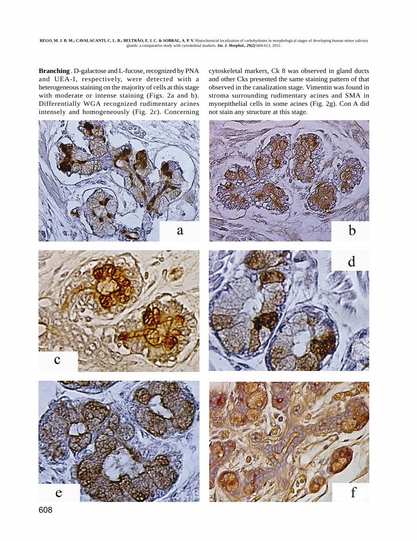
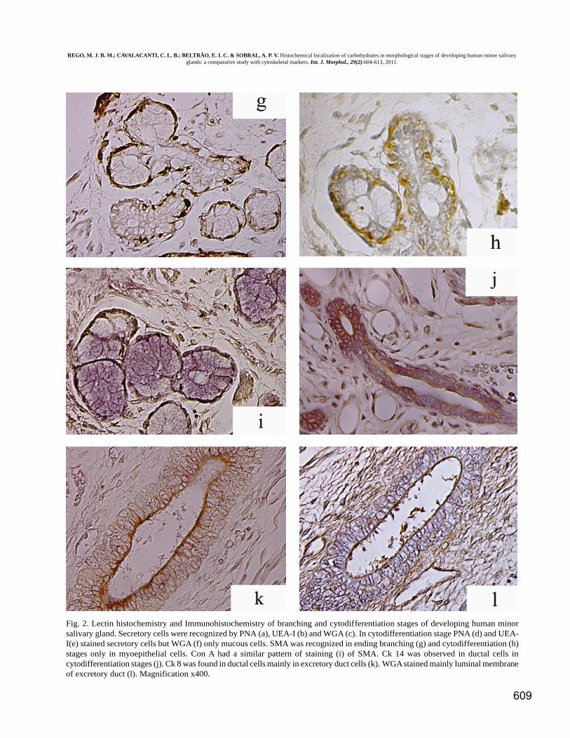
Branching . D-galactose and L-fucose, recognized by PNAand UEA-I, respectively, were detected with aheterogeneous staining on the majority of cells at this stagewith moderate or intense staining (Figs. 2a and b).Differentially WGA recognized rudimentary acinesintensely and homogeneously (Fig. 2c). Concerning
cytoskeletal markers, Ck 8 was observed in gland ductsand other Cks presented the same staining pattern of thatobserved in the canalization stage. Vimentin was found instroma surrounding rudimentary acines and SMA inmyoepithelial cells in some acines (Fig. 2g). Con A didnot stain any structure at this stage.
REGO, M. J. B. M.; CAVALACANTI, C. L. B.; BELTRÃO, E. I. C. & SOBRAL, A. P. V. Histochemical localization of carbohydrates in morphological stages of developing human minor salivaryglands: a comparative study with cytoskeletal markers. Int. J. Morphol., 29(2):604-613, 2011.
609
Fig. 2. Lectin histochemistry and Immunohistochemistry of branching and cytodifferentiation stages of developing human minorsalivary gland. Secretory cells were recognized by PNA (a), UEA-I (b) and WGA (c). In cytodifferentiation stage PNA (d) and UEA-I(e) stained secretory cells but WGA (f) only mucous cells. SMA was recognized in ending branching (g) and cytodifferentiation (h)stages only in myoepithelial cells. Con A had a similar pattern of staining (i) of SMA. Ck 14 was observed in ductal cells incytodifferentiation stages (j). Ck 8 was found in ductal cells mainly in excretory duct cells (k). WGA stained mainly luminal membraneof excretory duct (l). Magnification x400.
REGO, M. J. B. M.; CAVALACANTI, C. L. B.; BELTRÃO, E. I. C. & SOBRAL, A. P. V. Histochemical localization of carbohydrates in morphological stages of developing human minor salivaryglands: a comparative study with cytoskeletal markers. Int. J. Morphol., 29(2):604-613, 2011.

610
Stages Ck7 Ck8 Ck13 Ck14 Ck19 SMA V ConA UEA PNA WGA
Bud - - - - - - - - + + +
Canalicular
Peripheral cells - + - + + - - - - - -
Central cells - + - + + - - - + + +
Branching
Rudimentary acinar cells - - - - - + - - + + +
Ductal cells + + - + + - - - + + +
Ductal lumen - - - - - - - - + + +
Cytodifferentiation
Secretory cells - - - - - - - - + + +
Myoepithelial - - - - - + - + - - -
Ductal cells + + - + + - - - - - +
Ductal lumen - - - - - - - - + + +
Cytodifferentiation . PNA stained the cells of minor salivaryglands at this stage in a moderate to intense pattern (Fig.2d). Differing UEA-I homogeneously stained cytoplasm andmembrane of acinar mucous and serous cells sometimes anuclear membrane staining was also observed (Fig. 2e).WGA stained only mucous cells (Fig. 2f) and ductal cells(Fig. 2l) in a similar pattern observed to CK8. Con A wasthe only lectin capable of recognizing myoepithelial cells(Fig. 2h) presenting a similar staining pattern that observed
for SMA (Fig. 2i). Ck8 (Fig. 2k) and Ck7 stained ductalcells, Ck14 stained basal cells of excretory ducts and CK19(Fig. 2j) stained mainly intercalated ducts. Vimentine wasrecognized only in stroma cells (data not shown).Cytoskeletal proteins and lectins staining in thismorphological stage were similar to those observed in adults.Table III summarized the relationship between lectins andcytoskeleton proteins with stages and cell types of minorhuman salivary gland development.
Table III. Cytoskeleton (Immunohistochemistry) and saccharidies markers (lectin histochemistry) in relation with stages of human minorsalivary gland development
-; no staining, +; positive staining
DISCUSSION
During development the components of glycocalyxare optimally positioned to help cell communication withits environment. Carbohydrates in this structure are the mostprominent surface molecules by which cells face theirneighbors and molecules in the extracellular milieu (Gabius).Many papers have studied the molecular profile of proteinsand their function in human oral epithelium duringdevelopment (Marins et al., 2002; Lourenço et al., 2007a,2007b, 2008). Immunohistochemistry of these proteinsshows changes in function and development of salivaryglands, where the pattern of expression of these biomoleculesvaries with the stage of development, differentiation anddegree of complexity of the epithelium (Martins et al., 2002).But few studies focus on their carbohydrate profile. Adi etal. (1995) analyzed the time-related staining of some lectinsin developing human minor salivary gland. However, theirstudy does not include the morphological stages ofdevelopment or the expression of glucose/mannose residues.
Until now very little is known about the mechanismsthat regulate human salivary glands development, includingcarbohydrates. Quantitative and qualitative changes inglycosyl components of the cell surface and cytoplasm playsignificant roles in normal development and pathology(Ambrosi et al.).
In our results the reactivity of all intermediary filamentantibodies was negative in the epithelium of the salivaryglands during the bud stage. In this stage of development theepithelial cells are undifferentiated and do not exhibit any ofthe characteristics of the various types of cells in adults(Alberts et al., 2002). Among lectins, WGA, UEA-I and PNArecognized their specific carbohydrates N-acetyl-glucosamine, L-fucose and D-galactose, respectively, in cellmembranes at this stage. This pattern is similar to themembrane protein b-1-integrin in the same developmentalstage of human minor salivary (Lourenço et al., 2007b). This
REGO, M. J. B. M.; CAVALACANTI, C. L. B.; BELTRÃO, E. I. C. & SOBRAL, A. P. V. Histochemical localization of carbohydrates in morphological stages of developing human minor salivaryglands: a comparative study with cytoskeletal markers. Int. J. Morphol., 29(2):604-613, 2011.

611
staining similarity could be related to the fact that integrinssurface are all major carriers of N-glycans and their biologicalfunction and modulation are played by glycosylation (Pochec´et al., 2003). In epithelial cells, there are N-glycans of integrinswith highly b1-6 N-acetyl-glucosamine branched and also hadL- fucose and galactose residues (Gu & Tabigushi, 2004).This could be related with our result of lectin staining.
In the canalization stage, Ck8 and 19 in cells ofluminal duct were found in accordance to previous studies(Martins et al.; Lourenço et al., 2007b). In the same stage, L-fucose, D-galactose and N-acetyl-glucosamine are expressedas evidenced by UEA-I, PNA and WGA. Similar results wereobserved in studies with developing human sublingual gland(Chisholm & Adi, 1995). The expression of these sugarresidues was observed in the early canalization stage,decreasing during the progression of this stage, indicating thatsugar is required at the moment when cellular apoptosis isintense. The expression of D-galactose and L-fucose wasobserved in most cells of branching stage; while WGA stainedrudimentary acines and Ck 8 was found in the ducts on thisstage. A reasonable working hypothesis is that addition of N-acetyl-glucosamine plays a role in mediating protein-proteininteractions involved in organization of the cytoskeleton(Hagmann et al., 1992). Furthermore, there is clear evidencethat transmembrane proteins in the endoplasmatic reticulumand the Golgi apparatus were O-GlcNA cylated on theircytosolic domains and that monosaccharides modify andmodulate their membrane proteins (Gerald, 1997). Theintracellular localization of endoplasmatic reticulum and Golgimembranes maybe, could explain the WGA staining next tonuclear membrane.
The cytodifferentiation of excretory cells within therudimentary glands begins after the first canalization andbranching has been established (Cutler, 1990). In our study,different patterns of cytokeratins and carbohydrates expressionwere observed in acinar cells. Initially these cells presentedan intense staining to Ck 8, 14 and 19 that decreased with theprogress of cytodifferentiation stage of development to a weakstaining. Such changes in the pattern of expression ofcytokeratin are directly related to the functional stage of thecycle of these cells (Araújo et al., 2000). In adults, acine cellsof minor and major glands exhibit the same pattern ofexpression of cytokeratins in mucous and serous cells, whichwas not observed in lectins. UEA-I and WGA intensely stainedmucous and serous cells while PNA stained moderatelymucous cells and intensely in serous cells.
Vimentin was not detected in any epithelial cell at anystage of salivary gland development but it was in stromal cells(Data not shown). Previous study indicated vimentin as anearly marker of differentiation in myoepithelial neoplastic
cells pointing to the need of its expression duringdifferentiation (Furuse et al., 2006).
In myoepithelial cells during development of salivaryglands we observed SMA immune staining as Martins et al.SMA is an indicator of myoepithelial differentiation and itsexpression seems to be closely associated with naturalcontractibility of cell. Con A was the only lectin used in thisstudy able to recognize their specific monosaccharides(glucose/mannose) in myoepithelial cells. These cells have ahigh capacity to synthesize basal lamina that is rich in glucoseand mannose residues recognizable by Con A (Chisholm &Adi). The potential link between Con A staining and SMApositivity in muscle structure/function and protein O-mannosylation was greatly strengthened by the identificationof O-mannose saccharides in ∂-dystroglycan. This proteinbridges the actin cytoskeleton to extracellular matrix in muscleand nerve tissues (Haltiwanger & Lowe).
Ck13 was not recognized in any structure during glan-dular development in accordance to other studies (Martins etal.; Poter & Lane, 2003). Con A stained gland cells in the lastweek of cytodifferentiation was also observed in completelydeveloped adult glands.
L-fucose residues were indentified mainly inbranching and cytodifferentiation stages such saccharide isindicated to play critical roles in physiological processes.Experiments with knockout mice in neonatal period regardingFUT-8 activity, an enzyme that catalyse ∂-1-6 fucosyl addition,showed that this enzyme is crucial in Epidermal Growth Fac-tor Receptor (EGFR) signalization implicating in possiblegrowth retardation (Tanigushi et al., 2006). Signaling viatransforming growth factor-β, previously detected byimmunohistochemistry of human minor salivary glands duringdevelopment Lourenço et al. (Lourenço et al., 2007b), involve∂-1-6 fucosylated glycoconjugates which regulatesextracellular matrix protein expression. N-glycans corefucosylation is required for the binding of EGF to its recep-tor. Such carbohydrate residue, L-fucose, mediates EGFRfunction and the responses to EGF and TGF-∂ stimulationwhere both regulate several biological functions includingcell growth and cell differentiation (Wang et al., 2006).
The present study showed morphological evidencesthat sugars play important roles in salivary minor glanddevelopment. Lectin histochemistry suggests that L-fucose,D-galactose e N-acetyl-glucosamin are intensely andpreviously expressed than the traditional cytoskeletal markersin human minor salivary gland development and ConA alsoas biomarker of myoepithelial cells on cytodifferentiation.Analyses of sugar moieties of glycoconjugates will help toreveal the biological importance of glycosylation.
REGO, M. J. B. M.; CAVALACANTI, C. L. B.; BELTRÃO, E. I. C. & SOBRAL, A. P. V. Histochemical localization of carbohydrates in morphological stages of developing human minor salivaryglands: a comparative study with cytoskeletal markers. Int. J. Morphol., 29(2):604-613, 2011.

612
REFERENCES
Adi, M. M.; Chisholm, D. M. & Waterhouse, J. P.Histochemical study of lectin binding in the human fe-tal minor salivary glands. J. Oral Pathol. Med.,24(3):130-5, 1995.
Alberts, B.; Johnson, A.; Lews, J.; Raff, M.; Roberts, K. &Walter, P. Molecular biology of the cell. 4th Ed. New York,Garlard Publishing, 2002.
Ambrosi, M.; Cameron, N. R. & Davis, B. G. Lectins: toolsfor the molecular understanding of the glycocode. Org.Biomol. Chem., 3(9):1593-608, 2005.
Araújo, V. C.; Souza, S. O. M.; Carvalho, Y. R. & Araújo,N. S. Application of immunohistochemistry to the diag-nosis of salivary gland tumors. Appl. Immunohistochem.Mol. Morphol., 8(3):195-202, 2000.
Campos, L. M.; Cavalcanti, C. L. B.; Lima-Filho, J. L.;Carvalho-Junior, L. B. & Beltrão, E. I. C. Acridiniumester conjugated to lectin as chemiluminescenthistochemistry marker. Biomarkers, 11(5):480-4,2006.
Chisholm, D. M. & Adi, M. M. A histological lectin and S-1OO histochemical study of the developing prenatalhuman sublingual salvary gland. Arch. Oral Biol.,40(11):1073-6, 1995.
Cutler, L. S. The role of extracellular matrix in themorphogenesis and differentiation of salivary gland. Adv.Dent. Res., 4:27-33, 1990.
Dale, A. C. Salivary gland. In: Ten Cate, A. R. (ed). OralHistology. 7th ed. St. Louis, Mosby, 1994. pp. 312-33.
Denny, P. C.; Ball, W. D. & Redman, R. S. Salivary glands:a paradigm for diversity of gland development. Crit. Rev.Oral Biol. Med., 8(1):51-75, 1997.
Furuse, C.; Cury, P. R.; Araújo, N. S. & Araújo, V. C.Application of two different clones of vimentin to thediagnosis of salivary gland tumors. Appl.Immunohistochem. Mol. Morphol., 14(2):217-9, 2006.
Gabius, H. J. Biological information beyond the geneticcode: the sugar code. Naturwissenschaften, 87(3):108-21, 2000.
Gabius, H. J.; Siebert, H. C.; André, S.; Jiménez-Barbero, J.& Rüdiger, H. Chemical biology of the sugar code.Chembiochem, 5(6):741-64, 2004.
Gemeiner, P.; Mislovicˇová, D.; Tkácˇ, J.; Sˇvitel, J.;Pätoprst‡, V.; Hrabárová, E.; Kogan, G. & Kozár, T.Lectinomics. II: A highway to biomedical/clinicaldiagnostics. Biotechnol. Advanc., 27(1):1-15, 2009.
REGO, M. J. B. M.; CAVALACANTI, C. L. B.; BELTRÃO, E. I. C. & SOBRAL, A. P. V. Localización histoquímica de carbohidratosen el desarrollo de glándulas salivales menores humanas: un estudio comparativo con los marcadores del citoesqueleto. Int. J. Morphol.,29(2):604-613, 2011.
RESUMEN: Los hidratos de carbono tienen un papel crítico en muchos procesos celulares, como la enfermedad, el crecimientoy el desarrollo. Fueron utilizadasas lectinas, proteínas que reconocen los hidratos de carbono libres o conjugados, como sondas delocalización histoquímica de los carbohidratos en el desarrollo humano de la glándula salival menor. Se realizó inmunohistoquímica delos marcadores tradicionales del citoesqueleto (CKs 7, 8, 13, 14, 19, SMA y vimentina) y posterior comparación con la histoquímica delectinas para PNA, WGA, ConA y la UEA-I, específicas para D-galactosa, N-acetil-glucosamina, glucosa/manosa y L-fucosa, respecti-vamente. Para ello, se obtuvieron muestras de la lengua y de los labios de 15 fetos humanos entre 10-28 semanas de gestación. Ningunode los marcadores inmunológicos del citoesqueleto se identificaron en la primera etapa del desarrollo, diferente de los marcadores dehidratos de carbono. UEA-I, WGA y PNA reconocen sus residuos específicos de hidratos de carbono en todas las etapas analizadasvariando la intensidad de la tinción y los tipos de células. CK8 y N-acetil-glucosamina se expresaron en etapas de canalización, ramifi-cación y citodiferenciación mientras que SMA y la glucosa/manosa se observaron solamente en la etapa de citodiferenciación. ConA sólose reconoció en las células mioepiteliales en etapas de citodiferenciación. Así, debido a esta especificidad, ConA podría utilizarse comomarcador biológico de las células mioepiteliales en la citodiferenciación. La histoquímica de lectinas sugiere que L-fucosa, D-galactosay N-acetil-glucosamina son intensamente expresadas durante el desarrollo como los marcadores tradicionales del citoesqueleto humanosen las glándulas salivales menores .
PALABRAS CLAVE: Localización histoquímica de los carbohidratos, Inmunohistoquímica; Desarrollo de las glándulassalivares menores.
REGO, M. J. B. M.; CAVALACANTI, C. L. B.; BELTRÃO, E. I. C. & SOBRAL, A. P. V. Histochemical localization of carbohydrates in morphological stages of developing human minor salivaryglands: a comparative study with cytoskeletal markers. Int. J. Morphol., 29(2):604-613, 2011.
613
Gerald, W. H. Dynamic o-lined glycosylation of nuclear andcytoskeletal proteins. Annu. Rev. Biochem., 66:315-35,1997.
Gu, J. & Tabiguchi, N. Regulation of integrin functions byN-glycans. Glycocoj. J., 21(1-2):9-15, 2004.
Hagmann, J.; Grob, M. & Burger, M. M. The cytoskeletalproteins talin s o-glycosylated. J. Biol. Chem.,267(20):14424-8, 1992.
Haltiwanger, R. S. & Lowe, J. B. Role of Glycosylation inDevelopment. Annu. Rev. Biochem., 73: 491-537, 2004.
Klein, R. M. Development, structure, and function of salivaryglands. In: Avery, J. K. (Ed). Oral development andhistology. 3rd edn. New York, Thieme, 2002. pp.352-79.
Kuno, A.; Uchiyama, N.; Koseki-Kuno, S.; Ebe, Y.;Takashima, S.; Yamada, M. & Hirabayashi, J.Evanescent-field fluorescence-assisted lectin microarray:a new strategy for glycan profiling. Nat. Methods,2(11):851-6, 2005.
Lourenço, S. V.; Lima, D. M. L.; Uyekita, S. H. & Soares, F.A. Expression of beta-1 integrin in human developingsalivary glands and its parallel relation with maturationmarkers: In situ hybridization and immunofluorescencestudy. Arch. Oral Biol., 52(11):1064-71, 2007a.
Lourenço, S. V.; Uckita, S. H.; Lima, D. M. C. & Soares, F.A. Developing human minor salivary glands:morphological parallel relation between the expressionof TGF-beta. Virchows Arch., 452(4):427-34, 2008.
Lourenço, S. V.; Coutinho-Camillo, C. M.; Buim, M. E.;Uyekita, S. H. & Soares, F. A. Human salivary glandbranching morphogenesis: morphological localization ofclaudins and its parallel relation with developmentalstages revealed by expression of cytoskeleton andsecretion markers. Histochem. Cell Biol., 128(4):361-9,2007b.
Martins, M. D.; Cavalcanti deAraujo, V.; Raitz, R. Soaresde Araújo, N. Expression of cytoskeletal proteins indeveloping humam minor salivary glands. Eur. J. OralSci., 110(4):316-21, 2002.
Nangia-Makker, P.; Conklin, J.; Hogan, V. & Raz, A.Carbohydrate-binding proteins in cancer, and theirligands as therapeutic agents. Trends Mol. Med.,8(4):187-92, 2002.
Pochec´, E.; Litynsa, A.; Amoresano, A. & Casbarra, A. Aglycosylation profile of integrin alpha 3 beta 1 changeswith melanoma progression. Biochim. Biophys. Acta,1643(1-3):113-23, 2003.
Poter, R. M. & Lane, E. B. Phenotypes, genotypes and theircontribution to understanding keratin function. TrendsGenet., 19(5):278-85, 2003.
Tanigushi, N.; Miyoshi, E.; Gu, J.; Honke, K. & Matsumoto,A. Decoding sugar function by identifying targetglycoproteins. Curr. Opin. Struc. Biol., 16(5):561-6,2006.
Tucker, A. S. Salivary gland development. Semin. Cell Dev.Biol., 18(2):237-44, 2007.
Wang, X.; Gu, J.; Ihara, H.; Miyoshi, E.; Honke, K. &Taniguchi, N. Core fucosylation regulates epidermalgrowth factor receptor-mediated intracellular signaling.J. Biol. Chem., 281(5):2572-7, 2006.
Correspondence to:Moacyr Jesus Barreto de Melo RêgoRua Professor Moraes Rêbgo s/nCidade UniversitáriaRecife - PE50670-901BRAZIL Fax: + 55 81 21268484Tel: + 55 81 21012504
Email: [email protected]
Received: 15-12-2010Accepted: 25-03-2011
REGO, M. J. B. M.; CAVALACANTI, C. L. B.; BELTRÃO, E. I. C. & SOBRAL, A. P. V. Histochemical localization of carbohydrates in morphological stages of developing human minor salivaryglands: a comparative study with cytoskeletal markers. Int. J. Morphol., 29(2):604-613, 2011.