Embed Size (px)
Citation preview

Bull. Org. mond. Sant( 1972, 46, 813-819Bull. Wid Hith Org.
Histochemical investigation of the activity ofoxidoreductases in the skin lesions of lepromatousleprosy patients *G. F. 2URAVLEVA1
Thispaper reports an investigation ofthe activity ofthree basic groups ofoxidoreductasesin lepromatous leprosy: specific dehydrogenases, flavoprotein enzymes, and cytochrome oxi-dase. The activity of the enzymes was studied before treatment, at various stages of treat-ment during exacerbations, and in the stage of regression. The data obtained are of impor-tance for evaluating metabolic process in the cells of the specific infiltrates and the dermalconnective tissue in leprosy, for determining the nature and intensity of the inflammatoryprocess, andfor control purposes in cases of regression.
A determination of the activity of the oxido-reductases is useful in evaluating the level of metabo-lic activity of various organs. Highly complex enzymesystems are involved in tissue respiration, and deter-mination of their activity to some extent enables usto assess disturbances in the fine mechanisms ofmetabolism in the cells (Shelton & Schneider, 1952;Bamett & Palade, 1957, 1958; Portugalov & Jakov-lev, 1958; Scarpelli et al., 1958; Sedar & Rosa,1958; Kaplan & Novikoff, 1958; Pearse, 1960;Holt & Hicks, 1961; Strukov, 1963; Strukov et al.,1967; and others).
It has been established (Petrun', 1960; Hashimotoet al., 1964; Serbakov, 1966) that in the skin ofpersons suffering from certain dermatoses there isan increase in the activity of the aerobic tricarboxylicacid cycle and of the basic biochemical oxidationprocesses-tissue respiration and aerobic glycolysis.Using biochemical methods, Novosel (1968) andGolovcenko (1968) made the further discovery thatthere is intensified cytochrome oxidase activity in theaffected areas of skin in persons with eczema andpsoriasis. In their opinion, this is evidence of anacceleration of the oxidation-reduction processes inthe skin, brought about by intensive oxygen absorp-tion. They consider that, at the same time, a certain
* This investigation was financed by the World HealthOrganization.
1 Institute for the Study of Leprosy, Astrakhan, andDepartment of Morbid Anatomy, First Moscow MedicalInstitute, Moscow, USSR.
degree of oxygen deficiency is detectable in the skinand an oxygen deficit is produced in the affectedarea. As the skin lesions are resorbed, a fall in therate of tissue respiration is observed.
In a biochemical study of oxidizing enzymes inleprosy, Naylor (1958) discovered that the amount ofdehydrogenases in a leproma was noticeably greaterbefore treatment than during the course oftreatment. Visneveckij (1969) noted that active spe-cific infiltrates of the skin of patients with lepro-matous leprosy show a high cytochrome oxidase," NAD diaphorase ", and " NADP diaphorase "content but no succinate dehydrogenase activity.Under the influence of treatment for leprosy, theactivity of these enzymes in the skin decreases andtissue respiration is depressed. We have found noother data on the state of activity of oxidoreduc-tases in the skin of leprosy patients.
MATERIALS AND METHODS
We have studied the state of activity of all threegroups of oxidoreductases in cases of lepromatousleprosy, i.e., (1) the specific dehydrogenases, includ-ing the basic oxidoreductases of the Krebs cycle(succinate, malate, isocitrate, glutamate, glycerol-phosphate, glucose-6-phosphate, lactate, and alco-hol dehydrogenases), (2) the flavoprotein enzymes" NAD diaphorase " and NADP diaphorase ", and(3) cytochrome oxidase.The general principle of the histochemical method
for demonstrating dehydrogenase and " diaphorase "
2861 - 813
10

814 G. F. 2URAVLEVA
activity is that colourless tetrazolium salts are, as itwere, substituted for the substance to be reduced andbecome acceptors for the hydrogen, which is splitoff by enzyme action from the substance to be oxi-dized (Pearse, 1960). The tetrazolium salts are re-duced by addition of the hydrogen and convertedinto coloured formazans, which are precipitatedat sites of high enzyme activity. The activity ofoxidoreductases is best demonstrated with nitro-bluetetrazolium.1The activity of enzymes was determined in non-
fixed sections prepared in a cryostat at a temperatureof - 20°C, using nitro-blue tetrazolium as the hydro-gen acceptor and appropriate substrates, followingthe methods described by Pearse (1960). The de-hydrogenases were determined by the methods ofNachlas (1957), Hess et al. (1958), and Burstone (1959),using inter alia substrates of sodium succinate,malate, isocitrate, and lactate. The " diaphorases "were demonstrated by the method of Farber et al.(1954) using directly reduced forms of the coenzymesNADH and NADPH. Cytochrome oxidase wasdetermined by the method of Moog (1943) usingp-aminodimethylaniline oxalate as the substrate. Thesections were first treated with acetone for 30 se-conds to extract the fat droplets from the cell cyto-plasm. Control sections were incubated in solutionswithout substrates. The formation of dark-bluegranules of diformazan and aminophenol blue wereconsidered to be the index of enzyme activity, tak-ing into account their location, distribution, inten-sity, dimensions, and amount.Enzyme activity was estimated on the 5-point
system: very high, high, medium, low, and nil.Besides the histochemical methods of investigationdescribed, conventional histological staining tech-niques were also employed, i.e., haematoxylin-eosinfor general orientation of the histological structure,C.I. solvent red 23 (Sudan III) and C.I. solvent black(Sudan black B) for lipids, and the Ziehl-Neelsenmethod for leprosy bacilli.Enzyme activitywas studied systematically through-
out the course of the lepromatous process: beforetreatment, at various stages of treatment, duringexacerbations, and in the regressive stage. A totalof 69 fragrnents of skin obtained by biopsy frompatients with lepromatous leprosy were examinedtogether with a further 8 samples obtained during sur-gical operations on persons not suffering from leprosyas controls. The lepromatous cases were divided
1 3,3'-(3,3'-dimethoxy-4,4'-biphenylylene)bis[2-(p-nitro-phenyl)-S-phenyl-2H-tetrazolium]dichloride.
into 5 groups according to treatment and clinicalstatus: (1) untreated, with active manifestations ofleprosy (9 cases); (2) treated, but still exhibitingclinically active changes (13 cases); (3) patients in thestationary stage of development of the leprosy pro-cess (14 cases); (4) patients with lepra reaction(erythema nodosum) (10 cases); and (5) patients atthe regressive stage (23 cases).
RESULTS AND DISCUSSION
Histological investigation of the skin sections fromaffected areas in untreated active lepromatous pa-tients disclosed extensive areas of diffuse infiltration,consisting primarily of lepra cells containing lipidsand large numbers of homogeneous mycobacteria.Histoenzymological studies of the lepra cells showedvery high levels of activity of the Krebs' cycle en-zymes (succinate, malate, and isocitrate dehydro-genases), glutamate dehydrogenase, glycerophos-phate dehydrogenase, glucose-6-phosphate dehydro-genase, " NAD diaphorase ", " NADP diaphorase ",and cytochrome oxidase (Fig. 1-4). The dark-blueparticles of diformazan, indicative of dehydrogenaseand " diaphorase" activity, were fairly denselyscattered and were present in large quantities through-out the cytoplasm of the lepra cells; they were mostlyround and quite large. The diformazan granuleswere dispersed evenly throughout the cell. The cellnuclei were often difficult to distinguish since theywere masked by the diformazan grains. Intenseenzyme activity was noted in cells of the Malpi-ghian layer of the epidermis and in the vascularendothelium. A small number of fine particles ofdiformazan were scattered outside the areas of in-filtration, in the sweat-gland cells, in free fibroblasts,and in the fibrous structures of the dermis. In thesame patients, lactate and alcohol dehydrogenaseactivity was low. Grains of aminophenol blue, anindication of cytochrome oxidase activity, were seenin the cytoplasm of lepra cells, where they appearedlarge and intensely coloured; often the grains hadmerged together, forming quite considerable conglo-merates. In other cellular structures, cytochrome-oxidase activity was lower.
Histoenzymological investigation of lepromatouspatients already under treatment but still showinghistological signs of an active process disclosed in-tense persistent reactions for succinate, malate, iso-citrate, glutamate, glycerolphosphate, and glucose-6-phosphate dehydrogenases, " NAD diaphorase "," NADP diaphorase ", and cytochrome oxidase. Gra-

Fig. 1. Untreated lepromatous leprosyshowing very high succinatedehydrogenase activity in the lepra cellsof an infiltrate; x 1 00.
Fig. 2. Active lepromatous leprosyshowing high glutamate dehydrogenaseactivity in a granuloma; x 1 00
Fig. 3. Lepromatous leprosy lesion inthe active stage, showing highNAD diaphorase " activity in an
infiltrate; x 100
C~~~~~~~~or
1111iiiiZ
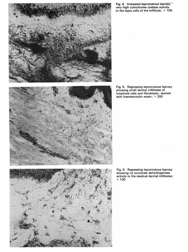
Fig. 4. Untreated lepromatous leprosy;very high cytochrome oxidase activityin the lepra cells of the infiltrate; x 100
rrr,[} ' Re I' t" 'S .'
a::a - >^rt'9~~~~~~~~~~~~~~~~~~~~~~~;.
Fig. 5. Regressing lepromatous leprosyshowing small dermal infiltrates oflymphoid cells and fibroblasts; stainedwith haematoxylin-eosin; x 200
Fig. 6. Regressing lepromatous leprosyshowing no succinate dehydrogenaseactivity in the residual dermal infiltrates;x 100
4 ..* -,..,W
.k0,..:Z k
... i; .-. j1. -6 :.

OXIDOREDUCTASE ACTIVITY IN LEPROMATOUS LEPROSY
nules of diformazan and aminophenol blue were stillevenly distributed in fairly large quantities throughoutthe cells in the areas of infiltration. Lactate and alco-hol dehydrogenase activity was low.
In those cases in which the leprous process was inthe stationary stage, histological examination reveal-ed moderately well-defined areas of infiltration in theskin, consisting of macrovacuolated lepra cells con-taining lipids and considerable numbers of granularleprosy bacilli. Histochemical examination of thelepra cells showed that the activity of all the de-hydrogenases investigated, except the lactate andalcohol dehydrogenases, and of " NAD diaphorase "," NADP diaphorase ", and cytochrome oxidase wasmoderate or, more rarely, low. Blue-stained particleswere found distributed evenly and in moderate num-bers in the lepra cells in the areas of infiltration. Inthe cells and fibres of dermal connective tissue, en-zyme activity was low. No cytochrome oxidase acti-vity was found in the epidermal cells.
In skin sections from erythema nodosum lesionsthe following phenomena were observed when ordi-nary staining techniques were used: dilatation of theblood vessels, swelling of the vascular endotheliumand thickening of the vessel walls, perivascularoedema and, in a number of cases, erythrocyte dia-pedesis. Small areas of infiltration consisting oflymphoid cells, fibroblasts, lepra cells and poly-nuclear cells were seen; the latter contained lipidsand a small quantity of mainly granular or, morerarely, homogeneous mycobacteria. In some cases,homogenization and fibrinoid degeneration of theconnective tissue had occurred. Histoenzymologicalstudies indicated moderate or low activity of succi-nate, malate, glutamate, glycerolphosphate, and glu-cose-6-phosphate dehydrogenases in areas of skininfiltration and in the epidermis. Lactate and alco-hol dehydrogenase activity in lymphoid cells, lepracells, and fibroblasts was low or completely absent.The cells and fibres of the granulomata showed amoderate level of " NAD diaphorase " and " NADPdiaphorase ". A high level of cytochrome oxidaseactivity was seen in the lepra cells in the areas ofinfiltration. As the clinical symptoms of the reactivephase subsided, enzyme activity also decreased.As regressive changes advanced in the areas of
dermal infiltration, there was a lowering of oxidore-ductase activity. Where there was marked regressionof the lepromatous granuloma, the staining of skinsections from previously affected sites with haemo-toxylin-eosin, C.I. solvent black, C.I. solvent red23, and by the Ziehl-Neelsen technique revealed
regressing residual infiltrates consisting mainly oflymphoid cells and fibroblasts; occasional disruptedmycobacteria were seen (Fig. 5). The activity of theKrebs cycle enzymes, the dehydrogenases, the " dia-phorase ", and cytochrome oxidase was determinedhistochemically and found to be low or to have dis-appeared completely. The blue particles of difor-mazan in the infiltrate cells became paler in colourand finely granular, giving the appearance of detrituswith an insignificant amount of stain (Fig. 6). Out-side the areas of infiltration, in the free fibroblasts,connective tissue fibres, skin appendages, nerves, andepidermis, the level of enzyme activity was relativelyhigh. No cytochrome oxidase activity was foundin the cells of the epidermis. These comparative dataon the activity of the oxidoreductases, processed bythe statistical method of Fisher (1952) as modified byMoncevi"jute-IEringene (1964), are shown in Table 1.The table shows that there is a very high and sta-
tistically significant level of activity of the oxidore-ductases investigated, except for lactate dehydrogen-ase and alcohol dehydrogenase, in the cells of activelepromatous granulomata of the skin. Since lactatedehydrogenase activity is an important index of theanaerobic consumption of glucose and the productionof lactic acid, it can be concluded that biologicaloxidation plays the most important role in the meta-bolic processes that occur in skin granulomata inleprosy. These results indicate that lepra cells arebiologically highly active, and this is in agreementwith the conclusions reached by Chatterjee (1958),Kosolapkina (1962) and Vdovina (1964) on the highribonucleoprotein and mucopolysaccharide contentof lepra cell protoplasm. The depression of dehydro-genase, " diaphorase ", and cytochrome oxidase acti-vity, and the simultaneous intensification of theiractivity in the connective tissue cells and the skinappendages, is evidence of a relative depression oftissue respiration in the areas of infiltration and ofa certain normalization of the oxidation-reductionprocesses in the skin of leprosy patients as a result oftreatment with dapsone.
Determination of enzyme activity (in conjunctionwith a number of other clinical and laboratoryindices) is of importance in evaluating the nature,intensity, and duration of the inflammatory processand in following up the regressive changes that occur.In this connexion, the data obtained can be of valuein both diagnosis and prognosis. The change in thelevel of activity of the respiratory enzymes in lepro-matous leprosy may be regarded as the result of adisturbance in the metabolism of the cells of specific
817

G. F. IURAVLEVA
Table 1. Levels of activity of respiratory enzymes in patients with untreated activelepromatous leprosy compared with levels in the regressive stage of the disease
Enzyme Ni Ml ± mib N2a M2 ± m2b tc p
succinate dehydrogenase 9 3.4 ± 0.27 23 0.6 ± 0.12 9.6 <0.1
malate dehydrogenase 9 3.4 ± 0.28 23 0.7 ± 0.11 9.0 <0.1
isocitrate dehydrogenase 8 3.2 ± 0.24 21 0.6 ± 0.13 9.6 <0.1
lactate dehydrogenase 8 0.8 ± 0.12 23 0.5 ± 0.13 1.6 12
glucose-6-phosphatedehydrogenase 9 3.3 + 0.25 23 0.7 ± 0.11 9.6 <0.1
alcohol dehydrogenase 9 1.4 ± 0.22 22 0.6 ± 0.15 2.8 1.1
glycerolphosphatedehydrogenase 9 3.4 ± 0.28 23 0.6 ± 0.13 9.0 <0.1
glutamate dehydrogenase 9 3.0 ± 0.64 23 0.6 ± 0.12 3.6 0.1' NAD-diaphorase 9 2.9 ± 0.16 23 0.7 ± 0.11 11.0 <0.1
- NADP-diaphorase - 9 3.3 ± 0.33 23 0.7 ± 0.11 7.0 <0.1
cytochrome oxidase 9 3.6 ± 0.2 21 0.6 ± 0.14 12.0 <0.1
a Ni = No. of observations on untreated lepromatous patients; N2 = No. of observations on treatedlepromatous patients in the regressive stage.
b MI, M2 = arithmetic mean; ml, m2 = standard error of the artithmetic mean.c t = Statistic for the difference between Mi and M2.
infiltrates. From the nature and intensity of enzymeactivity, it is possible to form some idea of the stateof tissue respiration and determine the nature of themetabolic activity of the cells; this is helpful instudying the pathogenesis of skin changes occurringin leprosy patients undergoing treatment with modemdrugs.
Consequently, in the pathogenesis of skin infec-tions in leprosy, disruption of the activity of theenzymes investigated is of considerable importance;it would appear to be caused by metabolic disturb-ances occurring not merely in the foci of infectionbut in the body as a whole. The use of modemhistoenzymological techniques considerably broadensour ideas on the nature of the morphogenesis andpathogenesis of the specific leprous involvement ofthe skin at the various stages of treatment.
Conclusions(1) A high degree of oxidation-reduction activity
can be demonstrated histochemically in the cells
of active lepromatous granulomata of the skin. Ittakes the form of a high level of activity of succinate,malate, glutamate, glycerolphosphate, and glucose-6-phosphate dehydrogenases, " NAD diaphorase","NADP diaphorase", and cytochrome oxidase.
(2) The low level of activity of lactate and alcoholdehydrogenases in the cells of active lepromatousinfiltrates points to a fall in anaerobic oxidationprocesses and a predominance of biological oxi-dation.
(3) Regressing lepromatous infiltrates show lowlevels of dehydrogenase, " diaphorase ", and cyto-chrome oxidase activity, which indicates that theoxidation-reduction processes in the cells of theseinfiltrates are inhibited.
(4) The data obtained from histoenzymologicalinvestigations are significant in evaluating the inten-sity of the specific inflammatory process and infollowing the regressive changes that occur inleproma-tous patients.
818

OXIDOREDUCTASE ACTIVITY IN LEPROMATOUS LEPROSY 819
RESUME
INVESTIGATIONS HISTOCHIMIQUES SUR L'ACTIVITE DES OXYDOREDUCTASES DANS LES LESIONSCUTANEES CHEZ DES MALADES ATTEINTS DE LEPRE LEPROMATEUSE
On a etudie a I'aide de methodes histochimiquesI'activite des deshydrogenases specifiques - dont notam-ment les principales oxydoreductases du cycle de Krebs ,des enzymes flavoproteiniques et de la cytochrome-oxy-dase chez des patients atteints de lepre lepromateuse adivers stades d'evolution de la maladie. Les examens ontporte sur 69 fragments de peau preleves par biopsiechez des malades et sur 6 fragments preleves chez dessujets non 1epreux. L'activit6 enzymatique a e mesur6esur des substrats appropries grace a l'emploi de bleu detetrazolium nitre.Chez les patients atteints de lepre lpromateuse evolu-
tive et non traitee, on note une forte activite de la cyto-chrome-oxydase, de la # NAD diaphorase #, de la #NADP
diaphorase >) et des diverses deshydrogenases, a l'exceptionde la lactate deshydrogenase et de l'alcool deshydrog&-nase. Dans les cas d'erythema nodosum, l'activite enzy-matique est moderee ou faible dans les zones d'infil-tration. Au fur et a mesure de la regression des lesions,l'inhibition ou l'abolition complete de l'activite des oxy-doreductases dans les cellules des infiltrats residuelscontraste avec le maintien d'une activite enzymatiqueelevee dans les fibroblastes, le tissu conjonctif, les annexesde la peau, les nerfs et l'epiderme.
Selon l'auteur, l'etude de l'activite des oxydoreductasesdans les lesions cutanees peut fournir des indicationssur l'evolution de la lepre lepromateuse et faciliter lediagnostic et le pronostic.
REFERENCES
Barnett, R. J. & Palade, G. B. (1957) J. biophys. bio-chem. Cytol., 3, 4, 577
Bamett, R. J. & Palade, G. B. (1958) J. Histochem.Cytochem., 6, 1, 1
Burstone, M. (1959) J. Histochem. Cytochem., 7, 112Chatterjee, K. R. (1958) Leprosy in India, 30, 30, 78Farber, E. et al. (1954) Proc. Soc. exp. Biol. (N.Y.),
86, 534Fisher, R. A. (1952) Statistical methods for research
workers, Edinburgh, Oliver & BoydGolovcenko, D. Ja. (1968 In: Aktual'nye voprosy derma-
tologii [Current problems in dermatology], Kiev, p. 117Hashimoto, K. et al. (1964) J. invest. Derm., 42, 61Hess, R. et al. (1958) J. biophys. biochem. Cytol., 4, 6, 753Holt, S. J. & Hicks, R. M. (1961) J. biophys. biochem.
Cytol., 11, 1, 47Kaplan, S. E. & Novikoff, A. B. (1959) J. Histochem.
Cytochem., 7, 295Kosolapkina, L. I. (1962) Ucenye zapiski Instituta po
izucYeniju lepry [Scientific notes of the Institute for theStudy of Leprosy], Astrakhan, 3 (8), 61
Moncevi"jute-Jtringene, E. V. (1964) Patologi6eskajaFiziologija i eksperimental'naja terapija, 4, 71
Moog, F. (1943) J. cell. comp.,Physiol., 22, 3, 223Nachlas, M. et al. (1957) J. Histochem. Cytochem., 5, 420Naylor, R. F. (1958) Int. J. Leprosy, 26, 4, 313Novosel, S. I. (1968) In: Aktual'nye voprosy dermatologii
[Current problems in dermatology], Kiev, p. 107
Pearse, A. G. E. (1960) Histochemistry, theoretical andapplied, London, Churchill
Petrun', N. M. (1960) Vestn. Derm. Vener., 4, 33Portugalov, V. V. & Jakovlev, V. A. (1950) In: Gisto-
himiceskie metody v normal'noj i patologiceskoj mor-fologii [Histochemical methods in normal and patho-logical morphology], Moscow, p. 28
Scarpelli, D. G. et al. (1958) J. biophys. biochem. Cytol.,4, 6, 747
scerbakov, A. P. (1966) IzuJenie gazoobmena c'erez kof*upri psoriaze [The study of gas exchange through theskin in psoriasis], Kiev (author's summary of a thesisfor a candidate's degree)
Sedar, A. W. & Rosa, C. G. (1958) Anat. Rec., 130, 2, 371Shelton, E. & Schneider, W. C. (1952) Anat. Rec., 112, 1, 61Strukov, A. I. (1963) In: Gistohimija v patologiceskoj
anatomii [Histochemistry in morbid anatomy], Moscow,p. 15
Strukov, A. I. et al. (1967) In: Gistohimija infarkta mio-karda [The histochemistry of myocardial infarction],Moscow
Vdovina, N. A. (1964) Ucenye zapiski Instituta poizuceniju lepry [Scientific notes of the Institute for theStudy of Leprosy], Astrakhan, vol. 4, p. 168
Visneveckij, F. E. (1969) Tezisy dokladov jubilejnojsessii Instituta po izuceniju lepry [Summaries ofpapersread at a Jubilee Session of the Institute for the Studyof Leprosy], Astrakhan, p. 17