Embed Size (px)
Citation preview

BASIC SCIENCE
Nanomedicine: Nanotechnology, Biology, and Medicine13 (2017) 853–861
Original Article
High-throughput bioaccumulation, biotransformation, and production ofsilver and selenium nanoparticles using genetically engineered
Pichia pastorisFatemeh Elahian, PhDa, Somayeh Reiisi, PhDb, Arman Shahidi, MScc,
Seyed Abbas Mirzaei, PhDa,⁎aCellular and Molecular Research Center, Shahrekord University of Medical Sciences, Shahrekord, Iran
bDepartment of Genetics, Faculty of Basic Sciences, University of Shahrekord, Shahrekord, IrancDepartment of Medical Biotechnology, School of Medicine, Zanjan University of Medical Sciences, Iran
Received 23 December 2015; accepted 17 October 2016
nanomedjournal.com
Abstract
A genetically modified Pichia pastoris strain overexpressing a metal-resistant variant of cytochrome b5 reductase enzyme was developedfor silver and selenium biosorption and for nanoparticle production. The maximum recombinant enzyme expression level was approximately31 IU/ml in the intercellular fluid after 24 h of incubation, and the capacity of the recombinant biomass for the biosorption of silver andselenium in aqueous batch models were measured as 163.90 and 63.71 mg/g, respectively. The ions were reduced in the presence of enzyme,leading to the formation of stable 70–180 nm metal nanoparticles. Various instrumental analyses confirmed the well-dispersed andcrystalline nature of the spherical nanometals. The purified silver and selenium nanoparticles exhibited at least 10-fold less cytotoxicitytoward HDF, EPG85–257, and T47D cells than silver nitrate and selenium dioxide. These results revealed that the engineered Pichia strainis an eco-friendly, rapid, high-throughput, and versatile reduction system for nanometal production.© 2016 Elsevier Inc. All rights reserved.
Key words: Biosorption; Cytochrome b5 reductase; Heavy metals; Nanoparticle; Recombinant yeast
Nanoparticles are defined as particles that are approximatelybetween 1 and 100 nanometers in size. Recently, the importance ofnanoparticles and nanomaterials has increased due to their uniquephysicochemical properties and their wide applicability in numerousfields, mainly electronics, chemistry, energy conservation, biosen-sors, and pioneer medicine.1 Commercial electrochemical methodsare widely used for the production of metal nanoparticles but can beunwieldy, water-inefficient, require the use of large amounts of
Funding sources: The authors are grateful for financial support from theZanjan University of Medical Sciences (grant numbers: A-12-543-5 andA-12-543-7) and Shahrekord University of Medical Sciences (grant number:1393-01-74-2373).
Conflict of interest: We certify that there is no financial conflict of interestinvolving any organization.
⁎Corresponding author at: Cellular and Molecular Research Center,Shahrekord University of Medical Sciences, Shahrekord, Iran.
E-mail addresses: [email protected], [email protected],[email protected] (S.A. Mirzaei).
http://dx.doi.org/10.1016/j.nano.2016.10.0091549-9634/© 2016 Elsevier Inc. All rights reserved.
Please cite this article as: Elahian F, et al, High-throughput bioaccumulation, bigenetically engineer.... Nanomedicine: NBM 2017;13:853-861, http://dx.doi.or
energy, and generate unwanted hazardous substances; higher levelsof initial investment are required to accrue sufficient return.2
However, the environmentally conscious synthesis of nanoparticlesbased on the use of microorganisms is obtaining favorableattention due to its simplicity, cost-effectiveness, environmentalsafety, eco-friendliness, robustness and high-speed, facile,room-temperature production.3,4
Metal nanoparticles are mainly biosynthesized in bacteria,actinomycetes, fungi, yeasts, and plants.5 Fungi and yeasts arebest choice for the synthesis of metallic nanoparticles based onsilver, gold, cadmium, palladium, and selenium because theysecrete enzymes and are easy to manipulate and culture onsimple media. Moreover, even in the presence of large amountsof metal ions, these microorganisms can survive and proliferatedue to their ability to resist metal stress.6,7 Chemical ecology isthe scientific study of the interactions and relationships betweenliving organisms and their microenvironment. Species thatdeveloped the enzymatic capacity to detoxify metal pollutionin their habitat survived while others became extinct. The
otransformation, and production of silver and selenium nanoparticles usingg/10.1016/j.nano.2016.10.009

854 F. Elahian et al / Nanomedicine: Nanotechnology, Biology, and Medicine 13 (2017) 853–861
mechanisms conferring these capacities include the overexpres-sion of efflux pumps; metal precipitation and eliminating metaltoxicity using oxidoreduction enzyme cascades; the bioaccumu-lation of toxic molecules in particular compartments; and thelack of special transporters.8–10
Silver and Selenium have historically been important due totheir broad-spectrum applications in chemistry, computers,cosmetics, electronics, industrial dyes, and textilemanufacturing11,12 and are among the essential trace supple-ments that act as successful antimicrobial, antifungal andantibiofilm agents, interact with proteins to inhibit cellulardamage, control thyroid function, modulate immune surveil-lance, inhibit tumor cell invasion and angiogenesis, promotedifferentiation, and regulate cell proliferation and death.13–15
Regardless of these benefits, window of therapeutic concentra-tion of Ag and Se ions is narrow; for this reason, studies are nowfocusing on how to overcome the disadvantages of high doses ofsilver and selenium using their nanoparticle counterparts.16,17
Silver and selenium nanoparticle biosynthesis by microor-ganisms and plant cells has been described in several studies.18
In addition to low-throughput intact cell strategies for nanoma-terial biosynthesis, other methods involving the use of cellextracts, cell-free supernatants, and pure enzymes have also beenemployed.19,20 Although pure oxidoreductive enzymes havebeen reported to catalyze high-quality and efficient metalbiotransformation, several laborious and costly enzyme purifi-cation, stabilization and immobilization processes are required.Furthermore, these enzymes cannot function alone, but requirecoenzymes. Preferential oxidoreductive coenzymes, such asNADH and NADPH are expensive and are seldom economicallyfeasible for use with current enzyme technologies.21
To the best of our knowledge, we report for the first time theintracellular biosynthesis of stable, less toxic and highlydispersed silver and selenium nanoparticles using geneticallymodified Pichia pastoris overexpressing Mucor racemosuscytochrome b5 reductase enzyme (Cyb5R). We previouslyisolated and characterized this filamentous fungus from pollutedindustrial effluents and sewage water; specific attention was paidto the Cyb5R enzyme due to its high resistance to heavymetals.21,22 Cloning this enzyme enabled Pichia to grow in thepresence of toxic ions through the intracellular reduction andsequestration of nanometals and led to the cost-efficienthigh-throughput biosorption and biotransformation of metals.The engineered yeast synthesizes the required coenzymes andcofactors in an eco-friendly manner. Such protocols wouldrevolutionize the mining industries, nanometal bioproduction,and the biological remediation of environmental pollution.
Methods
Cells, reagents and media
Silver nitrate (AgNO3) and selenium dioxide (SeO2) werepurchased from Sigma–Aldrich (St. Louis, USA). RPMI-1640medium, trypsin, fetal bovine serum (FBS), penicillin andstreptomycin were obtained from Gibco (Grand Island, NY,USA). Vectors, Zeocin, X-33 host strains, positive expressioncontrols, Pichia transformation reagents, and media components
for yeast culture were obtained from Invitrogen (NY, USA). Allother chemicals and solvents were of analytical grade and werepurchased from Merck (Darmstadt, Germany).
Codon optimization of soluble Cyb5R and Pichiacloning
Previously, the genome of Mucor racemosus and Pichiapastoris were studied, and the levels of codon usage weredetermined. The gene for the soluble isoform of MucorNADH-cytochrome b5 reductase (GI: 167,614,335) wascodon-optimized for expression in P. Pastoris and thensynthesized. The gene encoding 228 amino acids wassub-cloned in pPICZB between XhoI and XbaI for intracellulartag-free recombinant protein production. Transformation wasperformed according to Invitrogen's instructions. Briefly, afterthe corresponding plasmid pPICZB-Cyb5R was linearized usingSacI within the 5′ AOX1 region, the X-33 strain was chemicallytransformed. All putative multiple copy recombinants wereassayed for the Mut+ or MutS phenotypes using AOX1 primers.Intracellular enzyme activity was measured using theNADH-dependent ferricyanide reduction method; one unit ofCyb5R activity was defined as the amount of enzyme required toreduce 1 μmol ferricyanide in 1 min.22
Metal ion resistance and growth kinetics
The X33 parental strain and several stable and Mut+
recombinant counterpart clones were cultivated for 24 h in500-ml Erlenmeyer flasks containing 100 ml of YPD broth (1%w/v yeast extract, 2% w/v peptone, 2% w/v glucose).Subsequently, the cells were subcultured in fresh mediacontaining 0.5% methanol (which was used to activate theAOX1 promoter and express the recombinant enzyme) to reachan initial OD600 of approximately 0.01; AgNO3 and SeO2 stocksolutions were then added separately to the cell suspension toyield final concentrations of 0, 1, 2, 4, 6, 8, 10, 20, and 40 mM.The flasks were incubated for 96 h on an orbital shaker at 30 °Cand were sampled four times daily. The maximum specificgrowth rate (μmax) was estimated for all cultures according to thelinear least square method based on the natural logarithms of theoptical density values at 600 nm (measured at the beginning ofthe exponential phases) and several time points. The slope of thebest fitted line was considered to represent μmax.
Nanoparticle production kinetics
For the biosynthesis of Ag and Se nanoparticles (NPs), a freshculture of engineered Pichia pastoris was passaged in 500-mlconical flasks containing 100 ml of YPD and 0.5% methanol.The cultures were then cultivated overnight to an OD600 ofapproximately 1. Filter-sterile stock solutions of AgNO3 or SeO2
were added to the cell mixture at final concentrations of 6 mMand 4 mM, respectively. Negative controls were grown on heavymetal-free YPD medium. The biomass was sampled twice dailyfor 96 h and freeze dried separately. Subsamples (0.5 g) weredigested using microwave-assisted acid digestion according toEPA 3052 for biological samples. Briefly, 9 ml of concentratednitric acid, 1 ml of fuming HCl, and 2 ml of H2O2 were added tosamples in PTEF vessels. The reagents were allowed to react forapproximately 1 minute before sealing the vessels. The tubes

Table 1Maximum specific growth rate of biomass treated with various ion concentrations according to a mathematical model.
0 mM 1 mM 2 mM 4 mM 6 mM 8 mM 10 mM 20 mM 40 mM
Transformed yeast treated with Ag μ = 0.53a
R2= 0.96μ = 0.42b
R2 = 0.91μ = 0.41b
R2 = 0.93μ = 0.39b
R2 = 0.90μ = 0.40b
R2 = 0.90μ = 0.25c
R2 = 0.85μ = 0.21c
R2 = 0.81μ = 0.11c
R2 = 0.73GI
Non-transformed yeast treated with Ag μ = 0.51a
R2 = 0.97μ = 0.44b
R2 = 0.96μ = 0.12c
R2 = 0.76GI GI GI GI GI GI
Transformed yeast treated with Se μ = 0.53a
R2 = 0.96μ = 0.41b
R2 = 0.95μ = 0.40b
R2 = 0.94μ = 0.41b
R2 = 0.95μ = 0.35c
R2 = 0.96μ = 0.13d
R2 = 0.97μ = 0.12d
R2 = 0.81μ = 0.12d
R2 = 0.86GI
Non-transformed yeast treated with Se μ = 0.51a
R2 = 0.97μ = 0.33b
R2 = 0.87GI GI GI GI GI GI GI
μ: maximum specific growth rate average during the early exponential growth phase (1/h).R2 (R-squared): a regression coefficient that indicates how well the data fits the statistical model.GI: growth inhibition and the onset of yeast sporulation. No statistical model was fitted, or the R-squared value was less than 0.7.Values in the same rowmarked with the same letters are not significantly different (P N 0.05) according to the homogeneity of variance obtained using ANOVA analysis.
855F. Elahian et al / Nanomedicine: Nanotechnology, Biology, and Medicine 13 (2017) 853–861
were then placed in a microwave oven (Multiwave 3000™,PerkinElmer, USA) and heated to 180 °C for at least 15 minutes.After cooling, the metal contents were quantified by analyzingAg and Se in the TX-114-rich phase using ICP-OES (Optima,USA) according the manufacturer's protocol.
Purification of nanoparticles
A fresh overnight culture of recombinant Pichia was treatedwith 6 mM AgNO3 or 4 mM SeO2 and incubated for 3 days.Cells were then harvested by centrifugation at 5000×g for 10min. The pellet was washed twice with 0.8% w/v NaCl in 100mM phosphate buffer. The cells were then resuspended in fungallysis buffer (0.5% v/v Triton ×100; 1% w/v SDS; 100 mMNaCl;1 mM EDTA; 10 mM Tris pH = 8) and homogenized usingSilentCrusher™ (Heidolph, Germany). The suspension wascentrifuged at 14,000×g for at least 15 minutes. The resultingpellet containing metal nanoparticles was washed twice with 1.5M Tris/HCl buffer (pH 8.3) containing 1% SDS and thenresuspended in distilled water. The nanoparticles were purifiedthree times using a two-phase, water/octanol extraction system.Pure nanometal sediments at the tube bottom were washed withchloroform, ethyl alcohol, and distilled water, in that order, andthen freeze-dried.23
X-ray diffraction identity and particle size analyses
A small amount of the pure nanoparticles was resuspended indistilled water and homogenized using ultrasonication (75 watts)for 2 min. The particle size distribution and charge potential wereevaluated using dynamic light scattering and a zeta size analyzer(ZEN3600, Malvern, UK). X-ray diffraction spectra of pure Agand Se powders were recorded at the wide-angle range of 2θfrom 10 to 80 degrees at increments of 0.02-degree using apowder X-ray diffractometer (STOE, Germany).
Electron microscopy and atomic force microscopy
TEM measurements were recorded using an EM10C TEMsystem (EM10C-100KV, Zeiss, Germany) operating at a voltageof 120 keV. The samples were prepared on carbon-coated copperTEM grids by drop-coating with suspensions of homogenizedAg and Se. The suspension cell or pure colloidal nanoparticleswere smeared on a glass slide for high-resolution images using
scanning electron microscope (FESEM, Zeiss, Germany) at anaccelerating voltage of 15.0 kV. For atomic force microscopy(AFM), 10 μL of Ag and Se nanoparticle colloidal suspensionswere spread onto the surface of a dust-free cover slips, dried atroom temperature, and mounted on AFM (JPK, Deutschland)stubs. The size of the Ag and Se nanoparticles was evaluated, andthe thickness was measured using the package particle analysisfunction in the software according to standard protocols.
Human cell culture and cytotoxicity assay
Primary human dermal fibroblasts (HDF; NCBI NumberIndex: C646) were purchased from cell lines service (Pasteur,Iran). EPG85–257 (human gastric carcinoma) and T47D (humanepithelial breast cancer) cell lines were generously provided byProfessor Herman Lage (Molecular Pathology Department,University of Medicine Berlin, Germany). The cells werecultured in RPMI medium containing 2 mM L-glutamine, 10%(v/v) FBS, 100 IU/ml penicillin, and 100 mg/ml streptomycin ata constant temperature of 37 °C in a humidified CO2 incubator.Cells were seeded into 96-well tissue culture plates (1000 cellsper well) containing various concentrations of metal ions(0–1000 μM) or nanoparticles (0–10,000 μM) for 5 days.Afterward, the cells were treated with MTT (5 mg/ml) at 37 °Cfor 3 h. Formazan crystals were measured using a TECANmicroplate reader at 570 nm. IC50 values were defined as thedrug concentration that reduced the surviving fraction of cells ineach well by 50% compared to the untreated cells.24,25
Statistical analysis
All experiments were performed at least three times indepen-dently, and the results are expressed as the mean values ± SE. Thedata were compared using Student's t-test. P-values of less than 0.01were considered statistically significant.
Results
Cloning and evaluation of metal ion resistance
The gene coding for the full-length enzyme including asequence recognized by the restriction endonucleases XbaI andXhoI was synthesized. Cloning, transformation, and verificationof the clones were performed in E. coli strain DH5α and then the
Figure 1. Color changes in bioreduction batches using engineered Pichia.After 0 h (left), after 24 h of exposure to 6 mMAgNO3 (middle), and after 24h of exposure to 4 mM SeO2 (right).
Figure 2. Recombinant yeast growth and nanoparticle production. Recom-binant yeasts were treated with 6 mM silver nitrate (A) or 4 mM seleniumdioxide (B). Fresh samples were collected twice daily and subsequentlyanalyzed. Cell growth was monitored based on absorbance at 600 nm and drycell weight. Intracellular nanometal production was measured instrumentallyusing ICP-OES. The values represent the means of three independentexperiments (mean ± standard error).
856 F. Elahian et al / Nanomedicine: Nanotechnology, Biology, and Medicine 13 (2017) 853–861
right recombinant structure subcloned in X33-Pichia pastoris.Maximal total enzyme activity was measured as 31 IU/ml in theintercellular fluid after 24 h of incubation using 0.5% methanolfor expression induction. The growth kinetics for the parent andengineered yeast were monitored in the presence and absence ofvarious concentrations of AgNO3 and SeO2 in the range from 0to 40 mM (Table 1). Wild-type parental yeast strains did notresist ion concentrations of greater than 1 mM. Although thetransformants were resistant to metal ions even at concentrationsof 10 mM, the maximum specific growth rate and nanoparticleproduction were markedly decreased. The highest ion concen-trations that did not reduce the maximum specific growth ratesignificantly (P N 0.05) were 6 mM of AgNO3 (corresponding to647.2 μg/ml of elemental silver) and 4 mM of SeO2 (corre-sponding to 315.8 μg/ml of elemental selenium).
Nanoparticle production kinetics
The results obtained confirmed that recombinant Pichiapastoris efficiently reduced AgNO3 and SeO2 to unchargedmetal nanoparticles. The color of the medium changed due to thegeneration of nanoparticles (from initial light orange to metallicgray and red for Ag and Se production, respectively) after 24 h ofincubation (Figure 1). The maximum retention capacities wereapproximately 163.90 and 63.71 mg/g for silver and selenium,respectively (Figure 2). Silver and selenium production kineticsfollowed a natural logarithmic curve that was expressed as Y =12.05 × Ln(t) + 106.04 and Y = 4.38 × Ln(t) + 40.96 withsquared regression coefficients of 0.93 and 0.94, respectively(where Y denotes milligrams of nanometal per gram of driedyeast weight and t is the time in hours). Yeast growth kineticswere fitted to the following natural logarithmic equations:Ln(X) = 0.40 × t - 4.34 and Ln(X) = 0.41 × t – 4.20 for growthin 6 mM AgNO3 and 4 mM SeO2, respectively, with squaredregression coefficients of 0.90 and 0.95, respectively, during theearly exponential growth phase; in the equations, X representsbiomass, and t is the time elapsed in hours (SupplementaryFigure 1 and Table 1). Metal biosorption apparently occursduring the trophophasic phase of recombinant yeast growth,
coinciding with the production of recombinant Cyb5R. Biosorp-tion yield (%) is calculated by dividing the actual intracellularnanometal mass to the theoretical elemental mass added at timezero (Table 2); Percent yield is used to measure biosorptionreaction efficiency under certain conditions.
Identification and characterization of nanoparticles
X-ray diffraction spectra were obtained to determine thecrystalline nature and identity of the Ag and Se nanoparticles(Figure 3). The XRD-spectra showed four sharp extreme peaksin the total spectrum of 2θ values from 10 to 80; this resultreflects the crystalline structure of pure Ag and Se. Figure 4shows AFM and TEMmicrographs of the purified nanoparticles.

Table 2Percent yield of metal biosorption against time.
Time (h) 0 6 12 18 24 36 48 72 96
Silver biosorptionyield (%)a
0.00 ± 0.00 17.92 ± 2.11 23.42 ± 3.66 38.63 ± 2.31 65.78 ± 2.74 73.19 ± 1.38 76.04 ± 1.90 81.06 ± 1.94 84.12 ± 3.14
Selenium biosorptionyield (%)
0.00 ± 0.00 0.50 ± 3.86 41.44 ± 2.77 50.00 ± 2.06 63.38 ± 3.22 64.17 ± 3.12 67.79 ± 3.78 73.14 ± 3.01 72.82 ± 2.65
The data represent the mean ± standard error of three individual experiments.a Silver nitrate and selenium dioxide were present at 6 mM and 4 mM, respectively, in the cell reactors and biosorption yields were calculated by dividingactual yields by theoretical yields.
Figure 3. X-ray diffractogram of silver (A) and selenium (B) nanoparticles.Diffraction patterns confirmed the presence of nano-sized elemental silverand selenium as the major constituents. Sharp intensive peaks indicate smallcrystalline particle size.
857F. Elahian et al / Nanomedicine: Nanotechnology, Biology, and Medicine 13 (2017) 853–861
The Ag and Se NPs were spherical and ranged from 70 to 180 nmin diameter. The particle sizes of the Ag and Se particles wereassessed using TEM and AFM and were very similar (to thecrystallite size predicted using the particle size analyzer(Supplementary Figure 2).
Cytotoxicity Assay
The HDF, EPG85–257 and T47D cell lines were treated withincreasing doses of selenium dioxide, sliver nitrate, nanosilver,and nanoselenium. A dose–response curve was fitted to theresulting data, and the IC50 values were calculated after 5 days ofexposure in MTT cell viability assays (Figure 5). The cells werealso treated with mitoxantrone as a positive control for cell
cytotoxicity. The most cytotoxic effect was exerted by AgNO3
on the HDF line (P b 0.001; IC50 value, approximately 0.25μM). In contrast, nanoselenium appeared to have the leasttoxicity on the T47D line (IC50 N 5 mM elemental selenium).The obtained data showed that the cytotoxic effects of thesecompounds are dose-dependent and cell-specific (Table 3).
Discussion
Many chemical reactors are available commercially and aredesigned empirically to synthesize nanoparticles in the colloidaland aerosol phases. Unfortunately, the large-scale, economic,and eco-friendly synthesis of monodispersed nanoparticlesremains challenging.26,27 Recently, improved biological tech-nologies have attracted attention to the synthesis and applicationof metallic nanoparticles. Microorganisms (bacteria, yeasts andfungi) can interact with particular ions and reduce them to metalnanoparticles. In this regard Pichia pastoris proves to be a safeand important biological reactor for intracellular biosynthesis ofstable nanoparticles. This yeast is much more resistant to metaltoxicity, grows rapidly in inexpensive simple media, produceshigh yields of biomass and is easily manipulated genetically.Here a simple and element-specific reduction of metal ionscatalyzed enzymatically by NADH dependent cytochrome b5reductase. The bioproduction of silver and selenium nanoparti-cles can be visually followed by the medium color changes tometallic gray and red, respectively, during the early hours of ionsexposure (Figure 1). These colors arise from excitation of surfaceplasmon resonances in the nanometals.28
Previously native nitrate reductase, cytochrome c reductase,and cytochrome P450 reductase have been noticed for mild metalbioremediation. However, traditional enzyme technology costsremained high because in nature, organisms adjust the conditionsof their enzymes to produce an optimum rate of reaction, it istechnically very difficult to recover and immobilize activeenzymes, and much of the cost relates to cofactorsupplementation.19,20,29 On the hand overexpressing ametal-resistant variant of cytochrome b5 reductase in our studyresulted in Pichia resistance to high concentrations of heavymetal ions rather than native microorganisms and provided highyields of nanometals.
Based on literature review the magnitude of silver andselenium uptake capacity by native yeasts can be estimated as2–50 mg/g and 0.016–1.58 mg/g, respectively. In contrast, silveraccumulates in our engineered Pichia at 163.90 mg/g andselenium is biosorbed at 63.71 mg/g; these levels (Figure 2)

Figure 4. Examples of metal biotransformation using recombinant Pichia. (A, B) AFM image of silver nanometals. (C) AFM image of pure seleniumnanometals. (D, E) TEM image of silver and selenium nanometals. Microscopic analyses revealed the predominately spherical shape of the intracellularnanoparticles (average diameter, 70 to 180 nm).
858 F. Elahian et al / Nanomedicine: Nanotechnology, Biology, and Medicine 13 (2017) 853–861
represent the highest levels of these metals biosorption that havebeen reported.30–32 The levels of bioaccumulation of silver andselenium that were measured in this study are similar to thosemeasured in wild-type plants; however, yeast biomass apparentlyaccumulates more easily and more rapidly.33,34
Although the recombinant cells resisted metallic ion concen-trations approximately 20-fold higher than those that the parentalcells could withstand, the cell growth kinetics was affected.Cellular decrease in maximum specific growth rate (Table 1) inpresence of silver and selenium ions might have been due tothe general toxicity of the ions toward the metabolic pathwaysand is considered the main disadvantage of growing cellbiotransformations.23,35 Other reasons that might cause low
biomass yield in the presence of high ion levels in thisoverexpression/biocatalysis approach are the inhibition of yeastcell respiration and the high consumption of NADH and O2 inthe enzyme-based bioremediation technique.36
Figure 4 and Supplementary Figure 2 micrographs revealedthat the most of silver and selenium nanoparticles wereuniformly spherical and were in the range of 70–180 nm insize; hence, it could be understood that the nanometals exhibitedwell-defined dimensions and good monodispersity and theexperimental conditions such as metal ion concentrations,media compositions, fermentation conditions, and cofactoravailability were almost optimum. Particle size and monodis-persity were controlled more easily using this novel approach
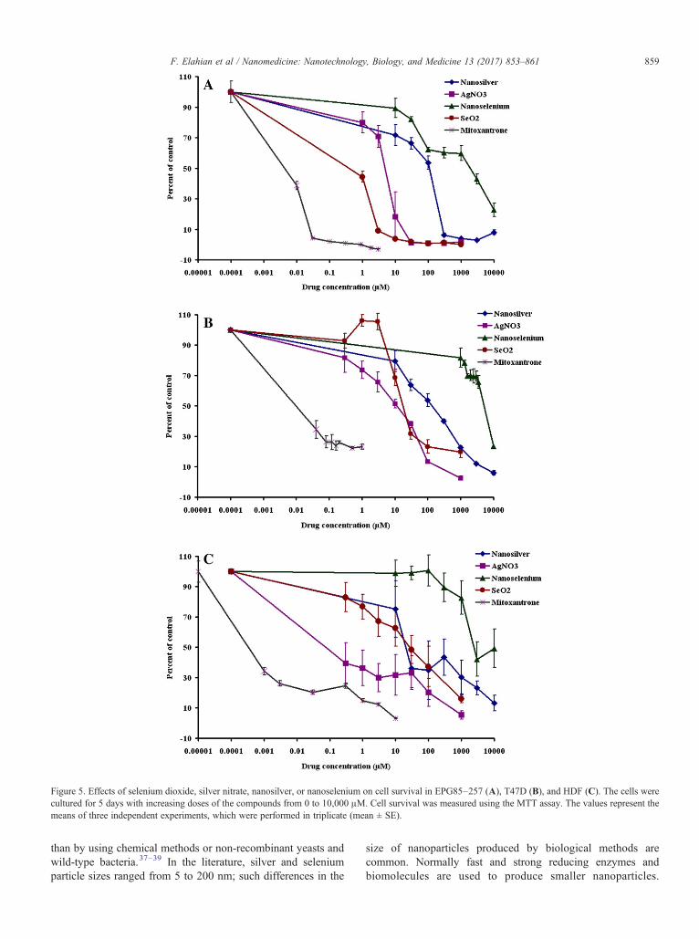
Figure 5. Effects of selenium dioxide, silver nitrate, nanosilver, or nanoselenium on cell survival in EPG85–257 (A), T47D (B), and HDF (C). The cells werecultured for 5 days with increasing doses of the compounds from 0 to 10,000 μM. Cell survival was measured using the MTT assay. The values represent themeans of three independent experiments, which were performed in triplicate (mean ± SE).
859F. Elahian et al / Nanomedicine: Nanotechnology, Biology, and Medicine 13 (2017) 853–861
than by using chemical methods or non-recombinant yeasts andwild-type bacteria.37–39 In the literature, silver and seleniumparticle sizes ranged from 5 to 200 nm; such differences in the
size of nanoparticles produced by biological methods arecommon. Normally fast and strong reducing enzymes andbiomolecules are used to produce smaller nanoparticles.

Table 3IC50 values of mitoxantrone, nanometals and their corresponding ions for the primary dermal fibroblasts and cancer cell lines.
Cells AgNO3
IC50a ± SE
NanosilverIC50 ± SE
MitoxantroneIC50 ± SE
SeO2
IC50 ± SENanoseleniumIC50 ± SE
EPG85–257 5.77 ± 0.414⁎⁎⁎ 115.29 ± 6.097⁎⁎⁎ 0.01 ± 0.004 0.85 ± 0.521⁎⁎⁎ 2152.49 ± 49.818⁎⁎⁎
T47D 12.32 ± 0.721⁎⁎⁎ 153.71 ± 10.107⁎⁎⁎ 0.03 ± 0.004 20.06 ± 1.613⁎⁎⁎ 5927.14 ± 50.203⁎⁎⁎
HDF 0.25 ± 0.137⁎⁎⁎ 22.81 ± 4.831⁎⁎⁎ 0.0007 ± 0.000 27.74 ± 4.012⁎⁎⁎ 2612.07 ± 113.328⁎⁎⁎
The data represent the mean ± standard error of three individual experiments.a The drug concentration (μM) required for 50% inhibition of cell growth after 5 days of drug exposure.⁎⁎⁎ Indicates statistical significance between the nanometal and its ion counterpart (P b 0.001).
860 F. Elahian et al / Nanomedicine: Nanotechnology, Biology, and Medicine 13 (2017) 853–861
Physicochemical production of nanoparticles is often performedin the presence of stabilizers in order to control of the nucleiformation and prevent unwanted agglomeration of the colloids;such methods involving chemical agents are associated withenvironmental toxicity and more difficult to be optimized.40,41
Low average values of Zeta potential (data not shown) aremainly caused by the high tendency of nanometals to aggregatein aqueous intracellular environments. Human albumin wassuccessfully used to reduce elemental aggregation in this study.A comparison of the measured IC50 values in Table 3 revealedthat nano-sized insoluble particles were at least 10-fold less toxictoward HDF, EPG85–257, and T47D cell lines rather than theirsoluble AgNO3 and SeO2. Normal HDF cells were moresensitive to the metal toxicity than cancerous T47D cells.Different responses in the activity of the key enzymes in metalmetabolism are the most possible explanation for differentialcytotoxicity in these cells. The cytotoxicity of the nanoparticlesand their ion counterparts showed a direct dose-dependence andcell viability was reduced at higher concentrations consistentwith many reports.
The results obtained here show that silver and seleniumnanoparticles were successfully biosynthesized using an engi-neered metal-resistant Pichia pastoris clone containing Cyb5R.The biosynthesis of nanoparticles using the engineered yeastrepresents a potential alternative to conventional biological andtraditional physicochemical methods; the advantages includehigh throughput, the production of less toxic waste,cost-effectiveness, simplicity, reduced time requirements,eco-friendliness, and the production of homogenous nanometals.
Author Contributions
S.A. Mirzaei coordinated the study, designed the cloning andbiotransformation experiments, and revised the final manuscript. F.Elahian designed the cytotoxicity study and performed the statisticalanalysis, wrote the corresponding portions of the manuscript, andparticipated in intellectual discussions of the data and manuscriptwriting. S. Reiisi engineered the Pichia clones and analyzed theenzyme activity. A. Shahidi performed most of the experiments aspart of his master's degree in medical biotechnology.
Appendix A. Supplementary data
Supplementary data to this article can be found online athttp://dx.doi.org/10.1016/j.nano.2016.10.009.
References
1. Daniel MC, Astruc D. Gold nanoparticles: assembly, supramolecularchemistry, quantum-size-related properties, and applications towardbiology, catalysis, and nanotechnology. Chem Rev 2004;104:293-346.
2. Dhillon GS, Brar SK, Kaur S, Verma M. Green approach fornanoparticle biosynthesis by fungi: current trends and applications.Crit Rev Biotechnol 2012;32:49-73.
3. Bansal V, Rautaray D, Bharde A, Ahire K, Sanyal A, Ahmad A, et al.Fungus-mediated biosynthesis of silica and titania particles. J MaterChem 2005;15:2583-9.
4. Azad AK, Amin L, Sidik NM. Genetically engineered organisms forbioremediation of pollutants in contaminated sites. Chin Sci Bull2014;59:703-14.
5. SinghP,KimYJ, ZhangD,YangDC.Biological synthesis of nanoparticlesfrom plants and microorganisms. Trends Biotechnol 2016;34:588-99.
6. Sastry M, Ahmad A, Islam Khan M, Kumar R. Biosynthesis of metalnanoparticles using fungi and actinomycete. Curr Sci 2003;85:162-70.
7. Balaji DS, Basavaraja S, Deshpande R, Mahesh DB, Prabhakar BK,Venkataraman A. Extracellular biosynthesis of functionalized silvernanoparticles by strains of Cladosporium cladosporioides fungus.Biointerfaces 2009;68:88-92.
8. Bhainsa KC, D'Souza SF. Extracellular biosynthesis of silver nanoparticlesusing the fungus aspergillus fumigatus. Biointerfaces 2006;47:160-4.
9. Bruins MR, Kapil S, Oehme FW. Microbial resistance to metals in theenvironment. Ecotoxicol Environ Saf 2000;45:198-207.
10. Iravani S. Green synthesis of metal nanoparticles using plants. GreenChem 2011;13:2638-50.
11. Lara HH, Garza-Trevino EN, Ixtepan-Turrent L, Singh DK. Silvernanoparticles are broad-spectrum bactericidal and virucidal compounds.J Nanobiotechnol 2011;9:30.
12. Tran PA, Webster TJ. Selenium nanoparticles inhibit Staphylococcusaureus growth. Nanomedicine 2011;6:1553-8.
13. Zeng H, Combs GF. Selenium as an anticancer nutrient: roles in cellproliferation and tumor cell invasion. J Nutr Biochem 2008;19:1-7.
14. Zhang R, Lee P, Lui VC, Chen Y, Liu X, Lok CN, et al. Silvernanoparticles promote osteogenesis of mesenchymal stem cells andimprove bone fracture healing in osteogenesis mechanism mouse model.Nanomedicine 2015;11:1949-59.
15. Fu X, Yang Y, Li X, Lai H, Huang Y, He L, et al. RGD peptide-conjugated selenium nanoparticles: antiangiogenesis by suppressingVEGF-VEGFR2-ERK/AKT pathway. Nanomedicine 2016;12:1627-39.
16. Ramamurthy C, Sampath KS, Arunkumar P, Kumar MS, Sujatha V,Premkumar K, et al. Green synthesis and characterization of seleniumnanoparticles and its augmented cytotoxicity with doxorubicin on cancercells. Bioprocess Biosyst Eng 2013;36:1131-9.
17. Tutaj K, Szlazak R, Szalapata K, Starzyk J, Luchowski R, Grudzinski W,et al. Amphotericin B-silver hybrid nanoparticles: synthesis, propertiesand antifungal activity. Nanomedicine 2016;12:1095-103.
18. Shikuo L, Yuhua S, Anjian X, Xuerong Y, Xiuzhen Z, Liangbao Y, et al.Rapid, room-temperature synthesis of amorphous selenium/proteincomposites using Capsicum annuum L extract. Nanotechnology2007;18:405101.

861F. Elahian et al / Nanomedicine: Nanotechnology, Biology, and Medicine 13 (2017) 853–861
19. Anil Kumar S, Abyaneh MK, Gosavi SW, Kulkarni SK, Pasricha R,Ahmad A, et al. Nitrate reductase-mediated synthesis of silvernanoparticles from AgNO3. Biotechnol Lett 2007;29:439-45.
20. Mukherjee P, Senapati S, Mandal D, Ahmad A, Khan MI, Kumar R, etal. Extracellular synthesis of gold nanoparticles by the fungus fusariumoxysporum. Chem Bio Chem 2002;3:461-3.
21. Elahian F, Sepehrizadeh Z, Moghimi B, Mirzaei SA. Human cytochromeb5 reductase: structure, function, and potential applications. Crit RevBiotechnol 2014;34:134-43.
22. Mirzaei SA, Yazdi MT. Sepehrizadeh Z secretory expression andpurification of a soluble NADH cytochrome b5 reductase enzyme fromMucor Racemosus in Pichia Pastoris based on codon usage adaptation.Biotechnol Lett 2010;32:1705-11.
23. Shakibaie M, Khorramizadeh MR, Faramarzi MA, Sabzevari O,Shahverdi AR. Biosynthesis and recovery of selenium nanoparticlesand the effects on matrix metalloproteinase-2 expression. BiotechnolAppl Biochem 2010;56:7-15.
24. Mansouri M, Mirzaei SA, Lage H, Mousavi SS, Elahian F. The cell cyclearrest and the anti-invasive effects of nitrogen-containing bispho-sphonates are not mediated by DBF4 in breast cancer cells. Biochimie2014;99:71-6.
25. Elahian F, Moghimi B, Dinmohammadi F, Ghamghami M, Hamidi M,Mirzaei SA. The anticancer agent prodigiosin is not a multidrugresistance protein substrate. DNA Cell Biol 2013;32:90-7.
26. Roco MC. Nanoparticles and nanotechnology research. J Nanopart Res1999;1:1-6.
27. Turner M, Golovko VB, Vaughan OP, Abdulkin P, Berenguer-MurciaA, Tikhov MS, et al. Selective oxidation with dioxygen by goldnanoparticle catalysts derived from 55-atom clusters. Nature2008;454:981-3.
28. Dar MA, Ingle A, Rai M. Enhanced antimicrobial activity of silvernanoparticles synthesized by Cryphonectria sp evaluated singly and incombination with antibiotics. Nanomedicine 2013;9:105-10.
29. Macdonald IDG, Smith WE. Orientation of cytochrome c adsorbed on acitrate-reduced silver colloid surface. Langmuir 1996;12:706-13.
30. Donmez G, Aksu Z. The effect of copper(II) ions on the growth andbioaccumulation properties of some yeasts. Process Biochem1999;35:135-42.
31. Kapoor A, Viraraghavan T. Fungal biosorption - an alternative treatmentoption for heavy metal bearing wastewaters: a review. BioresourTechnol 1995;53:195-206.
32. Wang J, Chen C. Biosorption of heavy metals by Saccharomycescerevisiae: a review. Biotechnol Adv 2006;24:427-51.
33. Nettem K, Almusallam AS. Equilibrium, kinetic, and thermodynamicstudies on the biosorption of selenium (IV) ions onto GanodermaLucidum biomass. Separ Sci Technol 2013;48:2293-301.
34. Wang J, Chen C. Biosorbents for heavy metals removal and their future.Biotechnol Adv 2009;27:195-226.
35. Quijano G, Couvert A, Amrane A, Darracq G, Couriol C, Le Cloirec P, etal. Toxicity and biodegradability of ionic liquids: new perspectivestowards whole-cell biotechnological applications. Chem Eng J2011;174:27-32.
36. Allen C, Boudet C, Hardacre C, Migaud M. Enhancement of whole celldioxygenase biotransformations of haloarenes by toxic ionic liquids.RSC Adv 2014;4:19916-24.
37. Mohanpuria P, Rana NK, Yadav SK. Biosynthesis of nanoparticles:technological concepts and future applications. J Nanopart Res2008;10:507-17.
38. Levard C, Hotze EM, Lowry GV, Brown GE. Environmentaltransformations of silver nanoparticles: impact on stability and toxicity.Environ Sci Technol 2012;46:6900-14.
39. Valls M, Atrian S, de Lorenzo V, Fernández LA. Engineering a mousemetallothionein on the cell surface of Ralstonia eutropha CH34 forimmobilization of heavy metals in soil. Nat Biotechnol 2000;18:661-5.
40. Sharma G, Sharma AR, Bhavesh R, Park J, Ganbold B, Nam JS, et al.Biomolecule-mediated synthesis of selenium nanoparticles using driedVitis vinifera (raisin) extract. Molecules 2014;19:2761-70.
41. Sharma VK, Yngard RA, Lin Y. Silver nanoparticles: green synthesisand their antimicrobial activities. Adv Colloid Interface Sci2009;145:83-96.