Embed Size (px)
Citation preview

Received: 6 October 2017 Revised: 22March 2018 Accepted: 22March 2018
DOI: 10.1002/JLB.3A1017-398RR
ART I C L E
𝜷2-Adrenoceptors inhibit neutrophil extracellular traps inhuman polymorphonuclear leukocytes
FrancaMarino1 Angela Scanzano1 Laura Pulze2 Monica Pinoli1
Emanuela Rasini1 Alessandra Luini1 Raffaella Bombelli1 Massimiliano Legnaro1
Magda de Eguileor2 Marco Cosentino1
1Center of Research inMedical Pharmacology,
Varese, Italy
2Department of Biotechnology and Life
Sciences, University of Insubria, Varese, Italy
Correspondence
FrancaMarino, PhD,Center forResearch in
Medical Pharmacology,University of Insubria,
Via,OttorinoRossi n. 9, 21100VareseVA, Italy.
Email: [email protected]
AbstractThis study tests the hypothesis that in isolated human polymorphonuclear leukocytes (PMN)
adrenergic ligands can affect neutrophil extracellular trap (NET) formation. We have previously
shown that, in PMN, adrenaline (A), through the activation of adrenergic receptors (AR), reduces
stimulus-dependent cell activation; we have, therefore, planned to investigate if AR are involved
inNETproduction. PMNwereobtained fromvenousbloodof healthy subject. The ability of adren-
ergic ligands to affect reactive oxygen species (ROS) production, NET production, and cell migra-
tion was investigated in cells cultured under resting conditions or after activation with N-formyl-
methionyl-leucyl-phenylalanine (fMLP), LPS, or IL-8. Stimuli-induced NET production measured
as ROS, microscopic evaluation, and elastase production was reverted by A and this effect was
blocked by the selective 𝛽2–AR antagonist ICI-118,551. The stimulus-induced ROS generation
andmigrationwas prevented by A and by isoprenaline (ISO), and these effects were counteracted
only by ICI-118,551 and not by the other two selective ligands for the 𝛽1 and 𝛽3–AR. Finally, the
presence of the 𝛽–ARs on PMNwas confirmed, by means of microscopy and flow cytometry. The
data of the present study suggest that adrenergic compounds, through the interaction of mainly
𝛽2–AR, are able to affect neutrophil functions. These data are suggestive of a possible therapeutic
role of 𝛽2–AR ligands (in addition to their classical use), promoting the possible therapeutic rele-
vance of adrenergic system in the modulation of innate immunity proposing their possible use as
anti-inflammatory drugs.
K EYWORDS
adrenergic receptor, adrenaline, neutrophils, neutrophil extracellular traps, reactive oxygen
species
1 INTRODUCTION
Polymorphonuclear neutrophils (PMN) are usually considered as the
first line of defense against invading microorganisms and as contribu-
tors to the orchestrated response after tissue invasion by pathogens.
PMN are able to migrate into inflamed tissues, and in loco provide
to the immune response through a number of different mechanisms,
including adhesion to endothelial layers, migration into tissues, and
subsequently phagocytosis, production of reactive oxygen species
(ROS) aswell as other pro-inflammatorymediators suchas chemokines
and cytokines, degranulation leading to release of neutrophil elastase
(NE) ormetalloproteases (MP), and formation of neutrophil extracellu-
lar traps (NET).1,2
During the last 3 decades, however, the involvement of these cells
in non-infectious diseases has been progressively unveiled, and now
PMN are considered pivotal cells in a wide range of inflammatory
diseases as, for example, cardiovascular and autoimmune diseases.3,4
In particular, although a key product of PMN, the NET, play a key
role in the killing of pathogens,1 compelling evidence indicates that
they are also involved in other clinical conditions such as auto-
immune diseases.2
Ultrastructural analysis shows that NET composition mainly
consists of nuclear chromatin, together with granular and cyto-
plasmic proteins.3 In particular, the presence inside the NET of
myeloperoxidase (MPO), NE, and cathepsin G suggests their involve-
ment in different inflammatory diseases, including cardiovascular
J Leukoc Biol. 2018;1–12. c©2018 Society for Leukocyte Biology 1www.jleukbio.org

2 MARINO ET AL.
diseases (CVD)5; in fact, MPO and NE are present in human
atherosclerotic plaques and contribute to their destabilization
and rupture.6,7 NET are also involved in auto-inflammatory diseases
such as systemic lupus erythematosus, Wegener’s granulomatosis,
and psoriasis.4,8,9
The catecholamines (CA) noradrenaline (NA), adrenaline (A), and
dopamine (DA) in addition to being key neurotransmitters and neu-
rohormones in the brain and in peripheral tissues are also trans-
mitters connecting the nervous and the immune system.10–14 Nerve
fibers positive for tyrosine hydroxylase (TH; enzyme limiting the syn-
thesis of CA) extensively innervate lymphoid organs.15,16 TH is also
expressed in immune cells,17,18 where it subserves endogenous pro-
duction of CA, which in turn affect immune cell functions.17,19 CA
can modulate hematopoiesis20–22 and their content in immune cells is
affected by several diseases19,23; cells of acquired and innate immu-
nity express both adrenergic receptors (AR) and dopaminergic recep-
tors (DR) and it has been shown that the activation of these recep-
tors results in changes of different cell functions.19,24–27 Several
studies have shown that these receptors are involved in immune-
mediated diseases.23,28,29
We have previously shown that, during activation with pro-
inflammatory stimuli, human PMN undergo morphological and func-
tional changes,30–35 including changes in AR expression.27 We have
also shown that these changes are followed by increased produc-
tion of chemokines, such as IL-8,30,31 and increased generation of
ROS.27 Finally, we have shown that these functional and morphologi-
cal changes can be prevented by pharmacological treatments31,33 and
involve the activation of specific AR-operated pathways.27
The aim of the present study was to investigate the ability of
adrenergic agents to affect NET production in human PMN and to
characterize the AR pathways eventually involved. Since in previ-
ous studies,27 we have identified a key role by 𝛽-AR-operated path-
ways, in the present study, we have particularly explored the ability
of A and of selective 𝛽-AR ligands to affect the NET production as
well as migration and ROS generation, two key functions involved in
the PMN-dependent tissue protection. Finally, by means of fluores-
cence microscopy and flow cytometry, we have investigated the 𝛽-
AR expression and the changes occurring following the exposition to
activating stimuli.
2 MATERIALS AND METHODS
2.1 PMN isolation
Experiments were performed on buffy coats of healthy subjects
obtained by the local blood bank (Circolo Hospital, Fondazione Mac-
chi, Varese, Italy). PMN were isolated by standard density-gradient
centrifugation as previously described.27 Cells were examined using
light microscopy in order to exclude the presence of platelets or
erythrocytes. Cell purity and viability were always assessed either
by light microscopic examination or by flow cytometric analysis and
experimentswereperformedonly in cellswhich presented apurity and
viability higher than 95%.
2.2 Cell culture
Cellswere cultured, according to our previous studies,27,34 under stan-
dard conditions, in RPMI medium, and subsequently, allowing to the
experimental protocols, harvested for the following analysis.
After harvesting, cells were stained with trypan blue and viability
was assessed by an automatic cell counter “Cellometer Auto T4” (Nex-
celomBsioscience LLC, Lawrence, MA, USA).
Stimuli employed to induce NET formation included (i) the chemo-
tactic peptide N-formyl-Met-Leu-Phe (fMLP, 0.1 𝜇M), which acts on
membrane receptors resulting in PMN activation36; (ii) LPS (1 𝜇g/ml;
extracted from E. coli serotype O127:B8), a strong inducer of NET and
able to induces an inflammatory response through the activationof dif-
ferent receptors, in particular the TLR437; and (iii) IL-8 (100 ng/mL),
a key physiological activator of PMN which induces extensive
NET-osis.3
2.3 Cell apoptosis
To evaluate the effect of A (1 nM–1 𝜇M) on PMN viability, cells
were cultured for 30 min or 3 h, then samples were recovered and
centrifuged (600 × g, 5 min, room temperature) to remove super-
natant, and washed with 1 mL of PBS. Apoptosis was evaluated by
means of flow cytofluorimetric assay using an FITC Annexin V detec-
tion Kit I (Becton Dickinson, Milan, Italy) according to the manufac-
turer’s instructions. Briefly, the cells were resuspended in 100 𝜇L of
Annexin V Binding Buffer (present in the kit) and stained with 5 𝜇L
of FITC-conjugated Annexin V (ANX-FITC) and 5 𝜇L of propidium
iodide staining solution (PI) in the dark for 15 min. After the incu-
bation, 250 𝜇L of binding buffer was added and samples were ana-
lyzed by a BD FACSCanto II Flow Cytometer (Becton Dickinson Italy,
Milano, Italy) and data were analyzed using BD FACSDiva software
(version 6.1.3). PMNwere identified based on FSC and SSC properties,
and at least 15.000 events were collected from each sample. Viable
(ANX−/PI−), early apoptotic (ANX+/PI−), late apoptotic (ANX+/PI+),necrotic (ANX−/PI+) PMNwere identifiedonabiparametric plotANX-
FITC versus PI with a log scale.
2.4 Morphological and quantitative evaluation of
NET production
2.4.1 ROS generation
Intracellular ROS levels were assessed in PMN under resting or acti-
vated conditions by using the redox sensitive dye C-DCDHF-DA
(Molecular Probes, Eugene, OR, USA) as previously described.27 Flu-
orescence was detected by means of a spectrofluorimeter (Perkin-
Elmer LS-50B, Perkin-Elmer Instruments, Bridgeport, CT, USA), with
excitationwavelength of 488 nm. Fluorescence emissionwas collected
at 525 nm and intracellular ROS levels were finally expressed as differ-
ence (Δ) between resting values measured at 60 s and after 30 min or
3 hmonitoring as area under the curve (AUC) of the complete intervals
of measurement. In particular, AUC represents the final AUC obtained
after the measurement of the complete 30 min (or 3 h) analysis, while
the delta represents the peak difference between values measured

MARINO ET AL. 3
after 60 s analysis and the final value reached after 30 min (or 3 h)
of evaluation.
The different time of data recording (30 or 180 min) were selected
according to the experimental procedures. In particular, NET-related
ROS evaluation were recorded for 180 min according to the other
procedures used to evaluate NET that were optical microscopy and
immunocytochemical characterization of myeloperoxidase expression
and elastase production (all of these data were recorded for 180 min).
On the contrary, the ability of adrenergic ligands to affect ROS gen-
eration through the activation of 𝛽-AR was evaluated during 30 min
according to our previous study.27
2.4.2 Optical microscopy
Isolated PMNwere seeded at the concentration of 8× 10⁴ cells/mL on
rounded glass coverslips (12 mm diameter, treated with 0.1% gelatin)
in RPMI-1640 medium and incubated for 3 h at 37◦C under resting
or activated conditions. After the incubation, cells were fixed with
4% paraformaldehyde (PFA) in PBS. In order to evaluate the pres-
ence of NET, PMN were stained with crystal violet and basic fuchsin.
Coverslips were mounted in Eukitt (medium for microscopy) and cells
were observed under a light microscope (Eclipse Nikon, Amsterdam,
the Netherlands).
2.4.3 Immunocytochemical characterization of
myeloperoxidase expression
PMN, cultured as above described for optical microscopy, were fixed
with 4%PFA on rounded glass coverslips and pre-incubated for 30min
with a blocking/permeabilizing solution made of PBS containing 2%
BSA (Sigma–Aldrich, Milan) and 0.1% Tween20 (Sigma–Aldrich). The
presence of MPO was assessed using anti-human MPO monoclonal
primary Ab (1:100 dilution; Cell Signaling Technology, the Nether-
lands). Incubation with appropriate secondary Ab conjugated with
cyanin3 (anti-rabbitCy31:300dilution, ThermoFisher Scientific,USA)
was performed for 1 h in a dark humid chamber at room temperature.
Nuclei were counterstained with DAPI (Sigma–Aldrich). All the anti-
bodies were diluted in blocking solution. In control samples, primary
Ab was omitted and cells were treated with BSA/Tween20 containing
PBS. Coverslips were mounted in CitiFluor (CitiFluor Ltd, London, UK)
and slides were observed under an Eclipse Nikonmicroscope.
2.4.4 Evaluation of NET elastase activity
PMNwere cultured for 3 h (5%CO2 and 37◦C) in RPMI 1640 enriched
with 1% BSA and 0.1% calcium chloride (NET buffer) and stimulated
as appropriate. At the end of culture, cells were washed with NET
buffer; NET DNA was digested by S7 nuclease (Roche 1010721001
from Sigma–Aldrich, Merck Group; 15 U/mL) for 15 min at 37◦C. The
nuclease was inactivated using EDTA (0.5 M). The suspension was
centrifuged and the supernatant was added to the elastase substrate
N-methoxysuccinyl-Ala-Ala-Pro-Val- p-nitroanilide (Cayman 17601–
50 from Vinci-Biochem srl, Florence, I; 0.52 mM), which is selectively
cleaved by elastase to yield a 4-nitroaniline product. The absorbance
was measured at 405 nm (spectrophotometric assay), and NET
elastase production was finally quantified by plotting the absorbance
versus a standard curve of elastase at different concentrations (Vinci-
Biochem srl, Florence, I; 0–36 mU/mL) and expressed as mU/mL con-
tent of the product.
2.5 Cell migration
Cell migration wasmeasured by a Boyden Chamber assay with modifi-
cations, as previously described.27 Migration was quantified by means
of optical microscopy, by measuring the distance (𝜇m) from the sur-
face of the filter to the leading front of cells. IL-8 (100 ng/mL) was used
as stimulus for cell migration (and added to the lower chamber) and
migration wasmeasured after 90min of incubation of PMN alone or in
the presence of IL-8 (and adrenergic ligands as appropriate, according
to the experimental design).
2.6 Characterization of 𝜷-AR expression on human
PMN
2.6.1 Real time PCR evaluation ofmRNA expression
𝛽-AR mRNA expression was evaluated in isolated PMN cultured for
3 h in the alone or in the presence of IL-8 or LPS. Total mRNA was
extracted from1× 106 cells by PerfectPure RNACell KitTM (Hamburg,
Germany) and quantity and quality of RNA extractedwas estimated by
spectrophotometrywith the 260/280 nm ratio. Total RNAwas reverse
transcribed using the high-capacity cDNAArchive Kit (Applied Biosys-
tems, Foster City, CA, USA) according to the manufacturer’s instruc-
tions and Real-time PCR was performed (StepOne R© System; Applied
Biosystems) using assay-on-demand kits. Gene expression level in a
given sample was represented as 2−ΔCt where ΔCt = [Ct (sample)
- Ct (housekeeping gene)]. Relative expression was determined by
normalization to 18S cDNA (analyzed by StepOne softwareTM 2.2.2,
Applied Biosystems).
2.6.2 Immunocytochemical characterization of 𝜷-AR
expression
PMN, fixed with 4% PFA on rounded glass coverslips, were pre-
incubated for 30 min with a blocking/permeabilizing solution made of
PBS containing 2% BSA (Sigma–Aldrich) and 0.1% Tween20 (Sigma–
Aldrich). The presence of the different 𝛽-ARwas assessed using the fol-
lowing primary Abs: anti-human 𝛽1-AR polyclonal Ab (1:100 dilution;
LifeSpan Biosciences, USA), anti-human 𝛽2−AR mAb (1:100 dilution;
Abcam, UK), anti-human 𝛽3-AR polyclonal Ab (1:100 dilution; Novus
Biologicals, USA). Incubations with appropriate secondary antibod-
ies conjugated with cyanin 3 (anti-rabbit Cy3 [1:300 dilution, Thermo
Fisher Scientific, USA]; anti-mouse Cy3 (1:200 dilution [KPL, USA])
were performed for 1 h in a dark humid chamber at room temperature.
Nuclei were counterstained with DAPI (Sigma–Aldrich, Milan). All the
antibodies were diluted in blocking solution. In control samples, pri-
maryAbswereomitted and cellswere treatedwithBSA/Tween20 con-
taining PBS. Coverslips were mounted in CitiFluor (CitiFluor Ltd, Lon-
don, UK) and slideswere observed under an EclipseNikonmicroscope.

4 MARINO ET AL.
2.6.3 Flow cytometric evaluation of 𝜷-AR
Expression of 𝛽1− and 𝛽2−AR on PMN was assessed in whole blood
samples, either under resting conditions or after stimulation with
fMLP, LPS, or IL-8, by using an indirect immunofluorescence stain-
ing and flow cytometry. Cells were first labeled with the following
primary Abs (first Ab): anti-human rabbit polyclonal Ab recognizing
an extracellular epitope of 𝛽1-AR (anti-𝛽1-AR, LifeSpan Biosciences,
USA) or anti-human mouse mAb directed against an extracellular epi-
tope of 𝛽2-AR (anti-𝛽2-AR, Abcam, UK), respectively. This step was fol-
lowed by a second labeling with fluorochrome-conjugated secondary
antibodies (second Ab): AlexaFluor647-conjugate F(ab’)2 donkey anti-
rabbit IgG specific for anti-𝛽1-AR (DAR-AF647, Biolegend, USA) and
eFluor660-conjugatedF(ab’)2 goat anti-mouse IgGspecific for anti-𝛽2-
AR (GAM-eFluor660, eBioscience-Invitrogen, USA). For PMN stimula-
tion the following procedure was performed: from each whole blood
sample, aliquots of 50 𝜇L were diluted with 950 𝜇L RPMI medium and
incubated for 30min and 3 h at 37◦C in amoist atmosphere of 5%CO2,
aloneorwith oneof the following stimuli: fMLP, LPS, or IL-8 (at the con-
centrations above detailed). At the end of the incubation time, samples
were centrifuged at 600 × g for 5 min at room temperature and pro-
cessed for the 𝛽-AR staining. To this end, blood samples were treated
with 3mLof a lysis buffer containing (g/L)NH4Cl (8.248), KHCO3 (1.0),
and EDTA (0.0368) in order to remove the erythrocytes and incubated
for 5 min at room temperature, during which the samples were gently
vortexed. After one centrifugation at 600 × g for 5 min, supernatants
were removed and the leukocytes were thenwashed once in 1mL PBS
(pH 7.4) and finally resuspended in 100 𝜇L PBS supplemented with 1%
BSA (PBS/BSA). Before first Ab labeling, the cells were incubated with
5 𝜇L of Human TruStain FcX-Fc Blocking Solution (Biolegend USA) for
10 min at room temperature to prevent unspecific binding to human
FcRs expressed on PMN. After this incubation and without washing
the cells, the labeling of the first Ab was performed by incubating the
samples with 10 𝜇L of anti-𝛽1-AR (final dilution 1:100) or anti-𝛽2-AR
(final dilution 1:50) for 30min at room temperature. The samples were
then washed with 1 mL PBS/BSA, centrifuged, resuspended in 100 𝜇L
PBS/BSA, and second Ab DAR-AF647 or GAM-eFluor660 (final dilu-
tion 1:400) were added and incubated for 30min at room temperature
in the dark. After the end of staining, the samples were washed again,
resuspended in 400 𝜇L PBS and kept on ice until acquisition. In each
evaluation, appropriate negative control samples in which primary Ab
were omitted were included. Acquisition and analysis were performed
on a BD FACSCanto II flow cytometer (Becton Dickinson Italy, Milan,
Italy) with BD FACSDiva software (version 6.1.3). PMN were identi-
fied by their classical morphological parameters by using a forward-
scatter (FSC) versus a side-scatter (SSC) biparametric dot-plot and at
least 20,000 cells from each sample were collected in the gate. Data
were analyzedwithBDFACSDiva software and resultswereexpressed
as both % of granulocytes positive for 𝛽1- and 𝛽2-AR, and ratio of the
median fluorescence intensity (MFI), corresponding to specific staining
for each 𝛽-AR, overMFI of a negative control (second Ab alone).
2.7 Statistical analysis
Data are presented asmeans± SEM or SD,with n indicating the number
of observations, as appropriate. Gaussian distributionwas analyzed by
means of D’Agostino & Pearson omnibus normality test. Parametric
continuous variables were compared by means of Mann–Whitney or
Wilcoxon test and statistical significancewas set at P< 0.05. Statistical
significance of concentration-response curve was evaluated by means
of one-way-ANOVA followed by t-test. Calculations were performed
using a commercial software (GraphPad Prism version 5.00 for Win-
dows, GraphPad Software, San Diego, CA, USA, www.graphpad.com).
3 RESULTS
3.1 Apoptosis
As shown in Table 1, culture of isolated PMN in the presence of differ-
ent concentrations ofA (1 nM–1𝜇M)did not induce appreciable differ-
ences in term of viability (ANX-/PI−), early apoptosis (ANX+/PI−), lateapoptosis (ANX+/PI+), and necrosis (ANX−/PI+; Table 1).
3.2 Morphological and quantitative evaluation of
NET production
ROS generation is functionally related with NET production.38 We
have investigated the ability of A to modulate NET production. To
this end, we have measured NET production through the evalua-
tion of ROS generation, optical microscopic evaluation of cell mor-
phology, MPO expression, elastase activity, and all these parameters
were analyzed after incubation of cells for 3 h with the different
activating stimuli.
TABLE 1 Effect of A on PMNviability (ANX-/PI−), early apoptosis (ANX+/PI−), late apoptosis (ANX+/PI+), and necrosis (ANX−/PI+) measuredby flow cytometry. PMNwere cultured for 30 min or 3 h alone or in the presence of A (10 nM–1 𝜇M). Data are expressed as percent of total cellsand are represented asmean± SD of 6 separate experiments
30min (% of positive cells) 3 h (% of positive cells)
ANX−/PI− ANX+ /PI- ANX+/PI+ ANX-/PI+ ANX−/PI− ANX+/PI− ANX+/PI+ ANX-/PI+
Control 84.6± 7.7 12.1± 7.3 1.7± 0.7 1.7± 1.0 81.9± 9.3 15.8± 9.3 1.6± 0.7 0.7± 0.6
A 10 nM 82.7± 9.3 13.3± 8.5 2.1± 0.8 1.9± 1.0 81.5± 7.1 15.6± 6.3 2.0± 1.1 0.9± 1.1
A 100 nM 83.2± 8.6 12.5± 7.4 2.1± 1.1 2.2± 2.1 81.7± 7.6 15.4± 6.6 2.0± 1.1 0.9± 1.0
A 1 𝜇M 81.3± 11.3 15.0± 9.9 2.0± 1.0 1.7± 0.9 80.3± 7.8 16.6± 6.4 2.2± 1.6 0.9± 1.1

MARINO ET AL. 5
F IGURE 1 Effect of A on NET-related ROSgeneration in isolated PMN. 3 h ROS generationexpressed as AUC (left panel) or Δ (right panel)in human PMN cultured alone (empty columns)or in the presence of A (hatched columns) underresting conditions (R) or during activation withfMLP, IL-8, and LPS. Data are presented asmean ± SEM of at least 3–6 separate experi-ments. Statistical analysis was performed usingMann–Whitney test and “D’Agostino & Pearsonomnibus normality test. *P < 0.05 and **P < 0.01versus absence of A; §versus R conditions
3.2.1 NET-related ROS generation
As shown in Fig. 1, the presence of A in the culture medium does not
significantly influence the 3 h resting ROS production (measured both
as AUC and delta [Δ]).Activation for 3 h with fMLP, increased the ROS generation, while
the effect of LPS and IL-8 although evident, did not reached the sta-
tistical significance. A (1 𝜇M) was able to prevents the fMLP and
IL-8 induced ROS generation (measured both as AUC and Δ) and only
slightly, but not significantly by LPS (Fig. 1).
In order to better evaluate the role of 𝛽-AR activation in the abil-
ity of A to prevent the NET-related ROS increase induced by acti-
vating stimuli, we have preincubated cells with the different 𝛽-AR
antagonist to counteract the effects of A. As shown in Fig. 2, the abil-
ity of A to decrease fMLP-induced ROS generation was contrasted
(both as AUC and Δ) by the nonselective 𝛽-AR antagonist propra-
nolol and by the selective 𝛽2-AR antagonist ICI-118,551, but not
by the 𝛽1-AR antagonist CGP-12177 and by the 𝛽3-AR antagonist
L-74,8337 (fig. 2).
3.2.2 Microscopic evaluation of cell morphology
Morphological analysis (Fig. 3) showed that, as expected, 3 h incuba-
tion with fMLP (panel C), IL-8 (panel E), and LPS (panel G) induced a
change in NETs production and a change in cellular phenotypes, com-
pared to resting (panel A). The pre-incubation with A (1 𝜇M, panel B)
although did not affect the resting conditions, partially prevents both
the NET formation and the morphological changes induced by the dif-
ferent stimuli (panel D, F, and H).
3.2.3 Immunocytochemical analysis of NET-related
myeloperoxidase expression
As shown in Fig. 4, MPO was expressed at low levels in neutrophils
under resting conditions (panel A); after 3 h activation with fMLP
(panel C), IL-8 (panel E), and LPS (G), a rise in MPO production was
observed both in the cytoplasm and also on NETs. The pre-incubation
with A (1 𝜇M) although did not affect the resting conditions, prevented
NETs formation andMPO expression, compared to resting conditions,
was slightly increased (panels D, F, and H).
3.2.4 NET elastase activity
The quantification of elastase, one of the NET component (Fig. 5),
corroborated the morphological data showing, that activation with
fMLP (exerting, in our model, the strongest PMN activation) induced
an increase in elastase production and that A was able to prevents
this effect; similarly, the pre-incubation with ICI-118,551 but not with
CGP-12177 reverted the A-induced inhibition of fMLP-induced elas-
tase production.
3.3 ROS generation
We have previously shown that, PMN when activated for 30 min
with fMLP, produce abundant ROS, and that the preincubation with
F IGURE 2 Effect of treatment with adren-ergic ligands on NET-related ROS generationin isolated PMN. Effect of A alone or in thepresence of selective 𝛽-AR antagonists on NET-related fMLP-inducedROSgenerationmeasuredas AUC (left panel) or Δ (right panel). Dataare represented as mean ± SEM of at least3–5 separate experiments. Statistical analysiswas performed using Mann–Whitney test and“D’Agostino & Pearson omnibus normality test.”*P < 0.05 and **P < 0.01 versus fMLP alone;#P< 0.05 versus A+fMLP

6 MARINO ET AL.
F IGURE 3 NET evaluation trough optical microscopy in PMNsunder resting and stimulated conditions. PMNs under resting con-ditions (A) or in the presence of A (B; 1 𝜇M) show similar pheno-type. Most of the cells are characterized by roundish profile (whitearrowheads) and noNET can be visualized. PMNs activated with fMLP(0.1 𝜇M; panel C), IL-8 (100 ng/mL; panel E), and LPS (1 𝜇g/mL; G) showchanges in morphology and the production of NETs (black arrows)can be observed. The treatment with A retains for the most part theunstimulated conditions and partially prevents the formation of NETs(D, F, and H)
A, was able to prevent this increase. In addition, we have shown
that the A-induced effects were counteracted by the nonselective
𝛽-AR antagonist propranolol.27 In the present study, we aimed to
deepen which kind of 𝛽-AR was specifically involved in the above
described effects. As shown in Fig. 6, the ability of A to prevent
the 30 min fMLP-induced ROS generation was contrasted only by
ICI-118,551 (selective 𝛽2-AR antagonist) while CGP-12177 (selective
𝛽1-AR antagonist) and L-74,8337 (selective 𝛽3-AR antagonist) were
ineffective.
3.4 Cell migration
In our previous paper, we have shown that A was able to prevent
the fMLP-induced cell migration and that this effect was reverted
by the 𝛽-AR antagonist propranolol and not by the 𝛼1 and -𝛼2-AR
F IGURE 4 Immunocytochemical characterization of myeloperox-idase expression. PMNs under resting conditions (A) or in the pres-ence of A (B) show low levels ofMPOexpression. PMNs activatedwithfMLP (0.1 𝜇M; panel C), IL-8 (100 ng/mL; panel E), and LPS (1 𝜇g/mL; G)show that NETs production (white arrowheads) and MPO expressionis higher both in the cytoplasm and also on NETs (white arrows). Thepre-incubation with A before fMLP (D), IL-8 (F), and LPS (H) preventsNETs formation and MPO levels are only slightly increased, comparedto control (panel B). MPO positivity is visible in red; nuclei, counter-stainedwith DAPI, are in blue
antagonists.27 Considering the key role of IL-8 as chemoattractant
stimulus for PMN and on the basis of the previous mentioned effects
exerted by propanolol, in the present study, we have explored the abil-
ity of A and of the 𝛽-AR agonist isoprenaline (ISO) to modulate IL-8-
induced PMNmigration.
Neither A nor ISO (1 𝜇M) were able to affected PMN spontaneous
migration (15 ± 7 𝜇m and 12 ± 2 𝜇m, respectively versus R; n = 4,
P > 0.05). As expected, IL-8 increased PMN migration and both A
and ISO prevented, in a concentration-dependent manner, the IL-8
induced migration (Fig. 7, panel A). According to our previous results,
we have investigated which kind of 𝛽-AR was involved in the ISO-
induced effects.
As shown in Fig. 7 (panel B), the ability of ISO (1 𝜇M) to
prevents the IL-8-induced cell migration was counteracted only
by propranolol and ICI-118,551, while CGP-12177 and L-74,8337
were ineffective.

MARINO ET AL. 7
F IGURE 5 Elastase production in isolated PMN. Elastase produc-tion in isolated neutrophils cultured alone or in the presence of fMLP,A, the 𝛽2 selective antagonist ICI-118,551, and the selective 𝛽1 antag-onist CGP-12177. Data are presented as mean ± SEM of 5 separateexperiments. Statistical analysis was performed using Mann–Whitneytest and “D’Agostino&Pearsonomnibus normality test” **P<0.01 ver-sus resting; #P< 0.005 and ##P< 0.01 versus fMLP alone
F IGURE 6 Effect of treatmentwith adrenergic ligands onROS gen-eration in isolated PMN. Treatment with A and selective 𝛽-AR antago-nists on 30 min fMLP-induced ROS generation. Data are presented asmean ± SEM of at least 3–6 separate experiments. Statistical analysiswas performed using Mann–Whitney test and “D′Agostino & Pearsonomnibus normality test.” **P < = 0.01 versus A + fMLP; #P < 0.05 and##P< 0.01 versus fMLP alone
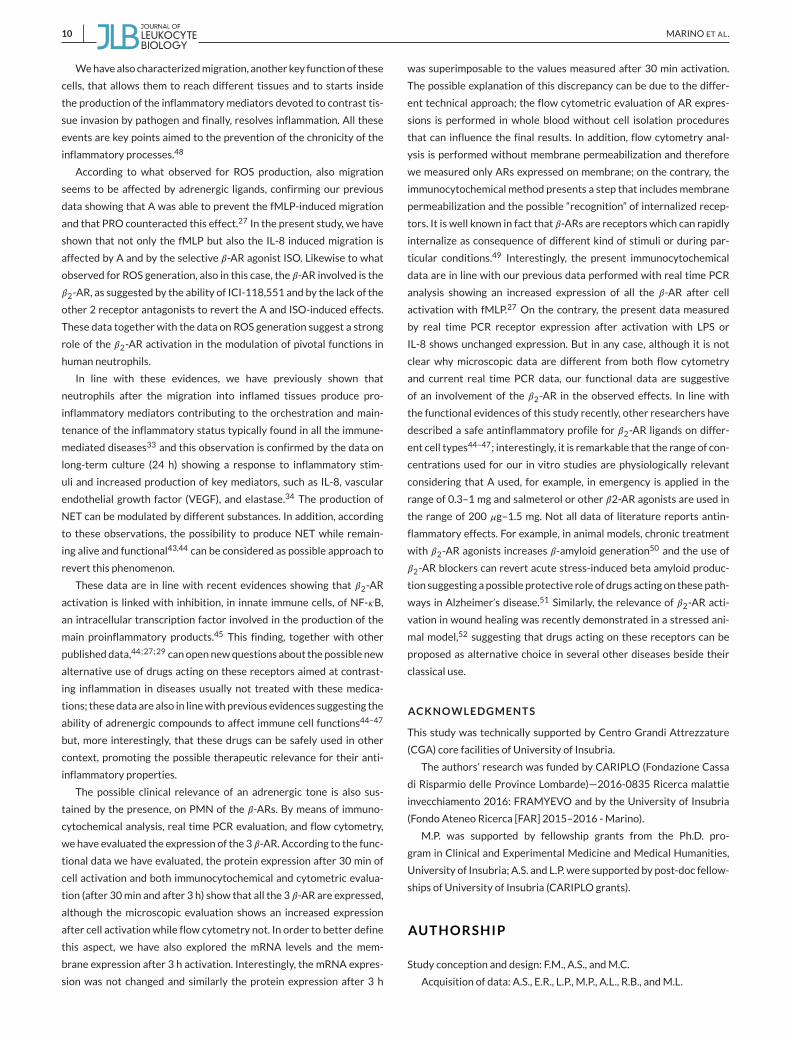
3.5 Characterization of 𝜷-AR expression on PMN
3.5.1 Real time PCR evaluation ofmRNA expression
In our previous study, we have shown that all the AR are expressed at
the mRNA level and in particular that the highest expression was for
the 𝛽-AR subtypes. We have also shown that the 3 h activation with
fMLP induced an increase in themRNAexpression of all the AR.27 Sim-
ilarly, our previous study showed that the effects exerted by A on PMN
functions were for the most part related to 𝛽-AR activation.27 In the
present study, starting from this observation, we have explored the
ability of other PMN activators namely IL-8 and LPS to influence the
𝛽-AR expression. As shown in Fig. 7, neither IL-8 (100 ng/mL) or LPS
(1𝜇g/mL)were able to significantly affect themRNA levels of the three
𝛽-ARs; in fact, both induce a slight increase of the expression for all the
3 receptors, but this increase did not reach the statistical significance.
3.5.2 Immunocytochemical analysis of 𝜷-AR expression
As shown in Fig. 9, 𝛽1-AR (A), 𝛽2-AR (E), and 𝛽3-AR (I) are expressed on
isolated PMN, but at low intensity; after activation with fMLP (panels
B,F, and J), IL-8 (panels C, G, and K), and LPS (panels D, H, and L), the
expression of all the three 𝛽-AR subtypes was increased.
3.5.3 Flow cytometric evaluation of 𝜷-AR expression
According to our previous data and on the basis of the present results,
we have evaluated the membrane receptor expression of the 𝛽1 and
𝛽2-ARbothafter30minand3h incubation.As shown inFig. 10, both 𝛽1and 𝛽2-ARare expressed andno substantial differenceswere observed
for the expression at the 2 times of incubation. The expression how-
ever was quite different for the two subtypes, showing higherMFI and
percent of positive cells for the 𝛽1-AR (P< 0.05) both after 30min and
180min of incubation.
Activation with fMLP, IL-8, or LPS did not significantly altered the
membrane expression, measured as MFI of both receptors with the
only exception of a slight decrease after activation with LPS for the
𝛽1-AR (P < 0.05 vs. resting) after 180 min of incubation both as MFI
and percent of positive cells (lower panels).
4 DISCUSSION
NET primarily identified, produced by neutrophils to kill microorgan-
isms and todefend against pathogens,1 are demonstrated tobe formed
also during inflammatory events,2 as for example, in cystic fibrosis,39
thrombosis,40 and chronic obstructive pulmonary disease.41 In the
last years, the list of diseases in which these neutrophil products are
demonstrated to be involved was certainly increased42 and a key role
ofNET in non-infective diseaseswas recently outlined.43 NETare com-
monly classified in 2 main types: lytic and vital NET44 and usually it is
documented that vital NET are produced rapidly by cells in which via-
bility and functional activities aremaintained.44
In the present study, we have shown that the stimulus-inducedNET
production can be modulated by A and other adrenergic ligands. In
particular, we have shown, for the first time, that A is able to pre-
vent the stimulus-induced NET production and this effect seems to
involve receptor activation, in particular the 𝛽-AR subtypes, as sug-
gested by the ability of 𝛽-AR antagonists to counteract the A and ISO
induced effects.

8 MARINO ET AL.
F IGURE 7 Effect of treatment with adren-ergic ligands on PMN migration. (A) Effect oftreatment with A and ISO on the IL-8 inducedcell migration. (B) Effect of pretreatment withthe selective 𝛽-AR antagonists on the abilityof ISO to prevent the IL-8-induced cell migra-tion. In both panels, data are presented asmean ± SEM of at least 3–7 separate experi-ments. Statistical analysis was performed usingMann–Whitney test and “D’Agostino & Pearsonomnibus normality test”. *P < 0.05, **P < 0.01,and ***P < 0.001 versus R; #P < 0.05, and##P < 0.01, and ### = P < 0.0001 versus IL-8;$P < 0.05 versus IL-8 alone; #P < 0.05 versusISO+ IL-8
F IGURE 8 mRNA levels of 𝜷-AR expressionin isolatedPMN. mRNA levels of 𝛽1-AR (left), 𝛽2-AR (middle), and 𝛽3-AR (right) in isolated PMNcultured for 3 h under resting conditions orafter activation with IL-8 (100 ng/mL) or LPS(1 𝜇g/mL). Data are presented as mean ± SD of4 separate experiments
F IGURE 9 Immunocytochemical characteri-zation of 𝜷-AR expression. 𝛽-AR expression inPMN under resting conditions (A, E, and I) andafter activation with fMLP (B, F, and J), IL-8 (C,G, and K), and LPS (D, H, and L). (A, E, and I): neu-trophils under resting conditions showing basallevel of 𝛽-AR expression. PMNs after activationwith different type of stimuli show increasedexpression of 𝛽1-AR (BC andD), 𝛽2-AR (F, G, andH), and 𝛽3-AR (J, K, and L). 𝛽-AR positivity is visi-ble in red; nuclei, counterstained with DAPI, arein blue

MARINO ET AL. 9
F IGURE 10 Flow cytometric evaluation of 𝜷1- and 𝜷2-AR expression. (A) Representative flow cytometric analysis of 𝛽1-AR or 𝛽2-AR fromwhole blood sample performed for each donor showing the morphological dot-plot (FSC vs. SSC signals) used to identify granulocyte population(red region) and the histogram overlays for 𝛽1-AR (left) or 𝛽2-AR (right). The ratio (MFI), calculated by dividing the MFI corresponding to specificstaining for 𝛽-AR over MFI corresponding to a negative control, was used to quantify the expression level of each 𝛽-AR and is shown above eachhistogram line.(B) Analysis of 𝛽1-AR (left) and 𝛽2-AR (right) expression after 30 or 180 min culture in the presence of fMLP (0.1 𝜇M), IL-8 (100 ng/mL), and LPS(1 𝜇g/mL). Data are expressed as ratio (MFI; in each time-set, upper panels) or percent of positive cells (in each time-set, lower panels) and arerepresented asmean± SEM of at least 4–5 separate experiments. Statistical analysis was performed usingMann–Whitney test and “D ′Agostino &Pearson omnibus normality test”. *P< 0.05 versus respective resting conditions
In our previous paper, we have extensively characterized, in human
neutrophils, the ROS-induced generation and the ability of A, and in
general of adrenergic compounds, to modulate this production.27
In the present study, we have investigated the ability of 𝛽-AR lig-
ands to affect NET production. To this end, we have measured the
3 h-induced ROS generation (ROS are necessary for NETosis3) related
to NET production and other different methods, such as microscopic
analysis and colorimetric assays. The main relevant finding is that the
3 h-NET-induced production was reduced by activation of 𝛽2-AR; in
particular, we have shown that A prevented the stimulus-induced NET
production; in addition we have shown, and to our knowledge this is
the first study showing a contribution of 𝛽2-AR in the modulation of
NET production, that this effect was receptor mediated; in fact, the
A-induced effect was reverted by the addition of the 𝛽2-AR antag-
onist ICI-118,551. We have considered the possibility that the abil-
ity of A to prevent the fMLP-induced effects can be due to an apop-
totic effect exerted by the CA in culture; we have shown no significant
apoptosis suggesting that the observed effects on NET, ROS genera-
tion, andmigration are receptor-mediated. Considering the recent evi-
dence showing thatNET can be produced by living neutrophils and this
function is not only an end stage of these cells43,44 or thatNET are pro-
duced only by a small part of these cells,43 this evidence supports the
idea that it is possible to interferes pharmacologicallywith thesemech-
anisms without affecting cell viability.
This result is of greater interest considering the pivotal role
of NET in several immune-mediated diseases and the possibility
offered by different safe drugs widely used in therapy known to
act through the interaction with these receptors (e.g., beta agonists
used in asthma and cardiovascular diseases) and this observation is
in line with literature data that propose 𝛽2-AR agonists as safe anti-
inflammatory drugs.27,45,47
ROS production can induce a negative process that can result
in chronic inflammation or cancer.48 According to our previous
data showing the ability of A to counteract the stimuli-induced
ROS generation and by the ability of propranolol (a nonselective
𝛽-AR antagonist) to counteract this effect, we have deepened this
aspect investigating specifically which kind of 𝛽-AR was involved in
this effect. The role of 𝛽2-AR in this effect is outlined by the fact that
only ICI-118,551 andnotCGP-12177 (𝛽1-AR antagonist) or L-74,8337
(𝛽3-AR antagonist) was able to revert the A-induced effects.
10 MARINO ET AL.
Wehavealso characterizedmigration, another key functionof these
cells, that allows them to reach different tissues and to starts inside
the production of the inflammatory mediators devoted to contrast tis-
sue invasion by pathogen and finally, resolves inflammation. All these
events are key points aimed to the prevention of the chronicity of the
inflammatory processes.48
According to what observed for ROS production, also migration
seems to be affected by adrenergic ligands, confirming our previous
data showing that A was able to prevent the fMLP-induced migration
and that PRO counteracted this effect.27 In the present study, we have
shown that not only the fMLP but also the IL-8 induced migration is
affected by A and by the selective 𝛽-AR agonist ISO. Likewise to what
observed for ROS generation, also in this case, the 𝛽-AR involved is the
𝛽2-AR, as suggested by the ability of ICI-118,551 and by the lack of the
other 2 receptor antagonists to revert the A and ISO-induced effects.
These data together with the data on ROS generation suggest a strong
role of the 𝛽2-AR activation in the modulation of pivotal functions in
human neutrophils.
In line with these evidences, we have previously shown that
neutrophils after the migration into inflamed tissues produce pro-
inflammatory mediators contributing to the orchestration and main-
tenance of the inflammatory status typically found in all the immune-
mediated diseases33 and this observation is confirmed by the data on
long-term culture (24 h) showing a response to inflammatory stim-
uli and increased production of key mediators, such as IL-8, vascular
endothelial growth factor (VEGF), and elastase.34 The production of
NET can be modulated by different substances. In addition, according
to these observations, the possibility to produce NET while remain-
ing alive and functional43,44 can be considered as possible approach to
revert this phenomenon.
These data are in line with recent evidences showing that 𝛽2-AR
activation is linked with inhibition, in innate immune cells, of NF-𝜅B,
an intracellular transcription factor involved in the production of the
main proinflammatory products.45 This finding, together with other
publisheddata,44;27;29 canopennewquestions about the possible new
alternative use of drugs acting on these receptors aimed at contrast-
ing inflammation in diseases usually not treated with these medica-
tions; these data are also in linewith previous evidences suggesting the
ability of adrenergic compounds to affect immune cell functions44–47
but, more interestingly, that these drugs can be safely used in other
context, promoting the possible therapeutic relevance for their anti-
inflammatory properties.
The possible clinical relevance of an adrenergic tone is also sus-
tained by the presence, on PMN of the 𝛽-ARs. By means of immuno-
cytochemical analysis, real time PCR evaluation, and flow cytometry,
we have evaluated the expression of the 3 𝛽-AR. According to the func-
tional data we have evaluated, the protein expression after 30 min of
cell activation and both immunocytochemical and cytometric evalua-
tion (after 30min and after 3 h) show that all the 3 𝛽-AR are expressed,
although the microscopic evaluation shows an increased expression
after cell activation while flow cytometry not. In order to better define
this aspect, we have also explored the mRNA levels and the mem-
brane expression after 3 h activation. Interestingly, the mRNA expres-
sion was not changed and similarly the protein expression after 3 h
was superimposable to the values measured after 30 min activation.
The possible explanation of this discrepancy can be due to the differ-
ent technical approach; the flow cytometric evaluation of AR expres-
sions is performed in whole blood without cell isolation procedures
that can influence the final results. In addition, flow cytometry anal-
ysis is performed without membrane permeabilization and therefore
we measured only ARs expressed on membrane; on the contrary, the
immunocytochemical method presents a step that includesmembrane
permeabilization and the possible “recognition” of internalized recep-
tors. It is well known in fact that 𝛽-ARs are receptors which can rapidly
internalize as consequence of different kind of stimuli or during par-
ticular conditions.49 Interestingly, the present immunocytochemical
data are in line with our previous data performed with real time PCR
analysis showing an increased expression of all the 𝛽-AR after cell
activation with fMLP.27 On the contrary, the present data measured
by real time PCR receptor expression after activation with LPS or
IL-8 shows unchanged expression. But in any case, although it is not
clear why microscopic data are different from both flow cytometry
and current real time PCR data, our functional data are suggestive
of an involvement of the 𝛽2-AR in the observed effects. In line with
the functional evidences of this study recently, other researchers have
described a safe antinflammatory profile for 𝛽2-AR ligands on differ-
ent cell types44–47; interestingly, it is remarkable that the range of con-
centrations used for our in vitro studies are physiologically relevant
considering that A used, for example, in emergency is applied in the
range of 0.3–1 mg and salmeterol or other 𝛽2-AR agonists are used in
the range of 200 𝜇g–1.5 mg. Not all data of literature reports antin-
flammatory effects. For example, in animal models, chronic treatment
with 𝛽2-AR agonists increases 𝛽-amyloid generation50 and the use of
𝛽2-AR blockers can revert acute stress-induced beta amyloid produc-
tion suggesting a possible protective role of drugs acting on these path-
ways in Alzheimer’s disease.51 Similarly, the relevance of 𝛽2-AR acti-
vation in wound healing was recently demonstrated in a stressed ani-
mal model,52 suggesting that drugs acting on these receptors can be
proposed as alternative choice in several other diseases beside their
classical use.
ACKNOWLEDGMENTS
This study was technically supported by Centro Grandi Attrezzature
(CGA) core facilities of University of Insubria.
The authors’ research was funded by CARIPLO (Fondazione Cassa
di Risparmio delle Province Lombarde)—2016-0835 Ricerca malattie
invecchiamento 2016: FRAMYEVO and by the University of Insubria
(Fondo Ateneo Ricerca [FAR] 2015–2016 -Marino).
M.P. was supported by fellowship grants from the Ph.D. pro-
gram in Clinical and Experimental Medicine and Medical Humanities,
University of Insubria; A.S. and L.P. were supported by post-doc fellow-
ships of University of Insubria (CARIPLO grants).
AUTHORSHIP
Study conception and design: F.M., A.S., andM.C.
Acquisition of data: A.S., E.R., L.P., M.P., A.L., R.B., andM.L.

MARINO ET AL. 11
Analysis and interpretation of data: E.R., R.B., A.S., M.P., M.L., and
M.D.E.
All authors were involved in drafting the article or revising it criti-
cally for important intellectual content, and all authors approved the
final version to be published.
All authors agree to be accountable for all aspects of the work in
ensuring that questions related to the accuracy or integrity of any part
of the work are appropriately investigated and resolved, and declare
to have confidence in the integrity of the contributions of their co-
authors.
DISCLOSURE
The authors declare no conflict of interests.
REFERENCES
1. Brinkmann V, Reichard U, Goosmann C, et al. Neutrophil extracellular
traps kill bacteria. Science. 2004;303:1532–1535.
2. Pinegin B, Vorobjeva N, Pinegin V. Neutrophil extracellular traps and
their role in the development of chronic inflammation and autoimmu-
nity. Autoimmun Rev. 2015;14:633–640.
3. Hansson GK, Hermansson A. The immune system in atherosclerosis.
Nat Immunol. 2011;12:204–212.
4. Kruger P, Saffarzadeh M, Weber AN, et al. Neutrophils: between
host defence, immune modulation, and tissue injury. PLoS Pathog.2015;11:e1004651.
5. Chistiakov DA, Bobryshev YV, Orekhov AN. Neutrophil’s weapons in
atherosclerosis. ExpMol Pathol. 2015;99:663–671.
6. van Leeuwen M, Duijvestijn A, Smook M, et al. Accumulation of
myeloperoxidase-positive neutrophils in atherosclerotic lesions in
LDLR-/- mice. Arterioscler Thromb Vasc Biol. 2008;28:84–89.
7. Marino F, TozziM, Schembri L, et al. Production of IL-8, VEGF and elas-
tase by circulating and intraplaque neutrophils in patients with carotid
atherosclerosis. PLoS One. 2015;10:e0124565.
8. Giaglis S, Hahn S, Hasler P. The NET outcome": are neutrophil extra-
cellular traps of any relevance to the pathophysiology of autoimmune
disorders in childhood. Front Pediatr. 2016;4:97.
9. HuSC, YuHS, YenFL, LinCL,ChenGS, LanCC.Neutrophil extracellular
trap formation is increased in psoriasis and induces human 𝛽-defensin-
2 production in epidermal keratinocytes. Sci Rep. 2016;5:31119.
10. Thureson-Klein A, Klein RL, Johansson O. Catecholamine-rich cells
and varicosities in bovine splenic nerve, vesicle contents and evidence
for exocytosis. J Neurobiol. 1979;10:309–324.
11. Bell C, Gillespie JS. Dopamine and noradrenaline levels in peripheral
tissues of severalmammalian species. J Neurochem. 1981;36:703–706.
12. Bergquist J, Tarkowski A, Ekman R, Ewing A. Discovery of endogenous
catecholamines in lymphocytes and evidence for catecholamine regu-
lation of lymphocyte function via an autocrine loop. Proc Natl Acad SciUSA. 1994;91:12912–12916.
13. Marino F, Cosentino M, Bombelli R, Ferrari M, Lecchini S, Frigo
GM. Endogenous catecholamine synthesis, metabolism, storage and
uptake in human peripheral blood mononuclear cells. Exp Hematol.1999;27:489–495.
14. Cosentino M, Marino F, Bombelli R, Ferrari M, Lecchini S, Frigo GM.
Endogenous catecholamine synthesis,metabolism, storage and uptake
in human neutrophils. Life Sci. 1999;64:975–981.
15. Williams JM, Felten DL. Sympathetic innervation of murine thy-
mus and spleen: a comparative histofluorescence study. Anat Rec.1981;199:531–542.
16. Carlson SL, Felten DL, Livnat S, Felten SY. Noradrenergic sympathetic
innervation of the spleen: acute drug-induced depletion of lympho-
cytes in the target fields of innervation results in redistribution of
noradrenergic fibers butmaintenance of compartmentation. JNeurosciRes. 1987;18:130–131.
17. Cosentino M, Zaffaroni M, Marino F, et al. Catecholamine production
and tyrosine hydroxylase expression in peripheral blood mononuclear
cells from multiple sclerosis patients: effect of cell stimulation and
possible relevance for activation-induced apoptosis. J Neuroimmunol.2002;133:233–240.
18. Cosentino M, Fietta AM, Ferrari M, et al. Human CD4+CD25+ reg-
ulatory T cells selectively express tyrosine hydroxylase and con-
tain endogenous catecholamines subserving an autocrine/paracrine
inhibitory functional loop. Blood. 2007;109:632–642.
19. Cosentino M, Marino F, Bombelli R, et al. Stimulation with phyto-
haemagglutinin induces the synthesis of catecholamines in human
peripheral blood mononuclear cells: role of protein kinase C and con-
tribution of intracellular calcium. J Neuroimmunol. 2002;125:125–133.
20. CosentinoM, FerrariM, KustrimovicN, Rasini E,Marino F. Influence of
dopamine receptor gene polymorphisms on circulating T lymphocytes:
a pilot study in healthy subjects.Hum Immunol. 2015;76:747–752.
21. Maestroni GJ, Conti A. Modulation of hematopoiesis via alpha
1-adrenergic receptors on bone marrow cells. Exp Hematol.1994;22:313–320.
22. Brown JE, Adamson JW. Modulation of in vitro erythropoiesis. The
influence of beta-adrenergic agonists on erythroid colony formation.
J Clin Invest. 1977;60:70–77.
23. Zaffaroni M, Marino F, Bombelli R, et al. Therapy with interferon-beta
modulates endogenous catecholamines in lymphocytes of patients
withmultiple sclerosis. Exp Neurol. 2008;214:315–321.
24. Prado C, Contreras F, González H, et al. Stimulation of dopamine
receptor D5 expressed on dendritic cells potentiates Th17-mediated
immunity. J Immunol. 2012;188:3062–3070.
25. Gaskill PJ, Yano HH, Kalpana GV, Javitch JA, Berman JW.
Dopamine receptor activation increases HIV entry into primary
human macrophages. PLoS One. 2014;9:e108232. https://doi.org/
10.1371/journal.pone.0108232.. Sep 30.
26. Kustrimovic N, Rasini E, Legnaro M, Marino F, Cosentino M. Expres-
sion of dopaminergic receptors on human CD4+ T lymphocytes: flow
cytometric analysis of naive andmemory subsets and relevance for the
neuroimmunology of neurodegenerative disease. J Neuroimmunephar-macol. 2014;9:302–312.
27. Scanzano A, Schembri L, Rasini E, et al. Adrenergic modulation
of migration, CD11b and CD18 expression, ROS and interleukin-8
production by human polymorphonuclear leukocytes. Inflamm Res.2015;64:127–135.
28. Cosentino M, Zaffaroni M, Legnaro M, et al. Dopaminergic receptors
and adrenoceptors in circulating lymphocytes as putative biomarkers
for the early onset and progression of multiple sclerosis. J Neuroim-munol. 2016;298:82–89.
29. ScanzanoA, CosentinoM. Adrenergic regulation of innate immunity: a
review. Front Pharmacol. 2015;6:171.
30. Guasti L, Marino F, CosentinoM, et al. Simvastatin treatmentmodifies
polymorphonuclear leukocyte function in high-risk subjects: a longitu-
dinal study. J Hypertens. 2006;24:2423–2430.
31. Guasti L,MarinoF,CosentinoM, et al. Prolonged statin-induced reduc-
tion in neutrophil reactive oxygen species and angiotensin II type 1
receptor expression: 1-year follow-up. Eur H J. 2008;29:1118–1126.

12 MARINO ET AL.
32. Marino F, Guasti L, Tozzi M, et al. Angiotensin type-1 receptor expres-
sion and interleukin-8 production in polymorphonuclear leukocytes
of patients with peripheral arterial disease. J Cardiovasc Pharmacol.2009;54:520–525.
33. Marino F, TozziM, Schembri L, et al. Production of IL-8, VEGF and elas-
tase by circulating and intraplaque neutrophils in patients with carotid
atherosclerosis. PLoS One. 2015;10:e0124565.
34. PinoliM, Schembri L, ScanzanoA, et al. Production of proinflammatory
mediators byhumanneutrophils during long-termculture. Int J Clin ExpPathol. 2016;9:1858–1866.
35. Pinoli M, Rasini E, Legnaro M, et al. Dopamine affects migration
and morphology of human neutrophils through D1-like dopaminergic
receptors. J Neuroimmune Pharmacol. 2016;11:S3–S61.
36. Ye RD, Boulay F,Wang JM, et al. International Union of Basic and Clin-
ical pharmacology. LXXIII: nomenclature for the formyl peptide recep-
tor (FPR) family. Pharmacol. Rev. 2009;61:119–161.
37. Pieterse E, Rother N, Yanginlar C, Hilbrands LK, van der Vlag J, Neu-
trophils discriminate between lipopolysaccharides of different bacte-
rial sources and selectively release neutrophil extracellular traps. FrontImmunol. 2016;7:484.
38. Hazeldine J, Harris P, Chapple IL, et al. Impaired neutrophil extracellu-
lar trap formation: a novel defect in the innate immune system of aged
individuals. Aging Cell. 2014;13:690–698.
39. Martínez-Alemán SR, Campos-García L, Palma-Nicolas JP,
Hernández-Bello R, González GM, Sánchez-González A. Under-
standing the entanglement: neutrophil extracellular traps
(NETs) in cystic fibrosis. Front Cell Infect Microbiol. 2017;7:104.
https://doi.org/10.3389/fcimb.2017.00104..
40. Jiménez-Alcázar M, Kim N, Fuchs TA. Circulating extracellular
DNA: cause or consequence of thrombosis? Semin Thromb Hemost.2017;43:553–561.
41. EchevarríaUrib, Leimgruber LC,GarcíaGJ, et al. Evidenceof eosinophil
extracellular trap cell death in COPD: does it represent the trigger
that switches on the disease. Int J Chron Obstruct Pulmon Dis. 2017;12:885–896.
42. TengTS, JiAL, Ji XY, Li YZ.Neutrophils and Immunity: frombactericidal
action to being conquered. J Immunol Res. 2017;2017:9671604.
43. Jorch SK, Kubes P. An emerging role for neutrophil extracellular traps
in noninfectious disease.NatMed. 2017;23:279–287.
44. Doring Y, Soehnlein O, Weber C. Neutrophil extracellular traps in
atherosclerosis and atherothrombosis. Circ Res. 2017;120:736–743.
45. Kolmus K, Tavernier J, Gerlo S. 𝛽2-Adrenergic receptors in immu-
nity and inflammation: stressing NF-𝜅B. Brain Behav Immun. 2015;45:297–310.
46. Keränen T, Hömmö T, Moilanen E, Korhonen R. 𝛽2-receptor ago-
nists salbutamol and terbutaline attenuated cytokine production by
suppressing ERK pathway through cAMP in macrophages. Cytokine.2017;94:1–7.
47. Dimitrov S, Hulteng E, Hong S. Inflammation and exercise: inhibition
of monocytic intracellular TNF production by acute exercise via 𝛽2-
adrenergic activation. Brain Behav Immun. 2017;61:60–68.
48. Szuster-CiesielskaA,Hryciuk-UmerE, StepulakA, KupiszK, Kandefer-
SzerszeńM. Reactive oxygen species production by blood neutrophils
of patientswith laryngeal carcinoma and antioxidative enzymeactivity
in their blood. Acta Oncol. 2004;43:252–258.
49. Peterson YK, Luttrell LM. The diverse roles of arrestin scaffolds in G
protein-coupled receptor signaling. Pharmacol Rev. 2017;69:256–297.
50. Chai GS, Wang YY, Yasheng A, Zhao P. Beta 2-adrenergic receptor
activation enhances neurogenesis in Alzheimer’s disease mice. NeuralRegen Res. 2016;11:1617–1624.
51. Ni Y, Zhao X, Bao G, et al. Activation of beta2-adrenergic receptor
stimulates gamma-secretase activity and accelerates amyloid plaque
formation.NatMed. 2006;12:1390–1396.
52. Kim MH, Gorouhi F, Ramirez S, et al. Catecholamine stress
alters neutrophil trafficking and impairs wound healing by 𝛽2-
adrenergic receptor-mediated upregulation of IL-6. J Invest Dermatol.2014;134:809–817.
How to cite this article: Marino F, Scanzano A, Pulze L,
et al. 𝛽2-Adrenoceptors inhibit neutrophil extracellular traps
in human polymorphonuclear leukocytes. J Leukoc Biol. 2018;1-
12. https://doi.org/10.1002/JLB.3A1017-398RR