Embed Size (px)
Citation preview
31-3-12
HHiittaacchhii RReeaall--ttiimmee TTiissssuuee
EEllaassttooggrraapphhyy::
PPuubblliiccaattiioonnss && IInntteerrnnaattiioonnaall
CCoommmmuunniiccaattiioonnss
CClliinniiccaall AAbbssttrraaccttss
31-3-12
HHiittaacchhii RReeaall--ttiimmee TTiissssuuee EEllaassttooggrraapphhyy ffoorr WWoommeenn’’ss HHeeaalltthh
31-3-12
DISTINGUISHING BENIGN FROM MALIGNANT MASSES AT BREAST US: COMBINED US ELASTOGRAPHY AND COLOR DOPPLER US—INFLUENCE ON RADIOLOGIST ACCURACY Nariya Cho, Mijung Jang, Chae Yeon Lyou, Jeong Seon Park, Hye Young Choi, and Woo Kyung Moon
Purpose: To investigate the effect of the combined use of ultrasonographic (US) elastography and color Doppler US on the accuracy of radiologists in distinguishing benign from malignant nonpalpable breast masses and in making the decision for biopsy recommendations at B-mode US.
Materials and Methods: This prospective study was conducted with institutional review board approval; written informed consent was obtained. A cohort of 367 biopsy-proved cases in 319 women (age range, 22–78 years; mean age, 48.6 years) with B-mode US, US elastographic, and Doppler US images was included. Five blinded readers independently scored the likelihood of malignancy for four data sets (ie, B-mode US alone, B-mode US and elastography, B-mode US and Doppler US, and B-mode US, US elastography, and Doppler US). The area under the receiver operating characteristic curve (Az) values, sensitivities, and specificities of each data set were compared.
Results: The Az of B-mode US, US elastography, and Doppler US (average, 0.844; range, 0.797–0.876) was greater than that of B-mode US alone (average, 0.771; range, 0.738–0.798) for all readers (P = .001 for readers 1, 2, and 3; P < .001 for reader 4; P = .002 for reader 5). When both elastography and Doppler scores were negative, leading to strict downgrading, the specificity increased for all readers from an average of 25.3% (75.4 of 298; range, 6.4%–40.9%) to 34.0% (101.2 of 298; range, 26.5%–48.7%) (P < .001 for readers 1, 2, 4, and 5; P = .016 for reader 3) without a significant change in sensitivity.
Conclusion: Combined use of US elastography and color Doppler US increases both the accuracy in distinguishing benign from malignant masses and the specificity in decision-making for biopsy recommendation at B-mode US.
Radiology 2012;262 80-90 ___________________
POSSIBILITIES OF REAL-TIME SONOELASTOGRAPHY IN LOCAL STAGING OF ENDOMETRIAL CANCER I.S. Belozerova, V.E. Gazhonova, S.O. Churkina, A.L. Lozovator, T.S. Kurganskaya, A.V. Zubarev; Moscow/RU Purpose: Possibilities of sonoelastography (SE) in local staging of endometrial cancer. Methods and Materials: 42 pts with proven EC underwent real-time sonoelastography before surgery (age range 37-72 y.o). US exams were performed on HI VISION Preirus and HI VISION 900 (Hitachi Medical Corporation). We used FIGO classification for local staging EC. US data were compared with final histopathology. The study was recorded and evaluated by 2 independent readers. Inter-observer agreement for tumour‘s location by walls (anterior, posterior, fundus, right, left), myometrial invasion (less than ½, more than ½), cervical and capsular involvement were studied. Sensitivity of SE in local staging was established. Results: All patients have been operated (17 total hysterectomies, 25 radical hysterectomies with lymph node dissection). Histopathology revealed 17/stage IA, 15/stage IB, 5/stage IIA, 3/IIB stage, 2/IIIAstage. The sensitivity of SE for local staging of EC: 89 % - stage IA, 90 % - stage IB, 84 % - stage IIA, 94 % - stage IIB, 93 % - stage IIIA. The Kappa value between SE and location by walls was poor (k ranged 0,3 to 0,5), between SE and myometrial invasion was good (k ranged 0,76 to 0,84) and between SE and cervical invasion was poor too (k 0,4-0,66). SE increased the sensitivity of US (from 83 % to 92,5%) and specificity (from 81 % to 90,3%) in local staging EC. Conclusion: Inclusion of SE in complex ultrasound scanning may help to evaluate myometrial invasion in pts with EC.
31-3-12
ECR 2012, March 2
nd – 5
th, Vienna, Austria
___________________
SONOELASTOGRAPHY IN PATIENTS WITH ENDOMETRIOSIS OF DIFFERENT LOCATION N.A. Vorontsova, V.E. Gazhonova, S.O. Churkina, E.B. Savinova, I.A. Ponomorenko, A.V. Zubarev; Moscow/RU Purpose: The aim of this study was to assess the usefulness of SE (sonoelastography) for evaluation of patients with endometriosis. Methods and Materials: 80 consecutive pts (with pelvic pain, dysmenorrhoea, urinary symptoms) range 18-45years with suspected diagnosis of endometriosis were referred for SE. All patients underwent conventional US and SE on HI VISION Preirus with an endocavity transducer (8-4 MHz frequency) and linear transducer (frequency 7,5-13 MHz). We used modified Tsukuba SE classification for evaluation of the SE data. 15 diagnostic biopsy, 39 diagnostic laparoscopies, 26 separate diagnostic scraping were performed with morphological study of the received material. US data (conventional B-mode, US angiography and SE) were assessed by comparing the findings with surgery results and MRI data. US data were retrospectively reviewed by 2 radiologists. Inter-observer agreement for endometriosis SE score, location of endometriosis (uterine, ovarium, cervix, urinary bladder‘s wall, soft tissue), endometriosis location by walls of the uterus, MRI data. Results: Pathomorphological examination revealed 32/endometriotic cysts, 7/endometriosis the uterine wall, 26/endometrial polyps, 6/endometriosis of urinary bladder‘s wall, and 9/endometrial infiltrations of pelvic soft tissue. Endometriosis was characterised by reversed score 1, score 3 and score 5 of Tsukuba classification for lesions on SE. SE showed good to moderate inter-observer agreement for endometriosis evaluation by scoring (k=0, 8-0, 95), for endometriosis locations by walls of the uterus (k= 0,78-0,94), for locations of endometriosis by organs (k =0,74 -0,87), poor to moderate inter-observer agreement for endometriosis evaluation by MRI data (k=0,21-0,35). Conclusion: US with SE offers a new possibility for definition of endometriosis. ECR 2012, March 2
nd – 5
th, Vienna, Austria
___________________
COMBINED USE OF US ELASTOGRAPHY AND CONVENTIONAL ULTRASONOGRAPHY FOR DIFFERENTIATION OF BENIGN AND MALIGNANT CIRCUMSCRIBED BREAST MASSES Soo-Yeon Kim , Jeong Seon Park PURPOSE To evaluate diagnostic performance of conventional ultrasonography (US) combined with US ELASTOGRAPHY for differentiation between benign and malignant breast lesions with circumscribed margins, with the pathology as reference standard. METHOD AND MATERIALS Between October 2008 and February 2010, we performed real-time US ELASTOGRAPHY in 263 consecutive women who were scheduled to undergo US-guided core biopsy. Among them, 100 women (mean age, 46 years; age range, 15-73 years) of 109 circumscribed breast masses (99 benign, 10 malignant) were included. Two radiologists retrospectively reviewed conventional and elastographic US images in consensus. We assessed the lesions according to BI-RADs classification (3 in 26 cases (23.9%), 4a in 44 cases (40.4%), 4b in 5 cases (4.5%) and 4c in 1 case (0.9%)) and then assigned conventional US scores from 1 to 4 corresponding on BI-RADs category 3, 4a, 4b and 4c. Elasticity scores were assessed by using a five-point scale (1-5). Summation of both conventional US and elasticity scores was defined as ‗combined score‘. The mean scores of benign and malignant lesions were compared with student t-test. The diagnostic performances of conventional US, elasticity and combined scoring were compared by using receiver operating characteristic (ROC) curve analysis.

31-3-12
RESULTS The mean conventional US core (2.6 vs. 1.7), mean elasticity score (3.7 vs. 1.9) and mean combined score (6.3 vs. 3.7) were significantly higher in malignancy than benign lesions (P<0.001). The area under the ROC curve (AUC) were 0.81 (95% CI, 0.72–0.88) for conventional US scoring, 0.94 (95% CI, 0.87–0.97) for elasticity scoring and 0.95 (95% CI, 0.89 – 0.99) for combined scoring. The AUC of combined scoring was significantly higher than conventional US scoring (P=0.03). There was no difference in AUC values between other modalities (P>0.05). The sensitivity and specificity of combined scoring were 90% and 89% at the cutoff value between 4 and 5, and 100% and 40% at the cutoff value between 3 and 4. Radiological Society of North America 97th Scientific Assembly and Annual Meeting November 27th – December 2nd, 2011, Chicago, USA
___________________ COMPARISON OF COMMERCIALLY AVAILABLE SHEAR WAVE AND STATIC US ELASTOGRAPHY SYSTEMS FOR DIFFERENTIATION OF BENIGN AND MALIGNANT BREAST MASSES Jung Min Chang, Woo Kyung Moon, Nariya Cho, Seung Ja Kim PURPOSE To prospectively compare the diagnostic performance of shear wave and static US elastography systems for differentiation of benign and malignant breast masses. METHOD AND MATERIALS Between March 2010 and April 2010, 125 women (mean age 47 years, range 22 – 75 years) with 156 breast masses (mean size 19 mm, range 4-80 mm) (76 malignant, 80 benign) underwent US elastographic examinations with both systems (shear wave and static US elastography) by one radiologist prior to biopsy. Probability of malignancy based on conventional US findings was recorded prior to US elastography. With shear wave system, quantitative elasticity values in kiloPascal units measured was recorded. For static elastography, the elasticity score (1-5) based on the degree and distribution of strain proposed by Itoh et al. (Radiology 2006; 239:341–350) was given. Diagnostic performance of the two systems in distinguishing benign from malignant masses was compared using receiver operating characteristic (ROC) curve analysis and McNemar‘s test using histological analysis as a reference standard. RESULTS The area under the ROC curve for the static elastography system (Az=0.948) was similar to that of the shear wave elastography system (Az=0.917) (difference between areas 0.02, 95% CI - 0.01-0.07, P=0.172). The best cut-off values, yielding the maximal sum of sensitivity and specificity, were between values in kiloPascal units of 57.7 and elasticity scores of 3 and 4. The sensitivity of the shear wave elastography system was higher than that of the static elastography system [98.7% (75 of 76) vs. 78.9% (60 of 76), P = 0.0001] and the specificity of the static elastography system was higher than that of the shearwave elastography system [96.3% (77 of 80) vs. 71.2% (57 of 80), P = 0.001]. CONCLUSION Two systems showed similar overall diagnostic performance. However the shearwave elastography system showed better sensitivity, and the static elastography system showed better specificity with the certain fixed cutoff values in distinguishing benign from malignant breast masses. CLINICAL RELEVANCE/APPLICATION Understanding the characteristics of both shear wave elastography and static elastography systems can be helpful in optimizing the diagnostic criteria for each system. Radiological Society of North America 97th Scientific Assembly and Annual Meeting November 27th – December 2nd, 2011, Chicago, USA
31-3-12
QUANTITATIVE ELASTOGRAPHIC ASSESSMENT OF NONPALPABLE BREAST NODULES BY MEASURING FAT-LESION STRAIN RATIO VS QUALITATIVE COLOUR ELASTOGRAPHY SCORES: COMPARISON OF DIAGNOSTIC PERFORMANCES BY A BLINDED PROSPECTIVE STUDY Vasanthakumar Venugopal, Ibne Ahmad , Ishrat Afshan PURPOSE The aim of this study is to compare the diagnostic performances of strain ratio measures and Ueno colour elasticity scores in nonpalable breast masses METHOD AND MATERIALS This prospective study included 117 solid lesions (88 benign, 29 malignant) in 102 consecutive patients (age range, 16-64 years) that were planned for biopsy based on B mode scanning.They were examined using ultrasound ELASTOGRAPHY (UE). The strain index (fat to lesion strain ratio) was calculated in all lesions.The elasticity scores according to Ueno colour scoring system were determined in all the lesions by a different set of blinded examiners. Biopsy results were the reference points in all cases. Receiver-operating characteristic curve analysis was done to determine the cut off point with regard to strain ratios. Sensitivity, specificity, positive and negative predictive values were calculated. The diagnostic performances of the two evaluation systems were compared by calculating the area under curve values and by McNemar's test. RESULTS Statistically significant difference was observed between the strain ratios of benign lesions (mean, 2.17 ± 1.54) and malignant lesions (mean, 8.52 ± 4.84). When 2.61 was taken as a cut-off value for malignant lesions, strain ratio evaluation had 93.1% sensitivity, and 78.4% specificity. The area under the curve for strain ratio-based elastographic analysis was 0.910(95% confidence interval [CI], 0.836–0.984), and the area under the curve for the colour elasticity scoring system was 0.865(95% confidence interval [CI], 0.785–0.945). The diagnostic performance of strain ratio-based elastographic analysis was better than that of the five-point scoring system with UE. Interobserver agreement was also better with strain ratio based evaluation (κ > 0.82 vs 0.65) CONCLUSION Despite the lack of standardised cut off values, strain ratio measurement has been consistently shown to be a better evaluation system compared to five point colour elasticity scoring system in various studies. Our study validates the point that strain ratio evaluation is more reliable than colour elasticity scoring system. CLINICAL RELEVANCE/APPLICATION Strain ratio measurements when used along with B-mode findings and /or colour elasticity scores gives a better diagnostic yield in non palpable breast lesions. Radiological Society of North America 97th Scientific Assembly and Annual Meeting November 27th – December 2nd, 2011, Chicago, USA
___________________ IMPACT OF ELASTOGRAPHY ON ASSESSING THE LIKELIHOOD OF MALIGNANCY OF ULTRASOUND LESIONS Jennifer Kohr, Janice Sung, Sharp Malak, Valencia King, Elizabeth Morris, Christopher Comstock PURPOSE To compare the accuracy of grey scale ultrasound (US) alone with grey scale US plus color elastography in assessing the likelihood of malignancy of ultrasound lesions.

31-3-12
METHOD AND MATERIALS A retrospective, IRB approved review of our database yielded 99 lesions with compressive elastography performed by ultrasound (US) technologists prior to US-guided biopsy on a Hitachi Hi Vision 900. Three blinded board-certified radiologists with breast imaging specialty training independently reviewed anonymized grey scale US images followed by elastography images for each lesion. Grey scale images alone were given BIRADS scores and rated on a 10 point level of suspicion scale. Grey scale and elastography US were then reviewed together. The quality of the elastography images were scored on a 5 point scale and those lesions with extremely poor quality images (score of 1) were excluded. The elastography images were assessed on a 5 point scale using graphic reference standard ranging from uniform high strain (1) to a mosaic pattern of strain to no strain (5). Overall BIRADS and level of suspicion scores were recorded based on the combined grey scale and elastography images. Sensitivity, specificity, PPV, NPV and level of suspicion ROC curves were calculated for grey scale alone and with elastography (combined US). Confidence intervals for each were calculated. RESULTS Of the 99 lesions 60 were benign and 39 were malignant, 12 were excluded because of poor image quality. For grey scale alone the sensitivity, specificity, PPV and NPV was 95.2%, 33.3%, 48.8% and 91.2% respectively for all readers. For combined US sensitivity, specificity, PPV, and NPV was 93.3%, 36.2%, 49.1% and 90.5% respectively. There was no statistically significant difference between grey scale US alone and combined US (all confidence intervals for each estimate overlapped). The area under the curve for level of suspicion was not significantly different between groups 0.830 (95% CI 0.711-0.919) and 0.809 (95% CI 0.714-0.904) for grey scale alone and combined US respectively. CONCLUSION In contrast to some published reports, our results suggest that elastography has no statistically significant impact on determining the level of suspicion of sonographic lesions or the final BIRADS assessment even after excluding poor quality images and applying a graphic reference standard. CLINICAL RELEVANCE/APPLICATION elastography may not significantly impact assessing the likelihood of malignancy. Radiological Society of North America 97th Scientific Assembly and Annual Meeting November 27th – December 2nd, 2011, Chicago, USA
___________________ ELASTIC MODULI OF BREAST CARCINOMA COMPARING US ELASTOGRAPHY FINDINGS T. Umemoto, E. Ueno,Y. Fujihara, T. Matsumura, T. Shiina, E. Tohno, T. Mitake, H. Bando, I. Morishima, H. Hara Purpose To evaluate the elastic moduli of the breast tissue and to contrast them with elasticity images for accurate interpretation of real-time US elastography. Material & Methods This study was approved by Human Subjects Institutional Review Board in Tsukuba Medical Center Hospital and University of Tsukuba. Written informed consent was obtained from all of the patients. Conventional US and Real-time Tissue Elastography were performed preoperatively in patients who had breast cancer. The slice of 5mm thickness including the lesion and the surrounding breast tissue was obtained from patient's specimen immediately after resection. Within 2 hours after surgical resection, elastic modulus of each region was measured using materials testing machine (Instron 3342) under the constant pre-compression and the controlled temperature. Results The elastic moduli obtained from the measurement were in increasing order of fat tissue, normal mammary gland, non-invasive and invasive breast cancer. The nonlinearity of the stress dependency was admitted in each region. Conclusion

31-3-12
In this study, we confirmed the values of elastic moduli of the breast carcinoma tissue were varied according to the histological structure of each lesion while those of fat and normal mammary gland tissue were almost constant. The nonlinearity of the stress dependency was also different in each region. These differences were demonstrated well on elasticity images. Ultrasound in Medicine and Biology, Volume 37, Issue 8, Supplement , Page S45, August 2011
___________________
SONOELASTOGRAPHY DURING PREGNANCY: AGE-RELATED CHANGES OF THE CERVIX AND CERVICAL INSUFFICIENCY A. Thomas, T. Slowinski , T. Fischer Purpose A real-time sonoelastography study was performed in a normal population to identify elastic tissue changes in relation to age and week of gestation. The findings were compared with a cervical insufficiency group. Material & Methods Sixty healthy unselected pregnant women at a mean of 28 weeks of gestation and 30 women with cervical insufficiency (26 weeks of gestation) were examined. The elastography scans were analyzed by means of a computer program (determination of thresholds for the colors red, blue and green) and by two independent readers using defined regions of interest (ROIs). The percentages of blue in correlation to the sum of red and green in the ROI served to calculate an elasticity tissue quotient (TQ). These quotients were correlated with age and week of gestation (Wilcoxon's test). Results The color distribution in the normal population showed that green was predominant (67.1 ± 12.5 %), followed by blue (26.5 ± 12.9 %) and red (6.4 ± 3.7 %). The TQ decreased significantly with increasing age (R = -0.311, p = 0.025), while tissue elasticity was not affected by the duration of pregnancy (R = 0.362, p = 0.008). The elastic portions were larger in women with cervical insufficiency as compared to the normal group (TQ 4.7 ± 3.2 versus 2.8 ± 1.8, p < 0.05). Conclusion The elastography findings did not change with the duration of pregnancy but with the women's age. An insufficient cervix was found to be ―softer‖ on elastography. Ultrasound in Medicine and Biology, Volume 37, Issue 8, Supplement , Page S122, August 2011
___________________
THE USEFULNESS OF SONOELASTOGRAPHY IN THE DIFFERENTIAL DIAGNOSIS OF SOLID BREAST LESIONS K. Dobruch-Sobczak, I. Sudoł-Szopińska Purpose To evaluate the usefulness of sonoelastography in a differential diagnosis of solid breast lesions: (1) comparing diagnostic value of B-mode imaging and sonoelastography, in relation to histological or cytological verification; (2) assessing the diagnostic value of BI-RADS classification and the Tsukuba scale; (3) calculating FLR ratio for breast lesions. Material & Methods The study was performed on 39 women aged between 23 and 83 years with 51 solid breast lesions. Ultrasound examinations were performed on the Hitachi EUB 7500. Visible changes in B-mode imaging were assessed according to the BI-RADS classification, and in elastography according to Tsukuba scale. For all changes FLR was calculated. Statistical analysis was performed to evaluate the sensitivity, specificity, ppv and npv of B-mode comparing with elastography. Results

31-3-12
Pathological evaluation revealed 26 malignant and 25 benign lesions. Sensitivity and specificity of B-mode imaging with the cut-off points BIRADS 4/5 were, respectively, 76.92% and 92.00%, while with the cut-off values BI-RADS 3/4 were 100% and 20%, respectively. The sensitivity and specificity with the cut-off point Tsukuba 3/4 on elastograms were, respectively, 57.69% and 96.00%. The value of FLR for malignancy was 4.18 and 1.54 for benign lesions. Conclusion Sonoelastography improves the specificity of B-mode. It may be useful in the diagnosis of benign lesions classified as BI-RADS 3 and 4. FLR index helps to differentiate the character of breast lesions. Ultrasound in Medicine and Biology, Volume 37, Issue 8, Supplement , Page S100, August 2011
___________________ SONOELASTOGRAPHY IN PATIENTS WITH ENDOMETRICAL CARCINOMA: WORK IN PROGRESS
I. Belozerova, T. Smirnova, V. Gazhonova, A. Lozovator, A. Zubarev Purpose To study the possibilities of sonoelastography (SE) in endometrial carcinoma (EC) and to compare findings with final histopathology. Material & Methods 30 patients with proven EC underwent SE (range 38-65 y.o). US examinations were performed on HI VISION Preirus HI VISION 900. The SE score was established using Tsukuba classification of the strain (5-point color scale: 1-3 benign, 4-5 malignant); 9 total hysterectomies and 29 radical hysterectomies with lymph node dissection were performed. US data were compared with final histopathology. The study was recorded and evaluated by 2 readers. Inter-observer agreement for tumor's SE score, tumor location by walls (anterior, posterior, fundus, right, left), myometrial invasion (less than ½, more than ½) and cervical involvement were studied. Results Histopathology revealed 9/stage IA, 16/stage IB, 3/stage IIA, 2/IIB stage. On SE the EC had score 4/9pts & score 5/21pts. SE showed good to moderate interobserver agreement for tumor evaluation by scoring (k = 0,8-0,93), poor to moderate for tumor location by walls (k = 0,3 -0,5), moderate to good for myometrial invasion (k = 0,76-0,84), moderate to poor for cervical invasion (k 0,4-0,66). Conclusion EC is characterized by scores 4 to 5 under Tsukuba classification for lesions on SE. SE helps to evaluate myometrial invasion. Unfortunately, a small groups of patients in IIA and IIB stages showed SE as inaccurate method for evaluating tumor location by walls and cervical involvement. Further studies should be performed. Ultrasound in Medicine and Biology, Volume 37, Issue 8, Supplement , Page S40, August 2011
___________________ SONOELASTOGRAPHY IN SMALL BENIGN AND MALIGNANT PAPILLARY OVARIAN TUMORS A. Fedorova, S. Churkina, V. Gazhonova Purpose To evaluate the diagnostic possibilities of endovaginal sonoelastography in diagnosis of small papillary ovarian cysts. Material & Methods 37 consecutive women with ovarian papillary cysts less than 4 sm in size were examined by conventional US with color-Doppler and endovaginal sonoelastography on HI VISION 900, HI VISION
31-3-12
Preirus (Hitachi Medical Corporation) with endocavital transducer with a high-frequency probe 8-4 MHz. All patients were operated. The Elasto-score was established using Tsukuba classification of the strain (5 point color scale: 1-3 benign, 4-5 malignant). US data was compared with final histopathology. Results All benign formations of ovaries charted as elastic type (green color characterized soft tissue) showed by EVSE in 96% of cases and malignant formations persistently stained with the dense type (dark blue color typical for solid, hard tissue) showed by EVSE in 98% of cases. In cases of benign lesions of ovarian papillary component mapped as elastic type, and in cases of malignant lesions papillary component persistently stained with the dense type. Sonoelastography increased the sensitivity (from 89% to 94,8%) & specificity (from 83% to 93%) of US. Pathomorphological results were compatible to sonoelastography data in most of the cases (k = 0.86, correlation 92%). Sonoelastography was more sensitive and more specific than standard ultrasound with color-Doppler. Conclusion EVSE in complex ultrasound increased the diagnostic confidence to differentiate between benign and malignant papillary ovarian tumors. Ultrasound in Medicine and Biology, Volume 37, Issue 8, Supplement , Page S40, August 2011
___________________
SONOELASTOGRAPHY IN THE DIFFERENTIATION OF HAEMORRHAGIC AND SEROUS CONTENT OF OVARIAN CYSTS
A. Fedorova, N. Vorontsova, S. Churkina, V. Gazhonova
Purpose The aim of this study was to evaluate the usefulness of endovaginal sonoelastography in the diagnosis of the fluid contents of the ovarian cysts. Material & Methods 46 pts were examined with complaints of acute lower abdomen pain and menorrhagia submitted to a Hospital urgently. All patients underwent conventional US and endovaginal sonoelastography on HI VISION 900, HI VISION Preirus with endocavital transducer with a high frequency probe 8-4 MHz. We used modified Tsukuba sonoelastography classification for evaluation of the EVSE data. 39 pts were operated. US data (conventional B-mode and SE) was assessed by comparing the findings with surgery results. US data were retrospectively reviewed by 2 radiologists. Inter-observer agreement was calculated. Results 21 patients proved to have functional cysts, 16 apoplexies of functional cysts, 6 cystadenomas with serous content, 6 haemorrhagic cysts and 3 endometrial cysts. SE found typical cystic RGB pattern in all cysts with serous contents and ―mirror‖ cystic type in all cases of haemorrhagic content. Interobserver agreement for RGB sign and ―mirror‖ RGB sign were high to moderate. EVSE increased the diagnostic confidence of US in the cases of ovarian apoplexy – in 71% of cases, in 94% with functional cysts, 62% cystadenomas with serous content haemorrhagic cysts 44%, endometrial cysts 78%. Conclusion SE can help in evaluation of the haemorrhagic or serous type of contents of the ovarian cysts. Ultrasound in Medicine and Biology, Volume 37, Issue 8, Supplement , Page S38, August 2011
___________________

31-3-12
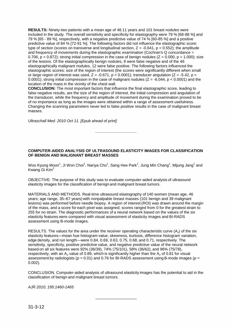
ELASTOSONOGRAPHY: A POSSIBLE NEW TOOL FOR DIAGNOSIS OF ADENOMYOSIS? Tessarolo M, Bonino L, Camanni M, Deltetto F.
OBJECTIVES:
Adenomyosis is a disorder defined by the presence of ectopic endometrial glands and stroma within the myometrium. Transvaginal ultrasound (TVU) is currently the first-line examination for this condition and the aim of this paper is to relate a pilot experience that was conducted using TVU to evaluate adenomyosis and which started from the assumption that tissues with anatomopathological differences show different elasticity values.
METHODS:
Using standard B-mode analysis and elastosonography, we evaluated 30 consecutive women with suspected uterine adenomyosis. In 15 cases the diagnosis was confirmed by histology.
RESULTS:
The adenomyotic area presented more softness (red and green) compared with the surrounding uterine tissue (blue); the borders of the adenomyotic area corresponded to the borders of the green area.
CONCLUSIONS:
These preliminary results suggest that elastosonography could be considered a useful tool in the diagnosis of adenomyosis because it is non-invasive, easy to understand, easy to perform, and has a short learning curve towards becoming skilled at the procedure.
Eur Radiol. 2011 Jul;21(7):1546-52. Epub 2011 Jan 26.
___________________ DIAGNOSTIC VALUE OF STRAIN RATIO MEASUREMENT IN THE DIFFERENTIATION OF MALIGNANT AND BENIGN BREAST LESIONS A. Farrokh, S. Wojcinski, F. Degenhardt
Abstract
Purpose: The aim of this study was to evaluate the strain ratio measurement of breast lesions, to calculate the diagnostic value and to provide practically oriented recommendations concerning execution.
Materials and Methods: 117 breast lesions in 98 patients were included in the study. All lesions were examined by B-mode ultrasound and elastography using strain ratio measurement. The preinterventional findings of the different methods were compared to the final histopathological results. The sensitivity, specificity, positive and negative predictive value and the diagnostic accuracy were calculated for each method.
Results: There was a significant difference between the strain ratio of malignant (mean 6.50; sd 3.03; 95 %-CI 5.68 - 7.33) and benign (mean 1.79; sd 3.83; 95 %-CI 0.92 - 2.75) lesions. The strain ratio showed a sensitivity of 92.6 % (95 %-CI 82.1 - 97.9) and a specificity of 95.2 % (95 %-CI 86.7 - 99.0). The positive and negative predictive values were 94.3 % and 93.7 %. B-mode ultrasound achieved a sensitivity of 94.4 % (95 %-CI 84.6 - 98.8) and a specificity of 87.3 % (95 %-CI 76.5 - 94.3). The positive and negative predictive values were 86.4 % and 94.8 %.
31-3-12
Conclusion: Strain ratio measurement of breast lesions is a standardized fast method for analyzing the stiffness inside the examined areas. Used as an additional tool to B-mode ultrasound, it helps to increase the specificity of the examination
Ultraschall in Med 2011; 32(4): 400-405
___________________
SONOELASTOGRAPHY FOR 1786 NON-PALPABLE BREAST MASSES: DIAGNOSTIC VALUE IN THE DECISION TO BIOPSY. Yi A, Cho N, Chang JM, Koo HR, La Yun B, Moon WK. OBJECTIVES: To evaluate the diagnostic value of sonoelastography by correlation with histopathology compared with conventional ultrasound on the decision to biopsy. METHODS: Prospectively determined BI-RADS categories of conventional ultrasound and elasticity scores from strain sonoelastography of 1786 non-palpable breast masses (1,523 benign and 263 malignant) in 1,538 women were correlated with histopathology. The sensitivity and specificity of two imaging techniques were compared regarding the decision to biopsy. We also investigated whether there was a subset of benign masses that were recommended for biopsy by B-mode ultrasound but that had a less than 2% malignancy rate with the addition of sonoelastography. RESULTS: The mean elasticity score of malignant lesions was higher than that of benign lesions (2.94 ± 1.10 vs. 1.78 ± 0.81) (P < 0.001). In the decision to biopsy, B-mode ultrasound had higher sensitivity than sonoelastography (98.5% vs. 93.2%) (P < 0.001), whereas sonoelastography had higher specificity than B-mode ultrasound (42.6% vs. 16.3%) (P < 0.001). BI-RADS category 4a lesions with an elasticity score of 1 had a malignancy rate of 0.8%. CONCLUSIONS: Sonoelastography has higher specificity than B-mode ultrasound in the differentiation between benign and malignant masses and has the potential to reduce biopsies with benign results. Eur Radiol. 2011 Nov 25. [Epub ahead of print]
___________________
THE ROLE OF SONOELASTOGRAPHY IN THE DIFFERENTIAL DIAGNOSIS OF BREAST LESIONS A. Gheonea, Z. Stoica, A. Bondari; Craiova/RO Purpose: Tissue elasticity imaging technology is expected to be a new modality for breast diagnosis, based on hardness as a tissue characteristic that is affected by tissue disease such as cancer. Methods and Materials: We introduced in this prospective study 59 patients diagnosed with breast lesions between January 2009 and January 2010. All the patients were examined in the supine position and the B mode ultrasound image was displayed alongside the elastography strain image. An EUS Hitachi EUB 8500 ultrasound system with an embedded elastography module (Hitachi Medical Systems Europe Holding AG, Zug, Switzerland) and a 6.5-MHz linear probe was used to obtain the B mode and elastography strain images. The elastography strain images were scored according to the Tsukuba elasticity score. Results: The average age of the women was 45 years. There were 28 benign (47.75%) and 29 malignant lesions (49.15%). The most common lesions of the benign nodules were fibroadenoma, cysts and fibrocystic change. Of the malignant nodules, the most common lesion was infiltrative ductal carcinoma For assessment of sonoelastography role in differential diagnosis of breast lesions, we performed ROC analysis, and we obtained a sensitivity of 89,7%, and a specificity of 92,9% (area under the ROC curve=0,924, 95 % CI =0,822-0,977 and p=0.0001).
31-3-12
Conclusion: Elastography is a fast, simple method which can complement the conventional US. Elastography is promising, and with future improvements in the technology, this imaging modality will become an invaluable tool for the diagnosis of breast diseases in the clinical setting. ECR 2011, March 4
th – 8
th, Vienna
___________________ DIAGNOSTIC ROLE OF A 5-LEVEL SCALE IN THE ELASTOGRAPHIC ANALYSIS OF BREAST LESIONS. A.Malich; Nordhausen/DE Purpose: Study aimed to analyze diagnostic value of semiautomated colour-coded elastography-analysis using a 5-level scale. Methods and Materials: 180 proven breast lesions were analyzed by two experiences radiologists in consensus using 14 Mhz-probe (Hitachi EUB 7500 HV). Elastographic data are given semiautomatically diversificated into red for the greatest strain and blue for the smallest strain (i.e. the hardest tissue) and green. Ueno-scaling was used. Results were matched to BI-RADS-classification and histopathology. Mean size was 10.4 mm (3mm-29mm). Strain ratio was calculated. Size groups were used: S1:<5mm (n=28); S2:<10mm (n=81); S3:<15mm (n=37); S4:<20mm (n=20); S5:<30mm (n=11). Results: Elastography-Scale 1-5 were observed in 6%; 42%; 6%; 12 % and 33 % of the malignant and 30%; 61%; 5%; 3 % and 1 % of the benign lesions, respectively. Summarizing scale 1 and 2 as benign, a sensitivity of 51.5% with a related specificity of 92.4% was measured. Scaling was clearly size-related. Mean value of the 5-level scales varied size dependent for malignant and benign lesions from 2.33/1.76(S1); 2.57/1.84(S2); 3.43/1.87(S3); 3.60/2.11(S4); 3.60/1.83(S5).If colour coded elastography reveals no blue signal, NPV was 92.7%. All cysts were characterized by a mixture of all three strain levels offering a characteristical colour distribution. Conclusion: Elastographic scales are influenced by lesion size and dignitiy of the lesions. The absence of smaller strain codings are highly predictive for benignity. Higher elastographic scales are predictive for malignancies. Larger lesions are characterized by a higher scaling. Cysts are characterized by a typical mosaic scale of all three semiquantitative elastographic strain levels. ECR 2011, March 4
th – 8
th, Vienna
___________________ COMPUTER-ASSISTED ANALYSIS OF STRAIN AT BREAST LESIONS: DIAGNOSTIC ROLE AND RELIABILITY OF CALCULATION A.Malich; Nordhausen/DE Purpose: Differential diagnosis of small breast lesions according to morphologic features in B-mode is still difficult. Study aimed to analyze diagnostic value and reliability of computer-assisted analysis of elastography. Methods and Materials: 180 proven breast lesions were analyzed by two experiences radiologists in consensus using 14 Mhz probe (Hitachi EUB 7500 HV) prior to verification (application pressure level 3-5; images with an average strain of the regular breast tissue only). Randomly selected lesions were repeatedly analyzed without and 1-3 times with the offered application aid. Results: Mean strain ratio for malignancies without application aid was 6.97 vs. 2.67 (benign lesions). Using the application aid mean strain ratio was 5.37 (malignancies) and 5.72 (benign lesions). Correlation coefficient of mean values of strain with/without application aid was 0.37, p<0.05, whereas this coefficient was 0.86 relating first and mean over 2nd and 3
rd calculation. Mean values of
strain ratio allow a discrimination of malignant vs. benign lesions with a diameter <15mm (SR 4.66 vs. 1.75) and <20mm (13.29 vs.2.32). Scars (4.44), calcifications (3.09) and fibroadenoma (2.47) are the benign entities with the highest SR in mean, whereas adenosis (1.05), cysts (1.36) and lymph nodes (1.65) are characterized by a significantly lower SR-value.

31-3-12
Conclusion: Strain ratio is a useful CAd-baed quantitative factor to evaluate dignity of breast lesions. Computer-assisted quantitative analysis of strain ratio is acceptably reliable and offers best discriminatory opportunities in lesions 10-20mm in largest diameter. Application aid is not useful to improve the diagnostic value. ECR 2011, March 4
th – 8
th, Vienna
___________________ QUALITATIVE AND SEMI-QUANTITATIVE EVALUATIONS OF SOLID BREAST LESIONS BY SONOELASTOGRAPHY H. Yerli, T. Yilmaz, T. Kaskati, H. Gulay; Izmir/TR Purpose: To determine whether the use of qualitative elasticity scoring method together with semi-quantitative strain index method by sonoelastography (SE) is useful to differentiate between benign and malignant breast masses. Methods and Materials: Some 78 lesions in 71 consecutive patients with solid breast masses (62 benign, 16 malignant) were prospectively included in this study. For each lesion, B-mode US and SE images were obtained. After elasticity scores had been determined with 5-point scoring method, strain indexes of the lesions were calculated using the same level and normal-appearing breast region as an internal reference by means of the method of strain ratio measurement. The findings were compared with histopathology. Considering the receiver operating curves, the diagnostic performances for the elasticity scoring and the strain index methods were determined. Results: The mean scores on SE were 2.69 ± 0.59 for benign lesions and 3.75 ± 0.68 for malignant lesions. The mean stiffness index values were 2.03 ± 2.67 for benign lesions and 5.97 ± 4.45 for malignant lesions. The area under the curve value was 0.864 for 5-point scoring method and 0.840 for strain index method (P = 706). Sensitivity and specificity for 5-point scoring method were 80 % and 95 %, respectively; 87.5% and 72.6% for B-mode US; and 80 % and 93 % for strain index method when a cutoff point of 3.52 was used. A semi-quantitative evaluation using the strain index method did not contribute to qualitative evaluation by scoring. Conclusions: After 5-point scoring by SE, additional measurement of the strain index is not mandatory to differentiate between benign and malignant breast masses. ECR 2011, March 4
th – 8
th, Vienna
___________________ AUTOMATED QUANTITATIVE COMPUTER-ASSISTED ANALYSIS OF ELASTOGRAPHY TO DISCRIMINATE BREAST LESIONS A. Malich, A. Kott, R. Gorna; Nordhausen/DE Purpose: This study aimed to verify the diagnostic value of strain ratio as a quantitative elastographic parameter in ultrasound of breast lesion and related influencing factors. Methods and Materials: 180 breast lesions were analysed sonographically (14MHz probe) by two experiences radiologists in consensus including elastography analysis (Ueno-scale) and computer-based calculation of strain ratio. Values were related to size and pathological outcome. The following size-related groups were used (largest available diameter): S1: <5mm; S2<10mm; 3<15mm; S4<20mm; S5>30mm. Mean overall size was 10.4mm. ROC-analysis for cut-off values was performed. Results: Strain ratio of malignant versus benign lesions was 6.36 versus 2.27. Mean strain ratios of benign lesions reflecting pathology were 0.98 (fibrolipoma); 1.05 (adenosis); 1.31 (fibrosis); 1.58 (cysts) 1.53 (intraglandular lymph nodes); 2.40 (fibroadenoma); 3.31 (calcificationsliponecrosis); 2.49 (papillomata); 4.44 (scars); and 2.06 (remaining benign entities). Size-related analysis of strain ratio was calculated for malignancies/fibroadenoma/cysts/other benign lesions as S1: 1.66/1.74/1.44/0.91; S2: 2.50/2.08/1.22/2.95; S3: 6.72/2.52/2.20/3.03; S4: 10.25/3.76/0.05/3.09; S5: 4.75/8.00/2.09/1.45. Best performing cut off values are (according to ROC-analysis) S2: 2.4; S3: 2.8; S4: 3.9. Conclusion: Strain ratio is influenced by size and histopathology. In the diagnostically most relevant
31-3-12
group of 5-20mm lesions, malignant lesions are characterised by a higher strain ratio versus all other entitites. Large malignant lesions are typically characterized by a lowered SR versus fibroadenomata (probably due to necrotic liquid components in cancer versus macrocalcifications in fibroadenomata). Typically fibroadenomas, scars and papillomas are characterised by increased SR-values as well. Elastography is of diagnostic use in the differential diagnosis of breast lesions. ECR 2011, March 4
th – 8
th, Vienna
___________________
REAL-TIME SONOELASTOGRAPHY PERFORMED IN ADDITION TO CONVENTIONAL ULTRASOUND: IMPROVED EARLY DETECTION OF ECTOPIC PREGNANCY V. Gazhonova, S. Churkina, A. Zubarev; Moscow/RU Purpose: To evaluate the possibilities of real-time sonoelastography in early detection of ectopic pregnancy. Methods and Materials: Endovaginal ultrasound with elastography was performed in 56 women (19-38 y.o). with a positive pregnancy test and with suspected complications of early pregnancy. Urinary and serum β-hCG levels were measured on the day of the patient‘s hospitalisation (Second International Units). Sonoelastography was performed with the HI VISION 900 and Preirus (Hitachi Medical Corporation) with an endocavity transducer, 8-4 MHz frequency (EUP - V53W, Hitachi). The elastographic images were assessed by 2 radiologists using a 4-point grading score for the presence or absence of ectopic pregnancy. Interobserver agreement and diagnostic confidence levels were calculated. We assessed the accuracy of sonoelastography for the detection of ectopic pregnancy by comparing the findings of sonoelastography with surgical results. Results: 25 women were proven to have an ectopic pregnancy. All 25 were accurately detected by endovaginal ultrasound with elastography. The ―blue eye‖ sonoelastographic sign was seen in every case of extrauterine pregnancy and had an 80 % diagnostic confidence of ectopic pregnancy in women with β-hCG levels lower than 1000 mIU/ml, and a 100 % diagnostic confidence when levels were above the discrimination point of 1000 mIU/ml. Inter-observer agreement revealed Kappa estimates ranging between 0,86 and 0,93, indicating almost perfect conformity in the assessment of pathological changes between reader 1 and reader 2. Conclusion: The ―blue eye‖ sign can be used for the detection of extrauterine pregnancy in doubtful cases of serum β-hSG levels lower than 1000 mIU/ml. The value of endovaginal sonoelastography (EVSE) in emergency gynaecological pathology ECR 2011, March 4
th – 8
th, Vienna
___________________ TO EVALUATE THE USEFULNESS OF EVSE IN THE DIAGNOSIS OF EMERGENCY GYNECOLOGICAL PATHOLOGY. S. Churkina, A. Fedorova, V. Gazhonova; Moscow/RU Methods and Materials: We study 140 women with acute lower abdomen pain and menorrhagia. All pts underwent conventional US and endovaginal sonoelastography on HI VISION 900, HI VISION Preirus (Hitachi Medical Corporation). We used modified Tsukuba sonoelastography classification for evaluation of the EVSE data. US results were compared with surgical and hystomorphological data. Results: The ―blue eye‖ sign was characteristic for ectopic pregnancy in 100 % of cases. The ―reverse cystic type‖ showed by EVSE in 100 % of cases with haemorrhagic continence. EVSE increased the diagnostic confidence of US in the cases of ectopic pregnancy in 38 % of cases, ovarian apoplexy - in 7 %, in 9 % with missed abortion, in 21 % with tuboovarian pathology, in 13 % with fibroid and ovarian torsion, in 20 % with pyosalpinx, in 18 % with hydrosalpinx, and in 21 % with haematometra. The overall sensitivity of US in the diagnosis of emergency gynaecological pathology increased from 77 % up to 95 % with EVSE. Conclusions: EVSE increased the diagnostic confidence of US in emergency gynaecological pathology,

31-3-12
especially in the cases with ectopic pregnancy and haemorrhage ECR 2011, March 4
th – 8
th, Vienna
___________________ AUTOMATIC SELECTION OF REPRESENTATIVE SLICE FROM CINE-LOOPS OF REAL-TIME SONOELASTOGRAPHY FOR CLASSIFYING SOLID BREAST MASSES Yeun-Chung Chang, Min-Chun Yang, Chiun-Sheng Huang, Shao-Chien Chang, Guan-Ying Huang, Woo Kyung Moon, Ruey-Feng Chang
This study aimed to evaluate the performance of automatic selection of representative slice from cine-loops of real-time sonoelastography for classifying benign and malignant breast masses. This retrospective study included 141 ultrasound elastographic studies (93 benign and 48 malignant masses). A novel computer-assisted system was developed for the automatic segmentation of the targeted lesion from cine-loops of real-time sonoelastography. Its hard ratio, defined as the ratio of the number of hard pixels within the tumor divided by the total number of pixels of the whole tumor, was also calculated. The targeted mass was segmented by edge-detection and region growing methods, with combined motion registration after manually defining the original seed. Signal-to-noise ratio (SNRe) and contrast-to-noise ratio (CNRe) of ultrasound elastogram were computed to obtain an optimum slice for differentiating benign and malignant lesions. The diagnostic results of automatic slice selection using maximum strain, maximum SNRe, maximum CNRe, maximum compression and the slices selected by radiologists were compared. Mann–Whitney U test, performance indexes and receiver operating characteristic (ROC) curves were used for statistical analysis. Performance using the maximum SNRe (accuracy 84.4%, sensitivity 83.3%, specificity 85.0% and Az value 0.90) was the best as compared with those of maximum CNRe (82.3%, 79.2%, 83.9% and 0.88, respectively), maximum compression (78.0%, 79.2%, 77.4% and 0.85, respectively), maximum strain (79.4%, 79.2%, 79.6% and 0.87, respectively) and radiologists‘ selection (77.3%, 77.1%, 77.4% and 0.80, respectively). Automatic selection of representative slice from the cine-loops of real-time sonoelastography is a practical, objective and accurate approach for classifying solid breast masses.
Ultrasound in Medicine and Biology, Volume 37, Issue 5, Pages 709-718 (May 2011)
___________________ ELASTOGRAPHY OF THE UTERINE CERVIX – IMPLICATIONS FOR DELIVERY ELASTOGRAPHY OF CERVIX
Malgorzata Swiatkowska-Freund, Krzysztof Preis
Objectives:
Elastography is widely used in radiology to diagnose tumors and to help to perform biopsies of liver, salivary gland and prostate tumors. The authors present a preliminary report pertaining to cervical assessment by elastography, in order to elucidate the ability of this method to show cervical consistency.
Methods:
Elastography of the uterine cervix was performed in 29 patients (in two of them twice) before labor induction with the internal os described using numeric scale called the Elastography Index (EI). A color map from purple to red was selected with the hardest tissues coded as purple and assigned 0 points, less hard tissues (blue) –1 point, green – 2 points, yellow – 3 points, and red (softest) – 4 points. Correlation between internal os EI and the success of labor induction were analyzed using the Pearson correlation test and the T-test.
Results:

31-3-12
A significant correlation between the EI of the internal os and labor induction success was observed (r = 0.71; p = 0.0004). The mean EI in the group of patients with successful induction was 1.23, while in the group with failed labor induction – 0.39 (T-Student test; p = 0.024).
Conclusions:
Elastography of the uterine cervix may be a method for objectively assessing internal os ripening before labor induction. Standardizing cervical properties seen in elastography during pregnancy may help to guide prostaglandins or oxytocin use in labor induction.
Ultrasound Obstet Gynecol. 2011 Apr 12. [Epub ahead of print]
___________________ ROLE OF SONOGRAPHIC ELASTOGRAPHY IN THE DIFFERENTIAL DIAGNOSIS OF AXILLARY LYMPH NODES IN BREAST CANCER Jae Jeong Choi, MD, Bong Joo Kang, MD, PhD, Sung Hun Kim, MD, Ji Hye Lee, MD, Seung Hee Jeong, MPH,Hyun Woo Yim, MD, PhD, Byung Joo Song, MD, Sang Seol Jung, MD Objectives—The purpose of this study was to evaluate the diagnostic utility of sonographic elastography in differentiating reactive and metastatic axillary lymph nodes in breast cancer. Methods—A total of 64 lymph nodes (reactive, n = 33; metastatic, n = 31) from 62 patients with breast cancer were examined by both B-mode sonography and elastography from April to July 2009. Two experienced radiologists retrospectively assessed B-mode sonograms by the sum of scores for 4 criteria: short diameter, shape, hilum, and cortical thickening. Elastographic images were given scores of 1 to 4 according to the percentage of high-elasticity areas in the lymph nodes. We compared the diagnostic performance of B-mode sonography, elastography, and combined examinations. We also calculated the strain ratio of the lymph node and subcutaneous fat tissue. Results—The elasticity score for malignant lymph nodes (mean, 3.1) was higher than the score for benign lymph nodes (mean, 2.2; P < .0001). With a cutoff between elasticity scores of 2 and 3, elastography showed 80.7% sensitivity, 66.7% specificity, and 73.4% accuracy. With a cutoff between B-mode sonographic scores of 1 and 2, B-mode sonography showed 74.2% sensitivity and 78.8% specificity. Combined B-mode and elastographic sonography showed higher sensitivity (87.1%) than B-mode sonography alone. With a strain ratio cutoff point of 2.3, sensitivity was 82.8%, and specificity was 56.3%. Conclusions—Sonographic elastography may increase the sensitivity of B-mode sonography in the detection of metastatic axillary lymph nodes. J Ultrasound Med 2011; 30:429–436
___________________ BREAST MASS EVALUATION: FACTORS INFLUENCING THE QUALITY OF US ELASTOGRAPHY Jung Min Chang, MD, Woo Kyung Moon, MD, Nariya Cho, MD and Seung Ja Kim, MD
Abstract
Purpose: To investigate factors influencing the quality of ultrasonographic (US) elastography in the
evaluation of suspicious breast masses.
Materials and Methods: This prospective study was conducted with institutional review board approval; written informed consent was obtained. Between January 2009 and February 2009, real-time US elastography of 312 breast masses (245 benign, 67 malignant) was performed in 268
31-3-12
consecutive patients (mean age, 45.7 years ± 10.2 [standard deviation]) prior to US-guided core biopsy. Five breast radiologists who had performed the examinations assessed the quality of elasticity images as inadequate, low, or high without histologic information. Age, body mass index (BMI), mammographic density, lesion size, lesion depth, and breast thickness at US were analyzed for their association with image quality by using the χ
2 test, Student t test, and multivariate analysis.
Sensitivities and specificities for the differentiation of benign from malignant masses on the basis of elastography were calculated and compared between groups of quality scores by using the logistic regression method.
Results: The quality of elasticity images was assessed as inadequate in 21 (6.7%) cases, low in 134 (42.9%), and high in 157 (50.3%). According to univariate analysis, smaller lesion size (P = .001), shallower lesion depth (P = .005), less breast thickness where the lesion was located (P < .0001), and benign pathologic finding (P = .004) were significantly associated with higher image quality. There was no correlation of image quality with age (P = .213), BMI (P = .191), mammographic density (P = .091), or distance from the nipple (P = .100). Multivariable analysis showed that breast thickness at the location of target lesions was the most important factor influencing elasticity image quality (P = .001). There were significant differences in sensitivity between higher-quality and lower-quality images (87.0% vs 56.8%, respectively; P = .015) in the differentiation of benign from malignant masses.
Conclusion: Breast thickness at the location of the lesion was the most important factor influencing image quality at US elastography. Sensitivity for classification of benign and malignant masses improved with higher quality scores.
Radiology 2011, 259, 59-64 ___________________
PREDICTIVE VALUE FOR MALIGNANCY OF SUSPICIOUS BREAST MASSES OF BI-RADS CATEGORIES 4 AND 5 USING ULTRASOUND ELASTOGRAPHY AND MR DIFFUSION-WEIGHTED IMAGING.
Satake H, Nishio A, Ikeda M, Ishigaki S, Shimamoto K, Hirano M, Naganawa S.
Department of Radiology, Nagoya University School of Medicine, 65 Tsuruma-cho, Showa-ku, Nagoya, Aichi 466-8550, Japan.
OBJECTIVE: The aim of this study is to evaluate the ability of ultrasound elastography and MR diffusion-weighted imaging (DWI) to predict malignancy of breast masses, with subsequent recommendation for biopsy. MATERIALS AND METHODS: For 115 breast masses classified as BI-RADS category 4 or 5, which were assessed according to combined findings of mammography, B-mode sonography, and dynamic contrast-enhanced MRI, two radiologists retrospectively evaluated the elasticity scores using ultrasound elastography and the apparent diffusion coefficient (ADC) values using MR DWI. The diagnostic abilities of these two techniques were analyzed by using univariate and multivariate logistic regression analysis. RESULTS: In the analysis of all 115 breast masses, the elasticity score was predictive of malignancy, whereas the ADC value was not independently predictive. In an analysis of the 52 masses assessed as BI-RADS category 4, the elasticity score was found to be a significant predictor of malignancy, compared with the ADC value, which was a nonsignificant predictor. In an analysis of the 63 masses assessed as BI-RADS category 5, neither the elasticity score nor the ADC value was a significant predictor of malignancy. CONCLUSION: Our results show that elasticity imaging provides relatively reliable predictions for malignancy, especially in BI-RADS category 4 masses, compared with MR DWI.
AJR Am J Roentgenol. 2011 Jan;196(1):202-9.
31-3-12
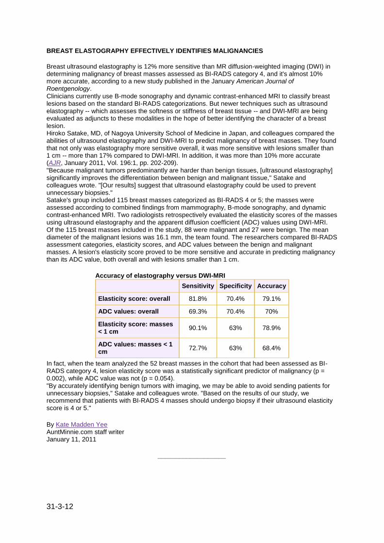
BREAST ELASTOGRAPHY EFFECTIVELY IDENTIFIES MALIGNANCIES
Breast ultrasound elastography is 12% more sensitive than MR diffusion-weighted imaging (DWI) in determining malignancy of breast masses assessed as BI-RADS category 4, and it's almost 10% more accurate, according to a new study published in the January American Journal of Roentgenology. Clinicians currently use B-mode sonography and dynamic contrast-enhanced MRI to classify breast lesions based on the standard BI-RADS categorizations. But newer techniques such as ultrasound elastography -- which assesses the softness or stiffness of breast tissue -- and DWI-MRI are being evaluated as adjuncts to these modalities in the hope of better identifying the character of a breast lesion. Hiroko Satake, MD, of Nagoya University School of Medicine in Japan, and colleagues compared the abilities of ultrasound elastography and DWI-MRI to predict malignancy of breast masses. They found that not only was elastography more sensitive overall, it was more sensitive with lesions smaller than 1 cm -- more than 17% compared to DWI-MRI. In addition, it was more than 10% more accurate (AJR, January 2011, Vol. 196:1, pp. 202-209). "Because malignant tumors predominantly are harder than benign tissues, [ultrasound elastography] significantly improves the differentiation between benign and malignant tissue," Satake and colleagues wrote. "[Our results] suggest that ultrasound elastography could be used to prevent unnecessary biopsies." Satake's group included 115 breast masses categorized as BI-RADS 4 or 5; the masses were assessed according to combined findings from mammography, B-mode sonography, and dynamic contrast-enhanced MRI. Two radiologists retrospectively evaluated the elasticity scores of the masses using ultrasound elastography and the apparent diffusion coefficient (ADC) values using DWI-MRI. Of the 115 breast masses included in the study, 88 were malignant and 27 were benign. The mean diameter of the malignant lesions was 16.1 mm, the team found. The researchers compared BI-RADS assessment categories, elasticity scores, and ADC values between the benign and malignant masses. A lesion's elasticity score proved to be more sensitive and accurate in predicting malignancy than its ADC value, both overall and with lesions smaller than 1 cm.
Accuracy of elastography versus DWI-MRI
Sensitivity Specificity Accuracy
Elasticity score: overall 81.8% 70.4% 79.1%
ADC values: overall 69.3% 70.4% 70%
Elasticity score: masses < 1 cm
90.1% 63% 78.9%
ADC values: masses < 1 cm
72.7% 63% 68.4%
In fact, when the team analyzed the 52 breast masses in the cohort that had been assessed as BI-RADS category 4, lesion elasticity score was a statistically significant predictor of malignancy (p = 0.002), while ADC value was not (p = 0.054). "By accurately identifying benign tumors with imaging, we may be able to avoid sending patients for unnecessary biopsies," Satake and colleagues wrote. "Based on the results of our study, we recommend that patients with BI-RADS 4 masses should undergo biopsy if their ultrasound elasticity score is 4 or 5."
By Kate Madden Yee AuntMinnie.com staff writer January 11, 2011
___________________

31-3-12
ALIASING ARTIFACT DEPICTED ON ULTRASOUND (US)-ELASTOGRAPHY FOR BREAST CYSTIC LESIONS MIMICKING SOLID MASSES Nariya Cho, Woo Kyung Moon, Jung Min Chang, Seung Ja Kim, Chae Yeon Lyou, Hye Young Choi Department of Radiology and Clinical Research Institute, Seoul National University Hospital and the Institute of Radiation Medicine, Seoul National University Medical Research Center, Seoul, Republic of Korea Background: It has been reported that ultrasound (US)-elastography is helpful in differentiation of benign and malignant solid masses and in reducing benign biopsy procedures for the supplemental breast US in addition to screening mammography. Furthermore, potential application of US-elastography in distinguishing cystic lesions which is known to be a major source of benign biopsy results has been suggested. Purpose: To describe the aliasing artifact on US-elastography for breast cystic lesions that mimic solid masses. Material and Methods: We retrospectively reviewed 13 lesions which showed a blue-green-red pattern artifact on US-elastography in 13 women (mean age 50 years; age range 3–66 years). They disappeared immediately after a needle biopsy. Breast composition, mammography and US findings, histology and follow-up imaging findings were analyzed. Results: All 13 patients showed heterogeneously dense (n = 5) or extremely dense breast parenchyma (n = 8). The most common US findings were an irregular shape (n = 7, 54%) and a circumscribed margin (n = 7, 54%). All 13 lesions had internal echogenicity and were initially considered as solid masses; 62% (n = 8) showed hypoechogenicity and 38% (n = 5) had echogenic and anechoic components. Posterior shadowing was seen in 31% (n = 4) of the lesions. All 13 lesions have been proven to be fibrocystic changes on biopsy histology. Follow-up US performed for 10 of 13 lesions showed no residual lesion (n = 9) or decreased its size (n = 1). Conclusion: An aliasing artifact that appears as a blue-green-red pattern in a breast mass as depicted on US-elastography is suggestive of a possible cystic breast lesion. Acta Radiologica 2011; 52: 3–7.
___________________ SONOELASTOGRAPHIC LESION STIFFNESS: PREOPERATIVE PREDICTOR FOR THE PRESENCE OF AN INVASIVE FOCUS IN NONPALPABLE DCIS DIAGNOSED BY CORE NEEDLE BIOPSY
Nariya Cho MD Woo Moon MD Jung Min Chang MD et al
PURPOSE
To evaluate the preoperative factors associated with upgrade to invasive cancers in patients with a core needle biopsy diagnosis of DCIS under US-guidance.
METHOD AND MATERIALS
Between June 2006 and May 2009, 3300 consecutive women underwent US-guided core biopsy and sonoELASTOGRAPHY examinations saved as video clips prior to biopsy. Histologic analysis yielded DCIS in 116 (3.5%) women. Fourteen women were excluded as correlation with surgical histology was not available. Finally, a total of 102 women (mean age 50, range 24-71 years) with 103 nonpalpable DCIS lesions (mean 15 mm, range 4–70 mm) formed our study population. Lesion type, lesion size, biopsy method, nuclear grade, and presence of comedonecrosis of biopsy histology were analyzed. B-mode US findings and elasticity scores from 1 to 5 based on the degree of lesion stiffness were determined by two radiologists after reviewing the video clips without histologic results. Fisher‘s exact test for univariate analysis and multivariable logistic regression model were used to determine the independent preoperative predicting factors of invasive cancer.
RESULTS
Elasticity score was the only independent feature for predicting presence of an invasive component.
31-3-12
Upgrade rates according to the elasticity score was 0% (0/18) for score 1, 18% (7/40) for score 2, 31% (8/26) for score 3, 47% (8/17) for score 4, and 50% (1/ 2) for score 5 (OR=1; OR=7.75, P=0.077; OR=13.11, P=0.023; OR=44.17, P=0.0008; OR=39.49, P=0.030). No difference was found in upgrade rates according to the lesion type [18% (10/57) for non-mass vs. 30% (14/46) for mass, P=0.161], lesion size [14% (6/42) for 4-10mm, 35% (13/37) for 11-20mm, 21% (5/24) for 21-70mm, P=0.088], biopsy method [18% (9/50) for 11G vs. 28% (15/53) for 14G, P=0.250], nuclear grade [16% (7/43) for low vs. 28% (17/60) for high, P=0.167], comedonecrosis [24% (14/58) for absence vs. 22% (10/45) for presence, P=1.0], and B-mode US findings.
CONCLUSION
Nonpalpable DCIS lesions diagnosed by US-guided core biopsy but having invasive components at surgical histology show higher sonoelastographic stiffness than pure DCIS regardless of the mammographic lesion type, lesion size, biopsy method, histologic variables, and B-mode US findings.
CLINICAL RELEVANCE/APPLICATION
Sonoelastographic lesion stiffness can be used as a guideline for the consideration of sentinel lymph node biopsy in patients with nonpalpable DCIS diagnosed by US-guided needle biopsy.
RSNA 2010, November 28th – December 2
nd, Chicago, USA
___________________ SONOELASTOGRAPHY HELPS IDENTIFY AGGRESSIVE DCIS LESIONS Determining which ductal carcinoma in situ (DCIS) lesions could turn into aggressive cancers is one of breast imaging's biggest challenges. Fortunately, sonoelastography may be able to help, according to South Korean researchers.
The researchers used sonoelastography to examine 102 women with 103 nonpalpable DCIS lesions. The mean size of the lesions was 15 mm, and patients were seen between June 2006 and May 2009. B-mode ultrasound scans and elastography studies were acquired, with lesion stiffness scored on a scale of 1 to 5.
Elasticity scores were the only independent feature for predicting an invasive component to DCIS lesions, the researchers found. They recommend that sonoelastographic lesion stiffness be used as a guideline to consider sentinel lymph node biopsy in patients with nonpalpable DCIS.
By: Brian Casey, AuntMinnie.com staff writer November 12, 2010
___________________
SONOELASTOGRAPHY PREDICTS WHETHER BREAST DCIS IS INVASIVE Sonoelastography can predict before surgery whether women with ductal carcinoma in situ (DCIS) have an invasive form of the disease, according to a study by Korean researchers presented at the recent RSNA meeting in Chicago. In a study of 103 nonpalpable DCIS lesions diagnosed on core-needle biopsy, a team from Seoul National University Hospital in South Korea found that a sonoelastography elasticity score of 1 predicted the absence of an invasive component of DCIS in 100% of cases, while a score of 4 or 5 accurately forecasted an invasive component in 47.4% of cases.
31-3-12
"Sonoelastography stiffness is an independent predictor of invasion in DCIS lesions [found] at core biopsy," said Nariya Cho, MD. DCIS diagnosed at core-needle biopsy is underestimated in 8% to 42% of cases in which the lesions are later found to be invasive and require further axillary lymph node sampling. And women with a high risk of invasive cancer need to have their axillary lymph node sampling planned preoperatively, Cho said. As a result, the researchers sought to retrospectively evaluate the preoperative factors associated with why DCIS cases are upgraded to invasive cancers, focusing on the usefulness of sonoelastography in patients with an ultrasound-guided core-needle biopsy diagnosis of nonpalpable DCIS. They reviewed the records of 3,510 consecutive women with 3,300 breast lesions who had undergone ultrasound-guided core biopsy and sonoelastography between June 2006 and May 2009. Histologic analysis found DCIS in 117 patients, but 15 were excluded from the study due to an unavailability of surgical histology. The remaining 102 women (mean age, 50) had a total of 103 DCIS lesions (mean, 15 mm; range, 4-70 mm). All patients received sonoelastography and surgery after ultrasound-guided needle localization. The mean interval between ultrasound-guided core-needle biopsy and surgery was 31 days. Histopathology diagnosis was made by a combination of core-needle biopsy and surgical excision. For data acquisition, one of five radiologists with three to seven years of experience in breast ultrasound performed B-mode ultrasound and sonoelastography on the patients and saved the cine clips in .avi format. Ultrasound-guided 11-gauge vacuum-assisted biopsy or 14- gauge automated core biopsy was then performed. After reviewing the video clips in random order and without access to the histologic results, two other radiologists provided their B-mode ultrasound findings and then elasticity scores (from 1 to 5) based on the degree of lesion stiffness. To determine the independent preoperative predicting factors of invasive cancer, the researchers utilized Fisher's exact test for univariate analysis and a multivariable logistic regression model. Factors such as lesion type, lesion size, biopsy method, nuclear grade, and the presence of comedonecrosis were analyzed. The researchers found that the elasticity score was the only independent feature for predicting the presence of an invasive component. Upgrade rates were as follows:
Elasticity score Upgrade rate No. patients upgraded
Odds ratio
1 0% 0/18 1
2 18% 7/40 7.75
3 31% 8/26 13.11
4 47% 8/17 44.17
5 50% 1/2 39.40
"Lesions with a higher elasticity score tended to have more invasive components," Cho said. The researchers did not find any difference in upgrade rates based on lesion type, lesion size, biopsy method, nuclear grade, comedonecrosis, and B-mode ultrasound findings. By Erik L. Ridley AuntMinnie staff writer December 13, 2010
___________________ ROLE AND CLINICAL USEFULNESS OF SONOELASTOGRAPHY IN SMALL BREAST MASSES Joo Hwa Myong MD Sung Hun Kim MD Ji-Hye Lee
PURPOSE
The purpose of this study was to evaluate the diagnostic performance of sonoelastography and B-mode ultrasonography and combination of sonoelastography and B-mode ultrasonography for differentiation of small breast masses.
METHOD AND MATERIALS
315 breast masses smaller than 1cm (267 benign and 48 malignant) in 278 patientswho had been

31-3-12
scheduled for a sonographically guided core biopsy were examined with B-mode ultrasonography and sonoelastography from March to October 2009. The histopathologic results were used as a reference standard. Two radiologists retrospectively evaluated the B-mode image according to ACR BIRADS and elastographic images according to classification proposed by Itoh et al. The strain ratio was calculated by dividing the strain value of the subcutaneous fat by that of the mass. The diagnostic performance of B-mode ultrasonography, sonoelastography and combination of two modalities were compared by receiver operating characteristic (ROC) curve analysis.
RESULTS
The elasticity score for malignant masses (3.02±1.33) was significantly higher than that for benign masses (1.72±0.78) (p < 0.001). No significant difference was found in the strain ratio between benign (2.03 ±2.04) and malignant (5.83±13.65) masses (P=0.060). Areas under the ROC curves(Az values) were 0.616 for B-mode ultrasonography, 0.671 for elasticity score, 0.668 for strain ratio, 0.727 for combination of B-mode ultrasonography and elastography score and 0.701 for combination of B-mode ultrasonography, elastography score and strain ratio. The sensitivity, specificity, positive predictive value, and negative predictive value were 93.8%, 51.7%, 25.9% and 97.9%, respectively, when elasticity score and B- mode ultrasonography was combined as follows; downgrade of final assessment category in case with elasticity score of 1, no change in case with that of 2,3 and upgrade in case with that of 4,5.
CONCLUSION
Elasticity score and strain ratio were comparable with B-mode ultrasonography in the diagnostic performance. Combination of B-mode ultrasonography and elasticity score showed better diagnostic performance than each modality.
CLINICAL RELEVANCE/APPLICATION Combination of B-mode ultrasonography and elasticity score would be helpful for for differentiation of benign and malignant lesions smaller than 1cm. Radiological Society of North America 96
th Scientific Assembly and Annual Meeting 2010, November
28th - December 2
nd, Chicago
___________________
BETTER TOGETHER IN THE BREAST: SONOELASTOGRAPHY AND B-MODE ULTRASOUND By: Brian Casey, AuntMinnie.com staff writer November 12, 2010 Combining sonoelastography with conventional B-mode ultrasound improves the differentiation of benign and malignant breast masses smaller than 1 cm, according to this presentation by Korean researchers. The group wanted to see if adding sonoelastography to B-mode ultrasound could improve differentiation of small masses, with the ultimate goal of reducing unnecessary biopsies. To this end, they examined 315 breast masses (267 benign and 48 malignant) in a population of 278 patients who had been scheduled for biopsy between March and October 2009. The performances of sonoelastography and B-mode ultrasound were analyzed independently and then in combination with each other. With elastography, radiologists calculated a strain ratio, thus producing elasticity scores. Elasticity scores for malignant masses were 3.02 ± 1.33, significantly higher than those of benign masses (1.72 ± 0.78). Combining B-mode ultrasound with elasticity scores produced a sensitivity of 93.8%, specificity of 51.7%, positive predictive value of 25.9%, and negative predictive value of 97.9%. The authors concluded that combining B-mode ultrasound with elasticity could reduce unnecessary biopsies of small suspicious breast masses, according to Sung Hun Kim, MD, who participated in the study.
___________________
31-3-12
SONOELASTOGRAPHIC LESION STIFFNESS: PREOPERATIVE PREDICTOR FOR THE PRESENCE OF AN INVASIVE FOCUS IN NONPALPABLE DCIS DIAGNOSED BY CORE NEEDLE BIOPSY Nariya Cho MD Woo Moon MD Jung Min Chang MD et al
PURPOSE
To evaluate the preoperative factors associated with upgrade to invasive cancers in patients with a core needle biopsy diagnosis of DCIS under US-guidance.
METHOD AND MATERIALS
Between June 2006 and May 2009, 3300 consecutive women underwent US-guided core biopsy and sonoelastography examinations saved as video clips prior to biopsy. Histologic analysis yielded DCIS in 116 (3.5%) women. Fourteen women were excluded as correlation with surgical histology was not available. Finally, a total of 102 women (mean age 50, range 24-71 years) with 103 nonpalpable DCIS lesions (mean 15 mm, range 4–70 mm) formed our study population. Lesion type, lesion size, biopsy method, nuclear grade, and presence of comedonecrosis of biopsy histology were analyzed. B-mode US findings and elasticity scores from 1 to 5 based on the degree of lesion stiffness were determined by two radiologists after reviewing the video clips without histologic results. Fisher‘s exact test for univariate analysis and multivariable logistic regression model were used to determine the independent preoperative predicting factors of invasive cancer.
RESULTS
Elasticity score was the only independent feature for predicting presence of an invasive component. Upgrade rates according to the elasticity score was 0% (0/18) for score 1, 18% (7/40) for score 2, 31% (8/26) for score 3, 47% (8/17) for score 4, and 50% (1/ 2) for score 5 (OR=1; OR=7.75, P=0.077; OR=13.11, P=0.023; OR=44.17, P=0.0008; OR=39.49, P=0.030). No difference was found in upgrade rates according to the lesion type [18% (10/57) for non-mass vs. 30% (14/46) for mass, P=0.161], lesion size [14% (6/42) for 4-10mm, 35% (13/37) for 11-20mm, 21% (5/24) for 21-70mm, P=0.088], biopsy method [18% (9/50) for 11G vs. 28% (15/53) for 14G, P=0.250], nuclear grade [16% (7/43) for low vs. 28% (17/60) for high, P=0.167], comedonecrosis [24% (14/58) for absence vs. 22% (10/45) for presence, P=1.0], and B-mode US findings.
CONCLUSION
Nonpalpable DCIS lesions diagnosed by US-guided core biopsy but having invasive components at surgical histology show higher sonoelastographic stiffness than pure DCIS regardless of the mammographic lesion type, lesion size, biopsy method, histologic variables, and B-mode US findings.
CLINICAL RELEVANCE/APPLICATION
Sonoelastographic lesion stiffness can be used as a guideline for the consideration of sentinel lymph node biopsy in patients with nonpalpable DCIS diagnosed by US-guided needle biopsy.
Radiological Society of North America 96th Scientific Assembly and Annual Meeting 2010, November
28th - December 2
nd, Chicago
___________________ SONOELASTOGRAPHY HELPS IDENTIFY AGGRESSIVE DCIS LESIONS By: Brian Casey, AuntMinnie.com staff writer November 12, 2010 Determining which ductal carcinoma in situ (DCIS) lesions could turn into aggressive cancers is one of breast imaging's biggest challenges. Fortunately, sonoelastography may be able to help, according to South Korean researchers. The researchers used sonoelastography to examine 102 women with 103 nonpalpable DCIS lesions. The mean size of the lesions was 15 mm, and patients were seen between June 2006 and May 2009. B-mode ultrasound scans and elastography studies were acquired, with lesion stiffness scored on a scale of 1 to 5. Elasticity scores were the only independent feature for predicting an invasive component to DCIS lesions, the researchers found. They recommend that sonoelastographic lesion stiffness be used as a guideline to consider sentinel lymph node biopsy in patients with nonpalpable DCIS.
31-3-12
SONOELASTOGRAPHY IN THE DIAGNOSIS OF MALIGNANT AND BENIGN BREAST LESIONS: INITIAL CLINICAL EXPERIENCES.
Hatzung G, Grunwald S, Zygmunt M, Geaid AA, Behrndt PO, Isermann R, Kohlmann T, Ohlinger R.
PURPOSE:
This prospective study aimed to compare sonoelastography, B-mode ultrasonography, and mammography in terms of their ability to distinguish benign from malignant breast lesions. We also assessed how the diagnostic value of sonoelastography differs between palpable and clinically occult lesions.
MATERIALS AND METHODS:
Evaluation revealed a total of 97 lesions (66 benign; 31 malignant) without histological confirmation at the time of the initial examination. The sensitivity, specificity, positive (PPV) and negative predictive value (NPV) as well as efficiency were calculated. These parameters were separately assessed for palpable lesions and for non-palpable lesions. We subsequently compared these results.
RESULTS:
Sonography had a sensitivity of 97% and a specificity of 82% (PPV: 71 %, NPV: 98%, efficiency: 87%). For mammography, the respective figures were 84% and 89% (PPV: 79%, NPV: 92%, efficiency: 88%). Sonoelastography had a sensitivity of 71% and a specificity of 48% (PPV: 39%, NPV: 78%, efficiency: 56%). The combination of sonography and sonoelastography yielded a sensitivity of 100% and a specificity of 38% (PPV: 43%, NPV: 100%, efficiency: 58%). The sensitivity and specificity were not statistically different between the groups of palpable and non-palpable lesions.
CONCLUSION:
Sonoelastography is easily performed and not very time-consuming. Used by itself, the method is not more efficacious than alternative techniques. When used in conjunction with B-mode ultrasonography, the latter's sensitivity was increased, albeit at the expense of specificity.
Ultraschall Med. 2010 Dec;31(6):596-603.
___________________
THE INFLUENCE OF TECHNICAL FACTORS ON SONOELASTOGRAPHIC ASSESSMENT OF SOLID BREAST NODULES.
Ciurea AI, Bolboaca SD, Ciortea CA, Botar-Jid C, Dudea SM.
Radiology, University of Medicine and Pharmacy Cluj-Napoca.
PURPOSE: The aim of the study was to assess the influence of technical factors and/or lesion characteristics on the final elastographic score in solid breast nodules. MATERIALS AND METHODS: Patients with solid breast masses examined between May 2007 and May 2008 in the Radiology Department of Cluj District University Hospital were included in the study. All lesions were examined with conventional ultrasound, Doppler ultrasound and sonoelastography, according to a preset protocol. The influence of the following factors on the elastographic score was evaluated: type of section (sagittal versus transverse); size of region of interest (small versus large); amplitude and frequency of movement; initial compression (light versus strong); angulation (perpendicular versus angulated transducer); characteristics of the lesion (size and location). The reference diagnosis was the histopathology diagnosis and, in twenty cases, short-term follow-up.
31-3-12
RESULTS: Ninety-two patients with a mean age of 48.11 years and 101 breast nodules were included in the study. The overall sensitivity and specificity for elastography were 79 % [68-88 %] and 79 % [65 - 89 %], respectively, with a negative predictive value of 74 % [60-85 %] and a positive predictive value of 84 % [72-91 %]. The following factors did not influence the elastographic score: type of section (scores on transverse and longitudinal section, Z = -0.641, p = 0.552); the amplitude and frequency of movements during the elastographic examination (Cochran's Q concordance = 0.706, p = 0.872); strong initial compression in the case of benign nodules (Z = 0.000, p = 1.000); size of the lesions. Of the elastographically benign nodules, 9 were false negative and of the 46 elastographically malignant nodules, 12 were false positive. The following factors influenced the elastographic scores: size of the region of interest (the scores were significantly different when small or large region of interest was used, Z = -0.671, p < 0.0001); transducer angulation (Z = -5.42, p < 0.0001); strong initial compression in the case of malignant nodules (Z = -6.044, p < 0.0001) and the location of the mass in the vicinity of the chest wall. CONCLUSION: The most important factors that influence the final elastographic score, leading to false negative results, are the size of the region of interest, the initial compression and angulation of the transducer, while the frequency and amplitude of movement during the examination proved to be of no importance as long as the images were obtained within a range of assessment usefulness. Changing the scanning parameters never led to false positive results in the case of malignant breast masses.
Ultraschall Med. 2010 Oct 11. [Epub ahead of print]
___________________
COMPUTER-AIDED ANALYSIS OF ULTRASOUND ELASTICITY IMAGES FOR CLASSIFICATION OF BENIGN AND MALIGNANT BREAST MASSES
Woo Kyung Moon1, Ji Won Choi
1, Nariya Cho
1, Sang Hee Park
1, Jung Min Chang
1, Mijung Jang
1 and
Kwang Gi Kim2
OBJECTIVE. The purpose of this study was to evaluate computer-aided analysis of ultrasound
elasticity images for the classification of benign and malignant breast tumors.
MATERIALS AND METHODS. Real-time ultrasound elastography of 140 women (mean age, 46
years; age range, 35–67 years) with nonpalpable breast masses (101 benign and 39 malignant
lesions) was performed before needle biopsy. A region of interest (ROI) was drawn around the margin
of the mass, and a score for each pixel was assigned; scores ranged from 0 for the greatest
strain to
255 for no strain. The diagnostic performances of a neural network based on the values of the six
elasticity features were compared with visual assessment of elasticity images and
BI-RADS
assessment using B-mode images.
RESULTS. The values for the area under the receiver operating characteristic curve (Az) of the six
elasticity features—mean hue histogram value, skewness, kurtosis, difference histogram variation,
edge density, and run length—were 0.84, 0.69, 0.63, 0.75, 0.68,
and 0.71, respectively. The
sensitivity, specificity, positive predictive value, and negative predictive value of the neural network
based on all six features were 92% (36/39), 74% (75/101), 58% (36/62),
and 96% (75/78),
respectively, with an Az value of 0.89, which is significantly higher than the Az of 0.81 for visual
assessment by radiologists (p = 0.01) and 0.76 for BI-RADS assessment using
B-mode images (p =
0.002).
CONCLUSION. Computer-aided analysis of ultrasound elasticity images has the potential to aid in the
classification of benign and malignant breast tumors.
AJR 2010; 195:1460-1465
___________________

31-3-12
ELASTOGRAPHY OF THE BREAST: TECHNIQUE AND PRACTICAL APPLICATIONS, BIRADS CORRELATION Giorgio Rizzatto Real-time elastography (RTE) of the breast may easily and quickly integrate conventional breast imaging. A mechanical force is applied to the tissue and sophisticated algorithms are used to estimate the tissue stiffness. Qualitative scores are derived from the estimate of the strain and help to differentiate soft benign lesions from malignancies. These are usually stiffer due to the secretion of collagen and fibronectin and the surrounding edema. The cysts almost always show a typical three-layered pattern. Strain scores are usually obtained with conventional scanners and transducers and may be integrated in fusion imaging; actually they are becorning very popular in breast imaging. Dedicated RTE technologies as an alternative track the shear wave propagation through tissues to obtain a quantitative evaluation of the acoustic modulus and promise to be the gold standard for the future applications. Even with qualitative strain scores clinical results are very accurate. RTE shows a very high specificity in benign lesions, including BIRADS category 3 lesions. With the best cutoff point between elasticity scores 3 and 4, the true negative predictive value is around 98%. RTE works better with small lesions, less than 10 mm in diameter. RTE is almost insensitive to the thickness and the echogenicity of the breast, and to the depth and the size of the lesion. RTE scores are well reproducible. Indexes of intra-observer (? =0.93) and inter-observer (? = 0.90) agreement are very good. Still RTE score is only a complementary descriptor that requires global experience in breast imaging and strict guidelines.
Euroson 2010, August 22 – 25th 2010, Copenhagen
___________________
MULTICENTER STUDY OF ULTRASOUND REAL-TIME TISSUE ELASTOGRAPHY IN 779 CASES FOR THE ASSESSMENT OF BREAST LESIONS: IMPROVED DIAGNOSTIC PERFORMANCE BY
COMBINING THE BI-RADS®-US CLASSIFICATION SYSTEM WITH SONOELASTOGRAPHY. Wojcinski S, Farrokh A, Weber S, Thomas A, Fischer T, Slowinski T, Schmidt W, Degenhardt F. Klinik für Frauenheilkunde und Geburtshilfe, Franziskus Hospital, Bielefeld, Germany. Abstract PURPOSE: Hitachi real-time tissue elastography (HI-RTE) is an ultrasound technique that facilitates the estimation of tissue elasticity. Our study evaluates whether sonoelastography improves the differentiation of benign and malignant breast lesions. MATERIALS AND METHODS: In a multicenter approach sonoelastography of focal breast lesions was carried out in 779 patients with subsequent histological confirmation. We present data from 3 study centers (Berlin, Bielefeld, Homburg/Saar) focusing on the sensitivity (SE), specificity (SP) and the positive (PPV) and negative predictive value (NPV) of sonoelastography. In addition we performed an analysis of the diagnostic performance, expressed by the pretest and posttest probability of disease (POD), in BI-RADS(R)-US 3 or 4 lesions as these categories can imply both malignant and benign lesions and a more precise prediction would be a preferable aim. RESULTS: Sonoelastography demonstrated an improved SP (89.5 %) and an excellent PPV (86.8 %) compared to B-mode ultrasound (76.1 % and 77.2 %). Especially in dense breasts ACR III-IV, the SP was even higher (92.8 %). In BI-RADS-US 3 lesions, a suspicious elastogram significantly modified the POD from 8.3 % to a posttest POD of 45.5 %. In BI-RADS-US 4 lesions, we found a pretest POD of 56.6 %. The posttest POD changed significantly to 24.2 % with a normal elastogram and to 81.5 % with a suspicious elastogram. CONCLUSION: Our data demonstrates that the complementary use of sonoelastography definitely improves the performance in breast diagnostics. Finally we present a protocol of how sonoelastography can be integrated into our daily practice.
Ultraschall Med. 2010 Apr 20. [Epub ahead of print]
___________________

31-3-12
USING REAL-TIME TISSUE ELASTOGRAPHY FOR BREAST LESION EVALUATION. OUR INITIAL EXPERIENCE Sughra Raza, MD, Ayodule Odulate, MD,Eugene M. W. Ong, MBBS, MRCP, FRCR,Sona Chikarmane, BS, Chad W. Harston, MD Objective. The purpose of this study was to prospectively assess the performance of real-time tissue elastography (RTE) in the evaluation of breast masses and correlate RTE and American College of Radiology Breast Imaging Reporting and Data System (BI-RADS) assessments with pathologic findings. Methods. Informed consent was obtained from all patients for this Health Insurance Portability and Accountability Act–compliant, Institutional Review Board–approved study. Patients with sonographically visible breast lesions for which a biopsy was recommended were considered potential study participants. Between October 2006 and February 2008, 186 consecutive women with 200 lesions were enrolled. Twelve lesions in 11 patients were excluded, resulting in a study population of 188 lesions in 175 women. After routine B-mode sonographic examination, RTE was performed using a manual freehand compression technique. Study lesions were assigned elasticity scores (ES) based on the system proposed by Itoh et al (Radiology 2006; 239:341–350), where 1 is normal and 5 represents abnormal strain. The lesion size on RTE and B-mode imaging was compared. Results were correlated with BI-RADS assessment and pathologic findings. Results. Pathologic examination revealed 61 of 188 malignancies (32.4%) and 127 of 188 benign lesions (67.6%). Of the malignant lesions, 84% had ES of 5 and 4, whereas 76% of benign lesions had ES of 1 and 2. The sensitivity of RTE was 92.7%, and specificity was 85.8%, with 4 false-negative and 16 false-positive results. Of the biopsy-proven benign BI-RADS 4A lesions, 63 of 76 (82.9%) had ES of 1 and 2, consistent with normal tissue. Conclusions. Real-time tissue elastography may provide additional characterization of breast lesions, improving specificity, particularly for low-suspicion lesions. J Ultrasound Med 2010; 29:551–563
___________________
SONOGRAPHIC ELASTOGRAPHY IN PAPILLARY LESIONS OF THE BREAST Jae Jeong Choi (1), BJ Kang (2), SH Kim (2), JH Lee (3), HWYim (4), SHJeong (4), YR Shin (2) (1) Yeouido St. Msry's Hospital, Catholic University of Korea, Korea (2) Seoul St. Mary's Hospital, Catholic University of Korea, Korea (3) St. Vincent's Hospital, Catholic University of Korea, Korea (4) Clinical Research Coordinating Center, Catholic Medical Center, Korea Objects: The purpose of this study was to evaluate the diagnostic utility of sonographic elastography in differentiating benign and malignant papillary lesions in breast. Methods: Conventional US and elastographic images were available in 85 women with 95 papillary lesions (benign, n=69; atypical, n=20; malignant, n=6) diagnosed at surgical excision or vacuum-assisted removal. Elastographic images were assigned an elasticity score (1 to 5) according to the degree and distribution of stain induced by mild compression. Conventional US findings were classified into three types (solid, intraductal and intracystic) and analyzed according to the Breast Imaging Reporting and Data System classification. We assessed diagnostic performance using the area under the curve. We correlated elastographic and conventional US findings with pathologic results. Results: For 71 solid-type papillary lesions, mean elasticity score was significantly higher for malignant lesions than for benign and atypical lesions (p=0.049). The area under the ROC curve was 0.78 for elastography and 0.87 for conventional US. Among 48 papillary lesions with elasticity score of 1 or 2, there was no malignant lesion. In differentiation benign lesions from atypical and malignant papillary lesions, no significant difference was found in the elasticity score (p=O.504). Conclusion: Sonographic elastography may be helpful in differentiation benign and atypical lesions from malignant papillary lesions. However, differentiation between benign and atypical papillary lesions needs surgical excision. Euroson 2010, August 22
nd – 25
th, Copenhagen

31-3-12
SIGNIFICANT DIFFERENTIATION OF FOCAL BREAST LESIONS: CALCULATION OF STRAIN RATIO IN BREAST SONOELASTOGRAPHY. Thomas A, Degenhardt F, Farrokh A, Wojcinski S, Slowinski T, Fischer T. Department of Obstetrics and Gynecology, Charité-Universitätsmedizin Berlin, Berlin, Germany. [email protected] Abstract RATIONALE AND OBJECTIVES: Initial data suggest that elastography can improve the specificity of ultrasound in differentiating benign and malignant breast lesions. The aim of this study was to compare elastography and B-mode ultrasound to determine whether the calculation of strain ratios (SRs) can further improve the differentiation of focal breast lesions. MATERIALS AND METHODS: A total of 227 women with histologically proven focal breast lesions (113 benign, 114 malignant) were included at two German breast centers. The women underwent a standardized ultrasound procedure using a high-end ultrasound system with a 9-MHz broadband linear transducer. B-mode scans and sonoelastograms were analyzed by two experienced readers using the Breast Imaging Reporting and Data System criteria. SRs were calculated from a tumor-adjusted region of interest (mean color pixel density) and a comparable region of interest placed in the lateral fatty tissue. Sensitivity, specificity, and cutoff values were calculated for SRs (receiver-operating characteristic analysis). RESULTS: The women had a mean age of 54 years (range, 19-87 years). The mean lesion diameter was 1.6 +/- 0.9 cm. Sensitivity and specificity were 96% and 56% for B-mode scanning, 81% and 89% for elastography, and 90% and 89% for SRs. An SR cutoff value of 2.45 (area under the curve, 0.949) allowed significant differentiation (P < .001) of malignant (mean, 5.1 +/- 4.2) and benign (mean, 1.6 +/- 1.0) lesions. The quantitative method of SR calculation was superior to subjective interpretation of sonoelastograms and B-mode scans, with a positive predictive value of 89% compared to 68% and 84% for the other two methods. CONCLUSIONS: Calculation of SRs contributes to the standardization of sonoelastography with high sensitivity and allows significant differentiation of benign and malignant breast lesions with higher specificity compared to B-mode ultrasound but not elastography. Acad Radiol. 2010 May;17(5):558-63. Epub 2010 Feb 20.
___________________
SIGNIFICANT DIFFERENTIATION OF FOCAL BREAST LESIONS: USE OF STRAIN RATIO IN BREAST ULTRASOUND T. Fischer
1, F. Degenhardt
2, A. Farrokh
2, S. Wojcinski
2, T. Slowinski
1, A. Thomas
1;
1Berlin/DE,
2Bielefeld/DE
Purpose: Does calculation of strain ratios (SRs) improve differentiation of sonographically detected focal breast lesions compared with B-mode scanning and elastography? Methods and Materials: A total of 227 women with histologically proven focal breast lesions (113 benign, 114 malignant) were included in a study at two German Breast Centers and underwent standardized imaging using a high-end ultrasound system (HITACHI HV 900). The B-mode ultrasound scans and elastograms were interpreted by two experienced examiners in consensus using BI-RADS criteria. A tumor-adjusted ROI (mean color pixel density) was related to a comparable ROI placed in lateral fatty tissue to calculate SRs. Sensitivity, specificity, and cut-off value of SRs (ROC analysis) were calculated. Results: The women had a mean age of 54 years (range, 19-87 years). Tumor diameter was 16±8.5 mm. Sensitivity and specificity were 96/55% for B-mode imaging, 81/89% for elastography, and 90/89% for SR. An SR cut-off value of 2.45 (AUC 0.949) significantly separated (p<0.05) malignant (mean 5.1±4.2) from benign (mean 1.6±1.0) focal breast lesions. SR had a positive predictive value of 89%, which was superior to B-mode scanning (68%) and elastography (84%). Conclusion: Calculation of strain ratios can contribute to further standardization of elastography and significantly differentiates benign and malignant breast lesions with high specificity. European Congress of Radiology, 2010, March 5
th – 9
th, Vienna, Austria
___________________

31-3-12
INTEROBSERVER VARIABILITY OF US ELASTOGRAPHY: HOW IT AFFECTS THE DIAGNOSIS OF BREAST LESIONS J. Yoon, M. Kim, M. Kim, E. Kim, J. Kwak, H. Moon, H. Koo, J. Kim; Seoul/KR Purpose: To evaluate the interobserver variability of elastography in the diagnosis of breast lesions on real time US, and how elastography improves the agreement of final assessment on conventional ultrasound (US). Methods and Materials: From April to May 2008, 65 breast lesions of 54 patients (mean age: 42.6 years, range: 18-72 years) with US-guided core biopsy were included in this study. Conventional US and elastography images were obtained prior to biopsy. US elastography images were obtained and prospectively analyzed by three radiologists with individual real-time image scanning. Each radiologist recorded final US BI-RADS assessments of conventional US and final assessment combined US elastography following the lesion to fat elasticity ratio and elasticity score. The histopathologic results obtained from US-guided core biopsy were used as reference standard. Interobserver variabilities of US elastography, and the final assessment of conventional and combined US elastography were evaluated. Results: Of the 65 lesions with US-guided core biopsy, 43 (66.2%) were diagnosed as benign, and 22 (33.8%) as malignant. For interobserver agreement of elastography on real time performance, fair agreement were obtained for both lesion to fat elasticity ratio (intraclass correlation coefficient (ICC) score: 0.26) and elasticity score (κ = 0.28). The interobserver agreement of final assessment with combination of conventional US and US elastography (κ = 0.27) was not improved when compared with that with conventional US only (κ = 0.37). Conclusion: Significant variability exists with elastography and this does not significantly reduce the interobserver variability of final assessment of breast lesions on US. European Congress of Radiology, 2010, March 5
th – 9
th, Vienna, Austria
___________________
STRAIN RATIO MEASUREMENT METHOD: A MORE OBJECTIVE BREAST LESION DIAGNOSIS METHOD WITH UE H. Zhi, B.-M. Luo, X.-Y. Xiao, H.-Y. Yang, B. Ou, Y.-Y. Wen; Guangzhou/CN Purpose: To evaluate the difference of stiffness of benign and malignant breast lesions for finding a more objective diagnostic method with ultrasonic elastography (UE). Methods and Materials: From January 2008 to April 2009, 559 solid lesions (415 benign, 144 malignant) from 428 consecutive patients (age range 12-77 years) were diagnosed by UE. By using the strain ratio measurement method together with the ultrasound machine, the strain ratio of the lesion was calculated. Final diagnosis was confirmed by histopathology. The study was approved by the ethics committee of the hospital. The area under the curve (AUC) and cutoff point, both of which were obtained by using a receiver operating characteristic curve analysis, were used to assess diagnostic performance. And the diagnostic performances were further compared with that of 5-point scoring system by Z test. Sensitivity, specificity, and accuracy were compared by using MecNemar test. Results: The strain ratios of benign lesions (mean 1.83, SD 1.22) and malignant lesions (mean 8.38, SD 7.65) were different (P = 0.000). When a cutoff point of 3.05 was used, UE had 92.4% sensitivity, 91.1% specificity, and 91.4% accuracy. With this method, UE had higher sensitivity than 5-point scoring system (P < 0.05). The AUC of the strain ratio measurement method was 0.944; the AUC of the 5-point scoring system was 0.885. The diagnostic performance of the strain ratio measurement method was better than the 5-point scoring system with UE (P < 0.05). Conclusion: Strain ratio measurement method would provide a new, more objectively diagnostic method besides 5-point scoring system for UE.
European Congress of Radiology, 2010, March 5
th – 9
th, Vienna, Austria
___________________
REAL-TIME SONOELASTOGRAPHY IMPROVED EARLY DETECTION OF ECTOPIC PREGNANCY. V.Gazhonova, S.Churkina, A.Zubarev Purpose: To study possibilities of Real-time Sonoelastography in early detection of ectopic pregnancy.

31-3-12
Materials and Methods: Endovaginal ultrasound with elastography was performed in 56 women (19-38 years, mean 32) with a positive pregnancy tests and with suspected complications of an early pregnancy.
Urinary and serum -hSG levels were measured at the day of patient‘s hospitalization. Sonoelastography were performed with EUB-HI VISION 900 (Hitachi Medical Corporation) with endocavity transducer 8-4 MHz (EUP – V53W, Hitachi). Elastographic images were rated by 2 radiologists under the 3 point grading score for the presence of ectopic pregnancy or it‘s absence. Inter-observer agreement and diagnostic confidence were calculated. The accuracy of sonoelastography for ectopic pregnancy was assessed by comparing the findings of sonoelastography with surgery results. Results: 25 women proved to have an ectopic pregnancy. All 25 were carefully detected by Endovaginal ultrasound with elastography. The ―blue eye‖ sonoelastographic sign was showed in every case of extrauterine pregnancy and absent in the normal intrauterine pregnancy. Inter-observer agreement revealed Kappa estimates ranging between 0,86 and 0,93 indicating almost perfect conformity in the depiction of pathological changes between reader 1 and reader 2. Conclusion: Real-time Sonoelastography is a promising additional method in early detection of ectopic
pregnancy. We proposed to use the sign ―blue eye‖ in the doubtful cases of serum -hSG levels lower than 1500 IU/ml for detection of extrauterine pregnancy.
European Congress of Radiology, 2010, March 5
th – 9
th, Vienna, Austria
___________________
EVALUATING OF THE BREAST TUMOR EXPANSION WITH SONOELASTOGRAPHY E. Khokhlova, T. Sherstneva, N. Rozhkova, A. Zubarev; Moscow/RU Purpose: To evaluate the diagnostic performance of sonoelastography in assessment of breast tumor expansion and to determine the accuracy of using a comparison of the strain of an adjacent muscle area to strain of the lymph node - Strain Ratio (SR). Methods and Materials: 72 malignant tumors and 85 axillary lymph nodes were analyzed with the EUB 6500 and EUB 900 (Hitachi Medical, Japan) and a linear-array transduser of 7,5-13 MHz. The US data were compared with histopathologic findings after radical surgery. Elastographic patterns of lymph nodes were determined on the distribution and percentage of the lymph node area with high (low) elasticity. The cut-off point for normal versus metastatic was set between patterns 2 and 3. SR is obtained by dividing strain of muscle tissues with that of the lymph node. Statistical analysis included sensitivity, specificity, accuracy, positive predictive value (PPV), negative predictive value (NPV). Results: Metastatic lymph node was identified in 31 patients. The lymph node sonoelastography was in agreement with histology for 74 nodes. Sensitivity, specificity, accuracy, PPV, NPV of combined evaluation of lymph nodes (B-mode and elastography) were 80,6%, 90,7%, 87,0%, 83,3%, 89,0%. SR has low values (0,1-3,6) in reactive and normal lymph nodes, SR has higher values in metastatic lymph nodes (2,9-23,5). Using a cut-off of 3,3, SR accuracy was 87,3%. The tumor sonoelastography was in agreement with histology for 64 lesions (sensitivity 88,9%). Conclusion: The combination of real-time sonoelastography with conventional ultrasound has the potential to further improve the diagnosis of breast tumor expansion.
European Congress of Radiology, 2010, March 5
th – 9
th, Vienna, Austria
___________________
DIAGNOSTIC POSSIBILITIES OF REAL-TIME SONOELASTOGRAPHY IN DIFFERENTIATION OF SMALL BENIGN AND MALIGNANT OVARIAN TUMORS. S. Churkina, V. Gazhonova, H. Khokhlova, A. Zubarev; Moscow/RU Purpose: The objective of the study was to examine strain pattern of different ovarian tumors.Methods and Materials: Real-time US strain imaging was performed in 43 consecutive ovarian tumors less than 3 sm im maximum size that were referred for surgery (15 solid, 17 cystic, 11 papillary). A EUB 900 High Vision (Hitachi Medical, Japan) and a convex transducer 8-4 MHz (EUP-V53W, Hitachi) were used. US data was

31-3-12
compared with final histopathology. Elasticity images of the ovaries with the tumor were obtained during manual freehand compressions of the tissue (compr. quality 3-4). Elasticity score according to the degree and distribution of the strain was established (5 point color scale: 1-3 benign, 4-5 malignant). Results: Based on the knowledge of sonoelastography 17 hysterectomies, 10 isolated resections of the tumors and 16 right or left ovariectomies were performed. 26 benign and 17 malignant ovarian tumors were revealed by final histopathology. Sonoelastography increased the sensitivity (from 89% to 94,8%) & specificity (from 83% to 93%) of US. Sonoelastography showed accurate differentiation of mucinous, serous and haemorrhagic cysts that appeared equal on B-mode imaging. Pathomorphological results were compatible to sonoelastography data in most of cases (k=0.86, correlation 92%). Clinical impact of sonoelastography in 46% cases increased diagnostic confidence helped to change the final diagnosis, in 32% of cases helped to more easily diagnose the tumor but didn‘t change the diagnosis. Conclusion: Real-time sonoelastography is a valuable tool that increases diagnostic confidence of stiffed malignant component in cystadenocarcinomas.
European Congress of Radiology, 2010, March 5
th – 9
th, Vienna, Austria
___________________
SONOELASTOGRAPHIC STRAIN INDEX FOR DIFFERENTIATION OF BENIGN AND MALIGNANT NONPALPABLE BREAST MASSES.
Cho N, Moon WK, Kim HY, Chang JM, Park SH, Lyou CY.
Department of Radiology and Clinical Research Institute,Seoul National University Hospital, 101 Daehangno, Jongno-gu, Seoul 110-744, Korea.
OBJECTIVE: The purpose of this study was to evaluate the diagnostic potential of the sonoelastographic strain index for differentiation of nonpalpable breast masses. METHODS: Ninety-nine nonpalpable breast masses (79 benign and 20 malignant) in 94 women (mean age, 45 years; range, 21-68 years) who had been scheduled for a sonographically guided core biopsy were examined with B-mode sonography and sonoelastography. Radiologists who had performed the biopsies analyzed the B-mode sonograms and provided American College of Radiology Breast Imaging Reporting and Data System categories. The strain index (fat to lesion strain ratio) was calculated by dividing the strain value of the subcutaneous fat by that of the mass. The histologic result from the sonographically guided core biopsy was used as a reference standard. The diagnostic performance of the strain index and that of B-mode sonography were compared by receiver operating characteristic (ROC) curve analysis. RESULTS: The mean strain index values +/- SD were 6.57 +/- 6.62 (range, 1.29-28.69) in malignant masses and 2.63 +/- 4.57 (range, 0.54-38.76) in benign masses (P = .019). The area under the ROC curve values were 0.835 (95% confidence interval [CI], 0.747-0.902) for B-mode sonography and 0.879 (95% CI, 0.798-0.936) for the strain index (P = .490). The sensitivity, specificity, positive predictive value, and negative predictive value were 95% (19 of 20), 75% (59 of 79), 48% (19 of 39), and 98% (59 of 60), respectively, when a best cutoff point of 2.24 was used. CONCLUSIONS: The strain index based on the fat to lesion strain ratio has diagnostic performance comparable with that of B-mode sonography for differentiation of benign and malignant breast masses.
J Ultrasound Med. 2010 Jan;29(1):1-7.
___________________
DIFFERENTIATION OF BENIGN FROM MALIGNANT NONPALPABLE BREAST MASSES: A COMPARISON OF COMPUTER-ASSISTED QUANTIFICATION AND VISUAL ASSESSMENT OF LESION STIFFNESS WITH THE USE OF SONOGRAPHIC ELASTOGRAPHY.
Chung SY, Moon WK, Choi JW, Cho N, Jang M, Kim KG.
Department of Radiology, Seoul Metropolitan Government Seoul National University Boramae Medical Center, Seoul, Korea.

31-3-12
BACKGROUND: Elastography has shown potential in differentiating benign from malignant breast tumors, but interobserver variability between experienced and inexperienced readers limits its wide usage. PURPOSE: To compare the diagnostic performance of computer-assisted quantification and visual assessment of lesion stiffness with the use of sonographic elastography for the differentiation of benign from malignant nonpalpable breast masses. MATERIAL AND METHODS: Sonographic elasticity images of 120 nonpalpable breast masses (70 benign and 50 malignant masses) were obtained in 120 women prior to performing a core biopsy. After subtraction of B-mode images from color elasticity images, the mean strain value of the lesion was computed. Elasticity images were also reviewed and were assigned a score on a five-point scale by two breast radiologists in consensus. Results were evaluated by using receiver operating characteristic (ROC) curve analysis. RESULTS: The mean +/- standard deviation values of strain were 221+/-18 for malignant lesions and 175+/-21 for benign lesions (P<0.001). For the elasticity score, the mean score was 3.5+/-0.1 for the malignant masses and 2.0+/-0.9 for the benign masses (P<0.001). The overall Pearson's correlation coefficient between the strain values and elasticity score was 0.689 (P<0.001). The area under the ROC curve (A(z)) value was 0.878 for use of the computer-assisted quantification method and 0.850 for visual assessment by the radiologists. The difference was not statistically significant (P=0.198). CONCLUSION: Computer-assisted quantification and visual assessment of lesion stiffness with the use of sonographic elasticity images had comparable diagnostic performance for the differentiation of nonpalpable breast masses.
Acta Radiol. 2010 Feb;51(1):9-14.
___________________
REAL-TIME TRANSVAGINAL ELASTOSONOGRAPHY OF UTERINE FIBROIDS O. AMI*†, F. LAMAZOU*, M. MABILLE‡, J. M. LEVAILLANT*, X. DEFFIEUX*, R. FRYDMAN*† and D. MUSSET‡ Services de *Gynecologie-Obstetrique et Medecine de la Reproduction and ‡Radiologie, AP-HP, Hopital Antoine Beclere, Universite Paris-Sud, and †INSERM - U782, Clamart, France Uterine fibroids are the most frequently occurring benign tumors originating in the uterus, and are usually round or partially rounded in shape (Figure 1). Although they are composed of the same smooth-muscle fibers as the uterine wall, they are many times more dense than is normal myometrium. This characteristic is frequently responsible for the poor visualization of fibroids on transvaginal ultrasonography, due to strong acoustic shadowing.
Figure 1 Conventional B-mode imaging with power Doppler demonstrating two uterine fibroids, measuring 23 mm (D1) and 31.9 mm (D2) in diameter. Peripheral vascularization of the fibroids is evident.
Because the distribution of fibroids can be difficult to determine on conventional B-mode ultrasonography, their number and size can be underestimated1. Three-dimensional ultrasound, computed tomography and magnetic resonance imaging have all been proposed for better fibroid visualization. However, a new, easy-to-use ultrasound tool, real-time transvaginal elastography2 has been suggested in two recent in-vitro studies as a potential method for evaluating fibroids3,4. The principle of the ultrasonographic technique is based on slight external tissue compression on the structures examined, which produces strain (displacement) within the tissue, with subsequent calculation of the strain profile along the axis of compression (using the Hitachi ECAM (extended combined utocorrelation method (Hitachi Medical Corporation, Tokyo, Japan)) algorithm to produce the elastography image). The strain profile is converted into an elastic modulus image, i.e. the tissue elasticity distribution, called an elastogram. The calculated elasticity values are then color-coded and superimposed on the translucent,
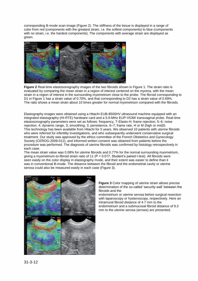
31-3-12
corresponding B-mode scan image (Figure 2). The stiffness of the tissue is displayed in a range of color from red (components with the greatest strain, i.e. the softest components) to blue (components with no strain, i.e. the hardest components). The components with average strain are displayed as green.
Figure 2 Real-time elastosonography images of the two fibroids shown in Figure 1. The strain ratio is evaluated by comparing the mean strain in a region of interest centered on the myoma, with the mean strain in a region of interest in the surrounding myometrium close to the probe. The fibroid corresponding to D1 in Figure 1 has a strain value of 0.70%, and that corresponding to D2 has a strain value of 0.69%. The ratio shows a mean strain about 10 times greater for normal myometrium compared with the fibroids. Elastography images were obtained using a Hitachi EUB-8500HV ultrasound machine equipped with an integrated elastography (HI-RTE) hardware card and a 5.9-MHz EUP-V53W transvaginal probe. Real-time elastosonography parameters were set as follows: frequency, T-Elasto-H; frame rejection, 5–6; noise rejection, 4; dynamic range, 3; smoothing, 3; persistence, 6–7; frame rate, H or M (high or mid)5. This technology has been available from Hitachi for 5 years. We observed 10 patients with uterine fibroids who were referred for infertility investigations, and who subsequently underwent conservative surgical treatment. Our study was approved by the ethics committee of the French Obstetrics and Gynecology Society (CEROG-2009-012), and informed written consent was obtained from patients before the procedure was performed. The diagnosis of uterine fibroids was confirmed by histology retrospectively in each case. The mean strain value was 0.08% for uterine fibroids and 0.77% for the normal surrounding myometrium, giving a myometrium-to-fibroid strain ratio of 11 (P = 0.017, Student‘s paired t-test). All fibroids were seen easily on the color display in elastography mode, and their extent was easier to define than it was in conventional B-mode. The distance between the fibroid and the endometrial cavity or uterine serosa could also be measured easily in each case (Figure 3).
Figure 3 Color mapping of uterine strain allows precise determination of the so-called ‗security wall‘ between the fibroids and the endometrium or uterine serosa before surgical resection with laparoscopy or hysteroscopy, respectively. Here an intramural fibroid distance of 4.7 mm to the endometrium and a submucosal fibroid distance of 9.3 mm to the uterine serosa (arrows) are presented.
31-3-12
Endovaginal ultrasonography is safe, accessible and inexpensive, and remains the primary imaging method for gynecological evaluation. Real-time elastosonography offers complementary diagnostic and mapping information. It is easy to perform, and the procedure requires only a few seconds of manipulation. With conventional ultrasound, the ultrasound beam is often strongly attenuated by the fibroid. As a result, with low gain, the posterior wall of the uterine myoma is poorly vizualized, but as the gain is increased, noise or artifactual echoes appear inside the mass, obscuring the image of the fibroid1. Real-time elastosonography provides an instantaneous color map that precisely delineates the fibroids, thus overcoming the limitations of conventional ultrasound. Real-time elastosonography encodes the ultrasonographic displacement of tissues under an external pressure constraint, using a temporal parameter which originates from an image sequence analysis and comparison over a short period. This temporal parameter is absent from conventional B-mode sonography, which displays only the instantaneous ability of tissues to reflect or absorb ultrasound waves, and this may explain why attenuation of ultrasound and shadowing artifacts affect elastography less than they do conventional B-mode ultrasound imaging. There are only a few reports in the literature on the in-vivo use of real-time transvaginal elastosonography in the field of gynecology. The usefulness of this technique has been documented for breast cancer and liver fibrosis, and has been of interest in the exploration of malignant tumors in the cervix, prostate and thyroid6–11. In this study, we have demonstrated the feasibility of in-vivo real-time transvaginal elastosonography of fibroids. In conclusion, real-time elastosonography is a promising tool that can provide detailed mapping and characterization of uterine fibroids. This could improve the gynecological ultrasound evaluation of size, volume and delineation of uterine fibroids before surgery or embolization. Future studies should aim to investigate strain contrast differences between fibroids and adenomyomas, and the characterization of uterine or adnexal pathologies. References 1. Vitiello D, McCarthy S. Diagnostic imaging of myomas. Obstet Gynecol Clin North Am 2006; 33: 85–95. 2. Janssen J. [(E)US elastography: current status and perspectives]. Z Gastroenterol 2008; 46: 572–579. 3. Hobson MA, Kiss MZ, Varghese T, Sommer AM, Kliewer MA, Zagzebski JA, Hall TJ, Harter J, Hartenbach EM, Madsen EL. In vitro uterine strain imaging: preliminary results. J Ultrasound Med 2007; 26: 899–908. 4. Kiss MZ, Hobson MA, Varghese T, Harter J, Kliewer MA, Hartenbach EM, Zagzebski JA. Frequency-dependent complex modulus of the uterus: preliminary results. Phys Med Biol 2006; 51: 3683–3695. 5. Havre RF, Elde E, Gilja OH, Odegaard S, Eide GE, Matre K, Nesje LB. Freehand real-time elastography: impact of scanning parameters on image quality and in vitro intra- and interobserver validations. Ultrasound Med Biol 2008; 34: 1638–1650. 6. Zhu QL, Jiang YX, Liu JB, Liu H, Sun Q, Dai Q, Chen X. Realtime ultrasound elastography: its potential role in assessment of breast lesions. Ultrasound Med Biol 2008; 34: 1232–1238. 7. Thomas A, Kummel S, Gemeinhardt O, Fischer T. Real-time sonoelastography of the cervix: tissue elasticity of the normal and abnormal cervix. Acad Radiol 2007; 14: 193–200. 8. Rubaltelli L, Corradin S, Dorigo A, Stabilito M, Tregnaghi A, Borsato S, Stramare R. Differential diagnosis of benign and malignant thyroid nodules at elastosonography. UltraschallMed 2009; 30: 175–179. 9. Tranquart F, Bleuzen A, Pierre-Renoult P, Chabrolle C, Sam Giao M, Lecomte P. [Elastosonography of thyroid lesions]. J Radiol 2008; 89: 35–39. 10. Salomon G, K¨ ollerman J, Thederan I, Chun FK, Bud¨aus L, Schlomm T, Isbarn H, Heinzer H, Huland H, Graefen M. Evaluation of prostate cancer detection with ultrasound realtime elastography: a comparison with step section pathological analysis after radical prostatectomy. Eur Urol 2008; 54: 1354–1362. 11. Brusseau E, Deprez JF, Duboeuf F, Basset O. [Biological tissue strain imaging with freehand elastography]. J Radiol 2007; 88:1823–1831. Ultrasound Obstet Gynecol 2009; 34: 486–488
___________________
31-3-12
FACTORS INFLUENCING THE QUALITY OF SONOELASTOGRAPHY FOR THE EVALUATION OF SUSPICIOUS BREAST LESIONS
Jung Min Chang, Woo Moon, Nariya Cho, Sanghee Park
PURPOSE
To investigate the factors influencing the quality of sonoELASTOGRAPHY for the evaluation of suspicious breast lesions.
METHOD AND MATERIALS
Between January 2009 and February 2009, real-time sonoELASTOGRAPHY of 312 breast masses (245 benign, 67 malignant) were obtained in 279 patients (mean age 46, range 16-74) with a EUB-8500 scanner (Hitachi Medical) prior to US-guided core biopsy. Five breast radiologists who performed the examinations prospectively assessed the quality of elasticity images on a 3 point scale (0=failure to obtain strain information, 1=valid strain information but high noise level, 2=valid strain information with low noise level), based on noise level and consistency of strain information. Diagnostic score of elasticity was also assigned on a 3 point scale (0=high stain, 1 =low strain, 2= no strain), based on the degree of strain in the hypoechoic lesion without histology information. The clinical and imaging findings of patients such as age, body mass index (BMI), mammographic density, breast thickness, lesion size, and lesion depth on US were analyzed for their association with the image quality using the χ2test, student t-test and Spearman rank correlation method. Sensitivities, specificities, PPV, and NPV for the classification of benign and malignant masses were calculated with a cutoff point between the diagnostic scores of 1 and 2 , and compared between the groups of different quality scores using the χ2test test.
RESULTS
The quality of elasticity images was assessed as score 0 in 21 cases (6.7%), score 1 in 134, and score 2 in 157. Quality score of elasticity images was negatively correlated with BMI (r =-.145, p=.010), breast thickness (r=-.266, p<.001), lesion size (r =-.175, p=.002), and lesion depth (r=-.145, p=0.011). However, the age and mammographic density was not associated with image quality. There were significant differences in sensitivities (87.0% vs. 59.5%, p=.041) and NPV (97.3% vs. 84.7%, p=.002) between the groups of quality score 1 and 2 for classification of benign and malignant masses.
CONCLUSION
Breast thickness, BMI, lesion depth, and lesion size was correlated with image quality of sonoELASTOGRAPHY. Sensitivity and NPV for classification of benign and malignant masses improved with higher quality scores.
CLINICAL RELEVANCE/APPLICATION
SonoELASTOGRAPHY seems to be favorable for the evaluation of small superficially-located lesions in women with smaller breasts and lower BMI.
Radiological Society of North America 95
th Scientific Assembly and Annual Meeting November 29
th –
December 4th, 2009, Chicago, USA
___________________
COMPARISON OF TWO COMMERCIALLY AVAILABLE SONOELASTOGRAPHY SYSTEMS IN DISTINGUISHING BENIGN FROM MALIGNANT BREAST MASSES
Nariya Cho, Woo Moon
PURPOSE
To retrospectively compare the diagnostic performance of two commercially available sonoELASTOGRAPHY systems in distinguishing benign from malignant breast masses.
31-3-12
METHOD AND MATERIALS
Between August 2008 and November 2008, 64 women (mean age 50 years, range 28 – 78 years) with 74 breast masses (mean size 16 mm, range 3-50 mm) (23 malignant, 51 benign) underwent sonoelastographic examinations with both systems (A and B) by one radiologist prior to biopsy. Probability of malignancy based on conventional US findings was recorded prior to sonoELASTOGRAPHY. Real time imaging files obtained from the two systems were saved as video clips, then masked, randomized and separately analyzed by two radiologists without information of final histology. For the A images, the lesion width ratio obtained by dividing the strain image measurements by the conventional US image measurements was calculated. For the B images, the elasticity score (1-5) based on the degree and distribution of strain was given in consensus. Diagnostic performance of the two systems in distinguishing benign from malignant masses was compared using receiver operating characteristic (ROC) curve analysis and McNemar‘s test.
RESULTS
The area under the ROC curve for the B system (Az=0.896) was higher than that of the A system (Az=0.722) (difference between areas 0.175, 95% CI 0.0589-0.291, P=0.003) and similar to that of conventional US (Az=0.916) (difference between areas 0.0196, 95% CI -0.0747-0.114, P=0.684). The best cut-off values, yielding the maximal sum of sensitivity and specificity, were between width ratios of 0.99 and 1 and elasticity scores of 3 and 4. The sensitivity of the A system was higher than that of the B system [87.0% (20 of 23) vs. 78.3% (18 of 23), P = 0.006] and the specificity of the B system was higher than that of the A system [90.2% (46 of 51) vs. 64.7% (33 of 51), P = 0.004].
CONCLUSION
The B system showed better overall diagnostic performance and specificity, and the A system showed better sensitivity in distinguishing benign from malignant breast masses.
CLINICAL RELEVANCE/APPLICATION
Understanding the characteristics of the two commercially available sonoELASTOGRAPHY systems can be helpful in optimizing the diagnostic criteria for each system.
Radiological Society of North America 95th Scientific Assembly and Annual Meeting November 29
th –
December 4th, 2009, Chicago, USA
___________________
ANALYSIS OF ELASTOGRAPHIC AND B-MODE FEATURES AT SONOELASTOGRAPHY FOR BREAST TUMOR CLASSIFICATION.
Woo Kyung Moon, Chiun-Sheng Huang, Wei-Chih Shen, Etsuo Takada, Ruey-Feng Chang, Juliwati Joe, Michiko Nakajima, and Masayuki Kobayashi Department of Radiology, College of Medicine, Seoul National University, Seoul, Korea.
The purpose of this study was to evaluate the accuracy of neural network analysis of elastographic features at sonoelastography for the classification of biopsy-proved benign and malignant breast tumors. Sonoelastography of 181 solid breast masses (113 benign and 68 malignant tumors) was performed for 181 patients (mean age, 47 years; range, 24-75 years). After the manual segmentation of the tumors, five elastographic features (strain difference, strain ratio, mean, median and mode) and six B-mode features (orientation, undulation, angularity, average gradient, gradient variance and intensity variance) were computed. A neural network was used to classify tumors by the use of these features. The Student's t test and receiver operating characteristic (ROC) curve analysis were used for statistical analysis. Area under ROC curve (Az) values of the three elastographic features- mean (0.87), median (0.86) and mode (0.83)-were significantly higher than the Az values for the six B-mode features (0.54-0.69) (p < 0.01). Accuracy,

31-3-12
sensitivity, specificity and Az of the neural network for the classification of solid breast tumors were 86.2% (156/181), 83.8% (57/68), 87.6% (99/113) and 0.84 for the elastographic features, respectively, and 82.3% (149/181), 70.6% (48/68), 89.4% (101/113) and 0.78 for the B-mode features, respectively, and 90.6% (164/181), 95.6% (65/68), 87.6% (99/113) and 0.92 for the combination of the elastographic and B-mode features, respectively. We conclude that sonoelastographic images and neural network analysis of features has the potential to increase the accuracy of the use of ultrasound for the classification of benign and malignant breast tumors. Ultrasound Med Biol, 2009 Nov;35(11):1794-802
___________________
DIAGNOSTIC PERFORMANCE OF FREEHAND ELASTOGRAPHY WITH STRAIN RATIO MEASUREMENT IN THE CHARACTERIZATION OF BREAST LESIONS REFERRED FOR ULTRASOUND GUIDED BIOPSY: INITIAL CLINICAL RESULTS AT A SINGLE CANCER REFERRAL CENTER. Todd R. Kumm, Alec Chau, Margaret Szabunio. H. Lee Moffitt Cancer Center & Research Institute, Tampa, USA. Background: While patients referred for biopsy of breast lesions represent a small percentage of the screening population, the resources consumed by this patient segment are disproportionately high [1]. Lesions biopsied yield a benign result in more than 75% of cases, therefore, a reliable, non–invasive method to characterize suspicious lesions would be valuable [1,2]. Elastography has been proposed as a potential tool for differentiation of benign from malignant lesions in the breast [2,3]. Our study included the use of strain ratio measurement in addition to elastography scoring of lesions. Aims: To determine the diagnostic performance of elastography (ES) and strain ratio (SR) measurement for breast lesions referred for biopsy. Methods: Patients referred for ultrasound guided biopsy of a suspicious breast lesion were included in this study after informed consent was given. Using the Hitachi Hi Vision 900 Ultrasound with integrated Sonoelastography software, elastography score (ES) images and strain ratio (SR) measurements were obtained for each lesion. Elastography system operators and elastogram readers were considered newcomers to the technique, but received specialized training from the equipment manufacturer prior to study initiation. Subjects were evaluated immediately prior to biopsy on the same date. Lesions were assigned an ES using the five–point visual scoring system (Figure 1) proposed by Itoh, et. al. [3]. The lesion was considered ES test negative if scored 0, 1 or 2; while a score of 3, 4 or 5 was considered ES test positive. Calculation of the SR value (Figure 2) was based upon the average strain measured in the lesion as compared to adjacent adipose tissue in the breast. Using proprietary software, the average strain of the lesion was determined by selecting a representative region of interest from the center of the lesion, and was expressed as ―ST–ave LESION‖. A corresponding ROI of adjacent adipose tissue was then selected, and was expressed as ―ST–ave FAT‖. The resultant SR value was expressed as a ratio according to the equation: ST–ave Fat /ST–ave Lesion = SR. A lesion was considered SR test negative if the ratio was <4.5; while a ratio of ≥4.5 was considered SR test positive, according to criteria provided by the manufacturer. Sensitivity, specificity, negative and positive predictive values and test accuracy were calculated using core needle or excisional biopsy result as the standard. Results: A total of 100 lesions from 85 patients were evaluated (Table 1). Combined ES and SR scoring counted the test as positive if either ES or SR score met the criteria for the positive test.
Table 1 Conclusions: Using strain ratio assessment provides additional information about a lesion‘s strain characteristics and may serve to complement elastography scoring using a visual scale. Combining SR assessment with ES improved sensitivity compared to either scoring system alone. Although combined scoring slightly decreased specificity, a single false negative result was excluded using this technique. Study limitations include a small sample size, operator experience and potential selection bias as our site is a cancer referral center. Ongoing study of these techniques may help to further clarify the value of combining these measurements and determine if the use of ES and SR scoring can help identify patients in which biopsy might be avoided in lieu of clinical follow up.

31-3-12
References: [1] Poplack SP, Carney PA, Weiss JE, Titus–Ernstoff L, Goodrich ME, Tosteson AN, Screening mammography: costs and use of screening–related services. Radiology, 234:79–85, 2005. [2] Burnside ES, Hall TJ, Sommer AM, et al., Differentiating Benign from Malignant Solid Breast Masses with US Strain Imaging. Radiology, 245:401–410, 2007. [3] Itoh A, Ueno E, Tohno E, et al., Breast disease: clinical application of US elastography for diagnosis. Radiology, 239:341–350, 2006.
Eighth International Conference on the Ultrasonic Measurement and Imaging of Tissue Elasticity, September 14 – 17, 2009, Vlissingen, The Netherlands
________________________
THE UTILITY OF REAL-TIME TISSUE ELASTOGRAPHY FOR THE BREAST
Assoc Prof Ei Ueno, University of Tsukuba, Japan The hardness of tumor tissue is an important finding in clinical examinations for breast cancer. A dynamic test has been used to evaluate hardness even in real-time ultrasonic examination. Elastography provides precise images of the strain produced when an extremely small pressure is applied to the breast. For this reason, it has been adopted as a very simple method for diagnosis. The hardness of the tissue is displayed in color tones, with increasing hardness presented in ascending order of red, yellow, green, and blue. The Tsukuba ultrasound group scores the hardness of the tissue on a scale of 1 to 5. The target region for comparison is the hypoechoic region of the lesion. Scores of 4 and score 5 both represent malignancies. A score of 4, depicted as a blue (No strain over the entire hypoechoic area). A score of 5, depicted as a blue over the entire hypoechoic area and in the surrounding. Scores of 1 and 2 are benign findings, and a score of 3 indicates that malignancy cannot be ruled out. A score of 1 is depicted as a green; 2, as a mosaic of green and blue; and 3, as a blue center with a green periphery. Altogether, these five scores basically correspond to the assessment categories of BIRADS. A cyst will appear in three color tones (blue, green, and red), due to artifact. This phenomenon, the so-called BGR sign, indicates the presence of liquid. A similar pattern of color tones is observed in an abscess. 12
th World Congress of the World Federation for Ultrasound in Medicine and Biology, 30
th August – 3
rd
September 2009, Sydney, Australia
________________________

31-3-12
ELASTOGRAPHY FOR THE CHARACTERIZATION OF NONPALPABLE BREAST LESIONS Assoc Prof Woo Kyung Moon, Seoul National University Hospital, Korea
We performed several prospective clinical studies to evaluate the role of sonoelastography for the characterization of nonpalpable breast lesions. First, 842 consecutive women who were scheduled to undergo US-guided core biopsy due to 864 breast lesions(79 cancers and 785 benign lesions) detected by supplemental screening US were examined with a commercialized sonoelastography. For the BI-RADS category 4a lesions, 28.5% (216 of 757) had a normal strain and 99.1% (214 of 216) of lesions with normal strain found out to be benign. Second, 300 women with 339 nonpalpable lesions were prospectively examined with sonoelastograpy and color Doppler US (CDUS) prior to biopsy. Of the 301 lesions with BI-RADS category 4a, 76 (25%) lesions showed normal strain on elastography and no vascularity on CDUS and all of them were found to be benign. Breast lesions diagnosed with DCIS by US-guided core biopsy with invasive components at surgery show less strain than pure DCIS at elastography regardless of the lesion size and B-mode US findings. Third, we evaluated the accuracy of neural network analysis of elastographic features at sonoelastography for the classification of 113 benign and 68 malignant breast tumors. Az values of the three elastographic features- mean (0.87), median (0.86) and mode (0.83)-were significantly higher than the Az values for the six B-mode features (0.54-0.69). In conclusion, addition of elastography to breast US has potential to reduce benign biopsy for BIRADS category 3 or 4a lesions that are detected by supplemental screening US.
12
th World Congress of the World Federation for Ultrasound in Medicine and Biology, 30
th August – 3
rd
September 2009, Sydney, Australia
________________________
FAT-LESION RATIO IN BREAST ELASTOGRAPHY Assoc Prof Ei Ueno, University of Tsukuba, Japan Elastography is a recommended technique for breast cancer specialists. With just short training, oncologists and physicians can acquire the same degree of competence as a ultrasound specialist in the ultrasonic diagnosis of breast cancer. Yet until recently, diagnoses based on strain imaging lacked objectivity. To provide greater objectivity in assessing elasticity, our group developed a new quantitative parameter called the Fat-Lesion Ratio (FLR). The FLR is defined as the ratio obtained by dividing the mean strain of fat by the mean strain of a hypo-echoic lesion. The strain of subcutaneous fat is determined from a circular area bound by the skin and mammary glands. The strain of a lesion, meanwhile, is determined from a circular area bound by the inner margin of the hypo-echoic area. The subjects of this study included 695 patients with hypo-echoic lesions (not over 2 cm in diameter) who underwent examinations by elastography between January 25, 2005 and September 26, 2008. Among these, patients, 305 had breast cancer and 390 had benign disease. The mean FLR in breast cancer (12.4) was considerably higher than the mean FLR in benign disease (4.2). When applying a cutoff point of 5.2, we obtained a sensitivity of 76.8%, specificity of 78.9%, and accuracy of 77.8%. The area under the ROC curve was high (0.832), with a sensitivity, specificity, and accuracy of 80.7%, 74.1%, and 77.8% respectively. FLR was effective for clinical diagnosis, with high diagnostic objectivity irrespective of the experience of the examiners
12
th World Congress of the World Federation for Ultrasound in Medicine and Biology, 30
th August – 3
rd
September 2009, Sydney, Australia
________________________
31-3-12
VALUE OF MODIFIED ELASTICITY SCORING SYSTEM IN THE DIAGNOSIS OF SOLID BREAST LESIONS Dr Xiaoyun Xiao, Dr Hui Zhi, Dr Haiyun Yang, Dr Bing Ou, Dr Yanling Wen, Dr Baoming Luo Objective. The purpose of the study was to evaluate the value of modified elasticity scoring system in the diagnosis of solid breast lesions. Methods. From September 2004 to August 2007, ultrasound strain imaging was performed on 575 patients with 626 solid lesions. All the breast lesions were analyzed with established and modified elasticity scoring system respectively. The established scoring system was the one proposed by Itoh A et al in 2004. The established one was proposed by our department. The diagnostic results were compared with histopathologic results. Receiver operating characteristic curves (ROC) were computed individually. The areas under the ROC (AUC) were calculated and compared. Results. There were 242 malignant and 384 benign lesions. 527 lesions could be scored according to both scoring systems. 99 lesions with untypical elasticity images could only be scored with modified scoring system. For the 527 lesions, AUC (area under ROC curve) of established elasticity scoring system was 0.914 and that of modified elasticity scoring system was 0.963. The difference between them was statistically significant. (Z=5.029, P<0.001) For 99 lesions, the AUC was 0.967 by using modified elasticity scoring system. Conclusions. Compared with the established one, modified elasticity scoring system is more accurate in the differential diagnosis of solid breast lesions. The system is convenient to handle and may be substitution for the established one. Since the limitation of UE, it‘s better to combine it with 2-D ultrasound for the differential diagnosis.
12
th World Congress of the World Federation for Ultrasound in Medicine and Biology, 30
th August – 3
rd
September 2009, Sydney, Australia
________________________
ULTRASOUND (US) - ELASTOGRAPHY OF SUSPICIOUS ABNORMAL BREAST LESIONS DETECTED BY SUPPLEMENTAL SCREENING US Dr Woo Kyung Moon, Dr Nariya Cho Seoul National University Hospital, Korea PURPOSE: To find out whether elastography is helpful in reducing the number of benign biopsies, using histological analysis as a reference standard. METHOD AND MATERIALS: A total of 864 lesions (mean size 1.0cm, range 0.3 - 3.0cm; BI-RADS category 4a /4b /4c /5; 757 / 66 / 28 /13 lesions) in 842 women (mean age 46, range 30-68) who were scheduled to undergo US-guided core biopsy formed our study group. Elastographic images were prospectively classified as positive (abnormal strain) and negative (normal strain) based on the degree of strain induced by light compression. RESULTS: Of the 864 suspicious abnormal lesions, 79 (9.1%) lesions were confirmed as cancers (77 positive, 2 negative at elastography). Of the 224 lesions with negative elastography (216 category 4a, 8 category 4b lesions), 2 lesions (0.9%) proved to be cancers (0.7 and 1.6cm, low grade DCIS). Of the 640 lesions with positive elastography, 77 lesions (12.0%) proved to be cancers (59 invasive ductal carcinomas, 18 DCIS) (p< 0.001). The rate of malignancy for BI-RADS category 4a / 4b / 4c / 5 lesions were 2.6% / 36.4% / 58.6% / 100%, respectively. For the BI-RADS category 4a lesions, 28.5% (216 of 757) had a normal strain and 99.1% (214 of 216) of lesions with normal strain found out to be benign. CONCLUSION: When a lesion categorized as BI-RADS category 4a has a normal strain on elastography, a biopsy may be averted.
12
th World Congress of the World Federation for Ultrasound in Medicine and Biology, 30
th August – 3
rd
September 2009, Sydney, Australia
________________________

31-3-12
HOW TO REDUCE FALSE POSITIVE RATES IN BREAST ULTRASOUND SCREENING Assoc Prof Woo Kyung Moon, Seoul National University Hospital, Korea Ultrasound detects cancers in about 0.40% of women with mammography-negative dense breasts, with a higher contribution in women younger than 50 years. However, false positives are a major concern in screening ultrasound. Several approaches to ultrasound interpretation and breast imaging management have been developed that substantially reduce the frequency of false-positive cases, involving both recall examinations and biopsies, without meaningfully reducing the detection of nonpalpable favorable-prognosis cancers. Successful methods to reduce the recall rate for screening ultrasound involve (1) obtaining clinical history and physical examination information, (2) confidently and correctly identifying some normal structures and artifacts, and (3) learning to ignore subtle sonographic findings of doubtful significance. Procedure-related changes including fat necrosis and foreign body can be ignored with the proper history. Fat lobule often mimics solid nodule and Cooper's ligament produces architectural distortion and shadowing. Dilated ducts without intraductal masses are incidental benign findings. Complicated cysts can mimic solid nodules. They are nonparallel orientation and often show curvilinear bright line of anterior wall. Oval circumscribed solid masses less than 1cm in size can be followed. Doppler ultrasound and elastography can help distinguish normal or benign from suspicious solid masses. When a breast lesion categorized as BI-RADS 3 or 4 shows a normal strain on elastography and no vascularity on color Doppler ultrasound, biopsy can be averted. Physicians performed the screening ultrasound could directly evaluate lesions in real-time and reduce patient anxiety and discomfort. By applying these approaches successfully, we will be able to demonstrate convincingly that the benefits of ultrasound far outweigh the risks of false-positive interpretations.
12
th World Congress of the World Federation for Ultrasound in Medicine and Biology, 30
th August – 3
rd
September 2009, Sydney, Australia
________________________
IS IT POSSIBLE TO DIFFERENTIATE BETWEEN PHYLLODES TUMOR AND FIBROADENOMA IN ULTRASONOGRAPHIC ELASTOGRAPHY? Mi Hee Jung
1, Dr Jin Hwa Lee
2, Soon Jeong Kyung
1, Seung Oh Kyung
1
1Kosin University College of Medicine, Korea,
2Dong-A University Hospital, Korea
Objective: The purpose of our study was to evaluate the US elastography of phyllodes tumors and to differentiate them from fibroadenomas. Materials and Methods: We retrospectively reviewed the US elastographic finding of phyllodes tumors and fibroadenomas. Elasticity images were assigned an elasticity score according to the degree and distribution of strain induced by light compression (five-point scale, Score : 1-3-benign, 4-5-malignant). Results: There was substantial overlap in the image characteristics of phyllodes tumors and fibroadenomas. The phyllodes tumors trend to have a score of 3 or 4. The fibroadenomas trend to have a score 1 or 2. Conclusion: Phyllodes tumor and fibroadenoma cannot be precisely differentiated on B-mode ultrasonography. US elastography may allow the differentiation of phyllodes tumor from fibroadenoma on the basis of differences in stiffness.
12
th World Congress of the World Federation for Ultrasound in Medicine and Biology, 30
th August – 3
rd
September 2009, Sydney, Australia
________________________

31-3-12
APPLICATION OF REAL-TIME SONOELASTOGRAPHY AND MAMMOGRAPHY IN DIAGNOSIS OF BREAST LESIONS Ying Xiao, Cheng Zhuang, Juan Pan, Fang Liu DEPARTMENT OF ULTRASONOGRAPHY, XIANGYA HOSPITAL, CENTRAL SOUTH UNIVERSITY, China Objective: To compare the diagnosis value of real-time sonoelastography and mammography in breast lesions. Methods: The instrument furnish used is the HITACH EUB 8500, Equipped with 7.5-13MHz high frequency linear- array transducer. 83 female patients including 101 lesions of breast disease were studied with color Doppler flow image and real-time sonoelastography. All cases were performed mammography,then verified by operation and Pathology. The target lesion was scored as 1 to 5, using the scoring system proposed by Itoh. The accuracy of sonoelastography and mammography for diagnosing breast diseases was compared. Receiver operating characteristic curves(ROC) were used to assess the diagnostic value of sonoelastography. Results: The sensitivity /specificity/accuracy of the aonoelastography diagnostic method were 87.06%/ 93.05%/91.08% respectively, of mammography were 72.4%,83.33%,80.19%. The positive and negetive-likelihood ratio for sonoelastography was 12.41(95%CI,5.90 to 26.88)and 0.14(95%CI,0.06 to 0.30); for mammography, was 4.34(95%CI,1.63-11.42)and 0.33(95%CI,0.19-0.63). Conclusions: Sonoelastography and mammography were both effective technology in the diagnosis of breast lesions. Sonoelastography has higher diagnostic value than mammography in breast lesions, especially,in the lesions which were no calcification. To combine sonoelastography with mammography can elevate the accuracy differentiating the benign from malignant breast lesions.
12
th World Congress of the World Federation for Ultrasound in Medicine and Biology, 30
th August – 3
rd
September 2009, Sydney, Australia
________________________
REAL-TIME SONOELASTOGRAPHY OF BENIGN AND MALIGNANT OVARIAN TUMORS Veronika Gazhonova, Svetlana Churkina, Alexander Zubarev Radiology Chair President Medical Center, Russia Objective: Many of ovarian tumors that are detected as pelvic mass may have similar B-mode and Doppler characteristics but require different treatment. The objective of the study was to examine strain pattern of different ovarian tumors for early differentiation and cancer detection. Materials and methods: Real-time sonoelastography was performed in 43 consecutive ovarian tumors less than 3 cm diameter that were referred for surgery (15 solid, 17 cystic, 11 papillary). A EUB 900 High Vision (Hitachi Medical, Japan) and a convex transducer 8-4 MHz (EUP-V53W, Hitachi) were used. US data was compared with final histopathology. Elasticity images were obtained during manual freehand compressions of the tissue (compr. quality 3-4). Elasticity score according to the degree and distribution of the strain was established (5 point color scale: 1-3 benign, 4-5 malignant). Results: 26 benign and 17 malignant ovarian tumors were revealed by final histopathology. Sonoelastography showed accurate differentiation of mucinous, serous and haemorrhagic cysts that appeared equal on B-mode imaging. Benign cystic papillary tumors showed less than 3point strain score. In the cases of malignancy papillary component appeared 4 or 5 point score. Benign teratomas and fibromas showed equal 4 point strain score. Based on the knowledge of sonoelastography 17 hysterectomies, 10 isolated resections of the tumors and 16 right or left ovariectomies were performed. Conclusion: Real-time sonoelastography is a valuable tool in differentiation of serous, mucinous and haemorrhagic cysts and increase diagnostic confidence of stiffed malignant component in cystadenocarcinomas. Further investigation is necessary to validate these results and calculate strain ratio of the ovarian tumors.

31-3-12
12th World Congress of the World Federation for Ultrasound in Medicine and Biology, 30
th August – 3
rd
September 2009, Sydney, Australia
________________________
REAL-TIME SONOELASTOGRAPHY IN DIFFERENTIATION OF THE SOLID BREAST LESIONS Ms Elena Khokhlova, Ms Elena Lukyanova, Presidential Scientific Center, Russia; Mss Nadezhda Rozhkova, Federal Mammology Center, Russia; Mr Alexander Zubarev, Presidential Scientific Center, Russia Purpose: To evaluate the diagnostic possibilities of ultrasound (US) elastography to differentiate benign from malignant breast lesions, with pathomorphological correlation. To determine the accuracy of using a comparison of the strain of an adjacent fat area to strain of the lesion – Strain Ratio (SR). Methods and Materials: 122 lesions (84 benign, 38 malignant) were analyzed with the EUB 8500 and EUB 900 (Hitachi Medical, Japan) and a linear-array transduser of 7,5-13 MHz. The US data were compared with histopathologic findings (fine needle aspiration, core or surgical biopsy). Elasticity images were assigned an elasticity score according to the degree and distribution of strain induced by light compression (five-point color scale, Tsukuba Elastography Score: 1-3 – benign, 4-5 – malignant). SR is obtained by dividing strain of subucutaneous fatty tissues with that of the lesion. Statistical analysis included sensitivity, specificity, accuracy, positive predictive value (PPV), negative predictive value (NPV). Results: The sonoelastography was in agreement with histology for 106 lesions, with 10 false-negative results and 6 false-positive results (sensitivity 73,7%, specificity 92,8%, PPV 82,3%, NPV 88,6%). SR shows low value in benign lesions and high value in malignant lesions. Using a cut-off of 4,3, SR achieved high percentage in specificity, sensitivity and accuracy: 95,2%, 78,9% and 90,1% respectively. Conclusion: Sonoelastography is an effective US technique for increasing specificity of conventional ultrasound methods. With the advanced SR measurement tool, additional quantification of strain data is possible by comparing the difference in stiffness between a mass and its surrounding tissue.
12
th World Congress of the World Federation for Ultrasound in Medicine and Biology, 30
th August – 3
rd
September 2009, Sydney, Australia
________________________
REAL-TIME SONOELASTOGRAPHY IN LOCAL STAGING OF ENDOMETRIAL CARCINOMA WITH PATHOMORPHOLOGICAL CORRELATIONS: WORK IN PROGRESS Mss Svetlana Churkina, Mss Veronica Gazhonova, Mss H Savinova, Mr Y Gribunov, Mss Ekaterina Panfilova, Mr Alexander Zubarev, Presidential Scientific Center, Russia Objective: Local stage of endometrial cancer strongly influence on surgical management, treatment choice, further prognosis. We propose real-time sonoelastography as a method of identification of myometrial, presence and extent of cervical invasion and presence of extrauterine disease based on the analysis of strain pattern which represents tissue hardness. The objective of the study was to evaluate the role of real-time sonoelastography in endometrial cancer local staging. Materials and methods: We performed real-time sonoelastography in 26 women reffered for surgery due to endometrial carcinoma. All patients underwent US examination on EUB 900 (Hitachi Medical Corporation) with built-in elastography software. The type of starin pattern of the myometrium in the junctional zone, cervical region and in parametrium was studied by 3 radiologists in consensus. US elasticity results were compared with pathomorphological and final hystopathology after surgery. Results: The tissue hardness in the region of tumor invasion appeared strongly blue on the strain pattern. The sensitivity of US in staging endometrial cancer with new technique was: for stage IA – 6/7 (86%), IB – 5/7 (71%), IC – 6/6 (100%), stage II – 4/4 (100%), stage III – 2/2 (100%). The sencitivity of elasticity imaging was less accurate in patients with concomitant benign disease. The overall sensitivity in local staging of endometrial cancer was 88,5%. Conclusion: Strain imaging analysis could be used as an adjunct to B-mode and Doppler imaging in local staging of endometrial carcinoma. Real-time sonoelastography is expected to play an important role in decision making of staging in endometrial carcinoma.

31-3-12
12
th World Congress of the World Federation for Ultrasound in Medicine and Biology, 30
th August – 3
rd
September 2009, Sydney, Australia
________________________
ELASTIC MODULI OF INVASIVE CARCINOMA OF THE BREAST COMPARED WITH US ELASTOGRAPHY FINDINGS Takeshi Umemoto, Ei Ueno, Tsukuba Medical Center Hospital, Japan; Youko Fujihara, Takeshi Matsumura, Hitachi Medical Corporation, Japan; Eriko Tohno, Hiroko Kiyomatsu, Tsukuba University Hospital, Japan; Makoto Yamakawa, Tsuyoshi Shiina, Graduate School of Medicine, University of Kyoto, Japan Purpose: To evaluate the elastic moduli of the breast tissue and to contrast them with elasticity images for accurate interpretation of real-time US elastography. Materials and Methods: All the patients gave informed consent. Conventional US and Real-time Tissue Elastography were performed preoperatively in 26 patients (1 man, 25 women; mean age 60.0 years ±13.8 [standard deviation]) who had invasive breast cancer. The slice of 5mm thickness including the lesion and the surrounding breast tissue was obtained from patient‘s specimen immediately after resection. Within 2 hours after surgical resection, elastic modulus of each region was measured using materials testing machine (Instron 3342) under the constant pre-compression. Result: The value of elastic moduli, the mean ± standard deviation was 2.2±1.9kPa for fat tissue, 5.1±2.0kPa for normal mammary gland, 26.4±20.1kPa for invasive breast cancer, respectively. The entire lesion showed the elasticity score of 3 or more (score of 3, 3 lesions; score of 4, 7 lesions; score of 5, 16 lesions) on elasticity images. Conclusion: The elastic moduli of the invasive breast cancer varied according to the histological structure of each lesion while those of fat and mammary gland were almost constant. These differences were demonstrated well on elasticity images. This study was approved by the University of Tsukuba Human Subjects Institutional Review Board.
12
th World Congress of the World Federation for Ultrasound in Medicine and Biology, 30
th August – 3
rd
September 2009, Sydney, Australia
________________________
PREDICTIVE VALUE FOR MALIGNANCY OF SUSPICIOUS BREAST MASSES OF BI-RADS CATEGORY 4-5 USING ULTRASOUND ELASTOGRAPHY AND MRI DIFFUSION-WEIGHTED IMAGING H. Satake, A. Nshio, M. Ikeda, S. Ishigaki, M. Kawamura, K. Shimamoto, H. Kawai, S. Naganawa; Nagoya/JP Purpose: To evaluate the predictive value for malignancy of ultrasound elastography (USE) and MRI diffusion-weighted imaging (MRI-DWI) regarding breast mass of BI-RADS category 4-5. Methods and Materials: The subjects were consecutive 63 suspicious breast masses classified as BI-RADS category 4 or 5. All patients were recommended for biopsy and were confirmed pathologically (19 benign, 44 malignant). In addition to the routine clinical examinations of mammography, ultrasound, and dynamic contrast-enhanced MRI, USE and MRI-DWI were also obtained. Two radiologists retrospectively evaluated elasticity score of USE, and calculated apparent diffusion coefficient (ADC) values on MRI-DWI. According to the several reports, the cut-off levels of elasticity score was defined as between scores 3 and 4. Optimal cut off levels of continuous ADC values were determined by estimating the receiver operating characteristic
curves.
The diagnostic
abilities to differentiate malignant from benign lesions were analyzed by using univariate and
multivariate logistic regression analyses. Results: The sensitivity, specificity, and accuracy were 84.0, 68.0, and 79.3% for elasticity score and 75.0, 74.0, and 74.6% for ADC values, respectively. All four cases with elasticity score 1 were proved to be benign pathologically. Although both elasticity score and ADC values were shown to provide the significant criteria for differentiating malignancy from benign in the univariate analysis, only elasticity

31-3-12
score was the significant predictor in the multivariate analysis. Conclusion: Our results show that USE is the examination that can add more reliable information to a recommendation for biopsy in the breast masses classified as BI-RADS category 4-5, compared with MRI-DWI. European Congress of Radiology, March 6 – 9
th, 2009, Vienna, Austria
________________________
IMAGING ASPECTS OF PAPILLARY BREAST PROLIFERATIONS: FROM MAMMOGRAPHY TO FREE HAND ELASTOGRAPHY A.G. Maglas, A. Chiorean, M. Duma, T.S. Serb, S.M. Dudea, S. Sfrangeu; Cluj Napoca/RO Learning Objectives: To present imaging aspects encountered with breast papillary proliferations. To highlight features that raise malignancy suspicion. Background: A retrospective analysis of cases diagnosed between a 4 year interval (2004-2008) in our radiology department was performed. Pathology was obtained using core or excision biopsies. 58 cases were included. 33 were benign papillary lesions and 25 were papillary carcinomas. Imaging examinations were carried out in accordance with the ACR guidelines. Galactography was performed in 6 cases and elastography in 12 cases. Imaging Findings: The most frequent mammographic appearance of invasive papillary carcinomas was that of an opacity with imprecise deliniation. On ultrasound, these tumours appeared as solid lesions (19), complex cysts (4) or intraductal proliferations (2). On ultrasound, the most frequent aspect of benign papillary lesions was that of dilated ducts with solid, intraductal component (21), followed by the nodular solid appearance in 8 cases and intracystic proliferations in 4 cases. Galactographically, there were visualized ductal obstruction, lacunary images or parietal irregularities. All elastographically assessed papillary proliferations proved to be more rigid than neighbouring breast parenchyma. Conclusion: Ultrasound may enable us to detect and appreciate the extent of disease in symptomatic and asymptomatic patients with negative standard mammographies. Differential diagnosis between benign or in situ papillary proliferations and the invasive ones is often impossible from an imaging point of view. A close appreciation of lesion‘s delineation and degree of vascularization may up-grade our level of suspicion. However, pathologic proof is mandatory for all papillary proliferations. European Congress of Radiology, March 6 – 9
th, 2009, Vienna, Austria
________________________
CORRELATION OF BREAST ULTRASOUND (US) ELASTOGRAPHY AND MR IMAGING: CORRELATION OF ELASTOGRAPHY SCORE (ES) WITH SHORT TAU INVERSION RECOVERY (STIR) MR IMAGES, ENHANCEMENT RATIO, APPARENT DIFFUSION COEFFICIENT (ADC) AND FIBROTIC CHANGES OF BREAST DISEASE R.N. Matsubayashi, M. Imanishi, K. Yasumori, T. Muranaka, S. Momosaki; Fukuoka/JP Purpose: US elastography provides information regarding tissue hardness, and is expected to become a novel diagnostic tool for breast disease. On the other hand, MR images reflect the tissue characteristics. Fibrosis of the stroma of breast diseases may affect the hardness of them. We investigated the correlation between ES and signal-intensity (SI) of STIR MR images, enhancement ratio, ADC, and the fibrosis in the breast lesions. Methods and Materials: We reviewed the findings of US elastography and MRI from 41 consecutive patients with breast lesions (25 invasive ductal carcinoma, 3 fibroadenoma, 1 phyllodes tumor, 2 ductal hyperplasia, 2 primary malignant lymphoma, 3 mastopathy, 1 metastasis, 1 tubular adenoma , 1 ductal carcinoma in situ, 1 diabetic matopathy and 1 intraductal papilloma). In each patient, elastography images were classified based on Tsukuba ES. We calculated the ratio of SI of the lesion to muscle on STIR images (L/M ratio), enhancement ratio of early to pre-contrast (E/P) and early to

31-3-12
delayed (E/D) images and ADC for each lesion. The ES and MR findings were correlated with the degree of fibrosis (based on Masson trichrome stain). Results: The ES significantly correlated with the L/M ratio (p=0.0306) and the ADC (p=0.0256). The stromal fibrosis also correlated with ES (p=0.0023), the L/M ratio (p=0.0344) and the E/D ratio (p=0.049). Conclusion: The ES and L/M ratio are correlated significantly each other, and they are correlated with the fibrosis. These results suggest that they will provide the information of fibrosis, and may help the diagnosis of breast lesions. European Congress of Radiology, March 6 – 9
th, 2009, Vienna, Austria
________________________
REAL-TIME US ELASTOGRAPHY IN THE DIFFERENTIATION OF SUSPICIOUS MICROCALCIFICATIONS ON MAMMOGRAPHY Cho N., Moon W.K., Park J.S The purpose of this study was to retrospectively evaluate the use of US elastography in the differentiation of mammographically detected suspicious microcalcifications, using histology as the reference standard. Between May 2006 and April 2007, real-time US elasticity images were obtained in 77 patients (age range, 24-67 years; mean, 46 years) with 77 mammographically detected areas of microcalcifications (42 benign and 35 malignant lesions) prior to needle biopsy. Two experienced radiologists reviewed cine clips of elasticity and B-mode images and assigned an elasticity score of 1 to 3 in consensus, based on the degree of strain in the hypoechoic lesion without information of mammography and histology. For the elasticity score, the mean +/- standard deviation was 1.5 +/- 0.7 for benign and 2.7 +/- 0.7 for malignant lesions (P < 0.001). When a cutoff point between elasticity scores of 1 and 2 was used, US elastography showed 97% (34/35) sensitivity, 62% (26/42) specificity, 68% (34/50) PPV, and 96% (26/27) NPV with an Az value of 0.852 (0.753-0.923, 95% confidence interval) in the differentiation of benign and malignant microcalcifications. Our results suggest that US elastography has the potential to differentiate benign and malignant lesions associated with microcalcifications detected at screening mammography. Eur Radiol. 2009 Jul;19(7):1621-8.
________________________
SONOELASTOGRAPHICALLY GUIDED PREOPERATIVE LOCALIZATION OF SUSPICIOUS BREAST MICROCALCIFICATIONS DETECTED WITH MAMMOGRAPHY Chiorean AR, Duma MM, Dudea SM,et al. Introduction Most breast cancers are stiffer than surrounding normal breast parenchyma, mainly due to the accompanying desmoplastic reaction of the affected tissue (Martegani A et al. ECR 2005; C-0209). The principle of sonoelastography relies on the relative resistance of stiff tissues to movement compared to softer adjacent ones when slight probe pressure is applied (Itoh A et al. Radiology 2006; 239: 341-350). Although the majority of microcalcifications are preoperatively localized by stereotactic or mammographic means, ultrasound-guided procedures are preferred whenever possible due to their relative ease-of-use and speed which appeal both to patients and physicians. Breast elastography has mainly focused in recent years on circumscribed, solid, and cystic lesions, while the case reported here focuses on a non-nodular abnormality (Hatzung G et al. Ultraschall in Med 2008; 29; Duma M et al. Ultraschall in Med 2008; 29). Case description A 54-year-old woman with an unremarkable medical history appeared for a first mammography screening at our breast unit. The patient reported no previous breast pain, nipple discharge, or palpable changes during self-examination. Breast cancer history among close relatives and the specialist's clinical examination were negative. Mammography was performed using an analog GE (General Electric) device. Each breast was X-rayed in two directions: medio-lateral-oblique (MLO) and

31-3-12
craniocaudal (CC). The standard mammographies displayed a predominantly adipose breast structure with no opacities or architectural distortions. Nevertheless, in the left upper outer quadrant of the breast, a nonspecific, segmental area of microcalcifications measuring 3.5 cm in diameter was detected. Complementary MLO and CC magnified views were recommended and revealed polymorphic and pulverulent clusters of microcalcifications orientated towards the nipple. The consultant radiologist classified this area as Bl-RADS 5, suspecting an extensive intraductal carcinoma (Fig.1).
Fig.l Magnified cranio-caudal view of the external quadrant of the left breast reveals clusters of polymorphic and pulverulent microcalcifications, orientated towards the nipple.
The consequent, standard approach would have been stereotactic preoperative localization of the mammographically detected area of microcalcifications using two or more "hook" needles. However, the long procedure times and the increased patient discomfort implied by the use of an analog mammography device, made the breast unit team determined to seek an alternative imaging guidance modality. Although the breasts were predominantly adipose, bilateral comparative ultrasound was indicated, using a Hitachi 8500 US device with an elastography option. The left upper outer quadrant was attentively scanned, but on conventional grayscale and Doppler US, no definite lesion, no abnormal vascular signal, and no change in ultrasound transmission were detected. By contrast, the elastographic assessment of the corresponding mammographically detected area of microcalcifications revealed breast parenchyma stiffer than the neighboring tissue. Sonoelastography of the contralateral breast quadrant displayed normal features (Fig.2a-d).
Fig.2 a, b Comparative sonoelastography of the external upper quadrants of the right and left breast revealed a stiffer parenchymal area in the left breast. corresponding to the suspicious microcalcifications detected with mammography. c, d Color and power Doppler US of the left external upper quadrant revealed no abnormal vascular signal within the area.
With the patient resting in the surgical position, a skin marker drawing (Fig.3a) of the abnormal, stiffer breast area was performed under sonoelastographic guidance. During surgery, a radiograph of the
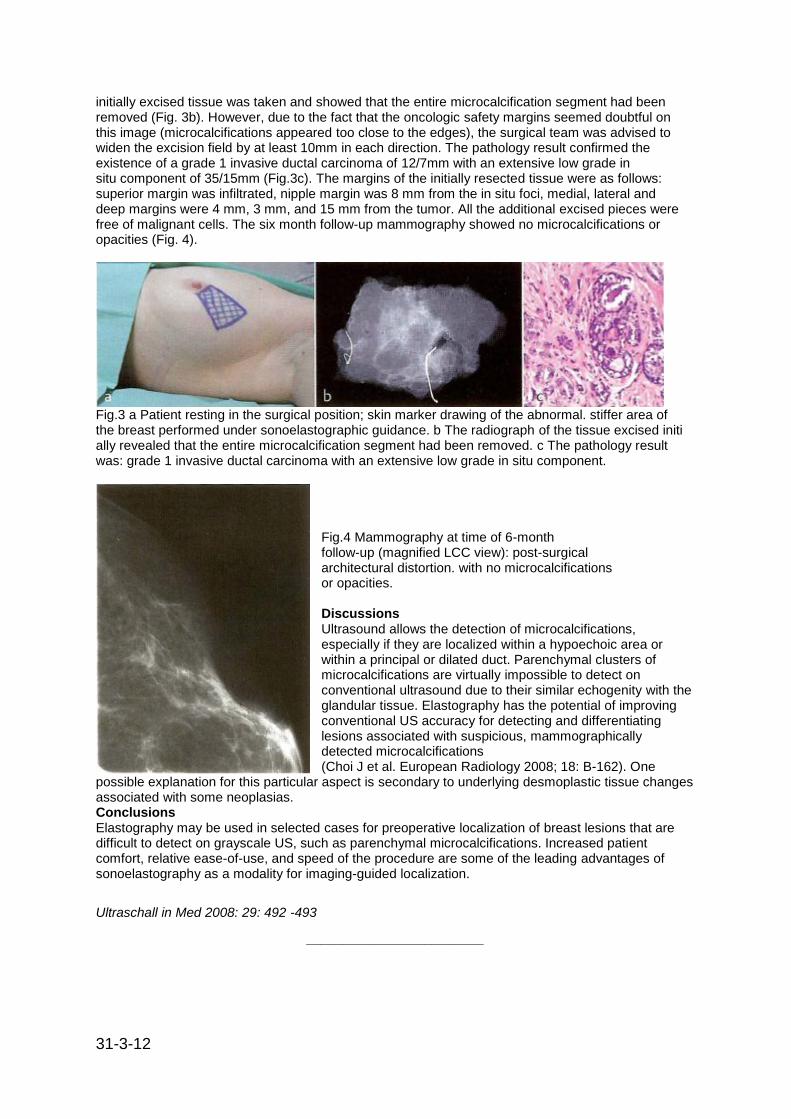
31-3-12
initially excised tissue was taken and showed that the entire microcalcification segment had been removed (Fig. 3b). However, due to the fact that the oncologic safety margins seemed doubtful on this image (microcalcifications appeared too close to the edges), the surgical team was advised to widen the excision field by at least 10mm in each direction. The pathology result confirmed the existence of a grade 1 invasive ductal carcinoma of 12/7mm with an extensive low grade in situ component of 35/15mm (Fig.3c). The margins of the initially resected tissue were as follows: superior margin was infiltrated, nipple margin was 8 mm from the in situ foci, medial, lateral and deep margins were 4 mm, 3 mm, and 15 mm from the tumor. All the additional excised pieces were free of malignant cells. The six month follow-up mammography showed no microcalcifications or opacities (Fig. 4).
Fig.3 a Patient resting in the surgical position; skin marker drawing of the abnormal. stiffer area of the breast performed under sonoelastographic guidance. b The radiograph of the tissue excised initi ally revealed that the entire microcalcification segment had been removed. c The pathology result was: grade 1 invasive ductal carcinoma with an extensive low grade in situ component.
Fig.4 Mammography at time of 6-month follow-up (magnified LCC view): post-surgical architectural distortion. with no microcalcifications or opacities. Discussions Ultrasound allows the detection of microcalcifications, especially if they are localized within a hypoechoic area or within a principal or dilated duct. Parenchymal clusters of microcalcifications are virtually impossible to detect on conventional ultrasound due to their similar echogenity with the glandular tissue. Elastography has the potential of improving conventional US accuracy for detecting and differentiating lesions associated with suspicious, mammographically detected microcalcifications (Choi J et al. European Radiology 2008; 18: B-162). One
possible explanation for this particular aspect is secondary to underlying desmoplastic tissue changes associated with some neoplasias. Conclusions Elastography may be used in selected cases for preoperative localization of breast lesions that are difficult to detect on grayscale US, such as parenchymal microcalcifications. Increased patient comfort, relative ease-of-use, and speed of the procedure are some of the leading advantages of sonoelastography as a modality for imaging-guided localization.
Ultraschall in Med 2008: 29: 492 -493
________________________

31-3-12
REAL-TIME ELASTICITY HELPS TO IMPROVE BREAST SPECIFICITY Anne Tardivon, Paris, France Elastography emerges as valuable adjunct technique to B-mode imaging for differentiation of masses Ultrasound has long been an efficient and useful adjunct technique for breast imaging. It is the first modality to be proposed in some situations: if a young or pregnant woman has a palpable mass, for example, or immediately after surgery. Ultrasound is usually performed during the diagnostic workup of masses, if architectural distortion is detected on mammograms, or following mammographic screening in individuals who are at high risk of breast cancer and have dense breast tissue. Ultrasound‘s primary role when imaging masses is to determine if the mass is cystic, and hence benign, or solid and potentially malignant. This differentiation is not always easy to make in practice, especially for complicated echoic cysts and lesions that are less than 7 mm in diameter. Characterization of solid masses is based mainly on morphological criteria: shape, spatial orientation, and margins. Interobserver variation can be high, however, especially for lesions that are BI-RADS category 3 (probably benign) or BI-RADS category 4a (low suspicion of malignancy). Most of these masses will be imaged again after a short time interval or referred for further investigation (fine-needle aspiration or core biopsy).
1
This all suggests that we need new tools to increase the specificity of B-mode ultrasound findings. Elastography is emerging as a promising candidate for this role. The technique has been reported in the literature since the 1980s, but it has not become routine. Now elastography is more user-friendly, and real-time images can be generated.
FIGURE 1. Elastography ultrasound (Hitachi software). Left: Strain ultrasound image shows soft lesion (green and red colors, score 2). Right: B-mode image shows hypoechoic nodule with microlobulated margins (BI-RADS 4a). Fine-needle aspiration revealed benign epithelial cells.
Essential Technique Elastography is currently offered by two vendors; Hitachi (EUB-8500 ultrasound scanner with integrated elastography software and a 6.5- to 13- MHz probe) and Siemens (Sonoline Elegra, 7.5L40 transducer at 7.2 MHz or VFX13-5 transducer at 10 MHz). The principle of the technique is essentially the same for both systems. Tissue compression produces displacement that is mainly in the longitudinal direction, the direction of the ultrasound beam. This can be used to calculate the strain in the tissue being compressed. Strain tends to be smaller in harder tissue than softer tissue. So working backwards, once the strain has been calculated, the tissue hardness can be evaluated as well. The influence of probe movement on the skin‘s surface in the lateral direction is minimized during measurement. Ultrasound elastography is performed at the same time as the standard imaging examination using the same probe. The lesion is first assessed using B-mode ultrasound. An adapter is then applied to the top of the probe to collect strain data, allowing perpendicular contact between the array and the skin. The operator identifies a region of interest around the lesion, ensuring that the target tissue occupies no more than one-third of the total ROI area. The lesion should be shown clearly on a double-screen display. The transducer is then used to apply weak repetitive pressure to the skin over the lesion (freehand compression technique). It is important that pressure applied during freehand compression is only light. The Hitachi system has a pressure indicator on the ultrasound screen, which should read between 2 and 3 during elastography (Figure 1). Higher readings indicate that the pressure being applied is too great. Excess pressure may induce nonlinear properties of tissue elastography and lead to misdiagnosis: the greater the compression over the tissues, the higher the elastographic values, which opens the possibility of potential false-positive results.
2 No published data are available to confirm this point, but in practice elastographic
mappings are modified and are more heterogeneous. Real-time color mapping inside the ROI and
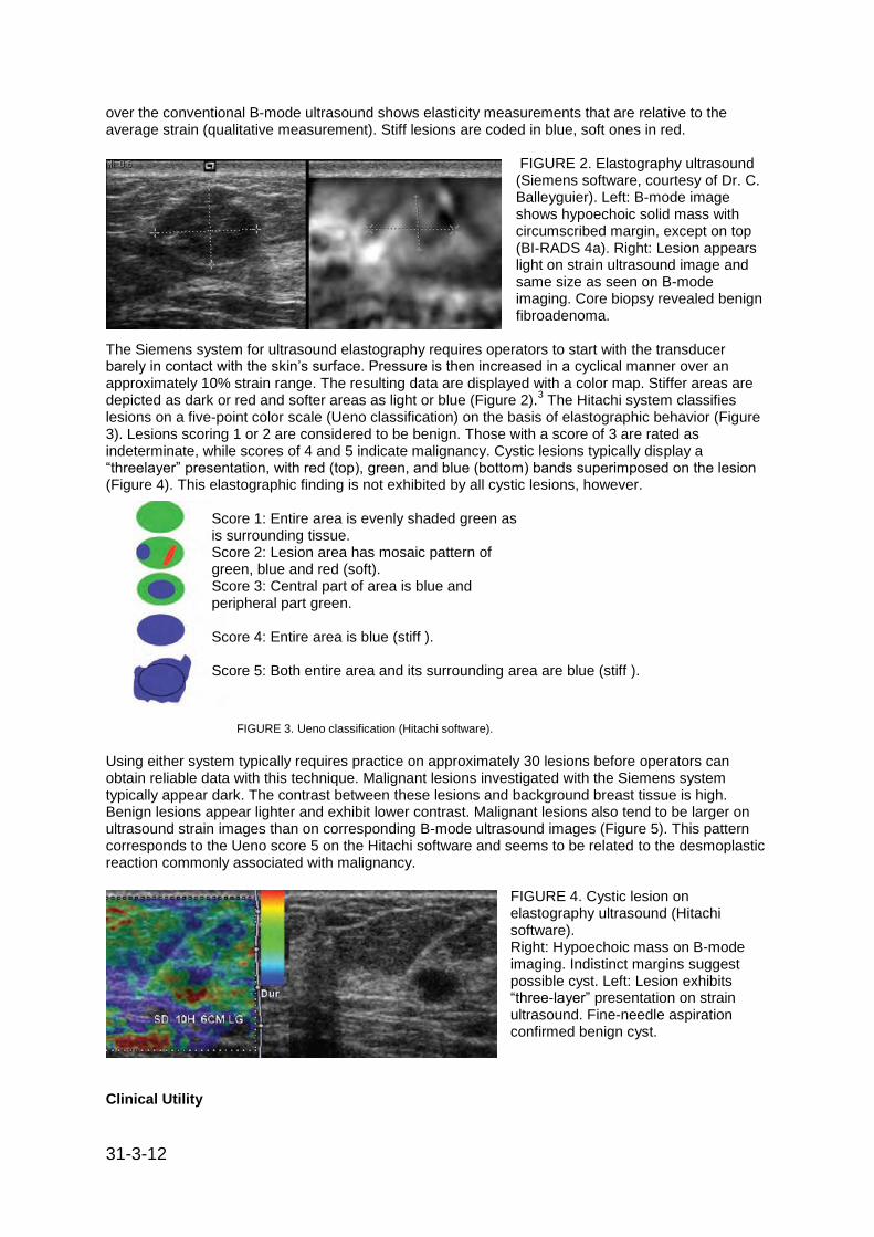
31-3-12
over the conventional B-mode ultrasound shows elasticity measurements that are relative to the average strain (qualitative measurement). Stiff lesions are coded in blue, soft ones in red.
FIGURE 2. Elastography ultrasound (Siemens software, courtesy of Dr. C. Balleyguier). Left: B-mode image shows hypoechoic solid mass with circumscribed margin, except on top (BI-RADS 4a). Right: Lesion appears light on strain ultrasound image and same size as seen on B-mode imaging. Core biopsy revealed benign fibroadenoma.
The Siemens system for ultrasound elastography requires operators to start with the transducer barely in contact with the skin‘s surface. Pressure is then increased in a cyclical manner over an approximately 10% strain range. The resulting data are displayed with a color map. Stiffer areas are depicted as dark or red and softer areas as light or blue (Figure 2).
3 The Hitachi system classifies
lesions on a five-point color scale (Ueno classification) on the basis of elastographic behavior (Figure 3). Lesions scoring 1 or 2 are considered to be benign. Those with a score of 3 are rated as indeterminate, while scores of 4 and 5 indicate malignancy. Cystic lesions typically display a ―threelayer‖ presentation, with red (top), green, and blue (bottom) bands superimposed on the lesion (Figure 4). This elastographic finding is not exhibited by all cystic lesions, however.
Score 1: Entire area is evenly shaded green as is surrounding tissue. Score 2: Lesion area has mosaic pattern of green, blue and red (soft). Score 3: Central part of area is blue and peripheral part green. Score 4: Entire area is blue (stiff ). Score 5: Both entire area and its surrounding area are blue (stiff ).
FIGURE 3. Ueno classification (Hitachi software). Using either system typically requires practice on approximately 30 lesions before operators can obtain reliable data with this technique. Malignant lesions investigated with the Siemens system typically appear dark. The contrast between these lesions and background breast tissue is high. Benign lesions appear lighter and exhibit lower contrast. Malignant lesions also tend to be larger on ultrasound strain images than on corresponding B-mode ultrasound images (Figure 5). This pattern corresponds to the Ueno score 5 on the Hitachi software and seems to be related to the desmoplastic reaction commonly associated with malignancy.
FIGURE 4. Cystic lesion on elastography ultrasound (Hitachi software). Right: Hypoechoic mass on B-mode imaging. Indistinct margins suggest possible cyst. Left: Lesion exhibits ―three-layer‖ presentation on strain ultrasound. Fine-needle aspiration confirmed benign cyst.
Clinical Utility

31-3-12
Many prospective studies have been published with comparable results. Elastography has been shown to increase the specificity of B-mode ultrasound to 85% to 98.5% and to increase the positive predictive value while slightly lowering or not affecting the sensitivity (78% to 87%).
4-8 Results from
one group suggest that elastography works better in lesions that have a diameter no greater than 15 mm, and that the best results are obtained in lesions less than 5 mm across.
9 Investigators from this
same group have modified the Ueno classification so that a score of 1 indicated a three-layer pattern, score 2 a lesion with an even elastic pattern (diffuse green), and score 3 a mostly elastic lesion with some small stiff areas (blue). The negative predictive value for cancer using this modified elastography classification was 98% (874 lesions). Researchers using the Hitachi system have reported high reproducibility of elastography data (good intra- and interobserver agreement). Interobserver reproducibility using the Siemens technique and measuring lesion size was not very good.
FIGURE 5. Elastography ultrasound (Siemens software). Left: B-mode image shows hypoechoic solid mass with indistinct margins (BI-RADS 4). Right: Mass is dark and appears larger on strain image (long axis = 15 mm; same measurement is 9 mm on B-mode image). Pathology: invasive ductal carcinoma, grade II.
This was the case even if the average area under the receiver operator characteristics curve after ultrasound strain imaging was greater than that after B-mode ultrasound alone.
3 If a lesion exhibits
characteristics that are typical of a malignant mass or a benign cyst on routine B-mode imaging, then elastography will not be useful for characterization. Elastography will, however, be extremely useful when characterizing complicated cystic lesions and benign solid masses because it can add another descriptor: soft lesion. This additional information may help operators decide which strategy to adopt when faced with lesions that are of low suspicion for malignancy (BI-RADS category 4a): intervention or repeat imaging after a short interval. Elastography can produce false negative and false-positive results. False-positive results have been reported for fibrous or calcified fibroadenomas and fibrous mastopathy. Meanwhile ductal carcinoma in situ and invasive cancers without desmoplastic reaction may appear soft on elastography (score 3, Ueno classification). This reinforces the message that an elastography score should be considered together with all other ultrasound findings and not used as a stand-alone diagnostic tool.
4,6
FIGURE 6. False-negative diagnosis. Left: Elastography image was given Ueno classification score of 3. Right: Ill-defined heterogeneous lesion. Pathology: ductal carcinoma in situ. Given reports of false-negative diagnoses in lesions rated ―score 3‖, this score should be used to indicate indeterminate lesions.
Emerging Techniques An alternative method is to take a quantitative approach to elastography. One ROI is placed over normal breast tissue (reference area, fat lobule), another over the target lesion, and the strains calculated for both. The strain produced in the fatty tissue is then divided by that of the lesion. This ratio will be low if the lesion is benign and high if it is malignant. In a study of 155 lesions (108 benign benign, 47 malignant) that used a cutoff value of 4.3 to divide the two groups, the method yielded a sensitivity of 89.4%, specificity of 88.8%, and accuracy of 89%. This type of quantification should increase the reproducibility of qualitative assessments. Elastography could potentially be used for many other applications besides differentiating between benign and malignant lesions. Such roles

31-3-12
have yet to be investigated and evaluated. Areas of interest include postradiotherapy imaging, local staging of breast cancer (searching for multifocality), staging of axillary lymph nodes, ―secondlook‖ imaging following breast MRI, improving localization of lesions, and investigating tumor response to neoadjuvant therapies. New elastography technologies are being investigated clinically as well. One promising method is supersonic shear wave elastography, which combines two innovative concepts. The same conventional ultrasound probe is used to manipulate the tissue and to image it. An ultrasonic focused beam creates acoustic pressure, removing the need for external compression. The resulting motion is imaged using an ultrafast ultrasound acquisition. Quantitative elastography information (kPa) is available using this technique.
10 Elastography ultrasound is emerging as a
valuable adjunct to B-mode ultrasound for the evaluation of breast lesions. Reliable information can be obtained from experienced operators. The findings must, however, always be integrated with other ultrasound findings. Guidelines on the proper use of elastography, based on the Hitachi system, have been defined:
9
• Elastography may increase the specificity of ultrasound in the evaluation of breast lesions. It is not indicated for surgical scars, diffuse lesions, or lesions larger than the transducer field-of-view. • Elastography interpretation requires global experience in breast imaging. Operators should scan and interpret at least 30 cases under the supervision of an expert before performing the technique on their own in clinical practice. • Elastography acquisition can be termed "correct" when the value on the monitor is at least 2 or 3. Color homogeneity throughout the scanning area around the lesion is another way of evaluating examination quality. • At least two correct elastography acquisitions lasting five seconds should be obtained for each lesion. The area scanned should cover almost all of the field-of-view. • Two elastography scores should be acquired through perpendicular scanning planes for lesions with a mixed texture on B-mode ultrasound. • The pressure applied with the probe must be constant and perpendicular to both the front margin of the lesion and the thoracic plane. Lateral movements must be avoided. DR. TARDIVON is a radiologist in the breast imaging section at the Institut Curie in Paris.
References 1. Lazarus E. Mainiero MB. Scbepps B, et aI. BI-RADS Iexicon for US and mammography: interobserver variability and positive predictive value. Radiology 2006:239(2);385-391. 2. Ueno E. Itoh A. Diagnosis of breast cancer by elasticity imaging.Eizo Joho Medical 2004;36(12):2-6 3. Burnside ES, Hall TJ, Sommer AM, et al Differentiating benign from malignant solid breast masses with US strain imaging. Radiology 2007;245(2):401-410. 4.Itoh A, Ueno E, Tohno E, et aL Breast disease: clinical application of US elastography for diagnosis. Radiology 2006:239(2):)41-350. 5. Thomas A, Fischer T, Frey H, et al. Real·time elastography-an advanced method of ultrasound: first results in 108 patients with breast lesions. Ultrasound Obstet Gynecol 2006;28(3):335-340. 6. Tardivon A. El Khoury C, Thibault F, et aI. {Elastography of the breast: a prospective study of 122 lesions.] J Radiol 2007;88(5 Pt 1):657-662. French 7. Zhi H, Ou B, Luo BM, et aI. Comparison of ultrasound elastography, mammography and sonography in the diagnosis of solid breast lesions. J Ultrasound Med 2007:26(6):807-815 8. Tan SM. Teh HS, Mancer JF. Poh WT. Improving B mode ultra·sound evalualion of breast lesions with real-time ultrasound elastography – a clinical approach. Breast 2008;17(3);252-257. 9. Rizzatto G. Real·time elastography of the breast in clinical practice- the Italian experience. Medix 2007(suppl): 12-15. 10. Tanter M, Bercoff J, Athanasiou A, etl al. Quantitative assessment of breast lesion viscoelasticity; initial clinical results using supersonic shear imaging- Ultrasound Med BioI 2008:34(9)l373 - 1386. Diagnostic Imaging Europe, December 2008/January 2009
________________________

31-3-12
ULTRASOUND ELASTOGRAPHY OF BREAST MASSES: NECESSITY OR LUXURY? Norran Said
PURPOSE
To investigate the added value of Elastography in, differentiating between benign and malignant breast masses, and deciding between biopsy & follow up (BI-RADS 3 & 4 categories).
METHOD AND MATERIALS
From July 2007 till February 2008, a total of 230 women (mean age 46.71, range 17 to 77 years) and 242 breast masses (61.6% benign , 38.4% malignant) were evaluated with real time grey scale ultrasound & Elastography (using the EUB 7500, Hitachi, Japan). BI-RADS categorization was applied followed by Elastography using visual assessment of strain images by a 5 point color scale (1-2 =benign, 3= indeterminate, 4-5 = malignant), and the Fat / lesion (strain ) ratio (< 4.8 = benign, > 4.8= malignant).
RESULTS
242 lesions with size range of 2 to 40mm were biopsied, and results were correlated with conventional ultrasound and Elastography findings. 70 cases were categorized as BI-RADS 3, and 79 cases as BI-RADS 4. Using Elastography with a visual score cut off value at 3, the results demonstrated a sensitivity & specificity of 95.7% & 76.51% , and a +ve PV, -ve PV of 71.77 & 96.61. Accuracy was 83.88. Using a cut off value at 4, the specificity increased to 85.9 %. When we added our Elastography findings to grey scale ultrasound findings, we had a total sensitivity & specificty of 100% & 78.52 % respectively, and a + PV & -PV of 74.40 & 100, while accuracy was 86.78.
CONCLUSION
Elastography can improve the diagnostic performance of ultrasound in evaluation of breast masses.
CLINICAL RELEVANCE/APPLICATION
Adding Elastography to breast ultrasound may improve the decision to confirm or eliminate the need for biopsy.
Radiological Society of North America 94th Scientific Assembly and Annual Meeting November 30
th –
December 5th, 2008, Chicago, USA
________________________
ULTRASOUND (US): ELASTOGRAPHY OF SUSPICIOUS ABNORMAL BREAST LESIONS DETECTED BY SUPPLEMENTAL SCREENING US Ha Young Kim (co-authors N Cho, W K Moon)
PURPOSE
To evaluate the negative predictive value of Elastography for suspicious abnormal lesions detected by supplemental screening US and to find out whether Elastography is helpful in reducing the number of benign biopsies, using histological analysis as a reference standard.
METHOD AND MATERIALS
Between May 2006 and March 2008, 1098 consecutive women who were scheduled to undergo US-guided core biopsy due to 1122 breast lesions detected by supplemental screening US were examined with a commercialized US-Elastography. All patients with US detected lesions had negative non-fatty mammogram. Lesions with category 3 (n= 224), atypical ductal hyperplasia (n=20), and larger than 3.0cm (n=13) were excluded. A total of 864 lesions (mean size 1.0cm, range 0.3 - 3.0cm; BI-RADS category 4a /4b /4c /5; 757 / 66 / 28 /13 lesions) in 842 women (mean age 46, range 30-68) formed our study group. Elastographic images were prospectively classified as positive (abnormal strain) and negative (normal strain) based on the degree of strain induced by light compression. We investigated whether there was a subset of benign lesions that were categorized as suspicious abnormality by conventional US, but as negative by Elastography.
RESULTS
Of the 864 suspicious abnormal lesions, 79 (9.1%) lesions were confirmed as cancers (77 positive, 2
31-3-12
negative at Elastography). Of the 224 lesions with negative Elastography (216 category 4a, 8 category 4b lesions), 2 lesions (0.9%) proved to be cancers (0.7 and 1.6cm, low grade DCIS). Of the 640 lesions with positive Elastography, 77 lesions (12.0%) proved to be cancers (59 invasive ductal carcinomas, 18 DCIS) (p< 0.001). The rate of malignancy for BI-RADS category 4a / 4b / 4c / 5 lesions were 2.6% / 36.4% / 58.6% / 100%, respectively. For the BI-RADS category 4a lesions, 28.5% (216 of 757) had a normal strain and 99.1% (214 of 216) of lesions with normal strain found out to be benign.
CONCLUSION
The negative predictive value of Elastography was 99.1% in 862 suspicious abnormal lesions detected by supplemental breast US. When a lesion categorized as BI-RADS category 4a has a normal strain on Elastography, a biopsy may be averted.
CLINICAL RELEVANCE/APPLICATION
Addition of US-Elastography to supplemental screening breast US has potential to reduce benign biopsy for BIRADS category 4a lesions that are detected by supplemental screening US. Radiological Society of North America 94
th Scientific Assembly and Annual Meeting November 30
th –
December 5th, 2008, Chicago, USA
________________________
BREAST ULTRASOUND ELASTOGRAPHY: RESULTS OF 193 BREAST LESIONS IN A PROSPECTIVE STUDY WITH HISTOPATHOLOGIC CORRELATION Fritz Schaefer
PURPOSE
To evaluate the diagnostic performance of ultrasound (US) Elastography in breast masses.
METHOD AND MATERIALS
193 lesions (benign, 129; malignant, 64) were analyzed with the EUB 8500 Logos ultrasonic unit (Hitachi Medical, Japan) using a linear-array transducer of 7.5-13 MHz. Reference standard was cytology (fine needle aspiration) or histology (core biopsy). US Elastography findings were classified according a six-point color scale (Ueno classification, 1-3=benign, 4-5=malignant). Conventional B-mode US findings were classified according the BI-RADS classification. Statistical analysis included sensitivity, specificity, ROC-analysis and kappa-values for intra- and interobserver reliability.
RESULTS
For malignant lesions the mean score in elasticity was 4.1+/-0.9, for benign lesions 2.1+/- 1.0 (p<0.001). With a best cut-off-point for Elastography between Ueno score 3 and 4, sensitivity was 96.9%, and specificity 76%, respectively. With a best cut-off-point for conventional US between BI-RADS 4 and 5, sensitivity was 57.8%, and specificity 96.1%, respectively. Elastography had higher sensitivity, and lower specificity than conventional US did. Two lesions scored Ueno 1 were false negative whereas no lesion scored BI-RADS 1-3 were false negative. ROC-curve for Elastography was 0.884 (p<0.001) and for conventional US 0.820 (p<0.001). Weighted kappa-value for intra-/interobserver reliability for BI-RADS-classification was 0.784/0.634 and for elasticity scores 0.720/0.561.
CONCLUSION
US Elastography has not the potential to replace conventional B-mode US for the detection of breast cancer, but can complement conventional US to improve the diagnostic performance.
CLINICAL RELEVANCE/APPLICATION First results of a new and innovative non-invasive technology for diagnosing breast cancer.
Radiological Society of North America 94th Scientific Assembly and Annual Meeting November 30
th –
December 5th, 2008, Chicago, USA
________________________
31-3-12
SEMI-QUANTITATING STIFFNESS OF BREAST SOLID LESIONS IN ULTRASONIC ELASTOGRAPHY Hui Zhi, MD, Xiaa-Yun Xiaa, MD, Hai-Yun Yang, MD, Van-Ling Wen, MD, Bing Qu, MD, Bao-Ming Lua, MD, Bi-Iing Liang, MD Rationale and Objectives. To explore whether strain ratio measurement could semi-quantitatively evaluate the stiffness of breast lesions. Materials and Methods. From January 2008 to May 2008, 148 patients with 254 solid lesions (183 benign, 71 malignant) in the breast were included in the study. Ultrasound sonography found the lesions and ultrasonic elastography obtained the strain images. By using the strain ratio measurement method together with the ultrasound machine, the strain index of the lesion was calculated. Different depths of breast tissue were selected as the reference. The strain indexes of malignant and benign solid lesions were calculated with the same level of breast tissue as the reference. Results. The strain indexes of breast lesions were different compared to the same depth of breast tissue and the superior level of fat tissue (P = 0.000). The strain indexes of breast lesions were different compared to different depths of breast glandular tissues (P = 0.003). At the same level of the breast lesions, 212 lesions were glandular tissue, 11 were fat tissue, and 40 were both. In the lesion plane, six lesions had almost no glandular tissue and 20 had almost no superior fat tissue. Compared to the same depth of breast tissue, the strain indexes of benign lesions (range, 0.62-11.07) and malignant lesions (range, 3.12-39.28) were different (P =0.000). Conclusion. Using the strain ratio measurement, stiffness of breast lesions could be semi-quantitated with the same depth of breast tissue as the reference. This method may provide another diagnostic method in addition to the 5-point scoring system used with ultrasonic elastography in the future.
Acad Radlol 2008; 15:1347-1353
________________________
REAL-TIME ULTRASOUND ELASTOGRAPHY: ITS POTENTIAL ROLE IN ASSESSMENT OF BREAST LESIONS. Zhu QL, Jiang YX, Liu JB, Liu H, Sun Q, Dai Q, Chen X.
Department of Diagnostic Ultrasound, Peking Union Medical College Hospital, Beijing, China.
We evaluated whether real-time ultrasound elastography (USE) performed in addition to conventional ultrasound (US) can improve the differentiation of benign from malignant breast lesions. Both conventional US and real-time USE were performed in 112 consecutive patients with 139 breast lesions using a Hitachi EUB-8500 US system. Each lesion was assigned an elasticity score according to the degree and distribution of strain induced manually by mild compression. The USE scores (1 to 5) were compared with the BI-RADS assessment categories (1 to 5) obtained with conventional US. Sensitivity, specificity and overall accuracy of each method were determined with surgical pathology as the gold standard. There were 70 benign and 69 malignant lesions. The mean elasticity score was significantly higher for malignant lesions than for benign lesions (4.33 +/- 0.11 vs. 2.10 +/- 0.13, p < 0.01). When a cutoff point of 4 was used, the sensitivity, specificity and accuracy were 85.5, 88.6 and 87% for USE and 94.2, 87.1 and 90.6% for conventional US, respectively. Of the 64 lesions assessed as BI-RADS 2 or 3(i.e., benign) based on conventional US, two were scored as 4 and 5 (i.e., malignant) using USE and were subsequently proven to be malignant. Of the 75 lesions with BI-RADS 4 or 5 category from conventional US, one was scored as a category 1 (benign) with USE and found to be benign by pathology. Our study results suggest that the addition of USE imaging to conventional US could be helpful in the detection and characterization of breast masses.
Ultrasound Med Biol. 2008 Aug;34(8):1232-8.
________________________
31-3-12
IMPROVING B MODE ULTRASOUND EVALUATION OF BREAST LESIONS WITH REAL-TIME ULTRASOUND ELASTOGRAPHY- A CLINICAL APPROACH
S.M. Tana, H.S. Teh
b, J.F. Kent Mancer
c, W.T. Poh
c
aDepartment of Surgery, Changi General Hospital, Singapore,
b Department of Radiology, Changi
General Hospital, Singapore, c Department of Pathology, Changi General Hospital, Singapore
Ultrasound elastography using the extended combined auto-correlation method of tissue elasticity allows for real-time strain image visualisation using a free-hand probe with concurrent conventional B mode imaging. Four hundred and fifteen consecutive women with 550 breast lesions confirmed on B mode ultrasound were assessed with elastography using the elasticity score. There were 119 malignant and 431 benign lesions. The elastography sensitivity was 78.0%, specificity was 98.5% and overall accuracy was 93.8%. The median score for malignancy was 5 and that for benign lesions was 2. There was good correlation with B mode BIRADS category. 98.6% of lesions with an elasticity score of 2 or below (95%CI = 96.8 - 99.4) were benign. BIRADS 3 lesions with an elasticity score of 2 or below may be re-classified as BIRADS 2 lesions. We found that 15.3% of BIRADS 2 and 3 lesions with an elasticity score of 3 were malignant. Real-time ultrasound elastography is user-friendly with a high accuracy rate, thereby improving B mode ultrasound assessment.
The Breast; 17 (2008): 252 – 257
________________________
ROLE OF SONOELASTOGRAPHY IN NON-PALPABLE BREAST LESIONS. Scaperrotta G, Ferranti C, Costa C, Mariani L, Marchesini M, Suman L, Folini C, Bergonzi S.
Department of Breast Imaging, Fondazione IRCCS Istituto Nazionale Tumori, Milan, Italy, [email protected].
The purpose of this study was to evaluate the diagnostic utility of sonoelastography in differentiating benign from malignant non-palpable breast lesions. A total of 293 BI-RADS 3-5 (Breast Imaging Reporting And Data System) impalpable breast lesions in 278 women was evaluated with B-mode ultrasound (US) and subsequently with sonoelastography (SE) before performing US-guided biopsy. Among the 293 lesions (size up to 2 cm), 110 (37.5%) were histologically malignant and 183 (62.5%) benign. Lesions that were malignant or showed atypical ductal hyperplasia were referred for surgical excision, as well as 32 benign lesions showing discordance between US/SE results and histology. All other benign lesions had US follow-up at 6/12 months, showing stability. Overall performance of SE was lower than US, with sensitivity and specificity of 80% and 80.9%, respectively, for SE as compared with 95.4% and 87.4% for US. Statistical analysis showed no improvement in the joint use of SE and US over the use of US alone, whose performance, however, was very high in our study. SE is a simple, fast and non-invasive diagnostic method that may be a useful aid to US for less experienced radiologists in the assessment of solid non-palpable breast lesions, especially BI-RADS 3, where specificity was higher (88.7%).
Eur Radiol. 2008:18(11);2381-9 (Full text available: http://www.springerlink.com/content/889352135p6418th)
________________________
BENIGN VERSUS MALIGNANT NODULAR BREAST LESION BY USING ELASTOSONOGRAPHY: PRELIMINARY REPORT Ciurea AI
I, Dumitriu D
I, Ciortea C
2, Nedevschi S
3, Bolboaca SD
1, Dudea SM
1
'University of Medicine and Pharmacy "1 Hatieganu" ClujNapoca; 2Emergency County Hospital, Cluj-Napoca;
3Technical University of Cluj-Napoca, Cluj-Napoca

31-3-12
Purpose: To assess the diagnostic accuracy of elastography in the characterization of nodular breast lesions. Material and Method: A sample of sixty-three patients with nodular lesions was analyzed between April and December 2007. The elastographic examinations were made using a Hitachi EUB 8500 device. In the study were included the patients with a pathological diagnosis and/or patients with known nodular lesion stable for at least two years. Lesions were classified and scored and the sensi-bility and specificity of elastography calculated. The strain ratio of breast lesions (fat lesion ratio) was calculated by using the software of Hitachi EUB 8500 device and also using the Image processing program. Results: The prevalence of malign breast lesion was of 60% with a 95% confidence interval (95%CI) of [48% - 72%]. The sensibility and specificity for elasography were almost 87% (95% CI [73% - 95%]) and 84% (95% CI [66% - 94%]), respectively. The accuracy of elasography was of 86% (95% CI [75 % - 93 %]), with a probability of a wrong positive test of 11 % (95 % CI[4% -24%] and a probability of wrong negative test of 19% (95% CI [8% -37%]. The ROC analysis of the strain ratio calculated with the Hitachi EUB 8500 device showed that the maximum sensibility (of 100%) and specificity (of 71 %) is obtained to a cutoff value of 5.06. The cutoff value for the maximum specificity (100%) and a corresponding sensibility of 12% revealed to be at 110.20. The ROC analysis of the strain ratio calculated with the ImageProcessing program showed that the maximum sensibility (of 100%) and specificity (of 50%) is obtained to a cutoff value of 1.59. The cutoff value for the maximum specificity (100%) and a corresponding sensibility of 12% revealed to be at 3.94. Conclusion: The sensibility and specificity of elastography is comparable with that of 2D ultrasonography. The diagnosis confidence could be increased by calculating the strain ratio. If the strain ratio is calculated with Hitachi EUB 8500 software a value less than 5.06 shown the benign nature of the lesion and a value greater than 97.21 shown malignancy. The corresponding values for the strain ratio calculated with ImageProcessing program were 1.63 (benignity) and 3.55 (malignancy) respectively. Acknowledgements: The horne made program was designed by Technical University of Cluj-Napoca as a part of a ElastoBreast research grant (CEEX/VIASAN/149j2006) Ultraschall in Med, 2008, suppl 1, OP2.9 XXth Congress of European Federation of Societies for Ultrasound in Medicine and Biology/XIth Romanian Conference of Ultrasound in Medicine and Biology, May 31 – June 3 2008, Timisoara, Romania .
________________________
BREAST LESIONS: CORRELATIONS BETWEEN ULTRASOUND BI-RADS CLASSIFICATION AND UENO-ITOH ELASTOGRAPHY SCORE Duma M
I, Chiorean A
2, Dudea S
2, Dumitriu D
2, lancu A
2, Roman R
2, Ciortea C
1, Ciurea A
2, Sfrangeu
S2, Bolboaca S
3
IER Clinical County Hospital Cluj-Napoca, Radiology Department;
2University of Medicine and
Pharmacy "luliu Hatieganu"; Radiology Depatment; 3University of Medicine and Pharmacy "luliu
Hatieganu", Statistics Department, Cluj Napoca Purpose: To establish the correlations between ultrasound (US) BI-RADS classification and Ueno-Itoh elastography score when assessing breast lesions. To determine which type of breast lesion (BI-RADS category) would benefit most from an elastographic assessment. Methodo!ogy: Our analysis was performed on 125 ultrasound detected breast lesions, in 80 patients examined with a Hitachi 8500 US device in our Breast Unit between May 2007 and February 2008. Each lesion was assessed according to BI-RADS and Ueno-ltoh elastography score. Histopathology was obtained by means of percutaneous biopsy or post-surgery. Fibroadenoma-like lesions unchanged over a period of 3 years were considered benign. Results: The 1, 2 and 6 Ueno-Itoh scores mostly correlated with BI-RADS 2 and 3 lesions such as cysts, hamartomas, lipomas, hematomas, non-palpable fibroadenomas. Palpable fibroadenomas initially included in BI-RADS 4a/b category, usually received benign elasticity scores (l or 2), the exception being represented by a minority of cases of old, fibrotic or calcified lesions (elastic score 3 or 4). Non-specific BIRADS 4a/b lesions, such as mastopathic nodules (frequent histologic feature: simple adenosis with apocrine methaplasia) demonstrated rather soft, elastic properties on
31-3-12
elastogram (score 1 or 2). The 4 and 5 Ueno-Itoh scores were predominantly correlated with BI-RADS 4c and 5 categories represented by high risk lesions (radial scar, papillomas, atypical epithelial ductal hyperplasia) and in situ or invasive carcinomas. Conclusions: Generally BI-RADS classification correlates well with Ueno-Itoh elasticity score, the main exception being represented by fibrotic, calcified lesions which falsely appear more suspicious post-elastography. In our opinion BI-RADS 3 and 4 lesions would benefit most from an elastographic assessment, a low Ueno-Itoh score allowing a less invasive approach; a high score imposing histopathologic evaluation. Ultraschall in Med, 2008, suppl 1, OP2.12 XXth Congress of European Federation of Societies for Ultrasound in Medicine and Biology/XIth Romanian Conference of Ultrasound in Medicine and Biology, May 31 – June 3 2008, Timisoara, Romania.
________________________
SHORT ANAIYSIS ON ELASTOGRAPHIC IMAGES OF BENIGN AND MALIGNANT BREAST LESIONS BASED ON COIOR AND HUE PARAMETERS Chiorean A
1, Duma M
2, Dudea S
1, Dumitriu D
1, lancu A
1, Roman R
1, Clonea C
2, Clurea A
1, Sfrangeu
S1, Bolboaca S
3
1University of Medicine and Pharmacy "luliu Hatieganu", Radiology Department, Cluj Napoca;
2ER
Clinical County HospitaL Cluj Napoca, Radiology Department; 2University of Medicine and Pharmacy
"luliu Hatieganu", Statistics Department, Cluj Napoca Purpose: To determine if color and hue analysis realized on elasto-graphic images of breast lesions can improve benign-malignant differentiation. Methodology: Our analysis was performed on 125 elastographic images of different breast lesions (malignant and benign) acquired from 80 patients examined in our Breast Unit using a Hitachi 8500 ultrasound device, during a period of 10 months (May 2007-February 2008). Histopathology was obtained by means of percutaneous biopsy or post-surgery. Fibroadenoma-like lesions unchanged over a period of 3 years were considered benign. Followed parameters (numeric values): average color (red, green, blue), color dispersion, average intensity, average hue, hue dispersion. Calculus modality: Image Processing Version 1.3, a program developed in collaboration with Technical University of Cluj Napoca. Results: Average Hue and Average Blue numeric values obtained for benign and malignant breast lesions were as follows: Avg Hue 142.01 for benign and 204.81 for malignant with a specificity of 83.12% calculated for a 180 cut off value; Avg Blue 59.60 for benign and 92.58 for malignant with a specificity of 71.43 % calculated for a 70 cut off value (p < 0.0001). Conclusion: In our opinion average Hue and average Blue could be some of the key parameters when evaluating elastographic images of breast lesions. Their numeric value becomes particularly useful in cases cataloged with 2 or 3 Ueno-Itoh scores, where "big numbers" for Avg Hue and Avg Blue point out to high risk lesions or neoplasias. Ultraschall in Med, 2008, suppl 1, OP2.13 XXth Congress of European Federation of Societies for Ultrasound in Medicine and Biology/XIth Romanian Conference of Ultrasound in Medicine and Biology, May 31 – June 3 2008, Timisoara, Romania.
________________________
FAT-LESION-RATIO VS. ELASTOGRAPHY SCORE: A NEW METHOD FOR SONOELASTOGRAPNY IN THE DIAGNOSTICS OF BREAST LESIONS Geaid AA. Grunwald S, Hatzung G, Behrndt PO, Ohlinger R University Clinic of Greifswald, Department of Gynecology and Obstetrics Objective: Pathological changes in the breast can influence the elasticity of tissues. The sonoelastography is a method, which displays the realtime tissue elasticity colour-coded in parallel
31-3-12
with the B-mode. The elastography-score (ESC) taken out of the colour-coded image was compared to the new sonoelastography measurement, the fat-Iesion~ratio (FLR) in terms of sensitivity and specificity. Materials and methods: From April to October 2007 patients of the breast consulting hours were examined each with one finding. The examination was accomplished with the Hitachi EUB 8500 HV. The ESC (score values 1 - 5) resulted from the determination of the examiner according to the ultrasound BI-RADS criteria whereas the FLR was measured by the elastography device via the selection of two regions of interest. This FLR is the result of the division of the average fat tissue elasticity through the average elasticity in the finding. The pathologic survey of each breast lesion happened before or after the elastography examination through the use of high-speed cut biopsy (n = 51/57, 89.5%), fine-needle aspiration cytology (n=5/57, 8.8%) or punction (n=1/57, 1.7%). The results were compared and evaluated with the ESC and FLR respectively. Results: The whole examination included 57 patients in the age of 20 to 81 (average 53 years). The pathological analysis assessed 27 benign and 30 malign lesions. With a cut-off value of 4.16 the FLR reached a sensitivity of 61.6% and a specifity of 76.0%. It turned out that high values appeared predominantly in malign and low values appeared more often in benign lesions. In contrast to the FLR the ESC had clearly a less sensitivity of 46.7% and a higher specifity of 92.3 %. Conclusion: The first results point out bigger advantages of the fat-lesion-ratio compared to the elastography score in the diagnostics of breast lesions. More results can be expected by increasing the patient population. Ultraschall in Med, 2008, suppl 1, OP2.15 XXth Congress of European Federation of Societies for Ultrasound in Medicine and Biology/XIth Romanian Conference of Ultrasound in Medicine and Biology, May 31 – June 3 2008, Timisoara, Romania.
________________________
SOURCES OF ERROR IN BREAST US ELASTOGRAPHY Ciurea A
I, Dumitriu D
I, Ciortea C
2, Dudea S
I
IUniversity of Medicine and Pharmacy"I Hatieganu" Cluj-Napoca;
2University Emergency Hospital Cluj-
Napoca Aims: The main purpose of this study has been to establish what factors determine the quality of an elastographic examination in breast pathology. Methods: In our ongoing study, which began in April 2007, we have examined 157 patients with focal breast lesions. All patients were examined using both 2D and Doppler ultrasound and elastography. The reference standard was the pathologic diagnosis. We tried to determine the importance of certain technical factors, such as: the type of section, the size of the region of interest, the degree of compression. We also studied in which way the characteristics of the lesion (size, distance to skin) influence the elastographic examination. Results: The elasticity score for the same lesion was sometimes different on sagittal and coronal sections - therefore it is important to examine the lesion using both types of sections and, probably. to take the highest elasticity score into account. If the region of interest was limited to the lesion, the elasticity score was not accurate, but if it included as much healthy tissue as possible, the score correlated better with the pathologic diagnosis. An inappropriate degree of compression influenced the result of the examination. The elasticity score was more accurate for smaIl masses than for large lesions, which occupy the whole field. It was also easier to determine for superficial lesions than for deeper-situated ones. Conclusions: Elastography is a useful tool in the uninvasive diagnosis of breast lesions, but it needs specific training, as weIl as acknowledging technical and pathological factors which may influence it. Ultraschall in Med, 2008, suppl 1, PP2.5 XXth Congress of European Federation of Societies for Ultrasound in Medicine and Biology/XIth Romanian Conference of Ultrasound in Medicine and Biology, May 31 – June 3 2008, Timisoara, Romania.
________________________

31-3-12
ELASTOGRAPHY: BETTER RESULTS IN THE DIAGNOSIS OF BREAST LESIONS COMPARED TO ULTRASOUND AND MAMMOGRAPHY? Hatzung G, Grunwald S, Geaid AA, Behrndt P0,Jager B, Ohlinger R Ernst Moritz Arndt University, Department of Gynecology and Obstetrics Objective: Some diseases, such as breast cancer, lead to a change of tissue hardness. Real-time elastography is a new method to estimate the tissue hardness. The purpose of the prospective study was to compare the dignity of elastography, ultrasound, and mammography in the diagnosis of breast lesions. The objective was to find out, if elastography shows better diagnostic accuracy in characterising nodular breast lesions. Furthermore it was examined, if elastography brings different results in application on different sizes of the diagnosed lesions. Material and Methods: Elastography was investigated in 99 lesions (27 carcinomas, 22 cysts, 19 fibroadenomas, et al.) in 78 patients (average age 55 years), using the high-end HITACHI EUB 8500 ultrasound system, equipped with a 13 - MHz linear transducer. All patients underwent mammographic and ultrasonic examination. The elastographic results were rated by the so called Ueno Score (1 - 2 = benign, 3 - 5 = malign), mammographic, and sonographic results according to the BIRADS criteria (1 - 3 = benign, 4 - 5 = malign). Afterwards, percutaneous samples for histologic diagnosis were taken. The elastographic, mammographic, and sonographic results were compared to those of the histological diagnosis. Results: 72 of 99 lesions were benign, 27 malign. Ultrasound had a sensitivity of 96%, specificity of 85%, respectively 81 % and 85 % for mammographic findings. The sensitivity of the elastography was 75%, the specificity 46%. A combination of elastography and ultrasound had the best sensitivity (100%) and specificity (94%). Elastography showed better results for lesions with a maximum diameter of 3 cm than for larger lesions. Conclusion: Elastography is easy and rapid to perform. Our initial results suggest that elastography is an additional method to assess the dignity of breast lesions. Due to the limited number of lesions included in the study, final conclusions are not yet possible. However, despite the small number of patient samples, these data provide an interesting contribution. The number of patients is currently being increased in order to get more significant results. Ultraschall in Med, 2008, suppl 1, OP6.1 XXth Congress of European Federation of Societies for Ultrasound in Medicine and Biology/XIth Romanian Conference of Ultrasound in Medicine and Biology, May 31 – June 3 2008, Timisoara, Romania.
________________________
NONPALPABLE BREAST MASSES: EVALUATION BY US ELASTOGRAPHY. N Cho, WK Moon, JS Park, JH Cha, M Jang, and MH Seong Department of Radiology and Clinical Research Institute, Seoul National University Hospital and the Institute of Radiation Medicine, Seoul National University Medical Research Center, Seoul, Korea. [email protected]
OBJECTIVE: To compare the diagnostic performances of conventional ultrasound (US) and US elastography for the differentiation of nonpalpable breast masses, and to evaluate whether elastography is helpful at reducing the number of benign biopsies, using histological analysis as a reference standard. MATERIALS AND METHODS: Conventional US and real-time elastographic images were obtained for 100 women who had been scheduled for a US-guided core biopsy of 100 nonpalpable breast masses (83 benign, 17 malignant). Two experienced radiologists unaware of the biopsy and clinical findings analyzed conventional US and elastographic images by consensus, and classified lesions based on degree of suspicion regarding the probability of malignancy. Results were evaluated by receiver operating characteristic curve analysis. In addition, the authors investigated whether a subset of lesions was categorized as suspicious by conventional US, but as benign by elastography. RESULTS: Areas under the ROC curves (Az values) were 0.901 for conventional US and 0.916 for elastography (p = 0.808). For BI-RADS category 4a lesions, 44% (22 of 50) had an elasticity score of 1 and all were found to be benign.

31-3-12
CONCLUSION: Elastography was found to have a diagnostic performance comparable to that of conventional US for the differentiation of nonpalpable breast masses. The authors conclude that BI-RADS category 4a lesions with an elasticity score of 1 probably do not require biopsy.
Korean J Radiol, March 1, 2008; 9(2): 111-8.
________________________
ULTRASOUND ELASTOGRAPHY: RESULTS OF A EUROPEAN MULTICENTRIC STUDY OF 429 BREAST LESIONS F. Schafer
1, A. Athanasiou
2, I. Leconte
3, R. Salvador
4, S. Ganau Macias
5, M. Wallis
6, C. Plancher
2,
A.A. Tardivon2;
1Kiel/DE,
2Paris/FR,
3Bruxelles/BE,
4Barcelona/ES,
5Sabadell/ES,
6Cambridge/GB
Purpose: To evaluate the performance of ultrasound elastography in breast lesions. Methods and Materials: 429 lesions (six centers, 268 benign, 161 malignant, 46.4% ≤10 mm) were evaluated by B-mode (BI-RADS classification) and elastography (EUB 8500 Logos unit, Hitachi, Japan). Diagnosis was obtained by FNA, core or surgical biopsies (84%), follow-up or comparison with previous studies (benign lesions). The elastic score was classified according to a five-point color scale (Ueno classification, 1-3=benign and 4-5=malignant). Intra- and inter-observer variables were evaluated in each center (kappa, 399 lesions). Results: With B-mode, sensitivity, specificity, positive predictive value, negative predictive value of BI-RADS classification (2-3=benign and 4-5=malignant) were 100, 51.5, 55.3 and 100%, respectively. When category 4 was subdivided into 4 low (one pejorative sign) and 4 high (two pejorative signs), specificity increased to 91% with a sensitivity of 85.7%. For elastography, sensitivity, specificity, positive predictive value, negative predictive value were: 72, 89.5, 84.6 and 79.9%, respectively. When the elastographic score 3 (central part of the mass stiffer than the peripheral normal tissue) was considered as malignant, these values were 83.2 (14 false negative results), 80.5, 77.5 and 85.6%, respectively. Intra- and inter-observer reproducibility were kappa=0.8 and 0.73. Conclusion: US elastography is an effective and reproducible US technique for increasing specificity of B-mode ultrasound. By adding new criteria for benignity, this technique may avoid unnecessary diagnostic procedures especially in breast masses categorized as BI-RADS categories 3 and 4 low.
European Congress of Radiology, March 7
th – 10
th 2008, Vienna, Austria
________________________
IS SONOELASTOGRAPHY HELPFUL IN BI-RADS III-IV (PROBABLY BENIGN-PROBABLY MALIGNANT) LESIONS? N.M. Abdel Razek, L. Ezzat, M. Shaalan; Cairo/EG Purpose: The goal of the study is to investigate the role of sonoelastography in evaluation of probably benign and probably malignant lesions (BI-RADS category 3 & 4) in sono-mammography and its contribution in patient managment. Methods and Materials: In this study 120 patients with BI-RADS 3 & 4 lesions in sono-mammography were investigated by sonoelastography (using the EUB 7500, Hitachi, Japan) and their elastography scores were analysed. All lesions were histopathologically proven after FNB, CNB, mammotome or operative biopsy. The elastography scores and results were correlated to the histopathology. Results: A total of 120 patients participated in the study. Their mean age was 48 yrs. 35% were histopathologically malignant lesions (including DCIS, lobular carcinoma, invasive duct carcinoma and medullary carcinoma). 65% were proven benign lesions icluding fibroadenomata, cysts, scars and papillomas as also sclerosing adenosis. Sonoelastography has increased the sensitivity and specificity of sono-mammography from 85 and 78% to 95 and 99%; however, as a sole diagnostic test, it has a sensitivity of 66% compared to a sensitivity of 58% of US in diagnosis of BI-RADS 3 & 4 lesions. The difference is not significant. It was found that the poor results were obtained with large lesions. Conclusion: In evaluation of BI-RADS 3 & 4 lesions, sonoelastography offers additional information

31-3-12
that increased the sensitivity and specificity of sono-mammography in evaluating these probable lesions. The accuracy of the test is increased in small lesions. European Congress of Radiology, March 7
th – 10
th 2008, Vienna, Austria
________________________
BREAST ULTRASOUND ELASTOGRAPHY: RESULTS OF 193 BREAST LESIONS IN A PROSPECTIVE STUDY WITH HISTOPATHOLOGIC CORRELATION F.K.W. Schaefer, S. Osterholz, B. Order, D. Fuerst, P.J. Schaefer, N. Hofheinz, J. Hedderich, W. Jonat, I. Schreer; Kiel/DE Purpose: To evaluate the diagnostic performance of ultrasound elastography in breast masses. Methods and Materials: 193 lesions (129 benign, 64 malignant) were analyzed with the EUB 8500 Logos ultrasonic unit (Hitachi Medical, Japan) and a linear-array transducer of 7.5-13 MHz. Standard of reference was cytology (FNA) or histology (core-biopsy). The elastic score was classified according a 6-point color scale (Ueno classification, 1-3=benign, 4-5=malignant). Conventional B-mode-US findings were classified according to the BI-RADS classification. Statistical analysis included sensitivity, specificity, ROC-analysis and kappa-values for intra- and interobserver reliability. Results: For malignant lesions, the mean score in elasticity was 4.1±0.9, for benign lesions 2.1±1.0 (p<0.001). With a best cut-off-point between score 3 and 4 in elastography, sensitivity was 96.9%, specificity 76%. When a best cut-off-point for conventional US was set between BI-RADS 4 and 5, sensitivity was 57.8%, specificity 96.1%. Elastography had higher sensitivity, and lower specificity than conventional US, but for two lesions of elastic-score 1 were false negative whereas no lesion that scored BI-RADS 1-3 was false negative. ROC-curve for elastography was 0.884 (p<0.001) and for conventional US 0.820 (p<0.001). Weighted kappa-value for intra-/interobserver reliability for BI-RADS-classification was 0.784/0.634 and for elasticity scores 0.720/0.561. Conclusion: Elastography has no potential to replace conventional B-mode-US for the detection of breast cancer, but can complement conventional US to improve the diagnostic performance. European Congress of Radiology, March 7
th – 10
th 2008, Vienna, Austria
________________________
ULTRASOUND (US) ELASTOGRAPHY OF BREAST LESIONS ASSOCIATED WITH MAMMOGRAPHICALLY DETECTED SUSPICIOUS MICROCALCIFICATIONS J. Choi, N. Cho, W.K. Moon; Seoul/KR Purpose: To evaluate the difference in strain between US lesions associated with malignant and benign microcalcifications detected at screening mammography by using US elastography. Methods and Materials: 50 consecutive women who had been scheduled to undergo US-guided vacuum assisted biopsy due to suspicious microcalcification cluster (larger than 1.0 cm) detected at screening mammography were examined with a commercialized US elastography. A total of 50 lesions (25 DCIS lesions and 25 fibrocystic changes) were found. Real-time imaging files were saved as video clips in avi format. Two experienced radiologists who had not performed the examinations analyzed in consensus the randomly ordered video clips without knowledge of the histology or mammographic findings and provided the elasticity score (1-5) according to the degree of strain induced by light
compression.
Results: For the elasticity score, the mean was 3.0±1.1 for malignant lesions and 1.3±0.5 for benign
lesions (p<0.001). When a cutoff point between 1 and 2 was used, elastography had 96% sensitivity,
72% specificity, 77% PPV, and 95% NPV. When a cutoff point between 2 and 3 was used,
elastography had 64% sensitivity, 100% specificity, 100% PPV, and 74% NPV. Mean area under the ROC curve was 0.930 (0.860-1.0, 95% confidence interval). Conclusion: Breast lesions associated with malignant microcalcifications tend to have less strain than benign microcalcifications at US elastography. US elastography has the potential to improve the accuracy of gray-scale US for the detection and differentiation of breast lesions associated with screening mammographically detected suspicious microcalcifications.
31-3-12
European Congress of Radiology, March 7
th – 10
th 2008, Vienna, Austria
________________________
LESION CHARACTERISATION M. Locatelli; Gorizia/IT The final objective in women with breast pathology is a longer disease-free survival and better quality of life, a challenge that can be tackled by improving surgical, adjunctive therapies and technical imaging. In technical imaging, more detailed and novel information are available for the diagnostic process and new techniques should allow the diagnostic process to be as physically and psychologically invasive as possible for the patient. The complex ensemble of sonographic signs and color Doppler patterns (lesion shape, orientation, margin, boundary, echo pattern, posterior acoustic wave, surrounding tissue, vascularity) has found a synthetic standardized language in the Breast Imaging Report and Data System (BI-RADS) developed by the American College of Radiology. This system is gradually becoming the official language in breast lesion management. It solves efficiently lesions BI-RADS 2, 5 and 6. On the other hand, lesions BI-RADS 3 and 4 still require full agreement in their management. Advanced technologies for lesion assessment, like real-time sono-elastography and volumetric acquisition with multiplanar reconstruction, are being introduced to clinical practice and may be of greatest benefit (diagnostic power) for lesions BI-RADS 3 and 4. Learning Objectives: 1. To show B-mode and color-flow algorithms. 2. To understand BI-RADS categories. 3. To review updated approaches for characterisation. European Congress of Radiology, March 7
th – 10
th 2008, Vienna, Austria
________________________
BREAST ELASTOGRAPHY TECHNIQUES BREAK NEW GROUND Diagnostic Imaging ECR 2008, (March 08, 2008)
By: H. A. Abella
Two new ultrasound elastography techniques show promise for the diagnosis and characterization of breast lesions, according to researchers from France and Korea. They could complement standard gray-scale sonography, evaluate suspicious microcalcifications detected with conventional mammography, and do away with unnecessary, painful needle biopsies.
Dr. Alexandra Athanasiu from the Institut Curie in Paris released preliminary results of "supersonic shear wave" sonoelastography for the characterization of breast lesions at the ECR in Vienna Friday. The technique combines the "palpation" effect of the ultrasound beam with a fast imaging sequence that produces a quantitative measurement of tissue elasticity in real time. It can be done with a conventional probe and is easily reproducible and operator-independent, according to Athanasiu.
Athanasiu and colleagues prospectively assessed 36 nodules from 34 consecutive patients that were correlated with pathology results. They found supersonic shear wave imaging detected all suspicious lesions, including small isoechoic ones, and could reliably characterize benign from malignant nodules. The elasticity values of malignant lesions proved significantly different from benign ones (170 kPa versus 62 kPa, respectively). The technique also characterized correctly the cystic component of simple or complicated cysts detected by B-mode sonography.
"Supersonic shear wave sonoelastography is an innovative operator-independent technique insensible to patient movements that could be a valuable complementary tool for characterizing benign versus malignant lesions," Athanasiu said. "It can avoid unnecessary short-term follow-ups and fine-needle aspiration biopsies."

31-3-12
In the other study, researchers from Seoul led by Dr. Nariya Cho used elastography to evaluate the difference in tissue strain associated with benign and malignant microcalcifications detected at screening mammography. They enrolled 50 consecutive women scheduled to undergo ultrasound-guided biopsies. The researchers found that breast lesions associated with malignant microcalcifications tend to demonstrate less strain than benign microcalcifications.
Two experienced radiologists blinded to mammographic and pathology findings interpreted the real-time elastograms of 50 lesions (25 ductal carcinoma in situ and 25 fibrotic cysts) and provided elasticity scores of 1 to 5 according to the degree of strain induced by light compression. Malignant lesions had a mean score of 3, while benign ones scored an average of 1.3. The difference was statistically significant (p<0.001).
"Ultrasound elastography has the potential to improve the accuracy of gray-scale sonography for the detection and differentiation of breast lesions associated with suspicious microcalcifications detected with screening mammography," Cho said.
________________________
BREAST SONOELASTOGRAPHY AIDS IN EVALUATING BREAST LESIONS
Aunt Minnie Report 28/1/08 (www.auntminnie.com)
Real-time breast sonoelastography supports other imaging modalities in the evaluation of breast
lesions, and shows promise for reducing biopsy rates, according to results from an Italian multicenter
study.
"(Sonoelastography) complements conventional ultrasound and mammography in the evaluation of
the breast lesion, mainly (in) BI-RADS 3 (patients)," said Dr. Giorgio Rizzatto of General Hospital in
Gorizia, Italy. In BI-RADS 3 patients, "we have understood that you can move the follow-up schedule
from six to 12 months, and also reduce the biopsy rate."
He presented the research during a scientific session at the 2007 RSNA meeting in Chicago.
To determine the clinical value of sonoelastography in the differential diagnosis of breast lesions in
daily clinical practice, eight Italian institutions performed high-resolution ultrasound and
sonoelastography using equipment from Hitachi Medical of Tokyo on a total of 784 women with a
mean age of 52.5 years. The women had 874 lesions with a definitive diagnosis; 614 were benign,
while 260 were malignant.
The ultrasound images were classified according to the BI-RADS criteria, while the sonoelastography
images were assigned an elastographic score from 1 to 5 based on the distribution and degree of
strain induced by light compression, according to Rizzatto.
Under the Italian classification system, a score of 1 indicated a three-layered pattern. A score of 2
indicated a lesion with even elastic pattern, while a score of 3 indicated a lesion with a mostly even
elastic pattern, but with some areas of no strain. A score of 4 indicated that most of the lesion has no
strain, while 5 represented a lesion with no strain.
Statistical analysis was performed by an independent institution. While the receiver operating curves
(ROCs) showed a slightly better performance for ultrasound (area under the curve of 0.94 for BI-
RADS) than for sonoelastography (0.90), the ROC curves demonstrated that elastography works
better in lesions with a diameter of 15 mm or smaller, he said. The best results were obtained in
lesions smaller than 5 mm in diameter.
The study team found that sonoelastography showed a very high specificity for benign lesions,
including BI-RADS 3 lesions. When using the best cutoff point found between elasticity scores 3 and

31-3-12
4, the technique's negative predictive value was 98% for the whole series, 96.3% for all the BI-RADS
3 lesions, and 100% for those with a size of 5mm or smaller, according to Rizzatto.
The researchers also noted that elastography scores were insensitive to the thickness and
echogenicity of the breast, as well as the depth and size of the lesion. Intraobserver agreement (k
index of 0.93) and interobserver agreement (k index of 0.90) were also very good, he said.
It's important to note, however, that elastography cannot be relied upon alone to evaluate the
pathology, Rizzatto said.
"You must integrate the results of elastography with all of the tomographic data," he said.
Based on the results of the study, the Italian researchers have developed new guidelines for the
standard acquisition and interpretation of breast elastography scanning and interpretation.
First, elastography is not indicated for surgical scars, diffuse lesions, or lesions larger than the
transducer field-of-view, Rizzatto said. Elastography interpretation also requires global experience in
breast imaging and the scanning and interpretation of at least 30 cases under the supervision of an
expert, he said.
A minimum of two acquisitions of five seconds should be obtained for each lesion, which must be in
the center of the scanning area. Also, with lesions showing mixed texture on B-mode, two
elastography scores must be acquired through perpendicular scanning planes, according to Rizzatto.
In addition, the pressure applied to the ultrasound probe must be constant and perpendicular to both
the front margin of the lesion and the thoracic plane, with no lateral movements, Rizzatto said.
Acquisition should be considered correct when the value of the reference LEDs on the monitor is
constant, with a value of at least 2 or 3.
________________________
TISSUE ELASTOGRAPHY IMAGING OF THE UTERINE CERVIX DURING PREGNANCY Syun-ichi YAMAGUCHI · Yoshimasa KAMEI Shiro KOZUMA · Yuji TAKETANI Introduction: Although the rate of spontaneous preterm delivery has gradually increased in many countries, no effective reproducible method for preventing spontaneous preterm delivery has been demonstrated. On the other hand, in terms of prediction and early detection of preterm labor, numerous studies have shown an inverse relation between cervical length measured by transvaginal ultrasonography and frequency of preterm delivery.1 Measurement of cervical length by transvaginal ultrasonography is now used to predict the risk of preterm labor. However, some patients whose cervical length is short do not have a preterm delivery, and the predictive value for preterm delivery is sometimes unsatisfactory. Until transvaginal ultrasonography (US) was used in the clinical setting, internal examination had been the only way to predict the risk of preterm delivery. However, palpitation of the cervix is a highly subjective method, with the accuracy depending on the skill of the practitioner. The ability to evaluate the probability of preterm delivery by using an image analyzing system would also provide objective information about cervical maturation as well as short cervical length, and might be a powerful compensatory tool for differentiating between patients with short cervical length at higher risk and at lower risk of preterm delivery. Discussion: Tissue elastography is a recently developed tissue characterization method for estimating tissue stiffness. We investigated the feasibility of using US tissue elastography by evaluating uterine cervical maturation during pregnancy. The principle underlying elastography is that tissue compression produces strain (displacement) within the tissue, and that less strain occurs in hard tissue than in soft tissue. Tissue elastography thus enables the estimation of tissue stiffness by measuring compression-induced tissue strain. Malignant tumors are generally known to be harder than benign tumors in the breast, thyroid, and prostate. Various studies
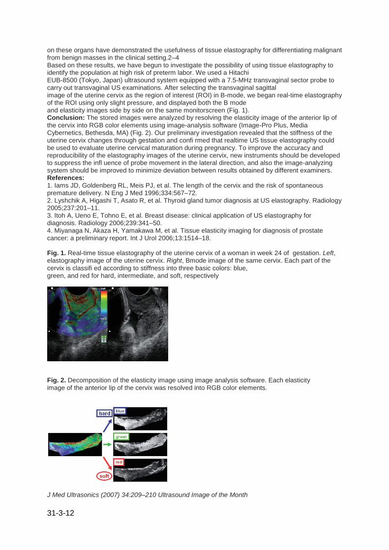
31-3-12
on these organs have demonstrated the usefulness of tissue elastography for differentiating malignant from benign masses in the clinical setting.2–4 Based on these results, we have begun to investigate the possibility of using tissue elastography to identify the population at high risk of preterm labor. We used a Hitachi EUB-8500 (Tokyo, Japan) ultrasound system equipped with a 7.5-MHz transvaginal sector probe to carry out transvaginal US examinations. After selecting the transvaginal sagittal image of the uterine cervix as the region of interest (ROI) in B-mode, we began real-time elastography of the ROI using only slight pressure, and displayed both the B mode and elasticity images side by side on the same monitorscreen (Fig. 1). Conclusion: The stored images were analyzed by resolving the elasticity image of the anterior lip of the cervix into RGB color elements using image-analysis software (Image-Pro Plus, Media Cybernetics, Bethesda, MA) (Fig. 2). Our preliminary investigation revealed that the stiffness of the uterine cervix changes through gestation and confi rmed that realtime US tissue elastography could be used to evaluate uterine cervical maturation during pregnancy. To improve the accuracy and reproducibility of the elastography images of the uterine cervix, new instruments should be developed to suppress the infl uence of probe movement in the lateral direction, and also the image-analyzing system should be improved to minimize deviation between results obtained by different examiners. References: 1. Iams JD, Goldenberg RL, Meis PJ, et al. The length of the cervix and the risk of spontaneous premature delivery. N Eng J Med 1996;334:567–72. 2. Lyshchik A, Higashi T, Asato R, et al. Thyroid gland tumor diagnosis at US elastography. Radiology 2005;237:201–11. 3. Itoh A, Ueno E, Tohno E, et al. Breast disease: clinical application of US elastography for diagnosis. Radiology 2006;239:341–50. 4. Miyanaga N, Akaza H, Yamakawa M, et al. Tissue elasticity imaging for diagnosis of prostate cancer: a preliminary report. Int J Urol 2006;13:1514–18. Fig. 1. Real-time tissue elastography of the uterine cervix of a woman in week 24 of gestation. Left, elastography image of the uterine cervix. Right, Bmode image of the same cervix. Each part of the cervix is classifi ed according to stiffness into three basic colors: blue, green, and red for hard, intermediate, and soft, respectively
Fig. 2. Decomposition of the elasticity image using image analysis software. Each elasticity image of the anterior lip of the cervix was resolved into RGB color elements.
J Med Ultrasonics (2007) 34:209–210 Ultrasound Image of the Month
31-3-12
ASSESSMENT OF BREAST TUMOR WITH US ELASTOGRAPHY: A NEW DECISION INDEX IN BREAST CANCER SCREENING Dr Youji Tsuchi, Shimane Medical University Purpose: We evaluate US elastography findings of benign and malignant breast tumors and exhibit the boon that introduction of US elastography provides for breast cancer screening. Method and Materials: Subjects of this study were 2529 females who had breast cancer screening in our institution from October, 2005 to March, 2007. All subjects received both palpation and mammography, and ultrasonography was added to subjects having breast mass lesion. Conventional US and US elastography was performed with digital US scanner (EUB-8500; Hitachi Medical, Tokyo, Japan) and 7.5MHz liner electric probe (EUP-L54M; Hitachi Medical) with a stabilizer was adapted to obtain the adequate elasticity images. Results: B-mode US to 212 breast tumors revealed necessity of biopsy in 42 (19.8%) subjects and malignant lesion was confirmed pathologically in 13 (6%) subjects. In US elastography findings of 13 malignant tumors, the intratumoral elastic decay area occupied more than 50% of all tumors area in all cases. However, in 38% (11/29) of benign tumors, the intratumoral elastic decay area was less than 50% of all tumor area. When this US elastography criteria is introduced into our breast cancer screening, 38% of the subjects with benign breast tumor may be relieved from unnecessarily invasive biopsy. Conclusion: US elastography becomes a new index of breast tumor diagnosis and provides a boon for the handling of the benign breast tumor patient in breast cancer screening. Clinical relevance/application: US elastography becomes a new index of breast tumor diagnosis and introduction of US elastography to breast cancer screening provides a boon for benign breast tumor patients. Radiological Society of North America 93
rd Scientific Assembly and Annual Meeting November 25
th –
30th, 2007, Chicago, USA
________________________
REAL-TIME BREAST SONOELASTOGRAPHY: RESULTS OF THE ITALIAN MULTICENTER STUDY (874 CASES) Presenter: Giorgio Rizzatto, Gorizia, Italy Purpose: To determine the clinical value of real-time sonoelastography (RTSE) in the differential diagnosis of breast lesions in the daily clinical practice.
Method and materials: At 8 institutions high-resolution ultrasound (US) and RTSE were performed with the same technology (Hitachi Medical, Japan) in 784 women (mean age, 52.5 years) who had 874 lesions with a definitive diagnosis (614 benign, 260 malignant). The size was =10 mm in 59% and =5 mm in 13,2% of the lesions. US images were classified according the BI-RADS criteria for US; RTSE images were assigned an elastographic score (1 to 5) according to the distribution and degree of strain induced by light compression. Scores were related both to solid and cystic lesions. Statistical analysis was performed by an independent institution.
Results: Considering the receiver operating curves the overall diagnostic performance of US was slightly better than RTSE (area under the curve 0.94 for BI-RADS and 0.90 for RTSE respectively). But RTSE showed a very high specificity in benign lesions, including BI-RADS 3 lesions (329 lesions, 37,6%). Almost all the cystic lesions, including those with slightly irregular margins and echogenic pattern on B-mode, showed a three-layered pattern (RTSE score 1). With the best cut off point between elasticity scores 3 and 4 the negative predictive value was 98% for the whole set, 96.3% for all the BI-RADS 3 lesions, and 100% for those with a size =5 mm. RTSE scores were insensitive to the thickness and the echogenicity of the breast, and to the depth and the size of the lesion. K indexes of intraobserver (0,93) and interobserver (0,90) agreement were very good.
Conclusion: RTSE scores are accurate and reproducible. Diagnostic scores are acquired in almost all patients in a few minutes and after a short learning curve. They help conventional US in characterizing small breast lesions. If incorporated in the diagnostic flow chart RTSE scores may avoid the use of biopsy in BI-RADS 3 for US and may postpone to 1 year the follow-up schedule.

31-3-12
Radiological Society of North America 93rd
Scientific Assembly and Annual Meeting November 25th –
30th, 2007, Chicago, USA
________________________
NEW QUANTITATIVE METHOD IN BREAST ELASTOGRAPHY: FAT LESION RATIO (FLR) Prof E. Ueno, Tsukuba University Purpose: In 2003, we completed the development of elastography which allows diagnosis by physicians even which little experience in ultrasound, and this technique has been commercialized and marketed since 2004. The performance of diagnosis with elastography is superior or equivalent to the conventional ―B-mode diagnosis + color Doppler‖ modality, with high reliability. It has the additional advantage of being simple and convenient. However one of the issues was the lack of the objectivity in diagnosis based on strain imaging. In the next step we developed a new quantitative method: Fat-Lesion Ratio (FLR) which provides greater objectivity in assessing elastography (strain image). Method and materials: FLR is defined as the ratio value obtained by dividing the mean strain at fat by the mean strain at hypo-echoic lesion. The strain for subcutaneous fat was determined from a circle bounded by the skin and mammary glands, and the strain for the lesion was determined from a circle bounded by the inner margin of the hypo-echoic area. Subjects were 408 cases with hypo-echoic lesions not over 2cm in diameter who have been examined elastography from January 25 2005. Among these subjects were 135 cases of breast cancer (mean age 54.6 ± 11.1, lesion diameter 12.1 ± 4.4 mm) and 271 cases of benign disease (mean age 45.9 ± 10.6, lesion diameter 9.6 ± 4.1). Results: The mean value for FLR in breast cancer (14.8) was considerably higher than the mean value for FLR in benign disease (4.47). Using a cut-off point of 4.8, we obtained sensitivity of 6.6%, specificity of 76.8%, and accuracy of 76.7%. Area under the ROC curve was high 0.818, while those of B-mode are 85.9%, 84.1% and 84.7% respectively. Although the accuracy of FLR is lower than that of B-mode, it is significant that FLR alone provided highly reliable diagnostic accuracy. Conclusion: It is concluded that FLR showed sufficient clinical performance with high diagnostic objectivity not depended on the experience of examiners. Clinical relevance/application: Just with a short training for FLR method, non-specialized examiners can acquire the equivalent degree of competence for diagnosis as specialists in ultrasonic diagnosis for breast cancer. Radiological Society of North America 93
rd Scientific Assembly and Annual Meeting November 25
th –
30th, 2007, Chicago, USA
________________________
ULTRASOUND (US) ELASTICITY IMAGES AND COMPUTER-AIDED ANALYSIS FOR CLASSIFICATION OF NONPALPABLE BENIGN AND MALIGNANT BREAST MASSES Ji Won Choi Co-author: Dr Moon, Soeul University, Korea Purpose: To retrospectively evaluate the performance of US elasticity images and computer-aided analysis for classification of biopsy-proved nonpalpable benign and malignant breast tumors Method and materials: Real-time US elastography was performed in 140 women (mean age,47 yrs;range,24–67 yrs) scheduled for US-guided core biopsy(101 benign,39 malignant tumors). Representative gray-scale and elasticity images of transverse and longitudinal scans were saved as bitmap files. After subtraction of gray-scale images from elasticity images,a region of interest drawn around the margin of mass on gray-scale image was loaded on subtracted color-scale images. The score of each pixel was assigned as from 0 for greatest strain(red) to 255 for no strain(blue). Average, skewness, kurtosis, difference histogram variation(DHV), edge density(ED), and run length were calculated. A neural network was used to classify tumors using these six features. Two breast radiologists provided elasticity score(1-5) by consensus without histologic information. The performance of neural network and radiologists were compared by ROC curve analysis. Results: The mean values of six elasticity features were different from malignant and benign masses as follows: 235±18 vs 194±38 in average, 264±6 vs 96 ±5 in skewness, 8861±6162 vs 3924±4381 in
31-3-12
kurtosis, 7157±4747 vs 109707±64920 in DHV, 1018±9 vs 1004±40 in ED, and 661±133 vs 734±77 in run length(P<.01 in all six features). The sensitivity, specificity, and PPV and NPV were 91%,74%,58%,and 95% for neural network based on all six elasticity features and 97%,40%,38%,and 97% at cutoff score between 2 and 3 and 54%,91%,70%,and 84% at cutoff score between 3 and 4 for radiologists. The Az value was 0.89 for neural network and 0.81 for radiologists and the difference was significant(P<.02). Conclusion: Computer-aided analysis of US elasticity images showed better performance than radiologists for classification of nonpalpable benign and malignant breast tumors. Clinical relevance/application: Computer-aided analysis of US elasticity images can be used an objective method to evaluate tissue strain and can aid in classification of nonpalpable benign and malignant breast tumors. Radiological Society of North America 93
rd Scientific Assembly and Annual Meeting November 25
th –
30th, 2007, Chicago, USA
________________________
ULTRASOUND (US) ELASTOGRAPHY OF BREAST LESIONS ASSOCIATED WITH SUSPICIOUS MICROCALCIFICATIONS DETECTED AT MAMMOGRAPHY Nariyo Cho Co-authors: Dr Moon, Soeul University, Korea Purpose: To evaluate the difference in strain between US lesions associated with malignant and benign microcalcifications detected at screening mammography by using real-time free-hand US elastography Method and materials: Between June 2006 and February 2007, 70 consecutive women who were scheduled to undergo US-guided vacuum assisted biopsy due to suspicious microcalcification cluster (median 1.6cm, range 1.0-4.0 cm) detected at screening mammography were examined with a commercialized US elastography. Lesions with mass on US (n=9), invasive cancer (n=6), atypical ductal hyperplasia (n=3), and negative calcification on specimen radiograph (n=2) were excluded. A total of 50 lesions (25 DCIS lesions and 25 fibrocystic changes) in 50 women (median, 46 years; range, 24–67 years) were included in this study. Real-time imaging files were saved as video clips in avi format. Two experienced radiologists who did not perform the examinations analyzed randomly ordered video clips without knowledge of the histology or mammographic findings and provided the elasticity score (1-5) in consensus according to the degree of strain induced by light compression. The sensitivity, specificity, positive predictive value (PPV), and negative predictive value (NPV) were used to assess the diagnostic performance. Results: For the elasticity score, the mean ± standard deviation was 3.0 ± 1.1 for malignant and 1.3 ± 0.5 for benign lesions (P < .001). When a cutoff point between 1 and 2 was used, elastography had 96% (24/25) sensitivity, 72% (18/25) specificity, 77% (24/31) PPV, and 95% (18/19) NPV. When a cutoff point between 2 and 3 was used, elastography had 64% (16/25) sensitivity, 100% (25/25) specificity, 100% (16/16) PPV, and 74% (25/34) NPV. Conclusion: Breast lesions associated with malignant microcalcifications tend to have less strain (harder) than benign microcalcifications at US elastography. Clinical relevance/application: US elastography has potential to improve the accuracy of gray-scale US for the detection and differentiation of breast lesions associated with suspicious microcalcifications detected at mammography. Radiological Society of North America 93
rd Scientific Assembly and Annual Meeting November 25
th –
30th, 2007, Chicago, USA
________________________

31-3-12
INTER AND INTRAOBSERVER AGREEMENT IN THE INTERPRETATION OF ULTRASOUND (US) ELASTOGRAPHY OF BREAST LESIONS S. Park Co-authors: Dr Moon, Soeul University, Korea Purpose: To evaluate inter- and intraobserver agreement of breast radiologists for the visual assessments of strain images obtained with real-time free-hand US elastography Method and materials: During the last 5 months, 130 consecutive women (mean, 55 years; range, 27-78 years) who had been scheduled to undergo US-guided core biopsy were examined with a commercialized US elastography. BIRADS category based on gray-scale US was 3 in 15% (20/130), 4 in 62% (80/130), and 5 in 23% (30/130). Representative real-time gray-scale and elasticity image files were saved as video clips for a total of 130 lesions (80 benign, 50 malignant, mean size 1.2cm, range 0.4-3.7cm). Three experienced radiologists independently analyzed the video clips without knowledge of the histology and provided the elasticity score by 5-point scale according to the degree and distribution of strain induced by light compression. The analysis was done two times with 1 month interval. Results were evaluated by using weighted κ statistics and receiver operating characteristic (ROC) curve analysis. Results: Three readers showed moderate to substantial interobserver agreement (mean κ (mκ), 0.644; range, 0.580-0.687) and substantial to almost perfect intraobserver agreement (mk, 0.821; range, 0.742-0.918). Interobserver agreement of malignant lesions (mκ, 0.520; range, 0.480-0.583) was lower than that of benign lesions (mκ, 0.606; range, 0.520-0.668). There was no significant difference in inter- and intraobserver agreements according to lesion size. Mean area under the ROC curve was 0.817 (0.770-0.845, 95% confidence interval). The sensitivity, specificity, positive predictive value and negative predictive values at cutoff score between 3 and 4 were 50.3%, 92.3%, 80.0%, and 75.0%, and those at 2 and 3 were 81.0%, 62.9%, 58.4%, and 85.8%, respectively. Conclusion: Interobserver agreement of breast radiologists was moderate to substantial and intraobserver agreement was substantial to perfect for the visual assessments of strain images obtained with real-time free-hand US elastography. Clinical relevance/application: Relatively low interobserver agreement of visual assessments for US-elastography enhances need for more standardized assessment method.
Radiological Society of North America 93
rd Scientific Assembly and Annual Meeting November 25
th –
30th, 2007, Chicago, USA
________________________
AVOIDING POTENTIAL SOURCES OF ERROR IN BREAST ULTRASOUND ELASTOGRAPHY Ciurea A, Dumitriu D, Chiorean A, Ciortea C, Botar-Jid C, Dudea S Romania Objectives: The main purpose of this study was to establish several factors which influence the quality of an elastographic examination in breast pathology. Methods: In our ongoing study, which began in April 2007, we have examined 40 patients with focal breast lesions. All patients were examined using both 2D and Doppler ultrasound and elastography. The reference standard was the pathologic diagnosis. We tried to determine the importance of certain technical factors, such as: the type of section, the size of the region of interest, the degree of compression. We also studied in which way the characteristics of the lesion influence the elastogram. Results: The elasticity score for the same lesion was sometimes different on sagittal and coronal sections – therefore it is important to examine the lesion using both types of sections and, probably, to take the highest elasticity score into account. If the region of interest was limited to the lesion, the elasticity score was not accurate, but if it included as much healthy tissue as possible, the score correlated better with the pathologic diagnosis. An inappropriate degree of compression also influenced the result of the examination. The elasticity score was more accurate for small masses than for large lesions, which occupied the whole field, and also for superficial lesions compared to deeper-situated ones. Conclusions: Elastography is a useful tool in the non-invasive diagnosis of breast lesions, but it needs specific training, as well as acknowledging technical and pathological factors which may influence its sensitivity and specificity.

31-3-12
XIXth Congress of European Federation of Societies for Ultrasound in Medicine and Biology &
Dreiländertreffen, 24th – 27
th October 2007, Leipzig, Germany
________________________
THE VALUE OF THE SONOELASTOGRAPHY IN BREAST DIAGNOSIS: A PROSPECTIVE COMPARISON BETWEEN FAT/LESION RATIO AND ELASTOGRAPHY SCORING.
AA Geaid, S Grunwald, M Zygmunt, R Ohlinger
Ernst-Moritz-Arndt-Universität Greifswald, Klinik und Poliklinik für Frauenheilkunde und Geburtshilfe, Greifswald, Germany
(Translated from German)
Objectives: Focal pathological changes of the breast can change the elasticity of the tissue. SonoElastography is a modality that displays in real-time the tissue elasticity as a colour overlay of the B-image. The so-called Elastography Score, ―ESC‖, acquired in colour has been compared with the new SonoElastography measurement, Fat/Lesion Ratio (FLR), in regard to sensitivity and specificity.
Material and methods: Since April 2007 all patients attending for breast consultation that had at least one finding have been examined. The examination was performed with the Hitachi EUB-8500 HV. The Elastography Score (ESC 1-5) was determined subjectively by the physician in a similar way to the ultrasound BI-RADS-Criteria, whereas the Fat/Lesion-Ratio (FLR) has been calculated by the ultrasound machine from the strain values and displayed as a measurement. The FLR was calculated by comparing the mean strain value of the fat tissues to the mean strain value in the lesion. The examinations were performed either before or after a Highspeed core biopsy (n=30/34, 88%) or with fine needle aspiration (n=4/34, 12%). The histolopathological results obtained were compared with both the ESC and FLR.
Results: Altogether 34 patients between 20 and 73 years old have been examined (average 52 years). 16 out of 34 breast lesions were histologically confirmed as benign and 18 as malignant. With a Cut-off value of 2.17, the FLR had a sensitivity of 77. 8% and a specificity of 75%. Higher values were obtained for malignant lesions, and lower values for benign lesions. However the ESC showed a significantly lower sensitivity of 38, 9% and almost the same specificity of 77, 8%.
Conclusion: These preliminary results showed a greater advantage for the Fat/Lesion-Ratio compared with the Elastography Score for the diagnosis of breast lesions. Further patient studies need to be performed.
XIXth Congress of European Federation of Societies for Ultrasound in Medicine and Biology & Dreiländertreffen, 24
th – 27
th October 2007, Leipzig, Germany
________________________
COMPARISON OF ULTRASOUND ELASTOGRAPHY, MAMMOGRAPHY, AND SONOGRAPHY IN THE DIAGNOSIS OF SOLID BREAST LESIONS Hui Zhi, MD, Bing Ou, MD, Bao-Ming Luo, MD,Xia Feng, MD, Yan-Ling Wen, MD, Hai-Yun Yang, MD Objective. The purpose of this study was to evaluate the value of ultrasound elastography (UE) in differentiating benign versus malignant lesions in the breast and compare it with conventional sonography and mammography. Methods. From September 2004 to May 2005, 296 solid lesions from 232 consecutive patients were diagnosed as benign or malignant by mammography and sonography and further analyzed with UE.

31-3-12
The diagnostic results were compared with histopathologic findings. The sensitivity, specificity, accuracy, positive and negative predictive values, and false-positive and -negative rates were calculated for each modality and the combination of UE and sonography. Results. Of 296 lesions, 87 were histologically malignant, and 209 were benign. Ultrasound elastography was the most specific (95.7%) and had the lowest false-positive rate (4.3%) of the 3 modalities. The accuracy (88.2%) and positive predictive value (87.1%) of UE were higher than those of sonography (72.6% and 52.5%, respectively). The sensitivity values, negative predictive values, and false negative rates of the 3 modalities had no differences. A combination of UE and sonography had the best sensitivity (89.7%) and accuracy (93.9%) and the lowest false-negative rate (9.2%). The specificity (95.7%) and positive predictive value (89.7%) of the combination were better, and the false-positive rate (4.3%) of the combination was lower than those of mammography and sonography. Conclusions. In a clinical trial with Chinese women, UE was superior to sonography and equal or superior to mammography in differentiating benign and malignant lesions in the breast. A combination of UE and sonography had the best results in detecting cancer and potentially could reduce unnecessary biopsy. Ultrasound elastography is a promising technique for evaluating breast lesions. Key words: breast carcinoma; mammography; sonography; ultrasound elastography. J Ultrasound Med 2007; 26:807–815
________________________
REAL-TIME SONOELASTOGRAPHY OF THE CERVIX: TISSUE ELASTICITY OF THE NORMAL AND ABNORMAL CERVIX Anke Thomas, MD, Sherko Kümmel, MD, Ole Gemeinhardt, Thomas Fischer, MD Rationale and Objectives. First study to investigate the basic tissue elastic properties of the cervix in pre- and postmenopausal healthy women and to compare these normal findings with the results in a group of patients with focal pathology of the cervix. Materials and Methods. A total of 113 patients underwent transvaginal ultrasound, among them 24 with cervical pathology. The real-time elastography (Hitachi) information was color-coded and superimposed on the B-mode scan. The elastography images were analyzed by means of a software tool to identify thresholds for the colors red (soft), blue (hard), and green (medium hard), and the percentages of the three colors of the total area were determined. The results were correlated with age. In addition, scans were evaluated subjectively on an analogue scale from 1 (definitely normal) to 5 (definitely abnormal). Statistical analysis was performed using Anova, Wilcoxon‘s test, and Pearson‘s correlation. Results. Computer-assisted generation of the color spectrum showed green to be predominant in both the normal group (67_13 %) and in the group with cervical pathology (64_15 %) without a significant difference between both groups (p__0.05). Significant differences (p_0.05) in the blue color spectrum (hard tissue) were found between the 13 cervical tumor patients (34_15 %) and the normal group (26_13 %) but not between the CIN patients and normal women (19_12 %) (p_0.05). Subjective tumor characterization also showed significant differences (p_0.05) among the groups and good correlation with the histologic diagnosis (r2_0.744). There were no significant changes in color distribution with patient age (p_0.05). Conclusion. Computer-assisted and subjective evaluation of cervical elastography allows differentiation of malignancy from normal findings. CIN cannot be identified with this modality. Elastographically, cervical tissue is of medium hardness and does not change with age. Acad Radiol 2007; 14:193–200
________________________
31-3-12
ULTRASOUND AND BREAST DISEASE: US-ADVANCED TECHNICAL ASPECTS AND FUTURE TRENDS G. Rizzatto; Gorizia/IT Major breakthrough to help radiologists in breast US is mostly linked to the increasing computing capability of new systems. CAD applications use sophisticated image analysis scheme and advanced decision engine to help differentiate benign and malignant lesions. They may integrate a variety of features such as automated segmentation, characterization, classification, annotation and report generation. Elastographic score is a new feature, easy and quick to be integrated with all other US and imaging signs. Adequate patterns are obtained in over 96% of all patients and k index shows a very high reproducibility. Significant increase in diagnostic accuracy is obtained mainly in US BI-RADS™ 3. Real-time sonoelastography significantly reduces the biopsy rate in small cystic lesions and may suggest appropriate interventions for malignancies with less typical basic features. Perfusion ultrasound with continuous low mechanical index and contrast agents may add useful information in: 1. Advanced cancers treated preoperatively and receptor-positive cancers in elderly patients. Perfusion shows changing in vascular clusters and kinetics that are predictive for outcome parameters and are detectable earlier than volume reduction. 2. Patients with metastatic nodes. Tumor foci mostly result in a lack of perfusion; smallest detected lesions are 3 mm. Sentinel node procedure may be saved in 25% of patients. Positive cases are straight scheduled for axillary dissection; the use of operating room is maximized. Multi-modality fusion breast imaging systems are developed to co-register different modalities to both increase the sensitivity to breast cancer and to facilitate all the procedures performed under US guidance. Learning Objectives:
1. To discuss the new potentials of computerised analysis. 2. To evaluate the clinical impact of new methods (contrast enhancers, elastography, etc). 3. To describe new fusion imaging methods.
European Congress of Radiology, March 9
th – 12
th 2007, Vienna, Austria
________________________
CHARACTERIZATION OF BREAST LESIONS WITH REAL-TIME SONOELASTOGRAPHY: RESULTS FROM THE ITALIAN MULTICENTER CLINICAL TRIAL M. Locatelli
1, G. Rizzatto
1, L. Aiani
2, A. Martegani
2, S. Baldassarre
3, G.M. Giuseppetti
3, A. Bulzacchi
4,
C. Di Maggio4, S.W. Della Sala
5, G. Mangialavori
6, P. Monno
6, E. Lattanzio
6, G. Scaperrotta
7, I.
Floriani7;
1Gorizia/IT,
2Como/IT,
3Ancona/IT,
4Padova/IT,
5Trento/IT,
6Bari/IT,
7Milan/IT
Purpose: To determine the clinical value of real-time sonoelastography (RTSE) in the differential diagnosis of breast lesions. Methods and Materials: At 8 institutions, high-resolution ultrasound (US) and RTSE were performed in 784 women (mean age, 52.5 years) who had 874 lesions with a definitive diagnosis (614 benign, 260 malignant). The size was ≤10 mm in 59% and ≤5 mm in 13.2% of the lesions. US images were classified according the BI-RADS criteria for US; RTSE images were assigned an elastographic score (1 to 5) according to the distribution and degree of strain induced by light compression. Statistical analysis was performed by an independent institution. Results: Considering the receiver operating curves, the overall diagnostic performance of US was slightly better than RTSE (area under the curve 0.94 for BI-RADS and 0.90 for RTSE, respectively). But RTSE showed a very high specificity in benign lesions, including BI-RADS 3 lesions (329 lesions, 37.6%). With the best cutoff point between elasticity scores 3 and 4, the negative predictive value was 98% for the whole series, 96.3% for all the BI-RADS 3 lesions, and 100% for those with a size ≤5 mm. RTSE scores were insensitive to the thickness and the echogenicity of the breast, and to the depth and the size of the lesion. K indexes showed a very high reproducibility. Conclusion: RTSE scores are accurate and reproducible. If incorporated in the diagnostic flow chart, they might avoid using biopsy in BI-RADS 3 for US and postpone to 1 year the follow-up schedule. European Congress of Radiology, March 9
th – 12
th 2007, Vienna, Austria

31-3-12
PERFORMED USING REAL-TIME SONOELASTOGRAPHY: A BETTER DIFFERENTIATION IN BREAST LESIONS? A. Thomas, T. Fischer; Berlin/DE Purpose: The goal of the present study was to compare the sensitivity and specificity of elastography with that of B-mode ultrasound (US) and mammography. Methods and Materials: A total of 300 patients with histologically confirmed breast lesions (168 benign, 132 malignant) were included. Evaluation was by means of the 3D finite-element method. The data is color-coded and superimposed on the B-mode US scan. The images were evaluated by2 independent readers. The results were compared with mammography, histology and the data obtained by previous ultrasound investigations. Sensitivities and specificities were calculated. Results: Sensitivity and specificity in the differentiation of benign and malignant lesions were 87% and 85%, respectively, for mammography, and 94% and 83% for B-mode ultrasound (94% / 83%). The 2 examiners were in very good agreement in their evaluation of the elastograms (kappa: 0.86). Elastography had a sensitivity of 82% and a specificity of 87%. Elastography was superior to B-mode US in diagnosing BI-RADS 3 lesions (92% vs. 82% specificity) and in lipomatous involution (80% vs. 69% specificity). Conclusion: Elastography in breast lesions showed a higher specificity and a lower sensitivity in comparison with B-mode sonography. Elastography may be beneficial in BI-RADS 3 lesions and in lipomatous involution. European Congress of Radiology, March 9
th – 12
th 2007, Vienna, Austria
________________________ ROLE OF ULTRASOUND ELASTOGRAPHY IN THE DIFFERENTIAL DIAGNOSIS OF BREAST LESIONS A. Ozdemir, S. Gultekin, I. Tuncbilek, T. Ugur, R. Erman, C. Yuce; Ankara/TR Purpose: To assess the adjunctive value of ultrasound elastography in the differential diagnosis of breast lesions. Methods and Materials: 401 breast lesions, 20 cystic and 381 solid (mean size: 14.9 mm), were included in the study. Following mammography and ultrasonography, the lesions were diagnosed as BI-RADS 2, 3, 4, 5, and 6 (n: 158, 81, 70, 79, 13). Elastography was performed with Hitachi EUB 8500 ultrasound equipment. Images were evaluated by 2 independent observers, with knowledge of final BI-RADS diagnostic categories but blind to the pathological diagnosis. An elastic score was assigned in solid lesions by referring a 5-point color scale. Elastic scores from 1 to 3 were accepted as benign, while scores 4 and 5 were malignant. In solid lesions, elastography and final BI-RADS diagnoses were compared to the final diagnosis. Results: Typical 3-layered appearance was observed in all cysts. Mean elastic score was 4.25 ± 1.12 in malignant (n: 94), and 2.32 ± 0.88 in benign solid lesions (n: 287) (p<0.05). Inter- and intra-observer agreement were 89% and 91%, respectively. Sensitivity, specificity, ppv and npv were 100%, 77%, 55% and 100%, respectively, for the final BI-RADS diagnostic assessment, and 82%, 91%, 76%, and 93%, respectively, for the elastography assessment. Sensitivity of elastography decreased substantially in lesions larger than 3 cm, compared to those smaller than 1 cm (42% vs 93%). Conclusion: US elastography is a valuable adjunct to standard breast imaging modalities, by increasing the final specificity and ppv significantly. Its impact is significantly more evident in lesions smaller than 1 cm. European Congress of Radiology, March 9
th – 12
th 2007, Vienna, Austria
________________________

31-3-12
NEW ULTRASOUND TECHNIQUES FOR THE DETECTION AND CHARACTERISATION OF FOCAL BREAST LESIONS T. Fischer; Berlin/DE In recent years, advanced techniques have revolutionized breast ultrasound (US). The new possibilities comprise techniques for optimizing image quality such as differential tissue harmonic imaging (THI) and frequency compounding (FC), tools for post-processing US raw data such as strain imaging (elastography), and the use of US contrast agents. The presentation will focus on characteristic features of malignant breast lesions using these state-of-the-art US techniques. The participants will learn to use new US techniques for 1. Classification of focal lesions according to BI-RADS; presentation of examples to work out the benefits of differential THI, 3D US, and elastography. 2. Lymph node staging and monitoring of neoadjuvant chemotherapy of breast cancer with US contrast agents. 3. Puncture and postoperative quality control using 3D and 4D US techniques. 4. Detection of focal liver lesions and monitoring of ablation therapy for liver metastases. 5. New software tools for US detection of microcalcifications. European Congress of Radiology, March 9
th – 12
th 2007, Vienna, Austria
________________________
REAL-TIME SONOELASTOGRAPHY OF THE CERVIX: IDENTIFY THE CHARACTERISTICS OF PHYSIOLOGICAL AND PATHOLOGICAL TISSUE ELASTICITY A. Thomas, Berlin/DE Purpose: A real-time sonoelastography in an initial clinical study was performed in a normal population to identify elastic tissue changes in pre- and post-menopausal healthy women and to compare these normal findings with the results in a group of patients with focal pathology of the cervix. Methods and Materials: 113 unselected women were examined, 89 healthy patients and 24 with focal pathology of the cervix. In the dual mode, the real-time elastography information was superimposed in color on the B-mode scan (Hitachi). The elastography scans were analyzed by means of a computer program and by 2 independent readers using defined regions of interest (ROIs) and an analogue scale from 1 (definitive normal) to 5 (definitive abnormal). These quotients were correlated with age (ANOVA, Wilcoxon‘s test). Results: The color distribution in the normal population showed that green was predominant, there was no significant difference in comparison to the patients with focal pathology (p>0.05) The TQ was not significant with the variation of age (p>0.05). The elastic changes in the blue-colored spectrum (harder tissue) were different in patients with cervical carcinoma as compared with the normal group (p<0.05). Analysis of the subjective data of the 2 readers indicated that there was a strong correlation between malignancy of the tumor and the ranking of the analogue scale (r
2=0.744).
Conclusion: The normal cervix was found to be ―softer‖ on elastography in comparison with patients with cancer of the cervix. The blue-colored spectrum of the elastogramm indicates cervical cancer. European Congress of Radiology, March 9
th – 12
th 2007, Vienna, Austria
________________________
REAL-TIME SONOELASTOGRAPHY PERFORMED IN ADDITION TO B-MODE ULTRASOUND AND MAMMOGRAPHY: IMPROVED DIFFERENTIATION OF BREAST LESIONS? Thomas A, Kümmel S, Fritzsche F, Warm M, Ebert B, Hamm B, Fischer T.
Department of Obstetrics and Gynecology, Chariteplatz, Charité - Universitätsmedizin, Berlin 10117, Germany. [email protected]
RATIONALE AND OBJECTIVES: The goal of the present study was to compare the sensitivity and specificity of elastography with that of B-mode ultrasound (US) and mammography.
31-3-12
MATERIALS AND METHODS: A total of 300 patients with histologically confirmed breast lesions (168 benign, 132 malignant) were included. Evaluation was by means of the three-dimensional finite-element method. The data are color-coded and superimposed on the B-mode US scan. The images were evaluated by two independent readers. The results were compared with mammography, histology, and the data obtained by previous US investigations. Sensitivities and specificities were calculated.
RESULTS: Sensitivity and specificity in the differentiation of benign and malignant lesions were 87% and 85%, respectively, for mammography and 94% and 83% for B-mode US. The two examiners were in very good agreement in their evaluation of the elastograms (kappa: 0.86). Elastography had a sensitivity of 82% and a specificity of 87%. Elastography was superior to B-mode US in diagnosing Breast Imaging Reporting and Data System (BI-RADS) 3 lesions (92% vs. 82% specificity) and in lipomatous involution (80% vs. 69% specificity).
CONCLUSION: Elastography in breast lesions showed a higher specificity and a lower sensitivity in comparison with B-mode sonography. Elastography may be beneficial in BI-RADS 3 lesions and in lipomatous involution.
Acad Radiol. 2006 Dec;13(12):1496-504
________________________
AN ADVANCED METHOD OF ULTRASOUND: REAL-TIME ELASTOGRAPHY—FIRST EXPERIENCE IN 300 PATIENTS WITH BREAST LESIONS Anke Thomas, Thomas Fischer, Berlin GERMANY PURPOSE The goal of the present study was to compare the sensitivity and specificity of elastography with that of B-mode ultrasound (US) and mammography. METHOD AND MATERIALS A total of 300 patients with histologically confirmed breast lesions (168 benign, 132 malignant) were included. Evaluation was by means of the 3D finite-element method (Hitachi). The data were color-coded and superimposed on the B-mode US scan. The images were evaluated by two independent readers. The results were compared with mammography, histology and the data obtained by previous ultrasound investigations. Sensitivities and specificities were calculated. RESULTS Sensitivity and specificity in the differentiation of benign and malignant lesions were 87% and 85%, respectively, for mammography and 94% and 83% for B-mode ultrasound. The two examiners were in very good agreement in their evaluation of the elastograms (kappa: 0.86). Elastography had a sensitivity of 82% and a specificity of 87%. Elastography was superior to B-mode US in diagnosing BI-RADS 3 lesions (92% vs. 82% specificity) and in lipomatous involution (80% vs. 69% specificity). CONCLUSION Elastography in breast lesions showed a higher specificity and a lower sensitivity in comparison with B-mode sonography. Elastography may be beneficial in BI-RADS 3 lesions and in lipomatous involution. CLINICAL RELEVANCE/APPLICATION The aim of the study presented here was to evaluate the new technique of real-time sonoelastography in terms of sensitivity and specificity as compared with mammography and B-mode US. In summary, real-time sonoelastography is a promising new approach to differentiate benign and malignant focal lesions of the breast. There is very good interobserver agreement in evaluating elastograms, which makes the method amenable to standardised interpretation. Elastography was found to be superior to B-mode US in evaluating BI-RADS 3 benign lesions and in the presence of lipomatous involution, where elastography is comparable to mammography in terms of sensitivity and specificity. Radiological Society of North America 92
nd Scientific Assembly and Annual Meeting November 26
th –
December 1st, 2006, Chicago, USA

31-3-12
CHARACTERIZATION OF BREAST LESIONS WITH REAL-TIME SONOELASTOGRAPHY: RESULTS FROM THE ITALIAN MULTICENTER CLINICAL TRIAL Rizzatto G, Aiani L, Baldassarre S, Butzacchi A, Della Sala S, Locatelli M, et al, ITALY PURPOSE To determine the clinical value of real-time sonoelastography (RTSE) in the differential diagnosis of breast lesions. METHOD AND MATERIALS At 8 institutions, high-resolution ultrasound (US) and RTSE were performed in 784 women (mean age, 52.5 years) who had 874 lesions with a definitive diagnosis (614 benign, 260 malignant). The size was ≤10 mm in 59% and ≤5 mm in 13,2% of the lesions. US images were classified according the BI-RADS criteria for US; RTSE images were assigned an elastographic score (1 to 5) according to the distribution and degree of strain induced by light compression. Statistical analysis was performed by an independent institution. RESULTS Considering the receiver operating curves the overall diagnostic performance of US was slightly better than RTSE (area under the curve 0.94 for BI-RADS and 0.90 for RTSE respectively). But RTSE showed a very high specificity in benign lesions, including BI-RADS 3 lesions (329 lesions, 37,6%). With the best cutoff point between elasticity scores 3 and 4 the negative predictive value was 98% for the whole set, 96.3% for all the BI-RADS 3 lesions, and 100% for those with a size ≤5 mm. RTSE scores were insensitive to the thickness and the echogenicity of the breast, and to the depth and the size of the lesion. K indexes of intraobserver (0,93) and interobserver (0,90) agreement were very good. CONCLUSION RTSE scores are accurate and reproducible. They help conventional US in characterizing small breast lesions. CLINICAL RELEVANCE/APPLICATION If incorporated in the diagnostic flow chart RTSE scores might avoid using biopsy in BI-RADS 3 for US and postpone to 1 year the follow-up schedule.
Radiological Society of North America 92
nd Scientific Assembly and Annual Meeting November 26
th –
December 1st, 2006, Chicago, USA
________________________
ULTRASOUND ELASTOGRAPHY: RESULTS OF A PROSPECTIVE MULTICENTRE STUDY OF 408 BREAST NODULES A Athanasiou, A tardivon, B Barreau, F Thibault, El Khoury, A Delignette, V Boussion, B Baratie, L Levy, P David, A Le Mouel, C Balu-Maestro (Translated from French) Objectives: To evaluate ultrasound elastography for characterization of breast lesions and to measure its reproducibility. Materials and Method: 408 lesions (369 patients, 59% < 10mm, 65.4% benign) were evaluated using Ultrasound elastography (Hitachi, Ueno classification, scores 1-3 = benign, scores 4-5 malignant). The diagnosis was obtained by FNA, core or surgical biopsies for 326 lesions (79.9%), or follow-up or comparison with previous US studies. Results: The sensitivity, specificity, PPV, NPV, and accuracy was 78%, 92.9%, 85.3%, 88.9%, and 87.7%, respectively. There were 19 false positives (fibrous lesions) and 31 false negatives (58% ACR Bi-Rads category 5, cancers in situ, poorly differentiated IDC and ILC). The sensitivity was greatest for lesions < 10mm (95.8%). For the new cases included (63 lesions), 32 lesions were classified as category 3 or weak 4; elastography confirmed the benign nature in 30 cases (94%, I false positive, 1 false negative). In the first 30 cases, the intra-observer agreement was 100% and inter-observer agreement 90% (2 readers). Conclusion: Ultrasound elastography is a useful diagnostic tool that can reduce biopsies in benign lesions and has shown to be reproducible.
31-3-12
Journées Francaises de Radiologie, October 21st – 25
th, 2006, Paris, France
________________________
ELASTOGRAPHY STRETCHES HORIZONS OF BREAST ULTRASOUND Diagnostic Imaging Europe, October 2006
Initial results convince Europeans experimental technique may reduce benign breast biopsy
rates Researchers at ECR 2006 hailed the potential of ultrasound elastography to dramatically reduce benign breast biopsy rates. Elastography refers to the measurement of elastic properties of tissues, based on the well-established principle that malignant tissue is harder than benign tissue. The technique is typically performed with ultrasound, but research with MR is also under way. The procedure, also known as elasticity, was born in the early 1990s. The University of Texas Medical School in the U.S. holds multiple patents on the technique with ultrasound (see www.elastography.com for details). Images are acquired on high-end ultrasound devices equipped with additional software and hardware. Prototype commercial systems became available to researchers after 2000, and vendors have since introduced commercial products, accelerating interest. Using a research system from Siemens, which is not yet commercially available, U.K radiologists re-ported results from four years of routine elastography in breast screening. They concluded that the technique could help halve the rate of benign breast biopsies. "We can decrease the biopsy rate in benign lesions. This has great importance given the increased incidence of screening abnormalities picked up on breast ultrasound," said Dr. William Svensson, a consultant radiologist at Charing Cross Hospital in London. With elastography, raw ultrasound is obtained before and after a slight compression of tissue, typically achieved with an ultrasound transducer. Compression may also be performed using vibrations in a technique known as sonoelastography. Elastography measures and displays strain; that is, the change in the dimension of tissue elements at various locations in the region of interest. An estimate of tissue strain is obtained at each point in the field-of-view, and strain values are then displayed as an image. The strain is estimated from minute differences between these two images. Research has shown that normal tissue and fat have smaller elasticity profiles, while hard areas, such as cancers, are larger than the gray-scale appearance. "The hypothesis was that the size of cancer is larger in elasticity imaging than B-mode and with benign lesions the reverse is the case," said Svensson. The U.K study included women who were referred for routine breast ultrasound for focal breast abnormalities. In cases where abnormalities were identified on ultrasound, women underwent breast strain imaging. Of 260 lesions identified, 71 were malignant, nine were intermediate with malignant potential, and the majority, 180 lesions, were benign. U.K researchers noted that lesions with an elasticity to gray-scale ratio of less than .75 were benign. Based on this ratio, all of the malignant lesions would have been identified, aside from a few intermediate lesions. That result equates to a sensitivity of 96%. In the study, specificity reached just 53%, meaning that if the ratio is applied, half of the benign lesions might not need to be biopsied, Svensson said. In addition to the obvious clinical value, there are also economic advantages. "Benign biopsies cost us a tot of money," he said. "If we can reduce them, that would bring significant savings for the health service." In another study presented at the ECR, French researchers reported positive findings for ultrasound elastography in a multicenter prospective study of 345 breast lesions in 314 patients. Patients were imaged on the Hitachi EUB 8500 Logos ultrasound unit with a technique similar to the one used in the UK study. Detected lesions were categorized by size and BI-RADS category. Researchers used the color classification system developed by Dr. Ei Ueno to score elastography images.
For example, elastography images that are completely green (soft) are typically benign with a Ueno
score 1, while those that are completely blue (stiff) are thought to be malignant and have a Ueno
score 5.

31-3-12
For lesions of all sizes, ultrasound elastography achieved sensitivity of 80%, specificity of 93%, positive predictive value of 85.3%, and negative predictive value of 90.3%. Sensitivity was highest for lesions less than 5 mm (90%), while specificity was best for lesions over 10 mm (95%). For lesions in BI - RADS categories 3 and 4, sensitivity was 68% and specificity was 90%. Researchers also reported 16 false positives with elastography (such as fibrous mastopathy and scle-rosis adenosis) and 26 false-negative findings (such as DCIS). The technique is most useful for lesions in the indeterminate BI - RADS categories and less useful for lesions in BI-RADS category 5, as false negatives might occur in these lesions. In obvious, suspicious nodules on Bmode imaging, elastographic assessment is either of very little use or not needed, said Dr. Anne Tardivon of the Institut Curie in Paris. Elastography is easy to perform once practitioners are trained, Tardivon said. It provides good visualization and is not time-consuming. "Elastography does not replace standard B-mode imaging, but, clearly, it is a useful complementary tool and may reduce biopsies of benign lesions," she said. At this time, the modality is being used for breast nodules detected on standard ultrasound studies, she said. Its potential role in evaluating patients without an abnormality on B-mode imaging remains to be evaluated. .
-By Emily Hayes
________________________
REAL-TIME ELASTOGRAPHY - AN ADVANCED METHOD OF ULTRASOUND: FIRST RESULTS IN 108 PATIENTS WITH BREAST LESIONS
Thomas A, Fischer T, Frey H, Ohinger R, Grunwald S, Blohmer JU, Winzer KJ, Weber S, Kristiansen G, Ebert B, Kummel S Department of Obstetrics and Gynecology, Charite - Universitatsmedizin Berlin, Charite Campus Mitte, Berlin, Germany.
OBJECTIVES: To evaluate whether real-time elastography, a new, non-invasive method for the diagnosis of breast cancer, improves the differentiation and characterization of benign and malignant breast lesions.
METHODS: Real-time elastography was carried out in 108 potential breast tumor patients with cytologically or histologically confirmed focal breast lesions (59 benign, 49 malignant; median age, 53.9 years; range, 16-84 years). Tumor and healthy tissue were differentiated by measurement of elasticity based on the correlation between tissue properties and elasticity modulus. Evaluation was performed using the three-dimensional (3D) finite element method, in which the information is color-coded and superimposed on the B-mode ultrasound image. A second observer evaluated the elastography images, in order to improve the objectivity of the method. The results of B-mode scan and elastography were compared with those of histology and previous sonographic findings. Sensitivities and specificities were calculated, taking histology as the gold standard.
RESULTS: B-mode ultrasound had a sensitivity of 91.8% and a specificity of 78%, compared with sensitivities of 77.6% and 79.6% and specificities of 91.5% and 84.7%, respectively, for the two observers evaluating elastography. Agreement between B-mode ultrasound and elastography was good, yielding a weighted kappa of 0.67.
CONCLUSIONS: Our initial clinical results suggest that real-time elastography improves the specificity of breast lesion diagnosis and is a promising new approach for the diagnosis of breast cancer. Elastography provides additional information for differentiating malignant BI-RADS (breast imaging reporting and data system) category IV lesions.
Ultrasound Obstet Gynecol, 2006, Sep;28 (3): 335-340
________________________

31-3-12
PICTURE OF THE MONTH: IMAGING OF THE CERVIX USING SONOELASTOGRAPHY A. Thomas Department of Obstetrics and Gynaecology, Charite, University of Berlin, Berlin, Germany A reliable diagnostic tool for the early identification of cervical insufficiency would be highly desirable. Numerous studies have demonstrated that the length of the cervix decreases during the course of pregnancy, while its width increases. Moreover, it has been shown that the probability of premature delivery increases when the cervix is short during the second trimester and the internal os is wide (funneling)
1–5. Based on the assumption that premature cervical softening
and premature delivery constitute a multifactorial process, we hypothesized that there may be underlying gestational and/or maternal age-related changes in tissue elasticity. Tissue elasticity can be determined from stretching and compression parameters of a tissue of interest. While stretching values can be derived directly from high-frequency echo signals, compression values cannot be determined directly. This is why assessment of compression should be performed under standardized conditions. Advances have been achieved through the advent of real-time sonoelastography
6. This technique generates images in which the elasticity values are superimposed
in color on conventional B-mode images in real time. The technique is similar to color Doppler and does not lengthen the scan time. Initial results with this new technique for different organ systems are very promising
7–10.
Based on these results, we have begun to investigate whether elastography can be used to identify typical changes in cervical tissue elasticity that correlate with the week of gestation or the age of the pregnant woman. Using a high-end ultrasound device (Hitachi EUB-8500, Wiesbaden, Germany), measurements of tissue elasticity were performed using the same probe as that used for transvaginal B-mode imaging. A region of interest was selected and the elasticity information presented in color with blue indicating harder tissue and red deformable, soft tissue (Figure 1). Elastography measures echo frequency patterns along the ultrasound beam over time before and after compression of a tissue area. At the same time, the echo frequency waves of neighboring ultrasound waves can be compared in order to take lateral deviations around the tissue area into account. This involves use of the so-called extended combined autocorrelation method, which allows for more precise calculations after compression because it takes into account tissue displacement in all spatial directions and can be applied immediately. Three basic colors of the elasticity spectrum are present in the cervix (green, red and blue). The percentages of red and green can be used to calculate an elasticity tissue quotient (TQ) using the formula: TQ = % red/% green. Correlation of the TQ with age and duration of pregnancy (week of gestation) showed, in our preliminary investigations, that TQ did not vary with the duration of pregnancy but that it did with maternal age. Our experience suggests that sonoelastography of the pregnant cervix is easy to perform and that it shows age-related differences in cervical elasticity. Future research will determine whether sonoelastography has the potential to provide insight into cervical insufficiency and premature delivery.
Figure 1 (a) Cervical elastography in a woman at 28 weeks‘ gestation showing intermediate tissue
elasticity (green) surrounding the soft cervical canal (red-yellow). (b) B-mode image of the same cervix.

31-3-12
References 1. Arabin B, H¨ ubener M, van Eyck J. Zervixinsuffizienz – diagnostische, pr¨aventive und therapeutische Ans¨ atze. DerGyn¨akologe 2001; 34: 715–725. 2. Gomez R, Galasso M, Romero R, Mazor M, Sorokin Y, Goncalves L, Treadwell M. Ultrasonographic examination of the uterine cervix is better than cervical digital examination as a predictor of the likelihood of premature delivery in patients with preterm labor and intact membranes. Am JObstetGynecol 1994; 171: 956–964. 3. Iams JD, Goldenberg RL, Meis PJ, Mercer BM, Moawad A, Das A, Thom E, McNellis D, Copper RL, Johnson F, Roberts JM. The length of the cervix and the risk of spontaneous premature delivery. N Engl JMed 1996; 334: 567–572. 4. Guzman ER,Mellon C, Vintzileos AM, Ananth CV, Walters C, Gipson K. Longitudinal assessment of endocervical canal length between 15 and 24 weeks‘ gestation in women at risk for pregnancy loss or preterm birth. Obstet Gynecol 1998; 92: 31–37. 5. Doyle NM, Monga M. Role of ultrasound in screening patients at risk for preterm delivery. Obstet Gynecol Clin N Am 2004; 31: 125–139. 6. Frey H. Realtime elastography. A new ultrasound procedure for the reconstruction of tissue elasticity. Radiologe 2003; 43: 850–854. 7. Finite Elemente Analyse f ¨ ur Ingenieure. Hanser Fachbuchverlag: Munich. 8. Shiina T, Doyley MM, Bamber JC. Strain imaging combined RF and envelope autocorrelation processing. Proc IEEE Ultrasonic Symp 1996; 2: 1331–1336. 9. Yamakawa M, Shiina T. Tissue elasticity reconstruction based on 3-dimensional finite-element model. Jpn J Appl Phys 1999; 38: 3393–3398. 10. Yamakawa M, Shiina T. Evaluation of a method for ultrasonic elastic imaging using 3-D tissue model. J Acoust Soc Am 1998; 923–924.
Ultrasound Obstet Gynecol, 2006, Sep;28 (3): 356-357
________________________
BREAST DISEASE: CLINICAL APPLICATION OF US ELASTOGRAPHY FOR DIAGNOSIS
Ako Itoh, MD, Ei Ueno, MD, PhD, Eriko Tohno, MD, PhD, Hiroshi Kamma, MD, PhD, Hideto Takahashi, PhD, Tsuyoshi Shiina, PhD, Makoto Yamakawa, PhD and Takeshi Matsumura, MS
Address correspondence to A.I., Department of Surgery, Hitachi General Hospital, 2-1-1 Jonan-Cho, Hitachi City, Ibaraki-ken, 317-0077, Japan (e-mail: [email protected]
Purpose: To evaluate the diagnostic performance of real-time freehand elastography by using the
extended combined autocorrelation method (CAM) to differentiate benign from malignant breast
lesions, with pathologic diagnosis as the reference standard.
Materials and Methods: This study was approved by the University of Tsukuba Human Subjects
Institutional Review Board; all patients gave informed consent. Conventional ultrasonography (US)
and real-time US elastography with CAM were performed in 111 women (mean age, 49.4
years; age
range, 27–91 years) who had breast lesions (59 benign, 52 malignant). Elasticity images were
assigned an elasticity score according to the degree and distribution of strain induced by light
compression. The area under the curve and cutoff point, both of which were obtained by using a
31-3-12
receiver operating characteristic curve analysis, were used to assess diagnostic performance. Mean
scores were examined by using a Student t test. Sensitivity, specificity,
and accuracy were compared
by using the standard proportion difference test or the -equivalent test.
Results: For elasticity score, the mean ± standard deviation was 4.2 ± 0.9 for malignant lesions and
2.1 ± 1.0 for benign lesions (P < .001). When a cutoff point of between 3 and
4 was used,
elastography had 86.5% sensitivity, 89.8% specificity, and 88.3% accuracy. When a best cutoff point
of between 4 and 5 was used, conventional US had 71.2% sensitivity, 96.6% specificity, and
84.7%
accuracy. Elastography had higher sensitivity than conventional US (P < .05). By using equivalence
bands for noninferiority or equivalence, it was shown that the specificity of elastography was
not
inferior to that of conventional US and that the accuracy of elastography was equivalent to that of
conventional US.
Conclusion: For assessing breast lesions, US elastography with the proposed imaging classification,
which was simple compared with that of the Breast Imaging Recording and Data System
classification, had almost the same diagnostic performance as conventional US.
Radiology 2006;239:341-350
________________________
STREAMING US, ELASTOGRAPHY US GIVES CLEARER PICTURE OF BREAST LESIONS - Aunt Minnie Report: 6/16/2006 (www.auntminnie.com)
Advanced breast ultrasound applications offer valuable information on lesion elasticity and fluidity, helping clinicians differentiate between masses and cysts, as well as benign and malignant lesions. In two recent studies, U.S. radiologists used streaming detection to further evaluate indeterminate sonographic breast masses, while a group from Japan explored the diagnostic application of ultrasound elastography.
Streaming for cysts
On ultrasound, simple cysts are aptly named with their easily identifiable traits -- they are anechoic, well-circumscribed masses with imperceptible walls and posterior acoustic enhancement. Solid masses, on the other hand, present a diagnostic conundrum as their features can overlap with those of cysts. The designation of a simple cyst versus a complicated cyst versus a solid mass can determine whether the patient should undergo aspiration or follow-up imaging, according to Dr. Mary Scott Soo and colleagues from Duke University in Durham, NC.
Soo's group and Siemens Medical Solutions tested streaming sonography for separating cysts from solid masses. "Streaming detection is a novel ultrasonic method that uses acoustic energy to induce fluid flow or acoustic streaming in cyst fluid, which can then be detected with Doppler methods," they explained (American Journal of Roentgenology, May 2006, Vol. 186:5, pp. 1335-1341).
US elastography
Recent studies have reported the success of elastography for evaluating hepatic fibrosis in HIV and cirrhosis in chronic liver disease. Now, Japanese researchers are stating that ultrasound elastography can be used to distinguish benign from malignant breast lesions. Their particular spin on this technique involves a unique tissue strain measurement that they call the combined autocorrelation method (CAM).
"The principle of elastography is that tissue compression produces (displacement) within the tissue.... The CAM method enables rapid and accurate detection of longitudinal displacement by using phase-domain processing without aliasing," explained Dr. Ako Itoh and colleagues at the University of

31-3-12
Tsukuba in Tsukuba, Japan, and Hitachi Medical in Kashiwa City, Japan (Radiology, May 2006, Vol. 239:2, pp. 341-350).
For this research, the group performed real-time freehand US elastography in 135 women with 76 benign lesions and 56 malignant lesions. Lesions that were classified as malignant included ductal carcinoma in situ (DCIS) and invasive ductal carcinoma of nonscirrhous type. Benign lesions included intraductal papilloma and fibroadenoma.
All elasticity images were generated with a digital ultrasound scanner and a 7.5-MHz linear transducer (EUB-6500, EUP-L53, Hitachi Medical Systems, Tokyo). "To obtain images that were appropriate for analysis, we applied the probe with only light pressure, which we defined as a level of pressure that maintained contact with the skin and permitted imaging conditions for which the association between pressure and strain were essentially proportional," the authors wrote.
The region of interest (ROI) on elasticity images was drawn to include subcutaneous fat at the top and pectoral muscle at the bottom. Lateral boundaries were set more than 5 mm from the lesion's boundary. Under CAM, each pixel of the elasticity image was assigned one of 256 colors with red representing the greatest strain, blue the least amount of strain, and green the average in the ROI. The color patterns were then scored from 1-5, with the latter indication no strain in the entire hypoechoic lesion.
The final pathologic diagnosis was 59 benign lesions, according to the results. The group found that the mean elasticity score was significantly higher for malignant lesions (4.2) than for benign ones (2.1). Of the malignant lesions, 86% had a CAM score of 4 or 5. Of the benign lesions, 68% had a CAM score of 1 or 2.
Fibroadenoma with elasticity score of 2 in 39-year-old woman. US images were obtained in transverse plane. Left: On conventional B-mode image, lesion was classified as BI-RADS category 3.
The area under the receiver operation characteristics (ROC) curve for elastography was 0.9185 versus 0.9153 for traditional ultrasound.
Elastography's sensitivity was 86.5%, specificity was 89.8%, and accuracy was 88.3%. For conventional ultrasound, the sensitivity was 71.2%, specificity was 96.6%, and accuracy was 84.7%.
"The specificity of elastography was not inferior to (i.e., not more than 15% different than) and the accuracy was equivalent to (i.e., within 13% of) that of conventional US," the authors stated.
They highlighted some of the advantages of US elastography with CAM: It was a simple system that provided a higher frame rate while maintaining high image quality. It also allowed for more rapid longitudinal displacement, as well as more robust lateral movement of the probe.

31-3-12
On elasticity image, hypoechoic lesion shows mosaic pattern of green and blue. Figure 3. Ako Itoh A, Ueno E, Tohno E, et al, "Breast Disease: Clinical Application of US Elastography for Diagnosis" (Radiology 2006;239:341-350).
However, they did note some limitations to this research: Their study population was biased toward cancer patients because they are a referral center. Also, the lesions assessed were predominantly larger ones. Finally, the main pitfall of elastography was that image and the scoring system were influenced by tissue compression. The authors emphasized that it takes practice to apply the right amount of pressure to the breast.
Nonetheless, they predicted that with future improvements, US elastography will become "invaluable" in the clinical setting. "With our system, lesions can be easily found because translucent color scale elasticity images are superimposed on the corresponding B-mode images," they stated. "We believe that, with concomitant use of elastography and conventional US, it may be possible to downgrade some BI-RADS category 3 and 4 lesions to BI-RADS category 2 lesions."
By Shalmali Pal
________________________
ULTRASOUND ELASTOGRAPHY: RESULTS OF A FRENCH MULTICENTRIC PROSPECTIVE STUDY ABOUT 345 BREAST LESIONS A.A. Tardivon
1, A. Delignette
2, S. Lemery
3, B. Baratte
4, L. Levy
1, P. David
5, A. Le Mouel
6, C. Balu-
Maestro7, B. Scheffer
8;
1Paris/FR,
2Dijon/FR,
3Clermont-Ferrand/FR,
4Amiens/FR,
5Albertville/FR,
6Besancon/FR,
7Nice/FR,
8Nantes/FR
Purpose : To evaluate the performance of ultrasound elastography in breast lesions. Methods and Materials : 345 lesions (228 benign, 117 malignant, 61% < 10 mm in size) were analyzed with the EUB 8500 Logos ultrasonic unit (Hitachi, Japan) and a linear array transducer of 7.5-13 MHz. Diagnosis was obtained by FNA, core or surgical biopsies, follow-up or comparison with previous US studies (benign lesions). The elastic score was classified according a five-point color scale (Ueno classification, 1-3 = benign and 4 -5 = malignant). Results : Sensitivity, specificity, positive predictive value, negative predictive value and accuracy were respectively 79.5, 93, 85.3, 89.8 and 88.4% . There were 11 stiff benign lesions (fibrous mastopathy and sclerosis adenosis) and 22 soft malignant lesions (DCIS, mucinous subtype, poor-differentiated IDC, and ILC). If the11 soft malignant lesions categorized as Bi-Rads category 5 were excluded, the sensitivity of elastography was 88%. When the elastographic score 3 (central part of the mass stiffer than the peripheral normal tissue) was considered as malignant, sensitivity, specificity, positive predictive value, negative predictive value and accuracy were respectively 90, 81, 70.5, 93.9

31-3-12
and 83.8%. When only masses categorized as Bi-Rads category 3 and 4 were analyzed, the sensitivity of US elastography was 90% (malignancy = scores 3-4-5) with a specificity of 72%. Conclusion: US elastography is a valuable adjunct to increase the specificity of the conventional B-mode, especially in breast masses categorized as Bi-Rads category 3 and 4 by adding new criteria for benignity and avoiding unnecessary diagnostic procedures. European Congress of Radiology, March 3
rd – 7
th 2006, Vienna, Austria
________________________
ULTRASOUND AND BREAST DISEASE: US-ADVANCED TECHNICAL ASPECTS AND FUTURE TRENDS
G. Rizzatto; Gorizia/IT Major breakthrough to help radiologists in breast US is mostly linked to the increasing computing capability of new systems and their digital scans. CAD applications use sophisticated image analysis scheme and advanced decision engine to help differentiate benign and malignant lesions. They may integrate a variety of features such as automated segmentation, characterization, classification, annotation and report generation. Elastographic score is a new feature, easy and quick to be integrated with all other US and imaging signs. Adequate patterns are obtained in over 96% of all patients. Significant increase in diagnostic accuracy is obtained mainly in BI-RADS™ 3 and 4. Real time sonoelastography significantly reduces the biopsy rate in cystic lesions and may suggest appropriate intervention for malignancies with less typical basic features. Perfusion ultrasound with continuous low mechanical index and contrast agents may add useful information in: 1.Advanced cancers treated preoperatively and receptor positive cancers in elderly patients. Perfusion shows changings in vascular clusters and kinetics that are predictive for outcome parameters and are earlier detectable than volume reduction. 2. Patients with metastatic lymph nodes. Tumor foci mostly result in a lack of perfusion; smallest detected lesions are 3 mm. Sentinel node procedure may be saved in 25% of patients. Positive cases are straight scheduled for axillary dissection; the use of operating room is maximized. Multi-modality "fusion" breast imaging systems are developed to co-register different modalities (US, MRI, mammography, optical imaging) to increase both the sensitivity to breast cancer and to maximize the procedures performed under US guidance. European Congress of Radiology, March 3
rd – 7
th 2006, Vienna, Austria
________________________
ELASTOSONOGRAPHY OF FLUID BREAST LESIONS: PARTICULAR MORPHOLOGIC FINDINGS Alberto Martegani, Como, Italy PURPOSE: The aim of our presentation is to evaluate if elastosonography (ES) is able to characterize breast fluid lesions, reducing the necessity of biopsy in atypical ultrasound features. METHODS AND MATERIALS: Fluid breast lesions, especially if mixed echotextured, may have an ultrasound (US) indeterminate appearance, that often requires a cytologic confirmation. Since April 2004 to February 2005, 79 Patients affected by 98 fluid breast lesions were examined with high resolution US and ES performed with the same equipment. Lesions were classified at baseline US as typical or indeterminate according to their content (type 1:homogeneously anechoic; type 2: mixed echotextured; type 3: intracystic nodules), to posterior acoustic transmission (absent or present), to wall thickness (< or > 1 mm). Lesion diameter (<1 cm, 1-2 cm, > 2 cm) was considered as well. Elastosonograms were considered as ―typically cystic‖ when a three-layered appearance was detected (blue, green, red) and ―atypical‖ (no three-layered appearance). Fine needle agobiopsy and cytology were considered as gold standard only in US indeterminate lesions.
31-3-12
RESULTS: All US typical cyst lesions (64/98) had a three-layered ES appearance. 26 US indeterminate lesions among 34 (76.5%) presented the three-stratified sign at ES and were classified as cysts by gold standard. 3/8 US indeterminate lesions with no ES three-stratified appearance were defined as cyst by gold standard. In characterization of indeterminate US lesions, sensitivity of ES was 89%, specificity 100% and diagnostic accuracy 91%. Lesion diameter did not impair ES specificity, as no false positive was detected in any group of lesions CONCLUSION: The three-layered ES appearance of a breast lesion may be considered as a reliable sign of cyst, not depending on lesion dimension nor on conventional US aspect. Radiological Society of North America 91
st Scientific Assembly and Annual Meeting, November 27
th –
30th
2005, Chicago, USA
________________________
REAL TIME SONOELASTOGRAPHY (RTSE) OF BREAST LESIONS: A PRACTICAL TOOL TO INCREASE THE DIAGNOSTIC CONFIDENCE AND TO REDUCE THE BIOPSY RATE Martina Locatelli, Gorizia, Italy LEARNING OBJECTIVES: At the conclusion of the session the learner will be able to: gain an appreciation for the indications and use of RTSE in breast practice; understand how technology is working; learn how a breast study is performed; learn how breast images are interpreted and quantified ABSTRACT: 140 lesions were examined with RTSE (Hitachi Logos EUB 8500) and verified with biopsy or extended follow up. RTSE added useful information in: 1. Small sized and complicated cysts which exhibited a typical three-layered pattern in 87% of the cases. 2. Strongly attenuating lesions in which only cancers and slerosing adenosis showed a suspicious pattern, requiring extended biopsy. 3. Malignancies with less typical features that always presented with a stiff behaviour. Elastographic score is a new feature, easy and quick to be integrated with all other ultrasound and imaging signs. Adequate patterns are obtained in over 96% of all patients. Significant increase in diagnostic accuracy is obtained mainly in BIRADS 3 and 4. RTSE significantly reduces the biopsy rate in cystic lesions and may suggest appropriate interventions for malignancies with less typical basic features. Radiological Society of North America 91
st Scientific Assembly and Annual Meeting, November 27
th –
30th
2005, Chicago, USA
________________________
SIGNIFICANT CLINICAL RESULTS IN THE DIAGNOSIS OF BREAST LESION BY MEANS OF REAL–TIME ELASTOGRAPHY.
Anke Thomas1, Sherko Kümmel
1, Holger Frey
2, Glen Kristiansen
1, Thomas Fischer
1.
1Charité – CCM, University Berlin, Berlin, GERMANY;
2Hitachi Medical Systems, Wiesbaden,
GERMANY. Aims: The goal of the study consisted of testing the suitability of the method for breast tumour diagnosis in clinical practice. Background: Real-time elastography was carried out in 108 candidate patients with histologically confirmed findings as a new, non-invasive method for the diagnosis of mammary carcinoma. Methods: Differences between normal and tumour tissue were visualised by measurements of elasticity properties, based on the elastic deformability of tissue after probe pressure. The study contained 108 female potential breast cancer patients with histologically confirmed focal signs (58 benign, 50 malignant) in the age range of 16 to 84 years old. Evaluation was made with the real-time elastography method (Hitachi EUB 8500) and was colour-coded and superimposed on the ultrasound scan in brightness mode (B-mode). The results were compared with the data of previous ultrasound investigations and histology and were statistically assessed with the aid of matrices and ROC curves. The ultrasound classification was based on BIRADS and input from Ei Ueno, MD. A second examiner was employed, in order to evaluate the objectivity of the method.

31-3-12
Results: The study showed that the correspondence between elastography and ultrasound in the BIRADS classification of mammary carcinoma was good. Assessment in McNemar‘s Test gave a weighted kappa of 0.5565 – 0.7751. Benign tissues were recognised with certainty, although malignant findings resulted in significant differences compared to the histological results which were moreover dependent on the examiner (p = 0.008/0.012). Overall, elastography possesses a sensitivity of 91% and a specificity of 83%. The area under the curve (AUC) gave 0.93 for ultrasound and 0.87 for the elastography. Both methods together showed in the ROC curve a greater AUC (0.94), but it is not significant. Conclusions: The conclusion is that measurement of tissue elasticity by means of real-time Elastography combined with the familiar B image improves diagnosis of breast lesions. Additionally, the method can be integrated easily into daily clinical practice. Fourth International Conference on the Ultrasonic Measurement and Imaging of Tissue Elasticity,
October 16th – 19
th 2005, Austin, Texas
________________________
REALTIME SONOELASTOGRAPHY OF 156 BREAST LESIONS IN A PROSPECTIVE CLINICAL SETTING. S. Weber
1, S. Wojcinski
1, K. Ertan
1, K. Remberger
2, U. Stein
2, R. Ohlinger³, A. Thomas
4, W. Schmidt
1.
1Department of Gynecology and Obstetrics,
2Institute for Pathology, University of Saarland,
Homburg/Saar, GERMANY; 3Department of Gynecology and Obstetrics, Ernst-Moritz-Arndt-
University, Greifswald, GERMANY; 4Department of Gynecology and Obstetrics, Charité – CCM,
University Berlin, Berlin, GERMANY. Aims: Ongoing technical progress has increased the accuracy of imaging in ultrasound mammography. Using a HITACHI EUB-L54M (50mm, 13-6MHz) and the HITACHI EUB-L53L (92mm, 10-5MHz) transducer, different criteria of dignity can be evaluated for validity. Some diseases, including cancer, can lead to a change of tissue hardness. Therefore, compressibility of a lesion can be used as a criterion. Real-time elasticity imaging is a precise ultrasound technique that can easily be performed with conventional ultrasound probes. Sonoelastography may provide more accurate discrimination of cancers from benign masses because it enables differences in tissue hardness to be detected. Methods: Over a period of 6 months, 156 breast tumors were examined using sonography by two independent examiners. The study was comprised of lesions detected either by mammography, ultrasound or manual palpation. An elastography score consisting of 5 grades has been proposed for the differential diagnosis of breast masses. This elastography score was determined and all images and videos were digitally archived and reviewed by a second examiner without having any other information about the patients. Finally, a biopsy was taken, and the data were then analyzed by previously defined criteria. Diagnostic validity was quantified by means of sensitivity, specificity, positive and negative predictive value, as well as the ODDS-ratio. Results: In total, 135 patients with 156 breast lesions participated in our study. Their mean age was 56 years (16-93 years). 75% of the patients were postmenopausal. The 156 lesions in these patients required biopsy either due to the mammographic, sonographic or clinical appearance. Pathological examination of the material led to the diagnosis of 67 malignant tumors (5 cases of ductal carcinoma in situ [DCIS], 2 cases of lobular carcinoma in situ [LCIS] and 60 cases of invasive carcinoma) and 89 benign diseases (32 mastopathic lesions, 18 fibroadenoma, 18 cysts, and 21 other findings – fat tissue necrosis, lymph nodes, scars, papilloma, mastitis). A higher elastography score was more frequently associated with malignant tumors. Using the L54 probe SonoElastography as a single method showed only a sensitivity of 64% (CI: 50%-77%) and a specificity of 89% (CI: 83%-95%). The US-BI-RADS classification had a sensitivity of 94% (95% CI: 91%-97%) and a specificity of 99% (CI: 96%-100%). Regarding only those lesions which were difficult to classify in B-mode sonography and therefore were assigned US-BI-RADS 3 or 4, there was no significant advantage in adding SonoElastography as the sensitivity was 55% for the US-BI-RADS 3 lesions and 67% for the US-BI-RADS 4 lesions compared with the overall sensitivity of 64% for SonoElastography.

31-3-12
Conclusions: The accuracy of SonoElastography was dependent on the histological subtype of the lesion. As known from pathological examinations, ductal carcinoma, often is the less lobular carcinoma, shows a desmoplastic stroma reaction, a dense cellular reaction with highly cross linked collagenous fibers. The more distinctive this reaction is, the harder the lesion gets. That proposes better results in SonoElastography for ductal carcinoma. This is represented by a higher specificity of 82% for ductal carcinoma, 100% for mixed type ductal/lobular carcinoma versus 64% for all tumors. Fourth International Conference on the Ultrasonic Measurement and Imaging of Tissue Elasticity,
October 16th – 19
th 2005, Austin, Texas
________________________
COMPUTER AIDED DIAGNOSIS OF BREAST CANCER BASED ON ELASTICITY IMAGES. Tsuyoshi Shiina
1, Makoto Yamakawa
1, Ako Itoh
2, Eriko Tohno
2, Ei Ueno
2, Takeshi Matsumura
3,
Tsuyoshi Mitake3.
1Graduate School of Systems and Information Engineering,
2Graduate School of Human Sciences,
University of Tsukuba, Tsukuba, JAPAN; 3Research & Development Center, Hitachi Medical
Corporation, Kashiwa, JAPAN. Background: Conventional sonogram, B-mode image, based on the magnitude of echoes often produces poor contrast between tumor and surrounding tissues. Consequently, the detection of cancerous breast tumors by B-mode images often requires a high degree of skill on the part of the operators. In other words, one must know how to detect an unclear boundary of a tumor and evaluate the smoothness of contour, to understand the intra-tumor texture in the image and subtle differences in brightness. In addition, some artifacts, such as shadow, make the diagnosis more difficult. On the other hand, ultrasonic tissue elasticity imaging can provide novel diagnostic information based on tissue hardness and, consequently, is expected to detect tumor with high contrast and also discriminate benign and malignant disease. Methods: We recently developed commercially based equipment for tissue elasticity imaging and have acquired tissue elasticity images (strain images) as well as B-mode images for 111 cases of beast tumor. In addition, we constituted scores of malignancy by comparing the hypoechoic region of B-mode and elasticity image, which is referred to as elasticity score and categorizes patterns of elasticity images into five classes from malignancy to benign. As a result of diagnosis based on the elasticity score, we found that it was possible for non-experienced doctors to attain high precision of diagnosis, that is, sensitivity, specificity and accuracy were 87%, 92% and 90%, respectively, while the accuracy attained by experts based on B-mode image was 88%. It should be noted that even a non-expert could attain a precise diagnosis based on the elasticity score as well as experts since the criteria on the elasticity score is much simpler than conventional B-mode images which requires the skill to recognize many complicated characteristics of the images. This means that the elasticity score is suited to be implemented into the computer-aided diagnosis (CAD) system. In this work, therefore, we tried to develop the CAD system based on the elasticity score. The CAD system extracted characteristics of malignancy from elasticity images based on the elasticity score and categorizes images to five classes by the following procedure. First, the region of tumor in the B-mode image is detected using an adaptive method for boundary detection. Next, by obtaining means and variance of intensity of elasticity image within the tumor region, elasticity images are classified to two major groups, that is, relative benign and malignant groups. Finally, by detecting the extension and pattern of low strain region, the two groups are classified to five classes. Results: The algorithm of the CAD was evaluated by using images for 86 cases of breast tumor such as intraductal carcinoma and fibroadenoma The result categorized by the CAD system showed the high coincidence, that is, 89% with those by experts. Conclusions: These results indicate that the CAD system based on elasticity images is promising as practical means for cancer diagnosis. Examples of processed images and results of scoring:
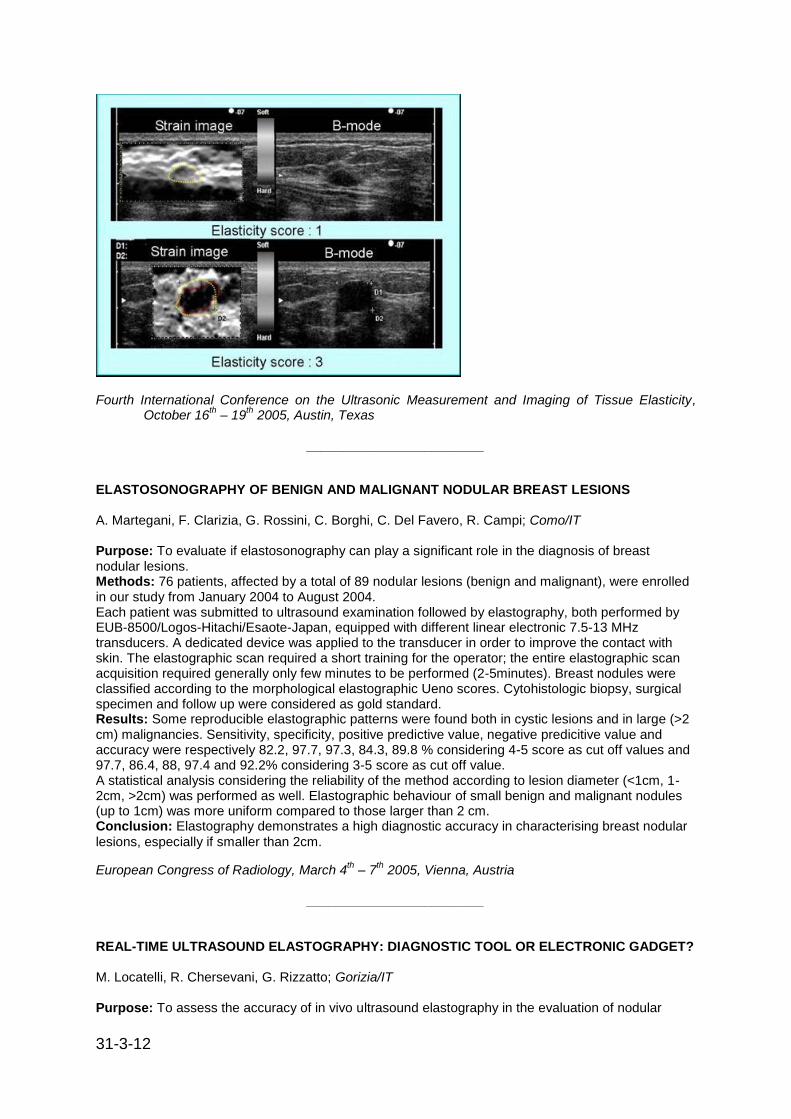
31-3-12
Fourth International Conference on the Ultrasonic Measurement and Imaging of Tissue Elasticity,
October 16th – 19
th 2005, Austin, Texas
________________________
ELASTOSONOGRAPHY OF BENIGN AND MALIGNANT NODULAR BREAST LESIONS A. Martegani, F. Clarizia, G. Rossini, C. Borghi, C. Del Favero, R. Campi; Como/IT Purpose: To evaluate if elastosonography can play a significant role in the diagnosis of breast nodular lesions. Methods: 76 patients, affected by a total of 89 nodular lesions (benign and malignant), were enrolled in our study from January 2004 to August 2004. Each patient was submitted to ultrasound examination followed by elastography, both performed by EUB-8500/Logos-Hitachi/Esaote-Japan, equipped with different linear electronic 7.5-13 MHz transducers. A dedicated device was applied to the transducer in order to improve the contact with skin. The elastographic scan required a short training for the operator; the entire elastographic scan acquisition required generally only few minutes to be performed (2-5minutes). Breast nodules were classified according to the morphological elastographic Ueno scores. Cytohistologic biopsy, surgical specimen and follow up were considered as gold standard. Results: Some reproducible elastographic patterns were found both in cystic lesions and in large (>2 cm) malignancies. Sensitivity, specificity, positive predictive value, negative predicitive value and accuracy were respectively 82.2, 97.7, 97.3, 84.3, 89.8 % considering 4-5 score as cut off values and 97.7, 86.4, 88, 97.4 and 92.2% considering 3-5 score as cut off value. A statistical analysis considering the reliability of the method according to lesion diameter (<1cm, 1-2cm, >2cm) was performed as well. Elastographic behaviour of small benign and malignant nodules (up to 1cm) was more uniform compared to those larger than 2 cm. Conclusion: Elastography demonstrates a high diagnostic accuracy in characterising breast nodular
lesions, especially if smaller than 2cm.
European Congress of Radiology, March 4th – 7
th 2005, Vienna, Austria
________________________
REAL-TIME ULTRASOUND ELASTOGRAPHY: DIAGNOSTIC TOOL OR ELECTRONIC GADGET? M. Locatelli, R. Chersevani, G. Rizzatto; Gorizia/IT Purpose: To assess the accuracy of in vivo ultrasound elastography in the evaluation of nodular
31-3-12
benign and malignant breast lesions. Methods and Materials: Ninety-seven breast lesions from 68 patients were studied with the EUB 8500 Logos ultrasonic unit (Hitachi, Japan) and a linear array transducer of 7.5-13 MHz . In 54% of lesions we obtained a pathologic diagnosis. All the other cases were well known benign lesions that we had monitored for at least two years. The elastic score was classified according a five-point color scale. The size of the lesions obtained by conventional B-mode was compared with the elastographic size. Results: 25 lesions resulted as very stiff and 23 of them turned out to be malignant (92%). 19 masses were classified as soft-mixed areas and 16 were fibroadenomas (84%). Typical three-layered areas were found in 93% of cysts. In 21 cases the estimated diameter of the lesion was greater with elastography than with B-mode; most were tumoral masses with infiltrating pathological behaviour. The exam was hampered in 4 cases (5.8%) because of the extremely stiff breast or because the lesion was located in a marginal area with insufficient surrounding tissues. Conclusions: Real-time elastography is a very quick and easy to perform diagnostic tool. It's online availability helps to increase the diagnostic confidence and to reduce unneccessary bioptical procedures in benign masses. Its ability to compare a lesion with the surrounding tissues might also be tested in the evaluation of the therapeutic efficacy for both tumors and inflammation. European Congress of Radiology, March 4
th – 7
th 2005, Vienna, Austria
________________________
US ELASTOGRAPHY SHOWS PROMISE AS MAMMOGRAPHY ADJUNCT - Aunt Minnie Report: 3/5/2005 ( http://www.auntminnie.com ) VIENNA - A pair of presentations at Friday's European Congress of Radiology (ECR) demonstrated good results for ultrasound elastography, a new technique that measures the stiffness of breast tissue as a possible indicator of tumor malignancy.
Ultrasound elastography is being pursued as a method for possibly reducing the number of biopsies that are performed on suspicious lesions detected on screening mammography. While conventional ultrasound is already being used in this role, ultrasound elastography, or elastosonography, adds an additional parameter for characterizing tissue by measuring the differences in tissue stiffness. Benign tissue is typically softer and less stiff, while malignant tissue is stiff on an elastography scan.
Elastosonography uses a conventional ultrasound scanner that is outfitted with a flat plate on the transducer head, according to Dr. Martina Locatelli of Vittorio Emmanuelle Hospital in Gorizia, Italy. The exam is conducted by compressing the probe against the breast repeatedly for about five minutes, producing real-time color-coded images that demonstrate the differences in tissue strain that occur during compression.
Locatelli's group had conducted in-vitro and ex-vivo elastosonography scans, but for their ECR paper decided to conduct an in-vivo study to assess the technique's performance in a clinical setting. They examined 98 patients using an EUB 8500 Logos scanner (Hitachi Medical Systems, Tokyo). The group used a linear-array transducer at 7-13.5 MHz, and compared the elastography images with conventional b-mode ultrasound.
There were 145 lesions in the patient population, with five lesions excluded for technical reasons, leaving a total of 140 lesions that were evaluated. The group used a five-point scoring algorithm, with score 1 corresponding to very soft tissue, such as that characterized by a liquid-filled body. Score 4 was indicative of a totally stiff lesion, while score 5 indicated an area in which both the target lesion and surrounding tissue were extremely stiff.
The vast majority of benign lesions were in the score 1, 2, and 3 categories, Locatelli said, while most of the malignant lesions were graded score 5. There was a mix of benign and malignant lesions that were graded as score 4. The group also compared the size of lesions on elastosonography versus b-mode ultrasound.

31-3-12
The technique produced sensitivity in the range of 92% for characterizing malignant lesions, and specificity of 84% in characterizing fibroadenomas or tissue with either soft or mixed characteristics. Some 93% of cysts demonstrated normal tissue characteristics on elastosonography.
Locatelli said her group had developed a diagnostic algorithm based on a combination of the elastosonography results and tissue attenuation as measured by b-mode ultrasound. "If we have ultrasound attention and a score 4 or 5 on elastosonography, we do the biopsy," Locatelli said. "If we have score 2 or 3 on elastosonography with ultrasound attenuation, we wait and we decide to do the biopsy on the basis of all ultrasound signs."
The advantages of the technique are that it is relatively easy to perform and can be performed with a conventional ultrasound scanner, she said. It is also a good complement to conventional ultrasound, and can reduce unnecessary biopsies.
"Elastosonography complements conventional ultrasound and mammography in the evaluation of breast lesions, mostly BI-RADS III and IV," Locatelli said. "Elastosonography reduces the biopsy rate in atypical cysts, and may suggest appropriate workup for cancers with atypical presentation. Elastosonography might increase the accuracy of ultrasound in both diagnosis and staging of carcinomas."
In another presentation in the same session, another Italian group also evaluated elastosonography for breast applications. They examined 76 patients from November 2003 to August 2004, finding 89 lesions, of which 45 were malignant and 44 benign. Like Locatelli's group, they used a five-point scale to score the lesions, according to Dr. Luca Aiani of Valduce Hospital in Como.
The group achieved a sensitivity of 82.2% and a specificity of 97.7%, with an accuracy of 89.8%. The group reported particularly good results for lesions smaller than 2 cm -- an important result considering that conventional ultrasound tends to perform better in characterizing larger lesions, Aiani said.
"The diagnostic performance of elastosonography is demonstrated to increase with a reduction of lesion dimension," he said. "On the other hand, the diagnostic performance of conventional ultrasound is directly related to the dimension of the lesion. So these two different methods, ultrasound and elastosonography, have a complementary diagnostic role."
Both Locatelli and Aiani acknowledged that due to the novelty of the technique and the small patient size of their studies, additional research was needed before they could recommend the routine clinical use of elastosonography.
________________________