Embed Size (px)
Citation preview

30-6-11
HHiittaacchhii RReeaall--ttiimmee TTiissssuuee
EEllaassttooggrraapphhyy::
PPuubblliiccaattiioonnss && IInntteerrnnaattiioonnaall
CCoommmmuunniiccaattiioonnss
CClliinniiccaall AAbbssttrraaccttss
30-6-11
HHiittaacchhii RReeaall--ttiimmee TTiissssuuee EEllaassttooggrraapphhyy ffoorr MMuussccuulloosskkeelleettaall
AApppplliiccaattiioonnss
30-6-11
REAL TIME ELASTOGRAPHY OF THE SYMPTOMATIC ACHILLES TENDON : WITH OR WITHOUT WATER PAD? S. Kjaer 1,*, T. Ellingsen 1, L. Bolvig 2, U. Fredberg 1 1Department of Internal medicine and Reumatology, Silkeborg Regions hospital, Silkeborg, 2Department of radiology, University Hospital of Aarhus, Aarhus, Denmark Background: Real-time ultrasound elastography (RTE) is a new technique used to evaluate the elasticity of muscle and tendon tissue. It has already been proven a feasible method in thyroid, cervix, breast, and liver diagnostics [1-4] but only a few studies have been made on tendon and muscle tissue [5-7]. Objectives: The aim of the study is to evaluate real-time freehand elastography of the symptomatic Achilles tendon with and without the use of water pad. Intra- and inter observer variability and a new grading system on RTE is described. Methods: 98 symptomatic Achilles tendons were examined with ultrasound RTE using freehand compression method with a 6-14Mhz transducer. Each tendon was examined with and without a 10mm water pad (Sonar Aid). A grading system was evaluated in which the tendons were evaluated and divided into a 9 points color grading scale (1-5 including ½ points) according to the most dominant color. Blue was graded 1, green 3 and red 5. Intra- and inter observer variability was made on recorded images to evaluate the grading system. Results: The grading ranged from 1.5 to 4.5. Mean grading in the 2 intra observations was without water pad 3.27 ± 0.72 vs. 3.26 ± 073 and with water pad 2.41 ± 0,62 vs 2.38 ± 0.63. In 67(68%) tendons the grading matched, in 30(31%) tendons there was a difference of 0.5. 1(1%) tendon was graded with a difference of 1. RTE with and without water pad showed a significant lower grading with a mean difference of 0,84 ± 0,66 (p<0,001) Coefficient of variation (CV) with and without water pad was 20.2% (range 0-64.3%). Conclusions: The use of water pad in RTE showed a highly significant difference to RTE without water pad with lower grading in the color scale as a result. Investigators should inform whether a water pad has been used or not in studies. The new grading system showed acceptable intra- and inter observer variability and is a feasible method to evaluate elastography on symptomatic Achilles tendons. References: 1: Itoh A, Ueno E, Tohno E, et al. Breast disease: clinical application of US elastography for diagnosis. Radiology 2006;239:341-50. 2: Miyanaga N, Akaza H, Yamakawa M, et al. Tissue elasticity imaging for diagnosis of prostate cancer: a preliminary report. Int J Urol 2006;13:1514-8. 3: Thomas A, Kümmel S, Gemeinhardt O, Fischer T. Real-time sonoelastography of the cervix: tissue elasticity of the normal and abnormal cervix. Acad Radiol 2007;14:193-200. 4: Friedrich-Rust M, Ong MF, Herrmann E, et al. Real-time elastography for noninvasive assessment of liver fibrosis in chronic viral hepatitis. AJR Am J Roentgenol 2007;188:758-64. 5: DrakonakiE, AllenG, WilsonG. Real-time ultrasound elastography of the normal Achilles tendon: reproducibility and pattern description. Clin Radiol 2009;64:1196-1202. 6: Zordo T, Fink C, Klauser A, et al. Real-Time Sonoelastography Findings in Healthy Achilles Tendons. AJR Am J Roentgenol. 2009;139:134-138. 7: Zordo T, Lill S, Klauser A, et al. Real-Time Sonoelastography of Lateral Epicondylitis: Comparison of Findings Between Patients and Healthy Volunteers. AJR Am J Roentgenol. 2009;193:180-185. EULAR 2011, London, 25 – 28 May
________________________

30-6-11
MUSCLE HARDNESS MEASUREMENT BY USING ULTRASOUND ELASTOGRAPHY: A FEASIBILITY STUDY Mamoru Niitsu1, Akie Michizaki1, Asako Endo1, Hitoshi Takei2 and Osamu Yanagisawa3 1Department of Radiology; 2Department of Physical Therapy, Faculty of Health Sciences, Tokyo Metropolitan University;3Faculty of Sport Sciences, Waseda University, Tokyo, Japan
Abstract Background: Muscle hardening, including delayed onset muscle soreness, can be found after vigorous exercise. Some techniques for measuring muscle hardness have been proposed. Freehand ultrasound (US) elastography has been developed and applied to breast imaging. Purpose: To evaluate the feasibility of US elastography for investigating changes in muscle hardness after eccentric exercise of the elbow flexor muscles. Material and Methods: Six healthy male volunteers performed eccentric contractions of the elbow flexor muscles of their non-dominant arms. US elastography was performed by using 14-6 MHz linear array transducer combined with a stabilizer and a gel pack of reference agent. Color-coded elasticity images and the relative strain ratio of the biceps brachii muscle to the reference were obtained. To verify the US elastography measurements, a commercially available durometer was also employed. Both measurements were performed before, immediately after, and 1– 4 days after exercise. The mean scores of strain ratios of the US elastography and durometer measurements were examined using repeated measures ANOVA. Results: US elastography demonstrated significant muscle hardening and recovery after exercise (P, 0.01). Muscle hardness increased up immediately after the exercise and continued to increase, peaking on day 2 post-exercise, and then decreased until day 4. The durometer indicated similar changes to US elastography. The control arms did not demonstrate any significant change. Conclusion: US elastography is feasible to measure muscle hardness and to produce a two-dimensional hardness map of the muscle.
Acta Radiologica 2011; 52: 99–105.
________________________
MUSCULOSKELETAL APPLICATION OF ULTRASOUND ELASTOGRAPHY: SOFT TISSUE
LIPOMA. Choi JY, Hong SH, Yoo HJ, Kim SJ. Department of Radiology, Seoul National University College of Medicine, Korea. [email protected] Abstract PURPOSE: To investigate the tissue elastic properties of soft tissue lipomas using real-time freehand elastography. MATERIALS AND METHODS: Conventional ultrasonography (US) and real-time freehand US elastography were performed in nine patients (M:F = 4:5; mean age, 53 years; 29-64 years) with soft-tissue lipoma confirmed by surgical resection. The elastogram was color-coded by 256 scales according to the degree of strain induced by light compression. The relative strains for lipoma and surrounding soft tissue were measured and mean strains were examined by using a Wilcoxon signed rank test. RESULTS: The elastograms showed red to sky-blue color in all lipomas and predominantly black in surrounding soft tissue. The mean relative strain (+/-standard deviation) was 67.9+/-28.5, 77.1+/-25.3, 63.3+/-31.2, and 15.7+/-18.3 for total, intramuscular, and subcutaneous lipoma, and surrounding soft tissue, respectively. The mean strain of the lipoma was significantly higher than one of surrounding soft tissue (p = .008, Wilcoxon signed rank test). CONCLUSION: Real-time elastography yields characteristic elastographic features of soft tissue lipoma distinguishing from those of adjacent soft tissues.
J Korean Soc Ultrasound Med. 2010 Dec;29(4):241-245
________________________
30-6-11
IS SONOELASTOGRAPHY OF VALUE IN ASSESSING TENDONS?
Klauser AS, Faschingbauer R, Jaschke WR.
Department of Diagnostic Radiology, Medical University Innsbruck, Innsbruck, Austria. [email protected]
Sonoelastography is a newly introduced ultrasound technique that evaluates tissue elasticity and thus provides additional information to that offered by conventional ultrasound images. In the musculoskeletal field, sonoelastography can help improve estimation of tendon stiffness. In this article, the principles and future developments of sonoelastography are discussed using the strongest and thickest tendon of the human body, the Achilles tendon, for illustrative purposes. Preliminary findings of sonoelastography in healthy and pathological Achilles tendons, technical considerations, examination technique and several limitations are addressed. The usefulness of elastography can be expected to increase rapidly in the musculoskeletal field, as soon as we learn to interpret elastographic artifacts as well as to take advantage of the new information provided by sonoelastography.
Semin Musculoskelet Radiol. 2010 Sep;14(3):323-33. Epub 2010 Jun 10.
________________________
ULTRASOUND ELASTOGRAPHY ASSESSMENT OF SKIN INVOLVEMENT IN SYSTEMIC SCLEROSIS: LIGHTS AND SHADOWS
1. ANNAMARIA IAGNOCCO, 2. OLGA KALOUDI, 3. CHIARA PERELLA, 4. FRANCESCA BANDINELLI, 5. VALERIA RICCIERI, 6. MASSIMILIANO VASILE, 7. FRANCESCO PORTA, 8. GUIDO VALESINI and 9. MARCO MATUCCI-CERINIC
Objective. To assess skin elasticity in systemic sclerosis (SSc) by using a new imaging modality, ultrasound elastography (UE).
Methods. Our study included 18 consecutive patients with SSc and 15 healthy controls. Modified Rodnan skin score, physical examination, and assessment of organ involvement were performed. UE was carried out on the middle forearm and on the fingers of the dominant arm. The echo signals recorded in real time during freehand operations of probe compression and relaxation produced images representing tissue elasticity, consisting of translucent colored bands superimposed on the B-mode ultrasonographic images. The color scale varied within a large band spectrum from red, indicative of soft and highly elastic tissue, to blue, which denoted hard and barely elastic tissue.
Results. On the forearm of all patients, UE showed a homogeneous blue area corresponding to the dermis visualized in a B-mode ultrasonographic image; in controls, a blue pattern was never detected and a predominance of green with sporadic areas of pale blue was observed. At sequential evaluations, UE of fingers produced inconstant and changeable colored areas.
Conclusion. The imaging pattern observed in the forearm of patients with SSc may represent the reduction of strain in the dermis due to loss of elasticity. The variable pattern obtained by finger evaluation demonstrated that UE can assess skin involvement in SSc only in those areas where the dermis is known to be thicker and where the bone hyperreflection is minimal. Further studies are needed to confirm our results and determine the validity of this new imaging modality.
J Rheumatol August 2010 37(8):1688-1691

30-6-11
EVALUATION OF FINDINGS IN SONOELASTOGRAPHY (SEL) IN FATTY ATROPHY IN ROTATOR CUFF COMPARED TO MAGNETIC RESONANCE TOMOGRAPHY (MRT) Ralph Faschingbauer, V Schreiber, G Feuchtner, P Soegner, VSmekal, W Jaschke, A Klauser Univeristy Hospital Innsbruck, Departement for Radiology, Austria Objective: To evaluate SEL in diagnosing fatty atrophy in rotator cuff tears, in comparison to MRT. Method and Materials: 20 patients with Supraspinatus (SSP) or Infraspinatus (ISP) tendon rupture were examined using SEL compared to MRT, using a grading modified accordingly to Goutallier. SEL was graded as grade 1 = <25% red colouring, grade 2 = 25-50% red colouring, grade 3 = 50-75% red colouring, grade 4 = > 75% red. Tissues of different elasticity show different colors in SEL, from blue (hard) to red (soft) Results: In MRT, grade 1 in SSP was found in 5 patients (26.3%), grade 2 in 10 patients (52.6%), grade 3 in 3 patients (15.8%), and grade 4 in 1 patient (5.3%). Correlating SEL showed 6 patients (31.6%) with grade 1, 9 patients (47.4%) with grade 2, 3 patient (15.8%) with grade 3, and 1 patient (5.3%) with grade 4 with R = 0.946 (P > 0.001). In MRT of the ISP, grade 1 was found in 16 patients (84.2%), grade 2 in 1 patient (5.3%) and grade 4 in 2 patients (10.5%). Correlating SEL showed 16 patients (84.2%) with grade 1, and 3 patients (15.8%) with grade 2 with R = 0.228 (P > 0.05). Overall correlation for SSP and ISP was R = 0.744 (P > 0.001) Conclusion: SEL demonstrates to be comparable to MRT in the detection of fatty atrophy in SSP and was helpful to assess muscle tissue with respect of elasticity and loss of elasticity in atrophied muscles. Euroson 2010, August 22 – 25, Copenhagen
________________________
PRELIMINARY FINDINGS OF SONOELASTOGRAPHY (SEL) IN PATELLAR TENDINOSIS COMPARED TO MAGNETIC RESONANCE TOMOGRAPHY (MRT) Ralph Faschingbauer(1), P Soegner (1), G Feuchtner (7), M Wick (1), WJaschke (1), A Klauser (1)
(1) Univeristy Hospital Innsbruck, Austria
Objective: To correlate MRT and SEL findings in patients presenting with patellar tendinosis. Methods and Materials: 10 healthy volunteers and 5 consecutive patients with an elective MRT examination of the knee, presenting with patellar tendinosis were included in this study and underwent SEL. SEL was performed on a Hitachi EUB 9000 US-scanner (EUP-L54M, 6-13 MHz). The principle of SEL is that tissue compression produces strain (displacement) within the tissue, and that strain is smaller in harder tissue (blue coloring) than in softer tissue (red coloring). The patellar tendon was scanned in a standardized 30° flexed position.The tendon was divided in 3 thirds, performed in longitudinal and axial planes. Results: 14/20 healthy tendons showed blue to green colouring, 6/20 presented with yellow parts within the tendon. No red colouring was detected in any healthy volunteer, interpreted as sign for harder tissue characteristics. In all 5 patients, presenting with typical alterations of tendinosis in MRT, tendons showed softer tissue characteristic using SEL, when compared to healthy volunteers. In detail. patients showed mainly yellow and red within the tendon and only minor blue to green coloring. Delineation of tendinosis amount was increased by using SEL when compared to MRT, mainly involving 2 tendon thirds (34/45). Conclusion: Our preliminary results show, that SEL seems to reliable in the detection of tendinosis when compared to MRI. However, tendinosis in terms of tissue softening might involve a greater tendon amount as suspected by MRI. Larger study population groups and further histological studies should prove these results. Euroson 2010, August 22 – 25, Copenhagen

30-6-11
THE CONTRIBUTION OF ULTRASONOGRAPHY AND SONOELASTOGRAPHY IN ASSESSMENT OF MYOSITIS Carolina Botar-Jid1, Laura Damian2, Sorin M. Dudea1, Dan Vasilescu1, Simona Rednic2, Radu Badea3 1 Radiology Department, „Iuliu Haţieganu” University of Medicine and Pharmacy Cluj-Napoca 2 Rheumatology Clinic, Emergency Clinical County Hospital, Cluj-Napoca 3 3rd Medical Clinic, „Iuliu Haţieganu” University of Medicine and Pharmacy Cluj-Napoca Abstract Aims: To analyze the utility of ultrasonography (US) in the assessment of inflammatory miopathies, and of sonoelastography in the assessment of the elasticity of skeletal muscle in myositis. Material and methods: The study group comprised 24 patients with musculoskeletal pathology examined using Hitachi 8500 EUB equipment with a 6.5-13 MHz transducer and software for elastography. The images were analyzed in conjunction with clinical and biochemical data. Using dedicated software for color information from the elastographic images, the average values for color intensity, hue and dispersion were calculated. Results: After the correlation of US images with clinical and paraclinical data the highest average values for color parameters were encountered on the superior third of the thigh. There was a proportional concordance between the average values of the color parameters and serum creatine kinase and serum lactic dehydrogenase and there was no significant agreement between the average values of the color parameters and the erythrocyte sedimentation rate, positive rheumatoid factor or positive antinuclear antibody. Conclusions: The correlation between the quantitative colour parameters from the elastographic images and the laborator studies suggest that sonoelastography could be an important tool in the management of the patients with myositis. Medical Ultrasonography 2010, Vol. 12, no. 2, 120-126
________________________
EVALUATION OF FINDINGS IN SONOELASTOGRAPHY IN FATTY ATROPHY IN ROTATOR CUFF COMPARED TO MAGNETIC RESONANCE TOMOGRAPHY R. Faschingbauer
1, V.M. Schreiber
2, G.M. Feuchtner
1, P. Soegner
1, V. Smekal
1, W. Jaschke
1, A.S.
Klauser1;
1Innsbruck/AT,
2Pittsburgh, PA/US
Purpose: To evaluate SonoElastography (SEL) in diagnosing fatty atrophy in rotator cuff tears compared to Magnetic Resonance Tomography (MRT). Methods and Materials: 20 patients (mean age: 59 years) with Supraspinatus (SSP) or Infraspinatus (ISP) tendon rupture were included, using a MRT grading modified according to Goutallier. SEL shows tissues with different elasticity in different colors, from red (soft) to blue (hard). SEL was graded as grade 1 = <25% red colouring, grade 2 = 25-50% red colouring, grade 3 = 50-75% red colouring, and grade 4 = > 75% red colouring. Results: MRT grade 1 in SSP was found in 5 patients (26.3%), grade 2 in 10 patients (52.6%), grade 3 in 3 patients (15.8%), and grade 4 in 1 patient (5.3%). SEL showed 6 patients (31.6%) with grade 1, 9 patients (47.4%) with grade 2, 3 patients (15.8%) with grade 3, and 1 patient (5.3%) with grade 4 with R = 0.946 (P ≤ 0.001). In MRT of the ISP, grade 1 was found in 16 patients (84.2%), grade 2 in 1 patient (5.3%) and grade 4 in 2 patients (10.5%). SEL showed 16 patients (84.2%) with grade 1, and 3 patients (15.8%) with grade 2 with R = 0.228 (P > 0.05). Overall correlation for SSP and ISP was R = 0.744 (P ≤ 0.001). Conclusion: SEL demonstrates to be comparable to MRT in detection of fatty atrophy in SSP. Furthermore, SEL was helpful to assess muscle tissue with respect to elasticity and loss of elasticity in atrophied muscles.
European Congress of Radiology 2010, Vienna, March 5
th – 9
th
________________________
30-6-11
MAGNETIC RESONANCE IMAGING, ULTRASOUND AND REAL-TIME ULTRASOUND ELASTOGRAPHY OF THE THIGH MUSCLES IN CONGENITAL MUSCLE DYSTROPHY. Drakonaki EE, Allen GM. Radiology Department, University of Crete, Heraklion, Greece. [email protected] Congenital muscle dystrophy includes a range of genetic disorders characterized by muscle weakness and contractures. We report the magnetic resonance (MR), ultrasound (US) and real-time sonoelastography (RTE) imaging findings of the thigh muscles of a 15-year-old boy with Bethlem myopathy diagnosed with clinical, electromyographic and histopathological criteria. Ultrasound and MR showed hyperechoic appearance and high signal intensity on T1- and T2-weighted sequences respectively at the periphery of the vastus lateralis and the long head of the biceps femoris muscles, and at a central area within the rectus femoris muscles. RTE was employed to examine the elastic properties of the muscle. The elastograms were presented as colour-coded maps superimposed on the B-mode images and revealed that the elastographic pattern correlated with the MR and US pattern of involvement. The abnormal muscle areas were stiffer (blue) than the normal-appearing areas (green), a finding that probably correlates with the presence of dystrophic collagen at the affected areas. This report suggests that RTE could be used as an additional imaging tool to evaluate the pattern of muscle changes in congenital myopathy. Further studies are needed to investigate the specificity and clinical value of RTE in the diagnosis and monitoring of neuromuscular disease. Skeletal Radiol. 2010 Apr;39(4):391-6.
________________________
REAL-TIME SONOELASTOGRAPHY: FINDINGS IN PATIENTS WITH SYMPTOMATIC ACHILLES TENDONS AND COMPARISON TO HEALTHY VOLUNTEERS.
De Zordo T, Chhem R, Smekal V, Feuchtner G, Reindl M, Fink C, Faschingbauer R, Jaschke W, Klauser AS.
Abstract PURPOSE: Real-time sonoelastography (SE), a newly introduced ultrasound technique, has already shown conclusive results in breast, prostate, and thyroid tumor diagnostics. This study investigated the performance of SE for the differentiation of Achilles tendon alterations of tendinopathy compared to clinical examination and conventional ultrasound (US).
MATERIALS AND METHODS: Achilles tendons in 25 consecutive patients with chronic Achilles tendinopathy and 25 healthy volunteers were examined clinically by US and by SE.
RESULTS: In the healthy volunteers, SE showed the tendon to be hard (93 %), while distinct softening was found in 57 % of the patients. SE showed more frequent involvement of the distal (64 %) and middle third (80 %) than the proximal third (28 %) of the Achilles tendon. Using SE a mean sensitivity of 94 %, specificity of 99 %, and accuracy of 97 % were found when clinical examination was used as the reference standard. The correlation to US was 0.89. Mild softening was found in 7 % of the healthy volunteers and in 11 % of the patients.
CONCLUSION: Our results emphasize that only distinct softening of Achilles tendons is comparable to clinical examination and US findings. However, mild softening might be explained by very early changes in tissue elasticity in the case of Achilles tendinopathy, which should be assessed in follow-up studies.
Ultraschall Med. 2010 Aug;31(4):394-400.
________________________
30-6-11
REAL-TIME ULTRASOUND ELASTOGRAPHY OF THE NORMAL ACHILLES TENDON: REPRODUCIBILITY AND PATTERN DESCRIPTION.
Drakonaki EE, Allen GM, Wilson DJ.
Department of Radiology, University Hospital, Heraklion, Greece. [email protected]
AIM: To investigate the feasibility and reproducibility of real-time freehand ultrasound elastography (RTE) of the normal Achilles tendon and to describe its elastographic appearances.
MATERIALS AND METHODS: Fifty normal Achilles tendons were prospectively examined using RTE performed by tissue compression using the hand-held transducer. The information was colour-coded (red=soft, green=medium, blue=hard) and superimposed on the B-mode image. Each tendon was examined three times transversely and longitudinally by two radiologists and the ratio between tendon and retro-Achilles fat strain (strain index) was calculated. The reproducibility of the elastograms was assessed qualitatively and quantitatively using the strain index inter and intra observer variation coefficient (intra/inter-CV and intra/inter-CC, respectively).
RESULTS: All tendons were clearly visualized on the elastograms. Nineteen tendons (19/50, 38%) appeared homogeneously green/blue (type 1). Thirty-one tendons (31/50, 62%) appeared green with longitudinal red stripes (type 2). The intra- and inter-CC values of the strain index were lower for the transverse plane than for the longitudinal plane (0.43, 0.45, 0.41 and 0.78, 0.66, 0.51, respectively). The intra-CV and inter-CV values were higher for the transverse than for the longitudinal plane measurements (39%, 37%, 30% and 30.50%, 30.10%, 29.60%, respectively).
CONCLUSION: RTE of the normal Achilles tendon is a feasible method. The reproducibility of the strain index is good and higher for longitudinal elastograms. Qualitative assessment enables the discrimination of two distinct elastographic patterns. Further studies are required to assess the clinical value of this method.
Clin Radiol. 2009 Dec;64(12):1196-202. Epub 2009 Oct 8.
________________________
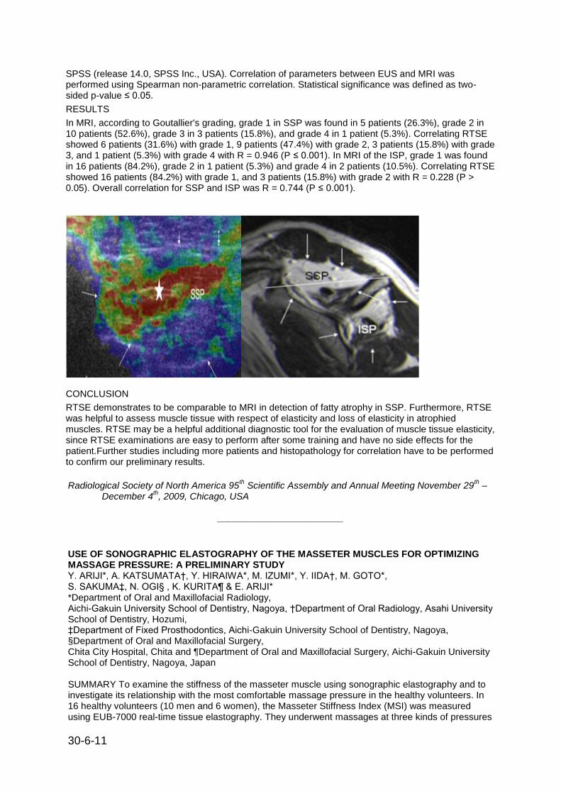
REAL-TIME SONOELASTOGRAPHY IN ROTATOR CUFF IMAGING AND COMPARISON TO MAGNETIC RESONANCE IMAGING AS GOLD STANDARD
Verena Schreiber, Vinzenz Smekal, Tobias De Zordo, Christian Fink, Gudrun Feuchtner, Andrea Klauser
PURPOSE
It was the purpose of this study to evaluate Realtime-SonoELASTOGRAPHY (RTSE) in diagnosing fatty atrophy in rotator cuff tears, in comparison to Magnetic Resonance Imaging (MRI) as the gold standard
METHOD AND MATERIALS
Ethics committee approval and informed written consent were obtained. 20 patients (45% F, 55% M, mean age: 59 years ± 7) with Supraspinatus (SSP) or Infraspinatus (ISP) tendon rupture were examined using RTSE (scanner frequency range 5 - 10 MHz). Patients underwent Magnetic Resonance Imaging (MRI) with sequences of T2-weighted tse-rst-sagittal and 4mm slice thickness. Grading was performed accordingly to Goutallier. RTSE was compared to MRI and graded as grade 1 = <25% red colouring , grade 2 = 25-50% red colouring, grade 3 = 50-75% red colouring, grade 4 = > 75% red. Tissues of different elasticity are shwon in different colors in RTSE with a spectrum ranging from red, representing soft tissue, to blue for hard tissue. Statistical analysis was performed using
30-6-11
SPSS (release 14.0, SPSS Inc., USA). Correlation of parameters between EUS and MRI was performed using Spearman non-parametric correlation. Statistical significance was defined as two-sided p-value ≤ 0.05.
RESULTS
In MRI, according to Goutallier's grading, grade 1 in SSP was found in 5 patients (26.3%), grade 2 in 10 patients (52.6%), grade 3 in 3 patients (15.8%), and grade 4 in 1 patient (5.3%). Correlating RTSE showed 6 patients (31.6%) with grade 1, 9 patients (47.4%) with grade 2, 3 patients (15.8%) with grade 3, and 1 patient (5.3%) with grade 4 with R = 0.946 (P ≤ 0.001). In MRI of the ISP, grade 1 was found in 16 patients (84.2%), grade 2 in 1 patient (5.3%) and grade 4 in 2 patients (10.5%). Correlating RTSE showed 16 patients (84.2%) with grade 1, and 3 patients (15.8%) with grade 2 with R = 0.228 (P > 0.05). Overall correlation for SSP and ISP was R = 0.744 (P ≤ 0.001).
CONCLUSION
RTSE demonstrates to be comparable to MRI in detection of fatty atrophy in SSP. Furthermore, RTSE was helpful to assess muscle tissue with respect of elasticity and loss of elasticity in atrophied muscles. RTSE may be a helpful additional diagnostic tool for the evaluation of muscle tissue elasticity, since RTSE examinations are easy to perform after some training and have no side effects for the patient.Further studies including more patients and histopathology for correlation have to be performed to confirm our preliminary results.
Radiological Society of North America 95
th Scientific Assembly and Annual Meeting November 29
th –
December 4th, 2009, Chicago, USA
________________________
USE OF SONOGRAPHIC ELASTOGRAPHY OF THE MASSETER MUSCLES FOR OPTIMIZING MASSAGE PRESSURE: A PRELIMINARY STUDY Y. ARIJI*, A. KATSUMATA†, Y. HIRAIWA*, M. IZUMI*, Y. IIDA†, M. GOTO*, S. SAKUMA‡, N. OGI§ , K. KURITA¶ & E. ARIJI* *Department of Oral and Maxillofacial Radiology, Aichi-Gakuin University School of Dentistry, Nagoya, †Department of Oral Radiology, Asahi University School of Dentistry, Hozumi, ‡Department of Fixed Prosthodontics, Aichi-Gakuin University School of Dentistry, Nagoya, §Department of Oral and Maxillofacial Surgery, Chita City Hospital, Chita and ¶Department of Oral and Maxillofacial Surgery, Aichi-Gakuin University School of Dentistry, Nagoya, Japan SUMMARY To examine the stiffness of the masseter muscle using sonographic elastography and to investigate its relationship with the most comfortable massage pressure in the healthy volunteers. In 16 healthy volunteers (10 men and 6 women), the Masseter Stiffness Index (MSI) was measured using EUB-7000 real-time tissue elastography. They underwent massages at three kinds of pressures
30-6-11
using the Oral Rehabilitation Robot (WAO-1). A subjective evaluation regarding the comfort of each massage was recorded on the visual analogue scale. Elastography was also performed in two patients with temporomandibular joint dysfunction with the myofascial pain. The mean MSI of the right and left muscles in the healthy volunteers were 0.85+/- 0.44 and 0.74 +/- 0.35 respectively. There was no significant difference between the right and left MSI in the healthy volunteers. The MSI was related to massage pressure at which the healthy men felt most comfortable. The two temporomandibular disorder patients had a large laterality in the MSI. The MSI was related to the most comfortable massage pressure in the healthy men. The MSI can be one index for determining the massage pressure. Journal of Oral Rehabilitation 2009 36; 627–635
________________________
SONOELASTOGRAPHIC EVALUATION OF ACHILLES TENDON IN AMATEUR SYMPTOMATIC RUNNERS Luca Sconfienza, Enzo Silvestri, Stefano Longo, Marco Cimmino Milan, Italy Purpose: Sonoelastography is a recently developed ultrasound (US) technique that allows in vivo assessment of tissue mechanical properties. Up to now, this technique has been mainly used to investigate prostatic tumours and breast masses. The aim of our paper is to use sonoelastography to evaluate Achilles tendon in amateurs symptomatic runners. Materials and Methods: Sixteen patients referred for unilateral Achilles tendon pain due to overuse associated with amateur sporting activities and 24 healthy controls were studied. US and sonoelastography were performed on 16 symptomatic tendons and 48 control tendons with a system equipped with a 10-6 MHz broadband linear array. The array was positioned at the calcaneal enthesis, retrocalcaneal bursa and in 3 different areas of the tendon body. The elastogram colour range was translated in a numeric score. Results were compared by the Kruskall Wallis test. Results: At grey scale US, symptomatic tendons showed a variety of basic changes in fibrillar pattern (2): increased tendon thickness (12), interruption (5), fragmentation (5), and disappearance of fibrillar echotexture (5). In the control group, we observed 1 case of increased tendon thickness and 5 cases of disappearance of fibrillar echotexture. By sonoelastography, no difference was observed between symptomatic and control tendons at the enthesis and bursa. Symptomatic tendons bodies were significantly harder than control ones, showing a prevalence of blue to green colour (p<0.0001). Conclusion: Sonoelastography shows increased stiffness in symptomatic enlarged Achilles tendons in comparison to normal ones. Long-term studies are needed to evaluate if these findings have a prognostic value. 12
th World Congress of the World Federation for Ultrasound in Medicine and Biology, 30
th August – 3
rd
September 2009, Sydney, Australia
________________________
REAL-TIME SONOELASTOGRAPHY FINDINGS IN HEALTHY ACHILLES TENDONS. De Zordo T, Fink C, Feuchtner GM, Smekal V, Reindl M, Klauser AS. Department of Radiology II, Medical University Innsbruck, Anichstrasse 35, 6020 Innsbruck, Austria. OBJECTIVE: Real-time sonoelastography is a new ultrasound-based technique able to assess tissue elasticity that has already shown feasibility in tumor diagnosis. The aim of this study was to assess the performance of real-time sonoelastography in depicting the Achilles tendons of healthy volunteers and to compare sonoelastography findings with conventional ultrasound findings. MATERIALS AND METHODS: Eighty asymptomatic Achilles tendons of 40 healthy volunteers (19 men, 21 women; mean age, 38 years; range, 20-76 years) were examined on real-time sonoelastography and ultrasound. The Achilles tendons were divided into the following thirds for image evaluation: proximal (musculotendinous junction), middle (2-6 cm above insertion at the calcaneus), and distal (insertion at the calcaneus). Longitudinal and axial images of each tendon third

30-6-11
were obtained using ultrasound and real-time sonoelastography. Real-time sonoelastography images were evaluated by reviewers using an experimentally proven color grading system. RESULTS: The Achilles tendons showed mainly a hard structured pattern (86.7%) (208/240 tendon thirds) on sonoelastography; however, mild softening was found in 12.1% (29/240) of the tendons. Distinct softening corresponding to alterations found also on ultrasound and, therefore, suggesting subclinical changes was detected in 1.3% (3/240). The overall correlation (kappa) between real-time sonoelastography and ultrasound findings was 1.00. CONCLUSION: In healthy volunteers, the Achilles tendon appeared hard on real-time sonoelastography with excellent correlation to ultrasound. Further investigation including pathologic tendons should be performed to prove the value of real-time sonoelastography in the assessment of Achilles tendinopathy
AJR Am J Roentgenol. 2009 Aug;193(2):W134-8.
________________________
REAL-TIME SONOELASTOGRAPHY OF LATERAL EPICONDYLITIS: COMPARISON OF FINDINGS BETWEEN PATIENTS AND HEALTHY VOLUNTEERS.
De Zordo T, Lill SR, Fink C, Feuchtner GM, Jaschke W, Bellmann-Weiler R, Klauser AS. Department of Radiology II, Medical University Innsbruck, Anichstrasse 35, 6020 Innsbruck, Austria. OBJECTIVE: The purpose of this study was to evaluate real-time sonoelastography in the assessment of the origins the common extensor tendon in healthy volunteers and in patients with symptoms of lateral epicondylitis. The findings were compared with those obtained at clinical examination, ultrasonography, and power Doppler sonography. SUBJECTS AND METHODS: Thirty-eight elbows of 32 consecutively registered patients with symptoms of lateral epicondylitis and 44 asymptomatic elbows of 28 healthy volunteers were assessed with ultrasound and real-time sonoelastography. A clinical examination was performed, and pain was classified with a visual analog scale. RESULTS: In healthy volunteers, real-time sonoelastographic images showed hard tendon structures in 96% of tendon thirds and mild alterations in 4%. Real-time sonoelastography of patients showed hard structures in 33% of tendon thirds but softening of different grades in 67%, a statistically significant difference in relation to the findings in healthy volunteers (p < 0.001). Lateral collateral ligament involvement and overlying fascial involvement were more commonly detected with real-time sonoelastography. The sensitivity of real-time sonoelastography was 100%, the specificity 89%, and the accuracy 94% with clinical examination as the reference standard. Good correlation with ultrasound findings was found (r > or = 0.900). No correlation was observed between ultrasound or real-time sonoelastographic findings and power Doppler sonographic findings, but power Doppler sonographic findings had a strong correlation with the visual analog scale score. CONCLUSION: Real-time sonoelastography is valuable in the detection of the intratendinous and peritendinous alterations of lateral epicondylitis and facilitates differentiation between healthy and symptomatic extensor tendon origins with excellent sensitivity and excellent correlation with ultrasound findings. AJR Am J Roentgenol. 2009 Jul;193(1):180-5
________________________
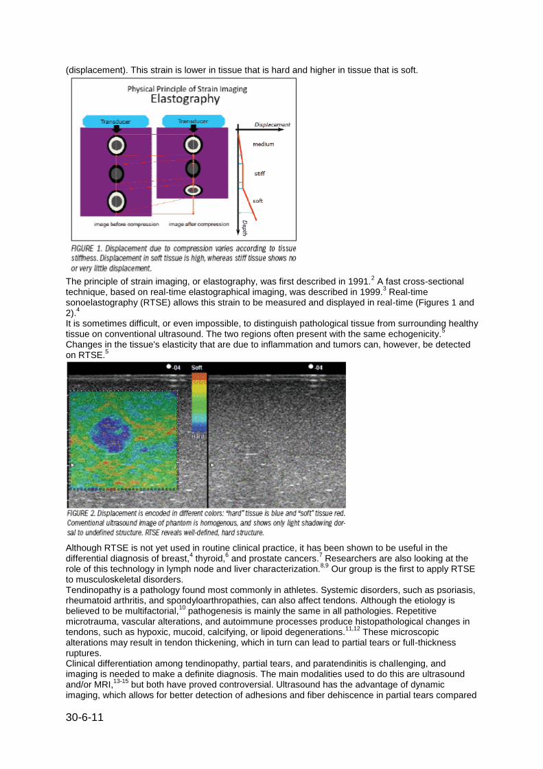
DISTINCT TISSUE SOFTENING LINKED TO MUSCULOSKELETAL DISORDERS CAN BE DETECTED IN REAL-TIME STRAIN IMAGING Diagnostic Imaging Europe, 01 June 2009 By Andrea S. Klauser, M.D., Tobias De Zordo, M.D., Ralph Faschingbauer, M.D. Real-time sonoelastography is a relatively new technique that can assess the elastic properties of tissues.
1 Elastography is based on the principle that the compression of tissue produces strain
30-6-11
(displacement). This strain is lower in tissue that is hard and higher in tissue that is soft.
The principle of strain imaging, or elastography, was first described in 1991.
2 A fast cross-sectional
technique, based on real-time elastographical imaging, was described in 1999.3 Real-time
sonoelastography (RTSE) allows this strain to be measured and displayed in real-time (Figures 1 and 2).
4
It is sometimes difficult, or even impossible, to distinguish pathological tissue from surrounding healthy tissue on conventional ultrasound. The two regions often present with the same echogenicity.
5
Changes in the tissue's elasticity that are due to inflammation and tumors can, however, be detected on RTSE.
5
Although RTSE is not yet used in routine clinical practice, it has been shown to be useful in the differential diagnosis of breast,
4 thyroid,
6 and prostate cancers.
7 Researchers are also looking at the
role of this technology in lymph node and liver characterization.8,9
Our group is the first to apply RTSE to musculoskeletal disorders. Tendinopathy is a pathology found most commonly in athletes. Systemic disorders, such as psoriasis, rheumatoid arthritis, and spondyloarthropathies, can also affect tendons. Although the etiology is believed to be multifactorial,
10 pathogenesis is mainly the same in all pathologies. Repetitive
microtrauma, vascular alterations, and autoimmune processes produce histopathological changes in tendons, such as hypoxic, mucoid, calcifying, or lipoid degenerations.
11,12 These microscopic
alterations may result in tendon thickening, which in turn can lead to partial tears or full-thickness ruptures. Clinical differentiation among tendinopathy, partial tears, and paratendinitis is challenging, and imaging is needed to make a definite diagnosis. The main modalities used to do this are ultrasound and/or MRI,
13-15 but both have proved controversial. Ultrasound has the advantage of dynamic
imaging, which allows for better detection of adhesions and fiber dehiscence in partial tears compared
30-6-11
with MRI. On the other hand, MRI enables the better detection of increased fluid (edema) that may be present in tendinosis. ASSESSMENT OF PERFORMANCE We investigated the performance of RTSE in the detection of Achilles tendinopathy and lateral epicondylitis (tennis elbow). All examinations were performed using a linear array transducer with a frequency of 6 to 13 MHz. We used an EUB-8500 ultrasound system when examining the Achilles tendon and an EUB-9000 system when studying the elbow (Hitachi Medical). Tissue elasticity distributions were calculated in real-time (up to 30 frames per second). Results were represented in color over the conventional B-mode image.
5
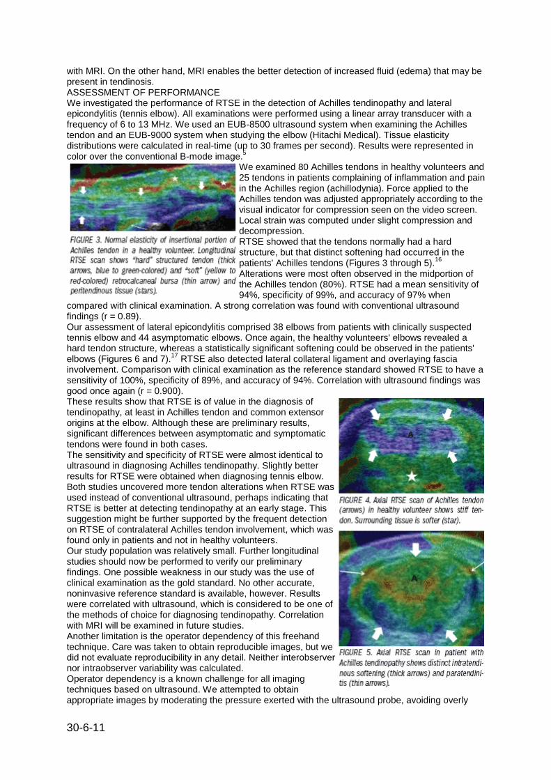
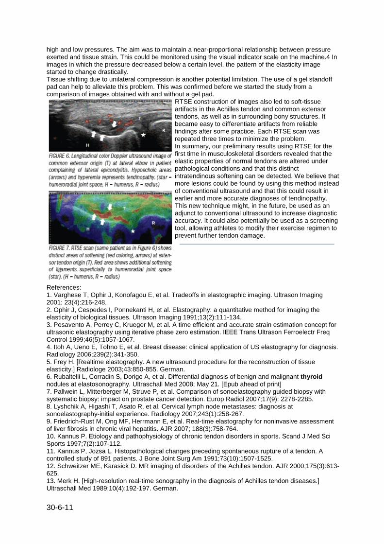
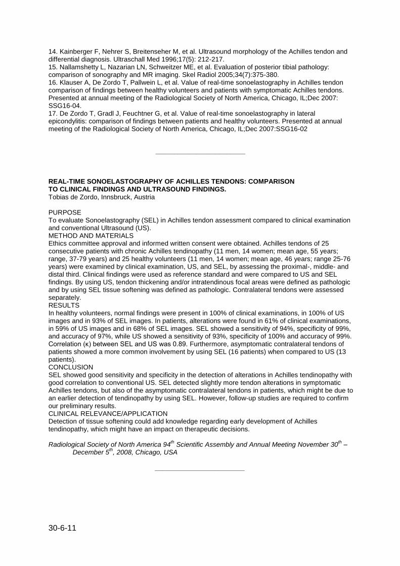
We examined 80 Achilles tendons in healthy volunteers and 25 tendons in patients complaining of inflammation and pain in the Achilles region (achillodynia). Force applied to the Achilles tendon was adjusted appropriately according to the visual indicator for compression seen on the video screen. Local strain was computed under slight compression and decompression. RTSE showed that the tendons normally had a hard structure, but that distinct softening had occurred in the patients' Achilles tendons (Figures 3 through 5).
16
Alterations were most often observed in the midportion of the Achilles tendon (80%). RTSE had a mean sensitivity of 94%, specificity of 99%, and accuracy of 97% when
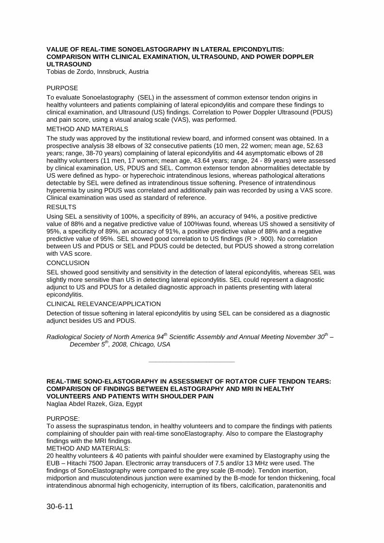
compared with clinical examination. A strong correlation was found with conventional ultrasound findings (r = 0.89). Our assessment of lateral epicondylitis comprised 38 elbows from patients with clinically suspected tennis elbow and 44 asymptomatic elbows. Once again, the healthy volunteers' elbows revealed a hard tendon structure, whereas a statistically significant softening could be observed in the patients' elbows (Figures 6 and 7).
17 RTSE also detected lateral collateral ligament and overlaying fascia
involvement. Comparison with clinical examination as the reference standard showed RTSE to have a sensitivity of 100%, specificity of 89%, and accuracy of 94%. Correlation with ultrasound findings was good once again (r = 0.900). These results show that RTSE is of value in the diagnosis of tendinopathy, at least in Achilles tendon and common extensor origins at the elbow. Although these are preliminary results, significant differences between asymptomatic and symptomatic tendons were found in both cases. The sensitivity and specificity of RTSE were almost identical to ultrasound in diagnosing Achilles tendinopathy. Slightly better results for RTSE were obtained when diagnosing tennis elbow. Both studies uncovered more tendon alterations when RTSE was used instead of conventional ultrasound, perhaps indicating that RTSE is better at detecting tendinopathy at an early stage. This suggestion might be further supported by the frequent detection on RTSE of contralateral Achilles tendon involvement, which was found only in patients and not in healthy volunteers. Our study population was relatively small. Further longitudinal studies should now be performed to verify our preliminary findings. One possible weakness in our study was the use of clinical examination as the gold standard. No other accurate, noninvasive reference standard is available, however. Results were correlated with ultrasound, which is considered to be one of the methods of choice for diagnosing tendinopathy. Correlation with MRI will be examined in future studies. Another limitation is the operator dependency of this freehand technique. Care was taken to obtain reproducible images, but we did not evaluate reproducibility in any detail. Neither interobserver nor intraobserver variability was calculated. Operator dependency is a known challenge for all imaging techniques based on ultrasound. We attempted to obtain appropriate images by moderating the pressure exerted with the ultrasound probe, avoiding overly
30-6-11
high and low pressures. The aim was to maintain a near-proportional relationship between pressure exerted and tissue strain. This could be monitored using the visual indicator scale on the machine.4 In images in which the pressure decreased below a certain level, the pattern of the elasticity image started to change drastically. Tissue shifting due to unilateral compression is another potential limitation. The use of a gel standoff pad can help to alleviate this problem. This was confirmed before we started the study from a comparison of images obtained with and without a gel pad.
RTSE construction of images also led to soft-tissue artifacts in the Achilles tendon and common extensor tendons, as well as in surrounding bony structures. It became easy to differentiate artifacts from reliable findings after some practice. Each RTSE scan was repeated three times to minimize the problem. In summary, our preliminary results using RTSE for the first time in musculoskeletal disorders revealed that the elastic properties of normal tendons are altered under pathological conditions and that this distinct intratendinous softening can be detected. We believe that more lesions could be found by using this method instead of conventional ultrasound and that this could result in earlier and more accurate diagnoses of tendinopathy. This new technique might, in the future, be used as an adjunct to conventional ultrasound to increase diagnostic accuracy. It could also potentially be used as a screening tool, allowing athletes to modify their exercise regimen to prevent further tendon damage.
References: 1. Varghese T, Ophir J, Konofagou E, et al. Tradeoffs in elastographic imaging. Ultrason Imaging 2001; 23(4):216-248. 2. Ophir J, Cespedes I, Ponnekanti H, et al. Elastography: a quantitative method for imaging the elasticity of biological tissues. Ultrason Imaging 1991;13(2):111-134. 3. Pesavento A, Perrey C, Krueger M, et al. A time efficient and accurate strain estimation concept for ultrasonic elastography using iterative phase zero estimation. IEEE Trans Ultrason Ferroelectr Freq Control 1999;46(5):1057-1067. 4. Itoh A, Ueno E, Tohno E, et al. Breast disease: clinical application of US elastography for diagnosis. Radiology 2006;239(2):341-350. 5. Frey H. [Realtime elastography. A new ultrasound procedure for the reconstruction of tissue elasticity.] Radiologe 2003;43:850-855. German. 6. Rubaltelli L, Corradin S, Dorigo A, et al. Differential diagnosis of benign and malignant thyroid nodules at elastosonography. Ultraschall Med 2008; May 21. [Epub ahead of print] 7. Pallwein L, Mitterberger M, Struve P, et al. Comparison of sonoelastography guided biopsy with systematic biopsy: impact on prostate cancer detection. Europ Radiol 2007;17(9): 2278-2285. 8. Lyshchik A, Higashi T, Asato R, et al. Cervical lymph node metastases: diagnosis at sonoelastography-initial experience. Radiology 2007;243(1):258-267. 9. Friedrich-Rust M, Ong MF, Herrmann E, et al. Real-time elastography for noninvasive assessment of liver fibrosis in chronic viral hepatitis. AJR 2007; 188(3):758-764. 10. Kannus P. Etiology and pathophysiology of chronic tendon disorders in sports. Scand J Med Sci Sports 1997;7(2):107-112. 11. Kannus P, Jozsa L. Histopathological changes preceding spontaneous rupture of a tendon. A controlled study of 891 patients. J Bone Joint Surg Am 1991;73(10):1507-1525. 12. Schweitzer ME, Karasick D. MR imaging of disorders of the Achilles tendon. AJR 2000;175(3):613-625. 13. Merk H. [High-resolution real-time sonography in the diagnosis of Achilles tendon diseases.] Ultraschall Med 1989;10(4):192-197. German.
30-6-11
14. Kainberger F, Nehrer S, Breitenseher M, et al. Ultrasound morphology of the Achilles tendon and differential diagnosis. Ultraschall Med 1996;17(5): 212-217. 15. Nallamshetty L, Nazarian LN, Schweitzer ME, et al. Evaluation of posterior tibial pathology: comparison of sonography and MR imaging. Skel Radiol 2005;34(7):375-380. 16. Klauser A, De Zordo T, Pallwein L, et al. Value of real-time sonoelastography in Achilles tendon comparison of findings between healthy volunteers and patients with symptomatic Achilles tendons. Presented at annual meeting of the Radiological Society of North America, Chicago, IL;Dec 2007: SSG16-04. 17. De Zordo T, Gradl J, Feuchtner G, et al. Value of real-time sonoelastography in lateral epicondylitis: comparison of findings between patients and healthy volunteers. Presented at annual meeting of the Radiological Society of North America, Chicago, IL;Dec 2007:SSG16-02
________________________
REAL-TIME SONOELASTOGRAPHY OF ACHILLES TENDONS: COMPARISON TO CLINICAL FINDINGS AND ULTRASOUND FINDINGS. Tobias de Zordo, Innsbruck, Austria PURPOSE To evaluate Sonoelastography (SEL) in Achilles tendon assessment compared to clinical examination and conventional Ultrasound (US). METHOD AND MATERIALS Ethics committee approval and informed written consent were obtained. Achilles tendons of 25 consecutive patients with chronic Achilles tendinopathy (11 men, 14 women; mean age, 55 years; range, 37-79 years) and 25 healthy volunteers (11 men, 14 women; mean age, 46 years; range 25-76 years) were examined by clinical examination, US, and SEL, by assessing the proximal-, middle- and distal third. Clinical findings were used as reference standard and were compared to US and SEL findings. By using US, tendon thickening and/or intratendinous focal areas were defined as pathologic and by using SEL tissue softening was defined as pathologic. Contralateral tendons were assessed separately. RESULTS In healthy volunteers, normal findings were present in 100% of clinical examinations, in 100% of US images and in 93% of SEL images. In patients, alterations were found in 61% of clinical examinations, in 59% of US images and in 68% of SEL images. SEL showed a sensitivity of 94%, specificity of 99%, and accuracy of 97%, while US showed a sensitivity of 93%, specificity of 100% and accuracy of 99%. Correlation (κ) between SEL and US was 0.89. Furthermore, asymptomatic contralateral tendons of patients showed a more common involvement by using SEL (16 patients) when compared to US (13 patients). CONCLUSION SEL showed good sensitivity and specificity in the detection of alterations in Achilles tendinopathy with good correlation to conventional US. SEL detected slightly more tendon alterations in symptomatic Achilles tendons, but also of the asymptomatic contralateral tendons in patients, which might be due to an earlier detection of tendinopathy by using SEL. However, follow-up studies are required to confirm our preliminary results. CLINICAL RELEVANCE/APPLICATION Detection of tissue softening could add knowledge regarding early development of Achilles tendinopathy, which might have an impact on therapeutic decisions. Radiological Society of North America 94
th Scientific Assembly and Annual Meeting November 30
th –
December 5th, 2008, Chicago, USA
________________________
30-6-11
VALUE OF REAL-TIME SONOELASTOGRAPHY IN LATERAL EPICONDYLITIS: COMPARISON WITH CLINICAL EXAMINATION, ULTRASOUND, AND POWER DOPPLER ULTRASOUND Tobias de Zordo, Innsbruck, Austria
PURPOSE
To evaluate Sonoelastography (SEL) in the assessment of common extensor tendon origins in healthy volunteers and patients complaining of lateral epicondylitis and compare these findings to clinical examination, and Ultrasound (US) findings. Correlation to Power Doppler Ultrasound (PDUS) and pain score, using a visual analog scale (VAS), was performed.
METHOD AND MATERIALS
The study was approved by the institutional review board, and informed consent was obtained. In a prospective analysis 38 elbows of 32 consecutive patients (10 men, 22 women; mean age, 52.63 years; range, 38-70 years) complaining of lateral epicondylitis and 44 asymptomatic elbows of 28 healthy volunteers (11 men, 17 women; mean age, 43.64 years; range, 24 - 89 years) were assessed by clinical examination, US, PDUS and SEL. Common extensor tendon abnormalities detectable by US were defined as hypo- or hyperechoic intratendinous lesions, whereas pathological alterations detectable by SEL were defined as intratendinous tissue softening. Presence of intratendinous hyperemia by using PDUS was correlated and additionally pain was recorded by using a VAS score. Clinical examination was used as standard of reference.
RESULTS
Using SEL a sensitivity of 100%, a specificity of 89%, an accuracy of 94%, a positive predictive value of 88% and a negative predictive value of 100%was found, whereas US showed a sensitivity of 95%, a specificity of 89%, an accuracy of 91%, a positive predictive value of 88% and a negative predictive value of 95%. SEL showed good correlation to US findings (R > .900). No correlation between US and PDUS or SEL and PDUS could be detected, but PDUS showed a strong correlation with VAS score.
CONCLUSION
SEL showed good sensitivity and sensitivity in the detection of lateral epicondylitis, whereas SEL was slightly more sensitive than US in detecting lateral epicondylitis. SEL could represent a diagnostic adjunct to US and PDUS for a detailed diagnostic approach in patients presenting with lateral epicondylitis.
CLINICAL RELEVANCE/APPLICATION
Detection of tissue softening in lateral epicondylitis by using SEL can be considered as a diagnostic adjunct besides US and PDUS.
Radiological Society of North America 94th Scientific Assembly and Annual Meeting November 30
th –
December 5th, 2008, Chicago, USA
________________________
REAL-TIME SONO-ELASTOGRAPHY IN ASSESSMENT OF ROTATOR CUFF TENDON TEARS: COMPARISON OF FINDINGS BETWEEN ELASTOGRAPHY AND MRI IN HEALTHY VOLUNTEERS AND PATIENTS WITH SHOULDER PAIN Naglaa Abdel Razek, Giza, Egypt PURPOSE: To assess the supraspinatus tendon, in healthy volunteers and to compare the findings with patients complaining of shoulder pain with real-time sonoElastography. Also to compare the Elastography findings with the MRI findings. METHOD AND MATERIALS: 20 healthy volunteers & 40 patients with painful shoulder were examined by Elastography using the EUB – Hitachi 7500 Japan. Electronic array transducers of 7.5 and/or 13 MHz were used. The findings of SonoElastography were compared to the grey scale (B-mode). Tendon insertion, midportion and musculotendinous junction were examined by the B-mode for tendon thickening, focal intratendinous abnormal high echogenicity, interruption of its fibers, calcification, paratenonitis and
30-6-11
bursitis . By SonoElastography, we have evaluated the tendon parts by a seminquantitative score of different colors representing stiff tissue (blue) to more soft tissue (green, yellow, red). The findings were compared to the findings of MRI in 20 patients. RESULTS: By Elastography, Tendons in healthy volunteers showed blue color allthrough, consistent with stiff normal tendon tissue and normal findings at gray scale. Patients showed intratendinous color alterations (green , yellow & red) not reaching the bursal or articular aspects in partial tear & reaching the bursal or articular surfaces in complete tear. These findings were matching with the gey scale findings of tendon thickening & focal hyperechogenic focus. In comparison to healthy volunteers, there was significant differences for tendon stiffness (P < 0.0001). Also there was a good correlation between the Elastography & MRI findings in 20 patients (P < 0.001). CONCLUSION: SonoElastography is a sensitive method for diagnosis of rotator cuff tears. Detection of tissue softening by Elastography might predict progressive tendinosis at different stages & can be used as a method as an easy reproducible follow up study to monitor treatment. CLINICAL RELEVANCE/APPLICATION: Sono-Elastography is a sensitive & reproducible method to diagnose tendon tears. It is a new evolving method in imaging the muskuloskeletal system. Radiological Society of North America 94
th Scientific Assembly and Annual Meeting November 30
th –
December 5th, 2008, Chicago, USA
________________________
SONOELASTOGRAPHY BREAKS NEW GROUND IN MUSCULOSKELETAL IMAGING Diagnostic Imaging RSNA 2008, (December 15, 2008) By: H. A. Abella Researchers from Austria, Italy, and Egypt are taking a leap of faith to evaluate several possible ultrasound elastography applications in musculoskeletal radiology. Everyone from weekend warriors to elite athletes may benefit if the test is proven effective, according to papers released at the 2008 RSNA meeting. Musculoskeletal radiologists seem keen on moving toward more quantitative, functional studies. But they need the right imaging tools to explain how MSK structures work instead of simply describing their appearance. Elastography -- performed by ultrasound or MRI -- has emerged as a way to characterize the mechanical properties of tissue. It has been praised as a useful diagnostic tool in breast, prostate, cervix, and thyroid applications. Now musculoskeletal radiologists could also use real-time sonoelastography for diagnosis of tissue softening or tears of heel, elbow, and shoulder tendons. Associate professor of radiology Dr. Andrea Klauser and colleagues at the Medical University of Innsbruck compared sonoelastography and standard sonography to assess Achilles tendons in 25 patients with chronic tendinopathy and 25 healthy subjects. They found elastography just as accurate to detect tendon abnormalities in symptomatic and asymptomatic patients. "Clinical differentiation between tendinopathies and other debilitating conditions is sometimes difficult," said abstract presenter Dr. Tobias De Zordo, a researcher with the Sonoelastography Project Innsbruck. "Sonoelastography showed good sensitivity and specificity in the detection of alterations of Achilles tendinopathy in good correlation with conventional ultrasound." During the same scientific session, investigators from the University of Genoa presented results of their own sonoelastography study of Achilles tendon degeneration in healthy athletes. They enrolled 16 patients referred for tendon pain associated with sport activity plus 24 healthy controls. They found sonoelastography useful for characterization of stiffness in symptomatic tendons compared to normal ones. In another study by the Sonoelastography Project Innsbruck, researchers used the modality to assess 32 patients previously diagnosed with elbow tendon lesions and 28 healthy volunteers. They compared results with those of the clinical exam plus standard and Doppler sonography. Sonoelastography provided sensitivity, specificity, accuracy, and positive and negative predictive values of 100%, 89%, 94%, 88%, and 100%, respectively, compared with 95%, 89%, 91%, 88%, and 95% for conventional sonography. Findings suggest that elastography could work as a diagnostic adjunct to power Doppler for a more detailed assessment of patients presenting with
30-6-11
elbow tendon lesions. Also getting a foothold in the MSK breakthrough, researchers from Giza, Egypt, used sonoelastography to assess the supraspinatus tendon in 20 healthy volunteers and 40 patients complaining of shoulder pain. They compared results with MRI and found that sonoelastography was a sensitive method for diagnosis of rotator cuff tears. Further studies will tell how effective sonoelastography would be in diagnosing pathology, said session chair Dr. Jon A. Jacobson. Researchers need to define the diagnostic and prognostic benefit of elastography over gray-scale, color, or power Doppler imaging. "There are potential applications," Jacobson told Diagnostic Imaging. "What remains to be seen is how much of that can be used clinically."
________________________
ULTRASOUND ELASTOGRAPHY SHOWS STRENGTH FOR DIAGNOSING ROTATOR CUFF TEARS By Erik L. Ridley, AuntMinnie staff writer, January 15, 2009
Ultrasound elastography can be a sensitive means of diagnosing rotator cuff tears in patients with painful shoulders, according to research from Alfa Scan Radiology Center in Giza, Egypt. "Detection of tissue softening by elastography might predict tendonitis at an early stage before MRI, as the examination can be done, unlike MRI, guided by the pain location," said Dr. Naglaa Abdel Razek. The research team sought to assess real-time ultrasound elastography for evaluating the supraspinatus tendon, studying 20 healthy volunteers and 40 patients presenting with shoulder pain. Elastography was performed using an EUB-7500 ultrasound system (Hitachi Medical, Tokyo) and electronic-array transducers of 7.5 and/or 13 MHz. Razek presented the research during a scientific session at the 2008 RSNA meeting in Chicago. Tendon parts were evaluated by a semiquantitative score of different colors representing stiff tissue (blue) to softer tissue (green, yellow, and red). In B-mode scanning, reviewers examined tendon insertion and the midportion and musculotendinous junction for tendon thickening, focal intratendinous abnormal high echogenicity, interruption of fibers, calcification, paratenonitis, and bursitis, according to Razek. The elastography findings were then compared with the B-mode results, and for 20 patients, elastography was also compared with MRI findings. Arthroscopy was performed only when elastography was positive and MRI had a negative finding, Razek said. In the 20 healthy volunteers, elastography showed blue color throughout the tendon, which is consistent with stiff normal tendon tissue and normal findings at grayscale, Razek said. In the patients with partial tears, elastography showed intratendinous color alterations (green, yellow, and red) not reaching the bursal or articular aspects. Patients with full tears showed color alterations reaching the bursal or articular surfaces. The differences in tendon stiffness between the healthy volunteers and the patients were statistically significant (p < 0.0001). The elastography and MRI findings also showed good correlation (p < 0.001), Razek said. In addition, elastography was able to diagnose tendinitis and mild synovial effusion in four cases (10%) that had false-negative findings on MRI. Elastography changed the diagnosis of partial tear into complete tear in two cases (5%), Razek said. "The sensitivity and negative predictive value has been increased from 95% to 97% and from 87% to 93% by adding elastography to the conventional ultrasound technique," she said. Elastography can also be used as an easy reproducible follow-up method to monitor treatment, Razek noted. "Elastography is suggested as a complementary study to conventional high-resolution ultrasound for diagnosis of rotator cuff tendon tears in patients with painful shoulder," she concluded.
_______________________
EVALUATION WITH SONOELASTOGRAPHY OF ACHILLES TENDON DAMAGE Sconfienza LM
1, Cimmino MA
1, D'auria MC
1, Minetti G
1, Garlaschi G
1, Silvestri E
2
1University of Genova, Italy;
2A.O. San Martina, Genova, Italy
Purpose: Sonoelastography is a recently developed ultrasound (US) technique that allows in vivo assessment of tissue mechanical properties. Up to now, this technique has been mainly used to investigate prostatic tumours and breast masses. The aim of our paper is to evaluate if damaged Achilles tendons show abnormal mechanical properties by sonoelastography.
30-6-11
Methods and materials: Twelve patients referred for unilateral Achilles tendon pain due to overuse associated with amateur sporting activities and 18 healthy controls were studied. US and sono-elastography were performed on 12 symptomatic tendons and 36 control tendons with a system equipped with a 10 - 6 MHz electronic broadband linear array. The array was positioned at the calcaneal enthesis, retro-calcaneal bursa and in 3 different areas of the tendon body. The elastogram colour range was translated in a numeric score. Results were compared by the KruskaII Wallis test. Results: At grey scale US, symptomatic tendons showed a variety of basic changes in fibrillar pattern (2): increased tendon thickness (12), interruption (5), fragmentation (5), and disappearance of fibrillar echo texture (5). In the control group, we observed 1 case of increased tendon thickness and 5 cases of disappearance of fibrillar echotexture. By sonoelastography, no difference was observed between symptomatic and control tendons at the enthesis and bursa. However, symptomatic tendons bodies were significantly harder than control ones, showing a prevalence of blue to green colour (p < 0.0001). Conclusions: SonoeIastography shows increased stiffness in symptomatic enlarged Achilles tendons in comparison to normal ones. Long-term studies are needed to evaluate if these findings have a prognostic value. Ultraschall in Med, 2008, suppl 1, OP9.5 XXth Congress of European Federation of Societies for Ultrasound in Medicine and Biology/XIth
Romanian Conference of Ultrasound in medicine and Biology, May 31st – June 3
rd 2008,
Timisoara, Romania.
________________________
ULTRASOUND ELASTOGRAPHY IN MUSCULOSKELETAL DISORDERS Botar-Jid C
1, Vasilescu D
1, Dudea SM
1, Damian L
2, Badea R
3
1Radiology Department, Iuliu Hatieganu University of Medicine and Pharmacy Cluj-Napoca;
2Rheumatology Clinic, Emergency Clinical County Hospital, Cluj-Napoca;
33rd Medical Clinic, Iuliu Hatieganu University of Medicine and Pharmacy Cluj-Napoca
Aim: The purpose of the study is to assess the ultrasound elastographic appearance of musculoskeletal disorders (traumatic lesions, miosytis, neuro-muscular disease, inflammatory lesions). Methods: The study group consists of 50 patients with musculoskeletal disorders who were examined in the interval May 2007 - January 2008. All patients were assessed using 2D ultrasound and elastography. The results were compared with clinical, biochemical and electromiographical analysis. The goal of the study was to characterize the elastographic appearance of the pathological changes of muscular tissue. Tissue stiffness was analyzed with custom developed hue analysis software. Results: In traumatic muscular lesions (12 patients), elastography allowed to differentiate hemorrhagic (elastic) from fibrotic (stiff) areas. In miosytis and neuromuscular diseases (22 patients), elastography revealed normal muscular elasticity in the early stages, while in the advanced stages the muscles showed increased stiffness, probably due to replacement by fibrotic tissues. In chronic inflammatory musculoskeletal disorders (pr, SA, 16 patients) elastography allows the assessment of elasticity of the ligaments and tendons. Elastography was used for the follow-up under therapy (25 patients). In cases with favorable evolution, elastography revealed normal elasticity, fibrous scar zones appear like stiff areas while in cases of cystic transformation, elastography reveals a "soft" appearance, as revealed by the increase, respectively decrease of the mean hue level. Conclusions: Elastography offers the opportunity to assess and grade the elasticity of the soft tissues of the musculoskeletal system. In this respect, it is a promising tool for the diagnosis and follow-up of musculoskeletal diseases, complementing the other imaging methods. Ultraschall in Med, 2008, suppl 1, OP9.9 XXth Congress of European Federation of Societies for Ultrasound in Medicine and Biology/XIth
Romanian Conference of Ultrasound in medicine and Biology, May 31st – June 3
rd 2008,
Timisoara, Romania. ________________________
30-6-11
VALUE OF REAL-TIME SONOELASTOGRAPHY IN LATERAL EPICONDYLITIS: COMPARISON OF FINDINGS BETWEEN PATIENTS AND HEALTHY VOLUNTEERS Presenter: Tobias De Zordo Abstract co-authors: Johann Gradl, Gudrun Feuchtner, Ammar Mallouhi, Paul Rhomberg, Andrea Klauser (Medical University, Innsbruck, Austria) Purpose: To assess extensor tendon insertion of the elbow in healthy volunteers and to compare the findings with patients complaining of lateral epicondylitis with real-time sonoelastography. Methods and materials: We studied extensor tendon insertion of 15 consecutive patients and 15 sex and age matched healthy volunteers by using real-time sonoelastography (Hitachi EUP-8500, L54M, 6-13 MHz) and compared it to findings in gray scale sonography (6-13 MHz). Presence of focal areas of degeneration, cleavage tears, involvement of the lateral collateral ligament, calcification and bony changes were evaluated by a seminquantitative score of different colours representing stiff tissue (blue) to more soft tissue (green, yellow, red). Results: Elbow extensor tendon insertion in healthy volunteers showed all blue to green coloring consistent with stiff normal tendon tissue and normal findings at gray scale US. In all patients extensor tendon insertion showed a significant higher detection of intratendinous color alterations detected by sonoelastography (yellow, red) in comparison to focal lesion detection by using gray scale US (P < 0.001) only. Comparison to healthy volunteers showed significant differences for tendon stiffness (P < 0.0001). Furthermore decreased differentiation of overlying soft tissue structures was found in patients compared to healthy volunteers (P < 0.001). Conclusions: Sonoelastography seems to be a sensitive method for assessment of extensor tendon insertion alterations in lateral epicondylitis, compared to conventional gray scales US. Clinical relevance/application: Detection of intratendinous tissue softening might predict progressive tendinosis at different stages. Furthermore decreased peritendinous differentiation of soft tissue layers might reflect peritendinous adhesions. Follow up studies or histopathology is needed for further evaluation of internal alterations detected by sonoelastography in painful lateral elbows. Radiological Society of North America 93
rd Scientific Assembly and Annual Meeting November 25
th –
30th, 2007, Chicago, USA
________________________
REAL-TIME SONOELASTOGRAPHY IN ACHILLES TENDON OF HEALTHY VOLUNTEERS AND PATIENTS WITH SYMPTOMATIC ACHILLES TENDONS: COMPARISON TO US AND MRI Presenter: Tobias De Zordo Abstract co-authors: Hannes Gradl, Gudrun Feuchtner, Paul Rhomberg, Matthias Schurich, Andrea Klauser (Medical University, Innsbruck, Austria) Purpose: To assess Achilles tendons in healthy volunteers and patients with achillodynia by using Sonoelastography (SEL) compared to B-mode Ultrasound (US) and MRI. Methods and materials: 50 Achilles tendons in 25 consecutive patients with unilateral complains and 50 Achilles tendons in 25 healthy sex age matched volunteers were examined using US and SEL (Hitachi EUP-8500, L54M, 6-13 MHz). Tendon insertion, mid-portion and musculotendinous junction were examined. 22/25 patients underwent MRI. Grading used for US and MRI was following: Grade 1: normal tendon, Grade 2: thickened, but homogeneous tendon, Grade 3: partial ruptures with or without thickening. Grading for SEL was accordingly: Grade 1: blue, green (hard tissue), Grade 2: (soft tissue), Grade 3: red (softest tissue). Interobserver variability was calculated. Results: SEL of healthy volunteers showed no Grade 3, but Grade 2 was found in 16% at mid-portion and in 4% at proximal tendon thirds. Patients showed Grade 3 in 64% (16/25) of the distal part, in 80% (20/25) of the mid-portion, and 28% (7/25) of the proximal part. SEL showed good correlation with US (P<0.001, R= 0.864) and MRI (P<0.001, R= 0.844). Asymptomatic contralateral side of patients showed an overall statistical significant difference (P<0.001) compared to healthy volunteers, located in the middle third by using both US (P <0.001) and SEL (P<0.001), for SEL alone in the distal part (p<0.001). Good Interobserver variability was found (2.9%). Conclusion: SEL detected sensitively alterations in symptomatic Achilles tendons and showed excellent correlation with MRI and US. SEL was more sensitive in detection of subclinical alterations in the proximal and distal tendon parts, and in contralateral Achilles tendons of patients complaining of achillodynia.
30-6-11
Clinical relevance/application: We suppose that SEL could be of help in detection of subclinical disease in Achilles tendons. SEL seems further to improve detection of tissue softening representing tendinosis at a higher stage in achillodynia. However, further studies are needed to prove if SEL can add prognostic information towards possible Achilles tendon rupture as an “End stage” of a degenerative process. Radiological Society of North America 93
rd Scientific Assembly and Annual Meeting November 25
th –
30th, 2007, Chicago, USA
________________________
ELASTOGRAPHY: IS IT USEFUL?
Professor A Klauser, Innsbruck, Austria
Ophir et al. first described the principle of strain imaging (“elastography”) in 1991. This imaging method is capable of visualizing displacements between US image pairs of tissue under axial compression. In order to reduce time consuming calculations Pesavento et al. developed a fast cross sectional technique, based on real-time elastographical imaging. Maximal compression can encode in Red, minimal compression can encode in Blue, between are green and yellow. In a preliminary study we assessed 18 Achilles tendons, Paratenon and Bursae in healthy volunteers and to compared the findings with 15 patients complaining of achillodynia with real-time sonoelastography. Tendon insertion, midportion and musculotendinous junction were examined and tendon abnormalities as thickening, focal intratendinous lesion, partial tears, calcification paratenonitis and bursitis were evaluated by a seminquantitative score of different colors representing stiff tissue (blue) to more soft tissue (green, yellow, red). Our results showed tendons in healthy volunteers all blue colored consistent with stiff normal tendon tissue and normal findings at gray scale. Patients in 10 patients and in all patients a significant higher detection of intratendinous color alterations detected by sonoeSlastography (green, yellow, red) in comparison to gray scale US (P< 0.001). Comparison to healthy volunteers showed significantdifferences for tendon stiffness (P < 0.0001). Detection of tendon thickening, partial tears and peritendinous alterations showed a good correlation with gray scale US (P < 0.001). In conclusion Sonoelastography seems to be a sensitive method for assessment of intratendinous Achilles tendon alterations in achillodynia, compared to conventional gray scales US. As clinical relevance detection of tissue softening in achyllodynia might predict progressive tendinosis at different stages. Follow up studies or histopathology will be perforemd for further evaluation of internal alterations detected by sonoelastography in painful Achilles tendons. Further MSK applications can be of value and will be discussed, where identical gray scale values should be differentiated regarding tissue softening as allowed by using Sonoelastography.
Musculoskeletal Ultrasound Society Meeting, August 25th – 28th, 2007, Paris, France
________________________
ELASTOSONOGRAPHY IN THE EVALUATION OF THE POST-TRAUMATIC MUSCULAR PATHOLOGY. G. Monetti
1, P. Minafra
2
1Istituto di Scienze Motorie Università degli Studi di Bologna - Italy
2 Cattedra di Medicina dello Sport Università degli Studi di Palermo - Italy
Objectives Assessing reliability of ultrasound examination complemented by elastosonography in the study of distraction muscular lesions resulting fibrotic-scarred. Materials and methods Three athletes practicing professional sports were evaluated in the steps after post-trauma muscular distraction of the inferior limb, using a state-of-the-art ultrasound equipment provided with

30-6-11
elastosonography method. Such method is known to provide information about the quality of soft tissues, assessing minor or major elasticity, which is significantly useful in the clinical and therapeutic follow-up of muscular lesions. The three athletes aged between 22 and 30, were assessed 4 weeks after trauma. Results In all patients the examination showed irregular areas and elastosonography assessed several degrees of altered intrinsic elasticity in site of the previous lesion and especially in peri-lesion areas, that in mere B-mode examination seem unaffected by post-traumatic problems, while in ultrasound they result extremely important to plan the functional recovery of the muscular tissue impaired. Conclusions Ultrasound method is universally acknowledged as a first standard examination in distraction muscular traumas, thus allowing immediately appreciating all the areas involved, especially thanks to the dynamic evaluation of the muscular components. The possibility of assessing also the elasticity degree of fibres once scar-formation has been completed is a further complementation of the ultrasound study, considering how difficult it is often to diagnose, in recovery, the real healing of impaired tissues. Therefore, elastosonography study is a valid support in the clinical and therapeutic follow-up of muscular lesions, allowing a more correct evaluation of the functional recovery in relation to the actual condition of muscular fibres involved in the repair process. Musculoskeletal Ultrasound Society Meeting, August 25th – 28th, 2007, Paris, France
________________________ REAL-TIME SONOELASTOGRAPHY IN ACHILLES TENDON: COMPARISON OF FINDINGS BETWEEN HEALTHY VOLUNTEERS AND PATIENTS WITH SYMPTOMATIC ACHILLES TENDONS A. Klauser
1, T. De Zordo
1, L. Pallwein
1, G. Feuchtner
1, V. Smekal
1, C. Dejacco
2, C. Hoser
1, C. Fink
1,
S.P. Mlekusch1;
1Innsbruck/AT,
2Klagenfurt/AT
Purpose: To assess Achilles tendons, Paratenon and Bursae in healthy volunteers and to compare the findings with patients complaining of achillodynia with real-time sonoelastography. Methods and Materials: Fifteen patients with 18 painful Achilles tendons and 18 tendons in healthy volunteers underwent real-time sonoelastography (Hitachi EUP-8500, L54M, 6-13 MHz) and compared it to findings in gray scale sonography (6-13 MHz). Tendon insertion, midportion and musculotendinous junction were examined and tendon thickening, focal intratendinous lesion, partial tears, calcification, paratenonitis and bursitis were evaluated by a seminquantitative score of different colors representing stiff tissue (blue) to more soft tissue (green, yellow, red). Results: Tendons in healthy volunteers showed all blue coloring, consistent with stiff normal tendon tissue and normal findings at gray scale. Patients showed tendon thickening in 16 tendons, alteration of gray scale echotexture in 10 patients and in all patients a significantly higher detection of intratendinous color alterations detected by sonoelastography (green, yellow, red) in comparison to gray scale US (P < 0.001). Comparison to healthy volunteers showed significant differences for tendon stiffness (P < 0.0001). Detection of tendon thickening, partial tears and peritendinous alterations showed a good correlation with gray scale US (P < 0.001). Conclusion: Sonoelastography seems to be a sensitive method for assessment of intratendinous alterations in achillodynia. Detection of tissue softening in achyllodynia might predict progressive tendinosis at different stages. Follow-up studies or histopathology is needed for further evaluation of internal alterations detected by sonoelastography in painful Achilles tendons. European Congress of Radiology, March 9
th – 12
th 2007, Vienna, Austria
________________________
30-6-11
VALUE OF REAL-TIME SONOELASTOGRAPHY IN ACHILLES TENDON COMPARISON OF FINDINGS BETWEEN HEALTHY VOLUNTEERS AND PATIENTS WITH SYMPTOMATIC ACHILLES TENDONS Klauser A, De Zordo T, Pallwein L, Feucltner G, Smekal V, Mallouhi A, Innsbruck AUSTRIA PURPOSE To assess Achilles tendons, Paratenon and Bursae in healthy volunteers and to compare the findings with patients complaining of achillodynia with real-time sonoelastography. METHOD AND MATERIALS We studied 15 patients with 18 painful Achilles tendons and 18 tendons in sex and age matched healthy volunteers by using real-time sonoelastography (Hitachi EUP-8500, L54M, 6-13 MHz) and compared it to findings in gray scale sonography (6-13 MHz). Tendon insertion, mid- portion and musculotendinous junction were examined and tendon abnormalities as thickening, focal intratendinous lesion, partial tears, calcification paratenonitis and bursitis were evaluated by a seminquantitative score of different colors representing stiff tissue (blue) to more soft tissue (green, yellow, red). RESULTS Tendons in healthy volunteers showed all blue coloring consistent with stiff normal tendon tissue and normal findings at gray scale. Patients showed tendon thickening in 16 tendons, alteration of gray scale echotexture in 10 patients and in all patients a significant higher detection of intratendinous color alterations detected by sonoelastography (green, yellow, red) in comparison to gray scale US (P < 0.001). Comparison to healthy volunteers showed significant differences for tendon stiffness (P < 0.0001). Detection of tendon thickening, partial tears and peritendinous alterations showed a good correlation with gray scale US (P < 0.001). CONCLUSION Sonoelastography seems to be a sensitive method for assessment of intratendinous Achilles tendon alterations in achillodynia, compared to conventional gray scales US. CLINICAL RELEVANCE/APPLICATION Detection of tissue softening in achyllodynia might predict progressive tendinosis at different stages. Follow up studies or histopathology is needed for further evaluation of internal alterations detected by sonoelastography in painful Achilles tendons Radiological Society of North America 92
nd Scientific Assembly and Annual Meeting November 26
th –
December 1st, 2006, Chicago, USA
________________________
CORRELATION BETWEEN MR AND SONOELASTOGRAPHY FOR THE EVALUATION OF ACUTE MUSCLE INJURY IN SPORTS PATIENTS D. Fábrega, L. Fouto; CREAR, Lisbon, Portugal. Purpose/Introduction: Muscle injuries are very common in sport. MRI can give a precise grading of lesions, distinguishing between muscle tears that are best treated conservatively and those requiring surgical intervention. Ultrasound studies are faster, more accessible and cheaper. Elasticity imaging has attracted attention as a technique which directly reveals the physical property of tissues and enables visualisation of the change in tissue hardness caused by acute lesions. SonoElastography is a new technique expected to be useful for diagnosis of muscle lesions. The aim of the study was to find correlation between the MR image and the SonoElastography study and to assess the practical clinical value of SonoElastography in assessment of acute muscle injury. Subjects and Methods: Patients-4 male and 1 female, age range 28 to 47yrs.; a runner, 2 soccer players, rugby player, no active sport with acute pain after sporting activity. MR study was performed in an open HITACHI 0.3T system and consisted of T2-weighted FSE (TR-3600/Te-125) axial, coronal and sagittal scans, slice thickness 6-8 mm. The patients were evaluated with HITACHI EUB-8500 system with integrated SonoElastography module and 13 MHz ultrasound probe. Elastogram was obtained using freehand manipulation, compressing areas which were more painful, where a distortion of the muscle fibres or of the contour of the fascia was seen, or where direct visualization indicated a tear. Results: MRI showed hyper intensive signals of lesions in injured muscle in all patients with

30-6-11
dimensions: case1-axial 30x20mm, case2-axial 30x25mm, case3 - longitudinal 80mm, case4- 100mm, case5- 150mm. In one case (patient 3, female- not active sportsman) a big fluid collection was visualized. In the elastography with compression, a soft lesion with a harder rim with distortion of the fibres was seen. The dimensions of the lesions were: 20x22, 22x10, 62, 67 and 130. In the female patient a strange pattern in the superficial fibres was observed, loss of compression and hyper intensive signal of fluid. Discussion/Conclusion: In all patients SonoElastography findings correlated with MR image. Elastography allowed visualization of the lesion and the dimensions were assessed. The size of the lesion in MR was larger because of the additional high signal of oedema. Elastography shows the dimensions of the lesion only. Elastography is a promising method for diagnosis of acute muscle injury and the quantification of the elasticity of the fibres. It can improve the discrimination of acute injury, is faster, more accessible, and a more economical method of diagnosis. European Society for Magnetic Resonance in Medicine and Biology, September 21
st – 23
rd 2006,
Warsaw, Poland
________________________