Embed Size (px)
Citation preview

Asian J Androl 2008; 10 (2): 291–297
.291.Tel: +86-21-5492-2824; Fax: +86-21-5492-2825; Shanghai, China
Herbal extracts counteract cisplatin-mediated cell death inrat testis
Amr Amin1, Alaaeldin A. Hamza1, Amr Kambal2, Sayel Daoud3
1Biology Department, United Arab Emirates University, Al-Ain 17551, United Arab Emirates2Hematology Laboratory, 3Histopathology Laboratory, Twam Hospital, Al-Ain 15258, United Arab Emirates
Abstract
Aim: To evaluate the protective effects of ginger (Gin) and roselle (Ros) against testicular damage and oxidativestress in a cisplatin (CIS)-induced rodent model. Their protective effects against CIS-induced apoptosis in testicularand epididymal sperms is also investigated. Methods: Ethanol extracts of Gin or Ros (1 g/kg·day) were given orallyto male albino rats for 26 days. This period began 21 days before a single CIS intraperitoneal injection (10 mg/kgbody weight). Results: Gin or Ros given orally significantly restored reproductive function. Both tested extractsnotably reduced the CIS-induced reproductive toxicity, as evidenced by restoring the testis normal morphology. InGin and Ros, the attenuation of CIS-induced damage was associated with less apoptotic cell death both in the testicu-lar tissue and in the sperms. CIS-induced alterations of testicular lipid peroxidation were markedly improved by theseplant extracts. Conclusion: The present results provide further insights into the mechanisms of protection againstCIS-induced reproductive toxicity and confirm the essential anti-oxidant potential of both examined extracts. (AsianJ Androl 2008 Mar; 10: 291–297)
Keywords: cisplatin; cell death; toxicity; flow cytometry; ginger; roselle
.Complementary Medicine .
DOI: 10.1111/j.1745-7262.2008.00379.xwww.asiaandro.com
© 2008, Asian Journal of Andrology, SIMM and SJTU. All rights reserved.
Correspondence to: Dr Amr Amin, Biology Department, UnitedArab Emirates University, Al-Ain 17551, United Arab Emirates.Tel: +971-3-7134-381 Fax: +971-3-7671-291E-mail: [email protected] address: Department of Zoology, Cairo University,Cairo 12613, Egypt.Received 2007-06-05 Accepted 2007-11-24
1 Introduction
Cis-diamminedichloroplatinum (II) or cisplatin (CIS)is a highly effective antineoplastic DNA alkylating agentused to treat many types of solid tumors includingtesticular, ovarian, breast, lung, bladder, and head andneck. However, adverse side-effects, including testicu-lar toxicity, limit its application [1]. Both short-term andlong-term effects of CIS treatment on testicular func-
tion have been previously documented in human [1] andin other animal models [2, 3]. Within days of CISinjection, animals develop severe testicular damagecharacterized by germ cell apoptosis, Leydig cell dys-function and testicular steroidogenic disorder [2, 4].Germ cell apoptosis has been reported to play an impor-tant role in CIS-induced testicular damage [2, 4]. CIS-induced DNA adduct formation in rat’s spermatozoa wasobserved after treatment with CIS at a dose of 10 mg/kgbody weight [5].
Free radicals have been reported to mediate reac-tions responsible for a wide range of CIS-induced side-effects. Consequently, anti-oxidants have been shown toprotect non-malignant cells and organs against damage byCIS [6]. CIS has previously been shown to induce lipidperoxidation (LP) with a concomitant decrease in the
.292.
Cell death mediates herbal reproductive protection
http://www.asiaandro.com; [email protected]
level of testicular anti-oxidants [3].Ginger rhizome (Gin, Zingiber officinale R., family
Zingiberaceae) is a spice commonly used as a digestiveaid and an anti-nausea remedy [7]. Gin extract has re-cently been shown to have a variety of biological activities,including anticancer, anti-oxidation, anti-inflammation andantimicrobial properties [8–10]. All Gin’s major activeingredients, such as zingerone, gingerdiol, zingibrene,gingerols and shogaols, are known to possess anti-oxi-dant activities [10, 11].
Roselle (ROS, Hibiscus sabdariffa L., familyMalvaceae) is an annual shrub commonly used to makejellies, jams and beverages. In folk medicine, Ros has com-monly been used for its antihypertension properties [12].The anthocyanin pigments that confer Ros’s color [13]make it a valuable food product. Many biological activities,such as anti-atherosclerosis, anticarcinogenic [14],hepatoprotective [15] and anti-oxidative properties [16],have been reported in Ros and its anthocyanin.
This investigation was set to evaluate the protectiveeffects of Gin and Ros against CIS-induced testiculartoxicity in male rats and to investigate whether apoptosismediates this protection.
2 Materials and methods
2.1 ChemicalsThe dried plants, Ros flowers and Gin roots, were
purchased from a local herbal store (Al-Ain, United ArabEmirates). CIS was purchased from Bristol-MyersSquibb (Hopewell, NJ, USA). Thiobarbituric acid, Folin’sreagent and bovine serum albumin were obtained fromSigma-Aldrich Chemicals (St. Louis, MO, USA). All otherchemicals were purchased from local commercialsuppliers.
2.2 AnimalsAlbino rats (150–200 g) of the Wistar strain were
obtained from the Animal House, United Arab EmiratesUniversity (Al-Ain, United Arab Emirates). They weremaintained on a standard pellet diet and tap water adlibitum and were kept in polycarbonate cages withwoodchip bedding under a 12 : 12 h light : dark cycleand room temperature 22–24ºC. Rats were acclimatizedto the environment for 2 weeks prior to experimental use.This study was conducted following the guidelines ofthe Animal Ethics Committee, United Arab EmiratesUniversity.
2.3 Plant extractionTo increase the yield of extraction in a shorter time
and a lower temperature, the liquid-phase microwave-assisted process was used for extraction of Gin and Rosaccording to the methods described by Alfaro et al. [17].These microwave-assisted extraction applications arebased on the selective heating of the matrix containingthe target extract when the matrix is immersed in a trans-parent solvent (ethanol and water). This solvent allowsfor selective heating of particular components within thematrix without using excessive heating. One-hundredgrams of dried plants, Ros flowers and Gin roots, weremixed in 1 000 mL of 70% ethanol. Mixtures were thenirradiated with microwaves for 2 min and extracts werefinally filtered through gauze and evaporated undervacuum at 40ºC using a rotary evaporator.
2.4 Experimental protocolCIS solution was freshly prepared, protected from
light in a saline solution, and was given in a volume of1 mL/100 g body weight. Control animals received anequivalent volume of saline based on body weight. Plantextracts were given orally by gavage at volumes of 1 g/kgbody weight. Rats were randomly divided into four groups(n = 5) and were subjected to the following treatment:The control group was treated daily with distilled water;In the CIS-treated group, animals were given a singleintraperitoneal dose of CIS (10 mg/kg body weight), usedpreviously to induce testicular toxicity in various animalspecies [2, 4]; Animals of the third group were givenGin extract for 21 days prior to CIS treatment and for5 days after CIS injection; The fourth group was fedRos extract daily for 21 days prior to CIS treatment andfor 5 days after CIS treatment. After 5 days of CIS treat-ment and 26 days of extracts and vehicle solutiontreatment, blood and testes were collected from allgroups.
2.5 Sample collection and preparationFollowing diethyl ether anesthesia, blood was collected
from the retro-orbital plexus. After the animals were killedthe testes were removed. For histopathologicalexamination, one testis was immediately fixed in 10% buff-ered formalin. For biochemical determination, the othertestis was homogenized in ice-cold KCl4 (150 mmol/L).The ratio of tissue weight to homogenization buffer was1:10. From the latter, suitable dilutions were preparedin different buffers to determine levels of malon-

Asian J Androl 2008; 10 (2): 291–297
.293.Tel: +86-21-5492-2824; Fax: +86-21-5492-2825; Shanghai, China
dialdehyde (MDA) and total proteins. To obtain serum,blood was collected in centrifuge tubes and centrifuged at1 300 × g for 20 min at 4ºC.
2.6 Biochemical assaysMDA is the most abundant individual aldehyde re-
sulting from LP breakdown in biological systems and isused as an indirect index of LP. Determination of MDAin biological materials, as described in Uchiyama andMihara [18], is based on its reaction with thiobarbituricacid to form a pink complex with the absorption maxi-mum at 535 nm.
The total protein content of testis was determined ac-cording to a modified Lowry’s method [19]. Absorbancewas recorded using a Shimadzu recording spectropho-tometer UV-160A (Tokyo, Japan) in all measurements.
2.7 HistologyFor the histological examinations, small pieces of tes-
tis were fixed in 10% neutral phosphate-buffered forma-lin and the hydrated 5 µm-thick sections were stainedwith hematoxylin and eosin. Sections were examinedunder a Leica DMRB/E light microscope (Heerbrugg,Switzerland).
2.8 ImmunohistochemistryApoptosis was assessed in deparaffinized sections
using the terminal deoxynucleotidyl transferase-mediatedtriphoshate nick-end labeling (TUNEL) technique. In thistechnique, the manufacturer’s protocol for the ApopTagPlus Peroxidase In Situ Apoptosis Detection Kit(Chemicon International, Temecula, CA, USA) wasfollowed. This method detects the DNA fragmentationassociated with apoptosis by labeling 3-OH DNA terminiwith digoxigenin nucleotides, a process facilitated by ter-minal deoxynucleotidyl transferase. The labeled frag-ments are then allowed to bind to anti-digoxigenin anti-body conjugated with peroxidase. Color was developedby adding sufficient peroxidase substrate to specimens.p53 protein expression was assessed on sections byimmunohistochemistry. Briefly, after deparaffinizationand rehydration; tissue sections were treated with 3%hydrogen peroxide for 20 min to diminish non-specificstaining. Sections were immersed in 10 mmol/L citratebuffer solution (pH 6.0) in a microwave oven twice for5 min then incubated with normal goat serum for 20 min.Sections were incubated overnight at 4ºC with the rabbitp53 primary antibody, then washed with phosphate-buff-
ered saline (PBS). Sections were exposed to the avi-din-biotin peroxidase complex (1/400; Dako, Glostrup,Denmark) for 1 h at room temperature. The chromoge-nic substrate of peroxidase was developed using a 0.05%solution of 3,3 diaminobenzidine tetrahydrochloride,0.03% hydrogen peroxide, and imidazole in Tris-HClbuffer (pH 7.6). Sections were counterstained withhematoxylin. The number of apoptotic and p53-posi-tive cells in each section was calculated by countingthe number of positive cells in 10 fields per slide at× 400 magnification. This was repeated for all five ani-mals in each group and the average was plotted.
2.9 Flow cytometryThe number of apoptotic sperms was analyzed by
flow cytometry. Sperms were washed twice and resus-pended in PBS (Nissui Pharmaceutical, Tokyo, Japan),and fixed with 70% ethanol. The suspension was treatedwith 10 mL PBS containing 2.5 mg RNase A (USB,Cleveland, OH, USA) and 10 µL Triton X-100 (Sigma-Aldrich, St. Louis, MO, USA) at 37ºC for 30 min. Thetreated nuclei were stained with 50 µg/mL propidium io-dide (Sigma-Aldrich) at room temperature (22 ± 2ºC) for30 min with gentle agitation. The dye fluorescence toreflect the relative DNA ploidies was measured usingFACSCalibur (Becton Dickinson, San Jose, CA, USA)equipped with an argon ion laser tuned at 488 nm at alow flow rate. Data were analyzed by Cell Quest soft-ware (Becton Dickinson, San Jose, CA, USA ).
2.10 Statistical analysisData are expressed as group mean ± SE. The statis-
tical analysis was carried out using ANOVA, with SPSSversion 10.0 (SPSS, Chicago, IL, USA). ANOVA wascarried out to detect differences between all variousgroups. When significant differences were detected,analysis of a difference between the means of the treatedand control groups was carried out using Dunnett’s t-test.
3 Results
3.1 Histological effects of Gin and RosControl rats showed normal testicular architecture
with an orderly arrangement of germinal and Sertoli cells.CIS treatment induced moderate to severe testicular at-rophy with degeneration of germ cells in seminiferoustubules (Figure 1). The tubules were shrunken and

.294.
Cell death mediates herbal reproductive protection
http://www.asiaandro.com; [email protected]
greatly depleted of germ cells. There were depleted num-bers of Leydig cells between the tubules. Sertoli cellswith few germ cells were observed in the lumen. Ani-mals pretreated with Gin or Ros showed normal testicu-lar morphology with irregular arrangement of germ cellsand slight degeneration of seminiferous epithelium andshedding of germ cells in some tubules.
3.2 Apoptotic cell deathTUNEL assay was used to identify apoptotic cells of
seminiferous tubules. Brown staining, indicating TUNEL-positive nuclei, was visible in seminiferous tubules ofcontrol and CIS-treated animals (Figure 2). However,TUNEL-positive cells were significantly (P < 0.001) in-creased in the CIS-treated group compared to the con-trol group (Figure 2B, E). Pretreatment with Gin or Ros
prior to CIS treatment significantly attenuated the increasein the number of TUNEL-positive cells in the CIS-treatedgroup (Figure 2C, D, E).
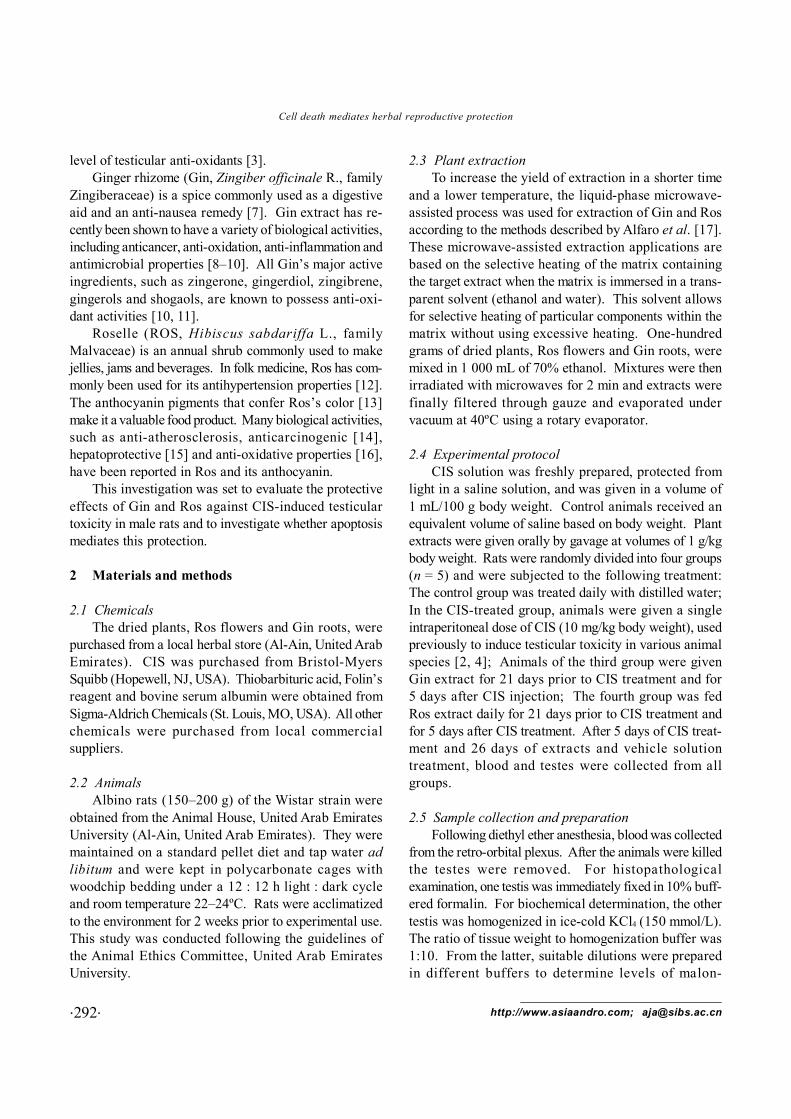
3.3 Effects on apoptosis in spermsThe effect of CIS on the percentage of apoptosis in
sperm was determined by flow cytometry. As shown inFigure 3, CIS induced significant increases in the per-
Figure 1. Photomicrograph of the seminiferous tubules of controlrats (A) showing the normal arrangement of germinal cells andSertoli cells. Testis of cisplatin-treated rats (B–D) showing tubularatrophy (asterisks) with extensive degeneration of germinalepithelium. The atrophic tubules contained degenerated Sertoli cellswith few germ cells. However, ginger pretreated (E) or roselle pre-treated (F) rats had less degeneration in some tubules and irregulararrangement of germ cells with shedding of cellular materials fromseminiferous epithelium in some tubules. Cells stained with hema-toxylin and eosin. Bars: (A)–(C), (E), (F) = 100 μm; (D) = 10 μm.
Figure 2. Terminal deoxynucleotidyl transferase-mediatedtriphoshate nick-end labeling (TUNEL)-positive cells in seminifer-ous tubules of rats treated with vehicle (control) (A), cisplatin(CIS) (B), ginger (Gin) + CIS (C), and roselle (Ros) + CIS (D).Photomicrographs (A–D) and the semiquantitative analysis (E)show variable levels of apoptosis in different experimental groups.The number of apoptotic cells in each section was calculated bycounting the number of TUNEL-positive cells in 10 fields per slideat × 400 magnification. This was repeated for all five animals ineach group and the average was plotted. Brown staining indicatesTUNEL-positive cells. TUNEL-positive cells are denoted by ar-rows (counterstained with hematoxylin, Bar = 10 μm). *P < 0.05,***P < 0.001, compared with control; +++P < 0.001, compared withCIS-treated group.
Asian J Androl 2008; 10 (2): 291–297
.295.Tel: +86-21-5492-2824; Fax: +86-21-5492-2825; Shanghai, China
centage of apoptosis in sperm. When rats were pre-treated with either Gin or Ros, this percentage of apopto-sis was significantly decreased as compared with theCIS group.
3.4 Effects on p53 protein expressionp53 protein expression was detected in seminiferous
tubules of both control and CIS-treated animals (Figure4A, B). Brown staining, indicating positive immuno-
stained cells, was significantly more (P < 0.001) in theCIS-treated group compared to the control group (Figure4B, E). The concomitant treatment with Gin or Rosbefore CIS treatment significantly prevented the increasein the number of p53-positive cells in the CIS-treatedgroup (Figure 4C, D, E).
3.5 Effects on testicular MDAA significant increase (P < 0.001) of testicular MDA
Figure 3. Flow cytometry DNA histogram of epididymal spermcells in rats. The histogram represents the counts for one represen-tative rat from each treatment group: control (A), cisplatin (CIS)(B), ginger (Gin) + CIS (C) and roselle (Ros) + CIS (D). Whenexcited with blue light, fluorescence stain associated with double-stranded DNA emits pink fluorescence, whereas single-strandedDNA emits blue fluorescence. (E): Effect of ginger (Gin) and roselle(Ros) on the percentage of apoptotic sperm cells in cisplatin (CIS)-treated rats. Each column represents the mean ± SE for five rats ineach treatment group. **P < 0.01, ***P < 0.001, compared withcontrol; +P < 0.05, +++P < 0.001, compared with CIS-treated group.
Figure 4. Protein expression of p53 in the seminiferous tubules ofrats treated with vehicle (Control) (A), cisplatin (CIS) (B), ginger(Gin) + CIS (C) and roselle (Ros) + CIS (D). Photomicrographs(A)–(D) and semiquantitative analysis (E) show the degree ofimmunostained cells in different experimental groups. The numberof p53-positive cells in each section was calculated by counting thenumber of immunostained cells in 10 fields per slide at × 400magnification. This was repeated for all five animals in each groupand the average was plotted. Brown staining indicates positive-stained cells. p53-positive cells are denoted by arrows (counter-stained with hematoxylin, Bar = 10 μm). ***P < 0.001, comparedwith control; +++P < 0.001, compared with CIS-treated group.

.296.
Cell death mediates herbal reproductive protection
http://www.asiaandro.com; [email protected]
Figure 5. Effect of ginger (Gin) and roselle (Ros) on testicularmalondialdehyde (MDA) levels in cisplatin (CIS)-treated rats.Each column represents the mean ± SE, for five rats in each group.***P < 0.001, compared with control; +++P < 0.001, compared withCIS-treated group.
expression. Elevation of p53 protein expression in re-sponse to DNA damage triggers either a transient cellcycle arrest or apoptosis [20, 21]. Sperms respond toexposure to a DNA-damaging agent by elevating p53protein levels [22]. It is therefore suggested here thatp53 is a necessary component in the CIS-mediatedapoptotic pathway of testicular epithelia.
Consistent with results reported elsewhere [3], thecurrent study shows that histological damage in testis isassociated with increase in testicular LP. CIS-treated ani-mals have shown an elevation in testicular MDA levelscompared with the control group. The decreased for-mation of anti-oxidants and the augmented activity offree radicals might account for the increase of MDA pro-duction in CIS-induced tissues. In our previous work,the levels of hepatic reduced glutathione as well as theenzyme activities of catalase and superoxide dismutasein the testis were lower in CIS-treated animals comparedto control animals. Several studies have shown that CIStoxicity in kidney is mediated by depletions of anti-oxi-dants and elevations of LP [23, 24]. CIS has also beensuggested to generate free radicals by interaction withDNA [25]. Therefore, overproduction of free radicalsand hence oxidative stress might account, at least in part,for testicular injury associated with CIS treatment.
Recently, much attention has been focused on the pro-tective effects of anti-oxidants and naturally occurringsubstances against oxidative stress damage. Gin or Rosextracts given before CIS treatment clearly attenuated thetesticular damage and decreased apoptotic damage bothin testes and sperms. It also retained the control value ofp53 protein expression in the testicular tissue. The pro-tective effect of plant extracts is accompanied by nor-malizing the increase of MDA. Gin crude extract and itsindividual constituents, such as zingerone, gingerdiol,zingibrene, gingerols, and shogaols, have been shown toprotect against LP in various established models [10, 11].Accumulating evidence suggests that the protective ef-fects of Ros against oxidative damage could be attrib-uted to its anti-oxidative properties [26–28]. The anti-oxidant activity of Ros could be attributed to its phenoliccontents, namely protocatechuic acid [28] and antho-cyanins [13, 16]. Ros has also been reported to preventor attenuate decrease in tissue anti-oxidant enzymes indifferent animal models and to provide cellular protec-tion against oxidative stress [3, 15]. In conclusion, thisstudy showed that apoptotic cell death might play animportant role in the development of CIS-induced tes-
was recorded after CIS treatment (Figure 5B). AlthoughMDA levels of pretreated animals (given Gin or Ros be-fore CIS treatment) did not return to the control level,there was no significant difference compared to thecontrol.
4 Discussion
Recent studies have shown the important role ofapoptosis in the pathogenesis of CIS testicular damage[1]. In the present study, the protective effect of Gin andRos against testicular damage induced by CIS was shownin rats. In addition to its role in normal testicular physiol-ogy [1], apoptosis of germ cells has been recently re-ported as a mechanism responsible for the toxic damageto spermatogenesis. CIS was reported to cause apoptosisto testicular germ cells and Sertoli cells [2, 4]. In thisstudy, apoptotic DNA fragmentation was determined intesticular tissue using the TUNEL technique and in epid-idymal sperm using cytometric assessment of DNAdamage. A single dose of CIS caused apoptosis in testes(germ cells and Sertoli cells) and in epididymal sperms.Consistent with the results of apoptosis, histologicalchanges were observed in the CIS-treated animal group.The high proportion of apoptosis in the present studysuggests that apoptosis is an important mechanism thatmight account for the marked loss of spermatogenic cellsin the CIS-intoxicated testes. The CIS-induced testicu-lar damage was also associated with upregulation of p53

Asian J Androl 2008; 10 (2): 291–297
.297.Tel: +86-21-5492-2824; Fax: +86-21-5492-2825; Shanghai, China
ticular damage. Both Gin and Ros are reported here tohave a potent protective effect on CIS-induced testiculardamage and apoptotic cell death in rats. The protectiveeffect of Gin and Ros might be due to their anti-oxidantproperties.
Acknowledgment
The authors are grateful to Ms. Karima Al-Mansouri(Biology Department, United Arab Emirates University)and for Mr. Moustafa A. Abdalla for their help in format-ting the manuscript.
References
1 Boekelheide K. Mechanisms of toxic damage to spermatogenesis.J Natl Cancer Inst Monogr 2005; 34: 6–8.
2 Cherry SM, Hunt PA, Hassold TJ. Cisplatin disrupts mam-malian spermatogenesis, but does not affect recombination orchromosome segregation. Mutat Res 2004; 564: 115–28.
3 Amin A, Hamza A. Effects of ginger and roselle on cisplatin-induced reproductive toxicity in rats. Asian J Androl 2006; 8:607–12.
4 Zhang X, Yamamoto N, Soramoto S, Takenaka I. Cisplatin-induced germ cell apoptosis in mouse testes. Arch Androl2001; 46: 43–9.
5 Hooser SB, van Dijk-Knijnenburg WC, Waalkens-BerendsenID, Smits-van Prooije AE, Snoeij NJ, Baan RA, Fichtinger-Schepman AM. Cisplatin-DNA adduct formation in rat sper-matozoa and its effects on fetal development. Cancer Lett2000; 151: 71–80.
6 Ateşşahin A, Karahan I, Türk G, Gür S, Yılmaz S, CeribasiAO. Protective role of lycopene on cisplatin-induced changesin sperm characteristics, testicular damage and oxidative stressin rats. Reprod Toxicol 2006; 21: 42–7.
7 Bryer E. A literature review of the effectiveness of ginger inalleviating mild-to-moderate nausea and vomiting of pregnancy.J Midwifery Womens Health 2005; 50: e1–3.
8 Lee E, Surh YJ. Induction of apoptosis in HL-60 cells bypungent vanilloids, [6]-gingerol and [6]-paradol. Cancer Lett1998; 134: 163–8.
9 Chung WY, Jung YJ, Surh YJ, Lee SS, Park KK. Antioxidativeand antitumor promoting effects of [6]-paradol and itshomologs. Mutat Res 2001; 496: 199–206.
10 Chrubasik S, Pittler MH, Roufogalis BD. Zingiberis rhizoma:a comprehensive review on the ginger effect and efficacyprofiles. Phytomedicine 2005; 12: 684–701.
11 Zancan KC, Marques MO, Petenate AJ, Meireles MA. Ex-traction of ginger (Zingiber officinale Roscoe) oleoresin withCO2 and co-solvents: a study of the antioxidant action of theextracts. J Supercrit Fluids 2002; 24: 57–76.
12 Herrera-Arellano A, Flores-Romero S, Chávez-Soto MA,Tortoriello J. Effectiveness and tolerability of a standardized
extract from Hibiscus sabdariffa in patients with mild to mod-erate hypertension: a controlled and randomized clinical trial.Phytomedicine 2004; 11: 375–82.
13 Tsai PJ, Huang HP. Effect of polymerization on the antioxi-dant capacity of anthocyanins in Roselle. Food Res Intern2004; 37: 313–8.
14 Tsai PJ, McIntosh J, Pearce P, Camden B, Jordan BR. Antho-cyanin and antioxidant capacity in Roselle (Hibiscus sabdariffaL.) extract. Food Res Intern 2002; 35: 351–6.
15 Amin A, Hamza AA. Hepatoprotective effects of Hibiscus,Rosmarinus and Salvia on azathioprine-induced toxicity inrats. Life Sci 2005; 77: 266–78.
16 Prenesti E, Berto S, Daniele PG, Toso S. Antioxidant powerquantification of decoction and cold infusions of Hibiscussabdariffa flowers. Food Chem 2007; 100: 433–8.
17 Alfaro MJ, Belanger JM, Padilla FC, Pare JR. Influence ofsolvent, matrix dielectric properties, and applied power onthe liquid-phase microwave-assisted processes (MAP) ex-traction of ginger (Zingiber officinale). Food Res Intern 2003;36: 499–504.
18 Uchiyama M, Mihara M. Determination of malonaldehydeprecursor in tissues by thiobarbituric acid test. Anal Biochem1978; 86: 271–8.
19 Peterson GL. A simplification of the protein assay method ofLowry which is more generally applicable. Anal Biochem1977; 83: 346–56.
20 Gomez-Lazaro M, Fernandez-Gomez FJ, Jordán J. P53:twenty-five years understanding the mechanism of genomeprotection. J Physiol Biochem 2004; 60: 287–307.
21 Szoke D, Sipos F, Spisák F, Molnár B, Tulassay Z. The p53gene and protein in 2005: new results, promising opportunities.Orv Hetil 2005; 146: 1587–94.
22 Wang J, Biju MP, Wang M, Haase VH, Dong Z. Cytoprotectiveeffects of hypoxia against cisplatin-induced tubular cellapoptosis: involvement of mitochondrial inhibition and p53suppression. J Am Soc Nephrol 2006; 17: 1875–85.
23 Priya SD, Devi CSS. Protective effect of quercetin in cisplatin-induced cell injury in the rat kidney. Indian J Pharmacol 1999;31: 422–6.
24 Yokozawa T, Nakagawa T, Lee KI, Cho EJ, Terasawa K,Takeuchi S. Effect of green tea tannin on cisplatin-inducednephropathy in LLC-PK1 cells and rats. J Pharm Pharmacol1999; 51: 1325–31.
25 Masuda H, Tanaka T, Takahama U. Cisplatin generates su-peroxide anion by interaction with DNA in a cell-free system.Biochem Biophys Res Commun 1994; 203: 1175–80.
26 Tseng T, Kao ES, Chu CY, Chou FP, Lin Wu HW, Wang CJ.Protective effects of dried flower extracts of Hibiscus sabdariffaL. against oxidative stress in rat primary hepatocytes. FoodChem Toxicol 1997; 35: 1159–64.
27 Wang C, Wang J, Lin W, Chu C, Chou F, Tseng T. Protectiveeffect of hisbiscus anthocyanins against tert-butyl hydroper-oxide-induced hepatic toxicity in rats. Food Chem Toxicol2000; 38: 411–6.
28 Liu C, Wang J, Chu C, Cheng M, Tseng T. In vivo protectiveeffect of protocatechuic acid on tert-butyl hydroperoxide-inducedrat hepatotoxicity. Food Chem Toxicol 2002; 40: 635–41.
Edited by Dr William Moorman


![ForeScout CounterACT Supplemental Administrative … Secure Acceptance, Installation, and Configuration ... CounterACT® Installation Guide Version 7.0.0 [2] CounterACT® Console User](https://img.dokumen.tips/doc/110x75/5b0d73937f8b9a685a8e27f5/forescout-counteract-supplemental-administrative-secure-acceptance-installation.jpg)