Embed Size (px)
Citation preview
319
Abstract: Hemifacial microsomia is a congenitalmalformation in which there is deficiency in the amountof hard and soft tissues on one side of the face. It isprimarily a syndrome of first and second branchialarches invo lv ing underdeve lopment o f thetemporomandibular joint, mandibular ramus,masticatory muscles, ears and occasionally defects infacial nerve and muscles. Here, we report three casesof hemifacial microsomia diagnosed based on clinicaland radiographic findings. All three cases had variablepresentations ranging from the mildest form thatincluded facial asymmetry and ear deformity to themost severe and unusual form with facial nerve paralysisand spine deformity. (J Oral Sci 52, 319-324, 2010)
Keywords: hemifacial microsomia; facial nerveparalysis; spine deformity.
IntroductionHemifacial microsomia (HFM) is the second most
common congenital facial anomaly after cleft lip/palate,with a reported incidence of about 1 in 5,600 live births(1). HFM was first described by German physician CarlFerdinand Von Arlt in 1881. Gorlin et al. used the termHFM to describe patients with unilateral microtia,macrostomia and malformation of mandibular ramus and
condyle, whereas Goldenhar syndrome was described asa variant, with vertebral anomalies and epibulbar dermoids.The name, craniofacial microsomia, was proposed byConverse et al. when cranial deformities were included.Other synonyms include first arch syndrome, first andsecond branchial arch syndrome, otomandibular dysostosis,oculo-auriculovertebral dysplasia and lateral facial dysplasia(2). Three case reports of hemifacial microsomia withvariable and uncommon features are discussed here.
Case ReportsCase 1
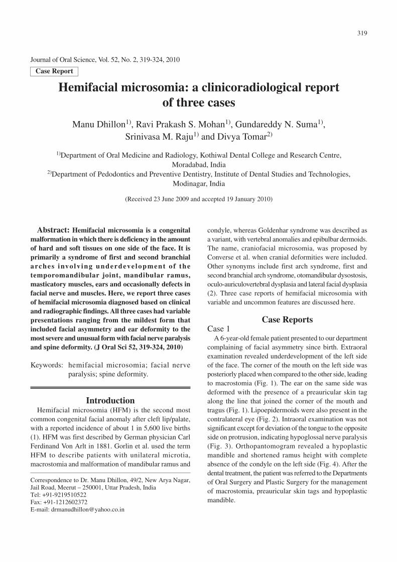
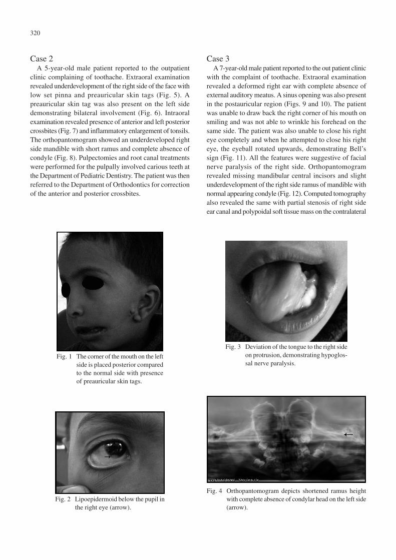
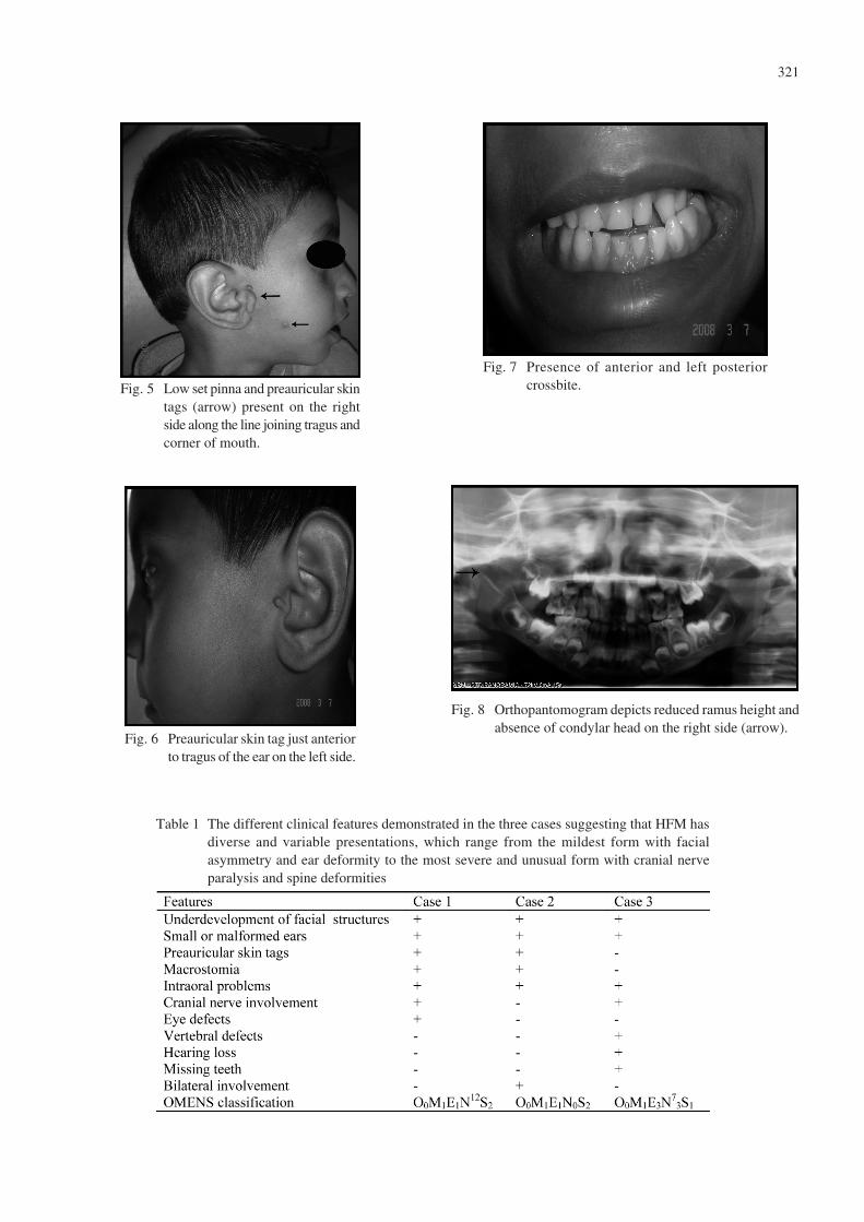
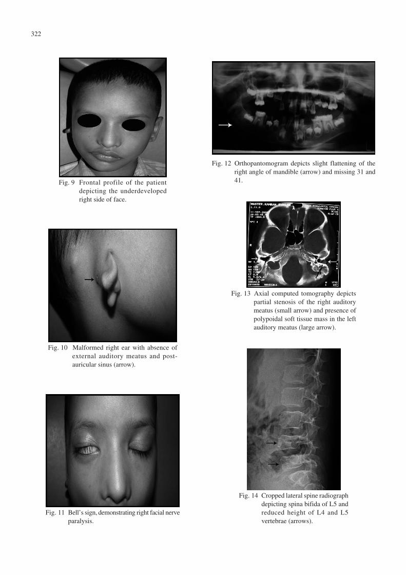
A 6-year-old female patient presented to our departmentcomplaining of facial asymmetry since birth. Extraoralexamination revealed underdevelopment of the left sideof the face. The corner of the mouth on the left side wasposteriorly placed when compared to the other side, leadingto macrostomia (Fig. 1). The ear on the same side wasdeformed with the presence of a preauricular skin tagalong the line that joined the corner of the mouth andtragus (Fig. 1). Lipoepidermoids were also present in thecontralateral eye (Fig. 2). Intraoral examination was notsignificant except for deviation of the tongue to the oppositeside on protrusion, indicating hypoglossal nerve paralysis(Fig. 3). Orthopantomogram revealed a hypoplasticmandible and shortened ramus height with completeabsence of the condyle on the left side (Fig. 4). After thedental treatment, the patient was referred to the Departmentsof Oral Surgery and Plastic Surgery for the managementof macrostomia, preauricular skin tags and hypoplasticmandible.
Journal of Oral Science, Vol. 52, No. 2, 319-324, 2010
Correspondence to Dr. Manu Dhillon, 49/2, New Arya Nagar,Jail Road, Meerut – 250001, Uttar Pradesh, IndiaTel: +91-9219510522Fax: +91-1212602372E-mail: [email protected]
Hemifacial microsomia: a clinicoradiological report of three cases
Manu Dhillon1), Ravi Prakash S. Mohan1), Gundareddy N. Suma1), Srinivasa M. Raju1) and Divya Tomar2)
1)Department of Oral Medicine and Radiology, Kothiwal Dental College and Research Centre, Moradabad, India
2)Department of Pedodontics and Preventive Dentistry, Institute of Dental Studies and Technologies,Modinagar, India
(Received 23 June 2009 and accepted 19 January 2010)
Case Report
320
Case 2A 5-year-old male patient reported to the outpatient
clinic complaining of toothache. Extraoral examinationrevealed underdevelopment of the right side of the face withlow set pinna and preauricular skin tags (Fig. 5). Apreauricular skin tag was also present on the left sidedemonstrating bilateral involvement (Fig. 6). Intraoralexamination revealed presence of anterior and left posteriorcrossbites (Fig. 7) and inflammatory enlargement of tonsils.The orthopantomogram showed an underdeveloped rightside mandible with short ramus and complete absence ofcondyle (Fig. 8). Pulpectomies and root canal treatmentswere performed for the pulpally involved carious teeth atthe Department of Pediatric Dentistry. The patient was thenreferred to the Department of Orthodontics for correctionof the anterior and posterior crossbites.
Case 3A 7-year-old male patient reported to the out patient clinic
with the complaint of toothache. Extraoral examinationrevealed a deformed right ear with complete absence ofexternal auditory meatus. A sinus opening was also presentin the postauricular region (Figs. 9 and 10). The patientwas unable to draw back the right corner of his mouth onsmiling and was not able to wrinkle his forehead on thesame side. The patient was also unable to close his righteye completely and when he attempted to close his righteye, the eyeball rotated upwards, demonstrating Bell’ssign (Fig. 11). All the features were suggestive of facialnerve paralysis of the right side. Orthopantomogramrevealed missing mandibular central incisors and slightunderdevelopment of the right side ramus of mandible withnormal appearing condyle (Fig. 12). Computed tomographyalso revealed the same with partial stenosis of right sideear canal and polypoidal soft tissue mass on the contralateral
Fig. 1 The corner of the mouth on the leftside is placed posterior comparedto the normal side with presenceof preauricular skin tags.
Fig. 2 Lipoepidermoid below the pupil inthe right eye (arrow).
Fig. 3 Deviation of the tongue to the right sideon protrusion, demonstrating hypoglos-sal nerve paralysis.
Fig. 4 Orthopantomogram depicts shortened ramus heightwith complete absence of condylar head on the left side(arrow).
321
Fig. 5 Low set pinna and preauricular skintags (arrow) present on the rightside along the line joining tragus andcorner of mouth.
Fig. 6 Preauricular skin tag just anteriorto tragus of the ear on the left side.
Fig. 7 Presence of anterior and left posteriorcrossbite.
Fig. 8 Orthopantomogram depicts reduced ramus height andabsence of condylar head on the right side (arrow).
Table 1 The different clinical features demonstrated in the three cases suggesting that HFM hasdiverse and variable presentations, which range from the mildest form with facialasymmetry and ear deformity to the most severe and unusual form with cranial nerveparalysis and spine deformities
322
Fig. 14 Cropped lateral spine radiographdepicting spina bifida of L5 andreduced height of L4 and L5vertebrae (arrows).
Fig. 11 Bell’s sign, demonstrating right facial nerveparalysis.
Fig. 10 Malformed right ear with absence ofexternal auditory meatus and post-auricular sinus (arrow).
Fig. 9 Frontal profile of the patientdepicting the underdevelopedright side of face.
Fig. 12 Orthopantomogram depicts slight flattening of theright angle of mandible (arrow) and missing 31 and41.
Fig. 13 Axial computed tomography depictspartial stenosis of the right auditorymeatus (small arrow) and presence ofpolypoidal soft tissue mass in the leftauditory meatus (large arrow).
323
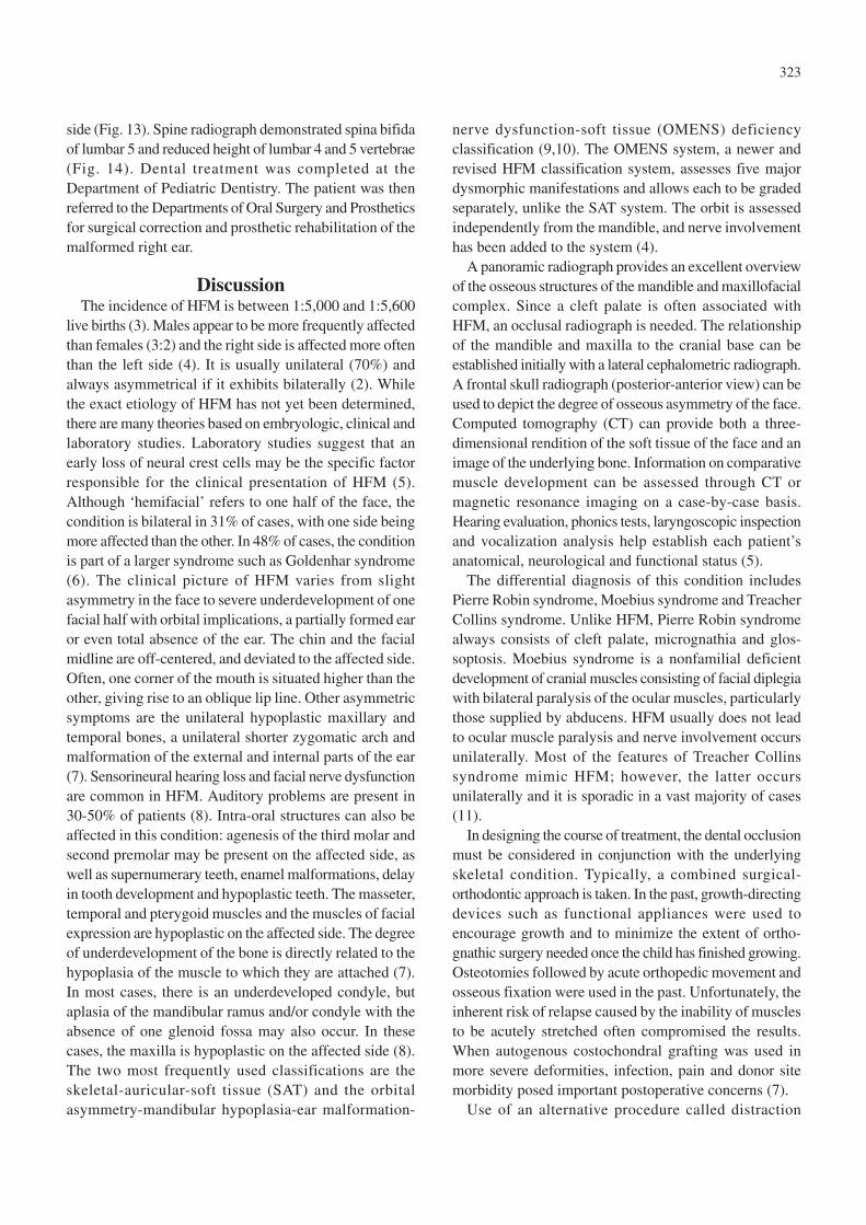
side (Fig. 13). Spine radiograph demonstrated spina bifidaof lumbar 5 and reduced height of lumbar 4 and 5 vertebrae(Fig. 14). Dental treatment was completed at theDepartment of Pediatric Dentistry. The patient was thenreferred to the Departments of Oral Surgery and Prostheticsfor surgical correction and prosthetic rehabilitation of themalformed right ear.
DiscussionThe incidence of HFM is between 1:5,000 and 1:5,600
live births (3). Males appear to be more frequently affectedthan females (3:2) and the right side is affected more oftenthan the left side (4). It is usually unilateral (70%) andalways asymmetrical if it exhibits bilaterally (2). Whilethe exact etiology of HFM has not yet been determined,there are many theories based on embryologic, clinical andlaboratory studies. Laboratory studies suggest that anearly loss of neural crest cells may be the specific factorresponsible for the clinical presentation of HFM (5).Although ‘hemifacial’ refers to one half of the face, thecondition is bilateral in 31% of cases, with one side beingmore affected than the other. In 48% of cases, the conditionis part of a larger syndrome such as Goldenhar syndrome(6). The clinical picture of HFM varies from slightasymmetry in the face to severe underdevelopment of onefacial half with orbital implications, a partially formed earor even total absence of the ear. The chin and the facialmidline are off-centered, and deviated to the affected side.Often, one corner of the mouth is situated higher than theother, giving rise to an oblique lip line. Other asymmetricsymptoms are the unilateral hypoplastic maxillary andtemporal bones, a unilateral shorter zygomatic arch andmalformation of the external and internal parts of the ear(7). Sensorineural hearing loss and facial nerve dysfunctionare common in HFM. Auditory problems are present in30-50% of patients (8). Intra-oral structures can also beaffected in this condition: agenesis of the third molar andsecond premolar may be present on the affected side, aswell as supernumerary teeth, enamel malformations, delayin tooth development and hypoplastic teeth. The masseter,temporal and pterygoid muscles and the muscles of facialexpression are hypoplastic on the affected side. The degreeof underdevelopment of the bone is directly related to thehypoplasia of the muscle to which they are attached (7).In most cases, there is an underdeveloped condyle, butaplasia of the mandibular ramus and/or condyle with theabsence of one glenoid fossa may also occur. In thesecases, the maxilla is hypoplastic on the affected side (8).The two most frequently used classifications are theskeletal-auricular-soft tissue (SAT) and the orbitalasymmetry-mandibular hypoplasia-ear malformation-
nerve dysfunction-soft tissue (OMENS) deficiencyclassification (9,10). The OMENS system, a newer andrevised HFM classification system, assesses five majordysmorphic manifestations and allows each to be gradedseparately, unlike the SAT system. The orbit is assessedindependently from the mandible, and nerve involvementhas been added to the system (4).
A panoramic radiograph provides an excellent overviewof the osseous structures of the mandible and maxillofacialcomplex. Since a cleft palate is often associated withHFM, an occlusal radiograph is needed. The relationshipof the mandible and maxilla to the cranial base can beestablished initially with a lateral cephalometric radiograph.A frontal skull radiograph (posterior-anterior view) can beused to depict the degree of osseous asymmetry of the face.Computed tomography (CT) can provide both a three-dimensional rendition of the soft tissue of the face and animage of the underlying bone. Information on comparativemuscle development can be assessed through CT ormagnetic resonance imaging on a case-by-case basis.Hearing evaluation, phonics tests, laryngoscopic inspectionand vocalization analysis help establish each patient’sanatomical, neurological and functional status (5).
The differential diagnosis of this condition includesPierre Robin syndrome, Moebius syndrome and TreacherCollins syndrome. Unlike HFM, Pierre Robin syndromealways consists of cleft palate, micrognathia and glos-soptosis. Moebius syndrome is a nonfamilial deficientdevelopment of cranial muscles consisting of facial diplegiawith bilateral paralysis of the ocular muscles, particularlythose supplied by abducens. HFM usually does not leadto ocular muscle paralysis and nerve involvement occursunilaterally. Most of the features of Treacher Collinssyndrome mimic HFM; however, the latter occursunilaterally and it is sporadic in a vast majority of cases(11).
In designing the course of treatment, the dental occlusionmust be considered in conjunction with the underlyingskeletal condition. Typically, a combined surgical-orthodontic approach is taken. In the past, growth-directingdevices such as functional appliances were used toencourage growth and to minimize the extent of ortho-gnathic surgery needed once the child has finished growing.Osteotomies followed by acute orthopedic movement andosseous fixation were used in the past. Unfortunately, theinherent risk of relapse caused by the inability of musclesto be acutely stretched often compromised the results.When autogenous costochondral grafting was used inmore severe deformities, infection, pain and donor sitemorbidity posed important postoperative concerns (7).
Use of an alternative procedure called distraction
324
osteogenesis is now widely accepted. It is a process in whichnew bone is formed between the surfaces of bone segmentsthat are gradually separated by incremental traction. Thisis a gradual method of creating bone after a surgicalcorticotomy sectioning of the cortical plates. Prosthetic earreconstruction can also be done for deformed ears (7).
Interestingly, the three cases reported here showedvariation from common features such as macrostomia,underdeveloped mandible and deformed ears to uncommonand unusual features like nerve paralysis, lipodermoids andvertebral defects. Dental surgeons should be aware ofvariable presentations of this syndrome which help todistinguish it from other syndromes so that proper treatmentcan be planned.
References1. Kapur R, Kapur R, Sheikh S, Jindal S, Kulkarni S
(2008) Hemifacial microsomia: a case report. JIndian Soc Pedod Prevent Dent 26, S34-S40.
2. Kalsotra P, Chowdhary A, Bhagat DR, Parihar SS,Prabhakar R (2006) Craniofacial microsomia. J KSci 8, 168-170.
3. Gorlin RJ, Cohen MM, Levin LS (1990) Syndromesof the head and neck. 3rd ed, Oxford UniversityPress, New York, 641-652.
4. Wang RR, Andres CJ (1999) Hemifacial microsomiaand treatment options for auricular replacement: a
review of the literature. J Prosthet Dent 82, 197-204.5. Monahan R, Seder K, Patel P, Alder M, Grud S,
O’Gara M (2001) Hemifacial microsomia: etiology,diagnosis and treatment. J Am Dent Assoc 132,1402-1408.
6. Singer SL, Haan E, Slee J, Goldblatt J (1994)Familial hemifacial microsomia due to autosomaldominant inheritance. Case reports. Aust Dent J39, 287-291.
7. Moulin-Romsée C, Verdonck A, Schoenaers J,Carels C (2004) Treatment of hemifacial microsomiain a growing child: the importance of co-operationbetween the orthodontist and the maxillofacialsurgeon. J Orthod 31, 190-200.
8. Carvalho GJ, Song CS, Vargervik K, Lalwani AK(1999) Auditory and facial nerve dysfunction inpatients with hemifacial microsomia. ArchOtolaryngol Head Neck Surg 125, 209-212.
9. David DJ, Mahatumarat C, Cooter RD (1987)Hemifacial microsomia: a multisystem classification.Plast Reconstr Surg 80, 525-535.
10. Vento AR, LaBrie RA, Mulliken JB (1991) TheO.M.E.N.S. classification of hemifacial microsomia.Cleft Palate Craniofac J 28, 68-77.
11. Rajendran R, Sivapathasundaram B (2006) Shafer’stextbook of oral pathology. 5th ed, Elsevier, NewDelhi, 986-989, 1189-1190.


![CaseReport …[8] E.M.Ongkosuwito,J.vanVooren,J.W.vanNecketal., “Changes of mandibular ramal height, during growth in unilateral hemifacial microsomia patients and unaected](https://img.dokumen.tips/doc/110x75/60c4829f23e96b545e31e549/casereport-8-emongkosuwitojvanvoorenjwvannecketal-aoechanges-of-mandibular.jpg)


![Review Article Hemifacial Spasm and Neurovascular Compressiondownloads.hindawi.com/journals/tswj/2014/349319.pdf · 2019-07-31 · improve hemifacial spasm-related headaches [ ]](https://img.dokumen.tips/doc/110x75/5f2bfcee1f6d0d036319a21e/review-article-hemifacial-spasm-and-neurovascular-2019-07-31-improve-hemifacial.jpg)