Embed Size (px)
Citation preview

1
Hematological profile of East African Short-Horn Zebu calves
from birth to 51 weeks of age
I. Conradie van Wyk, A. Goddard, B. M. de C. Bronsvoort, J. A. W. Coetzer,C. Booth, O. Hanotte, A. Jennings, H. Kiara, P. Mashego, C. Muller, G.Pretorius, E. J. Poole, S. M. Thumbi, P. G. Toye, M. E. J. Woolhouse &B. L. Penzhorn
I. C. van Wyk (*) : J. A. W. Coetzer : B. L. PenzhornDepartment of Veterinary Tropical Diseases,Faculty of Veterinary Science, University of Pretoria,Private Bag X4, Onderstepoort,Pretoria 0110, South Africae-mail: [email protected]
A. Goddard : C. Booth : P. Mashego : C. Muller : G. PretoriusClinical Pathology, Department Companion Animal Medicine,Faculty of Veterinary Science, University of Pretoria,Private Bag X4, Onderstepoort,Pretoria 0110, South Africa
B. M. de C. Bronsvoort : A. JenningsThe Roslin Institute, University of Edinburgh,Easter Bush,EH25 9RG, Edinburgh, UK
O. HanotteSchool of Biology, University of Nottingham,Nottingham NG7 2RD, UK
H. Kiara : E. J. Poole : P. G. ToyeInternational Livestock Research Institute,P.O. Box 30709-00100, Nairobi, Kenya
S. M. Thumbi : M. E. J. WoolhouseCentre for Immunology, Infection & Evolution,University of Edinburgh,Edinburgh EH9 3JT, UK
Abstract
This paper is the first attempt to accurately describe the hematological parameters for
any African breed of cattle, by capturing the changes in these parameters over the first
12 months of an animal’s life using a population-based sample of calves reared under
field conditions and natural disease challenge. Using a longitudinal study design, a
stratified clustered random sample of newborn calves was recruited into the IDEAL
study and monitored at 5-weekly intervals until 51 weeks of age. The blood cell

2
analysis performed at each visit included: packed cell volume; red cell count; red cell
distribution width; mean corpuscular volume; mean corpuscular hemoglobin
concentration; hemoglobin concentration; white cell count; absolute lymphocyte,
eosinophil, monocyte, and neutrophil counts; platelet count; mean platelet volume;
and total serum protein. The most significant age-related change in the red cell
parameters was a rise in red cell count and hemoglobin concentration during the
neonatal period. This is in contrast to what is reported for other ruminants, including
European cattle breeds where the neonatal period is marked by a fall in the red cell
parameters. There is a need to establish breed-specific reference ranges for blood
parameters for indigenous cattle breeds. The possible role of the postnatal rise in the
red cell parameters in the adaptability to environmental constraints and innate disease
resistance warrants further research into the dynamics of blood cell parameters of
these breeds.
Introduction
The sedentary mixed crop-livestock smallholding system encompasses >50% of poor people
resident in East Africa. These resource-poor subsistence farmers own only a few animals, yet
these animals are one of their principal capital assets. Subsistence farmers rely on livestock
for food security, financial income and social welfare. Indigenous breeds are regionally very
important and constitute up to 77% of the total Kenyan cattle population (Rege et al. 2001).
The small East African Short-horn Zebu, in particular, is the most common breed in eastern
and parts of south-central Africa (Mwacharo et al. 2006). Indigenous breeds are preferred by
farmers for their adaptability in terms of disease resistance, heat tolerance, and water and food
requirements (Mwacharo and Drucker 2005). Cattle are used for animal traction, milk and
meat, and the dung for fertilizer (Rege et al. 2001).

3
The tropical climate in western Kenya is conducive for the survival of many infectious
pathogens and vectors. The most economically important diseases of livestock in sub-Saharan
Africa are tick-borne diseases (East Coast fever (ECF), heartwater, anaplasmosis and
babesiosis), trypanosomosis (Uilenberg 1995; Minjauw and McLeod 2003; Maudlin 2006)
and helminthosis. Small-holder farmers are particularly vulnerable to the economic impact of
infectious diseases of livestock. Generally tick-borne infections do not affect trade in
livestock, but they are a significant cause of production losses (Perry and Young 1995).
Losses include lowered production rates, mortalities, decreased reproduction rates and costs
of treatment and control measures. These diseases also indirectly constrain livestock
production through limiting the use of the highly susceptible improved breeds of livestock
that are used in other countries to improve livestock productivity (Perry and Young 1995).
Baseline values of the parameters that can be used to measure an animal’s health status,
including hematological parameters, are lacking for indigenous cattle breeds. Most published
reference ranges for these parameters for cattle were compiled for European breeds (Jain
1993, Knowles et al. 2000; Mohri et al. 2007). Studies have indicated that baseline values for
hematological parameters differ between the various cattle breeds in the tropics (Oduye and
Okunaiya 1971). The aim of this study was to investigate the hematological profile of East
African Short-horn Zebu cattle living in tropical field conditions in Western Kenya, in
particular the age-related changes in the blood parameters from neonate to 51 weeks. This
paper is the first attempt to accurately describe the hematological parameters for any African
cattle breed capturing the changes in these parameters over the first 12 months of an animal’s
life using a population based sample of calves reared in their natural environment and disease
challenges.
Materials and Methods
The study was carried out in Busia district, Western Kenya (Fig. 1) and a full description of
the design is given by Bronsvoort et al. (2012) (in press). This site was considered to

4
represent of smallholder livestock farming systems in East Africa. The climate is tropical and
the main land use in the area is mixed crop/livestock-farming. The main crops cultivated
include maize, sugarcane, cotton, pigeon-peas and sisal. The main cattle breed kept is the
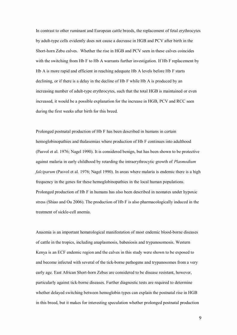
indigenous Zebu, of which the majority are East African Short-horn Zebus. These Short-horn
Zebus are a relatively small breed but are considered to be well adapted to the local
environmental constraints (Fig. 2). The calves recruited into the study remained with their
herd of origin, and were exposed to the natural pathogen challenge in the field. For the
duration of the study no interventions in terms of prevention or treatment (except on welfare
grounds in which case calves were censored) against infection were practiced.
A stratified 2-stage cluster sampling strategy was used. Stratification was by agro-ecological
zone (AEZ) (Jaetzold and Schmidt 1983) with 1st stage cluster (sub-location) selected using
stratified-random sampling with replacement and 2nd stage cluster (calf) selected using
ordinary random sampling without replacement. A total of 548 calves from sedentary mixed
crop-livestock smallholding farms were recruited between October 2007 and September 2009.
Calves were recruited within 7 days of birth and monitored routinely at 5-weekly intervals
until 51 weeks of age. At each visit a complete clinical examination of the calf was performed
and relevant samples collected. These samples included thin and thick peripheral smears of
blood collected from the marginal ear vein, jugular blood in 10 ml plain vacutainer tube for
the separation of serum, and jugular blood for hematology in 5 ml ethylene-diamine-tetra-
acetic acid (EDTA) plastic tubes.
Initial sample processing and analysis occurred at the local field laboratory, Busia, Kenya.
Packed cell volume (PCV) was measured manually using a Hawksley microhematocrit reader
(Jain 1993). Automated blood cell analysis was performed using the pocH-100iV Diff
(Sysmex© Europe GMBH). The hematological parameters investigated included: red cell
count (RCC), hemoglobin concentration (HGB), mean corpuscular volume (MCV), mean
corpuscular hemoglobin concentration (MCHC), red cell distribution width (RDW), total

5
white cell count (WCC), platelet count (Plt) and mean platelet volume (MPV). Total serum
protein (TSP) was measured from 100 μL serum using a refractometer (model RHC-200ATC,
Westover Scientific).
In addition to the automated blood analysis, thin blood smears were made from EDTA blood
for manual differential cell counts. These smears were transported to the University of
Pretoria, South Africa, where blood smears were stained with Diff Quick (Kyron South
Africa) for differential counts to calculate the absolute lymphocyte count (Lymph), absolute
eosinophil count (Eos), absolute neutrophil count (Neut) and absolute monocyte count
(Mono).
Descriptive statistics (mean and 95% confidence intervals) were used to describe the variation
and spread in each hematological parameter of the calf population at each sampling point.
Graphs are used to illustrate the changes in each parameter over time. The computation of the
results and the production of the graphs were done with R 2.8.1 (Ihaka and Gentleman 1996).
Results
Figures 3-13 illustrate the changes for all parameters for the total calf population between day
7 and day 364.
The calves showed a significant increase in PCV, RCC and HGB from day 7 (means of 30%,
7.73 x10⁶/μL and 10 g/dL, respectively) to day 42 (means of 36%, 10.23x10⁶/μL and 12 g/dL)
(Figs. 3-5). From day 42, the general trend for all three parameters was a decrease until day
364. The RDW was relatively high between day 7 to 42 (means of 34.9 and 35.9 fL,
respectively), indicating a significant variation in the size of red blood cells, which is
suggestive of a high number of immature red blood cells (Fig. 6). From day 42 to day 112 the
RDW decreased significantly, and then it remained between 31.2 and 31.8 fL to day 364. The
MCV (Fig. 7) was relatively high at day 7 (mean 39.8 fL), confirming the presence of a high

6
number of immature red blood cells, but decreased gradually from day 7 up to day 147 (mean
33.6 fL), from where it increased gradually until day 364 (mean 36.0 fL). The MCHC (Fig. 8)
showed a similar but more gradual trend, where it decreased between day 7 (mean 32.2 g/dL)
and day 77 (mean 31.6 g/dL), and then increased up to day 364 (mean 33.05 g/dL).
The WCC and Lymph (Figs. 9 and 10) showed very gradual increases from day 7 (mean 9.00
x103/uL and 3.7 x10³/μL) to day 364 (mean 11.40 x10³/μL and 7.6 x10³/μL). The Eos (Fig.
11) increased considerably from day 7 (mean 0.27 x10³/μL) to day 322 (mean 0.75 x10³/μL).
The Neut showed a gradual decrease from day 7 to day 112, after which it remained between
2.4-2.55 x10³/μL (mean) until day 287 and increased again to day 364.
The Plt (Fig. 12) also showed a steady decrease from day 7 (mean 611 x103/μL) up to day 364
(mean 385.5 x10³/μL). The MPV was relatively high at day 7 (mean 6.5 fL), decreased
gradually up to day 112 (mean 5.8 fL) and increased up to day 364 (mean 6.2 fL).
The TSP (Fig. 13) was relatively high at day 7 (mean 9.8 g/dL), after which it decreased up to
day 112 (mean 7.6 g/dL) but then remained between 7.6-8.0 g/dL (mean) up to day 364.
Discussion
This study showed that the hematological profile of the Short-horn Zebu differed both in
levels and age-related trends compared to reference ranges reported for European breeds. For
comparison, reported ranges for PCV for other indigenous cattle breeds are depicted in Table
1 and for European breed in Table 2, for which age-related changes are reported in more
detail.
The most significant age-related changes in the red blood cell parameters of the Short-horn
Zebu calves in this study occurred between day 7 and day 112. Concurrent low RCC and PCV
with a relatively high MCV and RDW in the first week suggest a macrocytic anemia. A high

7
RDW value is an indication of anisocytosis which is often seen in regenerative responses due
to a high number of reticulocytes. Following day 7 up to day 42 there was a significant
increase in PCV, HGB and RCC accompanied by a high RDW, suggestive of increased
erythropoiesis. The high MCV and a concurrent decrease in MCHC seen between day 7 and
day 42 also indicates an increase in the number of immature RBC which is indicative of
increased erythropoiesis. In neonatal Holstein calves erythrocytes were found to decrease in
size up to the age of 3-4 months (Mohri et al. 2007), resulting in a gradual decrease in MCV.
Immature red blood cells do not yet produce hemoglobin optimally since hemoglobin
production per cell increases as red blood cells mature (Harvey 1989), hence a lower MCHC.
After day 42 there was a decrease in PCV and HGB together with a continuing decrease in
MCV and MCHC, while RCC started decreasing from day 77. This may be suggestive of the
development of microcytic hypochromic anemia. Iron supplementation during the first four
weeks after birth corrected the drop in HGB in dairy calves (Mohri et al. 2004). The iron
levels of calf serum were not evaluated in this study but could possibly have added value to
explaining the results in this study.
The relatively low numbers of WCC and Lymph and high Neut around birth are consistent
with what is reported for other cattle breeds (Knowles et al. 2000; Mohri et al. 2007) and are
due to perinatal stress and high levels of cortisol during partus (Jain 1993). After the first
week, levels in WCC, Lymph and Eos increased as the immune system matured and the
animals became exposed to pathogens with resultant cellular immune responses. The Eos and
Mono of the Short-horn Zebu were considerably higher than published ranges for neonatal
calves of European breeds (Knowles et al. 2000; Mohri et al. 2007), but are comparable to
ranges reported for other indigenous African cattle breeds (Oduye and Okunaiya 1971) and
are possibly related to high parasite burdens under field conditions.

8
The platelet counts of the Short-horn Zebus were relatively high at day 7 and then decreased
significantly up to day 364. The MPV was also relatively high at week 1. Together with a
high RDW at day 7 this is suggestive of a regenerative response by the bone marrow at the
time around birth in both red cells and platelets.
The initial high levels in TSP in the first week were likely due to the uptake of
immunoglobulins from colostrum. The degradation of absorbed immunoglobulins likely
contributed to the age-related decrease in TSP after the first week. Chronic parasitic
infections, such as severe helminthosis, or malnutrition or a combination of both could
possibly also have contributed to the gradual decrease in TSP after day 7.
The increase found in the red cell parameters during the neonatal period in the Short-horn
Zebu calves in this study is in contrast to what is described for other ruminants, including
European cattle breeds (Karesh et al. 1986; Knowles et al. 2000; Mohri et al. 2007). For these
species and breeds there is a significant decrease in PCV, HGB and MCV in neonates up to
around 6 weeks that coincides with an increase in RCC, after which all three parameters
increase up to adult levels (Karesh et al. 1986; Knowles et al. 2000; Mohri et al. 2007) (Table
2). These changes, which are considered physiological, are ascribed to a decline in fetal
erythrocytes at a faster rate than the production of adult-type erythrocytes (Karesh et al.
1986). This also coincides with the replacement of fetal hemoglobin by adult hemoglobin
(Jain 1993; Mohri et al. 2007). Ruminants and primates have a distinct type of hemoglobin
during fetal life (Jain 1993). In these species embryonal hemoglobin is soon replaced by fetal
hemoglobin (Hb F), which in turn is eventually replaced by adult hemoglobin (Hb A).
Hemoglobin F has a higher affinity for oxygen than Hb A and its function is to maintain
partial pressure of oxygen of fetal blood. The replacement of Hb F with Hb A, referred as
“switching”, occurs within the first few weeks after birth in ruminants.
9
In contrast to other ruminant and European cattle breeds, the replacement of fetal erythrocytes
by adult-type cells evidently does not cause a decrease in HGB and PCV after birth in the
Short-horn Zebu calves. Whether the rise in HGB and PCV seen in these calves coincides
with the switching from Hb F to Hb A warrants further investigation. If Hb F replacement by
Hb A is more rapid and efficient in reaching adequate Hb A levels before Hb F starts
declining, or if there is a delay in the decline of Hb F while Hb A is produced by an
increasing number of adult-type erythrocytes, such that the total HGB is maintained or even
increased, it would be a possible explanation for the increase in HGB, PCV and RCC seen
during the first weeks after birth for this breed.
Prolonged postnatal production of Hb F has been described in humans in certain
hemoglobinopathies and thalasemias where production of Hb F continues into adulthood
(Pasvol et al. 1976; Nagel 1990). It is considered benign, but has been shown to be protective
against malaria in early childhood by retarding the intraerythrocytic growth of Plasmodium
falciparum (Pasvol et al. 1976; Nagel 1990). In areas where malaria is endemic there is a high
frequency in the genes for these hemoglobinopathies in the local human populations.
Prolonged production of Hb F in humans has also been described in neonates under hypoxic
stress (Shiao and Ou 2006). The production of Hb F is also pharmacologically induced in the
treatment of sickle-cell anemia.
Anaemia is an important hematological manifestation of most endemic blood-borne diseases
of cattle in the tropics, including anaplasmosis, babesiosis and trypanosomosis. Western
Kenya is an ECF endemic region and the calves in this study were shown to be exposed to
and become infected with several of the tick-borne pathogens and trypanosomes from a very
early age. East African Short-horn Zebus are considered to be disease resistant, however,
particularly against tick-borne diseases. Further diagnostic tests are required to determine
whether delayed switching between hemoglobin types can explain the postnatal rise in HGB
in this breed, but it makes for interesting speculation whether prolonged postnatal production

10
of HbF plays any part in the innate resistance of Zebu calves against blood-borne pathogens
during early calf-hood.
Compared to published ranges for European cattle breeds (Knowles et al. 2000; Mohri et al.
2007) TSP in these Short-horn Zebu calves was considerably elevated during the entire
monitoring period, but is comparable to ranges reported for other African Zebu breeds (Useh
et al. 2008). One possible explanation for higher TSP compared to European breeds is a high
level of antigenic stimulation resulting in high globulin levels. Even from an early age,
exposure to pathogens is considerable under field conditions in the tropical environment.
Unfortunately only total serum proteins were investigated. Without distinguishing between
different proteins, in particular albumin and the various immunoglobulins, it is difficult to
come to any conclusions with regard to the levels and trends of TSP levels in the calves in this
study.
Despite its economical importance as the main cattle breed not only in Kenya, but in all of
eastern Africa, little is known about baseline values of health parameters of the East African
Short-horn Zebu. There is a need to establish breed-specific reference ranges for blood
parameters for this breed. It is evident from this study that baseline values differ with age,
particularly in the neonate, and possibly contributes to the physiological adaptability to
environmental constraints and disease resistance of this breed. When one compares the age-
related changes in the red blood cell parameters for the calves in this study to the reported
values for other cattle breeds (Knowles et al. 2000; Mohri et al. 2007), there are considerable
differences in both the ranges for different age groups, as well as the trends in change over
time. The reference ranges for European breeds were established in cattle in environments
that are controlled for disease and nutrition. For this reason a direct comparison to the East
African Short-horn Zebu in this study is problematic to interpret, but it is of value since the
physiology of age-related changes in these breeds has been studied more extensively than in
indigenous African breeds. The changes in the red blood cell parameters of the calves under

11
study, especially during the neonatal period are not explained by what is known about the
physiology of other cattle breeds. This warrants further research into the dynamics of blood
cell parameters of the East African Short-horn Zebu and probably other indigenous cattle
breeds.
This research (project V027-09) was approved by the Research Committee of the Faculty of
Veterinary Science and the Animal Use and Care Committee of the University of Pretoria.
Acknowledgements
The work was done as part of a the IDEAL (Infectious Diseases of East African Livestock)
project, which is a collaboration between the University of Pretoria, University of Edinburgh,
University of Nottingham and the International Livestock Research Institute (ILRI), Nairobi,
Kenya. We would like to thank the Kenyan Department of Veterinary Services and the animal
health technicians for their logistical support, as well as the farmers who participated in this
study. The IDEAL project was generously funded by the Wellcome Trust (project no.
079445). The pocH-100iV Diff automated blood analyzer was kindly sponsored by Sysmex©
Europe GMBH.
Conflict of interest
There were no conflicts of interests of any kind in the study design, collection, analysis and
interpretation of data, writing of the manuscript, or the decision to submit the manuscript for
publication.
References
Brun-Hansen HC, Kampen A, Lund A (2006) Hematologic values in calves during the first 6
months of life. Vet Clin Path 35:182-187
Harvey JW (1989) Erythrocyte metabolism. In: Kaneko JJ (ed) Clinical biochemistry of
domestic animals, 3rd edn. Academic Press Inc., San Diago, pp 185-234

12
Ihaka R, Gentleman R (1996) R: A Language for data analysis and graphics. J Comput Graph
Stat 5:299-314
Jaetzold R, Schmidt H (1983) Farm management handbook of Kenya, Vol II. Natural
conditions and farm management information. Ministry of Agriculture, Kenya in cooperation
with the German Agricultural Team (GAT) of the German Agency for Technical Cooperation
(GTZ)
Jain NC (1993) Essentials of veterinary hematology. Lea & Febiger, Philadelphia
Karesh WB, Janssen DL, Oosterhuis JE (1986) Neonatal haematology of selected species of
Cervidae and Bovidae. J Zoo An Med 17:138-146
Knowles TG, Edwards JE, Bazeley KJ, Brown SN, Butterworth A, Warriss RD (2000)
Changes in the blood biochemical and haematological profile of neonatal calves with age. Vet
Rec 147:593-598
Lumsden JH, Mullen K, Rowe R (1980) Hematology and biochemistry reference values for
female Holstein cattle. Can J Comp Med 44:24-31
Magona JW, Walubengo JT, Odimin E (2008) Acute haemorraghic syndrome of bovine
trypanosomosis in Uganda. Acta Tropica 107:186-191
Maudlin I (2006) African trypanosomiasis. Ann Trop Med Parasitol 100:679−701
Minjauw B, McLeod A (2003) Tick-borne diseases and poverty. The impact of ticks and tick-
borne diseases on the livelihood of small-scale and marginal livestock owners in India and
eastern and southern Africa. Research report, DFID (Department for International
Development) Animal Health Programme, Centre for Tropical Veterinary Medicine,
University of Edinburgh, UK.
Mohri M, Sarrafzadeh F, Seifi A, Farzaneh N (2004) Effects of oral iron supplementation on
some haematological parameters and iron biochemistry in neonatal dairy calves. Comp Clin
Path 13:39−42
Mohri M, Sharifi K, Eidi S (2007) Hematology and serum biochemistry of Holstein dairy
calves: Age related changes and comparison with blood composition in adults. Res Vet Sci
83:30-39

13
Mwacharo JM, Drucker AG (2005) Production objectives and management strategies of
livestock keepers in south-east Kenya: implications for a breeding programme. Trop Anim
Health Prod 37:635–652
Mwacharo JM, Okeyo AM, Kamande GK, Rege JEO (2006) The small East African
Shorthorn zebu cows in Kenya. I: Linear body measurements. Trop Anim Health Prod 38:
65–74. doi:10.1007/s11250-006-4266-y
Nagel RL (1990) Innate resistance to malaria: the intraerythrocytic cycle. Blood Cells 16:321-
339
Oduye OO, Okunaiya OA (1971) Haematological studies on the white Fulani and N’Dama
breeds of cattle. Bull Epizoot Dis Afr 19:213-218
Pasvol G, Weatherall DJ, Wilson RJ, Smith DH, Gilles HM (1976) Fetal haemoglobin and
malaria. Lancet 12:1269-1272
Perry BD, Young AS (1995) The past and future roles of epidemiology and economics in the
control of tick-borne diseases of livestock in Africa: the case of theileriosis. Prev Vet Med
25:107−120
Rege JEO, Kahi AK, Okomo-Adhiambo M, Mwacharo J, Hanotte O (2001) Zebu cattle of
Kenya: Uses, performance, farmer preferences, measures of genetic diversity and options for
improved use. Animal Genetic Resources Research 1 International Livestock Research
Institute, Nairobi.
http://www.ilri.org/InfoServ/Webpub/fulldocs/AnGenResCD/docs/Zebucattle/. Accessed 14
Jan 2012
Saror D, Coles EH (1973) The blood picture of white Fulani (Zebu) and white Fulani/Friesian
(Crossbred) dairy cows. Bull Epizootic Dis Afr 21:485-487
Shiao SYP, Ou CN (2006) Accurate measurements of fetal haemoglobin for neonates with
different gestational ages. Hemoglobin 30:419-435
Uilenberg G (1995) International collaborative research: significance of tick-borne
hemoparasitic diseases to world animal health. Vet Par 57:19−41

14
Useh NM, Nok AJ, Ibrahim NDG, Adamu S, Esievo KAN (2008) Haematological and some
novel biochemical changes in Zebu cattle with Blackleg. Bulg J Vet Med 11:205−211
Van den Bossche P, Rowlands GJ (2001) The relationship between the parasitological
prevalence of trypanosomal infections in cattle and herd average packed cell volume. Acta
Tropica 78:163–170

15
List of figures
Fig. 1 Map of Kenya showing agroclimatic zones and highlighting the study area, Busia [adapted from
map source: www.fao.org/docrep/008/y5968e/y5968e10.htm (2005)]

16
Fig. 2 An East African Short-horn Zebu calf at 51 weeks of age (69kg body mass)
Fig. 3 Age-related changes in mean (95% CI) PCV of East African Short-horn Zebu calves from birth
to 51-weeks of age (n=548)

17
Fig. 4 Age-related changes in the mean (95% CI) RCC of East African Short-horn Zebu calves from
birth to 51-weeks of age (n=548)
Fig. 5 Age-related changes in mean (95% CI) HGB of East African Short-horn Zebu calves from birth
to 51-weeks of age (n=548)

18
Fig. 6 Age-related changes in mean (95% CI) RDW of East African Short-horn Zebu calves from birth
to 51-weeks of age (n=548)
Fig. 7 Age-related changes in mean (95% CI) MCV of East African Short-horn Zebu calves from birth
to 51-weeks of age (n=548)

19
Fig. 8 Age-related changes in mean (95% CI) MCHC of East African Short-horn Zebu calves from
birth to 51-weeks of age (n=548)
Fig. 9 Age-related changes in mean (95% CI) white cell counts of East African Short-horn Zebu calves
from birth to 51-weeks of age (n=548)

20
Fig. 10 Age-related changes in mean (95% CI) absolute lymphocyte counts of East African Short-horn
Zebu calves from birth to 51-weeks of age (n=548)
Fig. 11 Age-related changes in mean (95% CI) absolute eosinophil counts of East African Short-horn
Zebu calves from birth to 51-weeks of age (n=548)

21
Fig. 12 Age-related changes in mean (95% CI) platelet counts of East African Short-horn Zebu calves
from birth to 51-weeks of age (n=548)
Fig. 13 Age-related changes in mean (95% CI) total serum protein of East African Short-horn Zebu
calves from birth to 51-weeks of age (n=548)

22
List of tables
Table 1 Reported mean packed cell volume (%) of various African indigenous cattle breeds
Breed Country Age group Mean PCV (%) Reference
White Fulani
(Zebu)
Nigeria 0-6 months 30.91 Oduye and Okunaiya
(1971)7-12 months 28.51
13-24 months 29.11
White Fulani Nigeria Adult 36.6 ± 5.5 (SD) 2 Saror and Coles (1973)
Zebu Uganda 0-6 month 24.19 ± 1.9 (95%CI) Magona et al. (2008)
7-12 months 24.2 ± 1.5 (95%CI)
13-24 months 24.6 ± 1.1 (95%CI)
Angoni Zambia Adult 29.1-31.83 Van den Bossche and
Rowlands (2001)
N’dama Nigeria N.S. 37.9 ± 3.6 Oduye and Okunaiya
(1971)
1 range not reported; 2 during wet season; 3 range; N.S. not stated

23
Table 2 Reported mean packed cell volume (%) of various European cattle breeds
Breed Country Age group Mean PCV (%) Reference
Holstein Canada 1 – 14 days Range 17-47a Lumsden et al. (1980)
2 weeks – 6
months
Range 23-42a
6 months – 2
years
Range 26-48a
Holstein Iran 1-2 days 27 ± 3 (SE) adapted from Mohri et
al. (2007)4 weeks 24 ± 3 (SE)
6 weeks 25 ± 3 (SE)
12 weeks 31 ± 2 (SE)
Jersey USA 3.5-4.5 months 36.16 Jain (1993)
7.5-9 months 30.40
11-12 months 28.10
Norwegian Red Norway 1 week 35a (range 25-39) adapted from Brun-
Hansen et al. (2006)6-8 weeks 30a (range 19-37)
27-29 weeks 25a (range 22-28)
a Hematocrit (L/dL)