Embed Size (px)
Citation preview

HEART TUBEHEART TUBE&&
PERICARDIUMPERICARDIUM
Dr. Mujahid KhanDr. Mujahid Khan

Early Development of HeartEarly Development of Heart
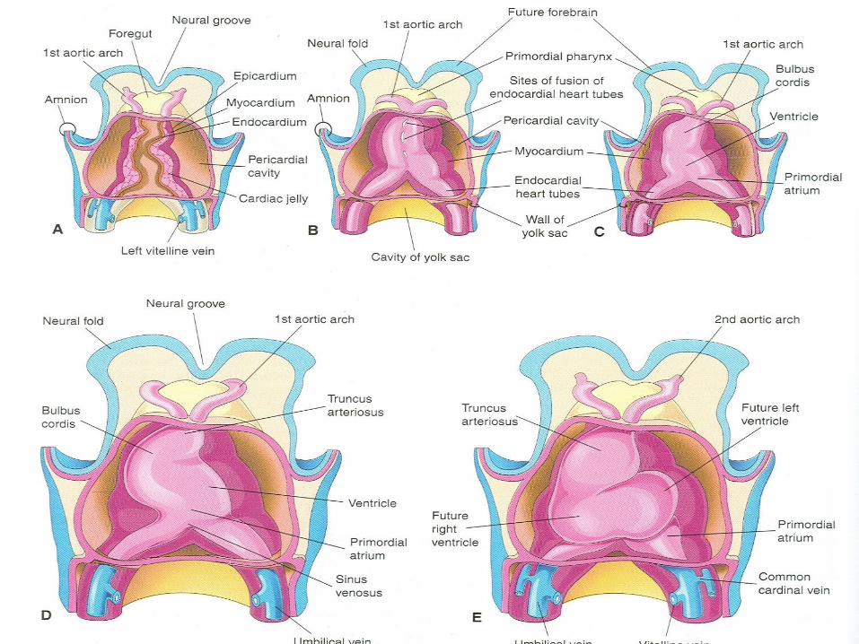
The earliest sign of heart is the appearance of The earliest sign of heart is the appearance of paired endothelial strands called angioblastic paired endothelial strands called angioblastic cordscords
They develop in the cardiogenic mesoderm They develop in the cardiogenic mesoderm during the third weekduring the third week
These cords canalize to form heart tubesThese cords canalize to form heart tubes
These cords fuse together to form the tubular These cords fuse together to form the tubular heart late in the third weekheart late in the third week

Early Development of HeartEarly Development of Heart
Primordium of heart is first evident at 18 Primordium of heart is first evident at 18 days in the cardiogenic areadays in the cardiogenic area
The heart begins to beat at 22-23 daysThe heart begins to beat at 22-23 days
Blood flow begins during the fourth week Blood flow begins during the fourth week and can be visualized by Doppler and can be visualized by Doppler ultrasonographyultrasonography

Development of HeartDevelopment of Heart
The endocardial heart tubes approach The endocardial heart tubes approach each other and fuse to form a single heart each other and fuse to form a single heart tube after lateral foldingtube after lateral folding
Fusion of tubes begins at the cranial end Fusion of tubes begins at the cranial end of the developing heart and extends of the developing heart and extends caudallycaudally

Primordial MyocardiumPrimordial Myocardium
As the heart tubes fuse, an external layer As the heart tubes fuse, an external layer of the embryonic heart, the primordial of the embryonic heart, the primordial myocardium is formed from splanchnic myocardium is formed from splanchnic mesoderm around pericardial coelommesoderm around pericardial coelom
At this stage the developing heart is At this stage the developing heart is composed of a thin endothelial tube, composed of a thin endothelial tube, separated from thick muscular tube by separated from thick muscular tube by gelatinous connective tissue, cardiac jellygelatinous connective tissue, cardiac jelly

EndocardiumEndocardium
The endothelial tube becomes the internal The endothelial tube becomes the internal endothelial lining of the heart, called endothelial lining of the heart, called endocardiumendocardium
The primordial myocardium becomes the The primordial myocardium becomes the muscular wall of the heart or myocardiummuscular wall of the heart or myocardium
The visceral pericardium or epicardium is The visceral pericardium or epicardium is derived from mesothelial cells and spread derived from mesothelial cells and spread over the myocardiumover the myocardium

After FoldingAfter Folding
As folding of head region occursAs folding of head region occurs
The heart and pericardial cavity come to The heart and pericardial cavity come to lie ventral to the foregut and caudal to the lie ventral to the foregut and caudal to the oropharyngeal membraneoropharyngeal membrane


Fate of Heart TubesFate of Heart Tubes
The tubular heart elongates and develops The tubular heart elongates and develops alternate dilations and constrictions:alternate dilations and constrictions:
Truncus ArteriosusTruncus Arteriosus Bulbus CordisBulbus Cordis VentricleVentricle AtriumAtrium Sinus venosusSinus venosus


Fate of Heart TubesFate of Heart Tubes
As the developing heart elongates and bends, it As the developing heart elongates and bends, it gradually invaginates into the pericardial cavitygradually invaginates into the pericardial cavity
Initially suspended from the dorsal wall by a Initially suspended from the dorsal wall by a mesentery, the dorsal mesocardiummesentery, the dorsal mesocardium
Central part of this mesentery soon degeneratesCentral part of this mesentery soon degenerates
Heart is now attached only at its cranial and Heart is now attached only at its cranial and caudal ends caudal ends

Truncus ArteriosusTruncus Arteriosus
Is continuous cranially with the aortic sac, from Is continuous cranially with the aortic sac, from which the aortic arches arisewhich the aortic arches arise
The sinus venosus receives umbilical, vitelline, The sinus venosus receives umbilical, vitelline, and common cardinal veins from the chorion, and common cardinal veins from the chorion, yolk sac, and embryo respectivelyyolk sac, and embryo respectively
Bulbus cordis and ventricle grow faster than Bulbus cordis and ventricle grow faster than other regions, the heart bends upon itself, other regions, the heart bends upon itself, forming bulboventricular loopforming bulboventricular loop


Truncus ArteriosusTruncus Arteriosus
As the primordial heart bends, the atrium As the primordial heart bends, the atrium and sinus venosus come to lie dorsal to and sinus venosus come to lie dorsal to the truncus arteriosus, bulbus cordis, and the truncus arteriosus, bulbus cordis, and ventricleventricle
By this stage the sinus venosus has By this stage the sinus venosus has developed lateral expansions, the right developed lateral expansions, the right and left horns of the sinus venosusand left horns of the sinus venosus


Pericardial CavityPericardial Cavity
As the heart elongates and bends, it gradually As the heart elongates and bends, it gradually invaginates into the pericardial cavityinvaginates into the pericardial cavity
The heart is initially suspended from the dorsal The heart is initially suspended from the dorsal wall by a mesentery, the dorsal mesocardiumwall by a mesentery, the dorsal mesocardium
The central part of the mesentery soon The central part of the mesentery soon degeneratesdegenerates
Forms a communication, the transverse Forms a communication, the transverse pericardial sinus between the right and left sides pericardial sinus between the right and left sides of the pericardial cavityof the pericardial cavity


Pericardial CavityPericardial Cavity
During the fourth week three well defined During the fourth week three well defined body cavities are formed:body cavities are formed:
Pericardial cavityPericardial cavity
2 pericardioperitoneal canals2 pericardioperitoneal canals
Peritoneal cavityPeritoneal cavity

Pleuropericardial MembranesPleuropericardial Membranes
As the pleuropericardial folds enlarge, they form As the pleuropericardial folds enlarge, they form partitions that separate the pericardial cavity partitions that separate the pericardial cavity from the pleural cavitiesfrom the pleural cavities
As the primordial pleural cavities expand As the primordial pleural cavities expand ventrally around the heart, they extend into the ventrally around the heart, they extend into the body wall, splitting the mesenchyme into:body wall, splitting the mesenchyme into:
An outer layer that becomes the thoracic wallAn outer layer that becomes the thoracic wall
An inner layer becomes the fibrous pericardium, An inner layer becomes the fibrous pericardium, the outer layer of the pericardial sac enclosing the outer layer of the pericardial sac enclosing the heartthe heart