Embed Size (px)
Citation preview
6-1
6. HEART AND CIRCULATORY SYSTEM I
Dr. Taube P. RothmanP&S [email protected]
RECOMMENDED READING: Larsen Human Embryology, 3rd Edition, pp. 195-199; 157-169top left; 172-174; bottom 181-182; 187-top 189, Simbryo-cardiovascular system
SUMMARY: The circulatory system, consisting of heart, blood vessels, and blood cells is the firstfunctional organ to develop. This lecture will focus on the formation of the embryonic vasculature, theorigin and formation of the early heart tube and primitive cardiac chambers, cardiac looping, and theprimitive circulation. Between the 5th - 8th week of embryonic development, the tubular heart isremodeled into a four chambered structure. We will see how right and left atrioventricular canalsconnect each atrium with its respective ventricle, and how the atrial septum and definitive right and leftatria form. We will also see why the great veins deliver blood to the right atrium while the pulmonaryveins empty into the left.
GLOSSARY:
Angioblasts: precursors of blood vesselsAngiogenesis: lengthening, branching, sprouting and remodeling of embryonic blood vesselsAortic arches: paired arteries surrounding the pharynx; portions will contribute to formation of thegreat arterial vesselsBlood islands: clusters of cells in the yolk sac, connecting stalk and chorionic villi that form primitiveblood vesselsCardiac jelly: gelatinous extracellular matrix that forms the middle layer of the heart tubeDuctus venosus: shunts most of the blood in the umbilical vein into the inferior vena cava (IVC) (by-passing the liver sinusoids)Hemangioblasts: earliest precursors of blood cellsIntussusception: incorporation of one structure into anotherVasculogenesis: formation of blood vessels in the embryonic disk
LEARNING OBJECTIVES:
At the end of the lecture you should be able to:1) Describe the origin and morphogenesis of the primitive embryonic cardiovascular system,
including the formation of embryonic blood vessels, the heart tube, cardiac looping,formation of primitive heart chambers and the early embryonic circulatory system.
2) Describe the structural remodeling of the primitive heart and circulatory system.
6-2
3) Explain why the inferior and superior vena cavae, as well as the coronary sinus, empty intothe right atrium and the pulmonary veins empty into the left atrium.
4) Describe the origin and role of endocardial cushions, the formation of the right and leftatrioventricular canals as well as the septum intermedium.
5) Discuss the formation, structure and function of the fetal atrial septum.
INTRODUCTION: Formation of the cardiovascular system begins during the 3rd week ofgestation and is completed by the end of the 8th week (refer to the chronological table in Chapter 7).The ventricular and atrial cardiac musculature, the endocardial cushions, the Purkinje conducting fibersand the endothelial lining of the heart (endocardium) are derived from mesoderm. Portions of the aorta,pulmonary trunk, and vascular walls of the aortic arch derivatives are formed from neural crest.
The first trimester:
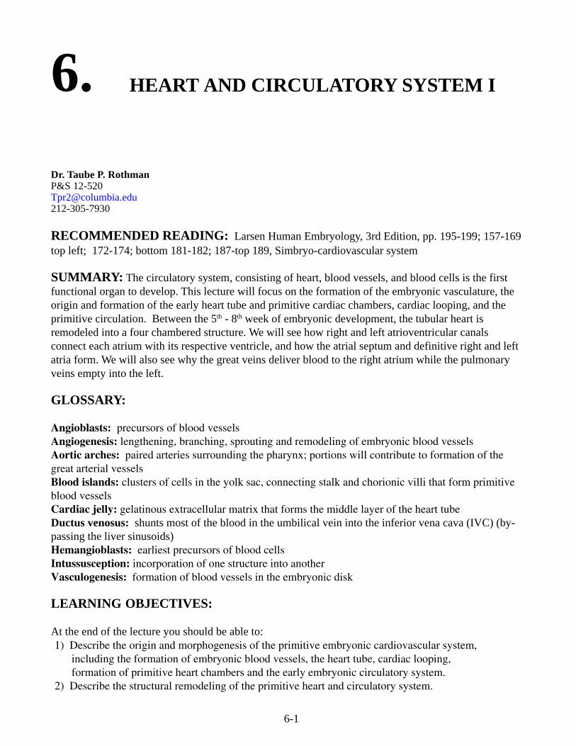
a) Vitelline stage (3rd – 4th week): Formation of blood and blood vessels begins in the mesodermalwall of the yolk sac as well as in the wall of the chorion outside the embryo proper (Figure 6-1).The yolk sac (the first hematopoietic region) provides nutrients via vitelline vessels while theplacenta is forming. Vasculogenesis begins in the splanchnic mesoderm of the embryonic disc.
b) By the beginning of the 5th week, the placenta provides nutrients, O2, etc. Cardiogenesis is wellunderway and the embryonic circulation is functional. The liver takes over the role ofhematopoiesis.
c) Structures designed to facilitate separate systemic and pulmonary circulations form between the5th and 8th weeks of gestation, although a dual pump is not operational until immediately afterbirth.
Fig. 6-1. Extra-embryonic bloodvessel formation in the chorion, theconnecting stalk, and the wall ofthe yolk sac in a presomite embryoof approximately 19 days(modified from Keibel and Elze).Note the position of the pericardialcavity and developing heart.
6-3
I. Blood vessel formation
During the third week of development,blood vessels form in blood islands in theextra-embryonic regions of the chorion, theconnecting stalk, and the wall of the yolksac, which is the first hematopoietic regionin the embryo (Figure 6-1). The primitiveblood vessels bring nutrients to the embryoand transport gases to and from sites ofrespiratory exchange. Blood vesselformation begins in the embryonic discprior to somite formation. Extra-embryonicblood vessels eventually merge with thoseof the embryo.
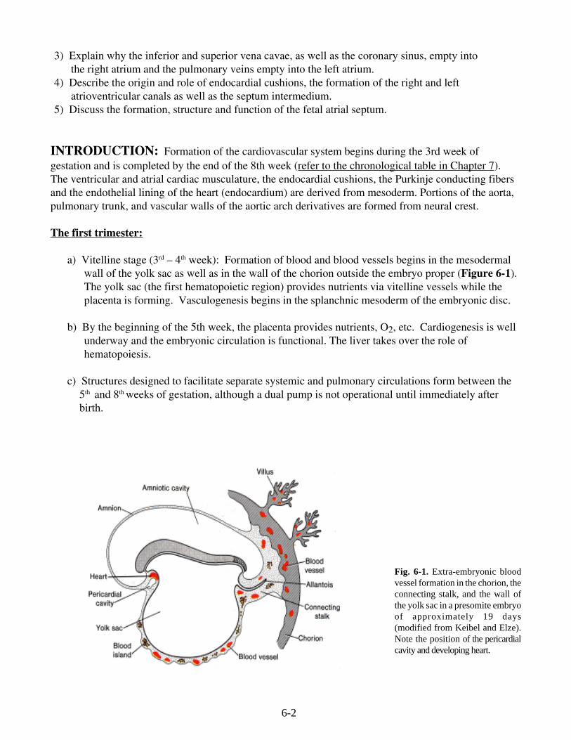
Blood vessels develop in three ways.a) Vasculogenesis: Fibroblast growth factor2 (FGF-2) binds to its receptor (FGFR) onsubpopulations of mesoderm cells andinduces them to form pluripotent
hemangioblasts (Figure 6-2A). Under the influence of vascular endothelial growth factor (VEGF)acting through two different receptors, the hemangioblasts segregate into an inner core of primitivenucleated blood cells (Figure 6-2B) surrounded by presumptive endothelial cells (Figure 6-2C) thatcoalesce to form primitive blood vessels.b) Angiogenesis: VEGF stimulates proliferation of endothelial cells at places wherevessels sprout from existing ones (Figure 6-2D).
c) Vascular precursors called angioblasts migrate into organs from other regions.
Fig. 6-2, A-C. Vasculogenesis: blood vessels and blood cellsform de novo. D. Angiogenesis: new vessels sprout fromexisting ones.
B C
D
Fig. 6-3. Cleavage of the lateral plate by the coelomreaches the cardiac region, bringing aboutdifferentiation of the splanchnopleure and thesomatopleure. These form the walls of the futurepericardial cavity.
A

6-4
Fig. 6-4. Transverse sections through embryos at different stages ofdevelopment, showing formation of a single heart tube from paired primordia.A. Early presomite embryo (17 days). B. Late presomite embryo (18 days). C.Eight-somite stage (22 days).
II. Formation of heart tube:
REVIEW (See lecture 2).Precardiac mesoderm emergesfrom the early primitive streak justposterior to the primitive node andreaches a position rostral andanterior to the prochordal plate(future buccopharyngealmembrane) forming a cardiacprimordium (cardiac crescent)(Figure 6-5A). Theintraembryonic coelom divides themesoderm into splanchnopleureand somatopleure. The cardiacprimordium is included insplanchnopleure (Figure 6-3).
The primitive heart tube:Vasculogenesis occurs within thecardiac primordium creatingbilateral endocardial tubes.Formation of the dorsal aortaeoccurs simultaneously, close to thenotochord. Transverse (Figure 6-4)and cephalo-caudal flexion (Figure6-5) results in formation of theforegut and intraembryoniccoelom. During this process, thetwo endocardial tubes are carriedventral to the closing gut tube.
Fig.6-5. A, Dorsal view of a late presomite embryo(about 18 days) after removal of the amnion.Vasculogenesis, occurring in cell clusters formed in thesplanchnic mesoderm in front of the neural plate on eachside of the embryo, is visible through the overlyingectoderm and somatic mesoderm layer. B,Cephalocaudal section through a similar embryoshowing the position of the presumptive pericardialcavity and cardiogenic area (18 days). C-E, Changes inthe position of the heart and pericardial cavity duringhead folding. The heart tube rotates 180o to reach aposition ventral to the foregut, caudal to theoropharyngeal membrane and suspended in the dorsalportion of the pericardial cavity (20-22 days).
6-5
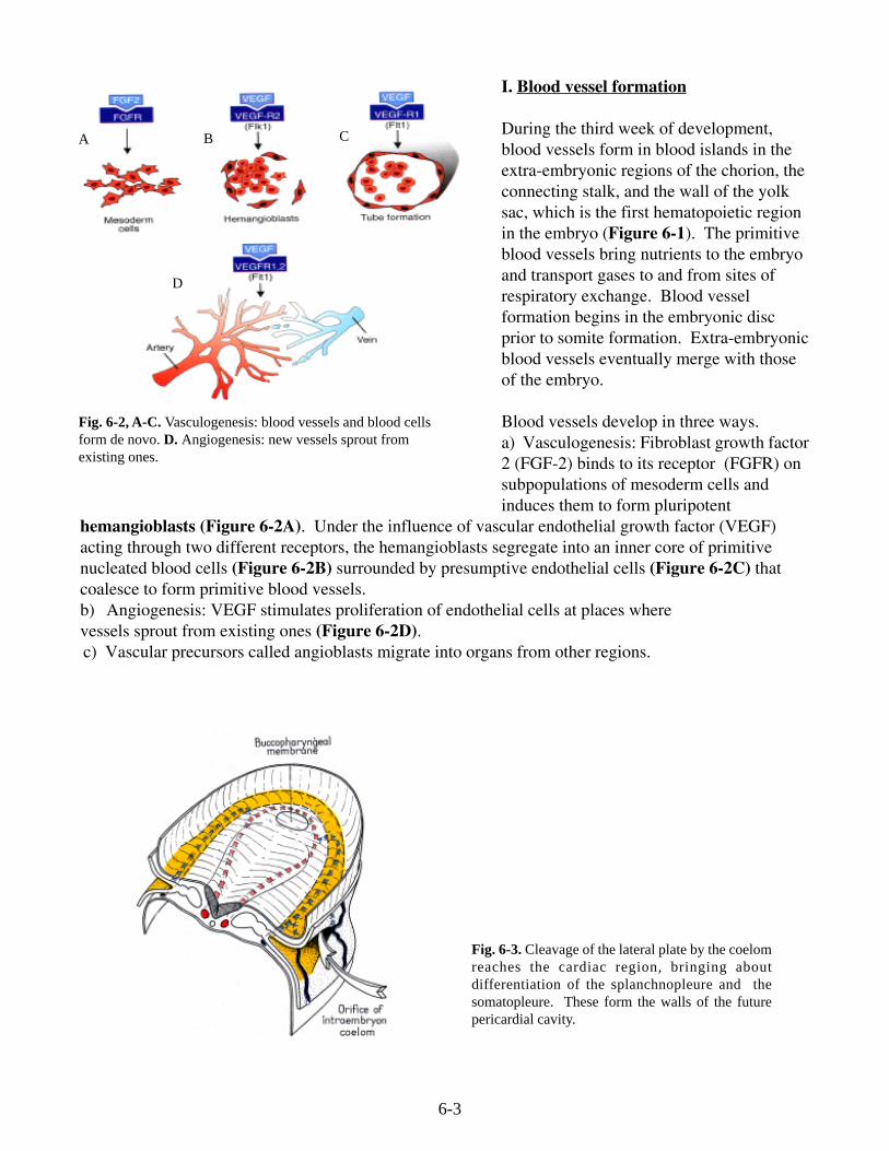
Fig. 6-6. Slide views of the heart: A, before, B,following loss of the mesocardium. a, common atrium;b, bulbus cordis; s, sinus venosus; t, truncus arteriosus;v, ventricle.
Apoptosis in portions of each of the tubes results in their fusion to form a single heart tubesuspended by a temporary dorsal mesocardium in the newly formed pericardial cavity (Figure 6-4).Fusion occurs in a cranial-caudal direction. Rapid growth of the central nervous system causesrotation of the heart tube and pericardial cavity 180o ventral and caudal to the oropharyngealmembrane (Figure 6-5).
b) The straight cardiac tube consists of an inner endocardocardial lining (endothelium) separatedfrom the cardiac mesoderm (future myocardium) by a gelatinous hyaluronate-rich cardiac jelly(Figure 6-4C). Cardiac mesoderm secretes the cardiac jelly that provides structural support andnutrients for the endocardial lining. BMP signaling promotes cardiac myogenesis (muscleformation)and is required later, during cardiac morphogenesis. Interestingly, the epicardium isderived from the proepicardial primordium, located near the dorsal mesocardium, and not from theheart tube. Cells migrating from the proepicardium cover the surface of the tubular heart. The dorsalmesocardium breaks down creating the transverse sinus of the pericardial cavity (Figure 6-6). Theheart tube is now attached to the pericardial cavity only at its caudal and rostral ends. At its caudalend, the endocardial tubes do not fuse, but extend toward the posterior part of the embryo as thevenous (afferent) inflow tracts of the heart. At its cranial end, the endothelial tube leading out fromthe heart produces pairs of vascular aortic arch arteries that loop from the ventral to the dorsal sideof the pharynx.
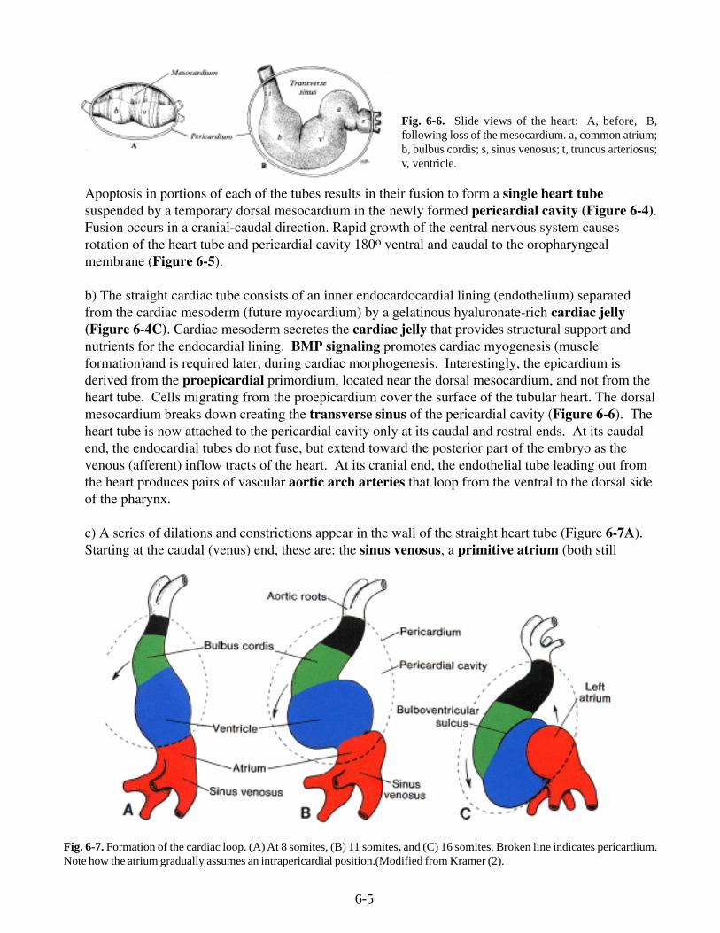
c) A series of dilations and constrictions appear in the wall of the straight heart tube (Figure 6-7A).Starting at the caudal (venus) end, these are: the sinus venosus, a primitive atrium (both still
Fig. 6-7. Formation of the cardiac loop. (A) At 8 somites, (B) 11 somites, and (C) 16 somites. Broken line indicates pericardium.Note how the atrium gradually assumes an intrapericardial position.(Modified from Kramer (2).
6-6
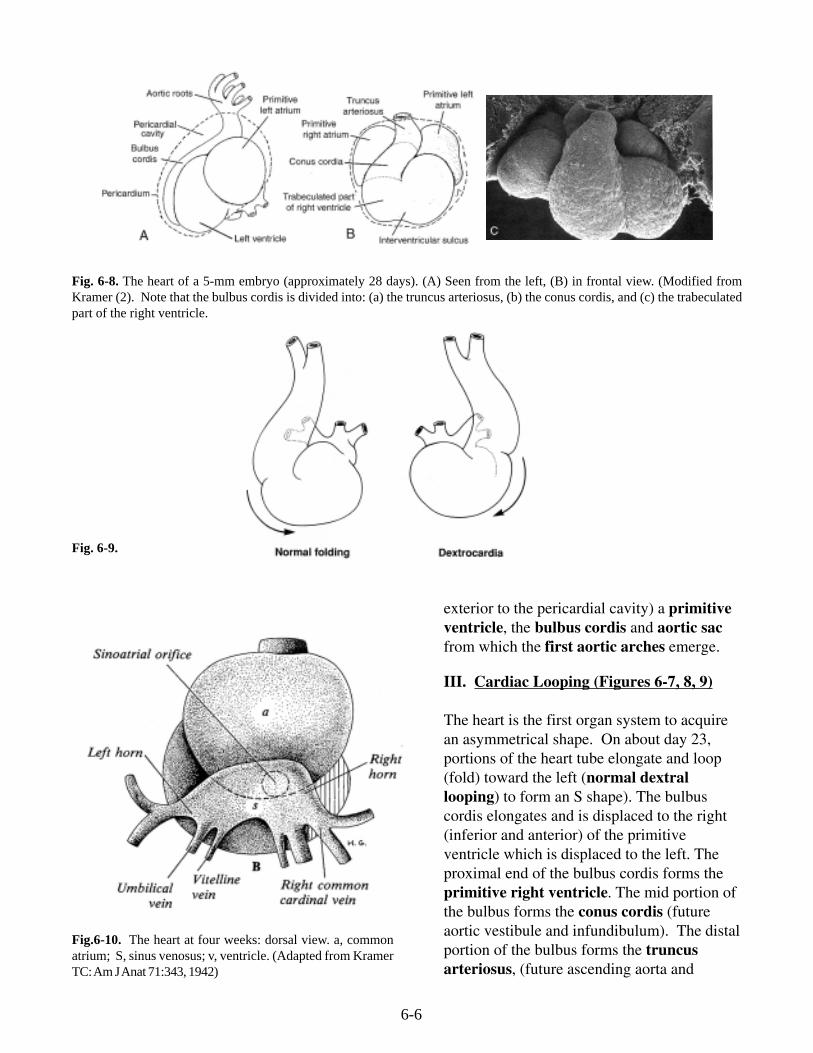
Fig. 6-8. The heart of a 5-mm embryo (approximately 28 days). (A) Seen from the left, (B) in frontal view. (Modified fromKramer (2). Note that the bulbus cordis is divided into: (a) the truncus arteriosus, (b) the conus cordis, and (c) the trabeculatedpart of the right ventricle.
exterior to the pericardial cavity) a primitiveventricle, the bulbus cordis and aortic sacfrom which the first aortic arches emerge.
III. Cardiac Looping (Figures 6-7, 8, 9)
The heart is the first organ system to acquirean asymmetrical shape. On about day 23,portions of the heart tube elongate and loop(fold) toward the left (normal dextrallooping) to form an S shape). The bulbuscordis elongates and is displaced to the right(inferior and anterior) of the primitiveventricle which is displaced to the left. Theproximal end of the bulbus cordis forms theprimitive right ventricle. The mid portion ofthe bulbus forms the conus cordis (futureaortic vestibule and infundibulum). The distalportion of the bulbus forms the truncusarteriosus, (future ascending aorta and
Fig. 6-9.
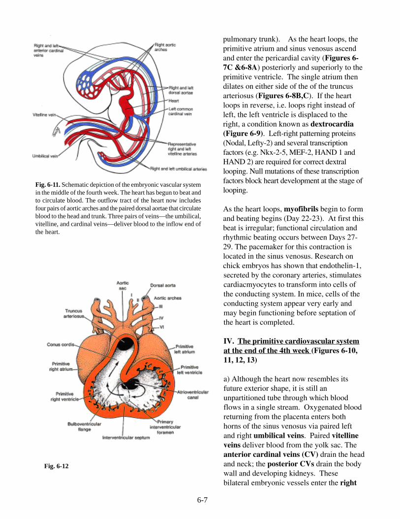
Fig.6-10. The heart at four weeks: dorsal view. a, commonatrium; S, sinus venosus; v, ventricle. (Adapted from KramerTC: Am J Anat 71:343, 1942)
6-7
Fig. 6-11. Schematic depiction of the embryonic vascular systemin the middle of the fourth week. The heart has begun to beat andto circulate blood. The outflow tract of the heart now includesfour pairs of aortic arches and the paired dorsal aortae that circulateblood to the head and trunk. Three pairs of veins—the umbilical,vitelline, and cardinal veins—deliver blood to the inflow end ofthe heart.
pulmonary trunk). As the heart loops, theprimitive atrium and sinus venosus ascendand enter the pericardial cavity (Figures 6-7C &6-8A) posteriorly and superiorly to theprimitive ventricle. The single atrium thendilates on either side of the of the truncusarteriosus (Figures 6-8B,C). If the heartloops in reverse, i.e. loops right instead ofleft, the left ventricle is displaced to theright, a condition known as dextrocardia(Figure 6-9). Left-right patterning proteins(Nodal, Lefty-2) and several transcriptionfactors (e.g. Nkx-2-5, MEF-2, HAND 1 andHAND 2) are required for correct dextrallooping. Null mutations of these transcriptionfactors block heart development at the stage oflooping.
As the heart loops, myofibrils begin to formand beating begins (Day 22-23). At first thisbeat is irregular; functional circulation andrhythmic beating occurs between Days 27-29. The pacemaker for this contraction islocated in the sinus venosus. Research onchick embryos has shown that endothelin-1,secreted by the coronary arteries, stimulatescardiacmyocytes to transform into cells ofthe conducting system. In mice, cells of theconducting system appear very early andmay begin functioning before septation ofthe heart is completed.
IV. The primitive cardiovascular systemat the end of the 4th week (Figures 6-10,11, 12, 13)
a) Although the heart now resembles itsfuture exterior shape, it is still anunpartitioned tube through which bloodflows in a single stream. Oxygenated bloodreturning from the placenta enters bothhorns of the sinus venosus via paired leftand right umbilical veins. Paired vitellineveins deliver blood from the yolk sac. Theanterior cardinal veins (CV) drain the headand neck; the posterior CVs drain the bodywall and developing kidneys. Thesebilateral embryonic vessels enter the right
Fig. 6-12
6-8
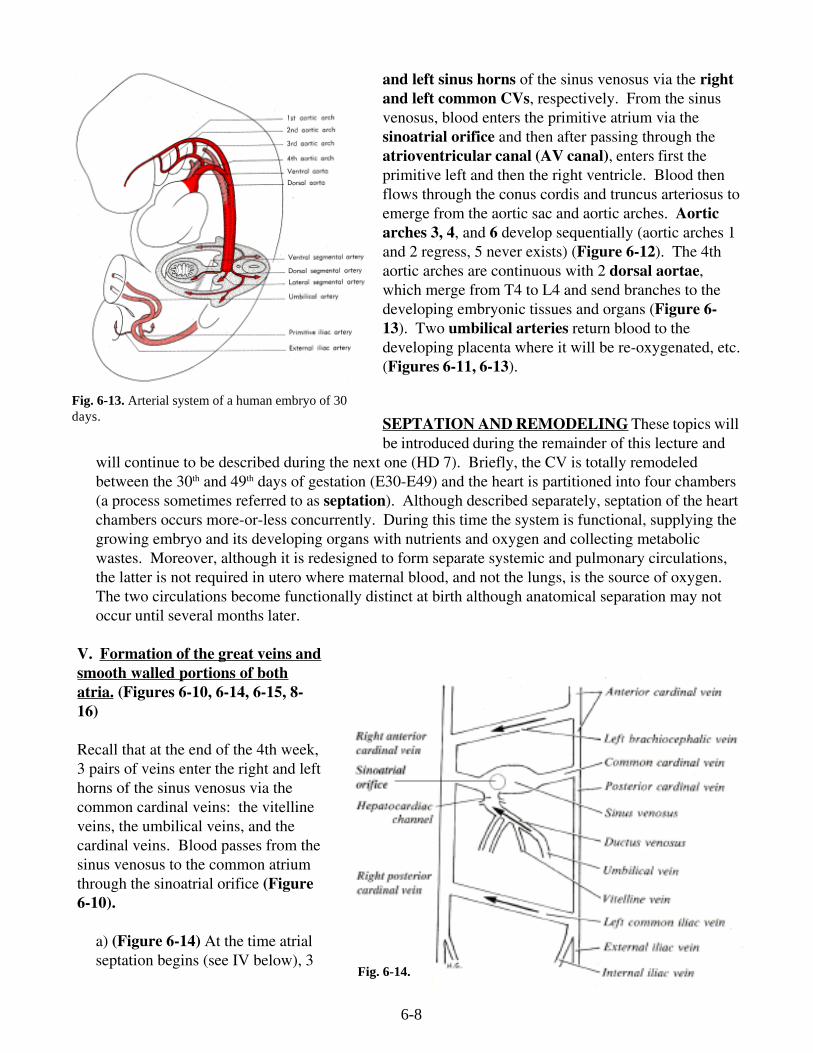
Fig. 6-13. Arterial system of a human embryo of 30days.
and left sinus horns of the sinus venosus via the rightand left common CVs, respectively. From the sinusvenosus, blood enters the primitive atrium via thesinoatrial orifice and then after passing through theatrioventricular canal (AV canal), enters first theprimitive left and then the right ventricle. Blood thenflows through the conus cordis and truncus arteriosus toemerge from the aortic sac and aortic arches. Aorticarches 3, 4, and 6 develop sequentially (aortic arches 1and 2 regress, 5 never exists) (Figure 6-12). The 4thaortic arches are continuous with 2 dorsal aortae,which merge from T4 to L4 and send branches to thedeveloping embryonic tissues and organs (Figure 6-13). Two umbilical arteries return blood to thedeveloping placenta where it will be re-oxygenated, etc.(Figures 6-11, 6-13).
SEPTATION AND REMODELING These topics willbe introduced during the remainder of this lecture and
will continue to be described during the next one (HD 7). Briefly, the CV is totally remodeledbetween the 30th and 49th days of gestation (E30-E49) and the heart is partitioned into four chambers(a process sometimes referred to as septation). Although described separately, septation of the heartchambers occurs more-or-less concurrently. During this time the system is functional, supplying thegrowing embryo and its developing organs with nutrients and oxygen and collecting metabolicwastes. Moreover, although it is redesigned to form separate systemic and pulmonary circulations,the latter is not required in utero where maternal blood, and not the lungs, is the source of oxygen.The two circulations become functionally distinct at birth although anatomical separation may notoccur until several months later.
V. Formation of the great veins andsmooth walled portions of bothatria. (Figures 6-10, 6-14, 6-15, 8-16)
Recall that at the end of the 4th week,3 pairs of veins enter the right and lefthorns of the sinus venosus via thecommon cardinal veins: the vitellineveins, the umbilical veins, and thecardinal veins. Blood passes from thesinus venosus to the common atriumthrough the sinoatrial orifice (Figure6-10).
a) (Figure 6-14) At the time atrialseptation begins (see IV below), 3
Fig. 6-14.
6-9
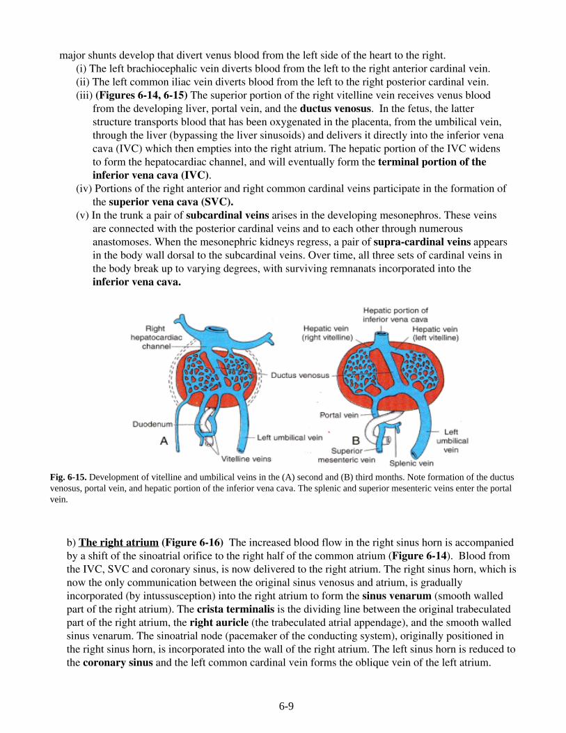
major shunts develop that divert venus blood from the left side of the heart to the right.(i) The left brachiocephalic vein diverts blood from the left to the right anterior cardinal vein.(ii) The left common iliac vein diverts blood from the left to the right posterior cardinal vein.(iii) (Figures 6-14, 6-15) The superior portion of the right vitelline vein receives venus blood
from the developing liver, portal vein, and the ductus venosus. In the fetus, the latterstructure transports blood that has been oxygenated in the placenta, from the umbilical vein,through the liver (bypassing the liver sinusoids) and delivers it directly into the inferior venacava (IVC) which then empties into the right atrium. The hepatic portion of the IVC widensto form the hepatocardiac channel, and will eventually form the terminal portion of theinferior vena cava (IVC).
(iv) Portions of the right anterior and right common cardinal veins participate in the formation ofthe superior vena cava (SVC).
(v) In the trunk a pair of subcardinal veins arises in the developing mesonephros. These veinsare connected with the posterior cardinal veins and to each other through numerousanastomoses. When the mesonephric kidneys regress, a pair of supra-cardinal veins appearsin the body wall dorsal to the subcardinal veins. Over time, all three sets of cardinal veins inthe body break up to varying degrees, with surviving remnanats incorporated into theinferior vena cava.
Fig. 6-15. Development of vitelline and umbilical veins in the (A) second and (B) third months. Note formation of the ductusvenosus, portal vein, and hepatic portion of the inferior vena cava. The splenic and superior mesenteric veins enter the portalvein.
b) The right atrium (Figure 6-16) The increased blood flow in the right sinus horn is accompaniedby a shift of the sinoatrial orifice to the right half of the common atrium (Figure 6-14). Blood fromthe IVC, SVC and coronary sinus, is now delivered to the right atrium. The right sinus horn, which isnow the only communication between the original sinus venosus and atrium, is graduallyincorporated (by intussusception) into the right atrium to form the sinus venarum (smooth walledpart of the right atrium). The crista terminalis is the dividing line between the original trabeculatedpart of the right atrium, the right auricle (the trabeculated atrial appendage), and the smooth walledsinus venarum. The sinoatrial node (pacemaker of the conducting system), originally positioned inthe right sinus horn, is incorporated into the wall of the right atrium. The left sinus horn is reduced tothe coronary sinus and the left common cardinal vein forms the oblique vein of the left atrium.
6-10
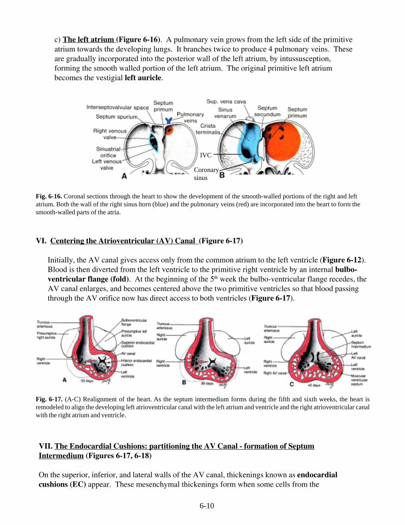
c) The left atrium (Figure 6-16). A pulmonary vein grows from the left side of the primitiveatrium towards the developing lungs. It branches twice to produce 4 pulmonary veins. Theseare gradually incorporated into the posterior wall of the left atrium, by intussusception,forming the smooth walled portion of the left atrium. The original primitive left atriumbecomes the vestigial left auricle.
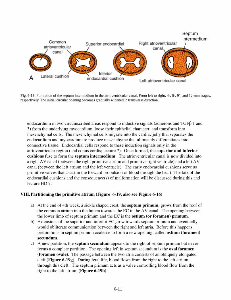
Fig. 6-17. (A-C) Realignment of the heart. As the septum intermedium forms during the fifth and sixth weeks, the heart isremodeled to align the developing left atrioventricular canal with the left atrium and ventricle and the right atrioventricular canalwith the right atrium and ventricle.
VII. The Endocardial Cushions: partitioning the AV Canal - formation of SeptumIntermedium (Figures 6-17, 6-18)
On the superior, inferior, and lateral walls of the AV canal, thickenings known as endocardialcushions (EC) appear. These mesenchymal thickenings form when some cells from the
Fig. 6-16. Coronal sections through the heart to show the development of the smooth-walled portions of the right and leftatrium. Both the wall of the right sinus horn (blue) and the pulmonary veins (red) are incorporated into the heart to form thesmooth-walled parts of the atria.
Coronarysinus
IVC
VI. Centering the Atrioventricular (AV) Canal (Figure 6-17)
Initially, the AV canal gives access only from the common atrium to the left ventricle (Figure 6-12).Blood is then diverted from the left ventricle to the primitive right ventricle by an internal bulbo-ventricular flange (fold). At the beginning of the 5th week the bulbo-ventricular flange recedes, theAV canal enlarges, and becomes centered above the two primitive ventricles so that blood passingthrough the AV orifice now has direct access to both ventricles (Figure 6-17).
6-11
endocardium in two circumscribed areas respond to inductive signals (adherons and TGFβ 1 and3) from the underlying myocardium, loose their epithelial character, and transform intomesenchymal cells. The mesenchymal cells migrate into the cardiac jelly that separates theendocardium and myocardium to produce mesenchyme that ultimately differentiates intoconnective tissue. Endocardial cells respond to these induction signals only in theatrioventricular region (and conus cordis; lecture 7). Once formed, the superior and inferiorcushions fuse to form the septum intermedium. The atrioventricular canal is now divided intoa right AV canal (between the right primitive atrium and primitive right ventricle) and a left AVcanal (between the left atrium and the left ventricle). The early endocardial cushions serve asprimitive valves that assist in the forward propulsion of blood through the heart. The fate of theendocardial cushions and the consequence(s) of malformation will be discussed during this andlecture HD 7.
VIII.Partitioning the primitive atrium (Figure 6-19, also see Figure 6-16)
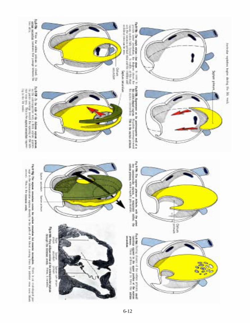
a) At the end of 4th week, a sickle shaped crest, the septum primum, grows from the roof ofthe common atrium into the lumen towards the EC in the AV canal. The opening betweenthe lower limb of septum primum and the EC is the ostium (or foramen) primum.
b) Extensions of the superior and inferior EC grow towards septum primum and eventuallywould obliterate communication between the right and left atria. Before this happens,perforations in septum primum coalesce to form a new opening, called ostium (foramen)secundum.
c) A new partition, the septum secundum appears to the right of septum primum but neverforms a complete partition. The opening left in septum secundum is the oval foramen(foramen ovale). The passage between the two atria consists of an obliquely elongatedcleft (Figure 6-19g). During fetal life, blood flows from the right to the left atriumthrough this cleft. The septum primum acts as a valve controlling blood flow from theright to the left atrium (Figure 6-19h)
Fig. 6-18. Formation of the septum intermedium in the atrioventricular canal. From left to right, 4-, 6-, 9", and 12-mm stages,respectively. The initial circular opening becomes gradually widened in transverse direction.
SeptumIntermedium
6-12