Embed Size (px)
Citation preview

An Initiation Codon Mutation (AUG -- GUG) of the Human al-Globin Gene
Structural Characterization and Evidence for a Mild Thalassemic Phenotype
Paolo Moi,* Faith E. Cash,t Stephen A. Liebhaber,$ Antonio Cao,1 and Mario Pirastull*Ospedale Regionale per le Microcitemie USL21, Cagliari; tHoward Hughes Medical Institute, and the Departments of Human
Genetics and Medicine, University of Pennsylvania, Philadelphia, Pennsylvania 19104; §Istituto di Clinica e Biologia Dell'EtaEvolutiva University Degli Studi Di Cagliari; IIlstituto di Ricerca Sulle Talassemie ed Anemie Mediterranee,CNR, Cagliari, Sardegna, Italy
Abstract
a-globin is encoded by two adjacent genes, al and a2. Recentevidence suggests that these genes are not equally expressedand that the a2-globin gene encodes the majority of a-globin.This finding would predict that a thalassemic mutation of thea2-globin gene would result in a more severe loss of a-chainsynthesis than a similar mutation in the al-globin gene. In aprevious study we described a nondeletion a-thalassemia de-fect in the a2-globin gene resulting from an AUG -- ACGinitiation codon mutation. In the present study we describe adifferent initiation codon mutation, AUG- GUG, present inthe al-globin gene. The al- and a2-globin gene initiationcodon mutations result in similarly lowered levels of encodedmRNA. Despite the similarity of these two mutations, the a2mutant results in a more severe loss of a-globin synthesis and amore severe clinical a-thalassemia phenotype than the corre-sponding al-globin gene mutation. This difference reflects thedominant role of a2-globin gene in overall a-globin synthesis.
Introduction
The a-globin gene cluster is located on the short arm of chro-mosome 16 (1). The genes in this cluster are arranged in theorder 5'-t- 4.-a-a2-a 1-3' (2-3). A variety of mutations withinthis cluster result in deficient or absent synthesis of a-globinand the consequent group of genetic disorders known as thea-thalassemias (4). Most commonly, a-thalassemia resultsfrom the deletion of one or both of the a-globin genes (5). Lesscommonly, a-globin gene function is altered by a defect thatdoes not produce a gross gene deletion (6-8). Several suchnondeletion a-thalassemias have now been defined at a molec-ular level. These include: termination codon mutations suchas Hb Constant Spring (9), a splicing defect caused by a 5-basepair (bp) deletion of the first intervening sequence (10),Hb Quong Sze, an extremely unstable a-globin structural var-iant (1 1, 12), a single nucleotide substitution of the polyaden-ylation site ( 1 3), a two nucleotide deletion at position -1 and-2 in the 5' untranslated region preceding the AUGcodon(14), an initiation codon mutation (AUG -- ACG) (15) and anonsense mutation (a 116 GAG-- UAG) (1 6). Apart from the-1, -2 deletion that occurs in a single a-globin gene chromo-some, each of these mutations affects the a2-globin gene.
Address reprint requests to Dr. Pirastu, Ospedale Regionale, Microci-temico, via Jenner 5N, 09100 Cagliari, Sardegna, Italy.
Receivedfor publication 7 April 1987 and in revisedform 29 June1987.
Hemoglobin H (HbH)' disease, the most severe form ofa-thalassemia compatible with life, commonly results from thedeletion of three of the four a-globin genes (17-23). Less com-monly, HbH disease can also result from the deletion of thetwo a-globin genes on one chromosome combined with anondeletion defect affecting one of the two a-globin genes onthe other chromosome. In Sardinians, the most commonnon-deletion a-thalassemia defect is the AUG-- ACGmutation inthe initiation codon of the a2-globin gene (15, 24-26). Thismutation destroys a recognition site of the enzyme NcoI.While screening a group of nondeletion HbHdisease patients(--/aTha) for this mutations by Southern blot analysis, wefound two siblings in whomthe NcoI map suggested a muta-tion at the initation codon of the a 1 rather than the a2 locus.The severity of a-thalassemia in these individuals was signifi-cantly less than in those with the NcoI mutation in the a2-glo-bin gene. In the present study, we define the structure of thisa 1 -globin gene mutation and characterize its impact upon a-globin mRNAand protein synthesis.
Methods
Hematologic analysis. Hematologic measurements were made with aCoulter Counter (model S, Coulter Electronics, Hialeah, FL). Electro-phoresis of hemoglobin was carried out on Titan III cellulose acetateplates, pH 8.6 (Helena Laboratories, Beaumont, TX). Hb A2 was de-termined by DE-52 microchromatography (27). Globin chain synthe-sis analysis was carried out according to the method of Kan et al. (28).
DNAanalysis. DNAwas extracted from peripheral leukocytes aspreviously described (29). The a-globin cluster was mapped by South-ern blot analysis using the restriction endonucleases BglII, HphI, NcoI,and HindIII in single and double digestion. The {-globin gene probewas a 1.8-kb Hinfl genomic fragment containing the entire pseudo-zeta gene excluding the 5' part of the first exon (30); the a-globin geneprobe was a 1 .5-kb PstI genomic fragment spanning the entire a-globingene.
Gene cloning and sequence analysis. Total genomic DNAisolatedfrom individual 111-2 (Fig. 1) was digested to completion with therestriction enzyme HindIII. The fraction containing 3.7 Kb fragmentswas collected by density sedimentation through a continuous sucrosegradient, ligated to Charon 28 vector in the HindIII site and packagedin vitro. The recombinant phages were propagated in Escherichia coli.Three phage clones containing the 3.7-kb a-globin gene fragment wereidentified out of 75,000 recombinant phages screened with a 32P-la-beled a-globin specific probe. The 3.7-kb fragment, which spans fromthe HindIlI site at codon 90-91 of the a2-globin gene to the HindIII siteat the same position within the a 1 -globin gene, was subcloned in theHindIII site of the pSP64 plasmid (New England Nuclear, Boston,MA). Sequencing was performed by the primer extension methoddirectly on supercoiled plasmid (31, 32) using as a primer for theKlenow fragment of DNApolymerase a 20-mer oligonucleotide (5'CCTTGACGTTGGTCTTGTCG3') complementary to the a-glo-
1. Abbreviations used in this paper: HbH, hemoglobin H.
1416 Moi, Cash, Liebhaber, Cao, and Pirastu
J. Clin. Invest.©The American Society for Clinical Investigation, Inc.0021-9738/87/1 1/1416/06 $2.00Volume 80, November 1987, 1416-1421

1 aaThCE - -
Figure 1. Pedigree of theindex family. The segrega-tion of the deletion andnondeletion a-thalassemicmutations is shown.
bin DNAsequence from nucleotide + 13 to +32 (the A of the AUGinitiation codon being designated as + 1).
RNAisolation and analysis. The RNAwas isolated from peripheralreticulocytes as previously detailed (33) and used for subsequent stud-ies without further purification. llug of total RNAwas translated inmicroccoccal nuclease treated rabbit reticulocyte lysate prepared fromNewZealand white rabbits as previously described (34). Translationswere done at 30°C in the presence of [35S]methionine exactly as pre-viously detailed (34). The labeled protein products were resolved on aTriton-acid-urea slab gel (35, 36) and visualized by autoradiography.The ratio of a2- to a I -globin mRNAwas established by primer exten-sion analysis as previously detailed (33) with the following exception:we have substituted for the previously described 31 nucleotide cDNAprimer, a 20 nucleotide synthetic oligonucleotide (3'-CACCCGCCGT--1---11Fil---1--5') that is complementary to the ter-minal 9 nucleotides of both a. I- and a2-globin mRNAsand to 11
adenosines of the contiguous poly-A tail. This oligomer was 5' end-la-beled with -4(32P)ATP (5,000 Ci/mM; Amersham Corp., ArlingtonHeights, IL) and polynucleotide kinase and purified by a single precipi-tation with three volumes of ethanol in the presence of 50 Mg/ml tRNAcarrier and 0.2 MNa acetate. 1 ng of the end-labeled oligonucleotideprimer was added to 0.5 Ag of reticulocyte RNAin a reverse transcrip-tion reaction containing 1 Al of AMVreverse transcriptase (LifeScience Associates, Bayport, NY, 15 U/MI). The subsequent HaeIIIrestriction nuclease digest of the 5' end-labeled single strand cDNA,and the 8% acrylamide/8 Murea gel analysis of the digest productswere all carried out exactly as previously detailed (33). Band intensitieson autoradiographs of the in vitro translation gels and the primerextension analysis gels were quantitated in the linear range by densi-tometry using a Zeineh soft laser scanner (model SL-504-XL;Biomed Instruments, Fullerton, CA).
Results
Family studies. Two sisters (111-2, III-3, indicated by thearrows in Fig. 1) with HbHdisease and their family were stud-
ied'at the hematology laboratory of the Ospedale Regionaleper le Micrositemie. The two patients presented with a similarhistory of chronic pallor and anemia. The spleen was barelypalpable, the liver was slightly enlarged, and the children wereof normal stature for their ages. There was no history of acutehemolytic crises. The results of the hematological evaluationof the family are shown in Table I.
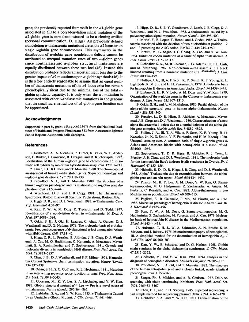
a-Globin gene mapping. The a-globin genotype of eachmember of the family was determined by Southern blot analy-sis. The presence of a double a-globin gene deletion[(--)chromosome] was inferred by digesting the genomicDNAwith Bg11I and using both the a-globin and {-globinprobes (8) (data not shown). The presence of the nondeletionmutation was detected by the loss of an NcoI restriction site.When normal DNAis cleaved with NcoI, three a-globin spe-cific fragments of 4.8, 3.7, and 2.1 kb are produced (Fig. 2).The 4.8 and 3.7 fragments contain the al- and the a2-globingenes, respectively. The 2. 1-kb fragment spans from the initia-tion codon of the a2-globin gene to a NcoI site located in the 5'flanking region. DNAfrom the patients investigated (111-2 and111-3) yielded the normal 2.1-kb fragment associated with anew 8.5-kb fragment that replaces the normal 3.7- and 4.8-kbfragments (Fig. 2). This pattern suggests the loss of the NcoIrestriction site located in the a I-globin gene. This interpreta-tion was confirmed by double digestion of the DNAwith NcoIand HindIII (data not shown). These findings indicate that ourpatient has a mutation in the a 1 -globin gene that abolishes theNcoI site normally located in the 5' part of the gene. NcoIdigestion of DNAisolated form the father and paternal grand-mother yielded the 8.5-kb abnormal fragment in addition tothe normal 4.8-, 3.7-, and 2.1-kb fragments which indicatedthe presence of the nondeletion defect in one chromosome anda normal a-globin gene complement in the other chromo-some. A summary of the a-thalassemia inheritance in thisfamily is presented in Fig. 1.
Sequence analysis of the al-globin gene initiation codonmutation. The absence of the normal NcoI site in the al-glo-bin gene raised the possibility that the previously reportedATG-. ACT initiation codon mutation found in the a2-glo-
bin gene of a Sardinian patient with HbH disease (15) mayhave been introduced at a homologous position in the aI geneby a gene conversion event (37). To determine the exact natureof the a 1-globin gene mutation that destroys the NcoI site, the
Table I. Hematologic Evaluation of a Family with Nondeletion HbHDisease
1-1 1-2 11-1 11-2 I11-1 111-2 III-3
Genotype aa/aa aa/aaFI ca/aa-aTh --/aa--laa"" --/aaTa
Age (yr) 86 84 58 53 22 12 10RBC(XJ02/liter) 4.42 4.62 5.28 5.21 6.11 5.92 6.64Hb (g/dl) 13.1 11.3 14.2 11.5 14.5 9.5 10.1Hct (%) 39 35.5 40.2 34.7 44 30 31.7MCV(fl) 87 75 77 67 72 52 49MCH(pg) 29.5 24.4 28 23 24 16 15MCHC(g/dl) 32.3 30.7 36 34 31 31 31Electrophoresis A+A2 A+A2 A+A2 A+A2 A+A2 H (3%)+A+A2 H(1.5%)+A+A2Hb A2 (%) 2.92 2.84 2.96 2.62 2.38 1.45 1.60Reticulocytes(%) 8 3 5 6 8 26 22RBCwith HbH inclusion
bodies per 1000 Absent 2 0.19 0.04 0.8 140 60
al-Globin Gene Initiation Codon Mutation 1417
II
III

Nco I - a globin
_M -8.5
am___wOno-o -4.8B_d _~__o _ 3.7
-2.1
1-t 2 11-1 11-2 III 1 111-2
5.~~~ ~~~am=>- =mCa2 aI
Nco 3'
* 3.7 v4I
8.5
mutant a 1-globin gene was cloned from individual III-2 andthe 5' end from nucleotides -35 to + 14 was sequenced. Thissequence analysis demonstrated a single nucleotide substitu-tion at the initiation codon:ATG -- GTG(Fig. 3).
1DNA
G*T
GGc
* ACIt
* AC
G*b A
* C
AG
S* AG
A C G T
RNA
U
GG 1 ValU
GC 2 LeuU
GU 3 SerC
U
C 4 Pro
Figure 3. Sequence analysisof the region of the mutanta 1 -globin gene surroundingthe initiation AUGcodon.The arrow indicates the po-sition of the A -- Gmuta-
tion.
Figure 2. Southern blot analysis of the a-globin genesin the index family using the restriction enzyme NcoIand the a-globin gene as a probe. At the bottom ofthe figure the NcoI map of the normal chromosome(aa) and of the nondeletion thalassemic chromosome(aaTh) is schematically represented. The result forsample 111-3 (not shown in this figure) is identical tothat of III-2.
mRNAanalysis. The relative levels of al- and a2-globinmRNAin total reticulocyte RNAwas measured by primerextension mapping. The results of studies on a normal aa/aa,a previously reported --/aTha individual, and one of the--a/aaTh individuals (III-2) are shown in Fig. 4. In normalreticulocytes the a2:al mRNAratio is 2.6 (33, 38). In fourunrelated Sardinian patients with nondeletion hemoglobin Hdisease in whomthe nondeletion defect was the T -- C substi-
tution in the ATGinitiation codon of the a2 gene (--/cTha)the average ratio was 0.9 (range 0.75-1.3) indicating that thelevel of steady state a2-globin mRNAwas decreased to ap-
proximately one-third of its normal level (Fig. 4 and Table II).This value is consistent with the previously reported value of1.0 (15). In the two sisters III-2 and 111-3 with the ATGGTGmutation in the al-globin gene (--/aaTh) the ao2/alratios of 12 and 14, respectively, indicated a reduction in thesteady-state level of a 1 mRNAto approximately one-fourth ofits normal level.
In vitro translation. Unfractionated mRNAwas translatedin vitro and the labeled translation products were resolved andquantitated by electrophoresis and densitometry. The resultsof this analysis are shown in Fig. 5 and summarized in Ta-ble II.
In vitro translation of normal reticulocyte mRNAgives an
a/l3 globin synthesis ratio (corrected for methionine content oftwo in a and one in ,) of 1.5 while in four unrelated individ-uals with HbH disease and the nondeletion (ATG -> ACG)defect in the a2-globin gene (--/aTha) had an average ratio of0.095 (range 0.04-0.16; Table II). The afl3 globin ratios of 111-2and III-3 (--/aaTh) were 0.22 and 0.27, respectively.
In a parallel set of experiments a/(3 synthesis was measuredin intact reticulocytes from each of these individuals. As in thein vitro translations the level of a-globin synthesized by thosewith the al mutation (--/aaTh) was greater than those with
1418 Moi, Cash, Liebhaber, Cao, and Pirastu
act v_ 2.1
aaT v 2.1
I -T.w T-------J r. .
J
- .I TI
B7
.

Figure 4. Primer extension analy-sis of the a2:a I globin mRNAratio in reticulocyte RNA. Sam-
12 3 ples of reticulocyte RNAwere iso-lated and cDNAwas synthesizedby extending a 32P end-labeledsynthetic oligonucleotide primer
n .t. complimentary to the 3' terminusof a-globin mRNA. The cDNAreverse transcripts were digested
104 (a1) )- with HaeIII and the products ana-lyzed on a denaturing gel. The po-
84 (al) - _ sition of the primer and of the al-and a2-globin specific bands are
72 (cr2) - indicated to the left of the autora-diograph and the size of eachband is noted in nucleotides (n.t.).The additional bands above the
49 (ac2) - 0 & primerappeartoresultfromre-verse transcription of mRNAde-gredation fragments as they comi-grate with short end-labeledDNAsnoted on gel analysis of thesample before HaeIII digestion
A (data not shown). The RNAorigi-nated from individuals with the
__~ _ following a-globin genotypes: lane(primer) - 1, aa/aa; lane 2, --/aacTh; lane 3,
--/a Tha.
the a2 mutation (--/aTba). The uniformly lower a/fl ratiosmeasured by in vitro translation as compared to the reticulo-cyte incubations may reflect the ability of proteolytic systemsin the intact cell to partially compensate for the chain imbal-ance. Such discrepancies between the al/( synthetic ratios ob-tained in vitro and during the labeling of intact reticulocytesare observed in a wide variety of deletion and nondeletiona-thalassemias (S. A. Liebhaber and F. E. Cash, unpublisheddata).
Discussion
In this study we have characterized a newly discovered muta-tion, ATG -- GTG, in the initiation codon of the al-globin
Table II. Synthesis of a- and fl-Globin in Individuals with HbHDisease
a/jS Protein Synthesis
Patient Genotype in vitro Reticulocytes a2:al mRNA
Normal aa/aa 1.5 1.00 2.6C.C. (III-2) --/aaTh 0.22 0.59 12.0R.C. (III-3) --/aaTh 0.22 0.75 14.0F.M. --/aTha 0.04 0.49 1.3V.A. --laTha 0.14 0.47 0.91A.L. --/aTha 0.04 0.22 0.75G.A. --/aTha 0.16 0.46 0.85
aTha, AUG--n'ACG mutation at the initiation codon of the a2-globingene. aaTh, AUG--GUGmutation at the initiation codon of the a 1-globin gene.
1 2 3
a-O_
4 Figure 5. In vitro translation of re-ticulocyte RNA. Total reticulocyteRNAisolated from each of threeindividuals was translated in arabbit reticulocyte lysate in thepresence of [35S]methionine andresolved on a Triton-acid-urea gel.The positions of #- and a-globinare indicated to the left of the au-toradiograph. The RNAorigi-nated from individuals with thefollowing a-globin genotypes: lane1, aa/aa; lane 2, --/aaTh; lane 3,--/aTa. Lane 4 is a controltranslation to which no exogenousRNAwas added.
gene that, when combined with deletion of both as-globin geneson the sister chromosome, results in the clinical phenotype ofa-thalassemia. In higher eucaryotes the ATGserves as the onlyfunctional initiation codon (39, 40). The only exception maybe the use of ACGin certain viral systems (41). Whether theATG GTGmutation in the a 1-globin gene, or the pre-viously described ATG -* ACGmutation in the a2-globingene completely abolishes a-globin mRNAtranslation has notbeen directly determined. However, the clinical phenotypes ofthe HbHdisease in both categories of patients (25; this study)suggest a functional loss of the affected a-globin gene. Theinitiation codon mutations in the a 1 and a2 loci are associatedwith similar three- to fourfold decreases in the steady statelevel of encoded mRNA. This comparable effect suggests thatthe two mutations may decrease the steady state level of theirencoded mRNAsby similar mechanisms. While effects upontranscription or RNAprocessing cannot be ruled out atpresent, mRNAinstability linked to the block in translationwould appear to be likely cause.
The severity of the a-thalassemia phenotype appears torelate both to the specific a-globin gene lost (a 1 or a2) and themanner in which it is lost (deletion versus nondeletion). a-glo-bin production during in vitro translation of reticulocyte RNAand during labeling of intact reticulocytes was less significantlydepressed in the --IaaTh patients (III-2 and 111-3), than in thepatients with a similar initiation codon mutation in the a2-globin gene (-_/aTha) (Fig. 5 and Table II). The mild pheno-type noted in these two patients with the --l/aa genotype isvery similar to that manifested by patients with the deletionalform of HbH disease (--I-a), and is significantly less severethan patients with HbH disease resulting from nondeletiondefects (ATG ACG, Hb Constant Spring) in the a2 gene(25, 42, 43). Since the a2-globin gene normally encodes two-to threefold higher steady state level of mRNA(33, 38, 44) andproduces two- to threefold more a-globin (45, 46) than theal-globin gene, a nondeletion defect in the a2-globin genewould be expected to result in the loss of two to three timesmore a-globin synthesis than a comparable mutation in the a 1gene. The rightward type a-thalassemia deletion (-aC3 7) wouldbe predicted to yield a mild phenotype comparable to an a 1nondeletion mutation since the loss of the a2-globin gene isassociated with a 1.8-fold compensatory increase in the ex-pression of the remaining al-globin gene (38).
The initiation codon mutation described herein is the firsta-thalassemia mutation to date identified in the a I -globin
al-Globin Gene Initiation Codon Mutation 1419
gene; the previously reported frameshift in the a 1-globin geneassociated in Cis to a polyadenylation signal mutation of thea2-globin gene is now demonstrated to be a cloning artifact(personal communication, D. Higgs). All previously definednondeletion a-thalassemia mutations are at the a2 locus or onsingle ac-globin gene chromosomes. This asymmetry in thedistribution of a-globin gene nondeletion defects cannot beattributed to unequal mutation rates of two a-globin genessince nonthalassemic a-globin structural mutations areequally distributed between these two loci (46). Instead thisdistribution probably reflects an ascertainment bias due to thegreater impact of a2 mutations upon a-globin synthesis (46). Itis therefore entirely reasonable to assume that an equal num-ber of thalassemia mutations of the a 1 locus exist but remainphenotypically silent due to the minimal loss of the total a-globin synthetic capacity. It is only when the a 1 mutation isassociated with other a-thalassemic mutations in the genomethat the small incremental loss of a 1 -globin gene function canbe appreciated.
Acknowledgments
Supported in part by grant l-Rol-AM-33975 from the National Insti-tutes of Health and Progetto Finalizzato 833 from Assessorato Igiene eSanita Regione Autonoma della Sardegna.
References
1. Deisseroth, A., A. Nienhuis, P. Turner, R. Valez, W. F. Ander-son, F. Ruddle, J. Lawrence, R. Creagan, and R. Kucherlapati. 1977.Localization of the human a-globin gene to chromosome 16 in so-matic cell hybrids by molecular hybridization assay. Cell. 12:205-211.
2. Lauer, J.; C. KJ. Shed, and T. Maniatis. 1980. The chromosomalarrangement of human a-like globin genes. Sequence homology anda-globin gene deletions. Cell. 20:119-130.
3. Proudfoot, N. J., and T. Maniatis, 1980. The structure of ahuman a-globin pseudogene and its relationship to a-globin gene du-plication. Cell. 21:537-44.
4. Weatherall, D. J., and J. B. Clegg. 1981. The ThalassaemiaSyndromes. Boston, Blackwell Scientific Publications, Boston, MA.
5. Higgs, D. R., and D. J. Weatherall. 1983. a-Thalassemia. Curr.Top. Haematol. 4:37-97.
6. Kan, Y. W., A. M. Dozy, R. Trecartin, and D. Todd. 1977.Identification of a nondeletion defect in a-thalassemia. N. Engl. J.Med. 297:1081-1084.
7. Orkin, S. H., J. Old, H. Lazarus, C. Altay, A. Gurgey, D. J.Weatherall, and D. G. Nathan. 1979. The molecular basis of a-thalas-semia; Frequent occurrence of dysfunctional a-loci among non-Asianswith HbHdisease. Cell. 17:33-42.
8. Higgs, D. R., L. Pressley, B. Aldridge, J. B. Clegg, D. J. Weath-erall, A. Cao, M. G. Hadjiminas, C. kattamis, A. Metaxatou-Mavro-mati, E. A. Rachmilewitz, and T. Sophocleous. 1981. Genetic andmolecular diversity in niondeletion HbHdisease. Proc. Natt. Acad. Sci.USA. 78:5833-5837.
9. Clegg, J. B., D. J. Weatherall, and P. F. Milner. 1971. Hemoglo-bin Costant Spring-a chain termination mutation. Nature (Lond.).234:337-339.
10. Orkin, S. H., S. C. Goff, and R. L. Hechtrhan. 1981. Mutationin an intervening sequence splice junction in man. Proc. Natl. Acad.Sci. USA. 78:5041-5045.
11. Goossens, M., K. Y. Lee, S. A. Liebhaber, and Y. W. Kan.1982. Globin structural mutant a 125 Leu -- Pro is a novel cause ofa-thalassemia. Nature (Lond.). 296:864-866.
12. Liebhaber, S. A., and Y. W. Kan. 1983. a-Thalassemia Causedby an Unstable a-Globin Mutant. J. Clin. Invest. 7 i:461-466.
13. Higgs, D. R., S. E. Y. Goodbourn, J. Lamb, J. B. Clegg, D. J.Weatherall, and N. J. Proudfoot. 1983. a-thalassaemia caused by apolyadenylation signal mutation. Nature (Lond.). 306:398-400.
14. Morle', F., B. Lopez, T. Henni, and J. Godet. 1985. a-Thalas-saemia associated with the deletion of two nucleotides at position -2and -3 preceding the AUGcodon. EMBOJ. 44:1245-1250.
15. Pirastu, M., G. Saglio, J. C. Chang, A. Cao, and Y. W. Kan.1984. Initiation codon mutation as a cause of alpha thalassaemia. J.Biol. Chem. 259:12315-12317.
16. Liebhaber, S. A., M. B. Coleman, J. G. Adams, III, F. E. Cash,and M. Steinberg. 1987. Non-deletion a-thalassemia in a blackkindred resulting from a nonsense mutation (a2ll6'Ao-UAG) J. Clin.Invest. 80:154-159.
17. Phillips, J. A., III, A. F. Scott, K. D. Smith, K. E. Young, K. L.Lightbody, R. M. Jiji, and H. H. Kazazian, Jr. 1979. A molecular basisfor hemoglobin H disease in American blacks. Blood. 54:1439-1445.
18. Embury, S. H., R. V. Lebo, A. M. Dozy, and Y. W. Kan. 1979.Organization of the a-globin genes in the Chinese a-thalassaemia syn-dromes. J. Clin. Invest. 63:1307-1310.
19. Orkin, S. H., and A. M. Michelson. 1980. Partial deletion of thealpha-globin structural gene in human alpha-thalassaemia. Nature(Lond.). 286:538-540.
20. Pressley, L., D. R. Higgs, B. Aldridge, A. Metaxatou-Mavro-mati, J. B. Clegg, and D. J. Weatherall. 1980. Characterization of a newalpha-thalassaemia- I defect due to a partial deletion of the alpha glo-bin gene complex. Nucleic Acids Res. 8:4889-4898.
21. Philips, J. A., III, T. A. Vik, A. F. Scott, K. E. Young, H. H.Kazazian, Jr., K. D. Smith, V. F. Fairbanks, and H. M. Koenig. 1980.Unequal crossing-over. A common basis of single a-globin genes inAsians and American blacks with hemoglobin H disease. Blood.55:1066-1069.
22. Sophocleous, T., D. R. Higgs, B. Aldridge, R. J. Trent, L.Pressley, J. B. Clegg, and D. J. Weatherall. 1981. The molecular basisfor the haemoglobin Bart's hydrops fetalis syndrome in Cyprus. Br. J.Haematol. 47:153-156.
23. Nicholls, R. D., D. R. Higgs, J. B. Clegg, and D. J. Weatherall.1985. Alpha°-Thalassemia due to recombination between the alpha-globin gene and an Alu repeat. Blood. 65:1434-1439.
24. Pirastu, M., K. Y. Lee, A. M. Dozy, Y. W. Kan, G. Stama-toyannopoulos, M. G. Hadjiminas, Z. Zachariades, A. Angius, M.Furbetta, C. Rosatelli, and A. Cao. 1982. Alpha-thalassemia in twoMediterranean populations. Blood. 60:509-515.
25. Paglietti, E., R. Galanello, P. Moi, M. Pirastu, and A. Cao.1986. Molecular pathology of hemoglobin Hdisease in Sardinians. Br.
J. Haerhatol. 63:485-496.26. Kan, Y. W., A. M. Dozy, G. Stamatoyannopoulos, M. G.
Hadjiminias, Z. Zachariades, M. Furgetta, and A. Cao. 1979. Molecu-lar basis of hemoglobin-H disease in the Mediterranean population.Blood. 54:1434-1438.
27. Huisman, T. H. J., W. A. Schroeder, A. N. Brodie, S. M.Mayson, and J. Jakway. 1975. Microchromatography of hemoglobins.III. A simplified method for the determination of hemoglobin A2. J.Lab Clin. Med. 86:700-703.
28. Kan, Y. W., E. Schwartz, and D. G. Nathan. 1968. Globinchain synthesis in the alpha thalassemia syndromes. J. Clin. Invest.47:2515-2522.
29. Goossens, M., and Y. W. Kan. 1981. DNAanalysis in thediagnosis of hemoglobin disorders. Methods Enzymol. 76:805-817.
30. Proudfoot, N. J., A. Gil, and T. Maniatis. 1982. The structureof the human zeta-globin gene and a closely linked, nearly identicalpseudogene. Cell. 1:553-563.
31. Sanger, Fr., S. Micklen, and A. R. Coulson. 1977. DNAse-quencing with chain-terminating inhibitors. Proc. Natl. Acad. Sci.USA. 74:5463-5467.
32. Chen, E. J., and P. H. Seeburg. 1985. Supercoil sequencing: afast simple method for sequencing plasmid DNA. DNA. 4:165-170.
33. Liebhaber, S. A., and Y. W. Kan. 1981. Differentiation of the
1420 Moi, Cash, Liebhaber, Cao, and Pirastu

mRNAtranscripts originating for the al- and a2-globin loci in nor-mals and a-thalassemics. J. Clin. Invest. 68:439-446.
34. Liebhaber, S. A., F. E. Cash, and S. H. Shakin. 1984. Transla-tionally associated helix-destabilizing activity in rabbit reticulocyte ly-sate. J. Biol. Chem. 259:15597-15602.
35. Alter, B. 1979. The Gy:Ay composition of fetal hemoglobin infetuses and newborns. Blood. 54:1158-1163.
36. Rovera, G., C. Magarian, and T. W. Borun. 1978. Resolution ofhemoglobin subunits by electrophoresis in acid urea polyacrylamidegels containing Triton X-100. Anal. Biochem. 85:506-518.
37. Liebhaber, S. A., E. F. Rappaport, F. E. Cash, S. K. Ballas, F. F.Schwartz, and S. Surrey. 1984. Hemoglobin I mutation encoded atboth a-globin loci on the same chromosome. Concerted evolution inthe human genome. Science (Wash. DC). 226:1949-1951.
38. Liebhaber, S. A., F. E. Cash, and D. M. Main. 1985. Compensa-tory increase in a l-globin gene expression in individuals heterozygousfor a-thalassemia-2 deletion. J. Clin. Invest. 76:1057-1064.
39. Kozak, M. 1983. Comparison of initiation of protein synthesisin procaryotes, eucaryotes, and organelles. Microbiol. Rev. 47: 1&45.
40. Brown, J. C., and A. E. Smith. 1970. Initiator codon in eukar-yotes. Nature (Lond.). 226:610-612.
41. Becerra, S. P., J. A. Rose, M. Hardy, B. M. Bardudy, and C. V.Anderson. 1985. Direct mapping of associated virus capsid proteins Band C' a possible ACGinitiation codon. Proc. Nati. Acad. Sci. USA.82:7919-7923.
42. Pongsamart, S., S. Pootrakul, P. Wasl, and S. Nanakorn. 1975.Hemoglobin Costant Spring: hemoglobin synthesis in heterozygousand homozygous states. Biochem. Biophys. Res. Commun. 63:681-686.
43. Winchagoon, P., P. Adironjnanon, and P. Wasl. 1980. Levels ofhaemoglobin H and proportion of red cells with inclusion bodies intwo types of haemoglobin H disease. Br. J. Haematol. 46:507-509.
44. Orkin, S. H., and S. C. Goff. 1981. The duplicated humana-globin genes: Their relative expression as measured by RNAanaly-sis. Cell. 24:345-352.
45. Shakin, S. H., and S. A. Liebhaber. 1986. Translational profilesof a 1, a2, and j-globin messenger ribonucleic acids in human reticulo-cytes. J. Clin. Invest. 78:1125-1129.
46. Liebhaber, S. A., F. E. Cash, and S. K. Ballas. 1986. Humana-globin gene expression. The dominant role of the a2-locus in mRNAand protein synthesis. J. Bio. Chem. 261:15327-15333.
al-Globin Gene Initiation Codon Mutation 1421