Embed Size (px)
Citation preview

Guidelines on
UrolithiasisC. Türk (chair), T. Knoll (vice-chair), A. Petrik,
K. Sarica, A. Skolarikos, M. Straub, C. Seitz
© European Association of Urology 2014

2 UROLITHIASIS - LIMITED UPDATE APRIL 2014
TABLE OF CONTENTS PAGE1. METHODOLOGY 7 1.1 Introduction 7 1.2 Data identification 7 1.3 Evidence sources 7 1.4 Level of evidence and grade of recommendation 7 1.5 Publication history 8 1.5.2 Potential conflict of interest statement 8 1.6 References 8
2. CLASSIFICATION OF STONES 9 2.1 Stone size 9 2.2 Stone location 9 2.3 X-ray characteristics 9 2.4 Aetiology of stone formation 9 2.5 Stone composition 9 2.6 Risk groups for stone formation 10 2.7 References 11
3. DIAGNOSIS 12 3.1 Diagnostic imaging 12 3.1.1 Evaluation of patients with acute flank pain 12 3.1.2 Evaluation of patients for whom further treatment of renal stones is planned 13 3.1.3 References 13 3.2 Diagnostics - metabolism-related 14 3.2.1 Basic laboratory analysis - non-emergency urolithiasis patients 15 3.2.2 Analysis of stone composition 15 3.3 References 16
4. TREATMENT OF PATIENTS WITH RENAL COLIC 16 4.1 Renal colic 16 4.1.1 Pain relief 16 4.1.2 Prevention of recurrent renal colic 16 4.1.3 Recommendations for analgesia during renal colic 17 4.1.4 References 17 4.2 Management of sepsis in obstructed kidney 18 4.2.1 Decompression 18 4.2.2 Further measures 18 4.2.3 References 18
5. STONE RELIEF 19 5.1 Observation of ureteral stones 19 5.1.1 Stone-passage rates 19 5.2 Observation of kidney stones 19 5.3 Medical expulsive therapy (MET) 20 5.3.1 Medical agents 20 5.3.2 Factors affecting success of medical expulsive therapy (tamsulosin) 20 5.3.2.1 Stone size 20 5.3.2.2 Stone location 20 5.3.2.3 Medical expulsive therapy after extracorporeal shock wave
lithotripsy (SWL) 20 5.3.2.4 Medical expulsive therapy after ureteroscopy 21 5.3.2.5 Medical expulsive therapy and ureteral stents 21 5.3.2.6 Duration of medical expulsive therapy treatment 21 5.3.2.7 Possible side-effects include retrograde ejaculation and hypotension 21 5.3.3 References 21 5.4 Chemolytic dissolution of stones 24 5.4.1 Percutaneous irrigation chemolysis 24 5.4.2 Oral chemolysis 24

UROLITHIASIS - LIMITED UPDATE APRIL 2014 3
5.4.3 References 25 5.5 Extracorporeal shock wave lithotripsy (SWL) 25 5.5.1 Contraindications of extracorporeal shock wave lithotripsy 25 5.5.2 Stenting before carrying out extracorporeal shock wave lithotripsy 25 5.5.2.1 Stenting in kidney stones 25 5.5.2.2 Stenting in ureteral stones 25 5.5.3 Best clinical practice 26 5.5.3.1 Pacemaker 26 5.5.3.2 Shock wave rate 26 5.5.3.3 Number of shock waves, energy setting and repeat treatment sessions 26 5.5.3.4 Improvement of acoustic coupling 26 5.5.3.5 Procedural control 26 5.5.3.6 Pain control 26 5.5.3.7 Antibiotic prophylaxis 26 5.5.3.8 Medical expulsive therapy after extracorporeal shock wave lithotripsy 27 5.5.4 Complications of extracorporeal shock wave lithotripsy 27 5.5.5 References 27 5.6 Endourology techniques 31 5.6.1 Percutaneous nephrolithotomy (PNL) 31 5.6.1.1 Intracorporeal lithotripsy 31 5.6.1.2 Extraction tools 31 5.6.1.3 Best clinical practice 31 5.6.1.3.1 Contraindications 31 5.6.1.3.2 Preoperative imaging 31 5.6.1.3.3 Positioning of the patient 32 5.6.1.3.4 Puncture 32 5.6.1.3.5 Dilatation 32 5.6.1.3.6 Nephrostomy and stents 32 5.6.1.4 Management of complications 32 5.6.2 Ureterorenoscopy (URS) (including retrograde access to renal collecting system) 33 5.6.2.1 Best clinical practice in URS 33 5.6.2.1.1 Preoperative work-up and preparations 33 5.6.2.1.2 Contraindications 33 5.6.2.1.3 Access to the upper urinary tract 33 5.6.2.1.4 Safety aspects 34 5.6.2.1.5 Ureteral access sheaths 34 5.6.2.1.6 Stone extraction 34 5.6.2.1.7 Intracorporeal lithotripsy 34 5.6.2.1.8 Stenting before and after URS 34 5.6.2.2 Complications 35 5.6.3 References 35 5.7 Open and laparoscopic surgery for removal of renal stones 38 5.7.1 Open surgery 38 5.7.1.1 Indications for open surgery 38 5.7.2 Laparoscopic surgery 39 5.7.2.1 Indications for laparoscopic stone surgery 39 5.7.3 References 40
6. INDICATION FOR ACTIVE STONE REMOVAL AND SELECTION OF PROCEDURE 42 6.1 Indications for active removal of ureteral stones 42 6.2 Indications for active removal of kidney stones 42 6.2.1 Natural history of caliceal stones 42 6.2.2 References 43 6.3 General recommendations and precautions for stone removal 43 6.3.1 Infections 43 6.3.2 Antithrombotic therapy and stone treatment 44 6.3.3 Obesity 44 6.3.4 Hard stones 44 6.3.5 Radiolucent stones 44 6.3.6 Steinstrasse 44

4 UROLITHIASIS - LIMITED UPDATE APRIL 2014
6.3.7 References 45 6.4 Selection of procedure for active removal of kidney stones 46 6.4.1 Stones in renal pelvis or upper/middle calices 46 6.4.2 Stones in the lower renal pole 46 6.4.3 References 48 6.5 Selection of procedure for active removal of ureteral stones 50 6.5.1 Methodology 50 6.5.2 Extracorporeal shock wave lithotripsy and ureteroscopy 50 6.5.2.1 Stone free rates (SFRs) 50 6.5.2.2 Complications 51 6.5.3 Percutaneous antegrade ureteroscopy 51 6.5.4 Other methods for ureteral stone removal 51 6.5.5 References 51
7. RESIDUAL STONES 52 7.1 Clinical evidence 52 7.2 Therapy 53 7.3 References 53
8. MANAGEMENT OF URINARY STONES AND RELATED PROBLEMS DURING PREGNANCY 54 8.1 Diagnostic imaging 54 8.2 Management 54 8.3 References 55
9. MANAGEMENT OF STONE PROBLEMS IN CHILDREN 56 9.1 Aetiology 56 9.2 Diagnostic imaging 56 9.2.1 Ultrasound 56 9.2.2 Plain films (KUB radiography) 57 9.2.3 Intravenous urography (IVU) 57 9.2.4 Helical computed tomography (CT) 57 9.2.5 Magnetic resonance urography (MRU) 57 9.2.6 Nuclear imaging 57 9.3 Stone removal 57 9.3.1 Medical expulsive therapy (MET) in children 57 9.3.2 Extracorporeal shock wave lithotripsy 57 9.3.3 Endourological procedures 58 9.3.3.1 Percutaneous nephrolithotripsy (PNL) 58 9.3.3.2 Ureteroscopy 58 9.3.4 Open or laparoscopic surgery 58 9.4 Special considerations on recurrence prevention 59 9.5 References 59
10. STONES IN URINARY DIVERSION AND OTHER VOIDING PROBLEMS 62 10.1 Management of stones in patients with urinary diversion 62 10.1.1 Aetiology 62 10.1.2 Management 62 10.1.3 Prevention 63 10.1.4 References 63 10.2 Management of stones in patients with neurogenic bladder 64 10.2.1 Aetiology, clinical presentation and diagnosis 64 10.2.2 Management 64 10.2.3 References 64 10.3 Management of stones in transplanted kidneys 64 10.3.1 Aetiology and clinical presentation 64 10.3.2 Management 65 10.3.3 References 65 10.4 Special problems in stone removal 66 10.5 References 66

UROLITHIASIS - LIMITED UPDATE APRIL 2014 5
11. METABOLIC EVALUATION AND RECURRENCE PREVENTION 67 11.1 General metabolic considerations for patient work-up 67 11.1.1 Evaluation of patient risk 67 11.1.2 Urine sampling 68 11.1.3 Timing of specific metabolic work-up 69 11.1.4 Reference ranges of laboratory values 69 11.1.5 Risk indices and additional diagnostic tools 69 11.1.6 References 71 11.2 General considerations for recurrence prevention 71 11.2.1 Fluid intake 72 11.2.2 Diet 72 11.2.3 Lifestyle 72 11.2.4 Recommendations for recurrence prevention 73 11.2.5 References 73 11.3 Stone-specific metabolic evaluation and pharmacological recurrence prevention 74 11.3.1 Introduction 74 11.3.2 References 75 11.4 Calcium oxalate stones 77 11.4.1 Diagnosis 77 11.4.2 Interpretation of results and aetiology 77 11.4.3 Specific treatment 79 11.4.4 Recommendations for pharmacological treatment of patients with specific
abnormalities in urine composition 79 11.4.5 References 79 11.5 Calcium phosphate stones 81 11.5.1 Diagnosis 81 11.5.2 Interpretation of results and aetiology 81 11.5.3 Pharmacological therapy 82 11.5.4 Recommendations for the treatment of calcium phosphate stones 82 11.5.5 References 82 11.6 Disorders and diseases related to calcium stones 83 11.6.1 Hyperparathyroidism 83 11.6.2 Granulomatous diseases 83 11.6.3 Primary hyperoxaluria 83 11.6.4 Enteric hyperoxaluria 83 11.6.5 Renal tubular acidosis 84 11.6.6 Nephrocalcinosis 85 11.6.6.1 Diagnosis 85 11.6.7 References 85 11.7 Uric acid and ammonium urate stones 86 11.7.1 Diagnosis 86 11.7.2 Interpretation of results 86 11.7.3 Specific treatment 86 11.7.4 References 87 11.8 Struvite and infection stones 88 11.8.1 Diagnosis 88 11.8.2 Specific treatment 88 11.8.3 Recommendations for therapeutic measures of infection stones 89 11.8.4 References 89 11.9 Cystine stones 91 11.9.1 Diagnosis 91 11.9.2 Specific treatment 92 11.9.2.1 Pharmacological treatment of cystine stones 92 11.9.3 Recommendations for the treatment of cystine stones 93 11.9.4 References 93 11.10 2,8-dihydroyadenine stones and xanthine stones 94 11.10.1 2,8-dihydroxyadenine stones 95 11.10.2 Xanthine stones 95 11.10.3 Fluid intake and diet 95 11.11 Drug stones 95

6 UROLITHIASIS - LIMITED UPDATE APRIL 2014
11.12 Unknown stone composition 95 11.13 References 96
12. ABBREVIATIONS USED IN THE TEXT 97

UROLITHIASIS - LIMITED UPDATE APRIL 2014 7
1. METHODOLOGY1.1 IntroductionThe European Association of Urology (EAU) Urolithiasis Guidelines Panel have prepared these guidelines to help urologists assess evidence-based management of stones/calculi and incorporate recommendations into clinical practice. The document covers most aspects of the disease, which is still a cause of significant morbidity despite technological and scientific advances. The Panel is aware of the geographical variations in healthcare provision.
1.2 Data identificationFor this 2014 print of the Urolithiasis guidelines, a scoping search, covering all content, was performed. Time frame of the search was October 16th 2012 through July 2013. This search was limited to level 1 evidence (systematic reviews [SRs] and meta-analyses of randomised controlled trials [RCTs]) and English language publications in peer-reviewed journals. Animal studies were excluded. The search identified 237 unique records. Additionally, lower level searches were performed for each chapter of the Urolithiasis guidelines, covering the past two years, with a cut-off date of November 25th, 2013. Selection of the papers was done through a consensus meeting of the Panel held in December 2013.
Annual scoping searches will be repeated as a standard procedure.
1.3 Evidence sourcesSearches were carried out in the Cochrane Library Database of Systematic Reviews, Cochrane Library of Controlled Clinical Trials, and Medline and Embase on the Dialog-Datastar platform. The searches used the controlled terminology and the use of free text ensured search sensitivity.
Randomised controlled trial strategies were based on Scottish Intercollegiate Guidelines Network (SIGN) and Modified McMaster/Health Information Research Unit (HIRU) filters for RCTs, systematic reviews and practice guidelines on the OVID platform and then translated into Datastar syntax.
There is a need for ongoing re-evaluation of the current guidelines by an expert panel. It must be emphasised that clinical guidelines present the best evidence available but following the recommendations will not necessarily result in the best outcome. Guidelines can never replace clinical expertise when making treatment decisions for individual patients - also taking personal values and preferences/individual circumstances of patients into account.
1.4 Level of evidence and grade of recommendationReferences in the text have been assessed according to their level of scientific evidence (Table 1.1), and guideline recommendations have been graded (Table 1.2) according to the Oxford Centre for Evidence-based Medicine Levels of Evidence (1). Grading aims to provide transparency between the underlying evidence and the recommendation given.
Table 1.1: Level of evidence (LE)*
Level Type of evidence1a Evidence obtained from meta-analysis of randomised controlled trials.1b Evidence obtained from at least one randomised trial.2a Evidence obtained from one well-designed controlled study without randomisation.2b Evidence obtained from at least one other type of well-designed quasi-experimental study.3 Evidence obtained from well-designed non-experimental studies, such as comparative studies,
correlation studies and case reports.4 Evidence obtained from expert committee reports or opinions or clinical experience of respected
authorities.* Modified (1).
When recommendations are graded, the link between the level of evidence and grade of recommendation is not directly linear. Availability of RCTs may not translate into a grade A recommendation when there are methodological limitations or disparity in published results.

8 UROLITHIASIS - LIMITED UPDATE APRIL 2014
Absence of high-level evidence does not necessarily preclude a grade A recommendation, if there is overwhelming clinical experience and consensus. There may be exceptions where corroborating studies cannot be performed, perhaps for ethical or other reasons, and unequivocal recommendations are considered helpful. Whenever this occurs, it is indicated in the text as “upgraded based on panel consensus”. The quality of the underlying scientific evidence must be balanced against benefits and burdens, values and preferences and cost when a grade is assigned (2-4).
The EAU Guidelines Office does not perform cost assessments, nor can it address local/national preferences systematically. The expert panels include this information whenever it is available.
Table 1.2: Grade of recommendation (GR)*
Grade Nature of recommendationsA Based on clinical studies of good quality and consistency addressing the specific recommendations
and including at least one randomised trial.B Based on well-conducted clinical studies, but without RCTs.C Made despite the absence of directly applicable clinical studies of good quality.
*Modified from. (1).
1.5 Publication historyThe current 2014 print presents a limited update of the 2013 publication of the EAU Urolithiasis Guidelines. Four sections of the text have been replaced (3.1 Diagnostic Imaging, 5.5 Extracorporeal Shockwave Lithotripsy, 6.3.2 Anticoagulation and 6.3.6 Steinstrasse). The flowcharts included in Chapter 11 (Metabolic evaluation and recurrence prevention) have been amended, with a revisit of all references. Recommendations have not changed, with the exception of section 6.3.2 Anticoagulation.
The first EAU Guidelines on Urolithiasis were published in 2000. Subsequent updates were presented in 2001 (limited update), 2005 (comprehensive update), 2008 (comprehensive update), 2009, 2010, 2011 (limited update), 2012 (comprehensive update) and 2013 (limited update).
A quick reference document presenting the main findings of the urolithiasis guidelines is also available alongside several scientific publications in European Urology and the Journal of Urology (5-7). All texts can be viewed and downloaded for personal use at the EAU website:http://www.uroweb.org/guidelines/online-guidelines/.
1.5.2 Potential conflict of interest statementThe expert panel have submitted potential conflict of interest statements which can be viewed on the EAU website: http://www.uroweb.org/guidelines/online-guidelines/.
1.6 References1. Oxford Centre for Evidence-based Medicine Levels of Evidence. Produced by Bob Phillips, Chris Ball,
Dave Sackett, Doug Badenoch, Sharon Straus, Brian Haynes, Martin Dawes since November 1998. Updated by Jeremy Howick March 2009.http://www.cebm.net/index.aspx?o=1025 [Access date March 2014]
2. Atkins D, Best D, Briss PA, et al; GRADE Working Group. Grading quality of evidence and strength of recommendations. BMJ 2004 Jun;328(7454):1490.http://www.ncbi.nlm.nih.gov/pubmed/15205295
3. Guyatt GH, Oxman AD, Vist GE, et al. GRADE: an emerging consensus on rating quality of evidence and strength of recommendations. BMJ 2008;336(7650):924-6.http://www.ncbi.nlm.nih.gov/pubmed/18436948
4. Guyatt GH, Oxman AD, Kunz R, et al; GRADE Working Group. Going from evidence to recommendations. BMJ 2008 May;336(7652):1049-51.http://www.ncbi.nlm.nih.gov/pmc/articles/PMC2376019/?http://www.gradeworkinggroup.org/publications/Grading_evidence_and_recommendations_BMJ.pdf
5. Tiselius HG, Ackermann D, Alken P, et al; Working Party on Lithiasis, European Association of Urology. Guidelines on Urolithiasis. Eur Urol 2001 Oct;40(4):362-71.http://www.ncbi.nlm.nih.gov/pubmed/11713390
6. Preminger GM, Tiselius HG, Assimos DG, et al; American Urological Association Education and Research, Inc; European Association of Urology. 2007 Guideline for the management of ureteral calculi. Eur Urol 2007 Dec;52(6):1610-31.http://www.ncbi.nlm.nih.gov/pubmed/18074433

UROLITHIASIS - LIMITED UPDATE APRIL 2014 9
7. Preminger GM, Tiselius HG, Assimos DG, et al; EAU/AUA Nephrolithiasis Guideline Panel. Guidelines on urolithiasis. J Urol 2007 Dec;178(6):2418-34.http://www.ncbi.nlm.nih.gov/pubmed/17993340
2. CLASSIFICATION OF STONESUrinary stones can be classified according to size, location, X-ray characteristics, aetiology of formation, composition, and risk of recurrence (1-4).
2.1 Stone sizeStone size is usually given in one or two dimensions, and stratified into those measuring up to 5, 5-10, 10-20, and > 20 mm in largest diameter.
2.2 Stone locationStones can be classified according to anatomical position: upper, middle or lower calyx; renal pelvis; upper,middle or distal ureter; and urinary bladder. Treatment of bladder stones is not discussed here.
2.3 X-ray characteristicsStones can be classified according to plain X-ray appearance [kidney-ureter-bladder (KUB) radiography] (Table 2.1), which varies according to mineral composition (3). Non-contrast-enhanced computer tomography (NCCT) can be used to classify stones according to density, inner structure and composition, which can affect treatment decisions (Section 6.3.4) (2,3).
Table 2.1: X-ray characteristics
Radiopaque Poor radiopacity RadiolucentCalcium oxalate dihydrate Magnesium ammonium phosphate Uric acidCalcium oxalate monohydrate Apatite Ammonium urateCalcium phosphates Cystine Xanthine
2,8-dihydroxyadenineDrug-stones (Section 11.11)
2.4 Aetiology of stone formationStones can be classified into those caused by: infection, or non-infectious causes (infection and non-infectionstones); genetic defects (5); or adverse drug effects (drug stones) (Table 2.2).
Table 2.2: Stones classified by aetiology*
Non-infection stones• Calcium oxalate• Calcium phosphate (including brushite and carbonate apatite)• Uric acidInfection stones• Magnesium ammonium phosphate• Carbonate apatite• Ammonium urateGenetic causes• Cystine• Xanthine• 2,8-dihydroxyadenineDrug stones
*See section 11.4.2
2.5 Stone compositionMetabolic aspects are important in stone formation, and metabolic evaluation is required to rule out anydisorders. Analysis in relation to metabolic disorders is the basis for further diagnostic and management
10 UROLITHIASIS - LIMITED UPDATE APRIL 2014
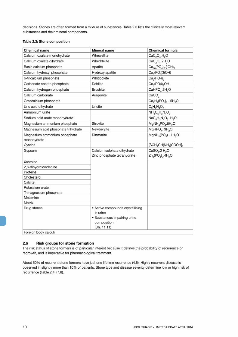
decisions. Stones are often formed from a mixture of substances. Table 2.3 lists the clinically most relevant substances and their mineral components.
Table 2.3: Stone composition
Chemical name Mineral name Chemical formulaCalcium oxalate monohydrate Whewellite CaC2O4.H2O
Calcium oxalate dihydrate Wheddelite CaC2O4.2H2O
Basic calcium phosphate Apatite Ca10(PO4)6.( OH)2Calcium hydroxyl phosphate Hydroxylapatite Ca5(PO3)3(OH)
b-tricalcium phosphate Whitlockite Ca3(PO4)2Carbonate apatite phosphate Dahllite Ca5(PO4)3OH
Calcium hydrogen phosphate Brushite CaHPO4.2H2O
Calcium carbonate Aragonite CaCO3
Octacalcium phosphate Ca8H2(PO4)6 . 5H2O
Uric acid dihydrate Uricite C5H4N4O3
Ammonium urate NH4C5H3N4O3
Sodium acid urate monohydrate NaC5H3N4O3. H2O
Magnesium ammonium phosphate Struvite MgNH4PO4.6H2O
Magnesium acid phosphate trihydrate Newberyite MgHPO4. 3H2O
Magnesium ammonium phosphate monohydrate
Dittmarite MgNH4(PO4) . 1H2O
Cystine [SCH2CH(NH2)COOH]2Gypsum Calcium sulphate dihydrate
Zinc phosphate tetrahydrateCaSO4.2 H2OZn3(PO4)2.4H2O
Xanthine2,8-dihydroxyadenineProteins Cholesterol Calcite Potassium urate Trimagnesium phosphate MelamineMatrixDrug stones • Active compounds crystallising
in urine• Substances impairing urine
composition (Ch. 11.11)
Foreign body calculi
2.6 Risk groups for stone formationThe risk status of stone formers is of particular interest because it defines the probability of recurrence orregrowth, and is imperative for pharmacological treatment.
About 50% of recurrent stone formers have just one lifetime recurrence (4,6). Highly recurrent disease isobserved in slightly more than 10% of patients. Stone type and disease severity determine low or high risk ofrecurrence (Table 2.4) (7,8).

UROLITHIASIS - LIMITED UPDATE APRIL 2014 11
Table 2.4: High-risk stone formers (7-13)
General factorsEarly onset of urolithiasis (especially children and teenagers)Familial stone formationBrushite-containing stones (CaHPO4. 2H2O)Uric acid and urate-containing stonesInfection stonesSolitary kidney (the kidney itself does not particularly increase risk of stone formation, but preventionof stone recurrence is of more importance)Diseases associated with stone formationHyperparathyroidismNephrocalcinosisGastrointestinal diseases (i.e., jejuno-ileal bypass, intestinal resection, Crohn’s disease, malabsorptiveconditions, enteric hyperoxaluria after urinary diversion) and bariatric surgerySarcoidosisGenetically determined stone formationCystinuria (type A, B and AB)Primary hyperoxaluria (PH)Renal tubular acidosis (RTA) type I2,8-dihydroxyadenineXanthinuriaLesch-Nyhan syndromeCystic fibrosisDrugs associated with stone formation Anatomical abnormalities associated with stone formationMedullary sponge kidney (tubular ectasia)Ureteropelvic junction (UPJ) obstructionCalyceal diverticulum, calyceal cystUreteral strictureVesico-uretero-renal refluxHorseshoe kidneyUreterocele
2.7 References1. Leusmann DB. Results of 5035 stone analyses: A contribution to epidemiology of urinary stone
disease. Scand J Urol Nephrol 1990;24:205-210. http://www.ncbi.nlm.nih.gov/pubmed/2237297
2. Leusmann DB. Whewellite, weddellite and company: where do all the strange names originate? BJU Int 2000 Sep;86(4):411-13.http://www.ncbi.nlm.nih.gov/pubmed/10971263
3. Kim SC, Burns EK, Lingeman JE, et al. Cystine calculi: correlation of CT-visible structure, CT number, and stone morphology with fragmentation by shock wave lithotripsy. Urol Res 2007 Dec;35(6):319-24.http://www.ncbi.nlm.nih.gov/pubmed/17965956
4. Hesse A, Brandle E, Wilbert D, et al. Study on the prevalence and incidence of urolithiasis in Germany comparing the years 1979 vs. 2000. Eur Urol 2003 Dec;44(6):709-13.http://www.ncbi.nlm.nih.gov/pubmed/14644124
5. Yasui T, Okada A, Usami M, et al. Association of the loci 5q35.3, 7q14.3, and 13.q14.1 with urolithiasis: A case-control study in the Japanese population. J Urol 2013 Apr;189(4 Suppl):e854.
6. Strohmaier WL. Course of calcium stone disease without treatment. What can we expect? Eur Urol 2000 Mar;37(3):339-44.http://www.ncbi.nlm.nih.gov/pubmed/10720863
7. Keoghane S, Walmsley B, Hodgson D. The natural history of untreated renal tract calculi. BJU Int 2010 Jun;105(12):1627-9.http://www.ncbi.nlm.nih.gov/pubmed/20438563
12 UROLITHIASIS - LIMITED UPDATE APRIL 2014
8. Straub M, Strohmaier WL, Berg W, et al. Diagnosis and metaphylaxis of stone disease Consensus concept of the National Working Committee on Stone Disease for the Upcoming German Urolithiasis Guideline. World J Urol 2005 Nov;23(5):309-23.http://www.ncbi.nlm.nih.gov/pubmed/16315051
9. Hesse AT, Tiselius H-G, Siener R, et al. (Eds). Urinary Stones, Diagnosis, Treatment and Prevention of Recurrence. 3rd edn. Basel, S. Karger AG, 2009. ISBN 978-3-8055-9149-2.
10. Basiri A, Shakhssalim N, Khoshdel AR, et al. Familial relations and recurrence pattern in nephrolithiasis: new words about old subjects. Urol J 2010 Jun;7(2):81-6.http://www.ncbi.nlm.nih.gov/pubmed/20535692
11. Goldfarb DS, Fischer ME, Keich Y, et al. A twin study of genetic and dietary influences on nephrolithiasis: a report from the Vietnam Era Twin (VET) Registry. Kidney Int 2005 Mar;67(3):1053-61.http://www.ncbi.nlm.nih.gov/pubmed/15698445
12. Durrani O, Morrisroe S, Jackman S, et al. Analysis of stone disease in morbidly obese patients undergoing gastric bypass surgery. J Endourol 2006 Oct;20(10):749-52.http://www.ncbi.nlm.nih.gov/pubmed/17094749
13. Asplin JR, Coe FL. Hyperoxaluria in kidney stone formers treated with modern bariatric surgery. J Urol 2007 Feb;177(2):565-9.http://www.ncbi.nlm.nih.gov/pubmed/17222634
3. DIAGNOSIS3.1 Diagnostic imagingPatients with urinary stones usually present with loin pain, vomiting, and sometimes fever, but may also be asymptomatic. Standard evaluation includes a detailed medical history and physical examination. The clinical diagnosis should be supported by appropriate imaging. If available, ultrasound (US) should be used as the primary diagnostic imaging tool, although pain relief, or any other emergency measures should not be delayed by imaging assessments. US is safe (no risk of radiation), reproducible and inexpensive. It can identify stones located in the calices, pelvis, and pyeloureteric and vesicoureteric junctions, as well as in patients with upper urinary tract dilatation. For all stones, US has a sensitivity of 19-93% and specificity of 84-100% (1). The sensitivity and specificity of KUB radiography is 44-77% and 80-87%, respectively (2). KUB radiography should not be performed if NCCT is considered (3), however, it is helpful in differentiating between radiolucent and radiopaque stones and for comparison during follow-up.
Recommendation LE GRWith fever or solitary kidney, and when diagnosis is doubtful, immediate imaging is indicated 4 A*
*Upgraded following panel consensus.
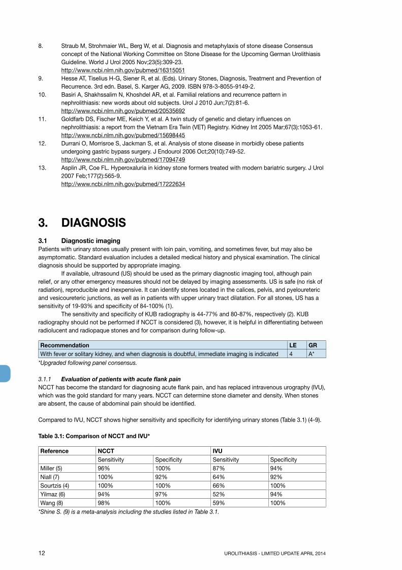
3.1.1 Evaluation of patients with acute flank painNCCT has become the standard for diagnosing acute flank pain, and has replaced intravenous urography (IVU), which was the gold standard for many years. NCCT can determine stone diameter and density. When stones are absent, the cause of abdominal pain should be identified.
Compared to IVU, NCCT shows higher sensitivity and specificity for identifying urinary stones (Table 3.1) (4-9).
Table 3.1: Comparison of NCCT and IVU*
Reference NCCT IVUSensitivity Specificity Sensitivity Specificity
Miller (5) 96% 100% 87% 94%Niall (7) 100% 92% 64% 92%Sourtzis (4) 100% 100% 66% 100%Yilmaz (6) 94% 97% 52% 94%Wang (8) 98% 100% 59% 100%
*Shine S. (9) is a meta-analysis including the studies listed in Table 3.1.
UROLITHIASIS - LIMITED UPDATE APRIL 2014 13
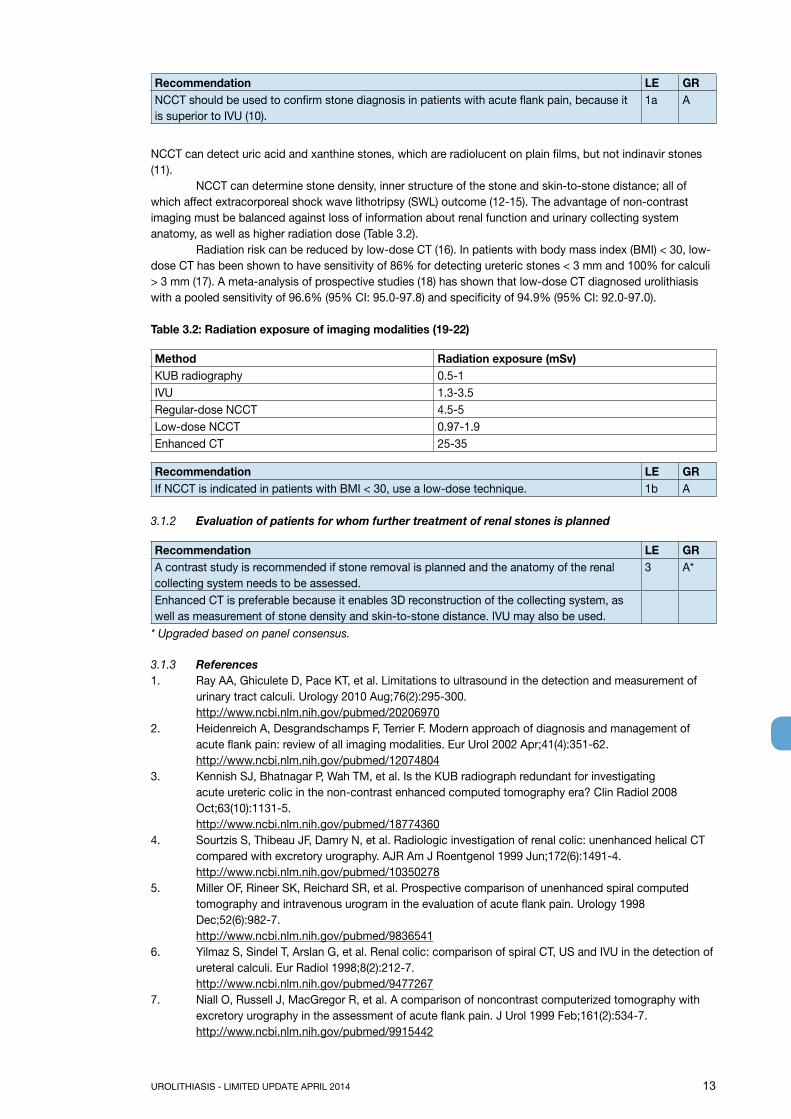
Recommendation LE GRNCCT should be used to confirm stone diagnosis in patients with acute flank pain, because it is superior to IVU (10).
1a A
NCCT can detect uric acid and xanthine stones, which are radiolucent on plain films, but not indinavir stones (11). NCCT can determine stone density, inner structure of the stone and skin-to-stone distance; all of which affect extracorporeal shock wave lithotripsy (SWL) outcome (12-15). The advantage of non-contrast imaging must be balanced against loss of information about renal function and urinary collecting system anatomy, as well as higher radiation dose (Table 3.2). Radiation risk can be reduced by low-dose CT (16). In patients with body mass index (BMI) < 30, low-dose CT has been shown to have sensitivity of 86% for detecting ureteric stones < 3 mm and 100% for calculi > 3 mm (17). A meta-analysis of prospective studies (18) has shown that low-dose CT diagnosed urolithiasis with a pooled sensitivity of 96.6% (95% CI: 95.0-97.8) and specificity of 94.9% (95% CI: 92.0-97.0).
Table 3.2: Radiation exposure of imaging modalities (19-22)
Method Radiation exposure (mSv)KUB radiography 0.5-1IVU 1.3-3.5Regular-dose NCCT 4.5-5Low-dose NCCT 0.97-1.9Enhanced CT 25-35
Recommendation LE GRIf NCCT is indicated in patients with BMI < 30, use a low-dose technique. 1b A
3.1.2 Evaluation of patients for whom further treatment of renal stones is planned
Recommendation LE GRA contrast study is recommended if stone removal is planned and the anatomy of the renal collecting system needs to be assessed.
3 A*
Enhanced CT is preferable because it enables 3D reconstruction of the collecting system, as well as measurement of stone density and skin-to-stone distance. IVU may also be used.
* Upgraded based on panel consensus.
3.1.3 References1. Ray AA, Ghiculete D, Pace KT, et al. Limitations to ultrasound in the detection and measurement of
urinary tract calculi. Urology 2010 Aug;76(2):295-300.http://www.ncbi.nlm.nih.gov/pubmed/20206970
2. Heidenreich A, Desgrandschamps F, Terrier F. Modern approach of diagnosis and management of acute flank pain: review of all imaging modalities. Eur Urol 2002 Apr;41(4):351-62.http://www.ncbi.nlm.nih.gov/pubmed/12074804
3. Kennish SJ, Bhatnagar P, Wah TM, et al. Is the KUB radiograph redundant for investigating acute ureteric colic in the non-contrast enhanced computed tomography era? Clin Radiol 2008 Oct;63(10):1131-5.http://www.ncbi.nlm.nih.gov/pubmed/18774360
4. Sourtzis S, Thibeau JF, Damry N, et al. Radiologic investigation of renal colic: unenhanced helical CT compared with excretory urography. AJR Am J Roentgenol 1999 Jun;172(6):1491-4.http://www.ncbi.nlm.nih.gov/pubmed/10350278
5. Miller OF, Rineer SK, Reichard SR, et al. Prospective comparison of unenhanced spiral computed tomography and intravenous urogram in the evaluation of acute flank pain. Urology 1998 Dec;52(6):982-7.http://www.ncbi.nlm.nih.gov/pubmed/9836541
6. Yilmaz S, Sindel T, Arslan G, et al. Renal colic: comparison of spiral CT, US and IVU in the detection of ureteral calculi. Eur Radiol 1998;8(2):212-7.http://www.ncbi.nlm.nih.gov/pubmed/9477267
7. Niall O, Russell J, MacGregor R, et al. A comparison of noncontrast computerized tomography with excretory urography in the assessment of acute flank pain. J Urol 1999 Feb;161(2):534-7.http://www.ncbi.nlm.nih.gov/pubmed/9915442

14 UROLITHIASIS - LIMITED UPDATE APRIL 2014
8. Wang JH, Shen SH, Huang SS, et al. Prospective comparison of unenhanced spiral computed tomography and intravenous urography in the evaluation of acute renal colic. J Chin Med Assoc 2008 Jan;71(1):30-6.http://www.ncbi.nlm.nih.gov/pubmed/18218557
9. Shine S. Urinary calculus: IVU vs. CT renal stone? A critically appraised topic. Abdom Imaging 2008 Jan-Feb;33(1):41-3.http://www.ncbi.nlm.nih.gov/pubmed/17786506
10. Worster A, Preyra I, Weaver B, et al. The accuracy of noncontrast helical computed tomography versus intravenous pyelography in the diagnosis of suspected acute urolithiasis: a meta-analysis. Ann Emerg Med 2002 Sep;40(3):280-6.http://www.ncbi.nlm.nih.gov/pubmed/12192351
11. Wu DS, Stoller ML. Indinavir urolithiasis. Curr Opin Urol 2000 Nov;10(6):557-61.http://www.ncbi.nlm.nih.gov/pubmed/11148725
12. El-Nahas AR, El-Assmy AM, Mansour O, et al. A prospective multivariate analysis of factors predicting stone disintegration by extracorporeal shock wave lithotripsy: the value of high-resolution noncontrast computed tomography. Eur Urol 2007 Jun;51(6):1688-93;discussion 93-4.http://www.ncbi.nlm.nih.gov/pubmed/17161522
13. Patel T, Kozakowski K, Hruby G, et al. Skin to stone distance is an independent predictor of stone-free status following shockwave lithotripsy. J Endourol 2009 Sep;23(9):1383-5.http://www.ncbi.nlm.nih.gov/pubmed/19694526
14. Kim SC, Burns EK, Lingeman JE, et al. Cystine calculi: correlation of CT-visible structure, CT number, and stone morphology with fragmentation by shock wave lithotripsy. Urol Res 2007 Dec;35(6):319-24.http://www.ncbi.nlm.nih.gov/pubmed/17965956
15. Zarse CA, Hameed TA, Jackson ME, et al. CT visible internal stone structure, but not Hounsfield unit value, of calcium oxalate monohydrate (COM) calculi predicts lithotripsy fragility in vitro. Urol Res 2007 Aug;35(4):201-6.http://www.ncbi.nlm.nih.gov/pubmed/17565491
16. Jellison FC, Smith JC, Heldt JP, et al. Effect of low dose radiation computerized tomography protocols on distal ureteral calculus detection. J Urol 2009 Dec;182(6):2762-7.http://www.ncbi.nlm.nih.gov/pubmed/19837431
17. Poletti PA, Platon A, Rutschmann OT, et al. Low-dose versus standard-dose CT protocol in patients with clinically suspected renal colic. AJR Am J Roentgenol 2007 Apr;188(4):927-33.http://www.ncbi.nlm.nih.gov/pubmed/17377025
18. Niemann T, Kollmann T, Bongartz G. Diagnostic performance of low-dose CT for the detection of urolithiasis: a meta-analysis. AJR Am J Roentgenol 2008 Aug;191(2):396-401.http://www.ncbi.nlm.nih.gov/pubmed/18647908
19. Kluner C, Hein PA, Gralla O, et al. Does ultra-low-dose CT with a radiation dose equivalent to that of KUB suffice to detect renal and ureteral calculi? J Comput Assist Tomogr 2006 Jan-Feb;30(1):44-50.http://www.ncbi.nlm.nih.gov/pubmed/16365571
20. Caoili EM, Cohan RH, Korobkin M, et al. Urinary tract abnormalities: initial experience with multi-detector row CT urography. Radiology 2002 Feb;222(2):353-60.http://www.ncbi.nlm.nih.gov/pubmed/11818599
21. Van Der Molen AJ, Cowan NC, Mueller-Lisse UG, et al. CT urography: definition, indications and techniques. A guideline for clinical practice. Eur Radiol 2008 Jan;18(1):4-17.http://www.ncbi.nlm.nih.gov/pubmed/17973110
22. Thomson JM, Glocer J, Abbott C, et al. Computed tomography versus intravenous urography in diagnosis of acute flank pain from urolithiasis: a randomized study comparing imaging costs and radiation dose. Australas Radiol 2001 Aug;45(3):291-7.http://www.ncbi.nlm.nih.gov/pubmed/11531751
3.2 Diagnostics - metabolism-relatedEach emergency patient with urolithiasis needs a succinct biochemical work-up of urine and blood besides imaging. At that point, no distinction is made between high- and low-risk patients.

UROLITHIASIS - LIMITED UPDATE APRIL 2014 15
Table 3.3: Recommendations: basic laboratory analysis - emergency urolithiasis patients (1-4)
Urine GRUrinary sediment/dipstick test of spot urine sample• red cells• white cells• nitrite• approximate urine pHUrine culture or microscopy
A*A
BloodSerum blood sample• creatinine• uric acid• ionised calcium• sodium• potassium
A*
Blood cell countCRP
A*
If intervention is likely or planned:Coagulation test (PTT and INR)
A*
* Upgraded based on panel consensus.CPR = C-reactive protein; INR = international normalised ratio; PTT = partial thromboplastin time.
3.2.1 Basic laboratory analysis - non-emergency urolithiasis patientsBiochemical work-up is similar for all stone patients. However, if no intervention is planned, examination of sodium, potassium, CRP, and blood coagulation time can be omitted. Only patients at high risk for stone recurrence should undergo a more specific analytical programme (4). Stone-specific metabolic evaluation is described in Chapter 11. The easiest means to achieve correct diagnosis is by analysis of a passed stone using a valid method as listed below (see 3.2.2). Once mineral composition is known, the potential metabolic disorders can be identified.
3.2.2 Analysis of stone compositionStone analysis should be performed in all first-time stone formers.In clinical practice, repeat stone analysis is needed in case of:• recurrence under pharmacological prevention;• early recurrence after interventional therapy with complete stone clearance;• late recurrence after a prolonged stone-free period (6).
Patients should be instructed to filter their urine to retrieve a concrement for analysis. Stone passage and restoration of normal renal function should be confirmed. The preferred analytical procedures are infrared spectroscopy (IRS) or X-ray diffraction (XRD) (5,7,8). Equivalent results can be obtained by polarisation microscopy, but only in centres with expertise. Chemical analysis (wet chemistry) is generally deemed to be obsolete (5).
Recommendations LE GRAlways perform stone analysis in first-time formers using a valid procedure (XRD or IRS). 2 ARepeat stone analysis in patients:• presenting with reccurent stones despite drug therapy;• with early recurrence after complete stone clearance;• with late recurrence after a long stone-free period because stone composition may
change (3).
2 B

16 UROLITHIASIS - LIMITED UPDATE APRIL 2014
3.3 References1. S-3 Guideline AWMF-Register-Nr. 043/044 Urinary Tract Infections. Epidemiology, diagnostics, therapy
and management of uncomplicated bacterial community acquired urinary tract infections in adults.http://www.awmf.org/leitlinien/detail/ll/043-044.html
2. Hesse AT, Tiselius H-G. Siener R, et al. (Eds). Urinary Stones, Diagnosis, Treatment and Prevention of Recurrence. 3rd edn. Basel, S.Karger AG, 2009. ISBN 978-3-8055-9149-2.
3. Pearle MS, Asplin JR, Coe FL, et al (Committee 3). Medical management of urolithiasis. In: 2nd International consultation on Stone Disease. Denstedt J, Khoury S. eds. pp. 57-84. Health Publications 2008, ISBN 0-9546956-7-4.http://www.icud.info/publications.html
4. Straub M, Strohmaier WL, Berg W, et al. Diagnosis and metaphylaxis of stone disease. Consensus concept of the National Working Committee on Stone Disease for the upcoming German Urolithiasis Guideline. World J Urol 2005 Nov;23(5):309-23.http://www.ncbi.nlm.nih.gov/pubmed/16315051
5. Hesse A, Kruse R, Geilenkeuser WJ, et al. Quality control in urinary stone analysis: results of 44 ring trials (1980-2001). Clin Chem Lab Med 2005;43(3):298-303.http://www.ncbi.nlm.nih.gov/pubmed/15843235
6. Mandel N, Mandel I, Fryjoff K, et al. Conversion of calcium oxalate to calcium phosphate with recurrent stone episodes. J Urol 2003 Jun;169(6):2026-9.http://www.ncbi.nlm.nih.gov/pubmed/12771710
7. Suror DJ, Scheidt S. Identification standards for human urinary calculus components, using crystallographic methods. Br J Urol 1968 Feb;40(1):22-8.http://www.ncbi.nlm.nih.gov/pubmed/5642759
8. Abdel-Halim RE, Abdel-Halim MR. A review of urinary stone analysis techniques. Saudi Med J 2006 Oct;27(10):1462-7.http://www.ncbi.nlm.nih.gov/pubmed/17013464
4. TREATMENT OF PATIENTS WITH RENAL COLIC4.1 Renal colic4.1.1 Pain reliefPain relief is the first therapeutic step in patients with an acute stone episode (1,2).
Non-steroidal anti-inflammatory drugs (NSAIDs) are effective in patients with acute stone colic (3-6), and have better analgesic efficacy than opioids. Patients receiving NSAIDs are less likely to require further analgesia in the short term.
Opioids, particularly pethidine, are associated with a high rate of vomiting compared to NSAIDs, and carry a greater likelihood of further analgesia being needed (7,8) (Section 4.1.3). If an opioid is used, it is recommended that it is not pethidine.
Statement LEFor symptomatic ureteral stones, urgent SWL as first-line treatment is a feasible option (9). 1b
Recommendations GRIn acute stone episodes, pain relief should be initiated immediately. AWhenever possible, an NSAID should be the first drug of choice. A
4.1.2 Prevention of recurrent renal colicFacilitation of passage of ureteral stones is discussed in Section 5.3.
For patients with ureteral stones that are expected to pass spontaneously, NSAID tablets or suppositories (e.g., diclofenac sodium, 100-150 mg/day, 3-10 days) may help reduce inflammation and risk of recurrent pain (8,10,11). Although diclofenac can affect renal function in patients with already reduced function, it has no effect in patients with normal kidney function (LE: 1b) (12). In a double-blind, placebo-controlled trial, recurrent pain episodes of stone colic were significantly fewer in patients treated with NSAIDs (as compared to no NSAIDs) during the first 7 days of treatment (11).

UROLITHIASIS - LIMITED UPDATE APRIL 2014 17
Daily α-blockers reduce recurrent colic (LE: 1a) (Section 5.3) (13,14).If analgesia cannot be achieved medically, drainage, using stenting or percutaneous nephrostomy, or stone removal, should be performed.
4.1.3 Recommendations for analgesia during renal colic
LE GRFirst choice: start with an NSAID, e.g. diclofenac*, indomethacin or ibuprofen**. 1b ASecond choice: hydromorphine, pentazocine or tramadol. 4 CUse α-blockers to reduce recurrent colics. 1a A
*Affects glomerular filtration rate (GFR) in patients with reduced renal function (15) (LE: 2a).**Recommended to counteract recurrent pain after ureteral colic.
4.1.4 References1. Phillips E, Kieley S, Johnson EB, et al. Emergency room management of ureteral calculi: current
practices. J Endourol 2009 Jun;23(6):1021-4.http://www.ncbi.nlm.nih.gov/pubmed/19445640
2. Micali S, Grande M, Sighinolfi MC, et al. Medical therapy of urolithiasis. J Endourol 2006 Nov;20(11):841-7.http://www.ncbi.nlm.nih.gov/pubmed/17144848
3. Ramos-Fernández M, Serrano LA. Evaluation and management of renal colic in the emergency department. Bol Asoc Med P R 2009 Jul-Sep;101(3):29-32.http://www.ncbi.nlm.nih.gov/pubmed/20120983
4. Engeler DS, Schmid S, Schmid HP. The ideal analgesic treatment for acute renal colic--theory and practice. Scand J Urol Nephrol 2008;42(2):137-42.http://www.ncbi.nlm.nih.gov/pubmed/17899475
5. Cohen E, Hafner R, Rotenberg Z, et al. Comparison of ketorolac and diclofenac in the treatment of renal colic. Eur J Clin Pharmacol 1998 Aug;54(6):455-8.http://www.ncbi.nlm.nih.gov/pubmed/9776434
6. Shokeir AA, Abdulmaaboud M, Farage Y, et al. Resistive index in renal colic: the effect of nonsteroidal anti-inflammatory drugs. BJU Int 1999 Aug;84(3):249-51.http://www.ncbi.nlm.nih.gov/pubmed/10468715
7. Holdgate A, Pollock T. Nonsteroidal anti-inflammatory drugs (NSAIDs) versus opioids for acute renal colic. Cochrane Database Syst Rev 2005 Apr;(2):CD004137.http://www.ncbi.nlm.nih.gov/pubmed/15846699
8. Ebell MH. NSAIDs vs. opiates for pain in acute renal colic. Am Fam Physician 2004 Nov;70(9):1682.http://www.ncbi.nlm.nih.gov/pubmed/15554485
9. Picozzi SC, Ricci C, Gaeta M, et al. Urgent shock wave lithotripsy as first-line treatment for ureteral stones: a meta-analysis of 570 patients. Urol Res 2012 Dec;40(6):725-31.http://www.ncbi.nlm.nih.gov/pubmed/22699356
10. Holdgate A, Pollock T. Systematic review of the relative efficacy of non-steroidal anti-inflammatory drugs and opioids in the treatment of acute renal colic. BMJ 2004 Jun;328(7453):1401.http://www.ncbi.nlm.nih.gov/pubmed/15178585
11. Laerum E, Ommundsen OE, Gronseth JE, et al. Oral diclofenac in the prophylactic treatment of recurrent renal colic. A double-blind comparison with placebo. Eur Urol 1995;28(2):108-11.http://www.ncbi.nlm.nih.gov/pubmed/8529732
12. Lee A, Cooper MG, Craig JC, et al. Effects of nonsteroidal anti-inflammatory drugs on postoperative renal function in adults with normal renal function. Cochrane Database Syst Rev 2007;18(2):CD002765.http://www.ncbi.nlm.nih.gov/pubmed/17443518
13. Dellabella M, Milanese G, Muzzonigro G. Randomized trial of the efficacy of tamsulosin, nifedipine and phloroglucinol in medical expulsive therapy for distal ureteral calculi. J Urol 2005 Jul;174(1):167-72.http://www.ncbi.nlm.nih.gov/pubmed/15947613
14. Resim S, Ekerbicer H, Ciftci A. Effect of tamsulosin on the number and intensity of ureteral colic in patients with lower ureteral calculus. Int J Urol 2005 Jul;12(7):615- 20.http://www.ncbi.nlm.nih.gov/pubmed/16045553
15. Walden M, Lahtinen J, Elvander E. Analgesic effect and tolerance of ketoprofen and diclofenac in acute ureteral colic. Scand J Urol Nephrol 1993;27(3):323-5.http://www.ncbi.nlm.nih.gov/pubmed/8290910

18 UROLITHIASIS - LIMITED UPDATE APRIL 2014
4.2 Management of sepsis in obstructed kidneyThe obstructed kidney with all signs of urinary tract infection (UTI) is a urological emergency. Urgent decompression is often necessary to prevent further complications in infected hydronephrosis secondary to stone-induced, unilateral or bilateral renal obstruction. The optimal method of decompression has yet to be established (1-3). However, it is known that compromised delivery of antibiotics into the obstructed kidney means that the collecting system must be drained to encourage resolution of infection.
4.2.1 DecompressionCurrently, there are two options for urgent decompression of obstructed collecting systems:• placement of an indwelling ureteral catheter;• percutaneous placement of a nephrostomy catheter.
There is little evidence to support the superiority of percutaneous nephrostomy over retrograde stenting for primary treatment of infected hydronephrosis. There is no good-quality evidence to suggest that ureteric stenting has more complications than percutaneous nephrostomy (1,4,5). Only two RCTs (2,5) have assessed decompression of acute infected hydronephrosis. The complications of percutaneous nephrostomy insertion have been reported consistently, but those of ureteric stent insertion are less well described (1). Definitive stone removal should be delayed until the infection is cleared following a complete course of antimicrobial therapy (6,7).
Emergency nephrectomy may become necessary in highly complicated cases to eliminate further complications.
Statement LEFor decompression of the renal collecting system, ureteral stents and percutaneous nephrostomy catheters are equally effective.
1b
Recommendations LE GRFor sepsis with obstructing stones, the collecting system should be urgently decompressed, using percutaneous drainage or ureteral stenting.
1b A
Definitive treatment of the stone should be delayed until sepsis is resolved. 1b A
4.2.2 Further measuresFollowing urgent decompression of the obstructed and infected urinary collecting system, both urine- and blood samples should be sent for culture-antibiogram sensitivity testing, and antibiotics should be initiated immediately thereafter. The regimen should be re-evaluated in the light of the culture-antibiogram test. Intensive care might become necessary.
Recommendations GRCollect urine for antibiogram test following decompression. A*Start antibiotics immediately thereafter (+ intensive care if necessary).Re-evaluate antibiotic regimen following antibiogram findings.
* Upgraded based on panel consensus.
4.2.3 References1. Ramsey S, Robertson A, Ablett MJ, et al. Evidence-based drainage of infected hydronephrosis
secondary to ureteric calculi. J Endourol 2010 Feb;24(2):185-9.http://www.ncbi.nlm.nih.gov/pubmed/20063999
2. Pearle MS, Pierce HL, Miller GL, et al. Optimal method of urgent decompression of the collecting system for obstruction and infection due to ureteral calculi. J Urol 1998 Oct;160(4):1260-4.http://www.ncbi.nlm.nih.gov/pubmed/9751331
3. Uppot RN. Emergent nephrostomy tube placement for acute urinary obstruction. Tech Vasc Interv Radiol 2009 Jun;12(2):154-61.http://www.ncbi.nlm.nih.gov/pubmed/19853233
4. Lynch MF, Anson KM, Patel U. Percutaneous nephrostomy and ureteric stent insertion for acute renal deobstruction. Consensus based guidelines. Br J Med Surg Urol 2008 Nov;1(3);120-5.http://www.bjmsu.com/article/S1875-9742(08)00095-5/abstract

UROLITHIASIS - LIMITED UPDATE APRIL 2014 19
5. Mokhmalji H, Braun PM, Portillo FJ, et al. Percutaneous nephrostomy versus ureteral stents for diversion of hydronephrosis caused by stones: A prospective, randomized clinical trial. J Urol 2001 Apr;165(4):1088-92.http://www.ncbi.nlm.nih.gov/pubmed/11257644
6. Klein LA, Koyle M, Berg S. The emergency management of patients with ureteral calculi and fever. J Urol 1983 May;129(5):938-40.http://www.ncbi.nlm.nih.gov/pubmed/6854761
7. Camúñez F, Echenagusia A, Prieto ML, et al. Percutaneous nephrostomy in pyonephrosis. Urol Radiol 1989;11(2):77-81.http://www.ncbi.nlm.nih.gov/pubmed/2667249
5. STONE RELIEFWhen deciding between active stone removal and conservative treatment with medical expulsive therapy(MET), it is important to consider all the patients’ circumstances that may affect treatment decisions (1).
5.1 Observation of ureteral stones5.1.1 Stone-passage ratesThere are only limited data about spontaneous stone passage according to size (2,3). A meta-analysis of 328 patients harbouring ureteral stones < 10 mm investigated the likelihood of ureteral stone passage (Table 5.1) (2). These studies had limitations including non-standardisation of stone size measurement, and lack of analysis of stone position, stone-passage history, and time to stone passage.
Table 5.1: Likelihood of ureteral stone passage of ureteral stones (2)
Stone size Average time to pass Percentage of passages (95% CI)< 5 mm (n = 224) 68% (46-85%)> 5 mm (n = 104) 47% (36-58%)< 2 mm 31 days2-4 mm 40 days4-6 mm 39 days
95% of stones up to 4 mm pass within 40 days (3).
Recommendations LE GRIn patients with newly diagnosed ureteral stones < 10 mm, and if active removal is not indicated (Chapter 6), observation with periodic evaluation is an optional initial treatment.
1a A
Such patients may be offered appropriate medical therapy to facilitate stone passage during observation.*
*see Section 5.3, Medical expusive therapy (MET).
5.2 Observation of kidney stonesObservation of kidney stones, especially in calices, depends on their natural history (Section 6.2.1).
Statement LEIt is still debatable whether kidney stones should be treated, or whether annual follow-up is sufficientfor asymptomatic caliceal stones that have remained stable for 6 months.
4
Recommendations GRKidney stones should be treated in case of growth, formation of de novo obstruction, associated infection, and acute or chronic pain.
A*
Comorbidity and patient preference need to be taken into consideration when making treatment decisions.
C
If kidney stones are not treated, periodic evaluation is needed. A** Upgraded based on panel consensus.

20 UROLITHIASIS - LIMITED UPDATE APRIL 2014
5.3 Medical expulsive therapy (MET)Drugs that expel stones might act by relaxing ureteral smooth muscle through inhibition of calcium channel pumps or α-1 receptor blockade (4,5). MET should only be used in patients who are comfortable with this approach and when there is no obvious advantage from immediate active stone removal.
Meta-analyses have shown that patients with ureteral stones treated with α-blockers or nifedipine are more likely to pass stones with fewer episodes of colic than those not receiving such therapy (4,5).
Statement LEThere is good evidence that MET accelerates spontaneous passage of ureteral stones and fragments generated with SWL, and limits pain (4-16).
1a
5.3.1 Medical agentsTamsulosin is one of the most commonly used α-blockers (4,6,17-20). However, one small study has suggested that tamsulosin, terazosin and doxazosin are equally effective, indicating a possible class effect (21). This is also indicated by several trials demonstrating increased stone expulsion using doxazosin (4,21,22), terazosin (21,23), alfuzosin (24-27) naftopidil (28,29), and silodosin (30,31).
Statement LESeveral trials have demonstrated an α-blocker class effect on stone expulsion rates. 1b
With regard to the class effect of calcium-channel blockers, only nifedipine has been investigated (LE = 1a) (4,9-11,32,33).
Administration of tamsulosin and nifedipine is safe and effective in patients with distal ureteral stones with renal colic. However, tamsulosin is significantly better than nifedipine in relieving renal colic and facilitating and accelerating ureteral stone expulsion (11,32,33). Based on studies with a limited number of patients (34,35) (LE: 1b), no recommendation for the use of corticosteroids in combination with α-blockers in MET can be made.
Statement LEThere is no evidence to support the use of corticosteroids as monotherapy for MET. Insufficient data exist to support the use of corticosteroids in combination with α-blockers as an accelerating adjunct (3,21,34,35).
1b
Recommendations for MET LE GRFor MET, α-blockers are recommended. 1a APatients should be counseled about the attendant risks of MET, including associated drug side effects, and should be informed that it is administered off-label†**.
A*
Patients, who elect for an attempt at spontaneous passage or MET, should have well-controlled pain, no clinical evidence of sepsis, and adequate renal functional reserve.
A
Patients should be followed once between 1 and 14 days to monitor stone position and be assessed for hydronephrosis.
4 A*
† It is not known if tamsulosin harms the human foetus or if it is found in breast milk.* Upgraded based on panel consensus.**MET in children cannot be recommended due to the limited data in this specific population.
5.3.2 Factors affecting success of medical expulsive therapy (tamsulosin)5.3.2.1 Stone sizeDue to the high likelihood of spontaneous passage of stones up to ~5 mm, MET is less likely to increase the stone-free rate (SFR) (5,36-39) (LE: 1b). However, MET does reduce the need for analgesics (4,6) (LE: 1a).
5.3.2.2 Stone locationThe vast majority of trials have investigated distal ureteral stones (4). One RCT has assessed the effect of tamsulosin on spontaneous passage of proximal ureteral calculi 5-10 mm. The main effect was to encourage stone migration to a more distal part of the ureter (40) (LE: 1b).
5.3.2.3 Medical expulsive therapy after extracorporeal shock wave lithotripsy (SWL)Clinical studies and several meta-analyses have shown that MET after SWL for ureteral or renal stones can

UROLITHIASIS - LIMITED UPDATE APRIL 2014 21
expedite expulsion and increase SFRs and reduce analgesic requirements (7,12,41-49) (LE: 1a).
5.3.2.4 Medical expulsive therapy after ureteroscopyMET following holmium:YAG laser lithotripsy increases SFRs and reduces colic episodes (50) (LE: 1b).
5.3.2.5 Medical expulsive therapy and ureteral stents (Section 5.6.2.1.8)
5.3.2.6 Duration of medical expulsive therapy treatmentMost studies have had a duration of 1 month or 30 days. No data are currently available to support other time-intervals.
5.3.2.7 Possible side-effects include retrograde ejaculation and hypotension (4).
5.3.3 References1. Skolarikos A, Laguna MP, Alivizatos G, et al. The role for active monitoring in urinary stones: a
systematic review. J Endourol 2010 Jun;24(6):923-30.http://www.ncbi.nlm.nih.gov/pubmed/20482232
2. Preminger GM, Tiselius HG, Assimos DG, et al. American Urological Association Education and Research, Inc; European Association of Urology. 2007 Guideline for the management of ureteral calculi. Eur Urol 2007 Dec;52(6):1610-31.http://www.ncbi.nlm.nih.gov/pubmed/18074433
3. Miller OF, Kane CJ. Time to stone passage for observed ureteral calculi: a guide for patient education. J Urol 1999 Sep;162(3 Pt 1):688-90;discussion 690-1.http://www.ncbi.nlm.nih.gov/pubmed/10458343
4. Seitz C, Liatsikos E, Porpiglia F, et al. Medical Therapy to Facilitate the Passage of Stones: What Is the Evidence? Eur Urol 2009 Sep;56(3):455-71.http://www.ncbi.nlm.nih.gov/pubmed/19560860
5. Liatsikos EN, Katsakiori PF, Assimakopoulos K, et al. Doxazosin for the management of distal-ureteral stones. J Endourol 2007 May;21(5):538-41.http://www.ncbi.nlm.nih.gov/pubmed/17523910
6. Hollingsworth JM, Rogers MA, Kaufman SR, et al. Medical therapy to facilitate urinary stone passage: a meta-analysis. Lancet 2006 Sep;368(9542):1171-9.http://www.ncbi.nlm.nih.gov/pubmed/17011944
7. Gravina GL, Costa AM, Ronchi P, et al. Tamsulosin treatment increases clinical success rate of single extracorporeal shock wave lithotripsy of renal stones. Urology 2005 Jul;66(1):24-8.http://www.ncbi.nlm.nih.gov/pubmed/15992885
8. Resim S, Ekerbicer HC, Ciftci A. Role of tamsulosin in treatment of patients with steinstrasse developing after extracorporeal shock wave lithotripsy. Urology 2005 Nov;66(5):945-8.http://www.ncbi.nlm.nih.gov/pubmed/16286100
9. Borghi L, Meschi T, Amato F, et al. Nifedipine and methylprednisolone in facilitating ureteral stone passage: a randomized, double-blind, placebo-controlled study. J Urol 1994 Oct;152(4):1095-8.http://www.ncbi.nlm.nih.gov/pubmed/8072071
10. Porpiglia F, Destefanis P, Fiori C, et al. Effectiveness of nifedipine and deflazacort in the management of distal ureter stones. Urology 2000 Oct;56(4):579-82.http://www.ncbi.nlm.nih.gov/pubmed/11018608
11. Dellabella M, Milanese G, Muzzonigro G. Randomized trial of the efficacy of tamsulosin, nifedipine and phloroglucinol in medical expulsive therapy for distal ureteral calculi. J Urol 2005 Jul;174(1):167-72.http://www.ncbi.nlm.nih.gov/pubmed/15947613
12. Naja V, Agarwal MM, Mandal AK, et al. Tamsulosin facilitates earlier clearance of stone fragments and reduces pain after shockwave lithotripsy for renal calculi; results from an open-label randomized study. Urology 2008 Nov;72(5):1006-11.http://www.ncbi.nlm.nih.gov/pubmed/18799202
13. Schuler TD, Shahani R, Honey RJ, et al. Medical expulsive therapy as an adjunct to improve shockwave lithotripsy outcomes: a systematic review and meta-analysis. J Endourol 2009 Mar;23(3):387-93.http://www.ncbi.nlm.nih.gov/pubmed/19245302
14. Parsons JK, Hergan LA, Sakamoto K, et al. Efficacy of alpha blockers for the treatment of ureteral stones. J Urol 2007 Mar;177(3):983-7.http://www.ncbi.nlm.nih.gov/pubmed/17296392

22 UROLITHIASIS - LIMITED UPDATE APRIL 2014
15. Singh A, Alter HJ, Littlepage A. A systematic review of medical therapy to facilitate passage of ureteral calculi. Ann Emerg Med 2007 Nov;50(5):552-63.http://www.ncbi.nlm.nih.gov/pubmed/17681643
16. Arrabal-Martin M, Valle-Diaz de la Guardia F, Arrabal-Polo MA, et al. Treatment of ureteral lithiasis with tamsulosin: literature review and meta-analysis. Urol Int 2010;84(3):254-9.http://www.ncbi.nlm.nih.gov/pubmed/20389151
17. Lojanapiwat B, Kochakarn W, Suparatchatpan N, et al. Effectiveness of low-dose and standard-dose tamsulosin in the treatment of distal ureteric stones: A randomized controlled study. J Int Med Res 2008 May-Jun;36(3):529-36.http://www.ncbi.nlm.nih.gov/pubmed/18534135
18. Wang CJ, Huang SW, Chang CH. Efficacy of an alpha1 blocker in expulsive therapy of lower ureteral stones. J Endourol 2008 Jan;22(1):41-6.http://www.ncbi.nlm.nih.gov/pubmed/18315472
19. Kaneko T, Matsushima H, Morimoto H, et al. Efficacy of low dose tamsulosin medical expulsive therapy for ureteral stones in Japanese male patients: a randomized controlled study. Int J Urol 2010 May;17(5):462-5.http://www.ncbi.nlm.nih.gov/pubmed/20202002
20. Al-Ansari A, Al-Naimi A, Alobaidy A, et al. Efficacy of tamsulosin in the management of lower ureteral stones: a randomized double-blind placebo-controlled study of 100 patients. Urology 2010 Jan;75(1):4-7.http://www.ncbi.nlm.nih.gov/pubmed/20109697
21. Yilmaz E, Batislam E, Basar MM, et al. The comparison and efficacy of 3 different alpha1-adrenergic blockers for distal ureteral stones. J Urol 2005 Jun;173(6):2010-2.http://www.ncbi.nlm.nih.gov/pubmed/15879806
22. Zehri AA, Ather MH, Abbas F, et al. Preliminary study of efficacy of doxazosin as a medical expulsive therapy of distal ureteric stones in a randomized clinical trial. Urology 2010 Jun;75(6):1285-8.http://www.ncbi.nlm.nih.gov/pubmed/20189226
23. Mohseni MG, Hosseini SR, Alizadeh F. Efficacy of terazosin as a facilitator agent for expulsion of the lower ureteral stones. Saudi Med J 2006 Jun;27(6):838-40.http://www.ncbi.nlm.nih.gov/pubmed/16758046
24. Agrawal M, Gupta M, Gupta A, et al. Prospective Randomized Trial Comparing Efficacy of Alfuzosin and Tamsulosin in Management of Lower Ureteral Stones. Urology 2009 Apr;73(4):706-9.http://www.ncbi.nlm.nih.gov/pubmed/19193417
25. Pedro RN, Hinck B, Hendlin K, et al. Alfuzosin stone expulsion therapy for distal ureteral calculi: a double-blind, placebo controlled study. J Urol 2008 Jun;179(6):2244-7, discussion 2247.http:/www.ncbi.nlm.nih.gov/pubmed/18423747
26. Ahmed AF, Al-Sayed AY. Tamsulosin versus Alfuzosin in the Treatment of Patients with Distal Ureteral Stones: Prospective, Randomized, Comparative Study. Korean J Urol 2010 Mar;51(3):193-7.http://www.ncbi.nlm.nih.gov/pubmed/20414396
27. Chau LH, Tai DC, Fung BT, et al. Medical expulsive therapy using alfuzosin for patient presenting with ureteral stone less than 10mm: a prospective randomized controlled trial. Int J Urol 2011 Jul;18(7): 510-4.http://www.ncbi.nlm.nih.gov/pubmed/21592234
28. Sun X, He L, Ge W, et al. Efficacy of selective alpha1D-Blocker Naftopidil as medical expulsive therapy for distal ureteral stones. J Urol 2009 Apr;181(4):1716-20.http://www.ncbi.nlm.nih.gov/pubmed/19233432
29. Zhou SG, Lu JL, Hui JH. Comparing efficacy of < (1)D-receptor antagonist naftopidil and < 1A/Dreceptor antagonist tamsulosin in management of distal ureteral stones. World J Urol 2011 Dec;29(6):767-71.http://www.ncbi.nlm.nih.gov/pubmed/21845472
30. Tsuzaka Y, Matsushima H, Kaneko T, et al. Naftopidil vs silodosin in medical expulsive therapy for ureteral stones: a randomized controlled study in Japanese male patients. Int J Urol 2011 Nov;18(11):792-5.http://www.ncbi.nlm.nih.gov/pubmed/21917021
31. Itoh Y, Okada A, Yasui T, et al. Efficacy of selective α1A adrenoceptor antagonist silodosin in the medical expulsive therapy for ureteral stones. Int J Urol 2011 Sep;18(9):672-4.http://www.ncbi.nlm.nih.gov/pubmed/21707766
32. Porpiglia F, Ghignone G, Fiori C, et al. Nifedipine versus tamsulosin for the management of lower ureteral stones. J Urol 2004 Aug;172(2):568-71.http://www.ncbi.nlm.nih.gov/pubmed/15247732

UROLITHIASIS - LIMITED UPDATE APRIL 2014 23
33. Ye Z, Yang H, Li H, et al. A multicentre, prospective, randomized trial: comparative efficacy of tamsulosin and nifedipine in medical expulsive therapy for distal ureteric stones with renal colic. BJU Int 2011 Jul;108(2):276-9.http://www.ncbi.nlm.nih.gov/pubmed/21083640
34. Porpiglia F, Vaccino D, Billia M, et al. Corticosteroids and tamsulosin in the medical expulsive therapy for symptomatic distal ureter stones: single drug or association? Eur Urol 2006 Aug;50(2):339-44.http://www.ncbi.nlm.nih.gov/pubmed/16574310
35. Dellabella M, Milanese G, Muzzonigro G. Medical-expulsive therapy for distal ureterolithiasis: randomized prospective study on role of corticosteroids used in combination with tamsulosin simplified treatment regimen and health-related quality of life. Urology 2005 Oct;66(4):712-5.http://www.ncbi.nlm.nih.gov/pubmed/16230122
36. Ferre RM, Wasielewski JN, Strout TD, et al. Tamsulosin for ureteral stones in the emergency department: a Randomized controlled trial. Ann Emerg Med 2009 Sep;54(3):432-9.http://www.ncbi.nlm.nih.gov/pubmed/19200622
37. Hermanns T, Sauermann P, Rufibach K, et al. Is there a role for tamsulosin in the treatment of distal ureteral stones of 7 mm or less? Results of a randomised, double-blind, placebo-controlled trial. Eur Urol 2009 Sep;56(3):407-12.http://www.ncbi.nlm.nih.gov/pubmed/19375849
38. Vincendeau S, Bellissant E, Houlgatte A, et al; Tamsulosin Study Group. Tamsulosin hydrochloride vs placebo for management of distal ureteral stones: a multicentric, randomized, double-blind trial. Arch Intern Med 2010 Dec;170(22):2021-7.http://www.ncbi.nlm.nih.gov/pubmed/21149761
39. Ochoa-Gómez R, Prieto-Díaz-Chávez E, Trujillo-Hernández B, et al. Tamsulosin does not have greater efficacy than conventional treatment for distal ureteral stone expulsion in Mexican patients. Urol Res 2011 Dec;39(6)491-5.http://www.ncbi.nlm.nih.gov/pubmed/21516496
40. Yencilek F, Erturhan S, Canguven O, et al. Does tamsulosin change the management of proximally located ureteral stones? Urol Res 2010 Jun;38(3):195-9.http://www.ncbi.nlm.nih.gov/pubmed/20182703
41. Bhagat SK, Chacko NK, Kekre NS, et al. Is there a role for tamsulosin in shock wave lithotripsy for renal and ureteral calculi? J Urol 2007 Jun;177(6):2185-8.http://www.ncbi.nlm.nih.gov/pubmed/17509314
42. Küpeli B, Irkilata L, Gürocak S, et al. Does tamsulosin enhance lower ureteral stone clearance with or without shock wave lithotripsy? Urology 2004 Dec;64(6):1111-5.http://www.ncbi.nlm.nih.gov/pubmed/15596181
43. Wang H, Liu K, Ji Z, et al. Effect of alpha1-adrenergic antagonists on lower ureteral stones with extracorporeal shock wave lithotripsy. Asian J Surg 2010 Jan;33(1):37-41.http://www.ncbi.nlm.nih.gov/pubmed/20497881
44. Zhu Y, Duijvesz D, Rovers MM, et al. alpha-Blockers to assist stone clearance after extracorporeal shock wave lithotripsy: a meta-analysis. BJU Int 2010 Jul;106(2):256-61.http://www.ncbi.nlm.nih.gov/pubmed/19889063
45. Hussein MM. Does tamsulosin increase stone clearance after shockwave lithotripsy of renal stones? A prospective, randomized controlled study. Scand J Urol Nephrol 2010 Feb;44(1):27-31.http://www.ncbi.nlm.nih.gov/pubmed/19947900
46. Singh SK, Pawar DS, Griwan MS, et al. Role of tamsulosin in clearance of upper ureteral calculi after extracorporeal shock wave lithotripsy: a randomized controlled trial. Urol J 2011 Winter;8(1):14-20.http://www.ncbi.nlm.nih.gov/pubmed/21404197
47. Zheng S, Liu LR, Yuan HC, et al. Tamsulosin as adjunctive treatment after shockwave lithotripsy in patients with upper urinary tract stones: a systematic review and meta-analysis. Scand J Urol Nephrol 2010 Dec;44(6):425-32.http://www.ncbi.nlm.nih.gov/pubmed/21080841
48. Falahatkar S, Khosropanah I, Vajary AD, et al. Is there a role for tamsulosin after shock wave lithotripsy in the treatment of renal and ureteral calculi? J Endourol 2011 Mar;25(3):495-8.http://www.ncbi.nlm.nih.gov/pubmed/21166579
49. Singh SK, Pawar DS, Griwan MS, et al. Role of tamsulosin in clearance of upper ureteral calculi after extracorporeal shock wave lithotripsy: a randomized controlled trial. Urol J 2011 Winter;8(1):14-20.http://www.ncbi.nlm.nih.gov/pubmed/21404197
50. John TT, Razdan S. Adjunctive tamsulosin improves stone free rate after ureteroscopic lithotripsy of large renal and ureteric calculi: a prospective randomized study. Urology 2010 May;75(5):1040-2.http://www.ncbi.nlm.nih.gov/pubmed/19819530

24 UROLITHIASIS - LIMITED UPDATE APRIL 2014
5.4 Chemolytic dissolution of stonesOral or percutaneous irrigation chemolysis of stones or their fragments can be useful first-line therapy. It may also be an adjunct to SWL, percutaneous nephrolithotomy (PNL), ureterorenoscopy (URS) or open surgery to support elimination of small residual fragments, considering that its use as first-line therapy may take several weeks to be effective.
Combined treatment with SWL and chemolysis is a minimally invasive option for patients with partial or complete infection staghorn stones who are not eligible for PNL. Stone fragmentation leads to increased stone surface area and improved efficacy of chemolitholysis. Chemolysis is possible only for the stone compositions listed below, therefore, knowledge of stone composition is mandatory before treatment.
5.4.1 Percutaneous irrigation chemolysisPercutaneous irrigation chemolysis may be an option for infection- and uric acid stones (1,2).
Recommendations GRIn percutaneous chemolysis, at least two nephrostomy catheters should be used to allow irrigation of the renal collecting system, while preventing chemolytic fluid draining into the bladder and reducing the risk of increased intrarenal pressure*.
A
Pressure- and flow-controlled systems should be used if available.
* Alternatively, one nephrostomy catheter with a JJ stent and bladder catheter can serve as a through-flowsystem preventing high pressure.
Table 5.2: Methods of percutaneous irrigation chemolysis
Stone composition Refs. Irrigation solution CommentsStruvite Carbon apatite
1-6 10% hemiacidrin, pH 3.5-4, Suby’s G
Combination with SWL for staghorn stones.Risk of cardiac arrest due to hypermagnesaemia.
Brushite 7 Hemiacidrin Suby’s G
Can be considered for residual fragments.
Cystine 8-13 Trihydroxymethyl aminomethane (THAM; 0.3 or 0.6 mol/L), pH 8.5-9.0, N-acetylcysteine (200 mg/L)
Takes significantly longer time than for uric acid stones.Used for elimination of residual fragments.
Uric acid 10,14-18 THAM (0.3 or 0.6 mol/L), pH 8.5-9.0
Oral chemolysis is the preferred option.
Irrigation chemolysis appears to the panel to be used rarely, probably because of the complexity of the technique and the possible side effects. 5.4.2 Oral chemolysisOral chemolitholysis is efficient only for uric acid calculi, and is based on alkalinisation of urine by application of alkaline citrate or sodium bicarbonate (3-6). When chemolitholysis is planned, the pH should be adjusted to 6.5-7.2. Within this range chemolysis is more effective at a higher pH, which, however, might lead to calcium phosphate stone formation. In case of uric acid obstruction of the collecting system, oral chemolysis in combination with urinary drainage is indicated (7). A combination of alkalinisation with tamsulosin seems to achieve the highest SFRs for distal ureteral stones (8).
Recommendations GRThe dosage of alkalising medication must be modified by the patient according to urine pH, which isa direct consequence of such medication.
A
Dipstick monitoring of urine pH by the patient is required at regular intervals during the day. Morningurine must be included.
A
The physician should clearly inform the patient of the significance of compliance. A

UROLITHIASIS - LIMITED UPDATE APRIL 2014 25
5.4.3 References1. Tiselius HG, Hellgren E, Andersson A, et al. Minimally invasive treatment of infection staghorn stones
with shock wave lithotripsy and chemolysis. Scand J Urol Nephrol 1999 Oct;33(5):286-90.http://www.ncbi.nlm.nih.gov/pubmed/10572989
2. Bernardo NO, Smith AD. Chemolysis of urinary calculi. Urol Clin North Am 2000 May;27(2):355-65.http://www.ncbi.nlm.nih.gov/pubmed/10778477
3. Honda M, Yamamoto K, Momohara C, et al. [Oral chemolysis of uric acid stones]. Hinyokika Kiyo 2003 Jun;49(6):307-10. [Article in Japanese]http://www.ncbi.nlm.nih.gov/pubmed/12894725
4. Chugtai MN, Khan FA, Kaleem M, et al. Management of uric acid stone. J Pak Med Assoc 1992 Jul;42(7):153-5.http://www.ncbi.nlm.nih.gov/pubmed/1404830
5. Rodman JS. Intermittent versus continuous alkaline therapy for Uric acid stones and urethral stones of uncertain composition. Urology 2002 Sep;60(3):378-82.http://www.ncbi.nlm.nih.gov/pubmed/12350465
6. Becker A. Uric acid stones. In: Nephrology 2007;12(s1):pp. S21-S25.http://onlinelibrary.wiley.com/doi/10.1111/j.1440-1797.2007.00774.x/abstract
7. Weirich W, Frohneberg D, Ackermann D, et al. [Practical experiences with antegrade local chemolysis of struvite/apatite, uric acid and cystine calculi in the kidney]. Urologe A 1984 Mar;23(2):95-8. [Article in German]http://www.ncbi.nlm.nih.gov/pubmed/6326367
8. El-Gamal O, El-Bendary M, Ragab M, et al. Role of combined use of potassium citrate and tamsulosin in the management of uric acid distal ureteral calculi. Urol Res 2012 Jun;40(3):219-24.http://www.ncbi.nlm.nih.gov/pubmed/21858663
5.5 Extracorporeal shock wave lithotripsy (SWL)Introduction of SWL in the early 1980s dramatically changed the management of urinary tract stones. The development of new lithotripters, modified indications, and treatment principles has also completely changed urolithiasis treatment. Modern lithotripters are smaller and usually included in uroradiological tables. They ensure application of SWL and other associated diagnostic and ancillary procedures.
More than 90% of stones in adults might be suitable for SWL treatment (1-3). However, success depends on the efficacy of the lithotripter and the following factors:• size, location (ureteral, pelvic or calyceal), and composition (hardness) of the stones (Chapter 6);• patient’s habitus (Chapter 6);• performance of SWL (best practice, see below).Each of these factors has an important influence on retreatment rate and final outcome of SWL.
5.5.1 Contraindications of extracorporeal shock wave lithotripsyThere are several contraindications to the use of extracorporeal SWL, including:• pregnancy, due to the potential effects on the foetus (4);• bleeding diatheses, which should be compensated for at least 24 h before and 48 h after treatment (5);• uncontrolled UTIs;• severe skeletal malformations and severe obesity, which prevent targeting of the stone;• arterial aneurysm in the vicinity of the stone (6);• anatomical obstruction distal to the stone.
5.5.2 Stenting before carrying out extracorporeal shock wave lithotripsy5.5.2.1 Stenting in kidney stonesRoutine use of internal stents before SWL does not improve SFR (7) (LE: 1b). A JJ stent reduces the risk of renal colic and obstruction, but does not reduce formation of steinstrasse or infective complications (8). However, stone particles may pass along stents while urine flows in and around the stent. This usually prevents obstruction and loss of ureteral contractions. Occasionally, stents do not efficiently drain purulent or mucoid material, increasing the risk of obstructive pyelonephritis. If fever occurs and lasts for a few days despite proven correct stent position, the stent must be removed and replaced by a new JJ stent or a percutaneous nephrostomy tube, even when US does not reveal any dilatation (panel consensus).
5.5.2.2 Stenting in ureteral stonesThe 2007 AUA/EAU Guidelines on the management of ureteral calculi state that routine stenting is not recommended as part of SWL (9). When the stent is inserted, patients often suffer from frequency, dysuria, urgency, and suprapubic pain (10).

26 UROLITHIASIS - LIMITED UPDATE APRIL 2014
Recommendation LE GRRoutine stenting is not recommended as part of SWL treatment of ureteral stones. 1b A
5.5.3 Best clinical practice5.5.3.1 PacemakerPatients with a pacemaker can be treated with SWL, provided that appropriate technical precautions are taken;patients with implanted cardioverter defibrillators must be managed with special care (firing mode temporarily reprogrammed during SWL treatment). However, this might not be necessary with new-generation lithotripters (11).
5.5.3.2 Shock wave rateLowering shock wave frequency from 120 to 60-90 shock waves/min improves SFR (12-17). Tissue damage increases with shock wave frequency (18-21).
Recommendation LE GRThe optimal shock wave frequency is 1.0-1.5 Hz (16). 1a A
5.5.3.3 Number of shock waves, energy setting and repeat treatment sessionsThe number of shock waves that can be delivered at each session depends on the type of lithotripter and shock wave power. There is no consensus on the maximum number of shock waves. Starting SWL on a lower energy setting with stepwise power (and SWL sequence) ramping can achieve vasoconstriction during treatment (22), which prevents renal injury (23,24). Animal studies (25) and a prospective randomised study (26) have shown better SFRS (96% vs. 72%) using stepwise power ramping, but no difference has been found for fragmentation or evidence of complications after SWL, irrespective of whether ramping was used (27).There are no conclusive data on the intervals required between repeated SWL sessions. However, clinical experience indicates that repeat sessions are feasible (within 1 day for ureteral stones).
Statement LEClinical experience has shown that repeat sessions are feasible (within 1 day for ureteral stones). 4
5.5.3.4 Improvement of acoustic couplingProper acoustic coupling between the cushion of the treatment head and the patient’s skin is important. Defects (air pockets) in the coupling gel reflect 99% of shock waves. A defect of only 2% in the gel layer covering the cushion reduces stone fragmentation by 20-40% (28). US gel is probably the optimum agent available for use as a lithotripsy coupling agent (29). To reduce air pockets, the gel should be applied to the water cushion straight from the container, rather than by hand (30).
Recommendation LE GREnsure correct use of the coupling gel because this is crucial for effective shock wave transportation (28).
2a B
5.5.3.5 Procedural controlResults of treatment are operator dependent, and better results are obtained by experienced urologists. During the procedure, careful imaging control of localisation contributes to outcome quality (31).
Recommendation LE GRMaintain careful fluoroscopic and/or ultrasonographic monitoring during the procedure. A*
* Upgraded based on panel consensus.
5.5.3.6 Pain controlCareful control of pain during treatment is necessary to limit pain-induced movements and excessive respiratory excursions (32-34).
Recommendation LE GRUse proper analgesia because it improves treatment results by limiting induced movements and excessive respiratory excursions.
4 C
5.5.3.7 Antibiotic prophylaxisNo standard antibiotic prophylaxis before SWL is recommended. However, prophylaxis is recommended in

UROLITHIASIS - LIMITED UPDATE APRIL 2014 27
case of internal stent placement ahead of anticipated treatments and in the presence of increased bacterial burden (e.g., indwelling catheter, nephrostomy tube, or infectious stones) (35-38).
Recommendation LE GRIn case of infected stones or bacteriuria, antibiotics should be given prior to SWL. 4 C
5.5.3.8 Medical expulsive therapy after extracorporeal shock wave lithotripsyMET after SWL for ureteral or renal stones can expedite expulsion and increase SFRs, as well as reduce additional analgesic requirements (39-47) (Section 5.3.2.3).
5.5.4 Complications of extracorporeal shock wave lithotripsyCompared to PNL and ureteroscopy, there are fewer overall complications with SWL (48,49) (Table 5.3).
Table 5.3: SWL-related complications (1,50-64)
Complications % Refs.Related to stone fragments
Steinstrasse 4 - 7 (50-52)Regrowth of residual fragments
21 - 59 (53,54)
Renal colic 2 - 4 (55)Infectious Bacteriuria in non-infection
stones7.7 - 23 (53,56)
Sepsis 1 - 2.7 (53,56)Tissue effect Renal Haematoma, symptomatic < 1 (1,57)
Haematoma, asymptomatic 4 - 19 (1,57)Cardiovascular Dysrhythmia 11 - 59 (53,58)
Morbid cardiac events Case reports (53,58)Gastrointestinal Bowel perforation Case reports (59-61)
Liver, spleen haematoma Case reports (61-64)
The relationship between SWL and hypertension or diabetes is unclear. Published data are contradictory andno conclusion can be reached (9,65-67).
5.5.5 References1. Wen CC, Nakada SY. Treatment selection and outcomes: renal calculi. Urol Clin North Am 2007
Aug;34(3):409-19.http://www.ncbi.nlm.nih.gov/pubmed/17678990
2. Miller NL, Lingeman JE. Management of kidney stones. BMJ 2007 Mar;334(7591):468-72.http://www.ncbi.nlm.nih.gov/pubmed/17332586
3. Galvin DJ, Pearle MS. The contemporary management of renal and ureteric calculi. BJU Int 2006 Dec;98(6):1283-8.http://www.ncbi.nlm.nih.gov/pubmed/17125486
4. Ohmori K, Matsuda T, Horii Y, et al. Effects of shock waves on the mouse fetus. J Urol 1994 Jan;151(1):255-8.http://www.ncbi.nlm.nih.gov/pubmed/8254823
5. Streem SB, Yost A. Extracorporeal shock wave lithotripsy in patients with bleeding diatheses. J Urol 1990 Dec;144(6):1347-8.http://www.ncbi.nlm.nih.gov/pubmed/2231922
6. Carey SW, Streem SB. Extracorporeal shock wave lithotripsy for patients with calcified ipsilateral renal arterial or abdominal aortic aneurysms. J Urol 1992 Jul;148(1):18-20.http://www.ncbi.nlm.nih.gov/pubmed/1613866
7. Musa AA. Use of double-J stents prior to extracorporeal shock wave lithotripsy is not beneficial: results of a prospective randomized study. Int Urol Nephrol 2008;40(1):19-22.http://www.ncbi.nlm.nih.gov/pubmed/17394095
8. Mohayuddin N, Malik HA, Hussain M, et al. The outcome of extracorporeal shockwave lithotripsy for renal pelvic stone with and without JJ stent--a comparative study. J Pak Med Assoc 2009 Mar;59(3):143-6.http://www.ncbi.nlm.nih.gov/pubmed/19288938

28 UROLITHIASIS - LIMITED UPDATE APRIL 2014
9. Preminger GM, Tiselius HG, Assimos DG, et al. 2007 Guideline for the management of ureteral calculi. Eur Urol 2007 Dec;52(6):1610-31. [No abstract available]http://www.ncbi.nlm.nih.gov/pubmed/18074433
10. Ghoneim IA, El-Ghoneimy MN, El-Naggar AE, et al. Extracorporeal shock wave lithotripsy in impacted upper ureteral stones: a prospective randomized comparison between stented and non-stented techniques. Urology 2010 Jan;75(1):45-50.http://www.ncbi.nlm.nih.gov/pubmed/19811806
11. Platonov MA, Gillis AM, Kavanagh KM. Pacemakers, implantable cardioverter/defibrillators, and extracorporeal shockwave lithotripsy: evidence-based guidelines for the modern era. J Endourol 2008 Feb;22(2):243-7.http://www.ncbi.nlm.nih.gov/pubmed/18294028
12. Yilmaz E, Batislam E, Basar M, et al. Optimal frequency in extracorporeal shock wave lithotripsy: prospective randomized study. Urology 2005 Dec;66(6):1160-4.http://www.ncbi.nlm.nih.gov/pubmed/16360432
13. Pace KT, Ghiculete D, Harju Met al. Shock wave lithotripsy at 60 or 120 shocks per minute: a randomized, double-blind trial. J Urol 2005 Aug;174(2):595-9.http://www.ncbi.nlm.nih.gov/pubmed/16006908
14. Madbouly K, El-Tiraifi AM, Seida M, et al. Slow versus fast shock wave lithotripsy rate for urolithiasis: a prospective randomized study. J Urol 2005 Jan;173(1):127-30.http://www.ncbi.nlm.nih.gov/pubmed/15592053
15. Li WM, Wu WJ, Chou YH, et al. Clinical predictors of stone fragmentation using slow-rate shock wave lithotripsy. Urol Int 2007;79(2):124-8.http://www.ncbi.nlm.nih.gov/pubmed/17851280
16. Semins MJ, Trock BJ, Matlaga BR. The effect of shock wave rate on the outcome of shock wave lithotripsy: a meta-analysis. J Urol 2008 Jan;179(1):194-7;discussion 7.http://www.ncbi.nlm.nih.gov/pubmed/18001796
17. Li K, Lin T, Zhang C, et al. Optimal frequency of shock wave lithotripsy in urolithiasis treatment: a systematic review and meta-analysis of randomized controlled trials. J Urol 2013 Oct;190(4):1260-7.http://www.ncbi.nlm.nih.gov/pubmed/23538240
18. Pishchalnikov YA, McAteer JA, Williams JC Jr., et al. Why stones break better at slow shockwave rates than at fast rates: in vitro study with a research electrohydraulic lithotripter. J Endourol 2006 Aug;20(8):537-41.http://www.ncbi.nlm.nih.gov/pubmed/16903810
19. Connors BA, Evan AP, Blomgren PM, et al. Extracorporeal shock wave lithotripsy at 60 shock waves/min reduces renal injury in a porcine model. BJU Int 2009 Oct;104(7):1004-8.http://www.ncbi.nlm.nih.gov/pubmed/19338532
20. Ng CF, Lo AK, Lee KW, et al. A prospective, randomized study of the clinical effects of shock wave delivery for unilateral kidney stones: 60 versus 120 shocks per minute. J Urol 2012 Sep;188(3):837-42.http://www.ncbi.nlm.nih.gov/pubmed/22819406
21. Moon KB, Lim GS, Hwang JS, et al. Optimal shock wave rate for shock wave lithotripsy in urolithiasis treatment: a prospective randomized study. Korean J Urol 2012 Nov;53(11):790-4.http://www.ncbi.nlm.nih.gov/pubmed/23185672
22. Handa RK, Bailey MR, Paun M, et al. Pretreatment with low-energy shock waves induces renal vasoconstriction during standard shock wave lithotripsy (SWL): a treatment protocol known to reduce SWL-induced renal injury. BJU Int 2009 May;103(9):1270-4.http://www.ncbi.nlm.nih.gov/pubmed/19154458
23. Connors BA, Evan AP, Blomgren PM, et al. Effect of initial shock wave voltage on shock wave lithotripsy-induced lesion size during step-wise voltage ramping. BJU Int 2009 Jan;103(1):104-7.http://www.ncbi.nlm.nih.gov/pubmed/18680494
24. Handa RK, McAteer JA, Connors BA, et al. Optimising an escalating shockwave amplitude treatment strategy to protect the kidney from injury during shockwave lithotripsy. BJU Int 2012 Dec;110(11):E1041-7.http://www.ncbi.nlm.nih.gov/pubmed/22612388
25. Maloney ME, Marguet CG, Zhou Y, et al. Progressive increase of lithotripter output produces better in-vivo stone comminution. J Endourol 2006 Sep;20(9):603-6.http://www.ncbi.nlm.nih.gov/pubmed/16999607
26. Demirci D, Sofikerim M, Yalcin E, et al. Comparison of conventional and step-wise shockwave lithotripsy in management of urinary calculi. J Endourol 2007 Dec;21(12):1407-10.http://www.ncbi.nlm.nih.gov/pubmed/18044996

UROLITHIASIS - LIMITED UPDATE APRIL 2014 29
27. Honey RJ, Ray AA, Ghiculete D, et al. Shock wave lithotripsy: a randomized, double-blind trial to compare immediate versus delayed voltage escalation. Urology 2010 Jan;75(1):38-43.http://www.ncbi.nlm.nih.gov/pubmed/19896176
28. Pishchalnikov YA, Neucks JS, VonDerHaar RJ, et al. Air pockets trapped during routine coupling in dry head lithotripsy can significantly decrease the delivery of shock wave energy. J Urol 2006 Dec;176 (6 Pt 1):2706-10.http://www.ncbi.nlm.nih.gov/pubmed/17085200
29. Cartledge JJ, Cross WR, Lloyd SN, et al. The efficacy of a range of contact media as coupling agents in extracorporeal shockwave lithotripsy. BJU Int 2001 Sep;88(4):321-4.http://www.ncbi.nlm.nih.gov/pubmed/11564013
30. Neucks JS, Pishchalnikov YA, Zancanaro AJ, et al. Improved acoustic coupling for shock wave lithotripsy. Urol Res 2008 Feb;36(1):61-6.http://www.ncbi.nlm.nih.gov/pubmed/18172634
31. Logarakis NF, Jewett MA, Luymes J, et al. Variation in clinical outcome following shock wave lithotripsy. J Urol 2000 Mar;163(3):721-5.http://www.ncbi.nlm.nih.gov/pubmed/10687964
32. Eichel L, Batzold P, Erturk E. Operator experience and adequate anesthesia improve treatment outcome with third-generation lithotripters. J Endourol 2001 Sep;15(7):671-3.http://www.ncbi.nlm.nih.gov/pubmed/11697394
33. Sorensen C, Chandhoke P, Moore M, et al. Comparison of intravenous sedation versus general anesthesia on the efficacy of the Doli 50 lithotriptor. J Urol 2002 Jul;168(1):35-7.http://www.ncbi.nlm.nih.gov/pubmed/12050487
34. Cleveland RO, Anglade R, Babayan RK. Effect of stone motion on in vitro comminution efficiency of Storz Modulith SLX. J Endourol 2004 Sep;18(7):629-33.http://www.ncbi.nlm.nih.gov/pubmed/15597649
35. Bierkens AF, Hendrikx AJ, Ezz el Din KE, et al. The value of antibiotic prophylaxis during extracorporeal shock wave lithotripsy in the prevention of urinary tract infections in patients with urine proven sterile prior to treatment. Eur Urol 1997;31(1):30-5.http://www.ncbi.nlm.nih.gov/pubmed/9032531
36. Deliveliotis C, Giftopoulos A, Koutsokalis G, et al. The necessity of prophylactic antibiotics during extracorporeal shock wave lithotripsy. Int Urol Nephrol 1997;29(5):517-21.http://www.ncbi.nlm.nih.gov/pubmed/9413755
37. Honey RJ, Ordon M, Ghiculete D, et al. A prospective study examining the incidence of bacteriuria and urinary tract infection after shock wave lithotripsy with targeted antibiotic prophylaxis. J Urol 2013 Jun;189(6):2112-7.http://www.ncbi.nlm.nih.gov/pubmed/23276509
38. Lu Y, Tianyong F, Ping H, et al. Antibiotic prophylaxis for shock wave lithotripsy in patients with sterile urine before treatment may be unnecessary: a systematic review and meta-analysis. J Urol 2012 Aug;188(2):441-8.http://www.ncbi.nlm.nih.gov/pubmed/22704118
39. Gravina GL, Costa AM, Ronchi P, et al. Tamsulosin treatment increases clinical success rate of single extracorporeal shock wave lithotripsy of renal stones. Urology 2005 Jul;66(1):24-8.http://www.ncbi.nlm.nih.gov/pubmed/15992885
40. Naja V, Agarwal MM, Mandal AK, et al. Tamsulosin facilitates earlier clearance of stone fragments and reduces pain after shockwave lithotripsy for renal calculi: results from an open-label randomized study. Urology 2008 Nov;72(5):1006-11.http://www.ncbi.nlm.nih.gov/pubmed/18799202
41. Bhagat SK, Chacko NK, Kekre NS, et al. Is there a role for tamsulosin in shock wave lithotripsy for renal and ureteral calculi? J Urol 2007 Jun;177(6):2185-8.http://www.ncbi.nlm.nih.gov/pubmed/17509314
42. Wang H, Liu K, Ji Z, Li H. Effect of alpha1-adrenergic antagonists on lower ureteral stones with extracorporeal shock wave lithotripsy. Asian J Surg 2010 Jan;33(1):37-41.http://www.ncbi.nlm.nih.gov/pubmed/20497881
43. Zhu Y, Duijvesz D, Rovers MM, et al. alpha-Blockers to assist stone clearance after extracorporeal shock wave lithotripsy: a meta-analysis. BJU Int 2010 Jul;106(2):256-61.http://www.ncbi.nlm.nih.gov/pubmed/19889063
44. Hussein MM. Does tamsulosin increase stone clearance after shockwave lithotripsy of renal stones? A prospective, randomized controlled study. Scand J Urol Nephrol 2010 Feb;44(1):27-31.http://www.ncbi.nlm.nih.gov/pubmed/19947900

30 UROLITHIASIS - LIMITED UPDATE APRIL 2014
45. Singh SK, Pawar DS, Griwan MS, et al. Role of tamsulosin in clearance of upper ureteral calculi after extracorporeal shock wave lithotripsy: a randomized controlled trial. Urol J 2011 Winter;8(1):14-20.http://www.ncbi.nlm.nih.gov/pubmed/21404197
46. Zheng S, Liu LR, Yuan HC, et al. Tamsulosin as adjunctive treatment after shockwave lithotripsy in patients with upper urinary tract stones: a systematic review and meta-analysis. Scand J Urol Nephrol 2010 Dec;44(6):425-32.http://www.ncbi.nlm.nih.gov/pubmed/21080841
47. Falahatkar S, Khosropanah I, Vajary AD, et al. Is there a role for tamsulosin after shock wave lithotripsy in the treatment of renal and ureteral calculi? J EndoUrol 2011 Mar;25(3):495-8.http://www.ncbi.nlm.nih.gov/pubmed/21166579
48. Pearle MS, Lingeman JE, Leveillee R, et al. Prospective, randomized trial comparing shock wave lithotripsy and ureteroscopy for lower pole caliceal calculi 1 cm or less. J Urol 2005 Jun;173(6):2005-9.http://www.ncbi.nlm.nih.gov/pubmed/15879805
49. Lingeman JE, Coury TA, Newman DM, et al. Comparison of results and morbidity of percutaneous nephrostolithotomy and extracorporeal shock wave lithotripsy. J Urol 1987 Sep;138(3):485-90.http://www.ncbi.nlm.nih.gov/pubmed/3625845
50. Sayed MA, el-Taher AM, Aboul-Ella HA, et al. Steinstrasse after extracorporeal shockwave lithotripsy: aetiology, prevention and management. BJU Int 2001 Nov;88(7):675-8.http://www.ncbi.nlm.nih.gov/pubmed/11890235
51. Madbouly K, Sheir KZ, Elsobky E, et al. Risk factors for the formation of a steinstrasse after extracorporeal shock wave lithotripsy: a statistical model. J Urol 2002 Mar;167(3):1239-42.http://www.ncbi.nlm.nih.gov/pubmed/11832705
52. Ather MH, Shrestha B, Mehmood A. Does ureteral stenting prior to shock wave lithotripsy influence the need for intervention in steinstrasse and related complications? Urol Int. 2009;83(2):222-5.http://www.ncbi.nlm.nih.gov/pubmed/19752621
53. Skolarikos A, Alivizatos G, de la Rosette J. Extracorporeal shock wave lithotripsy 25 years later: complications and their prevention. Eur Urol 2006 Nov;50(5):981-90;discussion 90.http://www.ncbi.nlm.nih.gov/pubmed/16481097
54. Osman MM, Alfano Y, Kamp S, et al. 5-year-follow-up of patients with clinically insignificant residual fragments after extracorporeal shockwave lithotripsy. Eur Urol 2005 Jun;47(6):860-4.http://www.ncbi.nlm.nih.gov/pubmed/15925084
55. Tan YM, Yip SK, Chong TW, et al. Clinical experience and results of ESWL treatment for 3,093 urinary calculi with the Storz Modulith SL 20 lithotripter at the Singapore general hospital. Scand J Urol Nephrol 2002;36(5):363-7.http://www.ncbi.nlm.nih.gov/pubmed/12487741
56. Muller-Mattheis VG, Schmale D, Seewald M, et al. Bacteremia during extracorporeal shock wave lithotripsy of renal calculi. J Urol 1991 Sep;146(3):733-6.http://www.ncbi.nlm.nih.gov/pubmed/1875482
57. Dhar NB, Thornton J, Karafa MT, et al. A multivariate analysis of risk factors associated with subcapsular hematoma formation following electromagnetic shock wave lithotripsy. J Urol 2004 Dec;172(6 Pt 1):2271-4.http://www.ncbi.nlm.nih.gov/pubmed/15538247
58. Zanetti G, Ostini F, Montanari E, et al. Cardiac dysrhythmias induced by extracorporeal shockwave lithotripsy. J EndoUrol 1999 Jul-Aug;13(6):409-12.http://www.ncbi.nlm.nih.gov/pubmed/10479005
59. Rodrigues Netto N, Jr., Ikonomidis JA, Longo JA, et al. Small-bowel perforation after shockwave lithotripsy. J EndoUrol 2003 Nov;17(9):719-20.http://www.ncbi.nlm.nih.gov/pubmed/14642028
60. Holmberg G, Spinnell S, Sjödin JG. Perforation of the bowel during SWL in prone position. J EndoUrol 1997 Oct;11(5):313-4.http://www.ncbi.nlm.nih.gov/pubmed/9355944
61. Maker V, Layke J. Gastrointestinal injury secondary to extracorporeal shock wave lithotripsy: a review of the literature since its inception. J Am Coll Surg 2004 Jan;198(1):128-35.http://www.ncbi.nlm.nih.gov/pubmed/14698320
62. Kim TB, Park HK, Lee KY, et al. Life-threatening complication after extracorporeal shock wave lithotripsy for a renal stone: a hepatic subcapsular hematoma. Korean J Urol 2010 Mar;51(3):212-5.http://www.ncbi.nlm.nih.gov/pubmed/20414400
63. Ng CF, Law VT, Chiu PK, et al. Hepatic haematoma after shockwave lithotripsy for renal stones. Urol Res 2012 Dec;40(6):785-9.http://www.ncbi.nlm.nih.gov/pubmed/22782117

UROLITHIASIS - LIMITED UPDATE APRIL 2014 31
64. Chen CS, Lai MK, Hsieh ML, et al. Subcapsular hematoma of spleen--a complication following extracorporeal shock wave lithotripsy for ureteral calculus. Changgeng Yi Xue Za Zhi 1992 Dec;15(4):215-9.http://www.ncbi.nlm.nih.gov/pubmed/1295657
65. Lingeman JE, Woods JR, Toth PD. Blood pressure changes following extracorporeal shock wave lithotripsy and other forms of treatment for nephrolithiasis. JAMA 1990 Apr;263(13):1789-94.http://www.ncbi.nlm.nih.gov/pubmed/2313851
66. Krambeck AE, Gettman MT, Rohlinger AL, et al. Diabetes mellitus and hypertension associated with shock wave lithotripsy of renal and proximal ureteral stones at 19 years of followup. J Urol 2006 May;175(5):1742-7.http://www.ncbi.nlm.nih.gov/pubmed/16600747
67. Eassa WA, Sheir KZ, Gad HM, et al. A. Prospective study of the long-term effects of shock wave lithotripsy on renal function and blood pressure. J Urol 2008 Mar;179(3):964-8;discussion 8-9.http://www.ncbi.nlm.nih.gov/pubmed/18207167
5.6 Endourology techniques5.6.1 Percutaneous nephrolithotomy (PNL)Since the 1980s PNL has been developed as the standard procedure for large renal calculi. Different rigid and flexible endoscopes are available and the selection is mainly based on the surgeon’s own preference. Standard access tracts are 24-30 F. Smaller access sheaths, < 18 French, were initially introduced for paediatric use, but are now increasingly popular in adults.
Access sheaths down to 4.8 French are now available. The benefits of such miniaturised instruments remain controversial, but recent literature support that reduction of tract size reduces bleeding complications and blood transfusions (1-4).
5.6.1.1 Intracorporeal lithotripsySeveral methods for intracorporal lithotripsy are available (the devices are discussed in Section 5.6.2.2.7). During PNL, ultrasonic and pneumatic systems are most commonly used for rigid nephroscopy. Flexible endoscopes require laser lithotripsy to maintain tip deflection and the Ho:YAG laser has become the standard, as for ureteroscopy (5). Electrohydraulic lithotripsy (EHL) is highly effective but is no longer considered as a first-line technique, due to frequent collateral damage (6).
Recommendations GRUltrasonic, ballistic and Ho:YAG devices are recommended for intracorporeal lithotripsy during PNL. A*When using flexible instruments, the Ho:YAG laser is currently the most effective device.
* Upgraded based on panel consensus.
5.6.1.2 Extraction toolsStones or stone fragments are extracted from the kidney through the access sheath of the nephroscope using forceps or baskets. Nitinol (nickel-titanium alloy) baskets provide additional advantages compared with steel wire baskets, such as increased flexibility. Tipless versions of nitinol baskets are also available for use in calices.
5.6.1.3 Best clinical practice5.6.1.3.1 ContraindicationsAll contraindications for general anaesthesia apply. Patients receiving anticoagulant therapy must be monitored carefully pre- and postoperatively. Anticoagulant therapy must be discontinued before PNL (7).
Other important contraindications include:• untreated UTI;• atypical bowel interposition;• tumour in the presumptive access tract area;• potential malignant kidney tumour;• pregnancy (Section 8.2).
5.6.1.3.2 Preoperative imagingPreprocedural evaluations are summarised in Chapter 3. In particular for PNL, US or CT of the kidney and the surrounding structures can provide information about interpositioned organs within the planned percutaneous path (e.g., spleen, liver, large bowel, pleura, and lung) (8).
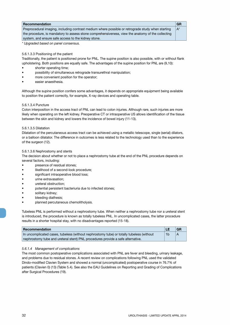
32 UROLITHIASIS - LIMITED UPDATE APRIL 2014
Recommendation GRPreprocedural imaging, including contrast medium where possible or retrograde study when starting the procedure, is mandatory to assess stone comprehensiveness, view the anatomy of the collecting system, and ensure safe access to the kidney stone.
A*
* Upgraded based on panel consensus.
5.6.1.3.3 Positioning of the patientTraditionally, the patient is positioned prone for PNL. The supine position is also possible, with or without flank upholstering. Both positions are equally safe. The advantages of the supine position for PNL are (9,10):• shorter operating time;• possibility of simultaneous retrograde transurethral manipulation;• more convenient position for the operator;• easier anaesthesia.
Although the supine position confers some advantages, it depends on appropriate equipment being available to position the patient correctly, for example, X-ray devices and operating table. 5.6.1.3.4 PunctureColon interposition in the access tract of PNL can lead to colon injuries. Although rare, such injuries are more likely when operating on the left kidney. Preoperative CT or intraoperative US allows identification of the tissue between the skin and kidney and lowers the incidence of bowel injury (11-13).
5.6.1.3.5 DilatationDilatation of the percutaneous access tract can be achieved using a metallic telescope, single (serial) dilators, or a balloon dilatator. The difference in outcomes is less related to the technology used than to the experience of the surgeon (12).
5.6.1.3.6 Nephrostomy and stentsThe decision about whether or not to place a nephrostomy tube at the end of the PNL procedure depends on several factors, including:• presence of residual stones;• likelihood of a second-look procedure;• significant intraoperative blood loss;• urine extravasation;• ureteral obstruction;• potential persistent bacteriuria due to infected stones;• solitary kidney;• bleeding diathesis;• planned percutaneous chemolitholysis.
Tubeless PNL is performed without a nephrostomy tube. When neither a nephrostomy tube nor a ureteral stent is introduced, the procedure is known as totally tubeless PNL. In uncomplicated cases, the latter procedure results in a shorter hospital stay, with no disadvantages reported (15-18).
Recommendation LE GRIn uncomplicated cases, tubeless (without nephrostomy tube) or totally tubeless (without nephrostomy tube and ureteral stent) PNL procedures provide a safe alternative.
1b A
5.6.1.4 Management of complicationsThe most common postoperative complications associated with PNL are fever and bleeding, urinary leakage, and problems due to residual stones. A recent review on complications following PNL used the validated Dindo-modified Clavien System and showed a normal (uncomplicated) postoperative course in 76.7% of patients (Clavien 0) (13) (Table 5.4). See also the EAU Guidelines on Reporting and Grading of Complications after Surgical Procedures (19).

UROLITHIASIS - LIMITED UPDATE APRIL 2014 33
Table 5.4: Complications following PNL
Complica-tions
Trans-fusion
Embolisation Urinoma Fever Sepsis Thoracic complication
Organ injury
Death LE
(Range) (0-20%) (0-1.5%) (0-1%) (0-32.1%)
(0.3-1.1%)
(0-11.6%) (0-1.7%)
(0-0.3%)
1a
N = 11,929 7% 0.4% 0.2% 10.8% 0.5% 1.5% 0.4%
Urinary leakage and stone clearance can be viewed endoscopically and by X-ray anlaysis. In doubtful cases, complications can be minimised by performing standard rather than totally tubeless PNL.
Perioperative fever can occur, even with a sterile preoperative urinary culture and perioperative antibiotic prophylaxis, because the kidney stones themselves may be a source of infection. Intraoperative kidney stone culture may therefore help to select postoperative antibiotics (20,21). Intraoperative irrigation pressure < 30 mm Hg and unobstructed postoperative urinary drainage may be important factors in preventing postoperative sepsis. Well-positioned or specially designed access sheaths can prevent high intrapelvic irrigation pressure (22).
Bleeding after PNL may be treated by brief clamping of the nephrostomy tube. Super-selective embolic occlusion of the artery may become necessary in case of severe bleeding.
5.6.2 Ureterorenoscopy (URS) (including retrograde access to renal collecting system)URS has dramatically changed the management of ureteral calculi. Major technical improvements include endoscope miniaturisation, enhanced optical quality and tools, and introduction of disposables. The current standard for rigid ureterorenoscopes are tip diameters of < 8 F. Rigid URS can be used for the whole ureter (23). Major technological progress has been achieved for retrograde intrarenal surgery [RIRS (flexible URS)], with improved deflection mechanisms, better durability, and recently, digital optical systems (24-26). Initial experience with digital scopes has demonstrated shorter operation times due to the improvement in image quality (27-29). In Europe, RIRS is mainly used for the renal collecting system and - in cases with difficult anatomy - the upper ureter.
5.6.2.1 Best clinical practice in ureterorenoscopy (URS)5.6.2.1.1 Preoperative work-up and preparationsBefore the procedure, the following information should be sought and actions taken (LE: 4):• patient history;• physical examination, because anatomical and congenital abnormalities may complicate or prevent
retrograde stone manipulation;• thrombocyte aggregation inhibitors/anticoagulants (antiplatelet drugs) should be discontinued if
possible, however URS can be performed in patients with bleeding disorders, with a moderate increase in complications (7,30);
• imaging.
Recommendation LE GRShort-term antibiotic prophylaxis should be administered (27). 4 A*
* Upgraded based on panel consensus.
5.6.2.1.2 ContraindicationsApart from general problems, for example, with general anaesthesia or untreated UTIs, URS can be performed in all patients without any specific contraindications. Specific problems such as ureteral strictures may prevent successful retrograde stone management.
5.6.2.1.3 Access to the upper urinary tractMost interventions are performed under general anaesthesia, although local or spinal anaesthesia is possible. Instrument miniaturisation means that intravenous sedation can be used to achieve the same outcome (31). Intravenous sedation with miniaturised instruments is especially suitable for female patients with distal ureteral stones. However, kidney movement is more pronounced with local or intravenous anaesthesia, which may hinder RIRS.
Antegrade URS is an option for large, impacted proximal ureteral calculi (32) (Section 6.5.3).

34 UROLITHIASIS - LIMITED UPDATE APRIL 2014
5.6.2.1.4 Safety aspectsFluoroscopic equipment must be available in the operating room. We recommend placement of a safety wire, even though some groups have demonstrated that URS can be performed without it (33,34). A safety wire prevents false passage in case of perforation, and ensures that a JJ stent can be inserted in difficult situations, thus avoiding more significant complications.
Retrograde access to the upper urinary tract is usually obtained under endoscopic guidance.
Balloon and plastic dilators are available if necessary. If insertion of a flexible URS is difficult, prior rigid ureteroscopy can be helpful for optical dilatation. If ureteral access is not possible, insertion of a JJ stent followed by URS after 7-14 days offers an alternative procedure.
Recommendation GRPlacement of a safety wire is recommended. A*
* Upgraded based on panel consensus.
5.6.2.1.5 Ureteral access sheathsHydrophilic-coated ureteral access sheaths, which are available in different calibres (inner diameter from 9 F upwards), can be inserted via a guide wire, with the tip placed in the proximal ureter. Ureteral access sheaths allow easy multiple access to the upper urinary tract and therefore significantly facilitate URS. The use of ureteral access sheaths improves vision by establishing a continuous outflow, decreasing intrarenal pressure, and potentially reducing operating time (35,36). Ureteral access sheaths allow continuous outflow of irrigation fluid, which improves visual quality and maintains a low-pressure system (37,38). The insertion of ureteral access sheaths may lead to ureteral damage, however, no data on long-term consequences are available (39). Use of ureteral access sheaths depends on the surgeon’s preference.
5.6.2.1.6 Stone extractionThe aim of URS is complete stone removal (especially ureteric stones). “Smash and go” strategies should be limited to the treatment of large renal stones. Stones can be extracted by endoscopic forceps or baskets. Forceps allow safe release of stone fragments if they become stuck within the ureter, but extraction takes longer than when using baskets. Only baskets made of nitinol can be used for RIRS (40).
StatementsNitinol baskets preserve the tip deflection of flexible ureterorenoscopes, and the tipless design reduces the risk of mucosal injury.Nitinol baskets are the only baskets suitable for use in RIRS.
Recommendation LE GRStone extraction using a basket without endoscopic visualisation of the stone (blind basketing) should not be performed.
4 A*
* Upgraded based on panel consensus.
Stones that cannot be extracted directly must be disintegrated. If it is difficult to access stones that need disintegration within the lower renal pole, it may help to displace them into a more accessible calyx (Section 6.4.2) (41).
5.6.2.1.7 Intracorporeal lithotripsyThe most effective lithotripsy system is the Ho:YAG laser, which has become the gold standard for ureteroscopy and flexible nephroscopy (Section 5.6.1.2), because it is effective for all stone types (5,42-44). Pneumatic and US systems can be used with high disintegration efficacy in rigid URS (45-46). However, stone migration into the kidney is a common problem, which can be prevented by placement of special tools proximal of the stone (47).
Recommendation LE GRHo:YAG laser lithotripsy is the preferred method for (flexible) URS. 3 B
5.6.2.1.8 Stenting before and after URSRoutine stenting is no longer necessary before URS. However, pre-stenting facilitates ureteroscopic

UROLITHIASIS - LIMITED UPDATE APRIL 2014 35
management of stones, improves the SFR, and reduces complications (48). Most urologists routinely insert a JJ stent following URS, although several randomised prospective trials have found that routine stenting after uncomplicated URS (complete stone removal) is not necessary; stenting might be associated with higher postoperative morbidity (49-51). A ureteric catheter with a shorter indwelling time (1 day) may be used as well, with similar results (52). Stents should be inserted in patients who are at increased risk of complications (e.g., residual fragments, bleeding, perforation, UTIs, or pregnancy), and in all doubtful cases, to avoid stressful emergencies.The ideal duration of stenting is not known. Most urologists favour 1-2 weeks after URS. Patients should be followed up with a plain abdominal film (KUB), CT or US.
α-Blockers reduce the morbidity of ureteral stents and increase tolerability (53). A recently published meta-analysis provides evidence for improvement of ureteral stent tolerability with tamsulosin (54).
Statement LEIn uncomplicated URS, a stent need not be inserted. 1a An α-blocker can reduce stent-related symptoms. 1a
5.6.2.2 ComplicationsThe overall complication rate after URS is 9-25% (23,55) (Table 5.5). Most are minor and do not require intervention. Ureteral avulsion and strictures used to be greatly feared, but nowadays are rare in experienced hands (< 1%). Previous perforations are the most important risk factor for complications.
Table 5.5: Complications of URS*
Rate (%)Intraoperative complications 3.6Mucosal injury 1.5Ureteral perforation 1.7Significant bleeding 0.1Ureteral avulsion 0.1Early complications 6.0Fever or urosepsis 1.1Persistent haematuria 2.0Renal colic 2.2Late complications 0.2Ureteral stricture 0.1Persistent vesicoureteral reflux 0.1
*From Geavlete, et al. (55).
5.6.3 References1. Mishra S, Sharma R, Garg C, et al. Prospective comparative study of miniperc and standard PNL for
treatment of 1 to 2 cm size renal stone. BJU Int 2011 Sep;108(6):896-9;discussion 899-900.http://www.ncbi.nlm.nih.gov/pubmed/21477212
2. Knoll T, Wezel F, Michel MS, et al. Do patients benefit from miniaturized tubeless percutaneous nephrolithotomy? A comparative prospective study. J Endourol 2010 Jul;24(7):1075-9.http://www.ncbi.nlm.nih.gov/pubmed/20575685
3. Sabnis RB, Ganesamoni A, Doshi AP, et al. Micropercutaneous nephrolithotomy (microperc) vs retrograde intrarenal surgery for the management of small renal calculi: A randomized controlled trial. BJU Int 2013 Aug;112(3):355-61.http://www.ncbi.nlm.nih.gov/pubmed/23826843
4. Yamaguchi A, Skolarikos A, Buchholz NP, et al; Clinical Research Office Of The Endourological Society Percutaneous Nephrolithotomy Study Group. Operating times and bleeding complications in percutaneous nephrolithotomy: a comparison of tract dilation methods in 5,537 patients in the Clinical Research Office of the Endourological Society Percutaneous Nephrolithotomy Global Study. J Endourol 2011 Jun;25(6):933-9. http://www.ncbi.nlm.nih.gov/pubmed/21568697
5. Gupta PK. Is the holmium: YAG laser the best intracorporeal lithotripter for the ureter? A 3-year retrospective study. J Endourol 2007 Mar;21(3):305-9.http://www.ncbi.nlm.nih.gov/pubmed/17444776

36 UROLITHIASIS - LIMITED UPDATE APRIL 2014
6. Hofbauer J, Hobarth K, Marberger M. Electrohydraulic versus pneumatic disintegration in the treatment of ureteral stones: a randomized, prospective trial. J Urol 1995 Mar;153(3 Pt 1):623-5.http://www.ncbi.nlm.nih.gov/pubmed/7861499
7. Turna B, Stein RJ, Smaldone MC, et al. Safety and efficacy of flexible ureterorenoscopy and holmium:YAG lithotripsy for intrarenal stones in anticoagulated cases. J Urol 2008 Apr;179(4):1415-9.http://www.ncbi.nlm.nih.gov/pubmed/18289567
8. Andonian S, Scoffone CM, Louie MK, et al. Does imaging modality used for percutaneous renal access make a difference? A matched case analysis. J Endourol 2013 Jan;27(1):24-8.http://www.ncbi.nlm.nih.gov/pubmed/22834999
9. De Sio M, Autorino R, Quarto G, et al. Modified supine versus prone position in percutaneous nephrolithotomy for renal stones treatable with a single percutaneous access: a prospective randomized trial. Eur Urol 2008;54(1):196-202.http://www.ncbi.nlm.nih.gov/pubmed/18262711
10 Valdivia JG, Scarpa RM, Duvdevani M, et al. Supine versus prone position during percutaneous nephrolithotomy: a report from the clinical research office of the endourological society percutaneous nephrolithotomy global study. J Endourol 2011 Oct;25(10):1619-25.http://www.ncbi.nlm.nih.gov/pubmed/21877911
11. El-Nahas AR, Shokeir AA, El-Assmy AM, et al. Colonic perforation during percutaneous nephrolithotomy: study of risk factors. Urology 2006 May;67(5):937-41.http://www.ncbi.nlm.nih.gov/pubmed/16635515
12. Osman M, Wendt-Nordahl G, Heger K, et al. Percutaneous nephrolithotomy with ultrasonography-guided renal access: experience from over 300 cases. BJU Int 2005 Oct;96(6):875-8.http://www.ncbi.nlm.nih.gov/pubmed/16153221
13. Jessen JP, Honeck P, Knoll T, et al. Percutaneous nephrolithotomy under combined sonographic/radiologic guided puncture: results of a learning curve using the modified Clavien grading system. World J Urol 2013 Dec;31(6):1599-603.http://www.ncbi.nlm.nih.gov/pubmed/23283412
14. Wezel F, Mamoulakis C, Rioja J, et al. Two contemporary series of percutaneous tract dilation for percutaneous nephrolithotomy. J Endourol 2009 Oct;23(10):1655-61.http://www.ncbi.nlm.nih.gov/pubmed/19558265
15. Singh I, Singh A, Mittal G. Tubeless percutaneous nephrolithotomy: is it really less morbid? J Endourol 2008 Mar;22(3):427-34.http://www.ncbi.nlm.nih.gov/pubmed/18355137
16. Kara C, Resorlu B, Bayindir M, et al. A randomized comparison of totally tubeless and standard percutaneous nephrolithotomy in elderly patients. Urology 2010 Aug;76(2):289-93.http://www.ncbi.nlm.nih.gov/pubmed/20299077
17. Istanbulluoglu MO, Ozturk B, Gonen M, et al. Effectiveness of totally tubeless percutaneous nephrolithotomy in selected patients: a prospective randomized study. Int Urol Nephrol 2009;41(3):541-5.http://www.ncbi.nlm.nih.gov/pubmed/19165617
18. Gonen M, Cicek T, Ozkardes H. Tubeless and stentless percutaneous nephrolithotomy in patients requiring supracostal access. Urol Int 2009;82(4):440-3.http://www.ncbi.nlm.nih.gov/pubmed/19506412
19. Mitropoulos D, Artibani W, Graefen M, et al. Reporting and grading of complications after urologic surgical procedures: an ad hoc EAU guidelines panel assessment and recommendations. Eur Urol 2012 Feb;61(2):341-9.http://www.ncbi.nlm.nih.gov/pubmed/22074761
20. Zanetti G, Paparella S, Trinchieri A, et al. Infections and urolithiasis: current clinical evidence in prophylaxis and antibiotic therapy. Arch Ital Urol Androl 2008 Mar;80(1):5-12.http://www.ncbi.nlm.nih.gov/pubmed/18533618
21. Gonen M, Turan H, Ozturk B, et al. Factors affecting fever following percutaneous nephrolithotomy: a prospective clinical study. J Endourol 2008 Sep;22(9):2135-8.http://www.ncbi.nlm.nih.gov/pubmed/18811569
22. Troxel SA, Low RK. Renal intrapelvic pressure during percutaneous nephrolithotomy and its correlation with the development of postoperative fever. J Urol 2002 Oct;168(4 Pt 1):1348-51.http://www.ncbi.nlm.nih.gov/pubmed/12352390
23. Preminger GM, Tiselius HG, Assimos DG, et al; American Urological Association Education and Research, Inc; European Association of Urology. 2007 Guideline for the management of ureteral calculi. Eur Urol 2007 Dec;52(6):1610-31.http://www.ncbi.nlm.nih.gov/pubmed/18074433

UROLITHIASIS - LIMITED UPDATE APRIL 2014 37
24. Wendt-Nordahl G, Mut T, Krombach P, et al. Do new generation flexible ureterorenoscopes offer a higher treatment success than their predecessors? Urol Res 2011 Jun;39(3):185-8.http://www.ncbi.nlm.nih.gov/pubmed/21052986
25. Knudsen B, Miyaoka R, Shah K, et al. Durability of the next-generation flexible fibreoptic ureteroscopes: a randomized prospective multi-institutional clinical trial. Urology 2010;75(3):534-8.http://www.ncbi.nlm.nih.gov/pubmed/19854494
26. Skolarikos AA, Papatsoris AG, Mitsogiannis IC, et al. Current status of ureteroscopic treatment for urolithiasis. Int J Urol 2009 Sep;16(9):713-7.http://www.ncbi.nlm.nih.gov/pubmed/19674169
27. Binbay M, Yuruk E, Akman T, et al. Is there a difference in outcomes between digital and fibreoptic flexible ureterorenoscopy procedures? J Endourol 2010 Dec;24(12)1929-34.http://www.ncbi.nlm.nih.gov/pubmed/21043835
28. Humphreys MR, Miller NL, Williams JC Jr, et al. A new world revealed: early experience with digital ureteroscopy. J Urol 2008 Mar;179(3):970-5.http://www.ncbi.nlm.nih.gov/pubmed/18207196
29. Mitchell S, Havranek E, Patel A. First digital flexible ureterorenoscope: initial experience. J Endourol 2008;22(1):47-50.http://www.ncbi.nlm.nih.gov/pubmed/18315473
30. Watterson JD, Girvan AR, Cook AJ, et al. Safety and efficacy of holmium: YAG laser lithotripsy in patients with bleeding diatheses. J Urol 2002 Aug;168(2):442-5.http://www.ncbi.nlm.nih.gov/pubmed/12131284
31. Cybulski PA, Joo H, Honey RJ. Ureteroscopy: anesthetic considerations. Urol Clin North Am 2004 Feb;31(1):43-7.http://www.ncbi.nlm.nih.gov/pubmed/15040400
32. Sun X, Xia S, Lu J, et al. Treatment of large impacted proximal ureteral stones: randomized comparison of percutaneous antegrade ureterolithotripsy versus retrograde ureterolithotripsy. J Endourol 2008 May;22(5):913-7.http://www.ncbi.nlm.nih.gov/pubmed/18429682
33. Dickstein RJ, Kreshover JE, Babayan RK, et al. Is a safety wire necessary during routine flexible ureteroscopy? J Endourol 2010 Oct;24(10):1589-92.http://www.ncbi.nlm.nih.gov/pubmed/20836719
34. Eandi JA, Hu B, Low RK. Evaluation of the impact and need for use of a safety guidewire during ureteroscopy. J Endourol 2008 Aug;22(8):1653-8.http://www.ncbi.nlm.nih.gov/pubmed/18721045
35. Stern JM, Yiee J, Park S. Safety and efficacy of ureteral access sheaths. J Endourol 2007 Feb;21(2):119-23.http://www.ncbi.nlm.nih.gov/pubmed/17338606
36. L’esperance JO, Ekeruo WO, Scales CD Jr, et al. Effect of ureteral access sheath on stone-free rates in patients undergoing ureteroscopic management of renal calculi. Urology 2005 Aug;66(2):252-5.http://www.ncbi.nlm.nih.gov/pubmed/16040093
37. Ng YH, Somani BK, Dennison A, et al. Irrigant flow and intrarenal pressure during flexible ureteroscopy: the effect of different access sheaths, working channel instruments, and hydrostatic pressure. J Endourol 2010 Dec;24(12):1915-20.http://www.ncbi.nlm.nih.gov/pubmed/21067276
38. Auge BK, Pietrow PK, Lallas CD, et al. Ureteral access sheath provides protection against elevated renal pressures during routine flexible ureteroscopic stone manipulation. J Endourol 2004 Feb;18(1):33-6.http://www.ncbi.nlm.nih.gov/pubmed/15006050
39. Traxer O, Thomas A. Prospective evaluation and classification of ureteral wall injuries resulting from insertion of a ureteral access sheath during retrograde intrarenal surgery. J Urol 2013 Feb;189(2): 580-4.http://www.ncbi.nlm.nih.gov/pubmed/22982421
40. Bach T, Geavlete B, Herrmann TR, et al. Working tools in flexible ureterorenoscopy--influence on flow and deflection: what does matter? J Endourol 2008 Aug;22(8):1639-43.http://www.ncbi.nlm.nih.gov/pubmed/18620506
41. Auge BK, Dahm P, Wu NZ, et al. Ureteroscopic management of lower-pole renal calculi: technique of calculus displacement. J Endourol 2001 Oct;15(8):835-8.http://www.ncbi.nlm.nih.gov/pubmed/11724125
42. Leijte JA, Oddens JR, Lock TM. Holmium laser lithotripsy for ureteral calculi: predictive factors for complications and success. J Endourol 2008 Feb;22(2):257-60.http://www.ncbi.nlm.nih.gov/pubmed/18294030

38 UROLITHIASIS - LIMITED UPDATE APRIL 2014
43. Marguet CG, Sung JC, Springhart WP, et al. In vitro comparison of stone retropulsion and fragmentation of the frequency doubled, double pulse nd:yag laser and the holmium:yag laser. J Urol 2005 May;173(5):1797-800.http://www.ncbi.nlm.nih.gov/pubmed/15821590
44. Pierre S, Preminger GM. Holmium laser for stone management. World J Urol 2007 Jun;25(3):235-9.http://www.ncbi.nlm.nih.gov/pubmed/17340157
45. Garg S, Mandal AK, Singh SK, et al. Ureteroscopic laser lithotripsy versus ballistic lithotripsy for treatment of ureteric stones: a prospective comparative study. Urol Int 2009;82(3):341-5.http://www.ncbi.nlm.nih.gov/pubmed/19440025
46. Binbay M, Tepeler A, Singh A, et al. Evaluation of pneumatic versus holmium:YAG laser lithotripsy for impacted ureteral stones. Int Urol Nephrol 2011 Dec;43(4):989-95.http://www.ncbi.nlm.nih.gov/pubmed/21479563
47. Ahmed M, Pedro RN, Kieley S, et al. Systematic evaluation of ureteral occlusion devices: insertion, deployment, stone migration, and extraction. Urology 2009 May;73(5):976-80.http://www.ncbi.nlm.nih.gov/pubmed/19394493
48. Rubenstein RA, Zhao LC, Loeb S, et al. Prestenting improves ureteroscopic stone-free rates. J Endourol 2007 Nov;21(11):1277-80.http://www.ncbi.nlm.nih.gov/pubmed/18042014
49. Song T, Liao B, Zheng S, Wei Q. Meta-analysis of postoperatively stenting or not in patients underwent ureteroscopic lithotripsy. Urol Res 2012 Feb;40(1):67-77.http://www.ncbi.nlm.nih.gov/pubmed/21573923
50. Haleblian G, Kijvikai K, de la Rosette J, et al. Ureteral stenting and urinary stone management: a systematic review. J Urol 2008 Feb;179(2):424-30.http://www.ncbi.nlm.nih.gov/pubmed/18076928
51. Nabi G, Cook J, N’Dow J, McClinton S. Outcomes of stenting after uncomplicated ureteroscopy: systematic review and meta-analysis. BMJ 2007 Mar;334(7593):572.http://www.ncbi.nlm.nih.gov/pubmed/17311851
52. Moon TD. Ureteral stenting--an obsolete procedure? J Urol 2002 May;167(5):1984.http://www.ncbi.nlm.nih.gov/pubmed/11956423
53. Wang CJ, Huang SW, Chang CH. Effects of specific alpha-1A/1D blocker on lower urinary tract symptoms due to double-J stent: a prospectively randomized study. Urol Res 2009 Jun;37(3):147-52.http://www.ncbi.nlm.nih.gov/pubmed/19277623
54. Lamb AD, Vowler SL, Johnston R, et al. Meta-analysis showing the beneficial effect of α-blockers on ureteric stent discomfort. BJU Int 2011 Dec;108(11):1894-902.http://www.ncbi.nlm.nih.gov/pubmed/21453351
55. Geavlete P, Georgescu D, Nita G, et al. Complications of 2735 retrograde semirigid ureteroscopy procedures: a single-center experience. J Endourol 2006 Mar;20(3):179-85.http://www.ncbi.nlm.nih.gov/pubmed/16548724
5.7 Open and laparoscopic surgery for removal of renal stones5.7.1 Open surgeryAdvances in SWL and endourological surgery (URS and PNL) have significantly decreased the indications for open stone surgery, which is now often a second- or third-line treatment option needed in only 1.0-5.4% of cases (1-5). The incidence of open stone surgery is ~1.5% of all stone removal interventions in developed countries, and in developing countries, it has dropped from 26% to 3.5% in recent years (3,5). However, open surgery is still needed for the most difficult stones, which supports the importance of maintaining proficiency, skills and expertise in open renal and ureteral surgical techniques such as extended pyelolithotomy, pyelonephrolithotomy, anatrophic nephrolithotomy, multiple radial nephrotomy, partial nephrectomy, and renal surgery under hypothermia (6-10) (Table 5.6).
Recently, intraoperative B-mode scanning and Doppler sonography (11,12) have been used to identify avascular areas in the renal parenchyma that are close to the stone or dilated calices. This allows removal of large staghorn stones by multiple small radial nephrotomy, without loss of kidney function.The efficacy of open surgery compared to less-invasive therapy in terms of SFRs, is based on historical data, but no comparative studies are available (13-16).
5.7.1.1 Indications for open surgeryThere is a consensus that most complex stones, including partial and complete staghorn stones, should be approached primarily with PNL or combined PNL and SWL. If a reasonable number of percutaneous approaches are not likely to be successful, or if multiple, endourological approaches have been performed unsuccessfully, open surgery may be a valid treatment option.

UROLITHIASIS - LIMITED UPDATE APRIL 2014 39
Table 5.6: Indications for open surgery
Kidney stones• Complex stone burden• Failure of SWL, PNL, or ureteroscopic procedure• Intrarenal anatomical abnormalities: infundibular stenosis; stone in the calyceal diverticulum (particularly in
an anterior calyx); obstruction of the ureteropelvic junction; and stricture if endourologic procedures have failed or are not promising
• Morbid obesity• Skeletal deformity, contractures and fixed deformities of hips and legs• Comorbidity• Concomitant open surgery• Non-functioning lower pole (partial nephrectomy), non-functioning kidney (nephrectomy)• Patient choice ( after failed minimally invasive procedures, a single procedure avoiding the risk of multiple
PNL procedures might be preffered by the case)• Stone in an ectopic kidney where percutaneous access and SWL may be difficult or impossible• For the paediatric population, the same considerations apply as for adultsUreteral stones • Large impacted ureteral stones• Cases requiring surgery for other concurrent conditions• In cases with failed other non-invasive or low-invasive procedures• For upper ureteral calculi, laparoscopic urolithomy has the highest stone-free rate compared to URS and
SWL (31) (LE: 1b)
5.7.2 Laparoscopic surgeryLaparoscopic urological surgery is increasingly replacing open surgery. Today laparoscopic surgery is used to remove renal and ureteric stones in certain situations, including complex stone burden, failed previous SWL and/or endourological procedures, anatomical abnormalities or morbid obesity, and planned nephrectomy of a stone-containing non-functioning kidney. Although surgical pyelolithotomy is rarely indicated (Table 5.7), laparoscopic removal of solitary large renal pelvic (17) as well as anterior caliceal diverticular stones is possible in selected cases (18). Stone-free rates are reported to be equal to PNL, but complications are more frequent, using laparoscopic retroperitoneal pyelolithotomy (17). Additionally, as a less-invasive option, laparoscopic anatrophic nephrolithotomy has been found to be effective for the removal of complex staghorn stones; however, PNL is still the method of choice and laparoscopic stone removal should be reserved for selected cases (19,20). Laparoscopic ureterolithotomy is relatively easy, with SFRs up to 100% provided expertise is available (21-24). It can replace open surgery in most situations (15,16). Retroperitoneal and transperitoneal laparoscopic access to all portions of the ureter has been reported (24-30), although laparoscopic ureterolithotomy in the distal ureter is less successful than in the middle and proximal ureter, but the size of the stone does not appear to influence outcome. Although highly effective, laparoscopic ureterolithotomy is not first-line therapy in most cases because of its invasiveness, longer recovery time, and greater risk of associated complications compared to SWL and URS (21-24) (Table 5.7).
5.7.2.1 Table 5.7: Indications for laparoscopic stone surgery
Indications for laparoscopic kidney-stone surgery include:• Complex stone burden• Failed previous SWL and/or endourological procedures• Anatomical abnormalities• Morbid obesity• Nephrectomy in case of non-functioning kidney.Indications for laparoscopic ureteral stone surgery include:• Large impacted ureteral stones• In cases of concurrent conditions requiring surgery• When other non-invasive or low-invasive procedures have failed• For upper ureteral calculi, laparoscopic urolithomy has the highest stone-free rate compared to URS and
SWL (31) (LE: 1b).

40 UROLITHIASIS - LIMITED UPDATE APRIL 2014
Recommendations LE GRLaparoscopic or open surgical stone removal may be considered in rare cases in which SWL, URS, and percutaneous URS fail or are unlikely to be successful.
3 C
When expertise is available, laparoscopic surgery should be the preferred option beforeproceeding to open surgery. An exception is complex renal stone burden and/or stonelocation.
3 C
For ureterolithotomy, laparoscopy is recommended for large impact stones or whenendoscopic lithotripsy or SWL has failed.
2 B
5.7.3 References1. Assimos DG, Boyce WH, Harrison LH, et al. The role of open stone surgery since extracorporeal shock
wave lithotripsy. J Urol 1989 Aug;142(2 Pt 1):263-7.http://www.ncbi.nlm.nih.gov/pubmed/2746742
2. Segura JW. Current surgical approaches to nephrolithiasis. Endocrinol Metab Clin North Am 1990 Dec;19(4):919-35.http://www.ncbi.nlm.nih.gov/pubmed/2081519
3. Honeck P, Wendt-Nordahl G, Krombach P, et al. Does open stone surgery still play a role in the treatment of urolithiasis? Data of a primary urolithiasis center. J Endourol 2009 Jul;23(7):1209-12.http://www.ncbi.nlm.nih.gov/pubmed/19538063
4. Bichler KH, Lahme S, Strohmaier WL. Indications for open stone removal of urinary calculi. Urol Int 1997;59(2):102-8.http://www.ncbi.nlm.nih.gov/pubmed/9392057
5. Paik ML, Resnick MI. Is there a role for open stone surgery? Urol Clin North Am 2000 May;27(2): 323-31.http://www.ncbi.nlm.nih.gov/pubmed/10778474
6. Matlaga BR, Assimos DG. Changing indications of open stone surgery. Urology 2002 Apr;59(4): 490-93;discussion 493-4.http://www.ncbi.nlm.nih.gov/pubmed/11927296
7. Ansari MS, Gupta NP. Impact of socioeconomic status in etiology and management of urinary stone disease. Urol Int 2003;70(4):255-61.http://www.ncbi.nlm.nih.gov/pubmed/12776701
8. Alivizatos G, Skolarikos A. Is there still a role for open surgery in the management of renal stones? Curr Opin Urol 2006 Mar;16(2):106-11.http://www.ncbi.nlm.nih.gov/pubmed/16479213
9. Kerbl K, Rehman J, Landman J, et al. Current management of urolithiasis: Progress or regress? J Endourol 2002 Jun;16(5):281-8.http://www.ncbi.nlm.nih.gov/pubmed/12184077
10. Preminger GM, Assimos DG, Lingeman JE, et al. Chapter 1: AUA guideline on management of staghorn calculi: Diagnosis and treatment recommendations. J Urol 2005 Jun;173(6):1991-2000.http://www.ncbi.nlm.nih.gov/pubmed/15879803
11. Thüroff JW, Frohneberg D, Riedmiller R, et al. Localization of segmental arteries in renal surgery by Doppler sonography. J Urol 1982 May;127(5):863-6.http://www.ncbi.nlm.nih.gov/pubmed/7086985
12. Alken P, Thüroff JW, Riedmiller H, et al. Doppler sonography and B-mode ultrasound scanning in renal stone surgery. Urology 1984 May;23(5):455-60.http://www.ncbi.nlm.nih.gov/pubmed/6719663
13. Kane CJ, Bolton DM, Stoller ML. Current indications for open stone surgery in an endourology center. Urology 1995 Feb;45(2):218-21.http://www.ncbi.nlm.nih.gov/pubmed/7855969
14. Sy FY, Wong MY, Foo KT. Current indications for open stone surgery in Singapore. Ann Acad Med Singapore 1999 Mar;28(2):241-4.http://www.ncbi.nlm.nih.gov/pubmed/10497675
15. Goel A, Hemal AK. Upper and mid-ureteric stones: a prospective unrandomized comparison of retroperitoneoscopic and open ureterolithotomy. BJU Int 2001 Nov;88(7):679-82.http://www.ncbi.nlm.nih.gov/pubmed/11890236
16. Skrepetis K, Doumas K, Siafakas I, et al. Laparoscopic versus open ureterolithotomy. A comparative study. Eur Urol 2001 Jul;40(1):32-6;discussion 37.http://www.ncbi.nlm.nih.gov/pubmed/11528174
UROLITHIASIS - LIMITED UPDATE APRIL 2014 41
17. Al-Hunayan A, Khalil M, Hassabo M, et al. Management of solitary renal pelvic stone: laparoscopic retroperitoneal pyelolithotomy versus percutaneous nephrolithotomy. J Endourol 2011 Jun;25(6): 975-8.http://www.ncbi.nlm.nih.gov/pubmed/21612433
18. Skolarikos A, Papatsoris AG, Albanis S, et al. Laparoscopic urinary stone surgery: an updated evidence based review. Urol Res 2010 Oct:38(5);337-44.http://www.ncbi.nlm.nih.gov/pubmed/20396871
19. Giedelman C, Arriaga J, Carmona O, et al. Laparoscopic anatrophic nephrolithotomy: developments of the technique in the era of minimally invasive surgery. J Endourol 2012 May;26(5):444-50. http://www.ncbi.nlm.nih.gov/pubmed/22142215
20. Zhou L, Xuan Q, Wu B, et al. Retroperitoneal laparoscopic anatrophic nephrolithotomy for large staghorn calculi. Int J Urol 2011 Feb;18(2):126-9.http://www.ncbi.nlm.nih.gov/pubmed/21198943
21. Fan T, Xian P, Yang L, et al. Experience and learning curve of retroperitoneal laparoscopic ureterolithotomy for upper ureteral calculi. J Endourol 2009 Nov;23(11):1867-70.http://www.ncbi.nlm.nih.gov/pubmed/19811058
22. Khaladkar S, Modi J, Bhansali M, et al. Which is the best option to treat large (> 1.5 cm) midureteric calculi? J Laparoendosc Adv Surg Tech A 2009 Aug;19(4):501-4.http://www.ncbi.nlm.nih.gov/pubmed/19670976
23. Jeong BC, Park HK, Byeon SS, et al. Retroperitoneal laparoscopic ureterolithotomy for upper ureter stones. J Korean Med Sci 2006 Jun;21(3):441-4.http://www.ncbi.nlm.nih.gov/pubmed/16778386
24. Hruza M, Zuazu JR, Goezen AS, et al. Laparoscopic and open stone surgery. Arch Ital Urol Androl 2010 Mar;82(1):64-71.http://www.ncbi.nlm.nih.gov/pubmed/20593725
25. El-Feel A, Abouel-Fettouh H, Abdel-Hakim AM. Laparoscopic transperitoneal ureterolithotomy. J Endourol 2007 Jan;21(1):50-4.http://www.ncbi.nlm.nih.gov/pubmed/17263607
26. Gaur DD, Trivedi S, Prabhudesai MR, Madhusudhana HR, et al. Laparoscopic ureterolithotomy: technical considerations and long term follow up. BJU Int 2002 Mar;89(4):339-43.http://www.ncbi.nlm.nih.gov/pubmed/11872020
27. Flasko T, Holman E, Kovacs G, et al. Laparoscopic ureterolithotomy: the method of choice in selected cases. J Laparoendosc Adv Surg Tech A 2005 Apr;15(2):149-52.http://www.ncbi.nlm.nih.gov/pubmed/15898906
28. Kijvikai K, Patcharatrakul S. Laparoscopic ureterolithotomy: its role and some controversial technical considerations. Int J Urol 2006 Mar;13(3):206-10.http://www.ncbi.nlm.nih.gov/pubmed/16643610
29. Wang Y, Hou J, Wen D, et al. Comparative analysis of upper ureteral stones (> 15 mm) treated with retroperitoneoscopic ureterolithotomy and ureteroscopic pneumatic lithotripsy. Int Urol Nephrol 2010 Dec;42(4):897-901.http://www.ncbi.nlm.nih.gov/pubmed/20169409
30. Lopes Neto AC, Korkes F, Silva JL 2nd, et al. Prospective randomized study of treatment of large proximal ureteral stones: extracorporeal shock wave lithotripsy versus ureterolithotripsy versus laparoscopy. J Urol 2012 Jan;187(1):164-8.http://www.ncbi.nlm.nih.gov/pubmed/22100003
31. Tefekli A, Tepeler A, Akman T, et al. The comparison of laparoscopic pyelolithotomy and percutaneous nephrolithotomy in the treatment of solitary large renal pelvic stones. Urol Res 2012 Oct;40(5):549-55. http://www.ncbi.nlm.nih.gov/pubmed/22307365

42 UROLITHIASIS - LIMITED UPDATE APRIL 2014
6. INDICATION FOR ACTIVE STONE REMOVAL AND SELECTION OF PROCEDUREAlthough kidney stones might be asymptomatic, ureteral stones cause acute renal colic in most cases.Treatment decisions for upper urinary tract calculi are based on several general aspects such as stone composition, stone size, and symptoms.
6.1 Indications for active removal of ureteral stones (1-3)- Stones with low likelihood of spontaneous passage.- Persistent pain despite adequate analgesic medication.- Persistent obstruction.- Renal insufficiency (renal failure, bilateral obstruction, or single kidney).
6.2 Indications for active removal of kidney stones (4)- Stone growth.- Stones in high-risk patients for stone formation.- Obstruction caused by stones.- Infection.- Symptomatic stones (e.g., Pain or haematuria).- Stones > 15 mm.- Stones < 15 mm if observation is not the option of choice.- Patient preference.- Comorbidity.- Social situation of the patient (e.g., Profession or travelling).- Choice of treatment.
6.2.1 Natural history of caliceal stonesNatural history of small, non-obstructing asymptomatic lower pole calculi is not well defined, and the risk of progression is unclear. There is still no consensus on the follow-up duration, and timing and type of intervention.
Statement LEAlthough the question of whether caliceal stones should be treated is still unanswered, stone growth, de novo obstruction, associated infection, and acute and/or chronic pain are indications for treatment (4-6).
3
Glowacki et al. have reported that the risk of a symptomatic episode or need for intervention was ~10% per year, with a cumulative 5-year event probability of 48.5% (7). In a recent retrospective study, 77% of asymptomatic patients with renal stones of all sizes experienced disease progression, with 26% requiring surgical intervention (8).
In a retrospective study, Hubner and Porpaczy have assumed that 83% of caliceal calculi require intervention within the first 5 years of diagnosis (9). Inci et al. have investigated lower pole caliceal stones, and observed that within a follow-up period of 52.3 months, nine (33.3%) patients had increased stone size, and three (11%) required intervention (10). However, in a prospective RCT with 2.2 years clinical follow-up, Keeley et al. have reported no significant difference between SWL and observation when they compared asymptomatic caliceal stones < 15 mm in terms of SFR, symptoms, requirement for additional treatment, quality of life, renal function, or hospital admission (11). Although some have recommended prophylaxis for these stones to prevent renal colic, haematuria, infection, or stone growth, conflicting data have been reported (7,9,12). In a follow-up period of almost 5 years after SWL, Osman et al. have demonstrated that 21.4% of patients with small residual fragments needed treatment. A similar figure is given by Rebuck et al. Although these studies are based on residuals after SWL and URS respectively, they may serve as information about natural history of renal stones (13,14). Excellent SFRs and pain relief have been reported after removal of small caliceal stones by SWL, PNL or URS, which indicates the need for removal of symptomatic caliceal stones (12-14).

UROLITHIASIS - LIMITED UPDATE APRIL 2014 43
Recommendations GRFor asymptomatic caliceal stones in general, active surveillance with annual follow-up of symptoms and stone status (KUB radiography, US, or NCCT) is an option for 2-3 years, whereas intervention should be considered after this period provided patients are adequately informed.
C
Observation might be associated with a greater risk of necessitating more invasive procedures.
6.2.2 References1. Preminger GM, Tiselius HG, Assimos DG, et al. American Urological Association Education and
Research, Inc; European Association of Urology. 2007 Guideline for the management of ureteral calculi. Eur Urol 2007 Dec;52(6):1610-31.http://www.ncbi.nlm.nih.gov/pubmed/18074433
2. Skolarikos A, Mitsogiannis H, Deliveliotis C. Indications, prediction of success and methods to improve outcome of shock wave lithotripsy of renal and upper ureteral calculi. Arch Ital Urol Androl 2010 Mar;82(1):56-63.http://www.ncbi.nlm.nih.gov/pubmed/20593724
3. Skolarikos A, Laguna MP, Alivizatos G, et al. The role for active monitoring in urinary stones: a systematic review. J Endourol 2010 Jun;24(6):923-30.http://www.ncbi.nlm.nih.gov/pubmed/20482232
4. Brandt B, Ostri P, Lange P, et al. Painful caliceal calculi. The treatment of small non-obstructing caliceal calculi in patients with symptoms. Scand J Urol Nephrol 1993;27(1):75-6.http://www.ncbi.nlm.nih.gov/pubmed/8493473
5. Andersson L, Sylven M. Small renal caliceal calculi as a cause of pain. J Urol 1983 Oct;130(4):752-3.http://www.ncbi.nlm.nih.gov/pubmed/6887409
6. Mee SL, Thuroff JW. Small caliceal stones: is extracorporeal shock wave lithotripsy justified? J Urol 1988 May;139(5):908-10.http://www.ncbi.nlm.nih.gov/pubmed/3361660
7. Glowacki LS, Beecroft ML, Cook RJ, et al. The natural history of asymptomatic urolithiasis. J Urol 1992 Feb;147(2):319-21.http://www.ncbi.nlm.nih.gov/pubmed/1732583
8. Burgher A, Beman M, Holtzman JL, et al. Progression of nephrolithiasis: long-term outcomes with observation of asymptomatic calculi. J Endourol 2004 Aug;18(6):534-9.http://www.ncbi.nlm.nih.gov/pubmed/15333216
9. Hubner W, Porpaczy P. Treatment of caliceal calculi. Br J Urol 1990 Jul;66(1):9-11.http://www.ncbi.nlm.nih.gov/pubmed/2393803
10. Inci K, Sahin A, Islamoglu E, et al. Prospective long−term followup of ptients with asymptomatic lower pole caliceal stones. J Urol 2007 Jun;177(6);2189−92.http://www.ncbi.nlm.nih.gov/pubmed/17509315
11. Keeley FX Jr, Tilling K, Elves A, et al. Preliminary results of a randomized controlled trial of prophylactic shock wave lithotripsy for small asymptomatic renal calyceal stones. BJU Int 2001 Jan;87(1):1-8.http://www.ncbi.nlm.nih.gov/pubmed/11121982
12. Collins JW, Keeley FX. Is there a role for prophylactic shock wave lithotripsy for asymptomatic calyceal stones? Curr Opin Urol 2002 Jul;12(4):281−6.http://www.ncbi.nlm.nih.gov/pubmed/12072647
13. Rebuck DA, Macejko A, Bhalani V, et al. The natural history of renal stone fragments following ureteroscopy. Urology 2011 Mar;77(3):564-8.http://www.ncbi.nlm.nih.gov/pubmed/21109293
14. Osman MM, Alfano Y, Kamp S, et al. 5-year-follow-up of patients with clinically insignificant residual fragments after extracorporeal shockwave lithotripsy. Eur Urol 2005 Jun;47(6):860-4. http://www.ncbi.nlm.nih.gov/pubmed/15925084
6.3 General recommendations and precautions for stone removal6.3.1 InfectionsUrinary tract infections should always be treated if stone removal is planned. In patients with clinically significant infection and obstruction, drainage should be performed for several days, via a stent or percutaneous nephrostomy, before starting stone removal.
Recommendation GRUrine culture or urinary microscopy is mandatory before any treatment is planned. A*
*Upgraded following panel consensus.

44 UROLITHIASIS - LIMITED UPDATE APRIL 2014
6.3.2 Antithrombotic therapy and stone treatmentPatients with a bleeding diathesis, or receiving antithrombotic therapy, should be referred to an internist for appropriate therapeutic measures before deciding on and during stone removal (1-5). In patients with an uncorrected bleeding diathesis, the following are at elevated risk of haemorrhage or perinephritic hematoma (PNH) (high- risk procedures):• SWL (hazard ratio of PNH up to 4.2 during anticoagulant/antiplatelet medication (LE: 2a) (6))• PNL;• percutaneous nephrostomy;• laparoscopic surgery;• open surgery (1,7,8)
SWL is feasible and safe after correction of the underlying coagulopathy (9-11). In case of an uncorrected bleeding disorder or continued antithrombotic therapy, URS, in contrast to SWL and PNL, might offer an alternative approach since it is associated with less morbidity (12-16). Only data on flexible ureteroscopy is available which support the superiority of URS in the treatment of proximal ureteric stones (12,17).
Recommendations LE GRIn patient at high risk for complications (due to antithrombotic therapy) in the presence of an asymptomatic caliceal stone, active surveillance should be offered.
C
Temporary discontinuation, or bridging of antithrombotic therapy in high-risk patients, should be decided in consultation with the internist.
3 B
Antithrombotic therapy should be stopped before stone removal after weighting the thrombotic risk.
3 B
If stone removal is essential and antithrombotic therapy cannot be discontinued, retrograde (flexible) ureterorenoscopy is the preferred approach since it is associated with less morbidity.
2a A*
* Upgraded based on panel consensus.
6.3.3 ObesityObesity can cause a higher risk due to anaesthesiological measurements, and a lower success rate after SWL and PNL (Section 5.5).
Statement LEIn case of severe obesity, URS is a more promising therapeutic option than SWL. 2b
6.3.4 Hard stonesStones composed of brushite, calcium oxalate monohydrate, or cystine are particularly hard (18). Percutaneos nephrolithotomy or RIRS are alternatives for removal of large SWL-resistant stones.
Recommendation LE GRConsider the stone composition before deciding on the method of removal (based on patients history, former stone analysis of the patient or HU in unenhanced CT. Stones with medium density > 1,000 HU on NCCT are less likely to be disintegrated by SWL) (18).
2a B
6.3.5 Radiolucent stonesStones composed of uric acid, but not sodium or ammonium urate, can be dissolved by oral chemolysis. Differentiation is done by urinary pH measurement (Section 5.4.2). Postoperative monitoring of radiolucent stones during therapy is the domain of US, however repeat NCCT might be necessary.
Recommendation GRCareful monitoring of radiolucent stones during/after therapy is imperative. A*
* Upgraded based on panel consensus.
6.3.6 SteinstrasseSteinstrasse is an accumulation of stone fragments or stone gravel in the ureter, which does not pass within a reasonable period of time, and interferes with the passage of urine (19,20). Steinstrasse occurs in 4-7% cases of SWL (21), and the major factor in steinstrasse formation is stone size (22).
Insertion of a ureteral stent before SWL prevents formation of steinstrasse in stones > 15 mm in diameter (23). Symptoms include flank pain, fever, nausea and vomiting, bladder irritation, or it may asymptomatic. A major problem of steinstrasse is ureter obstruction, which can be silent in 23% of cases (24).

UROLITHIASIS - LIMITED UPDATE APRIL 2014 45
When steinstrasse is asymptomatic, conservative treatment is an initial option, depending on patient preference and willingness to comply with close surveillance. Medical expulsion therapy significantly increases stone expulsion and reduces the need for endoscopic intervention (25,26).
Table 6.1: Treatment of steinstrasse
Asymptomatic LE Symptomatic LE Symptomatic + fever LE1. MET 1 1. URS 3 1. PCN 12. SWL 3 1. PCN 3 2. Stent 23. URS 3 1. SWL 3
2. Stent 3
Numbers 1,2, and 3 indicate first, second and third choice (Panel consensus) (27).
Statements LEMedical expulsion therapy increases the stone expulsion rate of steinstrasse (25). 1bWhen spontaneous passage is unlikely, further treatment of steinstrasse is indicated. 4SWL is indicated in asymptomatic and symptomatic cases, with no evidence of UTI, when large stone fragments are present.
4
Ureteroscopy is equally effective as SWL for treatment of steinstrasse (27,28). 3Placement of a percutaneous nephrostomy tube or ureteral stent is indicated for symptomatic ureteric obstruction with/without UTI.
4
Recommendations LE GRPercutaneous nephrostomy is indicated for steinstrasse associated with urinary tract infection/fever.
4 C
Shockwave lithotripsy is indicated for steinstrasse when large stone fragments are present. 4 CUreteroscopy is indicated for symptomatic steinstrasse and treatment failure. 4 C
6.3.7 References1. Watterson JD, Girvan AR, Cook AJ, et al. Safety and efficacy of holmium:YAG laser lithotripsy in
patients with bleeding diatheses. J Urol 2002 Aug;168(2):442-5.http://www.ncbi.nlm.nih.gov/pubmed/12131284
2. Kuo RL, Aslan P, Fitzgerald KB, et al. Use of ureteroscopy and holmium:YAG laser in patients with bleeding diatheses. Urology 1998 Oct;52(4):609-13.http://www.ncbi.nlm.nih.gov/pubmed/9763079
3. Kufer R, Thamasett S, Volkmer B, et al. New-generation lithotripters for treatment of patients with implantable cardioverter defibrillator: experimental approach and review of literature. J Endourol 2001 Jun;15(5):479-84.http://www.ncbi.nlm.nih.gov/pubmed/11465325
4. Rassweiler JJ, Renner C, Chaussy C, et al. Treatment of renal stones by extracorporeal shockwave lithotripsy: an update. Eur Urol 2001 Feb;39(2):187-99.http://www.ncbi.nlm.nih.gov/pubmed/11223679
5. Klingler HC, Kramer G, Lodde M, et al. Stone treatment and coagulopathy. Eur Urol 2003 Jan;43(1): 75-9.http://www.ncbi.nlm.nih.gov/pubmed/12507547
6. Fischer C, Wöhrle J, Pastor J, et al. [Extracorporeal shock-wave lithotripsy induced ultrastructural changes to the renal parenchyma under aspirin use. Electron microscopic findings in the rat kidney]. Urologe A 2007 Feb;46(2):150-5. [Article in German]http://www.ncbi.nlm.nih.gov/pubmed/17221245
7. Becopoulos T, Karayannis A, Mandalaki T, et al. Extracorporeal lithotripsy in patients with hemophilia. Eur Urol 1988;14(4):343-5.http://www.ncbi.nlm.nih.gov/pubmed/3169076
8. Ruiz Marcellán FJ, Mauri Cunill A, Cabré Fabré P, et al. [Extracorporeal shockwave lithotripsy in patients with coagulation disorders]. Arch Esp Urol 1992 Mar;45(2):135-7. [Article in Spanish]http://www.ncbi.nlm.nih.gov/pubmed/1567255
9. Ishikawa J, Okamoto M, Higashi Y, et al. Extracorporeal shock wave lithotripsy in von Willebrand’s disease. Int J Urol 1996 Jan;3(1):58-60.http://www.ncbi.nlm.nih.gov/pubmed/8646601

46 UROLITHIASIS - LIMITED UPDATE APRIL 2014
10. El-Nahas AR, El-Assmy AM, Mansour O, et al. A prospective multivariate analysis of factors predicting stone disintegration by extracorporeal shock wave lithotripsy: the value of high-resolution noncontrast computed tomography. Eur Urol 2007 Jun;51(6):1688-93;discussion 93-4.http://www.ncbi.nlm.nih.gov/pubmed/17161522
11. Coptcoat MJ, Webb DR, Kellet MJ, et al. The steinstrasse: a legacy of extracorporeal lithotripsy? Eur Urol 1988;14(2):93-5.http://www.ncbi.nlm.nih.gov/pubmed/3360043
12. Tolley DA. Consensus of lithotriptor terminology. World J Urol 1993;11(1):37-42.http://www.ncbi.nlm.nih.gov/pubmed/8490666
13. Ather MH, Shrestha B, Mehmood A. Does ureteral stenting prior to shock wave lithotripsy influence the need for intervention in steinstrasse and related complications? Urol Int 2009;83(2):222-5.http://www.ncbi.nlm.nih.gov/pubmed/19752621
14. Lucio J 2nd, Korkes F, Lopes-Neto AC, et al. Steinstrasse predictive factors and outcomes after extracorporeal shockwave lithotripsy. Int Braz J Urol 2011 Jul-Aug;37(4):477-82.http://www.ncbi.nlm.nih.gov/pubmed/21888699
15. Al-Awadi KA, Abdul Halim H, Kehinde EO, et al. Steinstrasse: a comparison of incidence with and without J stenting and the effect of J stenting on subsequent management. BJU Int 1999 Oct;84(6):618-21.http://www.ncbi.nlm.nih.gov/pubmed/10510104
16. Madbouly K, Sheir KZ, Elsobky E, et al. Risk factors for the formation of a steinstrasse after extracorporeal shock wave lithotripsy: a statistical model. J Urol 2002 Mar;167(3):1239-42.http://www.ncbi.nlm.nih.gov/pubmed/11832705
17. Hardy MR, McLeod DG. Silent renal obstruction with severe functional loss after extracorporeal shock wave lithotripsy: a report of 2 cases. J Urol 1987 Jan;137(1):91-2.http://www.ncbi.nlm.nih.gov/pubmed/3795373
18. Moursy E, Gamal WM, Abuzeid A. Tamsulosin as an expulsive therapy for steinstrasse after extracorporeal shock wave lithotripsy: a randomized controlled study. Scand J Urol Nephrol 2010 Nov;44(5):315-9.http://www.ncbi.nlm.nih.gov/pubmed/20560802
19. Resim S, Ekerbicer HC, Ciftci A. Role of tamsulosin in treatment of patients with steinstrasse developing after extracorporeal shock wave lithotripsy. Urology 2005 Nov;66(5):945-8.http://www.ncbi.nlm.nih.gov/pubmed/16286100
20. Sayed MA, el-Taher AM, Aboul-Ella HA, et al. Steinstrasse after extracorporeal shockwave lithotripsy: aetiology, prevention and management. BJU Int 2001 Nov;88(7):675-8.http://www.ncbi.nlm.nih.gov/pubmed/11890235
21. Goyal R, Dubey D, Khurana N, et al. Does the type of steinstrasse predict the outcome of expectant therapy? Indian J Urol 2006;22(2):135-8.http://www.indianjurol.com/text.asp?2006/22/2/135/26569
22. Rabbani SM. Treatment of steinstrasse by transureteral lithotripsy. Urol J 2008 Spring;5(2):89-93.http://www.ncbi.nlm.nih.gov/pubmed/18592460
6.4 Selection of procedure for active removal of kidney stones6.4.1 Stones in renal pelvis or upper/middle calicesShockwave lithotripsy, PNL and RIRS are available treatment modalities for renal calculi. Although PNL efficacy is hardly affected by stone size, the SFRs after SWL or URS are inversely proportional to stone size (1-4). Shockwave lithotripsy achieves excellent SFRs for stones up to 20 mm, except for those at the lower pole (3,5). Therefore, SWL remains the first method of choice for such stones. Larger stones > 20 mm should be treated primarily by PNL, because SWL often requires multiple treatments, and has the risk of ureteral obstruction (colic or steinstrasse) with the need for adjunctive procedures (Figure 6.1) (6). Retrograde renal surgery cannot be recommended as first-line treatment for stones > 20 mm, for which SFR is decreasing, and staged procedures have become necessary (7,8). However, RIRS can be successful in experienced hands in high-volume centres (4,9).
6.4.2 Stones in the lower renal poleThe stone clearance rate after SWL seems to be lower for stones in the inferior calyx than for other intrarenal locations. Although the disintegration efficacy of SWL is not limited compared to other locations, the fragments often remain in the calyx and cause recurrent stone formation. The reported SFR of SWL for lower pole calculiis 25-85%. The preferential use of endoscopic procedures is under discussion (1-6).
The following can impair successful stone treatment by SWL:
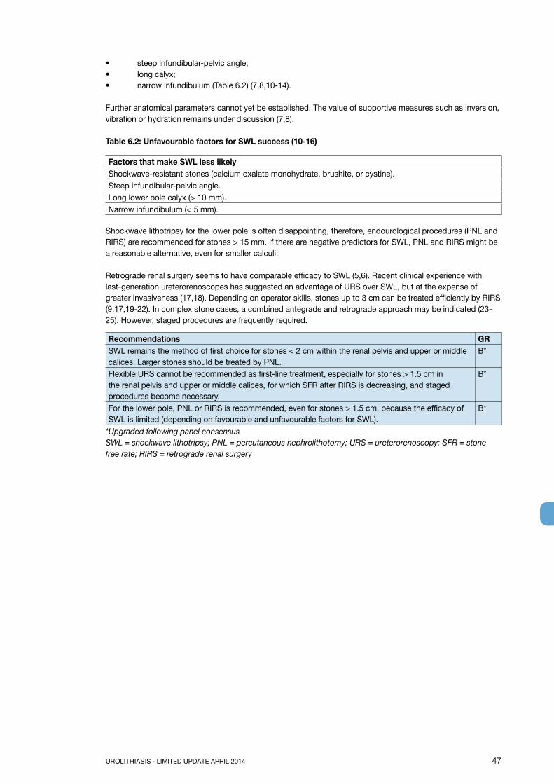
UROLITHIASIS - LIMITED UPDATE APRIL 2014 47
• steep infundibular-pelvic angle;• long calyx;• narrow infundibulum (Table 6.2) (7,8,10-14).
Further anatomical parameters cannot yet be established. The value of supportive measures such as inversion, vibration or hydration remains under discussion (7,8).
Table 6.2: Unfavourable factors for SWL success (10-16)
Factors that make SWL less likelyShockwave-resistant stones (calcium oxalate monohydrate, brushite, or cystine).Steep infundibular-pelvic angle.Long lower pole calyx (> 10 mm).Narrow infundibulum (< 5 mm).
Shockwave lithotripsy for the lower pole is often disappointing, therefore, endourological procedures (PNL and RIRS) are recommended for stones > 15 mm. If there are negative predictors for SWL, PNL and RIRS might be a reasonable alternative, even for smaller calculi.
Retrograde renal surgery seems to have comparable efficacy to SWL (5,6). Recent clinical experience with last-generation ureterorenoscopes has suggested an advantage of URS over SWL, but at the expense of greater invasiveness (17,18). Depending on operator skills, stones up to 3 cm can be treated efficiently by RIRS (9,17,19-22). In complex stone cases, a combined antegrade and retrograde approach may be indicated (23-25). However, staged procedures are frequently required.
Recommendations GRSWL remains the method of first choice for stones < 2 cm within the renal pelvis and upper or middle calices. Larger stones should be treated by PNL.
B*
Flexible URS cannot be recommended as first-line treatment, especially for stones > 1.5 cm in the renal pelvis and upper or middle calices, for which SFR after RIRS is decreasing, and staged procedures become necessary.
B*
For the lower pole, PNL or RIRS is recommended, even for stones > 1.5 cm, because the efficacy of SWL is limited (depending on favourable and unfavourable factors for SWL).
B*
*Upgraded following panel consensusSWL = shockwave lithotripsy; PNL = percutaneous nephrolithotomy; URS = ureterorenoscopy; SFR = stone free rate; RIRS = retrograde renal surgery

48 UROLITHIASIS - LIMITED UPDATE APRIL 2014
Figure 6.1: Treatment algorithm for renal calculi
In complex stone cases, open or laparocopic approaches are possible alternatives (see appropriate chapters).
6.4.3 References1. Argyropoulos AN, Tolley DA. Evaluation of outcome following lithotripsy. Curr Opin Urol 2010
Mar;20(2):154-8.http://www.ncbi.nlm.nih.gov/pubmed/19898239
2. Srisubat A, Potisat S, Lojanapiwat B, et al. Extracorporeal shock wave lithotripsy (ESWL) versus percutaneous nephrolithotomy (PCNL) or retrograde intrarenal surgery (RIRS) for kidney stones. Cochrane Database Syst Rev 2009 Oct;7(4):CD007044.http://www.ncbi.nlm.nih.gov/pubmed/19821393
3. Sahinkanat T, Ekerbicer H, Onal B, et al. Evaluation of the effects of relationships between main spatial lower pole calyceal anatomic factors on the success of shock-wave lithotripsy in patients with lower pole kidney stones. Urology 2008 May;71(5):801-5.http://www.ncbi.nlm.nih.gov/pubmed/18279941
4. Danuser H, Muller R, Descoeudres B, et al. Extracorporeal shock wave lithotripsy of lower calyx calculi: how much is treatment outcome influenced by the anatomy of the collecting system? Eur Urol 2007 Aug;52(2):539-46.http://www.ncbi.nlm.nih.gov/pubmed/17400366
Kidney stone (all but lower pole stone 10-20 mm)
Lower pole stone > 20 mm and < 10 mm: like above
1. PNL2. RIRS or SWL
SWL or Endourology
SWL or Endourology
1. SWL or RIRS2. PNL
1. Endourology2. SWL
10-20 mm
10-20 mmUnfavourable
factors for SWL (see table 6.2)
< 10 mm
> 20 mm
No
Yes

UROLITHIASIS - LIMITED UPDATE APRIL 2014 49
5. Preminger GM. Management of lower pole renal calculi: shock wave lithotripsy versus percutaneous nephrolithotomy versus flexible ureteroscopy. Urol Res 2006 Apr;34(2):108-11.http://www.ncbi.nlm.nih.gov/pubmed/16463145
6. Pearle MS, Lingeman JE, Leveillee R, et al. Prospective, randomized trial comparing shock wave lithotripsy and ureteroscopy for lower pole caliceal calculi 1 cm or less. J Urol 2005 Jun;173(6):2005-9.http://www.ncbi.nlm.nih.gov/pubmed/15879805
7. Albanis S, Ather HM, Papatsoris AG, et al. Inversion, hydration and diuresis during extracorporeal shock wave lithotripsy: does it improve the stone-free rate for lower pole stone clearance? Urol Int 2009;83(2):211-6.http://www.ncbi.nlm.nih.gov/pubmed/19752619
8. Kosar A, Ozturk A, Serel TA, et al. Effect of vibration massage therapy after extracorporeal shockwave lithotripsy in patients with lower caliceal stones. J Endourol 1999 Dec;13(10):705-7.http://www.ncbi.nlm.nih.gov/pubmed/10646674
9. Aboumarzouk OM, Monga M, Kata SG, et al. Flexible ureteroscopy and laser lithotripsy for stones >2 cm: a systematic review and meta-analysis. J Endourol 2012 Oct;26(10):1257-63.http://www.ncbi.nlm.nih.gov/pubmed/22642568
10. Handa RK, Bailey MR, Paun M, et al. Pretreatment with low-energy shock waves induces renal vasoconstriction during standard shock wave lithotripsy (SWL): a treatment protocol known to reduce SWL-induced renal injury. BJU Int 2009 May;103(9):1270-4.http://www.ncbi.nlm.nih.gov/pubmed/19154458
11. Manikandan R, Gall Z, Gunendran T, et al. Do anatomic factors pose a significant risk in the formation of lower pole stones? Urology 2007 Apr;69(4):620-4.http://www.ncbi.nlm.nih.gov/pubmed/17445636
12. Juan YS, Chuang SM, Wu WJ, et al. Impact of lower pole anatomy on stone clearance after shock wave lithotripsy. Kaohsiung J Med Sci 2005 Aug;21(8):358-64.http://www.ncbi.nlm.nih.gov/pubmed/16158878
13. Ruggera L, Beltrami P, Ballario R, et al. Impact of anatomical pielocaliceal topography in the treatment of renal lower calyces stones with extracorporeal shock wave lithotripsy. Int J Urol 2005 Jun;12(6): 525-32.http://www.ncbi.nlm.nih.gov/pubmed/15985072
14. Knoll T, Musial A, Trojan L, et al. Measurement of renal anatomy for prediction of lower-pole caliceal stone clearance: reproducibility of different parameters. J Endourol 2003 Sep;17(7):447-51.http://www.ncbi.nlm.nih.gov/pubmed/14565873
15. Sumino Y, Mimata H, Tasaki Y, et al. Predictors of lower pole renal stone clearance after extracorporeal shock wave lithotripsy. J Urol 2002 Oct;168(4 Pt 1):1344-7.http://www.ncbi.nlm.nih.gov/pubmed/12352389
16. Madbouly K, Sheir KZ, Elsobky E. Impact of lower pole renal anatomy on stone clearance after shock wave lithotripsy: fact or fiction? J Urol 2001 May;165(5):1415-8.http://www.ncbi.nlm.nih.gov/pubmed/11342888
17. Hussain M, Acher P, Penev B, et al. Redefining the limits of flexible ureterorenoscopy. J Endourol 2011 Jan;25(1):45-9.http://www.ncbi.nlm.nih.gov/pubmed/21050026
18. Wendt-Nordahl G, Mut T, Krombach P, et al. Do new generation flexible ureterorenoscopes offer a higher treatment success than their predecessors? Urol Res 2011 Jun;39(3):185-8.http://www.ncbi.nlm.nih.gov/pubmed/21052986
19. Hyams ES, Munver R, Bird VG, et al. Flexible ureterorenoscopy and holmium laser lithotripsy for the management of renal stone burdens that measure 2 to 3 cm: a multi-institutional experience. J Endourol 2010 Oct;24(10):1583-8.http://www.ncbi.nlm.nih.gov/pubmed/20629566
20. Prabhakar M. Retrograde ureteroscopic intrarenal surgery for large (1.6-3.5 cm) upper ureteric/renal calculus. Indian J Urol 2010 Jan-Mar;26(1):46-9.http://www.ncbi.nlm.nih.gov/pubmed/20535284
21. Riley JM, Stearman L, Troxel S. Retrograde ureteroscopy for renal stones larger than 2.5 cm. J Endourol 2009 Sep;23(9):1395-8.http://www.ncbi.nlm.nih.gov/pubmed/19694527
22. Aboumarzouk OM, Kata SG, Keeley FX, et al. Extracorporeal shock wave lithotripsy (ESWL) versus ureteroscopic management for ureteric calculi. Cochrane Database Syst Rev 2012;5:CD006029.http://www.ncbi.nlm.nih.gov/pubmed/22592707

50 UROLITHIASIS - LIMITED UPDATE APRIL 2014
23. Chang CH, Wang CJ, Huang SW. Totally tubeless percutaneous nephrolithotomy: a prospective randomized controlled study. Urol Res 2011 Dec;39(6):459-65.http://www.ncbi.nlm.nih.gov/pubmed/21331773
24. Agarwal M, Agrawal MS, Jaiswal A, et al. Safety and efficacy of ultrasonography as an adjunct to fluoroscopy for renal access in percutaneous nephrolithotomy (PCNL). BJU Int 2011 Oct;108(8): 1346-9.http://www.ncbi.nlm.nih.gov/pubmed/21251187
25. Deem S, Defade B, Modak A, et al. Percutaneous nephrolithotomy versus extracorporeal shock wave lithotripsy for moderate sized kidney stones. Urology 2011 Oct;78(4):739-43.http://www.ncbi.nlm.nih.gov/pubmed/21664653
6.5 Selection of procedure for active removal of ureteral stones6.5.1 MethodologyStone free rates were analysed for SWL and URS. If the study reported the SFR after all primary procedures, that rate was used for analysis. If not, and the study reported the SFR after the first procedure, then that rate was used. The Panel aimed to present an estimate of the number of primary procedures and the associated SFRs. There is a lack of uniformity in reporting the time to stone-free status, thereby limiting the ability to comment on the timing of this parameter.
6.5.2 Extracorporeal shock wave lithotripsy and ureteroscopyFor proximal stones, no difference in overall SFRs between SWL and URS was detected. However, after stratifying for stone size, in proximal ureteral stones < 10 mm (n = 1,285), SWL had a higher SFR than URS had. For stones > 10 mm (n = 819), URS had superior SFRs. This can be attributed to the fact that proximal ureteral stones treated with URS did not vary significantly with size, whereas the SFR following SWL negatively correlated with stone size.
For all mid-ureteral stones, URS appears superior to SWL, but after stratification for stone size, the small number of patients limits the significance. For all distal stones, URS yields better SFRs overall, compared to other methods for active stone removal, independent of stone size.
6.5.2.1 Stone free rates (SFRs)Table 6.3 shows the results of a meta-analysis of SFRs. The results are presented as medians of the posterior distribution (best central estimate) with 95% confidence intervals (CIs). This represents an update of the EAU/AUA Collaborative Guidelines Project (1). Outcomes show no significant changes.
Table 6.3: SFRs after primary treatment with SWL and URS in the overall population (1-5)*
Stone location and size SWL URSNo. of patients SFR/95% CI No. of patients SFR/95% CI
Distal ureter 7217 74% (73-75) 10,372 93% (93-94)< 10 mm 1684 86% (80-91) 2,013 97% (96-98)> 10 mm 966 74% (57-87) 668 93% (91-95)Mid ureter 1697 73% (71-75) 1,140 87% (85-89)< 10 mm 44 84% (65-95) 116 93% (88-98)> 10 mm 15 76% (36-97) 110 79% (71-87)Proximal ureter 6682 82% (81-83) 2,448 82% (81-84)< 10 mm 967 89% (87-91) 318 84% (80-88)> 10 mm 481 70% (66-74) 338 81% (77-85)
* Please note that in a few studies included in the meta-analysis different ranges of stone sizes were used and not an exact cut off of 10 mm.
Unfortunately, RCTs comparing these treatments have been lacking. However, the posterior distributions from the meta-analysis can be subtracted, which yields a distribution for the difference between the treatments. If the CI does not include zero, then the result can be considered to be significantly different. This operation is mathematically justifiable but operationally risky: if the patients receive different treatments or the outcome measures are different, the results might be meaningless. Nonetheless, the SFRs for URS remained significantly better than those for SWL for distal ureteral stones < 10 mm and > 10 mm and for proximal ureteral stones > 10 mm. The SFRs for mid-ureteral stones did not differ significantly between URS and SWL.

UROLITHIASIS - LIMITED UPDATE APRIL 2014 51
Although there are not sufficient data to compare flexible and rigid URS statistically for proximal ureteral stones, favourable SFRs have been reported using RIRS (87%) or rigid or semi-rigid URS (77%) (1). SFRs have probably continued to improve with the distribution and technical improvement of RIRS.
6.5.2.2 ComplicationsAlthough URS is effective for ureteric calculi, it has greater potential for complications. In the current endourological era, with access to newer and smaller rigid and flexible instruments, and use of small-calibre intracorporeal lithotripsy devices, the complication rate and morbidity of ureteroscopy have been significantly reduced (6).
Patients should be informed that URS has a better chance of achieving stone-free status with a single procedure, but has higher complication rates [Sections 5.5.4 (Complications of SWL) and 5.6.2.2 (Complications of URS)].
6.5.3 Percutaneous antegrade ureteroscopyPercutaneous antegrade removal of ureteral stones is a consideration in selected cases. For example, for very large (> 15 mm diameter) impacted stones in the proximal ureter between the ureteropelvic junction and the lower border of the fourth lumbar vertebra (7-10), or when the ureter is not amenable to retrograde manipulation (11-13). With SFRs of 85-100%, its superiority to standard techniques has been evaluated (7,10,11,14,15). The complication rate is low, and no different than for any other percutaneous procedure. However, percutaneous antegrade removal of ureteral stones is associated with longer operative times, hospital stay, and time to return to normal activities (10) (11-13).
Recommendation GRPercutaneous antegrade removal of ureteral stones is an alternative when SWL is not indicated or has failed, and when the upper urinary tract is not amenable to retrograde URS.
A
Table 6.4: Recommended treatment options (if indicated for active stone removal) (GR: A*)
Stone location and size First choice Second choiceProximal ureter < 10 mm SWL URSProximal ureter > 10 mm URS (retrograde or antegrade) or SWLDistal ureter < 10 mm URS or SWLDistal ureter > 10 mm URS SWL
*Upgraded following panel consensus.
Recommendation GRTreatment choices should be based on stone size and location, available equipment, and patient preference for stone removal.
A
6.5.4 Other methods for ureteral stone removalFew studies have reported laparoscopic stone removal (Section 5.7.2), and open surgery (Section 5.7.1). These procedures are usually reserved for special cases, therefore, the reported data could not be used to compare procedures with each other or with SWL or URS. These more invasive procedures have yielded high SFRs.
6.5.5 References1. Preminger GM, Tiselius HG, Assimos DG, et al. American Urological Association Education and
Research, Inc; European Association of Urology. 2007 Guideline for the management of ureteral calculi. Eur Urol 2007 Dec;52(6):1610-31.http://www.ncbi.nlm.nih.gov/pubmed/18074433
2. Tiselius HG. How efficient is extracorporeal shockwave lithotripsy with modern lithotripters for removal of ureteral stones? J Endourol 2008 Feb;22(2):249-55.http://www.ncbi.nlm.nih.gov/pubmed/18294029
3. Elashry OM, Elgamasy AK, Sabaa MA, et al. Ureteroscopic management of lower ureteric calculi: a 15-year single-centre experience. BJU Int 2008 Sep;102(8):1010-7.http://www.ncbi.nlm.nih.gov/pubmed/18485033
4. Fuganti PE, Pires S, Branco R, et al. Predictive factors for intraoperative complications in semirigid ureteroscopy: analysis of 1235 ballistic ureterolithotripsies. Urology 2008 Oct;72(4):770-4.http://www.ncbi.nlm.nih.gov/pubmed/18632141

52 UROLITHIASIS - LIMITED UPDATE APRIL 2014
5. Tugcu V, Tasci AI, Ozbek E, et al. Does stone dimension affect the effectiveness of ureteroscopic lithotripsy in distal ureteral stones? Int Urol Nephrol 2008;40(2):269-75.http://www.ncbi.nlm.nih.gov/pubmed/17899430
6. Hong YK, Park DS. Ureteroscopic lithotripsy using Swiss Lithoclast for treatment of ureteral calculi: 12-years experience. J Korean Med Sci 2009 Aug;24(4):690-4.http://www.ncbi.nlm.nih.gov/pubmed/19654954
7. Kumar V, Ahlawat R, Banjeree GK, et al. Percutaneous ureterolitholapaxy: the best bet to clear large bulk impacted upper ureteral calculi. Arch Esp Urol 1996 Jan-Feb;49(1):86-91.http://www.ncbi.nlm.nih.gov/pubmed/8678608
8. Goel R, Aron M, Kesarwani PK, et al. Percutaneous antegrade removal of impacted upper-ureteral calculi: still the treatment of choice in developing countries. J Endourol 2005 Jan-Feb;19(1):54-7.http://www.ncbi.nlm.nih.gov/pubmed/15735384
9. Berczi C, Flasko T, Lorincz L, et al. Results of percutaneous endoscopic ureterolithotomy compared to that of ureteroscopy. J Laparoendosc Adv Surg Tech A 2007 Jun;17(3):285-9.http://www.ncbi.nlm.nih.gov/pubmed/17570771
10. Sun X, Xia S, Lu J, et al. Treatment of Large Impacted Proximal Ureteral Stones: Randomized Comparison of Percutaneous Antegrade Ureterolithotripsy versus Retrograde Ureterolithotripsy J Endourol 2008 May;22(5):913-7.http://www.ncbi.nlm.nih.gov/pubmed/18429682
11. el-Nahas AR, Eraky I, el-Assmy AM, et al. Percutaneous treatment of large upper tract stones after urinary diversion. Urology 2006 Sep;68(3):500-4.http://www.ncbi.nlm.nih.gov/pubmed/16979745
12. El-Assmy A, El-Nahas AR, Mohsen T, et al. Extracorporeal shock wave lithotripsy of upper urinary tract calculi in patients with cystectomy and urinary diversion. Urology 2005 Sep;66(3):510-3.http://www.ncbi.nlm.nih.gov/pubmed/16140067
13. Rhee BK, Bretan PN Jr, Stoller ML. Urolithiasis in renal and combined pancreas/renal transplant recipients. J Urol 1999 May;161(5):1458-62.http://www.ncbi.nlm.nih.gov/pubmed/10210372
14. Karami H, Arbab AH, Hosseini SJ, et al. Impacted upper-ureteral calculi > 1 cm: bind access and totally tubeless percutaneous antegrade removal or retrograde approach? J Endourol 2006 Sep;20(9):616-9.http://www.ncbi.nlm.nih.gov/pubmed/16999610
15. Basiri A, Simforoosh N, Ziaee A, et al. Retrograde, antegrade, and laparoscopic approaches for the management of large, proximal ureteral stones: a randomized clinical trial. J Endourol 2008 Dec;22(12):2677-80.http://www.ncbi.nlm.nih.gov/pubmed/19025388
7. RESIDUAL STONES7.1 Clinical evidenceResidual fragments are commonly seen in the kidney (mostly in the lower calix) after SWL and sometimes after intracorporeal lithotripsy. Reports on residual fragments vary between institutions, according to imaging method. However, the clinical value of detecting very small concretions remains debatable.
The clinical problem of residual kidney stones is related to the risk of developing:• new stones from such nidi (heterogeneous nucleation);• persistent UTI;• dislocation of fragments with/without obstruction and symptoms (1-6).
Recommendations LE GRIdentification of biochemical risk factors and appropriate stone prevention is particularly indicated in patients with residual fragments or stones (3-5).
1b A
Patients with residual fragments or stones should be followed up regularly to monitor disease course.
4 C
Recurrence risk in patients with residual fragments after treatment of infection stones is higher than for

UROLITHIASIS - LIMITED UPDATE APRIL 2014 53
other stones. In a 2.2-year follow-up of 53 patients, 78% with stone fragments at 3 months after treatment experienced stone progression. The SFR was 20%, and the remaining 2% had stable disease (7). For all stone compositions, 21-59% of patients with residual stones required treatment within 5 years. Fragments > 5 mm are more likely than smaller ones to require intervention (2,3,5,8).
Table 7.1: Recommendations for the treatment of residual fragments
Residual fragments, stones (largest diameter)
Symptomatic residuals Asymptomatic residuals LE GR
< 4-5 mm Stone removal Reasonable follow-up (dependent on risk factors)
4 C
> 6-7 mm Stone removal
7.2 TherapyResidual fragments after PNL can be avoided by a second look using the existing percutaneous tract 1-3 days after the first procedure (9). To facilitate further clearance, medical and physical adjunctive therapy can be suggested. The indications for active stone removal and selection of the procedure are based on the same criteria as for primary stone treatment (Chapter 6) and includes repeat SWL (10). If intervention is not required, medical therapy according to stone analysis, patient risk group, and metabolic evaluation might help to prevent regrowth of residual fragments (11-14).
Statement LEFor well-disintegrated stone material in the lower calix, an inversion therapy with simultaneous mechanical percussion maneuver under enforced diuresis may facilitate stone clearance (15).
1b
Recommendation LE GRAfter SWL and URS, and in the presence of residual fragments, MET is recommended using an α-blocker to improve fragment clearance.
1a A
SWL = shockwave lithotripsy; URS = ureteronoscopy; MET = medical expulsive therapy
7.3 References1. Balaji KC, Menon M. Mechanism of stone formation. Urol Clin North Am 1997 Feb;24(1):1-11.
http://www.ncbi.nlm.nih.gov/pubmed/90488482. El-Nahas AR, El-Assmy AM, Madbouly K, et al. Predictors of clinical significance of residual fragments
after extracorporeal shockwave lithotripsy for renal stones. J Endourol 2006 Nov;20(11):870-4.http://www.ncbi.nlm.nih.gov/pubmed/17144853
3. Osman MM, Alfano Y, Kamp S, et al. 5-year-follow-up of patients with clinically insignificant residual fragments after extracorporeal shockwave lithotripsy. Eur Urol 2005 Jun;47(6):860-4.http://www.ncbi.nlm.nih.gov/pubmed/15925084
4. Buchholz NP, Meier-Padel S, Rutishauser G. Minor residual fragments after extracorporeal shockwave lithotripsy: spontaneous clearance or risk factor for recurrent stone formation? J Endourol 1997 Aug;11(4):227-32.http://www.ncbi.nlm.nih.gov/pubmed/9376838
5. Shigeta M, Kasaoka Y, Yasumoto H, et al. Fate of residual fragments after successful extracorporeal shock wave lithotripsy. Int J Urol 1999 Apr;6(4):169-72. http://www.ncbi.nlm.nih.gov/pubmed/10226832
6. Osman MM, Alfano Y, Kamp S, et al. 5-year-follow-up of patients with clinically insignificant residual fragments after extracorporeal shockwave lithotripsy. Eur Urol 2005 Jun;47(6):860-4.http://www.ncbi.nlm.nih.gov/pubmed/15925084
7. Beck EM, Riehle RA Jr. The fate of residual fragments after extracorporeal shock wave lithotripsy monotherapy of infection stones. J Urol 1991 Jan;145(1):6-9;discussion 9-10.http://www.ncbi.nlm.nih.gov/pubmed/1984100
8. Candau C, Saussine C, Lang H, et al. Natural history of residual renal stone fragments after ESWL. Eur Urol 2000 Jan;37(1):18-22.http://www.ncbi.nlm.nih.gov/pubmed/10671779
9. Acar C, Cal C. Impact of Residual Fragments following Endourological Treatments in Renal Stones. Adv Urol 2012;2012:813523.http://www.ncbi.nlm.nih.gov/pubmed/22829812

54 UROLITHIASIS - LIMITED UPDATE APRIL 2014
10. Krings F, Tuerk C, Steinkogler I, et al. Extracorporeal shock wave lithotripsy retreatment (”stir-up”) promotes discharge of persistent caliceal stone fragments after primary extracorporeal shock wave lithotripsy. J Urol 1992 Sep;148(3 Pt 2):1040-1;discussion 1041-2.http://www.ncbi.nlm.nih.gov/pubmed/1507326
11. Kang DE, Maloney MM, Haleblian GE, et al. Effect of medical management on recurrent stone formation following percutaneous nephrolithotomy. J Urol 2007 May;177(5):1785-8;discussion 1788-9.http://www.ncbi.nlm.nih.gov/pubmed/17437820
12. Fine JK, Pak CY, Preminger GM. Effect of medical management and residual fragments on recurrent stone formation following shock wave lithotripsy. J Urol 1995 Jan;153(1):27-32;discussion 32-3.http://www.ncbi.nlm.nih.gov/pubmed/7966783
13. Siener R, Glatz S, Nicolay C, et al. Prospective study on the efficacy of a selective treatment and risk factors for relapse in recurrent calcium oxalate stone patients. Eur Urol 2003 Oct;44(4):467-74.http://www.ncbi.nlm.nih.gov/pubmed/14499683
14. Cicerello E, Merlo F, Gambaro F, et al. Effect of alkaline citrate therapy on clearance of residual renal stone fragments after extracorporeal shock wave lithotripsy in sterile calcium and infection nephrolithiasis patients., J Urology 1994 Jan;151(1):5-9.http://www.ncbi.nlm.nih.gov/pubmed/8254832
15. Chiong E, Hwee ST, Kay LM, et al. Randomized controlled study of mechanical percussion, diuresis, and inversion therapy to assist passage of lower pole renal calculi after shock wave lithotripsy. Urology 2005 Jun;65(6):1070-4.http://www.ncbi.nlm.nih.gov/pubmed/15922429
8. MANAGEMENT OF URINARY STONES AND RELATED PROBLEMS DURING PREGNANCYUrolithiasis during pregnancy is a diagnostic and therapeutic challenge. In most cases, it becomes symptomatic in the second or third trimester (1,2).
8.1 Diagnostic imagingDiagnostic options in pregnant women are limited due to the possible teratogenic, carcinogenic, and mutagenic risk of foetal radiation exposure. The risk for the child crucially depends on gestational age and amount of radiation delivered. X-ray imaging during the first trimester should be reserved for diagnostic and therapeutic situations in which alternative imaging methods have failed (1,3,4). Ultrasound (when necessary using change in renal resistive index and transvaginal/transabdominal US with a full bladder) has become the primary radiological diagnostic tool when evaluating pregnant patients suspected of renal colic (1,5).
Statement LENormal physiological changes in pregnancy can mimic ureteral obstruction, therefore, US may not help to differentiate dilatation properly and has a limited role in acute obstruction.
3
X-ray imaging options in pregnancy are: limited excretory urography and NCCT (considering the higher dose of radiation exposure). Magnetic resonance urography (MRU) can be used to define the level of urinary tract obstruction, and to visualize stones as a filling defect. MRU studies avoid ionising radiation and iodinated contrast medium.However, findings are non-specific and there is little experience using this imaging modality during pregnancy (6,7).
Recommendation LE GRUltrasound is the method of choice for practical and safe evaluation of pregnant women. 1a A*
* Upgrade following panel consensus.
8.2 ManagementClinical management of a pregnant urolithiasis patient is complex and demands close collaboration between patient, obstetrician and urologist.
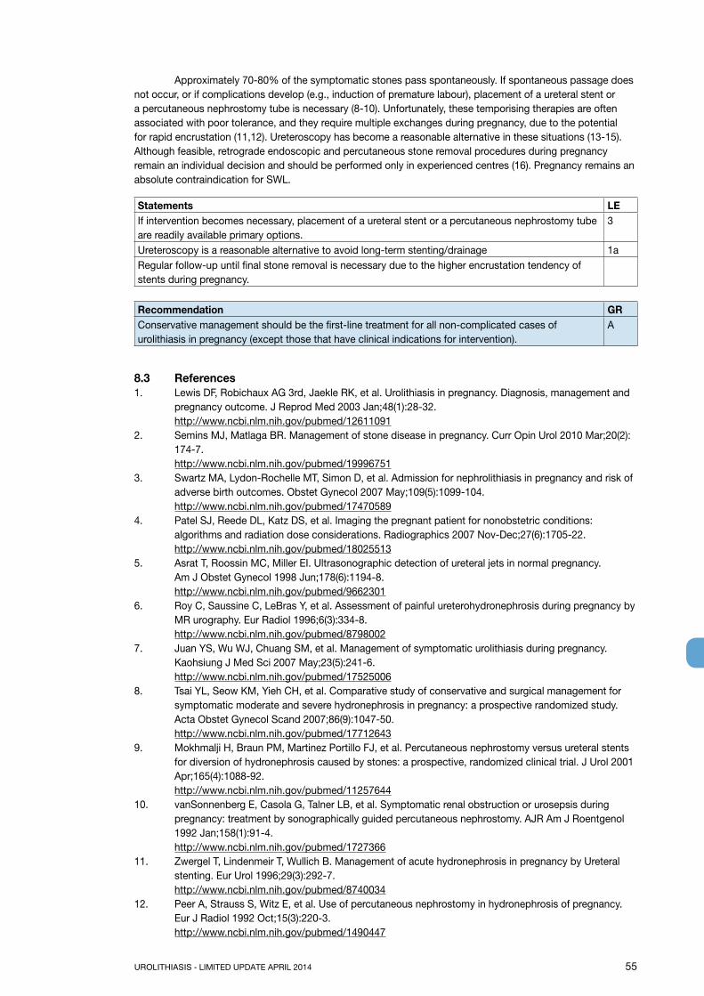
UROLITHIASIS - LIMITED UPDATE APRIL 2014 55
Approximately 70-80% of the symptomatic stones pass spontaneously. If spontaneous passage does not occur, or if complications develop (e.g., induction of premature labour), placement of a ureteral stent or a percutaneous nephrostomy tube is necessary (8-10). Unfortunately, these temporising therapies are often associated with poor tolerance, and they require multiple exchanges during pregnancy, due to the potential for rapid encrustation (11,12). Ureteroscopy has become a reasonable alternative in these situations (13-15). Although feasible, retrograde endoscopic and percutaneous stone removal procedures during pregnancy remain an individual decision and should be performed only in experienced centres (16). Pregnancy remains an absolute contraindication for SWL.
Statements LEIf intervention becomes necessary, placement of a ureteral stent or a percutaneous nephrostomy tube are readily available primary options.
3
Ureteroscopy is a reasonable alternative to avoid long-term stenting/drainage 1aRegular follow-up until final stone removal is necessary due to the higher encrustation tendency of stents during pregnancy.
Recommendation GRConservative management should be the first-line treatment for all non-complicated cases of urolithiasis in pregnancy (except those that have clinical indications for intervention).
A
8.3 References1. Lewis DF, Robichaux AG 3rd, Jaekle RK, et al. Urolithiasis in pregnancy. Diagnosis, management and
pregnancy outcome. J Reprod Med 2003 Jan;48(1):28-32.http://www.ncbi.nlm.nih.gov/pubmed/12611091
2. Semins MJ, Matlaga BR. Management of stone disease in pregnancy. Curr Opin Urol 2010 Mar;20(2): 174-7.http://www.ncbi.nlm.nih.gov/pubmed/19996751
3. Swartz MA, Lydon-Rochelle MT, Simon D, et al. Admission for nephrolithiasis in pregnancy and risk of adverse birth outcomes. Obstet Gynecol 2007 May;109(5):1099-104.http://www.ncbi.nlm.nih.gov/pubmed/17470589
4. Patel SJ, Reede DL, Katz DS, et al. Imaging the pregnant patient for nonobstetric conditions: algorithms and radiation dose considerations. Radiographics 2007 Nov-Dec;27(6):1705-22.http://www.ncbi.nlm.nih.gov/pubmed/18025513
5. Asrat T, Roossin MC, Miller EI. Ultrasonographic detection of ureteral jets in normal pregnancy. Am J Obstet Gynecol 1998 Jun;178(6):1194-8.http://www.ncbi.nlm.nih.gov/pubmed/9662301
6. Roy C, Saussine C, LeBras Y, et al. Assessment of painful ureterohydronephrosis during pregnancy by MR urography. Eur Radiol 1996;6(3):334-8.http://www.ncbi.nlm.nih.gov/pubmed/8798002
7. Juan YS, Wu WJ, Chuang SM, et al. Management of symptomatic urolithiasis during pregnancy. Kaohsiung J Med Sci 2007 May;23(5):241-6.http://www.ncbi.nlm.nih.gov/pubmed/17525006
8. Tsai YL, Seow KM, Yieh CH, et al. Comparative study of conservative and surgical management for symptomatic moderate and severe hydronephrosis in pregnancy: a prospective randomized study. Acta Obstet Gynecol Scand 2007;86(9):1047-50.http://www.ncbi.nlm.nih.gov/pubmed/17712643
9. Mokhmalji H, Braun PM, Martinez Portillo FJ, et al. Percutaneous nephrostomy versus ureteral stents for diversion of hydronephrosis caused by stones: a prospective, randomized clinical trial. J Urol 2001 Apr;165(4):1088-92.http://www.ncbi.nlm.nih.gov/pubmed/11257644
10. vanSonnenberg E, Casola G, Talner LB, et al. Symptomatic renal obstruction or urosepsis during pregnancy: treatment by sonographically guided percutaneous nephrostomy. AJR Am J Roentgenol 1992 Jan;158(1):91-4.http://www.ncbi.nlm.nih.gov/pubmed/1727366
11. Zwergel T, Lindenmeir T, Wullich B. Management of acute hydronephrosis in pregnancy by Ureteral stenting. Eur Urol 1996;29(3):292-7.http://www.ncbi.nlm.nih.gov/pubmed/8740034
12. Peer A, Strauss S, Witz E, et al. Use of percutaneous nephrostomy in hydronephrosis of pregnancy. Eur J Radiol 1992 Oct;15(3):220-3.http://www.ncbi.nlm.nih.gov/pubmed/1490447

56 UROLITHIASIS - LIMITED UPDATE APRIL 2014
13. Semins MJ, Trock BJ, Matlaga BR. The safety of ureteroscopy during pregnancy: a systematic review and meta-analysis. J Urol 2009 Jan;181(1):139-43.http://www.ncbi.nlm.nih.gov/pubmed/19012926
14. Rana AM, Aquil S, Khawaja AM. Semirigid ureteroscopy and pneumatic lithotripsy as definitive management of obstructive ureteral calculi during pregnancy. Urology 2009 May;73(5):964-7.http://www.ncbi.nlm.nih.gov/pubmed/19394491
15. Semins MJ, Trock BJ, Matlaga BR. The safety of ureteroscopy during pregnancy: a systematic review and meta-analysis. J Urol 2009 Jan;181(1);139-43.http://www.ncbi.nlm.nih.gov/pubmed/19012926
16. Toth C, Toth G, Varga A, et al. Percutaneous nephrolithotomy in early pregnancy. Int Urol Nephrol 2005;37(1):1-3.http://www.ncbi.nlm.nih.gov/pubmed/16132747
9. MANAGEMENT OF STONE PROBLEMS IN CHILDRENRates of urolithiasis have increased in developed countries, and there has been a shift in the age group experiencing a first stone episode (1-3). More than 1% of all urinary stones are seen in patients aged < 18 years. As a result of malnutrition and racial factors, paediatric urolithiasis remains an endemic disease in some areas (e.g., Turkey and the Far East); elsewhere, the rates are similar to those observed in developed countries (4-11).
9.1 AetiologyPaediatric patients forming urinary stones have a high risk of recurrence, therefore, standard diagnostic procedures for high-risk patients apply (Chapters 2.6 and 11).
Statement LEIn paediatric patients, the most common non-metabolic disorders are vesicoureteral reflux, ureteropelvic junction obstruction, neurogenic bladder, and other voiding difficulties (11,12).
4
Recommendations GRIn all paediatric patients, complete metabolic evaluation based on stone analysis (if available) is necessary.
A
All efforts should be made to collect stone material that then should be analysed to classify the stone type.
A*
*Upgrade following panel consensus.
9.2 Diagnostic imagingWhen selecting diagnostic procedures to identify urolithiasis in paediatric patients, it should be remembered that these patients might be uncooperative, require anaesthesia, or be sensitive to ionising radiation (13).
9.2.1 UltrasoundUltrasound (US) is the primary imaging technique (13) in paediatrics. Its advantages are absence of radiation and no need for anaesthesia. Ultrasound provides information about the presence, size and location of a stone, and the grade of dilatation/obstruction of the urinary collecting system and the severity of nephrocalcinosis. It also indicates anatomical abnormalities. Colour Doppler US shows differences in the ureteric jet (14) and resistive index of the arciform arteries of both kidneys, which are indicative of the grade of obstruction (15). Nevertheless, US fails to identify stones in > 40% of paediatric patients (16-19) (LE: 4), and provides no information about renal function.
Statement LEUS is the first choice for imaging in children and should include the kidney, filled bladder, and adjoining portions of the ureter (14,20).
2a

UROLITHIASIS - LIMITED UPDATE APRIL 2014 57
9.2.2 Plain films (KUB radiography)KUB radiography can help to identify stones and their radiopacity, and facilitate follow-up.
9.2.3 Intravenous urography (IVU)Intravenous urography is an important diagnostic tool. However, the need for contrast medium injection is a major drawback. The radiation dose for IVU is comparable to that for voiding cystourethrography (0.33 mSV) (21).
9.2.4 Helical computed tomography (CT)Recent CT protocols have been shown to reduce radiation exposure significantly (22). The principle of ALARA (as low as reasonable achievable) should always be observed. In adults it has a sensitivity of 94-100% and specificity of 92-100% (23). In children, only 5% of stones escape detection by NCCT (14,23,24). Sedation or anaesthesia is rarely needed with modern high-speed CT apparatus (11).
9.2.5 Magnetic resonance urography (MRU)Magnetic resonance urography cannot be used to detect urinary stones. However, it might provide detailed anatomical information about the urinary collecting system, the location of an obstruction or stenosis in the ureter, and renal parenchymal morphology (25).
9.2.6 Nuclear imaging99mTc-dimercaptosuccinyl acid scanning provides information about cortical abnormalities such as scarring, but does not aid primary diagnosis of urolithiasis. Diuretic renography with injection of a radiotracer (MAG3 [Mercaptoacetyltriglycin] or DPTA [Diethylentriaminpentaacetat]) and furosemide can be used to demonstrate renal function, identify obstruction in the kidney after injection of furosemide, and indicate the anatomical level of the obstruction (11,14).
Recommendations GRIn children, US is the first-line imaging modality when suspecting a stone. BIf US does not provide the required information, KUB radiography (or NCCT) should be performed. B
US = ultrasound; KUB = kidney, ureter, bladder; NCCT = non-contrast enhanced computed tomography.
9.3 Stone removalSeveral factors must be considered when selecting treatment procedures for children. Compared to adults, children pass fragments more rapidly after SWL (26). For endourological procedures, the smaller organs in children must be considered when selecting instruments for PNL or URS. Anticipation of the expected stone composition should be taken into account when selecting the appropriate procedure for stone removal (cystine stones are more resistant to SWL).
Statement LESpontaneous passage of a stone is more likely in children than adults (6,11,12). 4
9.3.1 Medical expulsive therapy (MET) in childrenMedical expulsive therapy has already been discussed in Section 5.3.2.6 but not addressing children. Although the use of α-blockers is very common in adults, there are few data to demonstrate their safety and efficacy in children, however Tamsulosin seems to support stone passage (27,28).
9.3.2 Extracorporeal shock wave lithotripsyExtracorporeal shock wave lithotripsy remains the least-invasive procedure for stone management in children (29-37).
SFRs of 67-93% in short-term and 57-92% in long-term follow-up studies have been reported. In children, compared with adults, SWL can achieve more effective disintegration of large stones, together with swifter and uncomplicated discharge of large fragments (33-35). Stones located in calices, as well as abnormal kidneys, and large stones, are more difficult to disintegrate and clear. The likelihood of urinary obstruction is higher in such cases, and children should be followed closely for the prolonged risk of urinary tract obstruction. The retreatment rate is 13.9-53.9%, and the need for ancillary procedures and/or additional interventions is 7-33% (33-35,37).
The need for general anaesthesia during SWL depends on patient age and the lithotripter used. General or

58 UROLITHIASIS - LIMITED UPDATE APRIL 2014
dissociative anaesthesia is administered in most children aged < 10 years, to avoid patient and stone motion and the need for repositioning (33,37). With modern lithotriptors, intravenous sedation or patient-controlled analgesia have been used in selected cooperative older children (38) (LE: 2b). There are concerns regarding the safety and potential biological effects of SWL on immature kidneys and surrounding organs in children. However, during short- and long-term follow-up, no irreversible functional or morphological side effects of high-energy shock waves have been demonstrated. In addition, when the potential deterioration of renal function is taken into account (although transient), restricting the number of shock waves and the energy used during each treatment session helps protect the kidneys (39-42).
If the stone burden requires a ureteral stent, alternative procedures should be considered. Ureteral stents are seldom needed following SWL of upper tract stones, ureteral pre-stenting decreases the SFR after initial treatment (29,31-33).
Statements LEIn children, the indications for SWL are similar to those in adults, however, they pass fragments more easily.
3
Children with renal stones of a diameter up to 20 mm (~300 mm2) are ideal candidates for SWL. 1b
9.3.3 Endourological proceduresImprovement in intracorporeal lithotripsy devices and development of smaller instruments facilitate PNL andURS in children.
9.3.3.1 Percutaneous nephrolithotripsy (PNL)Preoperative evaluation and indications for PNL in children are similar to those in adults. Provided appropriate-size instruments and US guidance, age is not a limiting factor, and PNL can be performed safely by experienced operators, with less radiation exposure, even for large and complex stones (43-47). SFRs are between 68% and 100% after a single session, and increase with adjunctive measures, such as second-look PNL, SWL and URS (43,44).
As for adults, tubeless PNL is safe in children, in well-selected cases (48).
Statement LEFor paediatric patients, the indications for PNL are similar to those in adults. 1a
Recommendation GRIn children, PNL is recommended for treatment of renal pelvic or caliceal stones with a diameter > 20 mm (~300 mm2).
C
9.3.3.2 UreteroscopyAlthough SWL still is the first-line treatment for most ureteral stones, it is unlikely to be successful for stones > 10 mm in diameter, or for impacted, calcium oxalate monohydrate or cystine stones, or stones in children with unfavourable anatomy and in whom localisation is difficult (49-52). If SWL is not promising, ureteroscopy can be used. With the clinical introduction of smaller-calibre instruments, this modality has become the treatment of choice for medium and larger distal ureteric stones in children (50-54). Different lithotripsy techniques, including ultrasonic, pneumatic and laser lithotripsy, are all safe and effective (Section 5.6.2.1.7) (55-57).
Recommendation LE GRFor intracorporeal lithotripsy, the same devices as in adults can be used (Ho:Yag laser, pneumatic and US lithotriptors).
3 C
Flexible ureteroscopy has become an efficacious treatment for paediatric upper urinary tract stones. It might be particularly effective for treatment of proximal ureteral calculi and for stones < 1.5 cm in the lower pole calices (58-60).
9.3.4 Open or laparoscopic surgeryMost stones in children can be managed by SWL and endoscopic techniques (59). Therefore, the rate of open procedure has dropped significantly (62-66). In some situations, open surgery is inevitable. Indications

UROLITHIASIS - LIMITED UPDATE APRIL 2014 59
for surgery include: failure of primary therapy for stone removal; very young children with complex stones; congenital obstruction that requires simultaneous surgical correction; severe orthopaedic deformities that limit positioning for endoscopic procedures; and abnormal kidney position (29,31,44,45). Open surgery can be replaced by laparoscopic procedures in experienced hands (64-66).
9.4 Special considerations on recurrence preventionAll paediatric stone formers need metabolic evaluation and recurrence prevention with respect to the detected stone type. In case of obstructive pathology in association with the established metabolic abnormalities, treatment should not be delayed. Children are in the high-risk group for stone recurrence (Chapter 11).
9.5 References1. Reis-Santos JM. Age of first stone episode. In: Rodgers AL, Hibbert BE, Hess B, Khan SR, Preminger
GM, eds. Urolithiasis. Cape Town: University of Cape Town, 2000, pp. 375-378.2. Robertson WG, Whitfield H, Unwin RJ, et al. Possible causes of the changing pattern of the age of
onset of urinary stone disease in the UK. In: Rodgers AL, Hibbert BE, Hess B, Khan SR, Preminger GM, eds. Urolithiasis. Cape Town: University of Cape Town, 2000, pp. 366-368.
3. Hesse A, Brandle E, Wilbert D, et al. Study on the prevalence and incidence of urolithiasis in Germany comparing the years 1979 vs. 2000. Eur Urol 2003 Dec;44(6):709-13.http://www.ncbi.nlm.nih.gov/pubmed/14644124
4. Djelloul Z, Djelloul A, Bedjaoui A, et al. [Urinary stones in Western Algeria: study of the composition of 1,354 urinary stones in relation to their anatomical site and the age and gender of the patients.] Prog Urol 2006 Jun;16(3):328-35. [Article in French]http://www.ncbi.nlm.nih.gov/pubmed/16821346
5. Sarica K. Pediatric urolithiasis: etiology, specific pathogenesis and medical treatment. Urol Res 2006 Apr;34(2):96-101.http://www.ncbi.nlm.nih.gov/pubmed/16432692
6. Mandeville JA, Nelson CP. Pediatric urolithiasis. Curr Opin Urol 2009 Jul;19(4):419-23.http://www.ncbi.nlm.nih.gov/pubmed/19440153
7. Sarica K. Medical aspect and minimal invasive treatment of urinary stones in children. Arch Ital Urol Androl 2008 Jun;80(2):43-9.http://www.ncbi.nlm.nih.gov/pubmed/18683808
8. Sayasone S, Odermatt P, Khammanivong K, et al. Bladder stones in childhood: a descriptive study in a rural setting in Saravan Province, Lao PDR1. Southeast Asian J Trop Med Public Health 2004;35 Suppl 2:50-2.http://www.ncbi.nlm.nih.gov/pubmed/15906634
9. Stamatelou KK, Francis ME, Jones CA, et al. Time trends in reported prevalence of kidney stones in the United States: 1976-1994. Kidney Int 2003 May;63(5):1817-23.http://www.ncbi.nlm.nih.gov/pubmed/12675858
10. DeFoor WR, Jackson E, Minevich E, et al. The risk of recurrent urolithiasis in children is dependent on urinary calcium and citrate. Urology 2010 Jul;76(1):242-5.http://www.ncbi.nlm.nih.gov/pubmed/20110113
11. Straub M, Strohmaier WL, Berg W, et al. Diagnosis and metaphylaxis of stone disease. Consensus concept of the National Working Committee on Stone Disease for the upcoming German Urolithiasis Guideline. World J Urol 2005 Nov;23(5):309-23.http://www.ncbi.nlm.nih.gov/pubmed/16315051
12. Sternberg K, Greenfield SP, Williot P, et al. Pediatric stone disease: an evolving experience. J Urol 2005 Oct;174(4 Pt 2):1711-4.http://www.ncbi.nlm.nih.gov/pubmed/16148688
13. Palmer LS. Pediatric urologic imaging. Urol Clin North Am 2006 Aug;33(3):409-23.http://www.ncbi.nlm.nih.gov/pubmed/16829274
14. Darge K, Heidemeier A. [Modern ultrasound technologies and their application in pediatric urinary tract imaging.] Radiologe 2005 Dec;45(12):1101-11. [Article in German]http://www.ncbi.nlm.nih.gov/pubmed/16086170
15. Pepe P, Motta L, Pennisi M, et al. Functional evaluation of the urinary tract by color-Doppler ultrasonography (CDU) in 100 patients with renal colic. Eur J Radiol 2005 Jan;53(1):131-5.http://www.ncbi.nlm.nih.gov/pubmed/15607864
16. Oner S, Oto A, Tekgul S, et al. Comparison of spiral CT and US in the evaluation of pediatric urolithiasis. JBR-BTR 2004 Sep-Oct;87(5):219-23.http://www.ncbi.nlm.nih.gov/pubmed/15587558

60 UROLITHIASIS - LIMITED UPDATE APRIL 2014
17. Palmer JS, Donaher ER, O’Riordan MA, et al. Diagnosis of pediatric urolithiasis: role of ultrasound and computerized tomography. J Urol 2005 Oct;174(4 Pt 1):1413-6.http://www.ncbi.nlm.nih.gov/pubmed/16145452
18. Riccabona M, Lindbichler F, Sinzig M. Conventional imaging in paediatric uroradiology. Eur J Radiol 2002 Aug;43(2):100-9.http://www.ncbi.nlm.nih.gov/pubmed/12127207
19. Chateil JF, Rouby C, Brun M, et al. [Practical measurement of radiation dose in pediatric radiology: use of the dose surface product in digital fluoroscopy and for neonatal chest radiographs.] J Radiol 2004 May;85(5 Pt 1):619-25. [Article in French]http://www.ncbi.nlm.nih.gov/pubmed/15205653
20. Riccabona M, Avni FE, Blickman JG, et al. Imaging recommendations in paediatric uroradiology. Minutes of the ESPR uroradiology task force session on childhood obstructive uropathy, high-grade fetal hydronephrosis, childhood haematuria, and urolithiasis in childhood. ESPR Annual Congress, Edinburgh, UK, June 2008. Pediatr Radiol 2009 Aug;39(8):891-8.http://www.ncbi.nlm.nih.gov/pubmed/19565235
21. Stratton KL, Pope JC 4th, Adams CM, et al. Implications of ionizing radiation in the pediatric urology patient. J Urol 2010 Jun;183(6):2137-42.http://www.ncbi.nlm.nih.gov/pubmed/20399463
22. Thomson JM, Glocer J, Abbott C, et al. Computed tomography versus intravenous urography in diagnosis of acute flank pain from urolithiasis: a randomized study comparing imaging costs and radiation dose. Australas Radiol 2001 Aug;45(3):291-7.http://www.ncbi.nlm.nih.gov/pubmed/11531751
23. Tamm EP, Silvermann PM, Shuman WP. Evaluation of the Patient with Flank Pain and Possible Ureteral Calculus. Radiology 2003 Aug;228(2):319-26.http://www.ncbi.nlm.nih.gov/pubmed/12819343
24. Cody DD, Moxley DM, Krugh KT, et al. Strategies for formulating appropriate MDCT techniques when imaging the chest, abdomen, and pelvis in pediatric patients AJR Am J Roentgenol 2004 Apr;182(4):849-59.http://www.ncbi.nlm.nih.gov/pubmed/15039151
25. Leppert A, Nadalin S, Schirg E, et al. Impact of magnetic resonance urography on preoperative diagnostic workup in children affected by hydronephrosis: should IVU be replaced? J Pediatr Surg 2002 Oct;37(10):1441-5.http://www.ncbi.nlm.nih.gov/pubmed/12378450
26. Hesse A, Kruse R, Geilenkeuser WJ, et al. Quality control in urinary stone analysis: results of 44 ring trials (1980-2001). Clin Chem Lab Med 2005;43(3):298-303.http://www.ncbi.nlm.nih.gov/pubmed/15843235
27. Aydogdu O, Burgu B, Gucuk A, et al. Effectiveness of doxazosin in treatment of distal ureteral stones in children. J Urol 2009 Dec;182(6):2880-4.
28. Mokhless I, Zahran AR, Youssif M, et al. Tamsulosin for the management of distal ureteral stones in children: a prospective randomized study. J Pediatr Urol 2012 Oct;8(5):544-8.http://www.ncbi.nlm.nih.gov/pubmed/22099477
29. Lahme S. Shockwave lithotripsy and endourological stone treatment in children. Urol Res 2006 Apr;34(2):112-7.http://www.ncbi.nlm.nih.gov/pubmed/16446980
30. Dogan HS, Tekgul S. Management of pediatric stone disease. Curr Urol Rep 2007 Mar;8(2):163-73.http://www.ncbi.nlm.nih.gov/pubmed/17303023
31. Smaldone MC, Docimo SG, Ost MC. Contemporary Surgical Management of Pediatric Urolithiasis. Urol Clin North Am 2010 May;37(2);253-67.http://www.ncbi.nlm.nih.gov/pubmed/20569803
32. Thomas BG. Management of stones in childhood. Curr Opin Urol 2010 Mar;20(2):159-62.http://www.ncbi.nlm.nih.gov/pubmed/19996750
33. Landau EH, Shenfeld OZ, Pode D, et al. Extracorporeal shock wave lithotripsy in prepubertal children: 22-year experience at a single institution with a single lithotriptor. J Urol 2009 Oct;182(4 Suppl): 1835-9.http://www.ncbi.nlm.nih.gov/pubmed/19692011
34. Landau EH, Gofrit ON, Shapiro A, et al. Extracorporeal shockwave lithotripsy is highly effective for ureteral calculi in children. J Urol 2001 Jun;165(6 Pt 2):2316-9.http://www.ncbi.nlm.nih.gov/pubmed/11371970
35. Tan AH, Al-Omar M, Watterson JD, et al. Results of shockwave lithotripsy for pediatric urolithiasis. J Endourol 2004 Aug;18(6):527-30.http://www.ncbi.nlm.nih.gov/pubmed/15333214

UROLITHIASIS - LIMITED UPDATE APRIL 2014 61
36. Frick J, Sarica K, Kohle R, et al. Long-term follow-up after extracorporeal shock wave lithotripsy in children. Eur Urol 1991;19(3):225-9.http://www.ncbi.nlm.nih.gov/pubmed/1855529
37. D’Addessi A, Bongiovanni L, Racioppi M, et al. Is extracorporeal shock wave lithotripsy in pediatrics a safe procedure? J Pediatr Surg 2008 Apr;43(4):591-6.http://www.ncbi.nlm.nih.gov/pubmed/18405701
38. Aldridge RD, Aldridge RC, Aldridge LM. Anesthesia for pediatric lithotripsy. Paediatr Anaesth 2006 Mar;16(3):236-41.http://www.ncbi.nlm.nih.gov/pubmed/16490086
39. Sarica K, Kupeli S, Sarica N, et al. Long-term follow-up of renal morphology and function in children after lithotripsy. Urol Int 1995;54(2):95-8.http://www.ncbi.nlm.nih.gov/pubmed/7747366
40. Griffin SJ, Margaryan M, Archambaud F, et al. Safety of Shock Wave Lithotripsy for Treatment of Pediatric Urolithiasis: 20-Year Experience. J Urol 2010 Jun;183(6):2332-6.http://www.ncbi.nlm.nih.gov/pubmed/20400129
41. Reisiger K, Vardi I, Yan Y, et al. Pediatric nephrolithiasis: does treatment affect renal growth? Urology 2007 Jun;69(6):1190-4.http://www.ncbi.nlm.nih.gov/pubmed/17572213
42. Kurien A, Symons S, Manohar T, et al. Extracorporeal shock wave lithotripsy in children: equivalent clearance rates to adults is achieved with fewer and lower energy shock waves. BJU Int 2009 Jan; 103(1):81-4.http://www.ncbi.nlm.nih.gov/pubmed/18727616
43. Desai M. Endoscopic management of stones in children. Curr Opin Urol 2005 Mar;15(2):107-12.http://www.ncbi.nlm.nih.gov/pubmed/15725934
44. Rizvi S, Nagvi S, Hussain Z, et al. Management of pediatric urolithiasis in Pakistan: experience with 1,440 children. J Urol 2003 Feb;169(2):634-7.http://www.ncbi.nlm.nih.gov/pubmed/12544331
45. Straub M, Gschwend J, Zorn C. Pediatric urolithiasis: the current surgical management. Pediatr Nephrol 2010 Jul;25(7):1239-44.http://www.ncbi.nlm.nih.gov/pubmed/20130924
46. Smaldone MC, Corcoran AT, Docimo SG, et al. Endourological management of pediatric stone disease: present status. J Urol 2009 Jan;181(1):17-28.http://www.ncbi.nlm.nih.gov/pubmed/19012920
47. Kapoor R, Solanki F, Singhania P, et al. Safety and efficacy of percutaneous nephrolithotomy in the pediatric population. J Endourol 2008 Apr;22(4):637-40.http://www.ncbi.nlm.nih.gov/pubmed/18338958
48. Samad L, Zaidi Z. Tubed vs tubeless PCNL in children. J Pak Med Assoc 2012 Sep;62(9):892-6.http://www.ncbi.nlm.nih.gov/pubmed/23139970
49. Gedik A, Orgen S, Akay AF, et al. Semi-rigid ureterorenoscopy in children without ureteral dilatation. Int Urol Nephrol 2008;40(1):11-4.http://www.ncbi.nlm.nih.gov/pubmed/17653831
50. Smaldone MC, Cannon GM Jr, Wu HY, et al. Is ureteroscopy first line treatment for pediatric stone disease? J Urol 2007 Nov;178(5):2128-31.http://www.ncbi.nlm.nih.gov/pubmed/17870124
51. Erturhan S, Yagci F, Sarica K. Ureteroscopic management of ureteral calculi in children. J Endourol 2007 Apr;21(4):397-400.http://www.ncbi.nlm.nih.gov/pubmed/17451329
52. Minevich E, Sheldon CA. The role of ureteroscopy in pediatric urology. Curr Opin Urol 2006 Jul; 16(4):295-8.http://www.ncbi.nlm.nih.gov/pubmed/16770131
53. Basiri A, Zare S, Tabibi A, et al. A multicenter, randomized, controlled trial of transureteral and shock wave lithotripsy-which is the best minimally invasive modality to treat distal ureteral calculi in children? J Urol 2010 Sep;184(3);1106-9.http://www.ncbi.nlm.nih.gov/pubmed/20650490
54. Basiri A, Zare S, Shakhssalim N, et al. Ureteral calculi in children: what is best as a minimally invasive modality? Urol J 2008 Spring;5(2);67-73.http://www.ncbi.nlm.nih.gov/pubmed/18592456
55. Safwat AS, Bissada NK, Kumar U, et al. Experience with ureteroscopic holmium laser lithotripsy in children. Pediatr Surg Int 2008 May;24(5):579-81.http://www.ncbi.nlm.nih.gov/pubmed/18365216

62 UROLITHIASIS - LIMITED UPDATE APRIL 2014
56. Gupta PK. Is the holmium: YAG laser the best intracorporeal lithotripter for the ureter? A 3-year retrospective study. J Endourol 2007 Mar;21(3):305-9.http://www.ncbi.nlm.nih.gov/pubmed/17444776
57. Erdenetsesteg G, Manohar T, Singh H, et al. Endourologic management of pediatric urolithiasis: proposed clinical guidelines. J Endourol 2006 Oct;20(10):737-48.http://www.ncbi.nlm.nih.gov/pubmed/17094748
58. Kim SS, Kolon TF, Canter D, et al. Pediatric Flexible Ureteroscopic Lithotripsy:The Children’s Hospital of Philadelphia Experience. J Urol 2008 Dec;180(6);2616-9.http://www.ncbi.nlm.nih.gov/pubmed/18950810
59. Lesani OA, Palmer JS. Retrograde proximal rigid ureteroscopy and pyeloscopy in prepubertal children: safe and effective. J Urol 2006 Oct;176(4 Pt 1):1570-3.http://www.ncbi.nlm.nih.gov/pubmed/16952683
60. Cannon GM, Smaldone MC, Wu HY, et al. Ureteroscopic management of lower-pole stones in a pediatric population. J Endourol 2007 Oct;21(10):1179-82.http://www.ncbi.nlm.nih.gov/pubmed/17949321
61. Sarica K, Erturhan S, Yurtseven C, et al. Effect of potassium citrate therapy on stone recurrence and regrowth after extracorporeal shockwave lithotripsy in children. J Endourol 2006 Nov;20(11):875-9.http://www.ncbi.nlm.nih.gov/pubmed/17144854
62. Muslumanoglu AY, Tefekli A, Sarilar O, et al. Extracorporeal shockwave lithotripsy as the first line treatment alternative for urinary tract stones in children: a large scale retrospective analysis. J Urol 2003 Dec;170(6 Pt 1):2405-8.http://www.ncbi.nlm.nih.gov/pubmed/14634438
63. Braun MP, Seif C, Jueneman KP, et al. Urolithiasis in children. Int Braz J Urol 2002 Nov-Dec;28(6); 539-44.http://www.ncbi.nlm.nih.gov/pubmed/15748404
64. Casale P, Grady RW, Joyner BD, et al. Transperitoneal laparoscopic pyelolithotomy after failed percutaneous access in the pediatric patient. J Urol 2004 Aug;172(2):680-3.http://www.ncbi.nlm.nih.gov/pubmed/15247760
65. Fragoso AC, Valla JS, Steyaert H, et al. Minimal access surgery in the management of pediatric urolithiasis. J Pediatr Urol 2009 Feb;5(1):42-6.http://www.ncbi.nlm.nih.gov/pubmed/18805739
66. Holman E, Toth C. Laparoscopically assisted percutaneous transperitoneal nephrolithotomy in pelvic dystopic kidneys: experience in 15 successful cases. J Laparoendosc Adv Surg Tech A 1998 Dec;8(6):431-5.http://www.ncbi.nlm.nih.gov/pubmed/9916597
10. STONES IN URINARY DIVERSION AND OTHER VOIDING PROBLEMS10.1 Management of stones in patients with urinary diversion10.1.1 AetiologyPatients with urinary diversion are at high risk for stone formation in the renal collecting system and ureter or in the conduit or continent reservoir (1-3). Metabolic factors (hypercalciuria, hyperoxaluria and hypocitraturia), infection with urease-producing bacteria, foreign bodies, mucus secretion, and urinary stasis are responsible for stone formation (4) (Chapter 2.6). One study has shown that the risk for recurrent upper-tract stones in patients with urinary diversion subjected to PNL was 63% at 5 years (5).
10.1.2 ManagementSome patients with smaller upper-tract stones can be treated effectively with SWL (6,7). However, in the majority, well-established endourological techniques are necessary to achieve stone-free status (8). An endoscopic approach might be difficult or impossible in individuals with long, tortuous conduits or with invisible ureter orifices.
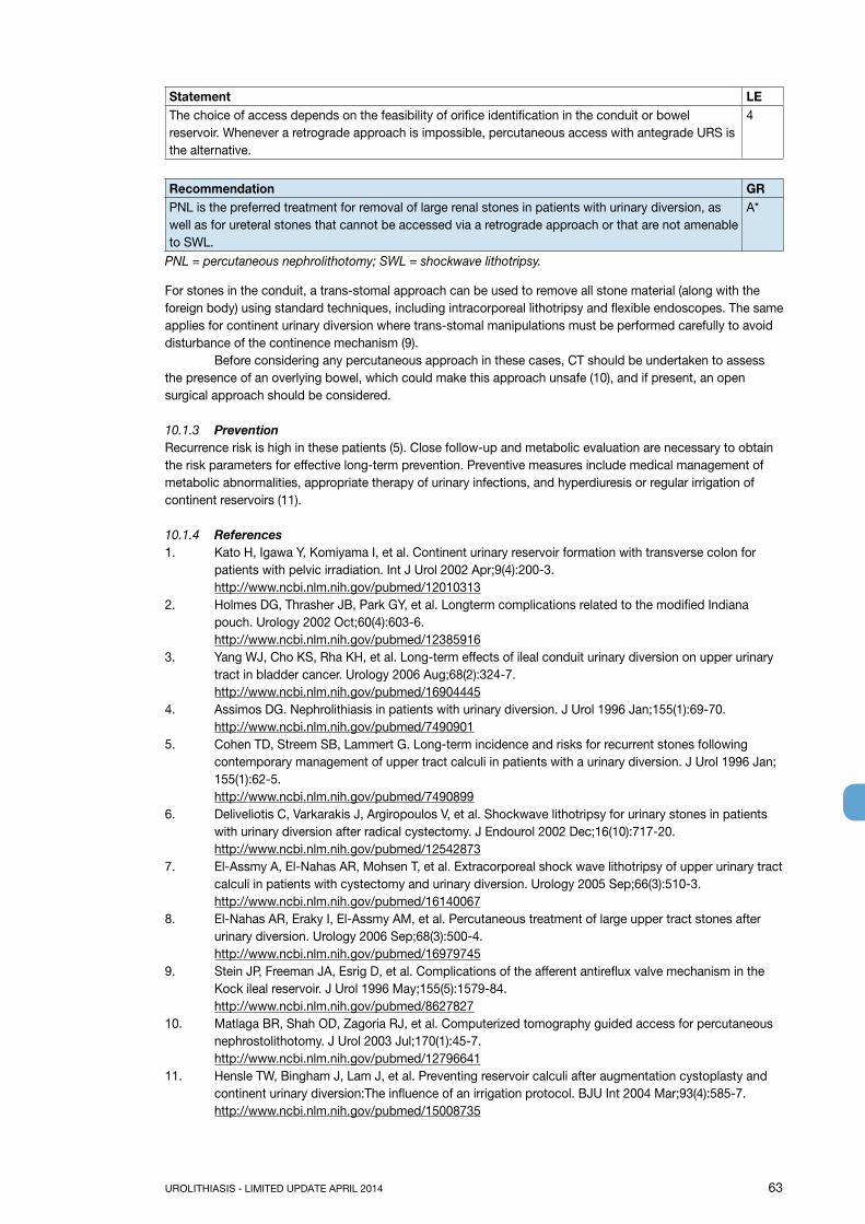
UROLITHIASIS - LIMITED UPDATE APRIL 2014 63
Statement LEThe choice of access depends on the feasibility of orifice identification in the conduit or bowel reservoir. Whenever a retrograde approach is impossible, percutaneous access with antegrade URS is the alternative.
4
Recommendation GRPNL is the preferred treatment for removal of large renal stones in patients with urinary diversion, as well as for ureteral stones that cannot be accessed via a retrograde approach or that are not amenable to SWL.
A*
PNL = percutaneous nephrolithotomy; SWL = shockwave lithotripsy.
For stones in the conduit, a trans-stomal approach can be used to remove all stone material (along with the foreign body) using standard techniques, including intracorporeal lithotripsy and flexible endoscopes. The same applies for continent urinary diversion where trans-stomal manipulations must be performed carefully to avoid disturbance of the continence mechanism (9). Before considering any percutaneous approach in these cases, CT should be undertaken to assess the presence of an overlying bowel, which could make this approach unsafe (10), and if present, an open surgical approach should be considered.
10.1.3 PreventionRecurrence risk is high in these patients (5). Close follow-up and metabolic evaluation are necessary to obtain the risk parameters for effective long-term prevention. Preventive measures include medical management of metabolic abnormalities, appropriate therapy of urinary infections, and hyperdiuresis or regular irrigation of continent reservoirs (11).
10.1.4 References1. Kato H, Igawa Y, Komiyama I, et al. Continent urinary reservoir formation with transverse colon for
patients with pelvic irradiation. Int J Urol 2002 Apr;9(4):200-3.http://www.ncbi.nlm.nih.gov/pubmed/12010313
2. Holmes DG, Thrasher JB, Park GY, et al. Longterm complications related to the modified Indiana pouch. Urology 2002 Oct;60(4):603-6.http://www.ncbi.nlm.nih.gov/pubmed/12385916
3. Yang WJ, Cho KS, Rha KH, et al. Long-term effects of ileal conduit urinary diversion on upper urinary tract in bladder cancer. Urology 2006 Aug;68(2):324-7.http://www.ncbi.nlm.nih.gov/pubmed/16904445
4. Assimos DG. Nephrolithiasis in patients with urinary diversion. J Urol 1996 Jan;155(1):69-70.http://www.ncbi.nlm.nih.gov/pubmed/7490901
5. Cohen TD, Streem SB, Lammert G. Long-term incidence and risks for recurrent stones following contemporary management of upper tract calculi in patients with a urinary diversion. J Urol 1996 Jan; 155(1):62-5.http://www.ncbi.nlm.nih.gov/pubmed/7490899
6. Deliveliotis C, Varkarakis J, Argiropoulos V, et al. Shockwave lithotripsy for urinary stones in patients with urinary diversion after radical cystectomy. J Endourol 2002 Dec;16(10):717-20.http://www.ncbi.nlm.nih.gov/pubmed/12542873
7. El-Assmy A, El-Nahas AR, Mohsen T, et al. Extracorporeal shock wave lithotripsy of upper urinary tract calculi in patients with cystectomy and urinary diversion. Urology 2005 Sep;66(3):510-3.http://www.ncbi.nlm.nih.gov/pubmed/16140067
8. El-Nahas AR, Eraky I, El-Assmy AM, et al. Percutaneous treatment of large upper tract stones after urinary diversion. Urology 2006 Sep;68(3):500-4.http://www.ncbi.nlm.nih.gov/pubmed/16979745
9. Stein JP, Freeman JA, Esrig D, et al. Complications of the afferent antireflux valve mechanism in the Kock ileal reservoir. J Urol 1996 May;155(5):1579-84.http://www.ncbi.nlm.nih.gov/pubmed/8627827
10. Matlaga BR, Shah OD, Zagoria RJ, et al. Computerized tomography guided access for percutaneous nephrostolithotomy. J Urol 2003 Jul;170(1):45-7.http://www.ncbi.nlm.nih.gov/pubmed/12796641
11. Hensle TW, Bingham J, Lam J, et al. Preventing reservoir calculi after augmentation cystoplasty and continent urinary diversion:The influence of an irrigation protocol. BJU Int 2004 Mar;93(4):585-7.http://www.ncbi.nlm.nih.gov/pubmed/15008735

64 UROLITHIASIS - LIMITED UPDATE APRIL 2014
10.2 Management of stones in patients with neurogenic bladder10.2.1 Aetiology, clinical presentation and diagnosisPatients with neurogenic bladder develop urinary calculi because of additional risk factors such as bacteriuria, pelvicalicectasis, vesicoureteral reflux, renal scarring, lower urinary tract reconstruction, and thoracic spinal defect (1). The main issues are urinary stasis and infection (Chapter 2.6). Indwelling catheters and surgical interposition of bowel segments for treatment of bladder dysfunction both facilitate UTI. Although calculi can form at any level of the urinary tract, they occur more frequently in the bladder; especially if bladder augmentation has been performed (2,3).
Diagnosis of stones may be difficult and late in the absence of clinical symptoms due to sensory impairment and vesicourethral dysfunction (4). Difficulties in self-catheterisation should lead to suspicion of bladder calculi. Imaging studies are needed (US, CT) to confirm clinical diagnosis prior to surgical intervention.
10.2.2 ManagementManagement of calculi in patients with neurogenic bladder is similar to that described in Section 10.1. In MMC (myelomeningocele-) patients, latex allergy is common, therefore, appropriate measures need to be taken regardless of the treatment (5). Any surgery in these patients must be performed under general anaesthesia because of the impossibility of using spinal anaesthesia. Bone deformities often complicate positioning on the operating table. The risk of stone formation after augmentation cystoplasty in immobile patients with sensory impairment can be significantly reduced by irrigation protocols (6). For efficient long-term stone prevention in patients with neurogenic bladder, correction of the metabolic disorder, appropriate infection control, and restoration of normal storing/voiding function of the bladder are needed.
Statement LEPatients undergoing urinary diversion and/or suffering from neurogenic bladder dysfunction are at risk for recurrent stone formation.
3
Recommendation GRIn myelomeningocele patients, latex allergy is common so that appropriate measures need to be taken regardless of the treatment.
B
10.2.3 References1. Raj GV, Bennett RT, Preminger GM, et al. The incidence of nephrolithiasis in patients with spinal neural
tube defects. J Urol 1999 Sep;162(3 Pt 2):1238-42.http://www.ncbi.nlm.nih.gov/pubmed/10458475
2. Gros DA, Thakkar RN, Lakshmanam Y, et al. Urolithiasis in spina bifida. Eur J Pediatr Surg 1998 Dec;8 Suppl 1:68-9.http://www.ncbi.nlm.nih.gov/pubmed/9926338
3. Kondo A, Gotoh M, Isobe Y, et al. Urolithiasis in those patients with myelodysplasia. Nihon Hinyokika Gakkai Zasshi 2003 Jan;94(1):15-9.http://www.ncbi.nlm.nih.gov/pubmed/12638200
4. Gacci M, Cai T, Travaglini F, et al. Giant stone in enterocystoplasty. Urol Int 2005;75(2):181-3.http://www.ncbi.nlm.nih.gov/pubmed/16123575
5. Rendeli C, Nucera E, Ausili E, et al. Latex sensitisation and allergy in children with myelomeningocele. Child’s Nerv Syst 2006 Jan;22(1):28-32.http://www.ncbi.nlm.nih.gov/pubmed/15703967
6. Hensle TW, Bingham J, Lam J, et al. Preventing reservoir calculi after augmentation cystoplasty and continent urinary diversion: the influence of an irrigation protocol. BJU Int 2004 Mar;93(4):585-7.http://www.ncbi.nlm.nih.gov/pubmed/15008735
10.3 Management of stones in transplanted kidneys10.3.1 Aetiology and clinical presentationTransplant patients depend on their solitary kidney for renal function. Impairment causing urinary stasis/obstruction therefore requires immediate intervention or drainage of the transplanted kidney. Risk factors in these patients are multifold:• Immunosuppression increases the infection risk, resulting in recurrent UTIs.• Hyperfiltration, excessively alkaline urine, renal tubular acidosis, and increased serum calcium caused

UROLITHIASIS - LIMITED UPDATE APRIL 2014 65
by persistent tertiary hyperparathyroidism (1) are biochemical risk factors.Stones in kidney allografts have a incidence of 0.2-1.7% (2-4).
Recommendation LE GRIn patients with transplanted kidneys, unexplained fever, or unexplained failure to thrive, US or NCCT should be performed to rule out calculi (5).
4 B
US = ultrasound; NCCT = non-contrast enhanced computed tomograpy.
10.3.2 ManagementTreatment decisions for selecting the appropriate technique for stone removal from a transplanted kidney are difficult. Although management principles are similar to those applied in other single renal units (6-9), additional factors such as transplant function, coagulative status, and anatomical obstacles due to the iliacal position of the organ, directly influence the surgical strategy. For large or ureteral stones, careful percutaneous access and subsequent antegrade endoscopy are more favourable. The introduction of small flexible ureteroscopes and holmium laser has made ureteroscopy a valid treatment option for transplant calculi. However, one must be aware of potential injury to adjacent organs (12-14). Retrograde access to transplanted kidneys is difficult due to the anterior location of the ureteral anastomosis, and ureteral tortuosity (15-17).
Statements LEConservative treatment for small asymptomatic stones is only possible under close surveillance and in absolutely compliant patientsSWL for small calyceal stones is an option with minimal complication risk, but localisation of the stone can be challenging and SFRs are poor (10,11).
4
Recommendations GRIn patients with transplanted kidneys, all contemporary treatment modalities, including shockwave therapy, (flexible) ureteroscopy, and percutaneous nephrolithotomy are management options.
B
Metabolic evaluation should be completed after stone removal. A**Upgraded following panel consensus.
10.3.3 References1. Harper JM, Samuell CT, Hallison PC, et al. Risk factors for calculus formation in patients with renal
transplants. Br J Urol 1994 Aug;74(2):147-50.http://www.ncbi.nlm.nih.gov/pubmed/7921929
2. Cho DK, Zackson DA, Cheigh J, et al. Urinary calculi in renal transplant recipients. Transplantation 1988 May;45(5):889-902.http://www.ncbi.nlm.nih.gov/pubmed/3285534
3. Hayes JM, Streem SB, Graneto D, et al. Renal transplant calculi: a re-evaluation of risk and management. Transplantation 1989 Jun;47(6):949-52.http://www.ncbi.nlm.nih.gov/pubmed/2660356
4. Shoskes DA, Hanbury D, Cranston D, et al. Urological complications in 1000 consecutive renal transplant recipients. J Urol 1995 Jan;153(1):18-21.http://www.ncbi.nlm.nih.gov/pubmed/7966766
5. Klingler HC, Kramer G, Lodde M, et al. Urolithiasis in allograft kidneys. Urology 2002 Mar;59(3):344-8.http://www.ncbi.nlm.nih.gov/pubmed/11880067
6. Trivedi A, Patel S, Devra A, et al. Management of Calculi in A Donor Kidney. Transplant Proc 2007 Apr;39(3):761-2.http://www.ncbi.nlm.nih.gov/pubmed/17445593
7. Yigit B, Aydın C, Titiz I, et al. Stone disease in kidney transplantation. Transplant Proc 2004 Jan-Feb; 36(1):187-9.http://www.ncbi.nlm.nih.gov/pubmed/15013342
8. Gupta M, Lee MW. Treatment of stones associated with complex or anomalous renal anatomy. Urol Clin North Am 2007 Aug;34(3):431-41.http://www.ncbi.nlm.nih.gov/pubmed/17678992
9. Challacombe B, Dasgupta P, Tiptaft R, et al. Multimodal management of urolithiasis in renal transplantation. BJU Int 2005 Aug;96(3):385-9.http://www.ncbi.nlm.nih.gov/pubmed/16042735

66 UROLITHIASIS - LIMITED UPDATE APRIL 2014
10. Rhoderik TM, Yang HC, Escobar FS, et al. Extracorporeal shock wave lithotripsy in the renal transplant patient: a case report and review of the literature. Clin Transplant 1992 Oct;6(5):375-8.http://www.ncbi.nlm.nih.gov/pubmed/10147926
11. Atala A, Steinbeck GS, Harty JI, et al. Extracorporeal shock-wave lithotripsy in transplanted kidney. Urology 1993 Jan;41(1):60-2.http://www.ncbi.nlm.nih.gov/pubmed/8420082
12. Rifaioglu MM, Berger AD, Pengune W, et al. Percutaneous management of stones in transplanted kidney. Urology 2008 Sep;72(3):508-12.http://www.ncbi.nlm.nih.gov/pubmed/18653217
13. Minon Cifuentes J, Garcia Tapia E, Garcia de la Pena E, et al. Percutaneous nephrolithotomy in transplanted kidney. Urology 1991 Sep;38(3):232-4.http://www.ncbi.nlm.nih.gov/pubmed/1887537
14. Wyatt J, Kolettis PN, Burns JR. Treatment outcomes for percutaneous nephrolithotomy in renal allografts. J Endourol 2009 Nov;23(11):1821-4.http://www.ncbi.nlm.nih.gov/pubmed/19814697
15. Del Pizzo JJ, Jacobs SC, Sklar GN. Ureteroscopic evaluation in renal transplant recipients. J Endourol 1998 Apr;12(2):135-8.http://www.ncbi.nlm.nih.gov/pubmed/9607439
16. Basiri A, Nikoobakht MR, Simforoosh N, et al. Ureteroscopic management of urological complications after renal transplantation. Scand J Urol Nephrol 2006;40(1):53-6.http://www.ncbi.nlm.nih.gov/pubmed/16452057
17. Lu HF, Shekarriz B, Stoller ML. Donor-gifted allograft urolithiasis: Early percutaneous management. Urology 2002 Jan;59(1):25-7.http://www.ncbi.nlm.nih.gov/pubmed/11796274
10.4 Special problems in stone removal
Table 10.1: Special problems in stone removal
Caliceal diverticulum stones • SWL, PNL (if possible) or RIRS.• Can also be removed using laparoscopic retroperitoneal
surgery (1-5)• Patients may become asymptomatic due to stone
disintegration (SWL) whilst well-disintegrated stone material remains in the original position due to narrow caliceal neck
Horseshoe kidneys • Can be treated in line with the options described above (6)• Passage of fragments after SWL might be poor
Stones in pelvic kidneys • SWL, RIRS or laparoscopic surgery• For obese patients, the options are SWL, PNL, RIRS or
open surgeryStones formed in a continent reservoir • Section 10.1
• Each stone problem must be considered and treated individually
Patients with obstruction of the ureteropelvic junction
• When outflow abnormality requires correction, stones can be removed by PNL together with percutaneous endopyelotomy or open/laparoscopic reconstructive surgery
• URS together with endopyelotomy with Ho:YAG.• Incision with an Acucise balloon catheter might be
considered.Provided the stones can be prevented from falling into the pelviureteral incision (7-10)
10.5 References1. Raboy A, Ferzli GS, Loffreda R, et al. Laparoscopic ureterolithotomy. Urology 1992 Mar;39(3):223-5.
http://www.ncbi.nlm.nih.gov/pubmed/15321022. Gaur DD. Retroperitoneal endoscopic ureterolithotomy: our experience in 12 patients. J Endourol 1993
Dec;7(6):501-3.http://www.ncbi.nlm.nih.gov/pubmed/8124346

UROLITHIASIS - LIMITED UPDATE APRIL 2014 67
3. Gaur DD. Retroperitoneal laparoscopic ureterolithotomy. World J Urol 1993;11(3):175-7.http://www.ncbi.nlm.nih.gov/pubmed/8401638
4. Gaur DD, Agarwal DK, Purohit KC, et al. Retroperitoneal laparoscopic pyelolithotomy. J Urol 1994 Apr;151(4):927-9.http://www.ncbi.nlm.nih.gov/pubmed/8126827
5. Escovar Diaz P, Rey Pacheco M, Lopez Escalante JR, et al. [Laparoscopic urelithotomy.] Arch Esp Urol 1993 Sep;46(7):633-7. [Article in Spanish]http://www.ncbi.nlm.nih.gov/pubmed/8239742
6. Locke DR, Newman RC, Steinbock GS, et al. Extracorporeal shock wave lithotripsy in horseshoe kidney. Urology 1990 May;35(5):407-11.http://www.ncbi.nlm.nih.gov/pubmed/2336770
7. Gelet A, Combe M, Ramackers JM, et al. Endopyelotomy with the Acucise cutting balloon device. Early clinical experience. Eur Urol 1997;31(4):389-93.http://www.ncbi.nlm.nih.gov/pubmed/9187895
8. Faerber GJ, Richardson TD, Farah N, et al. Retrograde treatment of ureteropelvic junction obstruction using the ureteral cutting balloon catheter. J Urol 1997 Feb;157(2):454-8.http://www.ncbi.nlm.nih.gov/pubmed/8996330
9. Berkman DS, Landman J, Gupta M. Treatment outcomes after endopyelotomy performed with or without simultaneous nephrolithotomy: 10-year experience. J Endourol 2009 Sep;23(9):1409-13.http://www.ncbi.nlm.nih.gov/pubmed/19694529
10. Nakada SY, Wolf JS Jr, Brink JA, et al. Retrospective analysis of the effect of crossing vessels on successful retrograde endopyelotomy outcomes using spiral computerized tomography angiography. J Urol 1998 Jan;159(1):62-5.http://www.ncbi.nlm.nih.gov/pubmed/9400437
11. METABOLIC EVALUATION AND RECURRENCE PREVENTION11.1 General metabolic considerations for patient work-up11.1.1 Evaluation of patient riskAfter stone passage, every patient should be assigned to a low- or high-risk group for stone formation (Figure 11.1).
For correct classification, two items are mandatory:• reliable stone analysis by infrared spectroscopy or X-ray diffraction;• basic analysis (Section 3.2).

68 UROLITHIASIS - LIMITED UPDATE APRIL 2014
Figure 11.1: Assignment of patients to low- or high-risk groups for stone formation
Only high-risk stone formers require specific metabolic evaluation. Stone type is the deciding factor for further diagnostic tests. The different stone types include:• calcium oxalate;• calcium phosphate;• uric acid;• ammonium urate;• struvite (and infection stones);• cystine;• xanthine;• 2,8-dihydroxyadenine;• drug stones;• unknown composition.
11.1.2 Urine samplingSpecific metabolic evaluation requires collection of two consecutive 24-h urine samples (1,2). The collecting bottles should be prepared with 5% thymol in isopropanol or stored at < 8°C during collection with the risk of spontaneous crystallisation in the urine (3,4). Preanalytical errors can be minimised by carrying out urinalysis immediately after collection. Alternatively boric acid (10 g powder per urine container) can also be used. The collecting method should be chosen in close cooperation with the particular laboratory. Urine pH should be assessed during collection of freshly voided urine four times daily (3,5) using sensitive pH-dipsticks or a pH-meter.
HCl can be used as a preservative in special situations to prevent precipitation of calcium oxalate and calcium phosphate. However, in samples preserved with HCl, pH measurement is impossible and uric acid precipitates immediately. Alkalinisation is needed to dissolve urate crystals if urate excretion is of interest (6).
Spot urine samples are an alternative method of sampling, particularly when 24-h urine collection is difficult, for example, in non-toilet trained children (7). Spot urine studies normally link the excretion rates to creatinine(7), but these are limited because the results may vary with collection time and patients’ sex, body weight and age.
STONE
Stone analysisunknown
Investigating a patient with unknown composition
(Table 11.12)
Stone analysisknown
Basic evaluation(Table 3.3,
section 3.2.1)
Specific metabolic evaluation
Stone specific recurrence prevention
General preventive measures
Risk factorspresentno yes
Low-riskstone former
High-riskstone former

UROLITHIASIS - LIMITED UPDATE APRIL 2014 69
11.1.3 Timing of specific metabolic work-upFor the initial specific metabolic work-up, the patient should stay on a self-determined diet under normal daily conditions and should ideally be stone free. A minimum of 20 days is recommended (3 months suggested) between stone expulsion or removal and 24-h urine collection (8).
Follow-up studies are necessary in patients receiving recurrent stone prophylaxis (9). The first follow-up 24-h urine measurement should be at 8-12 weeks after starting pharmacological prevention of stone recurrence. This enables drug dosage to be adjusted if urinary risk factors have not normalised, with further 24-h urine measurements if necessary. Once urinary parameters have been normalised, it is sufficient to perform 24-h urine evaluation every 12 months.The panel realise that on this issue there is only very limited published evidence.
11.1.4 Reference ranges of laboratory valuesTables 11.1 - 11.4 provide the internationally accepted reference ranges for the different laboratory values in serum and urine.
Table 11.1: Normal laboratory values for blood parameters in adults (5)
Blood parameter Reference rangeCreatinine 20-100 μmol/LSodium 135-145 mmol/LPotassium 3.5-5.5 mmol/LCalcium 2.0-2.5 mmol/L (total calcium)
1.12-1.32 mmol/L (ionised calcium)Uric acid 119-380 μmol/LChloride 98-112 mmol/LPhosphate 0.81-1.29 mmol/LBlood gas analysis pH 7.35-7.45
pO2 80-90 mmHg
pCO2 35-45 mmHg
HCO3 22-26 mmol/L
BE ± 2 mmol/LBE = base excess (loss of buffer base to neutralise acid).
11.1.5 Risk indices and additional diagnostic toolsSeveral risk indices have been developed to describe the crystallisation risk for calcium oxalate or calciumphosphate in urine:• APCaOxindex (10);• EQUIL, a computer program to calculate relative supersaturations (11);• Bonn Risk Index (12).
Another approach to risk assessment is the Joint Expert Speciation System (JESS), which is based on an extensive database of physiochemical constants and is similar to the EQUIL (13). However, clinical validation of these risk indices for recurrence prediction or therapy improvement is ongoing and the benefit remains controversial.

70 UROLITHIASIS - LIMITED UPDATE APRIL 2014
Table 11.2: Normal laboratory values for urinary parameters in adults
Urinary Parameters Reference ranges and limits for medical attentionpH Constantly > 5.8
Constantly > 7.0Constantly < 5.8
Specific weight > 1.010Creatinine 7-13 mmol/day females
13-18 mmol/day malesCalcium > 5.0 mmol/day
> 8.0 mmol/dayOxalate > 0.5 mmol/day
0.45-0.85 mmol/day> 1.0 mmol/day
Uric acid > 4.0 mmol/day (women), 5 mmol/day (men)Citrate < 2.5 mmol/dayMagnesium < 3.0 mmol/dayInorganic phosphate > 35 mmol/dayAmmonium > 50 mmol/dayCystine > 0.8 mmol/day
Table 11.3: Normal values for spot urine samples: creatinine ratios (solute/creatinine) (14)
Parameter/Patient age Ratio of solute to creatinineCalcium mol/mol mg/mg< 12 months < 2 0.811-3 years < 1.5 0.531-5 years < 1.1 0.395-7 years < 0.8 0.28> 7 years < 0.6 0.21Oxalate mmol/mol mg/g0-6 months < 325-360 288-2607-24 months < 132-174 110-1392-5 years < 98-101 805-14 years < 70-82 60-65> 16 years < 40 32Citrate mol/mol g/g0-5 years > 0.25 0.42> 5 years > 0.15 0.25Magnesium mol/mol g/g
> 0.63 > 0.13Uric acid < 0.56 mg/dl (33 ìmol/l) per GFR (ratio x plasma creatinine)> 2 years
Table 11.4: Urinary excretion of soluble excretion in 24-h urine samples (14)**
Calcium excretion Citrate excretion Cystine excretion Oxalate excretion Urate excretion
All age groups
< 0.1 mmol/kg/ 24 h< 4 mg/kg/24 h
All age groups
Boys> 1.9 mmol/1.73 m²/24 h> 365 mg/1.73 m²/24 h
< 10 y < 55 μmol/1.73 m²/24 h< 13 mg/1.73 m²/24 h
All age groups
< 0.5 mmol/1.73 m²/24 h< 45 mg /1.73 m²/24 h
< 1 y < 70 μmol/kg/ 24 h< 13 mg/kg/ 24 h
Girls> 1.6 mmol/1.73 m²/24 h> 310 mg/1.73 m²/24 h
> 10 y < 200 μmol/1.73 m²/24 h< 48 mg/1.73 m²/24 h
1-5 y < 65 μmol/kg/ 24 h< 11 mg/kg/ 24 h
> 5 y < 55 μmol/kg/ 24 h< 9.3mg/kg/ 24 h
**24h urine parameters are diet and gender dependent and may vary geographically.

UROLITHIASIS - LIMITED UPDATE APRIL 2014 71
11.1.6 References1. Parks JH, Goldfisher E, Asplin JR, et al. A single 24-hour urine collection is inadequate for the medical
evaluation of nephrolithiasis. J Urol 2002 Apr;167(4):1607-12.http://www.ncbi.nlm.nih.gov/pubmed/11912373
2. Nayan M, Elkoushy MA, Andonian S. Variations between two 24-hour urine collections in patients presenting to a tertiary stone clinic. Can Urol Assoc J 2012 Feb;6(1):30-3.http://www.ncbi.nlm.nih.gov/pubmed/22396364
3. Ferraz RR, Baxmann AC, Ferreira LG, et al. Preservation of urine samples for metabolic evaluation of stone-forming patients. Urol Res 2006 Oct;34(5):329-37. http://www.ncbi.nlm.nih.gov/pubmed/16896690
4. Yilmaz G, Yilmaz FM, Hakligor A, et al. Are preservatives necessary in 24-hour urine measurements? Clin Biochem 2008 Jul;41(10-11):899-901.http://www.ncbi.nlm.nih.gov/pubmed/18371307
5. Hesse A, Tiselius HG, Jahnen A. Urinary Stones: Diagnosis, Treatment and Prevention of Recurrence. In: Uric acid stones. Basel: S Karger AG, 2002, pp.73-91.
6. Cameron MA, Sakhaee K. Uric acid nephrolithiasis. Urol Clin North Am 2007 Aug;34(3):335-46.http://www.ncbi.nlm.nih.gov/pubmed/17678984
7. Coe FL, Evan A, Worcester E. Kidney stone disease. J Clin Invest 2005 Oct;115(10):2598-608.http://www.ncbi.nlm.nih.gov/pubmed/16200192
8. Norman RW, Bath SS, Robertson WG, et al. When should patients with symptomatic urinary stone disease be evaluated metabolically? J Urol 1984 Dec;132(6):1137-9.http://www.ncbi.nlm.nih.gov/pubmed/6502804
9. Assimos D. Urine evaluation. In. Assimos D. Chew B, Hatch M, Hautmann R, Holmes R, Williams J, Wolf JS. Evaluation of the stone former. In 2ND International Consultation on Stone Disease 2007, Denstedt J, Khoury S Eds, Health Publications-2008, ISBN 0-9546956-7-4
10. Tiselius HG. Standardized estimate of the ion activity product of calcium oxalate in urine from renal stone formers. Eur Urol 1989;16(1):48-50. http://www.ncbi.nlm.nih.gov/pubmed/2714318
11. Ackermann D, Brown C, Dunthorn M, et al. Use of the computer program EQUIL to estimate pH in model solutions and human urine. Urol Res 1989;17(3):157-61.http://www.ncbi.nlm.nih.gov/pubmed/2749945
12. Kavanagh JP, Laube N. Why does the Bonn Risk Index discriminate between calcium oxalate stone formers and healthy controls? J Urol 2006 Feb;175(2):766-70.http://www.ncbi.nlm.nih.gov/pubmed/16407047
13. Rodgers AL, Allie-Hamdulay S, Jackson GE. 2007 JESS: What can it teach us? In: AP Evan, JE Lingeman and JC Williams, Jr (Eds), IN: Proceedings of Renal Stone Disease 1st Annual International Urolithiasis Research Symposium, 203 November 2006, Indianapolis, Indiana, pp.183-191. Melville, New York: American Institute of Physics. ISBN 878-0-7354-0406-9.
14. Hoppe B, Kemper MJ. Diagnostic examination of the child with urolithiasis or nephrocalcinosis. Pediatr Nephrol 2010 Mar;25(3):403-13.http://www.ncbi.nlm.nih.gov/pubmed/19104842
11.2 General considerations for recurrence preventionAll stone formers, independent of their individual risk, should follow the preventive measures in Table 11.5. The main focus of these is normalisation of dietary habits and lifestyle risks. Stone formers at high risk need specific prophylaxis for recurrence, which is usually pharmacological treatment and based on stone analysis.
Table 11.5: General preventive measures
Fluid intake (drinking advice) Fluid amount: 2.5-3.0 L/dayCircadian drinkingNeutral pH beveragesDiuresis: 2.0-2.5 L/daySpecific weight of urine: < 1010
Nutritional advice for a balanced diet Balanced diet*Rich in vegetable and fibreNormal calcium content: 1-1.2 g/dayLimited NaCl content: 4-5 g/dayLimited animal protein content: 0.8-1.0 g/kg/day

72 UROLITHIASIS - LIMITED UPDATE APRIL 2014
Lifestyle advice to normalise general risk factors BMI: 18-25 kg/m2 (target adult value, not applicable to children)Stress limitation measuresAdequate physical activityBalancing of excessive fluid loss
Caution: The protein need is age-group dependent, therefore protein restriction in childhood should be handled carefully.* Avoid excessive consumption of vitamin supplements.
11.2.1 Fluid intakeAn inverse relationship between high fluid intake and stone formation has been repeatedly demonstrated (1-3). The effect of fruit juices is mainly determined by the presence of citrate or bicarbonate (4). If hydrogen ions are present, the net result is neutralisation. However, if potassium is present, both pH and citrate are increased (5,6). One large fair-quality RCT showed that soft drink consumption significantly reduced the risk for symptomatic recurrences in men with more than one past kidney stone of any type (3,7).
11.2.2 DietA common sense approach to diet should be taken, that is, a mixed balanced diet with contributions from all food groups, but without any excesses (3,8,9).
Fruits, vegetables and fibres: fruit and vegetable intake should be encouraged because of the beneficial effects of fibre, although the role of the later in preventing stone recurrences is debatable (10-12). The alkaline content of a vegetarian diet also increases urinary pH.
Oxalate: excessive intake of oxalate-rich products should be limited or avoided to prevent high oxalate load (4), particularly in patients who have high oxalate excretion.
Vitamin C: although vitamin C is a precursor of oxalate, its role as a risk factor in calcium oxalate stone formation remains controversial (13). However, it seems wise to advise calcium oxalate stone formers to avoid excessive intake.
Animal protein: should not be taken in excess (14,15) and limited to 0.8-1.0 g/kg body weight. Excessive consumption of animal protein has several effects that favour stone formation, including hypocitraturia, low urine pH, hyperoxaluria and hyperuricosuria.
Calcium intake: should not be restricted unless there are strong reasons because of the inverse relationship between dietary calcium and stone formation (11,16). The daily requirement for calcium is 1000 to 1200 mg (17). Calcium supplements are not recommended except in enteric hyperoxaluria, when additional calcium should be taken with meals to bind intestinal oxalate (3,15,18).
Sodium: the daily sodium (NaCl) intake should not exceed 3-5 g (17). High intake adversely affects urine composition:• calcium excretion is increased by reduced tubular reabsorption;• urinary citrate is reduced due to loss of bicarbonate;• increased risk of sodium urate crystal formation.
Calcium stone formation can be reduced by restricting sodium and animal protein (14,15). A positive correlation between sodium consumption and risk of first-time stone formation has been confirmed only in women (16,19). There have been no prospective clinical trials on the role of sodium restriction as an independent variable in reducing the risk of stone formation.
Urate: intake of urate-rich food should be restricted in patients with hyperuricosuric calcium oxalate (20,21) anduric acid stones. Intake should not exceed 500 mg/day (17).
11.2.3 LifestyleLifestyle factors may influence the risk of stone formation, for example, obesity (22) and arterial hypertension (23,24).

UROLITHIASIS - LIMITED UPDATE APRIL 2014 73
11.2.4 Recommendations for recurrence prevention
Recommendations LE GRThe aim should be to obtain a 24-h urine volume > 2.5 L. 1b AHyperoxaluria Oxalate restriction 2b BHigh sodium excretion Restricted intake of salt 1b ASmall urine volume Increased fluid intake 1b AUrea level indicating a high intake of animal protein
Avoid excessive intake of animal protein 1b A
11.2.5 References1. Borghi L, Meschi T, Amato F, et al. Urinary volume, water and recurrences in idiopathic calcium
nephrolithiasis: a 5-year randomized prospective study. J Urol 1996 Mar;155(3):839-43.http://www.ncbi.nlm.nih.gov/pubmed/8583588
2. Sarica K, Inal Y, Erturhan S, et al. The effect of calcium channel blockers on stone regrowth and recurrence after shock wave lithotripsy. Urol Res 2006 Jun;34(3):184-9.http://www.ncbi.nlm.nih.gov/pubmed/16463053
3. Fink HA, Wilt TW, Eidman KE, et al. Medical Management to prevent recurrent nephrolithiasis in adults: a systematic review fora n American College of Physicians clinical guideline. Ann Intern Med 2013 Apr;158(7):535-43.http://www.ncbi.nlm.nih.gov/pubmed/23546565
4. Siener R, Ebert D, Nicolay C, et al. Dietary risk factors for hyperoxaluria in calcium oxalate stone formers. Kidney Int 2003 Mar;63(3):1037-43.http://www.ncbi.nlm.nih.gov/pubmed/12631085
5. Wabner CL, Pak CY. Effect of orange juice consumption on urinary stone risk factors. J Urol 1993 Jun;149(6):1405-8.http://www.ncbi.nlm.nih.gov/pubmed/8501777
6. Gettman MT, Ogan K, Brinkley LJ, et al. Effect of cranberry juice consumption on urinary stone risk factors. J Urol 2005 Aug;174(2):590-4.http://www.ncbi.nlm.nih.gov/pubmed/16006907
7. Shuster J, Jenkins A, Logan C, et al. Soft drink consumption and urinary stone recurrence: a randomized prevention trial. J Clin Epidemiol 1992 Aug;45:911-6. http://www.ncbi.nlm.nih.gov/pubmed/1624973
8. Kocvara R, Plasgura P, Petrik A, et al. A prospective study of nonmedical prophylaxis after a first kidney stone. BJU Int 1999 Sep;84:393-8.http://www.ncbi.nlm.nih.gov/pubmed/10468751
9. Hess B, Mauron H, Ackermann D, et al. Effects of a ‘common sense diet’ on urinary composition and supersaturation in patients with idiopathic calcium urolithiasis. Eur Urol 1999 Aug;36(2):136-43.http://www.ncbi.nlm.nih.gov/pubmed/10420035
10. Ebisuno S, Morimoto S, Yasukawa S, et al. Results of long-term rice bran treatment on stone recurrence in hypercalciuric patients. Br J Urol 1991 Mar;67(3):237-40.http://www.ncbi.nlm.nih.gov/pubmed/1902388
11. Hiatt RA, Ettinger B, Caan B, et al. Randomized controlled trial of a low animal protein, high fiber diet in the pre- vention of recurrent calcium oxalate kidney stones. Am J Epidemiol 1996 Jul;144: 25-33. http://www.ncbi.nlm.nih.gov/pubmed/8659482
12. Dussol B, Iovanna C, Rotily M, et al. A randomized trial of low-animal-protein or high-fiber diets for secondary prevention of calcium nephrolithiasis. Nephron Clin Pract 2008;110:c185-94. http://www.ncbi.nlm.nih.gov/pubmed/18957869
13. Auer BL, Auer D, Rodger AL. The effects of ascorbic acid ingestion on the biochemical and physicochemical risk factors associated with calcium oxalate kidney stone formation. Clin Chem Lab Med 1998 Mar;36(3):143-7.http://www.ncbi.nlm.nih.gov/pubmed/9589801
14. Borghi L, Schianchi T, Meschi T, et al. Comparison of two diets for the prevention of recurrent stones in idiopathic hypercalciuria. N Engl J Med 2002 Jan;346(2):77-84.http://www.ncbi.nlm.nih.gov/pubmed/11784873
15. Fink HA, Akornor JW, Garimella PS, et al. Diet, fluid, or supplements for secondary prevention of nephrolithiasis: a systematic review and meta-analysis of randomized trials. Eur Urol 2009 Jul; 56(1):72-80.http://www.ncbi.nlm.nih.gov/pubmed/19321253

74 UROLITHIASIS - LIMITED UPDATE APRIL 2014
16. Curhan GC, Willett WC, Speizer FE, et al. Comparison of dietary calcium with supplemental calcium and other nutrients as factors affecting the risk for kidney stones in women. Ann Intern Med 1997 Apr;126(7):497-504.http://www.ncbi.nlm.nih.gov/pubmed/9092314
17. Hesse AT, Tiselius H-G. Siener R, Hoppe B. (Eds). Urinary Stones, Diagnosis, Treatment and Prevention of Recurrence. 3rd edn. Basel, S. Karger AG, 2009. ISBN 978-3-8055-9149-2.
18. von Unruh GE, Voss S, Sauerbruch T, et al. Dependence of oxalate absorption on the daily calcium intake. J Am Soc Nephrol 2004 Jun;15(6):1567-73.http://www.ncbi.nlm.nih.gov/pubmed/15153567
19. Curhan GC, Willett WC, Rimm EB, et al. A prospective study of dietary calcium and other nutrients and the risk of symptomatic kidney stones. N Engl J Med 1993 Mar;328(12):833-8.http://www.ncbi.nlm.nih.gov/pubmed/8441427
20. Coe FL. Hyperuricosuric calcium oxalate nephrolithiasis. Adv Exp Med Biol 1980;128:439-50.http://www.ncbi.nlm.nih.gov/pubmed/7424690
21. Ettinger B. Hyperuricosuric calcium stone disease. In: Coe FL, Favus MJ, Pak CYC, Parks JH, Preminger GM, eds. Kidney Stones: Medical and Surgical Management. Lippincott-Raven: Philadelphia, 1996, pp. 851-858.
22. Siener R, Glatz S, Nicolay C, et al. The role of overweight and obesity in calcium oxalate stone formation. Obes Res 2004 Jan;12(1):106-113.http://www.ncbi.nlm.nih.gov/pubmed/14742848
23. Madore F, Stampfer MJ, Rimm EB, et al. Nephrolithiasis and risk of hypertension. Am J Hypertens 1998 Jan;11(1 Pt 1):46-53.http://www.ncbi.nlm.nih.gov/pubmed/9504449
24. Madore F, Stampfer MJ, Willett WC, et al. Nephrolithiasis and risk of hypertension in women. Am J Kidney Dis 1998 Nov;32(5):802-7.http://www.ncbi.nlm.nih.gov/pubmed/9820450
11.3 Stone-specific metabolic evaluation and pharmacological recurrence prevention11.3.1 IntroductionPharmacological treatment is necessary in patients at high-risk for recurrent stone formation. The ideal drug should halt stone formation, have no side effects, and be easy to administer. Each of these aspects is important to achieve good compliance. Table 11.6 highlights the most important characteristics of commonly used medication.
Table 11.6: Pharmacological substances used for stone prevention - characteristics, specifics and dosage
Agent Rationale Dose Specifics and side effects
Stone type Ref
Alkaline citrates Alkalinisation
Hypocitraturia
Inhibition of calcium oxalate crystallisation
5-12 g/d (14-36 mmol/d)Children: 0.1-0.15 g/kg/d
Daily dose for alkalinisation depends on urine pH
Calcium oxalateUric acid Cystine
1-9
Allopurinol Hyperuricosuria
Hyperuricaemia
100-300 mg/d
Children:1-3 mg/kg/d
100 mg in isolated hyperuricosuriaRenal insufficiency demands dose correction
Calcium oxalateUric acidAmmonium urate2,8-Dihydroxyadenine
10-15
Calcium Enteric hyperoxaluria
500 mg/d Intake 30 min before the meals
Calcium oxalate 16-18
Captopril CystinuriaActive decrease of urinary cystine levels
75-150 mg Second-line option due to significant side effects
Cystine 19,20
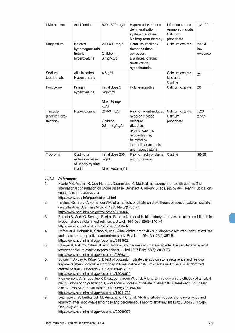
UROLITHIASIS - LIMITED UPDATE APRIL 2014 75
l-Methionine Acidification 600-1500 mg/d Hypercalciuria, bone demineralization, systemic acidosis.No long-term therapy.
Infection stonesAmmonium urateCalcium phosphate
1,21,22
Magnesium Isolated hypomagnesiuriaEnteric hyperoxaluria
200-400 mg/d
Children: 6 mg/kg/d
Renal insufficiency demands dose correction.Diarrhoea, chronic alkali losses, hypocitraturia.
Calcium oxalate 23-24low evidence
Sodium bicarbonate
AlkalinisationHypocitraturia
4.5 g/d Calcium oxalateUric acidCystine
25
Pyridoxine Primary hyperoxaluria
Initial dose 5 mg/kg/d
Max. 20 mg/kg/d
Polyneuropathia Calcium oxalate 26
Thiazide(Hydrochloro-thiazide)
Hypercalciuria 25-50 mg/d
Children: 0.5-1 mg/kg/d
Risk for agent-induced hypotonic blood pressure,diabetes, hyperuricaemia,hypokalaemia, followed by intracellular acidosis and hypocitraturia
Calcium oxalateCalcium phosphate
1,23, 27-35
Tiopronin CystinuriaActive decrease of urinary cystine levels
Initial dose 250 mg/d
Max. 2000 mg/d
Risk for tachyphylaxis and proteinuria.
Cystine 36-39
11.3.2 References1. Pearle MS, Asplin JR, Coe FL, et al. (Committee 3). Medical management of urolithiasis. In: 2nd
International consultation on Stone Disease, Denstedt J, Khoury S. eds. pp. 57-84. Health Publications 2008, ISBN 0-9546956-7-4.http://www.icud.info/publications.html
2. Tiselius HG, Berg C, Fornander AM, et al. Effects of citrate on the different phases of calcium oxalate crystallisation. Scanning Microsc 1993 Mar;7(1):381-9.http://www.ncbi.nlm.nih.gov/pubmed/8316807
3. Barcelo B, Wuhl O, Servitge E, et al. Randomized double-blind study of potassium citrate in idiopathic hypocitraturic calcium nephrolithiasis. J Urol 1993 Dec;150(6):1761-4.http://www.ncbi.nlm.nih.gov/pubmed/8230497
4. Hofbauer J, Hobarth K, Szabo N, et al. Alkali citrate prophylaxis in idiopathic recurrent calcium oxalate urolithiasis--a prospective randomized study. Br J Urol 1994 Apr;73(4):362-5.http://www.ncbi.nlm.nih.gov/pubmed/8199822
5. Ettinger B, Pak CY, Citron JT, et al. Potassium-magnesium citrate is an effective prophylaxis against recurrent calcium oxalate nephrolithiasis. J Urol 1997 Dec;158(6): 2069-73.http://www.ncbi.nlm.nih.gov/pubmed/9366314
6. Soygür T, Akbay A, Küpeli S. Effect of potassium citrate therapy on stone recurrence and residual fragments after shockwave lithotripsy in lower caliceal calcium oxalate urolithiasis: a randomized controlled trial. J Endourol 2002 Apr;16(3):149-52. http://www.ncbi.nlm.nih.gov/pubmed/12028622
7. Premgamone A, Sriboonlue P, Disatapornjaroen W, et al. A long-term study on the efficacy of a herbal plant, Orthosiphon grandiflorus, and sodium potassium citrate in renal calculi treatment. Southeast Asian J Trop Med Public Health 2001 Sep;32(3):654-60. http://www.ncbi.nlm.nih.gov/pubmed/11944733
8. Lojanapiwat B, Tanthanuch M, Pripathanont C, et al. Alkaline citrate reduces stone recurrence and regrowth after shockwave lithotripsy and percutaneous nephrolithotomy. Int Braz J Urol 2011 Sep-Oct;37(5):611-6.http://www.ncbi.nlm.nih.gov/pubmed/22099273

76 UROLITHIASIS - LIMITED UPDATE APRIL 2014
9. Fink HA, Wilt TW, Eidman KE, et al. Medical Management to prevent recurrent nephrolithiasis in adults: a systematic review fora n American College of Physicians clinical guideline. Ann Intern Med 2013 Apr;158(7):535-43.http://www.ncbi.nlm.nih.gov/pubmed/23546565
10. Favus MJ, Coe FL. The effects of allopurinol treatment on stone formation in hyperuricosuric calcium oxalate stone-formers. Scand J Urol Nephrol Suppl 1980;53:265-71.http://www.ncbi.nlm.nih.gov/pubmed/6938003
11. Miano L, Petta S, Galatioto GP, et al. A placebo controlled double-blind study of allopurinol in severe recurrent idiopathic renal lithiasis. In: Schwille PO, Smith LH, Robertson WG, Vahlensieck W, eds. Urolithiasis and Related Clinical Research. New York: Plenum Press, 1985, pp. 521-524.
12. Ettinger B, Tang A, Citron JT, et al. Randomized trial of allopurinol in the prevention of calcium oxalate calculi. N Engl J Med 1986 Nov;315(22):1386-9.http://www.ncbi.nlm.nih.gov/pubmed/3534570
13. Robertson WG, Peacock M, Sepby PL, et al. A multicentre trial to evaluate three treatments for recurrent idiopathic calcium stone disease—a preliminary report. New York: Plenum; 1985
14. Smith MJ. Placebo versus allopurinol for renal calculi. J Urol 1977 Jun;117(6): 690-2. http://www.ncbi.nlm.nih.gov/pubmed/875139
15. Smith MJ. Placebo versus allopurinol for recurrent urinary calculi. Proc Eur Dial Transplant Assoc 1983;20:422-6. [No abstract available]http://www.ncbi.nlm.nih.gov/pubmed/6361753
16. Curhan GC, Willett WC, Speizer FE, et al. Comparison of dietary calcium with supplemental calcium and other nutrients as factors affecting the risk for kidney stones in women. Ann Intern Med 1997 Apr;126(7):497-504.http://www.ncbi.nlm.nih.gov/pubmed/9092314
17. Fink HA, Akornor JW, Garimella PS, et al. Diet, fluid, or supplements for secondary prevention of nephrolithiasis: a systematic review and meta-analysis of randomized trials. Eur Urol 2009 Jul;56(1):72-80.http://www.ncbi.nlm.nih.gov/pubmed/19321253
18. von Unruh GE, Voss S, Sauerbruch T, et al. Dependence of oxalate absorption on the daily calcium intake. J Am Soc Nephrol 2004 Jun;15(6):1567-73.http://www.ncbi.nlm.nih.gov/pubmed/15153567
19. Cohen TD, Streem SB, Hall P. Clinical effect of captopril on the formation and growth of cystine calculi. J Urol 1995 Jul;154(1):164-6.http://www.ncbi.nlm.nih.gov/pubmed/7776415
20. Coulthard MG, Richardson J, Fleetwood A. The treatment of cystinuria with captopril. Am J Kidney Dis 1995 Apr;25(4):661-2.http://www.ncbi.nlm.nih.gov/pubmed/7702068
21. Jarrar K, Boedeker RH, Weidner W. Struvite stones: long term follow up under metaphylaxis. Ann Urol (Paris) 1996;30(3):112-7.http://www.ncbi.nlm.nih.gov/pubmed/8766146
22. Hesse A, Heimbach D. Causes of phosphate stone formation and the importance of metaphylaxis by urinary acidification: a review. World J Urol 1999 Oct;17(5):308-15.
23. Ettinger B, Citron JT, Livermore B, et al. Chlorthalidone reduces calcium oxalate calculous recurrence but magnesium hydroxide does not. J Urol 1988 Apr;139(4):679-84.http://www.ncbi.nlm.nih.gov/pubmed/3280829
24. Prien EL Sr, Gershoff SF. Magnesium oxide - pyridoxine therapy for recurrent calcium oxalate calculi. J Urol 1974 Oct;112(4):509-12.http://www.ncbi.nlm.nih.gov/pubmed/4414543
25. Pinheiro VB, Baxmann AC, Tisselius HG, et al. The effect of sodium bicarbonate upon urinary citrate excretion in calcium stone formers. Urology 2013;82(1):33-7.http://www.ncbi.nlm.nih.gov/pubmed/23602798
26. Hoppe B, Beck BB, Milliner DS. The primary hyperoxalurias. Kidney Int 2009 Jun;75(12):1264-71.http://www.ncbi.nlm.nih.gov/pubmed/19225556
27. Borghi L, Meshi T, Guerra A, et al. Randomized prospective study of a nonthiazide diuretic, indapamide, in preventing calcium stone recurrences. J Cardiovasc Pharmacol 1993;22 Suppl 6: S78-S86.http://www.ncbi.nlm.nih.gov/pubmed/7508066
28. Brocks P, Dahl C, Wolf H, et al. Do thiazides prevent recurrent idiopathic renal calcium stones? Lancet 1981 Jul;2(8238):124-5.http://www.ncbi.nlm.nih.gov/pubmed/6113485

UROLITHIASIS - LIMITED UPDATE APRIL 2014 77
29. Mortensen JT, Schultz A, Ostergaard AH. Thiazides in the prophylactic treatment of recurrent idiopathic kidney stones. Int Urol Nephrol 1986;18(3):265-9.http://www.ncbi.nlm.nih.gov/pubmed/3533825
30. Laerum S, Larsen S. Thiazide prophylaxis of urolithiasis. A double-blind study in general practice. Acta Med Scand 1984;215(4):383-9.http://www.ncbi.nlm.nih.gov/pubmed/6375276
31. Ohkawa M, Tokunaga S, Nakashima T, et al. Thiazide treatment for calcium urolithiasis in patients with idiopathic hypercalciuria. Br J Urol 1992 Jun;69(6):571-6.http://www.ncbi.nlm.nih.gov/pubmed/1638340
32. Scholz D, Schwille PO, Sigel A. Double-blind study with thiazide in recurrent calcium nephrolithiasis. J Urol 1982 Nov;128(5):903-7.http://www.ncbi.nlm.nih.gov/pubmed/7176047
33. Nicar MJ, Peterson R, Pak CY. Use of potassium citrate as potassium supplement during thiazide therapy of calcium nephrolithiasis. J Urol 1984 Mar;131(3):430-3.http://www.ncbi.nlm.nih.gov/pubmed/6699979
34. Fernández-Rodríguez A, Arrabal-Martíın M, García-Ruiz MJ, et al. [The role of thiazides in the prophylaxis of recurrent calcium lithiasis]. Actas Urol Esp 2006 Mar;30(3):305-9. [Article in Spanish]http://www.ncbi.nlm.nih.gov/pubmed/16749588
35. Ahlstrand C, Sandvall K, Tiselius HG, eds. Prophylactic treatment of cal- cium stone formers with hydrochlorothiazide and magnesium. Edsbruk, Sweden: Akademitryck; 1996.
36. Dolin DJ, Asplin JR, Flagel L, et al. Effect of cystinebinding thiol drugs on urinary cystine capacity in patients with cystinuria. J Endourol 2005 Apr;19(3):429-32.http://www.ncbi.nlm.nih.gov/pubmed/15865542
37. Chow GK, Streem SB. Medical treatment of cystinuria: results of contemporary clinical practice. J Urol 1996 Nov;156(5):1576-8.http://www.ncbi.nlm.nih.gov/pubmed/8863541
38. Pak CY, Fuller C, Sakhaee K, et al. Management of cystine nephrolithiasis with alphamercaptopropionylglycine. J Urol 1986 Nov;136(5):1003-8http://www.ncbi.nlm.nih.gov/pubmed/3534301
39. Tekin A, Tekgul S, Atsu N, et al. Cystine calculi in children: the results of a metabolic evaluation and response to medical therapy. J Urol 2001 Jun;165(6 Pt 2):2328-30.http://www.ncbi.nlm.nih.gov/pubmed/11371943
11.4 Calcium oxalate stonesThe criteria for identification of calcium oxalate stone formers with high recurrence risk are listed in Section 2.6.
11.4.1 DiagnosisBlood analysis requires measurement of creatinine, sodium, potassium, chloride, ionised calcium (or total calcium + albumin), uric acid, and parathyroid hormone (PTH) (and vitamin D) in case of increased calcium levels. Urinalysis requires measurement of urine volume, urine pH profile, specific weight, calcium, oxalate, uric acid, citrate, sodium and magnesium.
11.4.2 Interpretation of results and aetiologyThe diagnostic and therapeutic algorithm for calcium oxalate stones is shown in Figure 11.2 (1-25).

78 UROLITHIASIS - LIMITED UPDATE APRIL 2014
Figure 11.2: Diagnostic and therapeutic algorithm for calcium oxalate stones
Cal
cium
oxa
late
sto
ne
Bas
ic e
valu
atio
n
24 h
uri
ne c
olle
ctio
n
Hyp
erca
lcur
ia
5-8
mm
ol/d
2
Alc
alin
e C
itrat
e9-
12 g
/dor
Sod
ium
B
icar
bona
te1.
5 g
tid2,
4
> 8
mm
ol/d
Hyd
roch
loro
thia
zide
Initi
ally
25
mg/
dU
p to
50
mg/
d
< 2.
5 m
mol
/d
Alc
alin
eC
itrat
e9-
12 g
/d
> 0.
5 m
mol
/d
Cal
cium
> 5
00
mg/
d1
200-
400
mg/
d
> 1
mm
ol/d
Pyr
idox
ine
Initi
al 5
mg/
kg/d
Up
to 2
0 m
g/kg
/d
> 4
mm
ol/d
Alk
alin
e C
itrat
e9-
12 g
/dor
Sod
ium
B
icar
bona
te1.
5 g
tid2
PLU
SA
llopu
rinol
100
mg/
d
Hyp
erur
icos
uria
and
H
yper
uric
emia
> 3
80 µ
mol
Alk
alin
e C
itrat
e9-
12 g
/dP
LUS
Allo
purin
ol10
0-30
0 m
g/d4
< 3
mm
ol/d
Mag
nesi
um20
0-40
0 m
g/d3
Hyp
ocitr
atur
iaH
yper
oxal
uria
Hyp
erur
icos
uria
Hyp
omag
nesu
ria
1 B
e aw
are
of e
xces
s ca
lciu
m e
xcre
tion
2 tid
= th
ree
times
/day
(24h
) 3 N
o m
agne
sium
ther
apy
for p
atie
nts
with
ren
al in
suffi
cien
cy
4 Th
ere
is n
o ev
iden
ce th
at c
ombi
natio
n th
erap
y (th
iazi
de +
citr
ate)
(thi
azid
e +
allo
purin
ol) i
s su
perio
r to
thia
zide
ther
apy
alon
e (3
,4).

UROLITHIASIS - LIMITED UPDATE APRIL 2014 79
The most common metabolic abnormality associated with calcium stone formation are hypercalciuria, which affects 30-60% of adult stone formers, and hyperoxaluria (26-67%), followed by hyperuricosuria (15-46%), hypomagesuria (7-23%), and hypocitraturia (5-29%). However, ranges tend to differ based on ethnicity (1).• Elevated levels of ionised calcium in serum (or total calcium and albumin) require assessment of intact
PTH to confirm or exclude suspected hyperparathyroidism (HPT). • “Acidic arrest” (urine pH constantly < 6) may promote co-crystallisation of uric acid and calcium
oxalate. Similarly, increased uric acid excretion (> 4 mmol/day in adults or > 12 mg/kg/day in children) can act as a promoter.
• Urine pH levels constantly > 5.8 in the day profile indicate renal tubular acidosis (RTA), provided urinary tract infection (UTI) has been excluded. An ammonium chloride loading test confirms RTA and identifies RTA subtype (Section 11.6.5).
• Hypercalciuria may be associated with normocalcemia (idiopathic hypercalciuria, or granulomatous diseases) or hypercalcaemia (hyperparathyroidism, granulomatous diseases, vitamin D excess, or malignancy).
• Hypocitraturia (< 2.5 mmol/day) may be idiopathic or secondary to metabolic acidosis or hypokalaemia.
• Oxalate excretion > 0.5 mmol/day in adults (> 0.37 mmol/1.73 m2/day in children) confirms hyperoxaluria.
• primary hyperoxaluria (oxalate excretion mostly > 1 mmol/day), appears in three genetically determined forms;
• secondary hyperoxaluria (oxalate excretion > 0.5 mmol/day, usually < 1 mmol/day), occurs due to intestinal hyperabsorption of oxalate or extreme dietary oxalate intake;
• mild hyperoxaluria (oxalate excretion 0.45-0.85 mmol/day), commonly found in idiopathic calcium oxalate stone formers.
• Hypomagnesuria (< 3.0 mmol/day) may be related to poor dietary intake or to reduced intestinal absorption (chronic diarrhoea).
11.4.3 Specific treatmentGeneral preventive measures are recommended for fluid intake and diet. Hyperoxaluric stone formers should consume foods with low oxalate content, whereas hyperuricosuric stone formers benefit from daily dietary reduction of purine. Figure 11.2 summarises the diagnostic algorithm and the pharmacological treatment of calcium oxalate stones (1-25). There is only low level evidence on the efficacy of preventing stone recurrence through pre-treatment stone composition and biochemistry measures, or on-treatment biochemistry measures (24).
11.4.4 Recommendations for pharmacological treatment of patients with specific abnormalities in urine composition
Urinary risk factor Suggested treatment LE GRHypercalciuria Thiazide + potassium citrate 1a AHyperoxaluria Oxalate restriction 2b AEnteric hyperoxaluria Potassium citrate 3-4 C
Calcium supplement 2 BDiet reduced in fat and oxalate 3 B
Hypocitraturia Potassium citrate 1b AHypocitraturia Sodium bicarbonate if intolerant to potassium
citrate 1b A
Hyperuricosuria Allopurinol 1a AHigh sodium excretion Restricted intake of salt 1b ASmall urine volume Increased fluid intake 1b AUrea level indicating a high intake of animal protein
Avoid excessive intake of animal protein 1b A
No abnormality identified High fluid intake 2b B
11.4.5 References1. Worcester EM, Coe FL. New insights into the pathogenesis of idiopathic hypercalciuria. Semin Nephrol
2008;28:120-32.http://www.ncbi.nlm.nih.gov/pubmed/18359393
80 UROLITHIASIS - LIMITED UPDATE APRIL 2014
2. Pearle MS, Asplin JR, Coe FL, et al. (Committee 3). Medical management of urolithiasis. In: 2nd International consultation on Stone Disease, Denstedt J, Khoury S. eds. pp. 57-84. Health Publications 2008, ISBN 0-9546956-7-4.http://www.icud.info/publications.html
3. Borghi L, Meshi T, Guerra A, et al. Randomized prospective study of a nonthiazide diuretic, indapamide, in preventing calcium stone recurrences. J Cardiovasc Pharmacol 1993;22 Suppl 6: S78-S86.http://www.ncbi.nlm.nih.gov/pubmed/7508066
4. Fernández-Rodríguez A, Arrabal-Martíın M, García-Ruiz MJ, et al. [The role of thiazides in the prophylaxis of recurrent calcium lithiasis]. Actas Urol Esp 2006 Mar;30(3):305-9. [Article in Spanish]http://www.ncbi.nlm.nih.gov/pubmed/16749588
5. Brocks P, Dahl C, Wolf H, et al. Do thiazides prevent recurrent idiopathic renal calcium stones? Lancet 1981 Jul;2(8238):124-5.http://www.ncbi.nlm.nih.gov/pubmed/6113485
6. Ettinger B, Citron JT, Livermore B, et al. Chlorthalidone reduces calcium oxalate calculous recurrence but magnesium hydroxide does not. J Urol 1988 Apr;139(4):679-84.http://www.ncbi.nlm.nih.gov/pubmed/3280829
7. Mortensen JT, Schultz A, Ostergaard AH. Thiazides in the prophylactic treatment of recurrent idiopathic kidney stones. Int Urol Nephrol 1986;18(3):265-9.http://www.ncbi.nlm.nih.gov/pubmed/3533825
8. Laerum S, Larsen S. Thiazide prophylaxis of urolithiasis. A double-blind study in general practice. Acta Med Scand 1984;215(4):383-9.http://www.ncbi.nlm.nih.gov/pubmed/6375276
9. Ohkawa M, Tokunaga S, Nakashima T, et al. Thiazide treatment for calcium urolithiasis in patients with idiopathichypercalciuria. Br J Urol 1992 Jun;69(6):571-6.http://www.ncbi.nlm.nih.gov/pubmed/1638340
10. Wolf H, Brocks P, Dahl C. Do thiazides prevent recurrent idiopathic renal calcium oxalate stones? Proc Eur Dial Transplan Assoc 1983;20:477-80.http://www.ncbi.nlm.nih.gov/pubmed/6361755
11. Scholz D, Schwille PO, Sigel A. Double-blind study with thiazide in recurrent calcium nephrolithiasis. J Urol 1982 Nov;128(5):903-7.http://www.ncbi.nlm.nih.gov/pubmed/7176047
12. Nicar MJ, Peterson R, Pak CY. Use of potassium citrate as potassium supplement during thiazide therapy of calcium nephrolithiasis. J Urol 1984 Mar;131(3):430-3.http://www.ncbi.nlm.nih.gov/pubmed/6699979
13. Barcelo B, Wuhl O, Servitge E, et al. Randomized double-blind study of potassium citrate in idiopathic hypocitraturic calcium nephrolithiasis. J Urol 1993 Dec;150(6):1761-4.http://www.ncbi.nlm.nih.gov/pubmed/8230497
14. Hofbauer J, Hobarth K, Szabo N, et al. Alkali citrate prophylaxis in idiopathic recurrent calcium oxalate urolithiasis--a prospective randomized study. Br J Urol 1994 Apr;73(4):362-5.http://www.ncbi.nlm.nih.gov/pubmed/8199822
15. Ettinger B, Pak CY, Citron JT, et al. Potassium-magnesium citrate is an effective prophylaxis against recurrent calcium oxalate nephrolithiasis. J Urol 1997 Dec;158(6): 2069-73.http://www.ncbi.nlm.nih.gov/pubmed/9366314
16. Johansson G, Backman U, Danielson BG, et al. Effects of magnesium hydroxide in renal stone disease. J Am Coll Nutr 1982;1(2):179-85.http://www.ncbi.nlm.nih.gov/pubmed/6764473
17. Khan SR, Shevock PN, Hackett RL. Magnesium oxide administration and prevention of calcium oxalate nephrolithiasis. J Urol 1993 Feb;149(2):412-6.http://www.ncbi.nlm.nih.gov/pubmed/8426432
18. Ettinger B, Pak CY, Citron JT, et al. Potassium-magnesium citrate is an effective prophylaxis against recurrent calcium oxalate nephrolithiasis. J Urol 1997 Dec;158(6):2069-73.http://www.ncbi.nlm.nih.gov/pubmed
19. Prien EL Sr, Gershoff SF. Magnesium oxide - pyridoxine therapy for recurrent calcium oxalate calculi. J Urol 1974 Oct;112(4):509-12.http://www.ncbi.nlm.nih.gov/pubmed/4414543
20. Pearle MS, Roehrborn CG, Pak CY. Meta-analysis of randomized trials for medical prevention of calcium oxalate nephrolithiasis. J Endourol 1999 Nov;13(9):679-85.http://www.ncbi.nlm.nih.gov/pubmed/10608521

UROLITHIASIS - LIMITED UPDATE APRIL 2014 81
21. Favus MJ, Coe FL. The effects of allopurinol treatment on stone formation in hyperuricosuric calcium oxalate stone-formers. Scand J Urol Nephrol Suppl 1980;53:265-71.http://www.ncbi.nlm.nih.gov/pubmed/6938003
22. Miano L, Petta S, Galatioto GP, et al. A placebo controlled double-blind study of allopurinol in severe recurrent idiopathic renal lithiasis. In: Schwille PO, Smith LH, Robertson WG, Vahlensieck W, eds. Urolithiasis and Related Clinical Research. New York: Plenum Press, 1985, pp. 521-524.
23. Ettinger B, Tang A, Citron JT, et al. Randomized trial of allopurinol in the prevention of calcium oxalate calculi. N Engl J Med 1986 Nov;315(22):1386-9.http://www.ncbi.nlm.nih.gov/pubmed/3534570
24. Fink HA, Wilt TW, Eidman KE, et al. Medical Management to prevent recurrent nephrolithiasis in adults: a systematic review fora n American College of Physicians clinical guideline. Ann Intern Med 2013 Apr;158(7):535-43.http://www.ncbi.nlm.nih.gov/pubmed/23546565
25. Pinheiro VB, Baxmann AC, Tiselius HG, et al. The effect of sodium bicarbonate upon urinary citrate excretion in calcium stone formers. Urology 2013 Jul;82(1):33-7. http://www.ncbi.nlm.nih.gov/pubmed/23602798
11.5 Calcium phosphate stonesSome calcium phosphate stone formers are at high-risk of recurrence. Further information on identifying high-risk patients is given in Section 2.6. Calcium phosphate mainly appears in two completely different minerals: carbonate apatite and brushite. Carbonate apatite crystallisation occurs at a pH > 6.8 and may be associated with infection. Brushite crystallises occur at an optimum pH of 6.5-6.8, at high urinary concentrations of calcium (> 8 mmol/day) and phosphate (> 35 mmol/day). Its occurrence is not related to UTI. Possible causes of calcium phosphate stones include HPT, RTA and UTI; each of which requires different therapy.
11.5.1 DiagnosisDiagnosis requires blood analysis for: creatinine, sodium, potassium, chloride, ionised calcium (or total calcium + albumin), and PTH (in case of increased calcium levels). Urinalysis includes measurement of: volume, urine pH profile, specific weight, calcium, phosphate and citrate.
11.5.2 Interpretation of results and aetiologyGeneral preventive measures are recommended for fluid intake and diet. The diagnostic and therapeutic algorithm for calcium phosphate stones is shown in Figure 11.3.

82 UROLITHIASIS - LIMITED UPDATE APRIL 2014
Figure 11.3: Diagnostic and therapeutic algorithm for calcium phosphate stones
11.5.3 Pharmacological therapy (1-6)HPT and RTA are common causes of calcium phosphate stone formation. Although most patients with primary HPT require surgery, RTA can be corrected pharmacologically. If primary HPT and RTA have been excluded, pharmacotherapy for calcium phosphate calculi depends on effective reduction of urinary calcium levels using thiazides. If urine pH remains constantly > 6.2, urinary acidification with l-methionine may be helpful however it is not commonly used and needs monitoring for systemic acidosis development. For infection-associated calcium phosphate stones, it is important to consider the guidance given for infection stones.
11.5.4 Recommendations for the treatment of calcium phosphate stones
Urinary risk factor Suggested treatment LE GRHypercalciuria Thiazide 1a AInadequate urine pH Acidification 3-4 CUTI Antibiotics 3-4 C
11.5.5 References1. Pearle MS, Asplin JR, Coe FL, et al. (Committee 3). Medical management of urolithiasis. In: 2nd
International consultation on Stone Disease, Denstedt J, Khoury S. eds. pp. 57-84. Health Publications 2008, ISBN 0-9546956-7-4.http://www.icud.info/publications.html
2. Borghi L, Meshi T, Guerra A, et al. Randomized prospective study of a nonthiazide diuretic, indapamide, in preventing calcium stone recurrences. J Cardiovasc Pharmacol 1993;22 Suppl 6: S78-S86.http://www.ncbi.nlm.nih.gov/pubmed/7508066
3. Brocks P, Dahl C, Wolf H, et al. Do thiazides prevent recurrent idiopathic renal calcium stones? Lancet 1981 Jul;2(8238):124-5.http://www.ncbi.nlm.nih.gov/pubmed/6113485
4. Fink HA, Wilt TW, Eidman KE, et al. Medical Management to prevent recurrent nephrolithiasis in adults: a systematic review fora n American College of Physicians clinical guideline. Ann Intern Med 2013 Apr;158(7):535-43.http://www.ncbi.nlm.nih.gov/pubmed/23546565
Calcium phosphatestones
Brushite stones
Basic evaluation
Hypercalciuria> 8 mmol/d
Hydrochlorothiazideinitially 25 mg/dup to 50 mg/d
Carbonate apatitestones
Basic evaluation
Hypercalciuria> 8 mmol/d
Urinary pH > 5.8Elevated calcium
exclude HPTExclude HPT Exclude RTA
Adjust urinary pHbetween 5.8 and 6.2 with
L-methionine200-500 mg 3 times daily
Hydrochlorothiazideinitially 25 mg/dup to 50 mg/d
Exclude RTA Exclude UTI

UROLITHIASIS - LIMITED UPDATE APRIL 2014 83
5. Pearle MS, Roehrborn CG, Pak CY. Meta-analysis of randomized trials for medical prevention of calcium oxalate nephrolithiasis. J Endourol 1999 Nov;13(9):679-85.http://www.ncbi.nlm.nih.gov/pubmed/10608521
6. Scholz D, Schwille PO, Sigel A. Double-blind study with thiazide in recurrent calcium ephrolithiasis. J Urol 1982 Nov;128(5):903-7.http://www.ncbi.nlm.nih.gov/pubmed/7176047
11.6 Disorders and diseases related to calcium stones11.6.1 Hyperparathyroidism (1-4)Primary HPT is responsible for an estimated 5% of all calcium stone formation. Kidney stones occur in approximately 20% of patients with primary HPT. The clinical appearance of HPT typically comprises bone loss, gastric ulcers and urolithiasis. Elevated levels of PTH significantly increase calcium turnover, leading to hypercalcaemia and hypercalciuria. Serum calcium may be mildly elevated and serum PTH within the upper normal limits, therefore, repeated measurements may be needed; preferably with the patient fasting. Stones of PTH patients may contain both calcium oxalate and calcium phosphate. If HPT is suspected, neck exploration should be performed to confirm the diagnosis. Primary HPT can only be cured by surgery.
11.6.2 Granulomatous diseases (5,6)Granulomatous diseases, such as sarcoidosis, may be complicated by hypercalcaemia and hypercalciuria secondary to increased calcitriol production. The latter is independent of PTH control, leading to increased calcium absorption in the gastrointestinal tract and suppression of PTH. Treatment focusses on the activity of the granulomatous diseases and may require steroids, hydroxychloroquine or ketoconazole. It should be reserved to the specialist.
11.6.3 Primary hyperoxaluria (7)Patients with primary hyperoxaluria (PH) should be referred to specialised centres, because successful management requires an experienced interdisciplinary team. The main therapeutic aim is to reduce endogenous oxalate production, which is increased in patients with PH. In approximately one-third of patients with PH type I, pyridoxine therapy normalises or significantly reduces urinary oxalate excretion. The goal of adequate urine dilution is achieved by adjusting fluid intake to 3.5-4.0 L/day in adults (children 1.5 L/m2 body surface area) and following a circadian drinking regimen. Therapeutic options for preventing calcium oxalate crystallisation include hyperdiuresis, alkaline citrates and magnesium. However, in end-stage renal failure, PH requires simultaneous liver-kidney transplantation.
Treatment regimens are:• Pyridoxine in PH type I: 5-20 mg/kg/day according to urinary oxalate excretion and patient tolerance;• Alkaline citrate: 9-12 g/day in adults, 0.1-0.15 meq/kg/day in children;• Magnesium: 200-400 mg/day (no magnesium in case of renal insufficiency).
Urinary risk factor Suggested treatment LE GRPrimary hyperoxaluria Pyridoxine 3 B
11.6.4 Enteric hyperoxaluria (8-10)Enteric hyperoxaluria is a particularly problematic condition in patients with intestinal malabsorption of fat. This abnormality is associated with a high risk of stone formation, and is seen after intestinal resection and malabsorptive bariatric surgery and in Crohn’s disease and pancreas insufficiency. Intestinal loss of fatty acids is combined with loss of calcium. The normal complex formation between oxalate and calcium is therefore disturbed and oxalate absorption is increased. In addition to hyperoxaluria, these patients usually present with hypocitraturia because of loss of alkali. Urine pH is usually low, as are urinary calcium and urine volume. All these abnormalities contribute to high levels of supersaturation with calcium oxalate, crystalluria, and stone formation.
Specific preventive measures are:• Restricted intake of oxalate-rich foods;• Restricted fat intake;• Calcium supplementation at meal times to enable calcium oxalate complex formation in the intestine
(8,9);• Sufficient fluid intake to balance intestinal loss of water caused by diarrhoea;

84 UROLITHIASIS - LIMITED UPDATE APRIL 2014
• Alkaline citrates to raise urinary pH and citrate.
Urinary risk factor Suggested treatment LE GREnteric hyperoxaluria Potassium citrate 3-4 C
Calcium supplement 2 BDiet reduced in fat and oxalate 3 B
Small urine volume Increased fluid intake 1b A
11.6.5 Renal tubular acidosis (11-13)Renal tubular acidosis is caused by severe impairment of proton or bicarbonate handling along the nephron. Kidney stone formation most probably occurs in patients with distal RTA type I. Figure 11.4 outlines the diagnosis of RTA. Table 11.7 shows acquired and inherited causes of RTA.
Figure 11.4: Diagnosis of renal tubular acidosis
** An alternative Ammonium Chloride loading test using NH4Cl load with 0.05 g/kg body weight over 3 days might provide similar results and may be better tolerated by the patient (13).
Renal tubular acidosis can be acquired or inherited. Reasons for acquired RTA can be obstructive uropathy, recurrent pyelonephritis, acute tubular necrosis, renal transplantation, analgesic nephropathy, sarcoidosis, idiopathic hypercalciuria and primary parathyroidism, and drug-induced (e.g. zonisamide). Table 11.7 shows the inherited causes of RTA.
Table 11.7: Inherited causes of renal tubular acidosis
Type - inheritance Gene/gene product/function PhenotypeAutosomal dominant SLC4A1/AE1/Cl-bicarbonate
exchangerHypercalciuria, hypokalaemia, osteomalacia
Autosomal recessive with hearing loss
ATP6V1B1/B1 subunit of vacuolar H-ATPase/proton secretion
Hypercalciuria, hypokalaemia, rickets
Autosomal recessive ATP6V0A4/A4 subunit of vacuolar H-ATPase/proton secretion
Hypercalciuria, hypokalaemia, rickets
The main therapeutic aim is restoring a normal acid-base equilibrium. Despite the alkaline pH of urine in RTA, alkalinisation using alkaline citrates or sodium bicarbonate is key to normalising the metabolic changes (intracellular acidosis) responsible for stone formation (Table 11.8). The alkali load reduces tubular reabsorption
Urinary pHconstantly > 5.8
Ammonium chloride loading test**(NH4Cl 0.1 g/kg body weight)
except for patents with clinically confirmed metabolic acidosis
RTA Type Ipossible
Urine pH < 5.4RTA excluded!
Urine pH > 5.4RTA
Normal bicarbonate in BGARTA - incomplete
Low bicarbonate in BGARTA - complete

UROLITHIASIS - LIMITED UPDATE APRIL 2014 85
of citrate, which in turn normalises citrate excretion and simultaneously reduces calcium turnover. Therapeutic success can be monitored by venous blood gas analysis (base excess: ± 2.0 mmol/L) in complete RTA. If excessive calcium excretion (> 8 mmol/day) persists after re-establishing acid-base equilibrium, thiazides may lower urinary calcium excretion.
Table 11.8: Pharmacological treatment of renal tubular acidosis
Biochemical risk factor Rationale for pharmacological therapy
Medication
Hypercalciuria Calcium excretion > 8 mmol/day Hydrochlorothiazide, - in adults, 25 mg/day initially, up to 50
mg/day- in children, 0.5-1 mg/kg/day
Inadequate urine pH Intracellular acidosis in nephron Alkaline citrate, 9-12 g/day divided in 3 dosages OR Sodium bicarbonate, 1.5 g, 3 times daily
Urinary risk factor Suggested treatment LE GRDistal RTA Potassium citrate 2b BHypercalciuria Thiazide + potassium citrate 1a A
11.6.6 Nephrocalcinosis (14,15)Nephrocalcinosis (NC) refers to increased crystal deposition within the renal cortex or medulla, and occurs alone or in combination with kidney stones. There are various metabolic causes. The main risk factors are: HPT, PH, RTA, vitamin D metabolic disorders, idiopathic hypercalciuria and hypocitraturia, and genetic disorders, including Dent’s disease Bartter’s syndrome and Medullary sponge kidney. The many causes of NC means there is no single standard therapy. Therapeutic attention must focus on the underlying metabolic or genetic disease, while minimising the biochemical risk factors.
11.6.6.1 DiagnosisDiagnosis requires the following blood analysis: PTH (in case of increased calcium levels), vitamin D and metabolites, vitamin A, sodium, potassium, magnesium, chloride, and blood gas analysis. Urinalysis should investigate: urine pH profile (minimum 4 times daily), daily urine volume, specific weight of urine, and levels of calcium, oxalate, phosphate, uric acid, magnesium and citrate.
11.6.7 References1. Silverberg SJ, Shane E, Jacobs TP, et al. A 10-year prospective study of primary hyperparathyroidism
with or without parathyroid surgery. N Engl J Med 1999 Oct;341(17):1249-55.http://www.ncbi.nlm.nih.gov/pubmed/10528034
2. Evan AP, Lingeman JE, Coe FL, et al. Histopathology and surgical anatomy of patients with primary hyperparathyroidism and calcium phosphate stones. Kidney Int 2008 Jul;74(2):223-9,http://www.ncbi.nlm.nih.gov/pubmed/18449170
3. Mollerup CL, Vestergaard P, Frokjaer VG, et al. Risk of renal stone events in primary hyperparathyroidism before and after parathyroid surgery: controlled retrospective follow up study. BMJ 2002 Oct;325(7368):807.http://www.ncbi.nlm.nih.gov/pubmed/12376441
4. Rao DS, Phillips ER, Divine GW, et al. Randomized controlled clinical trial of surgery versus no surgery in patients with mild asymptomatic primary hyperparathyroidism. J Clin Endocrinol Metab 2004 Nov;89(11):5415-22.http://www.ncbi.nlm.nih.gov/pubmed/15531491
5. Rizzato C, Colombo P. Nephrolithiasis as a presenting feature of chronic sarcoidosis: a prospective study. Sarcoidosis Vasc Diffuse Lung Dis 1996 Sep;13(2):167-72.http://www.ncbi.nlm.nih.gov/pubmed/8893387
6. Sharma OP. Vitamin D, Calcium, and sarcoidosis. Chest 1996 Feb;109(2):535-9.http://www.ncbi.nlm.nih.gov/pubmed/8620732
7. Hoppe B, Beck BB, Milliner DS. The primary hyperoxalurias. Kidney Int 2009 Jun;75(12):1264-71.http://www.ncbi.nlm.nih.gov/pubmed/19225556

86 UROLITHIASIS - LIMITED UPDATE APRIL 2014
8. Takei K, Ito H, Masai M, et al. Oral calcium supplement decreases urinary oxalate excretion in patients with enteric hyperoxaluria. Urol Int 1998;61(3):192-5.http://www.ncbi.nlm.nih.gov/pubmed/9933846
9. von Unruh GE, Voss S, Sauerbruch T, et al. Dependence of oxalate absorption on the daily calcium intake. J Am Soc Nephrol 2004 Jun;15(6):1567-73.http://www.ncbi.nlm.nih.gov/pubmed/15153567
10. Hoppe B, Leumann E, von Unruh G, et al. Diagnostic and therapeutic approaches in patients with secondary hyperoxaluria. Front Biosci 2003 Sep;8:e43743.http://www.ncbi.nlm.nih.gov/pubmed/12957811
11. Domrongkitchaiporn S, Khositseth S, Stitchantrakul W, et al. Dosage of potassium citrate in the correction of urinary abnormalities in pediatric distal renal tubular acidosis patients. Am J Kidney Dis 2002 Feb;39(2):383-91.http://www.ncbi.nlm.nih.gov/pubmed/11840381
12. Maxwell AP. Genetic renal abnormalities. Medicine 2007;35(7):386-92.13. Hess B, Michel R, Takkinen R, et al. Risk factors for low urinary citrate in calcium nephrolithiasis:
low vegetable fibre intake and low urine volume to be added to the list. Nephrol Dial Transplant 1994;9(6):642-9.http://www.ncbi.nlm.nih.gov/pubmed/7970090
14. Schell-Feith EA, Moerdijk A, van Zwieten PH, et al. Does citrate prevent nephrocalcinosis in preterm neonates? Pediatr Nephrol 2006 Dec;21(12):1830-6.http://www.ncbi.nlm.nih.gov/pubmed/17039333
15. Hoppe B, Kemper MJ. Diagnostic examination of the child with urolithiasis or nephrocalcinosis. Pediatr Nephrol 2010 Mar;25(3):403-13.http://www.ncbi.nlm.nih.gov/pubmed/19104842
11.7 Uric acid and ammonium urate stonesAll uric acid and ammonium urate stone formers are considered to be at high risk of recurrence (1). Uric acid nephrolithiasis is responsible for approximately 10% of kidney stones (2). They are associated with hyperuricosuria or low urinary pH. Hyperuricosuria may be a result of dietary excess, endogenous overproduction (enzyme defects), myeloproliferative disorders, tumour lysis syndrome, drugs, gout or catabolism (3). Low urinary pH may be caused by decreased urinary ammonium excretion (insulin resistance or gout), increased endogenous acid production (insulin resistance, metabolic syndrome, or exercise-induced lactic acidosis), increased acid intake (high animal protein intake), or increased base loss (diarrhoea) (3).
Ammonium urate stones are extremely rare, comprising < 1% of all types of urinary stones. They are associated with UTI, malabsorption (inflammatory bowel disease and ileostomy diversion or laxative abuse), potassium deficiency, hypokalemia and malnutrition. Suggestions on uric acid and ammonium urate nephrolithiasis are based on level 3 and 4 evidence.
11.7.1 DiagnosisFigure 11.5 shows the diagnostic and therapeutic algorithm for uric acid and ammonium urate stones. Blood analysis requires measurement of creatinine, potassium and uric acid levels. Urinalysis requires measurement of urine volume, urine pH profile, specific weight of urine, and uric acid level. Urine culture is needed in case of ammonium urate stones.
11.7.2 Interpretation of resultsUric acid and ammonium urate stones form under completely different biochemical conditions. Acidic arrest(urine pH constantly < 5.8) promotes uric acid crystallisation.
Hyperuricosuria is defined as uric acid excretion > 4 mmol/day in adults or > 0.12 mmol/kg/day in children. Hyperuricaemia may be present, but there is only weak evidence for its association with stone formation. Hyperuricosuric calcium oxalate stone formation can be distinguished from uric acid stone formation by: urinary pH, which is usually > 5.5 in calcium oxalate stone formation and < 5.5 in uric acid stone formation and occasional absence of hyperuricosuria in patients with pure uric acid stones (4,5).Ammonium urate crystals form in urine at pH > 6.5, at high uric acid concentration and ammonium being present to serve as cation (6-8).
11.7.3 Specific treatmentGeneral preventive measures are recommended for fluid intake and diet. Hyperuricosuric stone formers benefit from purine reduction of their daily diet. Figure 11.5 describes pharmacological treatment (1-15). For uric acid

UROLITHIASIS - LIMITED UPDATE APRIL 2014 87
stones, allopurinol may change the stone composition distribution in patients with gout to a pattern similar to that in stone formers without gout (16).
Figure 11.5: Diagnostic and therapeutic algorithm for uric acid and ammonium urate stones
tid = three times a day. 1 d: day (24h)* A higher pH may lead to calcium phosphate stone formation.
11.7.4 References1. Hesse AT, Tiselius H-G. Siener R, Hoppe B. (Eds). Urinary Stones, Diagnosis, Treatment and
Prevention of Recurrence, 3rd edn. Basel, S.Karger AG; 2009. ISBN 978-3-8055-9149-2.2. Mandel NS, Mandel GS. Urinary tract stone disease in the United States veteran population. II.
Geographical analysis of variations in composition. J Urol 1989 Dec;142(6):1516-21.http://www.ncbi.nlm.nih.gov/pubmed/2585627
3. Cameron MA, Sakhaee K. Uric acid nephrolithiasis. Urol Clin North Am 2007 Aug;34(3):335-46.http://www.ncbi.nlm.nih.gov/pubmed/17678984
4. Millman S, Strauss AL, Parks JH, et al. Pathogenesis and clinical course of mixed calcium oxalate and uric acid nephrolithiasis. Kidney Int 1982 Oct;22(4):366-70.http://www.ncbi.nlm.nih.gov/pubmed/7176335
5. Pak CY, Poindexter JR, Peterson RD, et al. Biochemical distinction between hyperuricosuric calcium urolithiasis and gouty diathesis. Urology 2002 Nov;60(5):789-94.http://www.ncbi.nlm.nih.gov/pubmed/12429297
6. Chou YH, Huang CN, Li WM, et al. Clinical study of ammonium acid urate urolithiasis. Kaohsiung J Med Sci 2012 May;28(5):259-64.http://www.ncbi.nlm.nih.gov/pubmed/22531304
7. Wagner CA, Mohebbi N. Urinary pH and stone formation. J Nephrol 2010 Nov-Dec;23 Suppl 16: S165-9.http://www.ncbi.nlm.nih.gov/pubmed/21170875
Urate containing stones
Ammonium urate stoneUrid acid stone
Basic evaluation Basic evaluation
UrinepH > 6.5“Uric acid arrest”
Urine pH < 6
Alcaline citrate9-12 g/d2
OrSodium
bicarbonate1.5 g tid
L-methionine200-500 mg tidTarget urine-pH
5.8-6.2
Correction of factors
predisposing amm.urate stone
formation
Dose depends on targeted urine pH
Preventionurine pH 6.2-6.8
Chemolytholisisurine pH 6.5-7.2*
Allopurinol100-300 mg/d
UTI
Antibiotics
> 4.0 mmol/d > 4.0 mmol/dand
Hyperuricemia> 380 µmolAllopurinol
100 mg/d
Hyperuricosuria

88 UROLITHIASIS - LIMITED UPDATE APRIL 2014
8. Miano R, Germani S, Vespasiani G. Stones and urinary tract infections. Urol Int 2007;79 (Suppl 1): 32-6.http://www.ncbi.nlm.nih.gov/pubmed/17726350
9. Rodman JS, Sosa E, Lopez ML. Diagnosis and treatment of uric acid calculi. In: Coe FL, Favus MJ, Pak CYC, Parks JH, Preminger GM, eds. Kidney Stones. Medical and Surgical Management. Philadelphia: Lippincott-Raven, 1996, pp. 973-989.
10. Low RK, Stoller ML. Uric acid-related nephrolithiasis. Urol Clin North Am 1997 Feb;24(1):135-48.http://www.ncbi.nlm.nih.gov/pubmed/9048857
11. Shekarriz B, Stoller ML. Uric acid nephrolithiasis: current concepts and controversies. J Urol 2002 Oct;168(4 Pt 1):1307-14.http://www.ncbi.nlm.nih.gov/pubmed/12352383
12. Coe FL, Evan A, Worcester E. Kidney stone disease. J Clin Invest 2005 Oct;115(10):2598-608.http://www.ncbi.nlm.nih.gov/pubmed/16200192
13. Pak CY, Waters O, Arnold L, et al. Mechanism for calcium urolithiasis among patients with hyperuricosuria: supersaturation of urine with respect to monosodium urate. J Clin Invest 1977 Mar; 59(3):426-31.http://www.ncbi.nlm.nih.gov/pubmed/14173
14. Wilcox WR, Khalaf A, Weinberger A, et al. Solubility of uric acid and monosodium urate. Med BiolEng 1972 Jul;10(4):522-31.http://www.ncbi.nlm.nih.gov/pubmed/5074854
15. Mattle D, Hess B. Preventive treatment of nephrolithiasis with alkali citrate--a critical review. Urol Res 2005 May;33(2):73-9.http://www.ncbi.nlm.nih.gov/pubmed/15875173
16. Marchini GS, Sarkissian C, Tian D, et al. Gout, stone composition and urinary stone risk: a matched case comparative study. Urol 2013 Apr;189(4):1334-9. http://www.ncbi.nlm.nih.gov/pubmed/23022002
11.8 Struvite and infection stonesAll infection-stone formers are deemed at high risk of recurrence. Struvite stones represent 2-15% of the stones sent for analysis. Stones that contain struvite may originate de novo or grow on pre-existing stones, which are infected with urea-splitting bacteria (1,2). There are several factors predisposing patients to struvite stone formation (Table 11.9) (3,4).
11.8.1 DiagnosisBlood analysis requires measurement of creatinine, and urinalysis requires repeat urine pH measurements and urine culture.
InterpretationInfection stones contain the following minerals: struvite and/or carbonate apatite and/or ammonium urate.
Urine culture typically provides evidence for urease-producing bacteria, which increase ammonia ions and develop alkaline urine (Table 11.10). Carbonate apatite starts to crystallise at a urine pH level of 6.8. Struvite only precipitates at pH > 7.2 (4-7). Proteus mirabilis accounts for more than half of all urease-positive UTIs (8,9).
11.8.2 Specific treatmentGeneral preventive measures are recommended for fluid intake and diet. Specific measures include complete surgical stone removal (3,4), short- or long-term antibiotic treatment (10), urinary acidification using methionine (11) or ammonium chloride (12), and urease inhibition (13,14). For severe infections, acetohydroxamic acid may be an option (13,14) (Figure 11.6), however it is not licensed/available in all European countries.

UROLITHIASIS - LIMITED UPDATE APRIL 2014 89
11.8.3 Recommendations for therapeutic measures of infection stones
Recommendations for therapeutic measures LE GRSurgical removal of the stone material as completely as possible 3-4 A*Short-term antibiotic course 3 BLong-term antibiotic course 3 BUrinary acidification: ammonium chloride, 1 g, 2 or 3 times daily 3 BUrinary acidification: methionine, 200-500 mg, 1-3 times daily 3 BUrease inhibition 1b A
* upgraded following panel consensus.
11.8.4 References1. Rodman JS. Struvite stones. Nephron 1999;81 Suppl 1:50-9.
http://www.ncbi.nlm.nih.gov/pubmed/98732152. Kramer G, Klingler HC, Steiner GE. Role of bacteria in the development of kidney stones. Curr Opin
Urol 2000 Jan;10(1):35-8.http://www.ncbi.nlm.nih.gov/pubmed/10650513
3. Gettman MT, Segura JW. Struvite stones: diagnosis and current treatment concepts. J Endourol 1999 Nov;13(9):653-8.http://www.ncbi.nlm.nih.gov/pubmed/10608517
4. Straub M, Strohmaier WL, Berg W, et al. Diagnosis and metaphylaxis of stone disease Consensus concept of the National Working Committee on Stone Disease for the Upcoming German Urolithiasis Guideline. World J Urol 2005 Nov;23(5):309-23.http://www.ncbi.nlm.nih.gov/pubmed/16315051
5. Bichler KH, Eipper E, Naber K, et al. Urinary infection stones. Int J Antimicrob Agents 2002 Jun;19(6):488-98.http://www.ncbi.nlm.nih.gov/pubmed/12135839
6. Carpentier X, Daudon M, Traxer O, et al. Relationships between carbonation rate of carbapatite and morphologic characteristics of calcium phosphate stones and etiology. Urology 2009 May;73(5): 968-75.http://www.ncbi.nlm.nih.gov/pubmed/19394492
7. Schwartz BF, Stoller ML. Nonsurgical management of infection-related renal calculi. Urol Clin North Am 1999 Nov;26(4):765-78.http://www.ncbi.nlm.nih.gov/pubmed/10584617
8. Thompson RB, Stamey TA. Bacteriology of infected stones. Urology 1973 Dec;2(6):627-33.http://www.ncbi.nlm.nih.gov/pubmed/4587909
9. McLean RJC, Nickel JC, Cheng KJ, et al. The ecology and pathogenicity of urease-producing bacteria in the urinary tract. Crit Rev Microbiol 1988;16(1):37-79.http://www.ncbi.nlm.nih.gov/pubmed/3053050
10. Wong HY, Riedl CR, Griffith DP.Medical management and prevention of struvite stones. In: Coe FL, Favus MJ, Pak CYC, Parks JH, Preminger GM, eds. Kidney Stones: Medical and Surgical Management. Philadelphia: Lippincott-Raven, 1996, pp. 941-50.
11. Jarrar K, Boedeker RH, Weidner W. Struvite stones: long term follow up under metaphylaxis. Ann Urol (Paris) 1996;30(3):112-17.http://www.ncbi.nlm.nih.gov/pubmed/8766146
12. Wall I, Tiselius HG. Long-term acidification of urine in patients treated for infected renal stones. Urol Int 1990;45(6):336-41.http://www.ncbi.nlm.nih.gov/pubmed/2288050
13. Griffith DP, Gleeson MJ, Lee H, et al. Randomized double-blind trial of Lithostat (acetohydroxamic acid) in the palliative treatment of infection induced urinary calculi. Eur Urol 1991;20(3):243-7.http://www.ncbi.nlm.nih.gov/pubmed/1726639
14. Williams JJ, Rodman JS, Peterson CM. A randomized double blind study of acetohydroxamic acid in struvite nephrolithiasis. N Engl J Med 1984 Sep;311(12):760-4.http://www.ncbi.nlm.nih.gov/pubmed/6472365

90 UROLITHIASIS - LIMITED UPDATE APRIL 2014
Table 11.9: Factors predisposing to struvite stone formation
Neurogenic bladderSpinal cord injury/paralysisContinent urinary diversionHeal conduitForeign bodyStone diseaseIndwelling urinary catheterUrethral strictureBenign prostatic hyperplasiaBladder diverticulumCystoceleCaliceal diverticulumUreteropelvic junction obstruction
Table 11.10: Most important species of urease-producing bacteria
Obligate urease-producing bacteria (> 98 %)• Proteus spp.• Providencia rettgeri• Morganella morganii• Corynebacterium urealyticum• Ureaplasma urealyticumFacultative urease-producing bacteria• Enterobacter gergoviae• Klebsiella spp.• Providencia stuartii• Serratia marcescens• Staphylococcus spp.CAUTION: 0-5% of strains of Escherichia coli, Entercoccus spp. and Pseudomonas aeruginosa may produce urease.

UROLITHIASIS - LIMITED UPDATE APRIL 2014 91
Figure 11.6: Diagnostic and therapeutic algorithm for infection stones
1 Discussed with uric acid stones, 2 Acetohydroxamic acid* When nationally available.bid = twice a day; tid = three times a day.
11.9 Cystine stonesCystine stones account for 1-2% of all urinary stones in adults and 6-8% of the stones reported in paediatric studies (1,2). All cystine stone formers are deemed at high risk of recurrence.
11.9.1 DiagnosisBlood analysis includes measurement of creatinine, and urinalysis includes measurement of urine volume, pH profile, specific weight, and cystine.
Interpretation• Cystine is poorly soluble in urine and crystallises spontaneously within the physiological urinary pH
range.• Cystine solubility depends strongly on urine pH: at pH 6.0, the limit of solubility is 1.33 mmol/L.• Routine analysis of cystine is not suitable for therapeutic monitoring.• Regardless of phenotype or genotype of the cystinuric patient, the clinical manifestations are the same
(3).• There is no role for genotyping patients in the routine management of cystinuria (4-6).• Reductive therapy targets the disulphide binding in the cysteine molecule. For therapy monitoring,
it is essential to differentiate between cystine, cysteine and drug-cysteine complexes. Only high-performance liquid chromatography (HPLC)-based analysis differentiates between the different complexes formed by therapy.
• Diagnosis is established by stone analysis. The typical hexagonal crystals are detectable in only 20-25% of urine specimens from patients with cystinuria (7).
Infection stones(Struvite carbon apatite
ammonium urate1)
Basic evaluation
Urease producing bacteria
TreatmentUrinary pH
(Carbon apatite > 6.8Struvite > 7.2)
Complete surgical removal
is mandatoryAntibiotics
Urineacidification
Ureaseinhibition*
Percutaneous chemolysis
may be a useful adjunct
Short or long course
Ammonium chloride
1 g bid or tid
Methionine200-500 mg1-3 times/d
AHA2
15 mg/kg/day

92 UROLITHIASIS - LIMITED UPDATE APRIL 2014
• The cyanide nitroprusside colorimetric qualitative test detects the presence of cystine at a threshold concentration of 75 mg/L, with a sensitivity of 72% and specificity of 95%. False-positive results in patients with Fanconi’s syndrome, homocystinuria, or those taking various drugs, including ampicillin or sulfa-containing medication (8,9).
• Quantitative 24-h urinary cystine excretion confirms the diagnosis in the absence of stone analysis. Levels above 30 mg/day are considered abnormal (10-13).
11.9.2 Specific treatmentGeneral preventative measures for fluid intake and diet are recommended. A diet low in methionine may theoretically reduce urinary excretion of cystine; however, patients are unlikely to comply sufficiently with such a diet. A restricted intake of sodium is more easily achieved and is more effective in reducing urinary cystine. Patients are usually advised to avoid sodium consumption > 2 g/day (14). A high level of diuresis is of fundamental importance, aiming for a 24-h urine volume of > 3 L (15,16). A considerable fluid intake evenly distributed throughout the day is necessary.
11.9.2.1 Pharmacological treatment of cystine stonesThe main therapeutic option for avoiding cystine crystallisation is to maintain urine pH > 7.5, to improve cystine solubility and ensure appropriate hydration with a minimum of 3.5 L/day in adults, or 1.5 L/m2 body surface area in children. Free cystine concentration can be decreased by reductive substances, which act by splitting the disulphide binding of cysteine.
Tiopronin is currently the best choice for cystine reduction. However, side effects often lead to treatment termination, for example, when nephrotic syndrome develops, or poor compliance, especially with long-term use.
After carefully considering the risk of early tachyphylaxis, putting into place a dose-escape phenomenon for long-term use, and recurrence risk, tiopronin is recommended at cystine levels > 3.0 mmol/day or in the case of recurring stone formation, notwithstanding other preventive measures. Ascorbic acid (as effervescent tablets) can be used when cystine excretion is < 3.0 mmol/day. However, it has uncertain, limited reductive power and is estimated to lower urinary cystine levels by ~20% (17). The effectiveness and use of ascorbic acid as a standard therapeutic regimen are controversial (18). Results for the angiotensin-converting enzyme inhibitor, captopril, are controversial, and hypotonus and hyperkalaemia are possible side effects (19-21). Captopril remains a second-line option, for use when tiopronin is not feasible or unsuccessful.

UROLITHIASIS - LIMITED UPDATE APRIL 2014 93
Figure 11.7: Metabolic management of cystine stones
11.9.3 Recommendations for the treatment of cystine stones
Therapeutic measures LE GRUrine dilutionHigh fluid intake recommended so that 24-h urine volume exceeds 3 L.Intake should be > 150 mL/h.
3 B
AlkalinisationFor cystine excretion < 3 mmol/day: potassium citrate 3-10 mmol 2 or 3 times daily, to achieve pH > 7.5.
3 B
Complex formation with cystineFor patients with cystine excretion > 3 mmol/day, or when other measures are insufficient: Tiopronin, 250-2000 mg/day.Captopril, 75-150 mg/d, remains a second-line option if tiopronin is not feasible or unsuccessful.
3 B
11.9.4 References1. Leusmann DB, Blaschke R, Schmandt W. Results of 5035 stone analyses: A contribution to
epidemiology of urinary stone disease. Scand J Urol Nephrol 1990;24(3):205-10.http://www.ncbi.nlm.nih.gov/pubmed/2237297
2. Milliner DS, Murphy ME. Urolithiasis in pediatric patients. Mayo Clin Proc 1993 Mar;68(3):241-8.http://www.ncbi.nlm.nih.gov/pubmed/8474265
3. Rogers A, Kalakish S, Desai RA, et al. Management of cystinuria. Urol Clin North Am 2007 Aug;34(3):347-62.http://www.ncbi.nlm.nih.gov/pubmed/17678985
Basic evaluation
Appropriate hydration with > 3.5 L/d in adults and
1.5 L/m2 body surface in children
ANDAdjust urine pH
between 7.5. and 8.5 with
alkaline citrates or sodium bicarbonate
Cystine excretion< 3 mmol/d
Possible add. treatment with Tiopronin
(depending on recurrence)
Cystine excretion> 3 mmol/d
Additional treatment with Tiopronin 250 mg/d up to
2000 mg/d max. dos
Cystine stones

94 UROLITHIASIS - LIMITED UPDATE APRIL 2014
4. Dello Strologo L, Pras E, Pontesilli C, et al. Comparison between SLC3A1 and SLC7A9 cystinuria patients and carriers: a need for a new classification. J Am Soc Nephrol 2002 Oct;13(10):2547-53.http://www.ncbi.nlm.nih.gov/pubmed/12239244
5. Lee WS, Wells RG, Sabbag RV, et al. Cloning and chromosomal localization of a human kidney cDNA involved in cystine, dibasic, and neutral amino acid transport. J Clin Invest 1993 May;91(5):1959-63.http://www.ncbi.nlm.nih.gov/pubmed/8486766
6. Becker G; Caring for Australians with Renal Impairment (CARI). The CARI guidelines. Kidney stones: cystine stones. Nephrology (Carlton) 2007 Feb;12 Suppl 1:S4-10. [No abstract available].http://www.ncbi.nlm.nih.gov/pubmed/17316277
7. Knoll T, Zollner A, Wendt-Nordahl G, et al. Cystinuria in childhood and adolescence: recommendations for diagnosis, treatment, and follow-up. Pediatr Nephrol 2005 Jan;20(1):19-24.http://www.ncbi.nlm.nih.gov/pubmed/15602663
8. Finocchiaro R, D’Eufemia P, Celli M, et al. Usefulness of cyanide-nitroprusside test in detecting incomplete recessive heterozygotes for cystinuria: a standardized dilution procedure. Urol Res 1998; 26(6):401-5.http://www.ncbi.nlm.nih.gov/pubmed/9879820
9. Nakagawa Y, Coe FL. A modified cyanide-nitroprusside method for quantifying urinary cystine concentration that corrects for creatinine interference. Clin Chim Acta 1999 Nov;289(1-2):57-68.http://www.ncbi.nlm.nih.gov/pubmed/10556653
10. Nakagawa Y, Asplin JR, Goldfarb DS, et al. Clinical use of cystine supersaturation measurements. J Urol 2000 Nov;164(5):1481-5.http://www.ncbi.nlm.nih.gov/pubmed/11025687
11. Fjellstedt E, Denneberg T, Jeppsson JO, et al. Cystine analyses of separate day and night urine as a basis for the management of patients with homozygous cystinuria. Urol Res 2001 Oct;29(5):303-10.http://www.ncbi.nlm.nih.gov/pubmed/11762791
12. Rogers A, Kalakish S, Desai RA, et al. Management of cystinuria. Urol Clin North Am 2007 Aug; 34(3):347-62.http://www.ncbi.nlm.nih.gov/pubmed/17678985
13. Boutros M, Vicanek C, Rozen R, et al. Transient neonatal cystinuria. Kidney Int 2005 Feb;67(2):443-8.http://www.ncbi.nlm.nih.gov/pubmed/15673291
14. Ng CS, Streem SB. Contemporary management of cystinuria. J Endourol 1999 Nov;13(9):647-51.http://www.ncbi.nlm.nih.gov/pubmed/10608516
15. Biyani CS, Cartledge JJ. Cystinuria-Diagnosis and Management. EAU-EBU Update Series 4, issue 5. 2006:175-83.http://journals.elsevierhealth.com/periodicals/eeus/issues/contents
16. Dent CE, Senior B. Studies on the treatment of cystinuria. Br J Urol 1955 Dec;27(4):317-32.http://www.ncbi.nlm.nih.gov/pubmed/13276628
17. Birwe H, Schneeberger W, Hesse A. Investigations of the efficacy of ascorbic acid therapy in cystinuria. Urol Res 1991;19(3):199-201.http://www.ncbi.nlm.nih.gov/pubmed/1887529
18. Straub M, Strohmaier WL, Berg W, et al. Diagnosis and metaphylaxis of stone disease. Consensus concept of the National Working Committee on Stone Disease for the upcoming German Urolithiasis Guideline. World J Urol 2005 Nov;23(5):309-23.http://www.ncbi.nlm.nih.gov/pubmed/16315051
19. Pearle MS, Asplin JR, Coe FL, et al (Committee 3). Medical management of urolithiasis. In: 2nd International consultation on Stone Disease, Denstedt J, Khoury S. eds. pp. 57-84. Health Publications 2008, ISBN 0-9546956-7-4.http://www.icud.info/publications.html
20. Cohen TD, Streem SB, Hall P. Clinical effect of captopril on the formation and growth of cystine calculi. J Urol 1995 Jul;154(1):164-6.http://www.ncbi.nlm.nih.gov/pubmed/7776415
21. Coulthard MG, Richardson J, Fleetwood A. The treatment of cystinuria with captopril. Am J Kidney Dis 1995 Apr;25(4):661-2.http://www.ncbi.nlm.nih.gov/pubmed/7702068
11.10 2,8-dihydroyadenine stones and xanthine stones (1)All 2,8-dihydroxyadenine and xanthine stone formers are considered to be at high risk of recurrence. Both stone types are rare. Diagnosis and specific prevention are similar to those for uric acid stones.

UROLITHIASIS - LIMITED UPDATE APRIL 2014 95
11.10.1 2,8-dihydroxyadenine stonesA genetically determined defect of adenine phosphoribosyl transferase causes high urinary excretion of poorly soluble 2,8-dihydroxyadenine. High-dose allopurinol or febuxostat are important options, but should be given with regular monitoring.
11.10.2 Xanthine stonesPatients who form xanthine stones usually show decreased levels of serum uric acid. There is no available pharmacological intervention.
11.10.3 Fluid intake and dietRecommendations for general preventive measures apply. Pharmacological intervention is difficult, therefore, high fluid intake ensures optimal specific weight levels of urine < 1.010. A purine-reduced diet decreases the risk of spontaneous crystallisation in urine.
11.11 Drug stones (2)Drug stones are induced by pharmacological treatment (3) (Table 11.11). Two types exist:• stones formed by crystallised compounds of the drug;• stones formed due to unfavourable changes in urine composition under drug therapy.
Table 11.11: Compounds that cause drug stones
Active compounds crystallising in urine• Allopurinol/oxypurinol• Amoxicillin/ampicillin• Ceftriaxone• Quinolones• Ephedrine• Indinavir• Magnesium trisilicate• Sulphonamides• Triamterene• ZonisamideSubstances impairing urine composition• Acetazolamide• Allopurinol• Aluminium magnesium hydroxide• Ascorbic acid• Calcium• Furosemide• Laxatives• Methoxyflurane• Vitamin D• Topiramate
11.12 Unknown stone composition (4)An accurate medical history is the first step towards identifying risk factors (Table 11.12).
Diagnostic imaging begins with ultrasound (US) examination of both kidneys to establish whether the patient is stone free. Stone detection by US should be followed by KUB and unenhanced multislice CT in adults to differentiate between calcium-containing and non-calcium stones. Blood analysis demonstrates severe metabolic and organic disorders, such as renal insufficiency, HPT or other hypercalcaemic states and hyperuricaemia. In children, hyperoxalaemia is additionally screened. Urinalysis is performed routinely with a dipstick test as described above. Urine culture is required if there are signs of infection. Constant urine pH < 5.8 in the daily profile indicates acidic arrest, which may promote uric acid crystallisation. Persistent urine pH > 5.8 in the daily profile indicates RTA, if UTI is excluded. Microscopy of urinary sediment can help to discover rare stone types, because crystals of 2,8-dihydroxyadenine, cystine and xanthine are pathognomonic for the corresponding disease. In cases in which the presence of cystine is doubtful, a cyanide nitroprusside colorimetric qualitative test can be used to detect the presence of cystine in urine, with a sensitivity of 72% and specificity of 95%. False-positive results

96 UROLITHIASIS - LIMITED UPDATE APRIL 2014
are possible in patients with Fanconi’s syndrome or homocystinuria, or in those taking various drugs, including ampicillin or sulfa-containing medication (5,6).
Following this programme, the most probable stone type can be assumed and specific patient evaluation can follow. However, if any expulsed stone material is available, it should be analysed by diagnostic confirmation or correction.
Table 11.12: Investigating patients with stones of unknown composition
Investigation Rationale for investigationMedical history • Stone history (former stone events, family history)
• Dietary habits• Medication chart
Diagnostic imaging • Ultrasound in case of a suspected stone• Unenhanced helical CT• (Determination of Hounsfield units provides information about the possible stone
composition)Blood analysis • Creatinine
• Calcium (ionised calcium or total calcium + albumin)• Uric acid
Urinalysis • Urine pH profile (measurement after each voiding, minimum 4 times daily)• Dipstick test: leukocytes, erythrocytes, nitrite, protein, urine pH, specific weight• Urine culture• Microscopy of urinary sediment (morning urine)• Cyanide nitroprusside test (cystine exclusion)
Further examinations depend on the results of the investigations listed above
11.13 References1. Hesse AT, Tiselius HG, Siener R, Hoppe B. (Eds). Urinary Stones, Diagnosis, Treatment and Prevention
of Recurrence. 3rd edn. Basel, S.Karger AG; 2009. ISBN 978-3-8055-9149-2.2. Pearle MS, Asplin JR, Coe FL, Rodgers A, Worcester EM (Committee 3). Medical management of
urolithiasis. In: 2nd International consultation on Stone Disease, Denstedt J, Khoury S. eds. pp. 57-84. Health Publications 2008, ISBN 0-9546956-7-4.http://www.icud.info/publications.html
3. Matlaga BR, Shah OD, Assimos DG. Drug induced urinary calculi. Rev Urol 2003 Fall;5(4):227-31.http://www.ncbi.nlm.nih.gov/pubmed/16985842
4. Straub M, Strohmaier WL, Berg W, et al. Diagnosis and metaphylaxis of stone disease. Consensus concept of the National Working Committee on Stone Disease for the upcoming German Urolithiasis Guideline. World J Urol 2005 Nov;23(5):309-23.http://www.ncbi.nlm.nih.gov/pubmed/16315051
5. Finiochiarro R, D’Eufemia P, Celli M, et al. Usefulness of cyanide-nitroprusside test in detecting incomplete recessive heterozygotes for cystinuria: a standardized dilution procedure. Urol Res 1998; 26(6):401-5http://www.ncbi.nlm.nih.gov/pubmed/9879820
6. Nakagawa Y, Coe FL. A modified cyanidenitroprusside method for quantifying urinary cystine concentration that corrects for creatinine interference. Clin Chim Acta 1999 Nov;289(1-2):57-68.http://www.ncbi.nlm.nih.gov/pubmed/10556653

UROLITHIASIS - LIMITED UPDATE APRIL 2014 97
12. ABBREVIATIONS USED IN THE TEXT This list is not comprehensive for the most common abbreviations
AHA acetohydroxamic acidBFMZ bendroflumethiazideBMI body mass indexCI credible intervalsCT computed tomographyDPTA diethylene triamine pertaacetic acid (radiotracer)EAU European Association of UrologyGR grade of recommendationHCTZ hydrochlorothiazideHIRU Health Information Research UnitHo:YAG holmium:yttrium-aluminium-garnet (laser)HPT hyperparathyroidismINR international normalised ratioIRS infrared spectroscopyIVU intravenous urographyJESS joint expert speciation systemKUB kidney ureter bladderLE level of evidenceMAG 3 mercapto acetyltriglycine (radiotracer)MET medical expulsive therapyMMC myelomeningoceleMRU magnetic resonance urographyNC nephrocalcinosisNCCT non-contrast enhanced computed tomographyNSAIDs non-steroidal anti-inflammatory drugsPCN percutaneous nephrostomyPH primary hyperoxaluriaPNL percutaneous nephrolithotomyPTH parathyroid hormonePTT partial thrombolastin timeRCT randomised controlled trialRIRS retrograde renal surgeryRTA renal tubular acidosisSFR stone free rateSIGN Scottish Intercollegiate Guidelines NetworkSWL (extracorporeal) shock wave lithotripsyTHAM tris-hydroxymethyl-aminomethaneUPJ ureteropelvic junctionURS ureterorenoscopyUS ultrasoundUTI urinary tract infectionXRD X-ray diffraction
Conflict of interestAll members of the Urolithiasis Guidelines working group have provided disclosure statements of all relationships that they have that might be perceived as a potential source of a conflict of interest. This information is publicly accessible through the European Association of Urology website. This guidelines document was developed with the financial support of the European Association of Urology. No external sources of funding and support have been involved. The EAU is a non-profit organisation and funding is limited to administrative assistance and travel and meeting expenses. No honoraria or other reimbursements have been provided.

98 UROLITHIASIS - LIMITED UPDATE APRIL 2014