Embed Size (px)
Citation preview

Guidelines on Management of Head and Neck Paragangliomas
Jonathan Smout
Liverpool Vascular Endovascular Service

Parganglionomas (ata)
• Derived from the embryonic neural crest (neuroendocrine tumour)
• Closely related to pheochromocytomas
• Incidence Head and Neck Paraganglionoma HNPGL of around 0.5 per million
• Peaks of 30 and 50 years (familial forms younger age).
• The WHO classification HNPGL
• Carotid body paraganglioma - CBT (most common)
• Jugulotympanic paraganglioma
• Vagal paraganglioma
• Laryngeal paraganglioma &
• Miscellaneous

Natural History of HNPGL
• MAJORITY - solitary, benign and slow growing• Often asymptomatic until large
• MINORITY - more aggressive (genetic mutation)• Metastasize despite no differences in histological appearance
• Predictors of growth include:• Genetic status including positive family history (see below)• Age at presentation• Risk of malignancy is greater in Vagal PGLs > Carotid > jugular or tympanic
PGLs.


Guideline objectives
• These guidelines aim to provide succinct guidance for surgeons on the management of all forms of HNPGLs
• Summarises evidence
• Improve knowledge
• Standards for care
• Call for participants 2017
• Consensus meeting 25th January 2018
British Skull Base Society - Guidelines on Management of Head and Neck Paragangliomas (HNP)
• Otolaryngology
• Radiology
• Endocrine
• Vascular Surgery
• Neurosurgery
• Clinical Biochemistry
• Genetics
• Clinical Biochemistry
• Histopathology
• Nuclear Medicine
• Oncology
Guideline recommendationsHNPGL
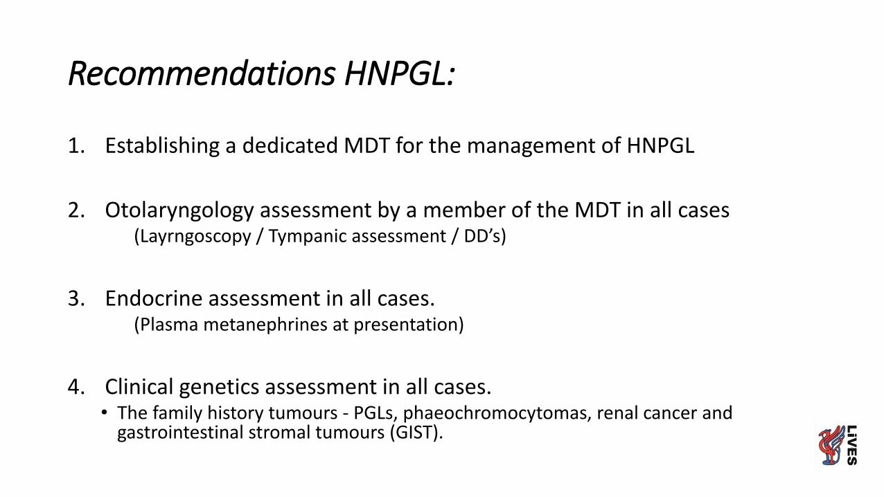
Recommendations HNPGL:
1. Establishing a dedicated MDT for the management of HNPGL
2. Otolaryngology assessment by a member of the MDT in all cases(Layrngoscopy / Tympanic assessment / DD’s)
3. Endocrine assessment in all cases. (Plasma metanephrines at presentation)
4. Clinical genetics assessment in all cases. • The family history tumours - PGLs, phaeochromocytomas, renal cancer and
gastrointestinal stromal tumours (GIST).

Recommendations HNPGL:
5. Imaging for Head and Neck PGL - HNPGL
• Contrast enhanced MRI of the head and neck
• Temporal bone PGLs (jugular and tympanic) CT
of the skull base.
• MRI (CT) thorax, abdomen and pelvis
• 123Iodine labelled metaiodobenzylguanidine scintigraphy (MIBG) or Positron Emission Tomography (PET) optimal for metastatic disease
• CTA pre- CBT surgery

PGL associated gene abnormalities

Guidelines - Management options:
Initial management:
• Active surveillance with serial imaging +/- plasma metanephrines(MAJORITY)
• EXCEPT - Tympanic, jugular with nerve impairment, Secretory, malignant disease, rapid growth, patient choice)
Treatment options
• Surgery
• Radiotherapy

Guidelines - Who to treat
• Tumours <4cm• A period of conservative management may be undertaken
• Risk of growth following conservative management is high compared to surgical control rates and the lifetime risk of complications.
• Control rates of surgery and radiotherapy are similar• ?Older age group who are less likely to develop long-term complications.
• Tumours >4cm• Because of the high-risk of complications when removing tumours larger than
4cm conservative management is often considered preferable in this group. Radiotherapy may be considered if there is tumour growth, especially in the older age group.
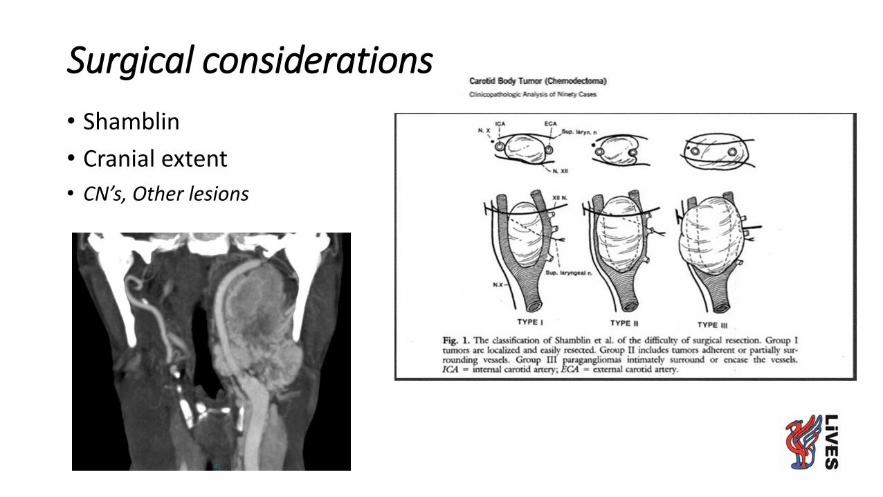
Surgical considerations
• Shamblin
• Cranial extent
• CN’s, Other lesions

Shamblin Stage 2 3
Shamblin 2.5?

CBT Case
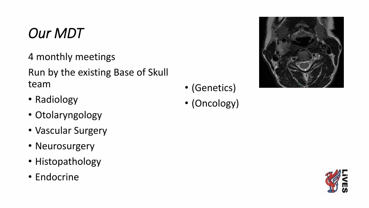
Our MDT
4 monthly meetings
Run by the existing Base of Skull team
• Radiology
• Otolaryngology
• Vascular Surgery
• Neurosurgery
• Histopathology
• Endocrine
• (Genetics)
• (Oncology)

MDT Review
ENT Vascular Endocrine Genetics
LayrngoscopyTympanic assessmment
CBT’sSurgically complex
Endocrine assessment and abdominal imaging
Counselling and relevant genetic tests
First Appointment
Serum metanephrine
Contrast MRI neck
ENT referral
Vascular (Carotid paraganglionoma)
Endocrine referral
Genetics referral
Paraganglionomareferral

Conclusions
• Establish your team
• Clear responsibilities for members
• Standardise your treatments
• Audit your results
• Don’t dabble