Embed Size (px)
Citation preview
REVIEWEndocrine-Related Cancer (2010) 17 R173–R193
Paraneoplastic syndromes secondaryto neuroendocrine tumours
Gregory Kaltsas, Ioannis I Androulakis, Wouter W de Herder1
and Ashley B Grossman2
Endocrine Unit, Department of Pathophysiology, National University of Athens, Mikras Asias 75, 11527 Athens, Greece1Department of Internal Medicine, Sector of Endocrinology, Erasmus MC, 3000 DR Rotterdam, The Netherlands2Department of Endocrinology, St Bartholomew’s Hospital, London EC1A 7BE, UK
(Correspondence should be addressed to G Kaltsas; Email: [email protected])
Abstract
Neuroendocrine tumours may be either benign or malignant tumours, and have the ability tosynthesise and secrete biologically active substances characteristic of the cell of origin thatcan cause distinct clinical syndromes. The term ‘paraneoplastic syndromes’ (PNSs) is used todenote syndromes secondary to substances secreted from tumours not related to their specificorgan or tissue of origin and/or production of autoantibodies against tumour cells; such syndromesare mainly associated with hormonal and neurological symptoms. Appreciation of the presence ofsuch syndromes is important as clinical presentation, if not identified, may delay the diagnosis ofthe underlying neoplasia. Conversely, early recognition can allow for more rapid diagnosis,particularly as the coexistence of a neoplasm with a clinical or biochemical marker offers anadditional determinant of tumour status/progression. PNSs can complicate the patient’s clinicalcourse, response to treatment, impact prognosis and even be confused as metastatic spread.Their diagnosis involves a multidisciplinary approach, and detailed endocrinological, neurological,radiological and histological studies are required. Correct diagnosis is essential as the treatment ofchoice will be different for each disorder, particularly in the case of malignant tumours; it istherefore important to develop appropriate means to correctly identify and localise these tumours.Clinical awareness and the incorporation into clinical practise of 111In-octreotide scintigraphy,chromogranin A and other evolving biochemical marker measurement techniques havesubstantially contributed to the identification of patients harbouring such syndromes. Disease-specific medical therapies are mandatory in order to prevent recurrence and/or further tumourgrowth. Owing to their rarity, central registration of these syndromes is very helpful in order to beable to provide evidence-based diagnostic and therapeutic approaches.
Endocrine-Related Cancer (2010) 17 R173–R193
Introduction
Neuroendocrine tumours (NETs) are derived fromcells that have the unique ability to synthesise, store
and secrete a variety of metabolically active sub-
stances, peptides and amines, which can cause distinct
clinical syndromes. These secretory products are
characteristic of the tissue of origin, and such secretory
tumours are denoted ‘functioning’ in order to be
distinguished from tumours originating from NE cells
not producing any substances associated with clear
clinical syndromes. The latter tumours are termed
‘non-functioning’ and cause symptoms, along with
functioning tumours, due to mass effects (Kaltsas et al.
Endocrine-Related Cancer (2010) 17 R173–R193
1351–0088/10/017–R173 q 2010 Society for Endocrinology Printed in Grea
2004b, Modlin et al. 2008), although they may in fact
also be secretory without causing any well-described
syndrome. The universal non-specific immunohisto-
chemical markers, chromogranin A (CgA) and
synaptophysin, have been used to substantiate the NE
nature of these tumours, and in this context, tumours
expressing these markers are regarded as NETs, and as
such will be considered in the present review (Table 1).
The recently introduced WHO and European Neuro-
endocrine Tumour Society (ENETS) classifications
have identified NETs as either benign, unknown
potential, well or poorly differentiated endocrine
t Britain
DOI: 10.1677/ERC-10-0024
Online version via http://www.endocrinology-journals.org
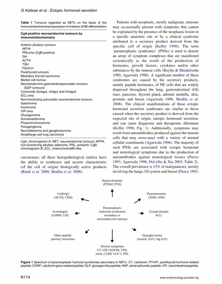
Table 1 Tumours regarded as NETs on the basis of the
immunohistochemical expression of markers of NE differentiation
CgA-positive neuroendocrine tumours by
immunohistochemistry
Anterior pituitary tumours
NFPA
PRLoma (CgB positive)
GH
ACTH
TSH
FSH/LH
Parathyroid tumours
Medullary thyroid carcinoma
Merkel cell tumour
Neuroendocrine gastroenteropancreatic tumours
(GEP tumours)
Carcinoids (foregut, midgut and hindgut)
ECL-oma
Non-functioning pancreatic neuroendocrine tumours
Gastrinoma
Insulinoma
VIP-oma
Glucagonoma
Somatostatinoma
Phaeochromocytoma
Paraganglioma
Neuroblastoma and ganglioneuroma
Small/large cell lung carcinoma
CgA, chromogranin A; NET, neuroendocrine tumours; NFPA,non-functioning pituitary adenoma; PRL, prolactin; CgB,chromogranin B; ECL, enterochromaffin-like.
G Kaltsas et al.: Ectopic hormonal secretion
carcinomas; all these histopathological entities have
the ability to synthesise and secrete characteristic
(of the cell of origin) biologically active products
(Rindi et al. 2000, Modlin et al. 2008).
Hyperca(PTHrP
Diverse sCT, VIP, LH
renin, CGRP,
Other peptidicpituitary hormones
Acromegaly(GHRH, GH)
Cushing's(ACTH, CRH)
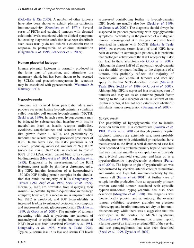
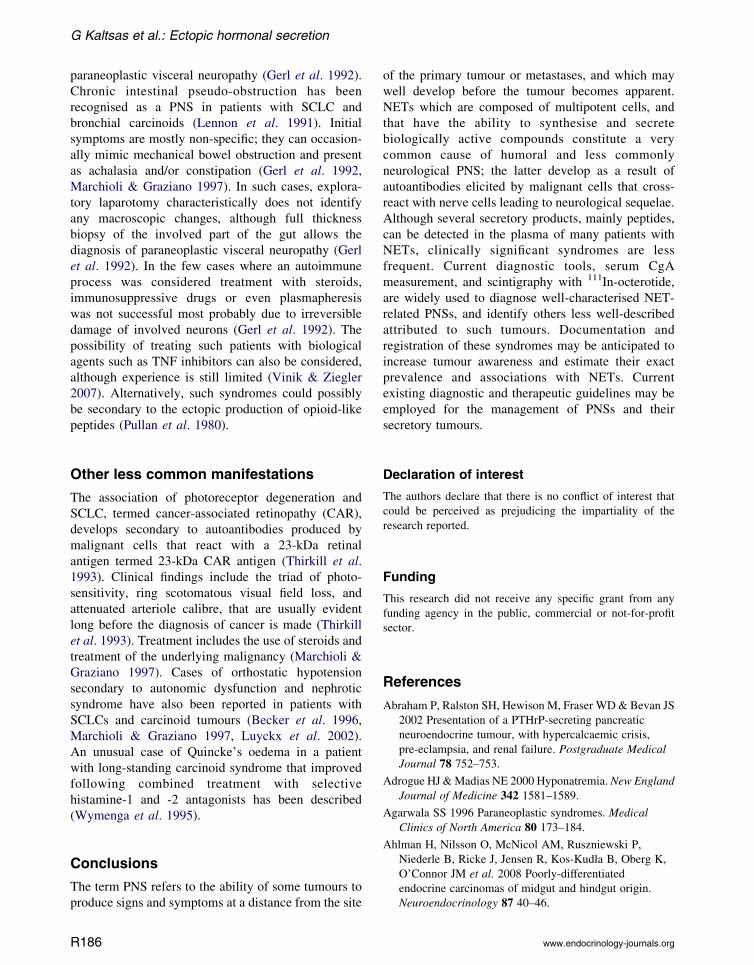
Paraneoendocrine
secondneuroendocr
Figure 1 Spectrum of paraneoplastic humoral syndromes secondapeptide; CGRP,calcitonin gene relatedpeptide; GLP, glucagon like p
R174
Patients with neoplastic, mostly malignant, tumours
may occasionally present with symptoms that cannot
be explained by the presence of the neoplastic lesion in
a specific anatomic site or by a clinical syndrome
attributed to a secretory product derived from the
specific cell of origin (Keffer 1996). The term
‘paraneoplastic syndromes’ (PNSs) is used to denote
an array of symptom complexes that are manifested
systemically as the result of the production of
hormones, growth factors, cytokines and/or other
substances by the tumour cells (Baylin & Mendelsohn
1980, Agarwala 1996). A significant number of these
syndromes are caused by the secretory products,
mainly peptide hormones, of NE cells that are widely
dispersed throughout the lung, gastrointestinal (GI)
tract, pancreas, thyroid gland, adrenal medulla, skin,
prostate and breast (Agarwala 1996, Modlin et al.
2008). The clinical manifestations of these ectopic
hormonal secretion syndromes are similar to those
caused when the secretory product is derived from the
expected site of origin, eutopic hormonal secretion,
and can cause diagnostic and therapeutic dilemmas
(Keffer 1996; Fig. 1). Additionally, symptoms may
result from autoantibodies produced against the tumour
cells that may cross-react with a variety of normal
cellular constituents (Agarwala 1996). The majority of
such PNSs are associated with ectopic hormonal
and neurological symptoms due to the production of
autoantibodies against neurological tissues (Pierce
1993, Agarwala 1996, DeLellis & Xia 2003; Table 2).
The overall prevalence is 15% of malignancies, mostly
involving the lungs, GI system and breast (Pierce 1993,
lcaemia, PTH)
Hyponatraemia(ADH, ANP)
GynaecomastiahCG
Hypoglycaemia(insulin, IGF1, big IGF)
ymptoms/FSH, TSH, GLP-1, PRL
plasticsyndromesary toine tumours
ry to NETs. CT, calcitonin; PTHrP, parathyroid hormone relatedeptide;ANP, atrial natriuretic peptide; VIP, vasointestinal peptide.
www.endocrinology-journals.org
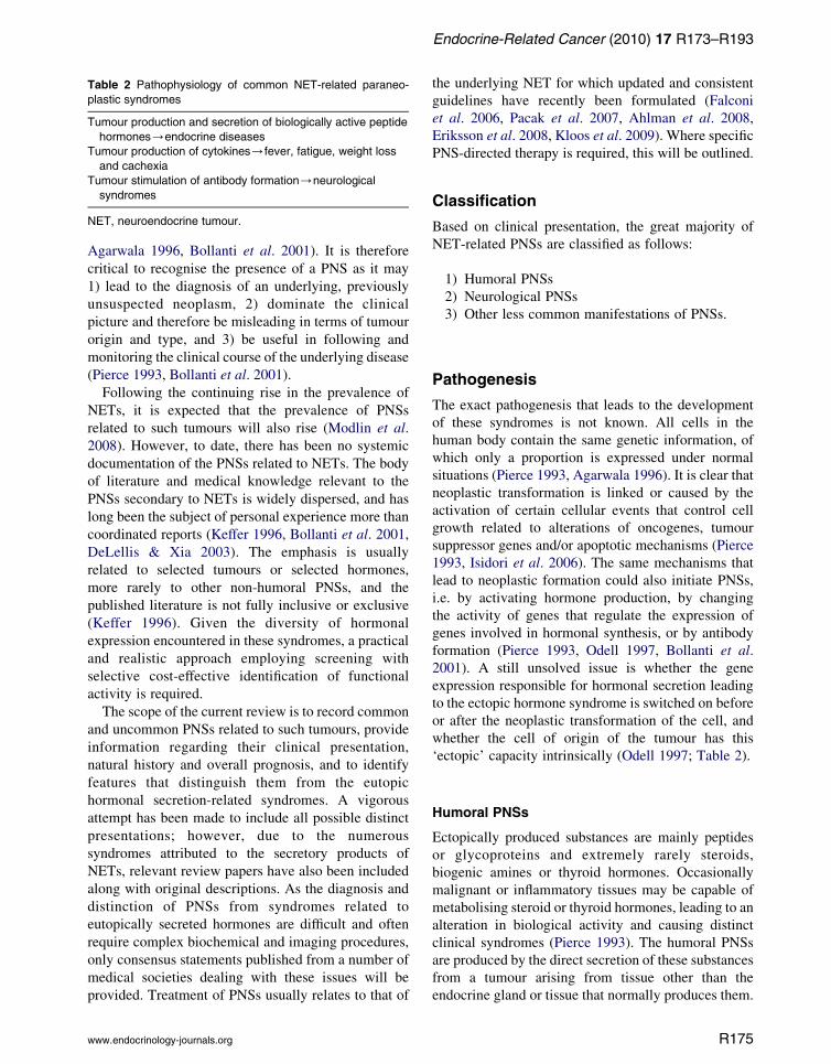
Table 2 Pathophysiology of common NET-related paraneo-
plastic syndromes
Tumour production and secretion of biologically active peptide
hormones/endocrine diseases
Tumour production of cytokines/fever, fatigue, weight loss
and cachexia
Tumour stimulation of antibody formation/neurological
syndromes
NET, neuroendocrine tumour.
Endocrine-Related Cancer (2010) 17 R173–R193
Agarwala 1996, Bollanti et al. 2001). It is therefore
critical to recognise the presence of a PNS as it may
1) lead to the diagnosis of an underlying, previously
unsuspected neoplasm, 2) dominate the clinical
picture and therefore be misleading in terms of tumour
origin and type, and 3) be useful in following and
monitoring the clinical course of the underlying disease
(Pierce 1993, Bollanti et al. 2001).
Following the continuing rise in the prevalence of
NETs, it is expected that the prevalence of PNSs
related to such tumours will also rise (Modlin et al.
2008). However, to date, there has been no systemic
documentation of the PNSs related to NETs. The body
of literature and medical knowledge relevant to the
PNSs secondary to NETs is widely dispersed, and has
long been the subject of personal experience more than
coordinated reports (Keffer 1996, Bollanti et al. 2001,
DeLellis & Xia 2003). The emphasis is usually
related to selected tumours or selected hormones,
more rarely to other non-humoral PNSs, and the
published literature is not fully inclusive or exclusive
(Keffer 1996). Given the diversity of hormonal
expression encountered in these syndromes, a practical
and realistic approach employing screening with
selective cost-effective identification of functional
activity is required.
The scope of the current review is to record common
and uncommon PNSs related to such tumours, provide
information regarding their clinical presentation,
natural history and overall prognosis, and to identify
features that distinguish them from the eutopic
hormonal secretion-related syndromes. A vigorous
attempt has been made to include all possible distinct
presentations; however, due to the numerous
syndromes attributed to the secretory products of
NETs, relevant review papers have also been included
along with original descriptions. As the diagnosis and
distinction of PNSs from syndromes related to
eutopically secreted hormones are difficult and often
require complex biochemical and imaging procedures,
only consensus statements published from a number of
medical societies dealing with these issues will be
provided. Treatment of PNSs usually relates to that of
www.endocrinology-journals.org
the underlying NET for which updated and consistent
guidelines have recently been formulated (Falconi
et al. 2006, Pacak et al. 2007, Ahlman et al. 2008,
Eriksson et al. 2008, Kloos et al. 2009). Where specific
PNS-directed therapy is required, this will be outlined.
Classification
Based on clinical presentation, the great majority of
NET-related PNSs are classified as follows:
1) Humoral PNSs
2) Neurological PNSs
3) Other less common manifestations of PNSs.
Pathogenesis
The exact pathogenesis that leads to the development
of these syndromes is not known. All cells in the
human body contain the same genetic information, of
which only a proportion is expressed under normal
situations (Pierce 1993, Agarwala 1996). It is clear that
neoplastic transformation is linked or caused by the
activation of certain cellular events that control cell
growth related to alterations of oncogenes, tumour
suppressor genes and/or apoptotic mechanisms (Pierce
1993, Isidori et al. 2006). The same mechanisms that
lead to neoplastic formation could also initiate PNSs,
i.e. by activating hormone production, by changing
the activity of genes that regulate the expression of
genes involved in hormonal synthesis, or by antibody
formation (Pierce 1993, Odell 1997, Bollanti et al.
2001). A still unsolved issue is whether the gene
expression responsible for hormonal secretion leading
to the ectopic hormone syndrome is switched on before
or after the neoplastic transformation of the cell, and
whether the cell of origin of the tumour has this
‘ectopic’ capacity intrinsically (Odell 1997; Table 2).
Humoral PNSs
Ectopically produced substances are mainly peptides
or glycoproteins and extremely rarely steroids,
biogenic amines or thyroid hormones. Occasionally
malignant or inflammatory tissues may be capable of
metabolising steroid or thyroid hormones, leading to an
alteration in biological activity and causing distinct
clinical syndromes (Pierce 1993). The humoral PNSs
are produced by the direct secretion of these substances
from a tumour arising from tissue other than the
endocrine gland or tissue that normally produces them.
R175
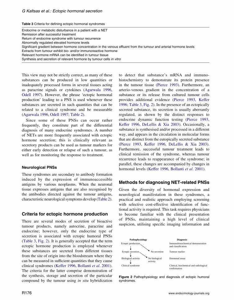
Table 3 Criteria for defining ectopic hormonal syndromes
Endocrine or metabolic disturbance in a patient with a NET
Remission after successful treatment
Return of endocrine syndrome with tumour recurrence
Abnormally regulated elevated hormone levels
Significant gradient between hormone concentration in the venous effluent from the tumour and arterial hormone levels
Extracts from tumour exhibit bio- and/or immunoreactive hormone
Relevant hormone mRNA can be identified in tumour tissue
Synthesis and secretion of relevant hormone by tumour cells in vitro
G Kaltsas et al.: Ectopic hormonal secretion
This view may not be strictly correct, as many of these
substances can be produced in low quantities or
inadequately processed forms in several tissues acting
as paracrine signals or cytokines (Agarwala 1996,
Odell 1997). However, the phrase ‘ectopic hormonal
production’ leading to a PNS is used whenever these
substances are secreted in such quantities that can be
related to a clinical syndrome and be measurable
(Agarwala 1996, Odell 1997; Table 2).
Since some of these PNSs can occur rather
frequently, they constitute part of the differential
diagnosis of many endocrine syndromes. A number
of NETs are more frequently associated with ectopic
hormone secretion; this is clinically relevant as
secretory products can be used as tumour markers for
either early detection or relapse of such a tumour, as
well as for monitoring the response to treatment.
Neurological PNSs
These syndromes are secondary to antibody formation
induced by the expression of immunoaccessible
antigens by various neoplasms. When the neuronal
tissue expresses antigens that are also recognised by
the antibodies directed against the tumour antigens,
characteristic neurological symptoms develop (Table 2).
Pathophysiology Diagnosis
Ectopic production Immunohistochemical demonstrationand classification
Ectopic secretion No secretion Tumour marker
Biological activity No biologicalactivity
Hormonal assay
Clinical syndrome Clinical, biochemical and radiologicalconfirmation
Figure 2 Pathophysiology and diagnosis of ectopic humoralsyndromes.
Criteria for ectopic hormone production
There are several modes of secretion of bioactive
tumour products, namely autocrine, paracrine and
endocrine; however, only the endocrine type of
secretion is associated with ectopic humoral PNSs
(Table 3, Fig. 2). It is generally accepted that the term
ectopic hormone production is employed whenever
these substances are secreted from different tissues
from the site of origin into the bloodstream where they
can be measured in sufficient quantities that they cause
clinical syndromes (Keffer 1996, Bollanti et al. 2001).
The criteria for the latter comprise demonstration of
the synthesis, storage and secretion of the particular
compound by the tumour using in situ hybridization
R176
to detect that substance’s mRNA and immuno-
histochemistry to demonstrate its protein presence
in the tumour tissue (Pierce 1993). Furthermore, an
arterio-venous gradient in the concentration of a
substance or its release from cultured tumour cells
provides additional evidence (Pierce 1993, Keffer
1996; Table 3, Fig. 2). In the presence of an ectopically
secreted substance, its secretion is usually aberrantly
regulated, as shown by the distinct responses to
endocrine dynamic function testing (Pierce 1993,
Keffer 1996, DeLellis & Xia 2003). Occasionally, a
substance is synthesised and/or processed in a different
way, and appears in the circulation in molecular forms
that are distinct from the eutopically secreted substance
(Pierce 1993, Keffer 1996, DeLellis & Xia 2003).
Furthermore, successful tumour treatment leads to
clinical remission of the syndrome, whereas tumour
recurrence leads to reappearance of the syndrome; in
parallel, these changes are accompanied by changes in
hormonal levels (Keffer 1996, Bollanti et al. 2001).
Methods for diagnosing NET-related PNSs
Given the diversity of hormonal expression and
neurological manifestation in these syndromes, a
practical and realistic approach employing screening
with selective cost-effective identification of func-
tional activity is required. This task requires physicians
to become familiar with the clinical presentation
of PNSs, maintaining a high level of clinical
suspicion, utilising specific imaging information and
www.endocrinology-journals.org
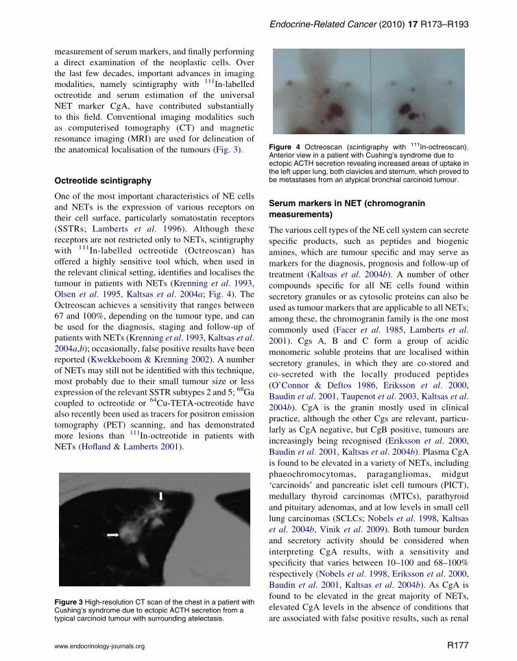
Figure 4 Octreoscan (scintigraphy with 111In-octreoscan).
Endocrine-Related Cancer (2010) 17 R173–R193
measurement of serum markers, and finally performing
a direct examination of the neoplastic cells. Over
the last few decades, important advances in imaging
modalities, namely scintigraphy with 111In-labelled
octreotide and serum estimation of the universal
NET marker CgA, have contributed substantially
to this field. Conventional imaging modalities such
as computerised tomography (CT) and magnetic
resonance imaging (MRI) are used for delineation of
the anatomical localisation of the tumours (Fig. 3).
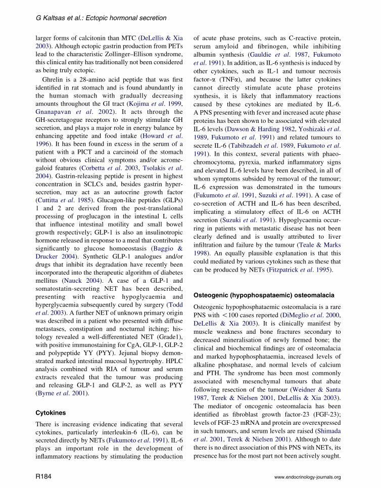
Anterior view in a patient with Cushing’s syndrome due toectopic ACTH secretion revealing increased areas of uptake inthe left upper lung, both clavicles and sternum, which proved tobe metastases from an atypical bronchial carcinoid tumour. Octreotide scintigraphyOne of the most important characteristics of NE cells
and NETs is the expression of various receptors on
their cell surface, particularly somatostatin receptors
(SSTRs; Lamberts et al. 1996). Although these
receptors are not restricted only to NETs, scintigraphy
with 111In-labelled octreotide (Octreoscan) has
offered a highly sensitive tool which, when used in
the relevant clinical setting, identifies and localises the
tumour in patients with NETs (Krenning et al. 1993,
Olsen et al. 1995, Kaltsas et al. 2004a; Fig. 4). The
Octreoscan achieves a sensitivity that ranges between
67 and 100%, depending on the tumour type, and can
be used for the diagnosis, staging and follow-up of
patients with NETs (Krenning et al. 1993, Kaltsas et al.
2004a,b); occasionally, false positive results have been
reported (Kwekkeboom & Krenning 2002). A number
of NETs may still not be identified with this technique,
most probably due to their small tumour size or less
expression of the relevant SSTR subtypes 2 and 5; 68Ga
coupled to octreotide or 64Cu-TETA-octreotide have
also recently been used as tracers for positron emission
tomography (PET) scanning, and has demonstrated
more lesions than 111In-octreotide in patients with
NETs (Hofland & Lamberts 2001).
Figure 3 High-resolution CT scan of the chest in a patient withCushing’s syndrome due to ectopic ACTH secretion from atypical carcinoid tumour with surrounding atelectasis.
www.endocrinology-journals.org
Serum markers in NET (chromogranin
measurements)
The various cell types of the NE cell system can secrete
specific products, such as peptides and biogenic
amines, which are tumour specific and may serve as
markers for the diagnosis, prognosis and follow-up of
treatment (Kaltsas et al. 2004b). A number of other
compounds specific for all NE cells found within
secretory granules or as cytosolic proteins can also be
used as tumour markers that are applicable to all NETs;
among these, the chromogranin family is the one most
commonly used (Facer et al. 1985, Lamberts et al.
2001). Cgs A, B and C form a group of acidic
monomeric soluble proteins that are localised within
secretory granules, in which they are co-stored and
co-secreted with the locally produced peptides
(O’Connor & Deftos 1986, Eriksson et al. 2000,
Baudin et al. 2001, Taupenot et al. 2003, Kaltsas et al.
2004b). CgA is the granin mostly used in clinical
practice, although the other Cgs are relevant, particu-
larly as CgA negative, but CgB positive, tumours are
increasingly being recognised (Eriksson et al. 2000,
Baudin et al. 2001, Kaltsas et al. 2004b). Plasma CgA
is found to be elevated in a variety of NETs, including
phaeochromocytomas, paragangliomas, midgut
‘carcinoids’ and pancreatic islet cell tumours (PICT),
medullary thyroid carcinomas (MTCs), parathyroid
and pituitary adenomas, and at low levels in small cell
lung carcinomas (SCLCs; Nobels et al. 1998, Kaltsas
et al. 2004b, Vinik et al. 2009). Both tumour burden
and secretory activity should be considered when
interpreting CgA results, with a sensitivity and
specificity that varies between 10–100 and 68–100%
respectively (Nobels et al. 1998, Eriksson et al. 2000,
Baudin et al. 2001, Kaltsas et al. 2004b). As CgA is
found to be elevated in the great majority of NETs,
elevated CgA levels in the absence of conditions that
are associated with false positive results, such as renal
R177
G Kaltsas et al.: Ectopic hormonal secretion
insufficiency and hypergastrinaemia (especially in
response to antacid preparations, where levels can be
remarkably elevated), constitute a useful indicator of
the presence of a PNS in a relevant clinical setting
(Baudin et al. 2001). Although CgA can also be used as
a prognostic factor as it correlates with tumour burden,
particularly in GI NETs, there is a lack of correlation
between absolute CgA levels and symptom frequency
and severity (Janson et al. 1997, Woltering et al. 2006).
In the presence of uncertainty, the highly specific and
sensitive pancreastatin assay can detect small tumour
load, being up to 100-fold more sensitive and specific
than CgA assays (O’Dorisio et al. 2010).
Specific antibodies for neurological PNSs
There are several autoantibodies that have been shown
to be of diagnostic significance in neurological PNS.
These will be discussed below.
Humoral PNSs in NET
Cushing’s syndrome
The association between Cushing’s syndrome (CS) and
various cancers was recognised as early as 1928, but
was first fully characterised by Liddle in 1965 (Table 4;
Brown 1928, Wajchenberg et al. 1994, Keffer 1996,
Bollanti et al. 2001). This PNS develops secondary to
tumoral ACTH and less often CRH production, and
accounts for 10–20% of the total cases of CS (Howlett
et al. 1986, Newell-Price et al. 2006). Large amounts
of biologically active ACTH are found in tumour
tissue, although immunoreactive ACTH may also be
found at high concentrations in tumour extracts
from patients without clinical manifestations of CS
(Howlett et al. 1986, Oldfield et al. 1991). ACTH is
usually produced in its high-molecular weight pre-
cursor form, ‘big-ACTH’, and/or involves abnormal
posttranslational processing of precursor peptides
with different bioactivity (Stewart et al. 1994).
Owing to excessive ACTH production, severe hyper-
cortisolism may occasionally be induced (Newell-Price
et al. 1998).
NETs associated with CS are often derived from the
lung, thymus, pancreas, thyroid (MTC), chromaffin
cell tumours (phaeochromocytomas, paragangliomas
and neuroblastomas) and rarely from the ovary or
prostate (Ilias et al. 2005, Isidori et al. 2006).
Bronchial carcinoids (typical and atypical) account
for 36–46% of these cases, whereas the highly
malignant SCLC accounts for 8–20% of clinically
R178
apparent cases (Limper et al. 1992, Newell-Price et al.
1998). However, up to 30% of SCLCs hypersecrete
ACTH that may be bio-inactive following incomplete
processing and thus not capable of inducing a clinical
syndrome, or possibly the time course may be
insufficiently long for the syndrome to be manifest
(Limper et al. 1992, Newell-Price et al. 1998).
Typically, bronchial carcinoids produce a clinical
and biochemical syndrome that resembles pituitary-
dependent CS (Cushing’s disease, CD). In contrast,
patients with CS secondary to SCLCs do not typically
exhibit the classical manifestations of prolonged and
sustained hypercortisolaemia but rather those of the
underlying malignancy (Bollanti et al. 2001, Ilias et al.
2005, Isidori et al. 2006). Weight loss rather than
weight gain, and slight changes in body fat distribution
and hyperpigmentation constitute early reported
symptoms/signs (Bollanti et al. 2001, Ilias et al.
2005, Isidori et al. 2006); due to excessive cortisol
secretion, mineralocorticoid effects and glucose intol-
erance are often present in patients with SCLCs (Ilias
et al. 2005, Isidori et al. 2006). In such cases, the
diagnosis is readily suspected due to the rapid onset of
symptoms, related to the degree of malignancy of the
tumour, and is based on clinical, biochemical and
radiological features (Newell-Price et al. 1998, Alwani
et al. 2009). However, in cases of CS due to bronchial
carcinoids where imaging does not immediately reveal
the source, ‘covert’ CS, several endocrine tests may be
necessary including inferior petrosal sinus sampling
(IPSS) to exclude CD; as such tumours may be small
and elude radiological detection, in w8–19% of cases,
medical or surgical control of hypercortisolaemia and
prolonged follow-up may be necessary to establish the
diagnosis (Oldfield et al. 1991, Kaltsas et al. 1998,
Tsagarakis et al. 2003, Ilias et al. 2005, Isidori et al.
2006, Newell-Price et al. 2006). In some cases, the
source may be hidden for many years or may never be
revealed, so-called ‘occult’ CS. The incidence of CS
secondary to MTCs is !1%, with w100 cases being
reported, whereas CS due to chromaffin cell tumours is
even rarer with !30 reported cases (Barbosa et al.
2005, Nijhoff et al. 2009). Occasionally, cyclic or
periodic production may render this diagnosis of the
PNS extremely difficult (van Dam et al. 2002, Arnaldi
et al. 2003).
Rarely, CS may result from CRH production from
SCLCs, MTCs, carcinoids, PETs, chromaffin cell
tumours or hypothalamic tumours (Upton & Amatruda1971, Oldfield et al. 1991, Muller & von Werder 1992,
DeLellis & Xia 2003, Markou et al. 2005); in many ofthese cases, tumours may produce both ACTH and
CRH (DeLellis & Xia 2003, Zangeneh et al. 2003).
www.endocrinology-journals.org
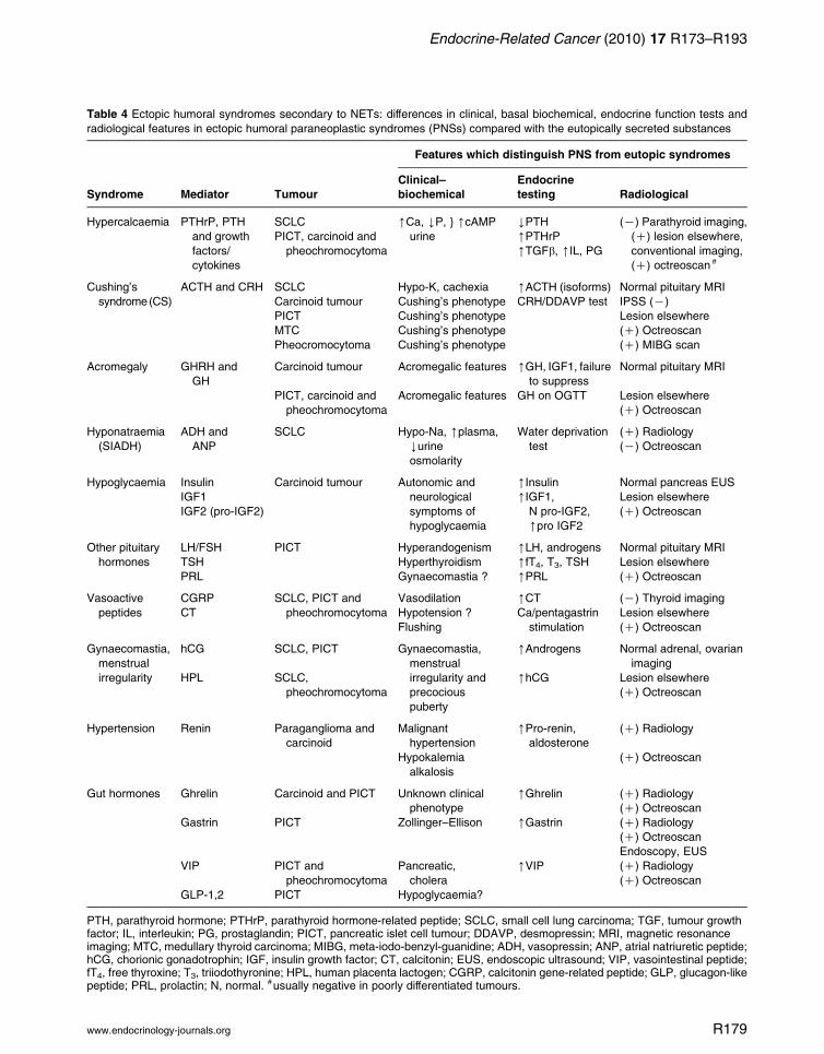
Table 4 Ectopic humoral syndromes secondary to NETs: differences in clinical, basal biochemical, endocrine function tests and
radiological features in ectopic humoral paraneoplastic syndromes (PNSs) compared with the eutopically secreted substances
Features which distinguish PNS from eutopic syndromes
Syndrome Mediator Tumour
Clinical–
biochemical
Endocrine
testing Radiological
Hypercalcaemia PTHrP, PTH
and growth
factors/
cytokines
SCLC [Ca, YP, } [cAMP
urine
YPTH (K) Parathyroid imaging,
(C) lesion elsewhere,
conventional imaging,
(C) octreoscan #
PICT, carcinoid and
pheochromocytoma
[PTHrP
[TGFb, [IL, PG
Cushing’s
syndrome(CS)
ACTH and CRH SCLC Hypo-K, cachexia [ACTH (isoforms) Normal pituitary MRI
Carcinoid tumour Cushing’s phenotype CRH/DDAVP test IPSS (K)
PICT Cushing’s phenotype Lesion elsewhere
MTC Cushing’s phenotype (C) Octreoscan
Pheocromocytoma Cushing’s phenotype (C) MIBG scan
Acromegaly GHRH and
GH
Carcinoid tumour Acromegalic features [GH, IGF1, failure
to suppress
Normal pituitary MRI
PICT, carcinoid and
pheochromocytoma
Acromegalic features GH on OGTT Lesion elsewhere
(C) Octreoscan
Hyponatraemia
(SIADH)
ADH and
ANP
SCLC Hypo-Na, [plasma,
Yurine
osmolarity
Water deprivation
test
(C) Radiology
(K) Octreoscan
Hypoglycaemia Insulin Carcinoid tumour Autonomic and
neurological
symptoms of
hypoglycaemia
[Insulin Normal pancreas EUS
IGF1 [IGF1,
N pro-IGF2,
[pro IGF2
Lesion elsewhere
IGF2 (pro-IGF2) (C) Octreoscan
Other pituitary
hormones
LH/FSH PICT Hyperandogenism [LH, androgens Normal pituitary MRI
TSH Hyperthyroidism [fT4, T3, TSH Lesion elsewhere
PRL Gynaecomastia ? [PRL (C) Octreoscan
Vasoactive
peptides
CGRP SCLC, PICT and
pheochromocytoma
Vasodilation [CT (K) Thyroid imaging
CT Hypotension ? Ca/pentagastrin
stimulation
Lesion elsewhere
Flushing (C) Octreoscan
Gynaecomastia,
menstrual
irregularity
hCG SCLC, PICT Gynaecomastia,
menstrual
irregularity and
precocious
puberty
[Androgens Normal adrenal, ovarian
imaging
HPL SCLC,
pheochromocytoma
[hCG Lesion elsewhere
(C) Octreoscan
Hypertension Renin Paraganglioma and
carcinoid
Malignant
hypertension
[Pro-renin,
aldosterone
(C) Radiology
Hypokalemia
alkalosis
(C) Octreoscan
Gut hormones Ghrelin Carcinoid and PICT Unknown clinical
phenotype
[Ghrelin (C) Radiology
(C) Octreoscan
Gastrin PICT Zollinger–Ellison [Gastrin (C) Radiology
(C) Octreoscan
Endoscopy, EUS
VIP PICT and
pheochromocytoma
Pancreatic,
cholera
[VIP (C) Radiology
(C) Octreoscan
GLP-1,2 PICT Hypoglycaemia?
PTH, parathyroid hormone; PTHrP, parathyroid hormone-related peptide; SCLC, small cell lung carcinoma; TGF, tumour growthfactor; IL, interleukin; PG, prostaglandin; PICT, pancreatic islet cell tumour; DDAVP, desmopressin; MRI, magnetic resonanceimaging; MTC, medullary thyroid carcinoma; MIBG, meta-iodo-benzyl-guanidine; ADH, vasopressin; ANP, atrial natriuretic peptide;hCG, chorionic gonadotrophin; IGF, insulin growth factor; CT, calcitonin; EUS, endoscopic ultrasound; VIP, vasointestinal peptide;fT4, free thyroxine; T3, triiodothyronine; HPL, human placenta lactogen; CGRP, calcitonin gene-related peptide; GLP, glucagon-likepeptide; PRL, prolactin; N, normal. #usually negative in poorly differentiated tumours.
Endocrine-Related Cancer (2010) 17 R173–R193
www.endocrinology-journals.org R179
G Kaltsas et al.: Ectopic hormonal secretion
In such cases, patients have high CRH levels in plasmaand tumour tissue, whereas plasma ACTH levels are
also increased; CS due to CRH production does not
have a distinctive presentation, and endocrine testingmay represent an interplay between ectopic and
eutopic production (DeLellis & Xia 2003, Zangeneh
et al. 2003, Markou et al. 2005).
It has been suggested that no single endocrine testand/or imaging procedure are accurate enough to
diagnose and localise ectopic ACTH/CRH-producing
bronchial carcinoids, particularly as false positive IPSSresults may occasionally be obtained, albeit very
rarely (Young et al. 1998, de Herder & Lamberts1999, Baudin et al. 2001, Loli et al. 2003). In such
cases, scintigraphy with 111In-octreotide, particularly
after correction of hypercortisolaemia, and PETusing several novel tracers can be used to reveal
confounding cases eluding localisation (de Herderet al. 1994, Tsagarakis et al. 2003, Kaltsas et al. 2004b,
Markou et al. 2005). The recent finding that dopamine
receptors are expressed in NETs associated with CS,and that the dopamine agonist cabergoline could be
effective in controlling cortisol excess in a subgroup ofsuch patients, provides further medical therapeutic
options to control the hypercortisolism while
awaiting definitive diagnosis (Newell-Price et al.2006, Pivonello et al. 2007). Mifepristone, an
antagonist of both progesterone and glucocorticoidreceptors, has also been used to control excessive
hypercortisolaemia secondary to disseminated NETs
(Cassier et al. 2008). However, as its effects can onlybe monitored clinically and it is associated with
hypertension and hypokalaemia, its use is generallylimited to the short term (Cassier et al. 2008).
Hypercalcaemia
Although humoral hypercalcaemia is one of the
commonest of the PNSs, and w5% of patients with
malignant tumours may develop hypercalcaemia, it has
not been commonly described in patients with NETs
(Pierce 1993, DeLellis & Xia 2003). As the main
secretory products of NETs are peptides or amines,
almost all cases of NET-related hypercalcaemia are
associated with hypophosphataemia suggesting a
parathyroid hormone (PTH)-like effect, implicating
as mediators either PTH or PTH-related protein
(PTHrP; DeLellis & Xia 2003). PTHrP was first
isolated in 1987 from cancer cell lines and a tumour
associated with hypercalcaemia, and is now considered
to be the main mediator of humoral hypercalcaemia
of malignancy (Suva et al. 1987, Strewler 2000).
PTHrP is present in a very wide variety of normal cells
and tissues, and has a wide spectrum of functions;
R180
hypomethylation of the promoter has been suggested
as a possible underlying mechanism by which the
gene is expressed (Strewler 2000). PTHrP has been
shown to be secreted by NETs. The first case of a
PTHrP-producing malignant NET was described in a
pancreatic islet cell tumour presenting with severe
hypercalcaemia during pregnancy (Abraham et al.
2002); however, benign phaeochromocytomas can also
secrete PTHrP and cause hypercalcaemia (Fukumoto
et al. 1991). Several reports have now demonstrated
either biochemical and/or immunohistochemical
PTHrP-related hypercalcaemia in w25 patients with
PICTs (Mao et al. 1995, Srirajaskanthan et al. 2009).
The majority of these tumours are well-differentiated
stage III–IV carcinomas, and this entity should be
considered in the differential diagnosis of all patients
with NETs presenting with hypercalcaemia and a
disproportionately low PTH. In contrast to other
PTHrP-secreting malignancies that have a mostly
abysmal prognosis, patients with NET-related PTHrP
secretion have a much better outcome (DeLellis & Xia
2003, Srirajaskanthan et al. 2009). It is possible that the
exact prevalence of PTHrP-secreting NETs may well
be underestimated as the assay is difficult to perform,
and may not always be requested (Srirajaskanthan
et al. 2009). Very few cases of ectopic PTH secretion
from NETs have been documented, two from an SCLC
and one from an MTC (Weiss et al. 2006, Demura et al.
2009). A further case secondary to a small cell
carcinoma of the ovary has also been described;
although hypercalcaemia is encountered in 70% of
patients harbouring such tumours, almost all are
secondary to PTHrP secretion (Chen et al. 2005).
Furthermore, hypercalcaemia due to ectopic PTH
secretion from a poorly differentiated pancreatic NET
has been described, in which transactivation of
the PTH gene was found (VanHouten et al. 2006).
Interestingly, serum PTHrP was also elevated and
detected immunohistochemically in the tumour speci-
men (VanHouten et al. 2006). Although successful
treatment of the underlying neoplasm usually suffices
to control the clinical symptoms and systemic sequelae
of hypercalcaemia, in cases of severe, residual or
recurrent disease, medical treatment of hypercalcaemia
is also required (Makras & Papapoulos 2009).
Acromegaly
Acromegaly secondary to non-pituitary tumours israre, and accounts for !1% of cases of acromegaly
(Melmed 2006). This PNS is more commonly related
to GHRH-hypersecretion and rarely to GH itself;however, its rarity may be related to the fact that
www.endocrinology-journals.org
Endocrine-Related Cancer (2010) 17 R173–R193
clinical presentation is usually subtle, and GHRHmeasurement is not widely available (Biermasz et al.
2007). NETs most commonly associated with GHRH
hypersecretion are carcinoids, PICTs, SCLCs andphaeochromocytomas, with w70 cases having been
described (Thorner et al. 1984, Faglia et al. 1992,Biermasz et al. 2007, Vieira et al. 2007). However,
GHRH tumoral immunoreactivity without clinically
obvious acromegaly is found in up to 17% ofgastroenteropancreatic NETs (Dayal et al. 1986).
Clinical presentation is not different to that of pituitaryorigin, although co-secretion of other substances by the
tumour may increase clinical suspicion (Losa et al.
1993); endocrine testing does not reliably distinguishthese tumours from pituitary adenomas (Biermasz
et al. 2007). In contrast to the presence of pituitaryadenomas in patients with classical acromegaly, the
pituitary pathology in patients with ectopic GHRH
secretion and acromegaly is GH-cell hyperplasia(except in patients with hypothalamic GHRH-
producing tumours where adenomatous lesions mayalso be found; Sano et al. 1988, Biermasz et al. 2007).
Only a few patients with GHRH-producing secondary
to PICTs from patients with the multiple endocrineneoplasia-1 (MEN-1) syndrome have been described
(Sano et al. 1987, Ramsay et al. 1988, Biermasz et al.
2007). A further two cases of ectopic GH secretion, onefrom a PICT, have also been described (Melmed et al.
1985, Beuschlein et al. 2000). Treatment is that of theunderlying tumour, and relates to the extent of the
disease; in GI NETs, long-acting somatostatin
analogues can also be useful for the treatment of boththe tumour and the PNS in the case of residual disease
(Kaltsas et al. 2004b).
Ectopic vasopressin and atrial natriuretic peptide
secretion
Vasopressin (ADH) is produced within the hypo-
thalamus and stored in nerve terminals of the posterior
pituitary and from a subset of normal pulmonary NE
cells (Gainer & Wray 1992). Additional processing of
ADH also occurs in SCLC cells that can also synthesise
and secrete oxytocin (OXT) and its corresponding
neurophysin; cosecretion of ADH and OXT may be
related to linkage of encoding genes (Sausville et al.
1985). Both ADH and OXT exert autocrine/paracrine
signalling activities, and have been implicated in the
initiation and growth of SCLCs (Pequeux et al. 2002,
DeLellis & Xia 2003). A syndrome of renal sodium
loss and hyponatraemia resulting from inappropriate
ADH secretion (SIADH) has been described since the
1950s (Schwartz et al. 1957). Although ADH levels are
increased in up to 50% of patients with SCLCs, only
www.endocrinology-journals.org
15% develop the syndrome; however, some patients
exhibit abnormalities following water loading (Moses
& Scheinman 1991). Increased plasma OXT levels
occur in up to 20% of patients with SCLCs. (Moses &
Scheinman 1991). Atrial natriuretic peptide (ANP) is a
peptide synthesised from the cardiac atria that can
cause natriuresis and hypotension (Kangawa et al.
1984, Marchioli & Graziano 1997). It has also been
implicated in the development of some cases of
hyponatraemia related to malignancy, and both ANP
and ADH can be synthesised and produced contribut-
ing to the hyponatraemia by the same tumour cells
(Shimizu et al. 1991); however, severe cases of
hyponatraemia are more clearly associated with
SIADH (Marchioli & Graziano 1997, DeLellis & Xia
2003). In contrast to the majority of chronic causes of
hyponatraemia that may develop gradually and be
relatively asymptomatic, hyponatraemia secondary to
ectopic hormonal production can develop abruptly and
be associated with severe symptoms (Adrogue &
Madias 2000). Although there are no established
guidelines for evaluating and treating such patients,
the diagnosis is readily made by demonstrating a
urinary osmolality that exceeds 100 mOsm/kg of water
in the presence of low effective plasma osmolality in
an euvolaemic individual (Adrogue & Madias 2000).
Treating the underlying neoplasm is the definitive
means of correcting the hyponatraemia (Ellison & Berl
2007). In the absence of symptoms, gradual correction
of the hyponatraemia is appropriate, and involves
adequate solute intake and fluid restriction (Adrogue &
Madias 2000, Ellison & Berl 2007). In the presence of
symptoms, increasing serum sodium by 0.5–1 mmol/l
per h for a total of 8 mmol/l during the first day
may suffice to render the patient asymptomatic; this can
be enhanced by promoting free-water excretion with
furosemide (Ellison & Berl 2007). Alternatively, the
management of SIADH may be enhanced by the recent
introduction of the ‘vaptans’, ADH antagonists;
however, the clinical experience with these agents
remains limited (Ghali et al. 2006, Schrier et al. 2006).
Ectopic calcitonin
Elevated calcitonin levels are considered a valuable
biochemical marker for both sporadic and familial
forms of MTCs (Kloos et al. 2009). An elevated basal
value O50 pg/ml, and/or a five- to tenfold peak
following pentagastrin or calcium stimulation are
both highly suggestive of an MTC (Kloos et al.
2009). However, in the majority of cases, such values
are not associated with a secretory syndrome and,
therefore, do not fulfil the prerequisites for a PNS
R181
G Kaltsas et al.: Ectopic hormonal secretion
(DeLellis & Xia 2003). A number of other tumours
have also been shown to exhibit plasma calcitonin
immunoreactivity (Coombes et al. 1974). Several
cases of PICTs and carcinoid tumours with elevated
calcitonin levels associated with no clinical symptoms
but causing diagnostic confusion have been described;
such cases usually do not exhibit a calcitonin rise in
response to pentagastrin or calcium stimulation
(Engelbach et al. 1998, Schneider et al. 2009).
Human placental lactogen
Human placental lactogen is normally produced in
the latter part of gestation, and stimulates the
mammary gland, but has been shown to be secreted
by SCLCs and phaeochromocytoma; its secretion
may be associated with gynaecomastia (Weintraub &
Kadesky 1971).
Hypoglycaemia
Tumours not derived from pancreatic islets may
produce recurrent fasting hypoglycaemia, a condition
called non-islet cell tumour hypoglycaemia (NICTH;
Seckl et al. 1999). In such cases, hypoglycaemia may
be induced by substances that interfere with insulin
metabolism (such as insulin receptor antibodies,
cytokines, catecholamines and secretion of insulin-
like growth factor 1, IGF1), and particularly by
tumours that secrete partially processed precursors of
IGF2. In the latter case, the IGF2 precursor is not
cleaved, producing increased amounts of ‘big IGF2’
(molecular mass, 10–17 kDa, in contrast to mature
IGF2 of 7.5 kDa), which cannot bind to its cognate-
binding protein (Megyesi et al. 1974, Daughaday et al.
1993). Diagnosis is by measurement of the IGF2
isoforms, most easily by thin-layer chromatography.
Big IGF2 impairs formation of a heterotrimeric
150 kDa IGF-binding protein complex in the circula-
tion that binds the majority of IGFs (Daughaday &
Trivedi 1992, Zapf et al. 1992, Seckl et al. 1999).
Normally, IGFs are prevented from displaying their
insulin-like potential by their sequestration in this large
complex; however, this mechanism is impaired when
big IGF2 is produced, and IGF bioavailability is
increased leading to enhanced peripheral consumption
and suppressed hepatic glucose production (Seckl et al.
1999, de Groot et al. 2007). The majority of tumours
presenting with such a syndrome are tumours of
mesenchymal or epithelial origin, but rare cases of
NETs have also been described (Gorden et al. 1981,
Daughaday et al. 1993, Marks & Teale 1998).
Typically, serum insulin is low and serum GH levels
R182
suppressed contributing further to hypoglycaemia;
IGF1 levels are usually also low (Seckl et al. 1999,
de Groot et al. 2007). The diagnosis should always be
suspected in patients presenting with hypoglycaemic
symptoms, particularly in the presence of a malignant
tumour; acromegaloid skin changes have also been
described in patients with NICTH (Marks & Teale
1998). As elevated serum levels of total IGF2 have
been described in acromegalic patients, it is probable
that prolonged activation of the IGF1 receptor by IGF2
can lead to these symptoms (de Groot et al. 2007).
Although in almost half of all patients, hypoglycaemia
was the initial symptom leading to the diagnosis of a
tumour, this probably reflects the majority of
mesenchymal and epithelial tumours and does not
apply for the few NETs described to date (Marks &
Teale 1998, Seckl et al. 1999, de Groot et al. 2007).
Although big IGF2 is expressed in a broad spectrum of
tumours and may act as an autocrine growth factor
binding to the IGF2 receptor or the A isoform of the
insulin receptor, it has not been established whether it
stimulates tumour progression (Baserga et al. 2003).
Ectopic insulin
The possibility of hypoglycaemia due to insulin
secretion from NICTs is controversial (Gorden et al.
1981, Furrer et al. 2001). Although primary hepatic
carcinoid tumours are extremely rare, most probably
reflecting tumours with unidentified primaries that have
metastasised to the liver, a well-documented case has
been described of a probable primary hepatic carcinoid
that was manifest initially as extrapituitary acromegaly
and a typical carcinoid syndrome, and later on as a
hyperinsulinaemic hypoglycaemic syndrome (Furrer
et al. 2001). The hepatic origin of hyperinsulinism was
demonstrated by selective arterial calcium stimulation
and insulin and C-peptide immunoreactivity by the
tumour cell (Furrer et al. 2001). A further case of
ectopic insulin production from an apparently primary
ovarian carcinoid tumour associated with episodic
hyperinsulinaemic hypoglycaemia has also been
described (Morgello et al. 1988). The diagnosis was
biochemically proven, and at autopsy, the ovarian
tumour exhibited secretory granules on electron
microscopy and insulin immunoreactivity on immuno-
histochemistry, while there was a suggestion that this
developed in the context of MEN-1 syndrome
(Morgello et al. 1988). Following that original report,
a further case of an insulin-secreting NET of the cervix,
and two paragangliomas, has also been described
(Seckl et al. 1999, Uysal et al. 2007).
www.endocrinology-journals.org
Endocrine-Related Cancer (2010) 17 R173–R193
Ectopic IGF1
A rare case of a large cell lung carcinoma with recurrent
hypoglycaemia, low insulin and big IGF2 levels, and
increased IGF1 levels, has recently been described
(Nauck et al. 2007). Although no acromegaloid features
were found in that particular patient, hypoglycaemia
did not recur and IGF1 levels decreased following
successful treatment (Nauck et al. 2007).
Treatment relates to that of the underlying
neoplasm, stage and grade of the disease. Patients
with NICTH may undergo complete remission
following surgical removal of the tumour; even partial
removal often may reduce or abolish the hypogly-
caemia (Marks & Teale 1998). This is followed by a
rise in IGF1 levels, restoration of the IGF2/IGF1 ratio,
blood glucose and plasma insulin levels (Perros et al.
1996, Marks & Teale 1998). Both hGH and
prednisolone can induce a substantial and seemingly
specific effect in alleviating the symptoms of
hypoglycaemia (Mitchell et al. 1968, Teale et al.
1992, Perros et al. 1996). Although a therapeutic role
of long-acting somatostatin analogues seems feasible,
they should be used with caution as they may inhibit
other counter-regulatory responses to hypoglycaemia
hormones (Kaltsas et al. 2004b).
Other ectopic pituitary hormone secretion
Although extremely rare, a few cases of ectopic
LH production from PICTs have been described
(Brignardello et al. 2004, Piaditis et al. 2005).
An interesting case was reported of ectopic bioactive
LH production from a PICT in a woman presenting
with symptoms/signs of hyperandrogenism and
markedly elevated serum androgen and LH levels,
leading to hyperthecosis and bilateral luteinised
granulosa–thecal cell tumours of the ovaries (Piaditis
et al. 2005). No definite case of ectopic TSH has
been clearly described, although some anecdotal
cases have been mentioned in the literature; ectopic
TSH-secreting pituitary adenomas have occasionally
been described (Bollanti et al. 2001, Pasquini et al.
2003). The paraneoplastic production of prolactin
has been reported in association with SCLCs
(Turkington 1971).
Ectopic secretion of other peptide hormones
Tumour-associated b-human chorionic gonadotrophin
(hCG) production has been demonstrated in SCLCs
and PICTs clinically associated with gynaecomastia in
men, menstrual irregularity and virilisation in women,
and precocious puberty in children (Braunstein et al.
www.endocrinology-journals.org
1972, DeLellis & Xia 2003, Yaturu et al. 2003, Mehta
et al. 2008). An hCG-like protein is also found in a
variety of normal tissue, and there is evidence to
suggest that the a-hCG subunit may exert a paracrine
effect on the growth of tumour cells (Rivera et al.
1989), whereas b-hCG has been thought to act as a
growth factor in SCLCs (Szturmowicz et al. 1995). An
interesting case of a patient with an ovarian metastasis
from an ACTH-producing carcinoid tumour and
androgen hypersecretion by steroid-producing cells of
the ovary has been described (Netea-Maier et al. 2006).
It was thought that high ACTH levels induced
androgen hypersecretion, which was gonadotrophin
sensitive (Netea-Maier et al. 2006).
Ectopic renin secretion
This PNS is extremely rare, and only very few cases
of renin hypersecretion related to NETs (SCLC,
paraganglioma and carcinoid) have been described
(Dayal et al. 1986). Clinically, these patients present
with hypertension and hypokalaemia, whereas an
increased ratio of pro-renin to renin is found due
to inefficient processing of renin by the tumours
(Leckie et al. 1994).
Ectopic gut hormonal and vasoactive peptide
secretion
Clinical syndromes associated with the ectopic
production of gut hormones by tumours are very rare,
but vasoactive instestinal polypeptide (VIP) causing
typical watery diarrhoea has been described
(Said 1976). Tumours of the lung (SCLC), MTC,
phaeochromocytoma and NETs arising from the
kidney have also been reported to produce VIP (Said
& Faloona 1975, Tischler et al. 1984). Calcitonin gene
related peptide (CGRP) is derived from the calcitonin
gene as a result of alternative processing of calcitonin
mRNA; this is widely distributed in the thyroid and
neural tissues of brain, gut and perivascular tissue
(Herrera et al. 1992), whereas VIP is the major product
in the central and peripheral nervous system (Herrera
et al. 1992). Both peptides are potent vasodilators, and
may produce flushing and hypotension (Sundler et al.
1988, Herrera et al. 1992). Although the presence of
peptide immunoreactivity is not always associated with
clinical manifestations, several cases of phaeochromo-
cytomas presenting with flushing, hypotension or
normal blood pressure plus excessive catecholamine
secretion and elevated CGRP and/or VIP levels have
been described (Fisher et al. 1987, Takami et al. 1990,
Herrera et al. 1992). CGRP-producing NETs secrete
R183
G Kaltsas et al.: Ectopic hormonal secretion
larger forms of calcitonin than MTC (DeLellis & Xia
2003). Although ectopic gastrin production from PETs
lead to the characteristic Zollinger–Ellison syndrome,
this clinical entity has traditionally not been considered
as being truly ectopic.
Ghrelin is a 28-amino acid peptide that was first
identified in rat stomach and is found abundantly in
the human stomach with gradually decreasing
amounts throughout the GI tract (Kojima et al. 1999,
Gnanapavan et al. 2002). It acts through the
GH-secretagogue receptors to strongly stimulate GH
secretion, and plays a major role in energy balance by
enhancing appetite and food intake (Howard et al.
1996). It has been found in excess in the serum of a
patient with a PICT and a carcinoid of the stomach
without obvious clinical symptoms and/or acrome-
galoid features (Corbetta et al. 2003, Tsolakis et al.
2004). Gastrin-releasing peptide is present in highest
concentration in SCLCs and, besides gastrin hyper-
secretion, may act as an autocrine growth factor
(Cuttitta et al. 1985). Glucagon-like peptides (GLPs)
1 and 2 are derived from the post-translational
processing of proglucagon in the intestinal L cells
that influence intestinal motility and small bowel
growth respectively; GLP-1 is also an insulinotropic
hormone released in response to a meal that contributes
significantly to glucose homoeostasis (Baggio &
Drucker 2004). Synthetic GLP-1 analogues and/or
drugs that inhibit its degradation have recently been
incorporated into the therapeutic algorithm of diabetes
mellitus (Nauck 2004). A case of a GLP-1 and
somatostatin-secreting NET has been described,
presenting with reactive hypoglycaemia and
hyperglycaemia subsequently cured by surgery (Todd
et al. 2003). A further NET of unknown primary origin
was described in a patient who presented with diffuse
metastases, constipation and nocturnal itching; his-
tology revealed a well-differentiated NET (Grade1),
with positive immunostaining for CgA, GLP-1, GLP-2
and polypeptide YY (PYY). Jejunal biopsy demon-
strated marked intestinal mucosal hypertrophy. HPLC
analysis combined with RIA of tumour and serum
extracts revealed that the tumour was producing
and releasing GLP-1 and GLP-2, as well as PYY
(Byrne et al. 2001).
Cytokines
There is increasing evidence indicating that several
cytokines, particularly interleukin-6 (IL-6), can be
secreted directly by NETs (Fukumoto et al. 1991). IL-6
plays an important role in the development of
inflammatory reactions by stimulating the production
R184
of acute phase proteins, such as C-reactive protein,
serum amyloid and fibrinogen, while inhibiting
albumin synthesis (Gauldie et al. 1987, Fukumoto
et al. 1991). In addition, as IL-6 synthesis is induced by
other cytokines, such as IL-1 and tumour necrosis
factor-a (TNFa), and because the latter cytokines
cannot directly stimulate acute phase proteins
synthesis, it is likely that inflammatory reactions
caused by these cytokines are mediated by IL-6.
A PNS presenting with fever and increased acute phase
proteins has been shown to be associated with elevated
IL-6 levels (Dawson & Harding 1982, Yoshizaki et al.
1989, Fukumoto et al. 1991) and related tumours to
secrete IL-6 (Tabibzadeh et al. 1989, Fukumoto et al.
1991). In this context, several patients with phaeo-
chromocytoma, pyrexia, marked inflammatory signs
and elevated IL-6 levels have been described, in all of
whom symptoms subsided by removal of the tumour;
IL-6 expression was demonstrated in the tumours
(Fukumoto et al. 1991, Suzuki et al. 1991). A case of
co-secretion of ACTH and IL-6 has been described,
implicating a stimulatory effect of IL-6 on ACTH
secretion (Suzuki et al. 1991). Hypoglycaemia occur-
ring in patients with metastatic disease has not been
clearly defined and is usually attributed to liver
infiltration and failure by the tumour (Teale & Marks
1998). An equally plausible explanation is that this
could mediated by various cytokines such as these that
can be produced by NETs (Fitzpatrick et al. 1995).
Osteogenic (hypophospataemic) osteomalacia
Osteogenic hypophosphataemic osteomalacia is a rare
PNS with !100 cases reported (DiMeglio et al. 2000,
DeLellis & Xia 2003). It is clinically manifest by
muscle weakness and bone fractures secondary to
decreased mineralisation of newly formed bone; the
clinical and biochemical findings are of osteomalacia
and marked hypophosphataemia, increased levels of
alkaline phosphatase, and normal levels of calcium
and PTH. The syndrome has been most commonly
associated with mesenchymal tumours that abate
following resection of the tumour (Weidner & Santa
1987, Terek & Nielsen 2001, DeLellis & Xia 2003).
The mediator of oncogenic osteomalacia has been
identified as fibroblast growth factor-23 (FGF-23);
levels of FGF-23 mRNA and protein are overexpressed
in such tumours, and serum levels are raised (Shimada
et al. 2001, Terek & Nielsen 2001). Although to date
there is no direct association of this PNS with NETs, its
presence has for the most part not been actively sought.
www.endocrinology-journals.org
Endocrine-Related Cancer (2010) 17 R173–R193
Neurological PNSs of NET
Lambert–Eaton myasthenic syndrome
More than 50% of well-documented cases of
Eaton–Lambert syndrome, an uncommon presynaptic
neuromuscular junction disorder, have been reported in
association with SCLC (Table 5; Marchioli &
Graziano 1997). This PNS presents as subacute or
chronic proximal muscle weakness, mainly of the
pelvic and shoulder girdle muscles, and more rarely
involvement of the cranial nerves, that may improve
with movement; it has been estimated that the
incidence of this syndrome may be as high as 3–6%
in such patients (Deleu & De Geeter 1991, Marchioli &
Graziano 1997). A very few cases of Lambert–Eaton
myasthenic syndrome in association with atypical
carcinoid tumours that remit following treatment
have been described (Burns et al. 1999). It has been
established that voltage-gated calcium channels, which
function in the release of acetylcholine from pre-
synaptic sites, particularly the P/Q-type, are the targets
of antibodies produced in the presence of the tumour
cells (Marchioli & Graziano 1997).
Paraneoplastic cerebellar degeneration
This PNS is rare, and usually presents as an ataxic gate,
which may make the patient unable to walk (Brain &
Wilkinson 1965). Other relevant symptoms such as
loss of coordination, dysarthria and nystagmus may
develop limiting ambulation, vision and coordination
(Brain & Wilkinson 1965, Posner 1993). It has mainly
been linked to SCLCs, and its pathogenesis relates to
autoantibody-induced destruction of Purkinje cells
(Posner 1993). Although occasionally patients may
respond to treatment of the underlying disease and/or
administration of i.v. immunoglobulin, the majority of
cases suffer considerable deficits due to rapid
cerebellar damage, unless the diagnosis is readily
made and treatment started at an early stage (Marchioli
& Graziano 1997). Very few cases of other non-SCLC
Table 5 Neurological paraneoplastic syndromes related to NETs
Neurological PNS Responsible A
Lambert–Eaton myasthenic syndrome (LEMS) Anti-voltage-ga
Cerebellar degeneration –
Limbic encephalitis Anti-Hu, anti-M
Visceral plexopathy Type 1 anti-neu
Cancer-associated retinopathy Anti-23 kDa CA
Autonomic dysfunction –
PNS, paraneoplasmatic syndromes; NET, neuroendocrine tumours
www.endocrinology-journals.org
NET-related paraneoplastic cerebellar degeneration
cases have been described (Balducci et al. 1999).
Limbic encephalitis
Limbic encephalitis (LE) is a multifocal inflammatory
disorder characterised by personality changes, irrit-
ability, memory loss, seizures and, in some cases,
dementia (Davis & Ravenel 2008). In more than 60%
of cases, this occurs as a PNS predating the diagnosis
of malignancy by an average of 3–5 months, and can be
associated with a number of malignancies, the most
common being SCLCs (Gultekin et al. 2000). In the
majority of cases, it results from an autoimmune
reaction to onconeural antigens including anti-
neuronal nuclear antibody type 1 (anti-Hu) antibodies
(SCLC) and anti-Ma2 antibodies (germ cell tumours;
Gultekin et al. 2000, Davis & Ravenel 2008). Recently,
a case of LE associated with a thymic carcinoid has
been described (Davis & Ravenel 2008). While thymic
carcinoid tumours may be clinically silent, they can
also be associated with humoral PNSs, the most
common being CS, whereas neurological PNSs are
distinctly unusual (Davis & Ravenel 2008). In general,
these tumours are relatively aggressive; about 80% of
cases show aggressive behaviour with local or distant
metastases, particularly in the presence of a PNS
(Davis & Ravenel 2008).
Although peripheral neuropathy has commonly
being the presenting or even preceding symptom in
patients with neoplasms, it has not distinctively been
described in patients with NETs. However, several
authors have recently identified a series of antibodies
reactive with neurons of the myenteric plexus in the
sera of patients with paraneoplastic intestinal obstruc-
tion, defined as a syndrome of GI obstruction in the
absence of mechanical blockage (Gerl et al. 1992).
The presence of enteric neuronal autoantibodies
(type 1 anti-neuronal nuclear antibodies; Lennon
et al. 1991), along with other autoantibodies in such
patients, suggests an autoimmune pathogenesis of the
uto-Ab NET
ted calcium channels (P/Q type) SCLC, carcinoid
SCLC
a2 SCLC, carcinoid
ronal nuclear antibodies SCLC
R antigen SCLC
SCLC, carcinoid
; SCLC, small cell lung carcinoma.
R185
G Kaltsas et al.: Ectopic hormonal secretion
paraneoplastic visceral neuropathy (Gerl et al. 1992).
Chronic intestinal pseudo-obstruction has been
recognised as a PNS in patients with SCLC and
bronchial carcinoids (Lennon et al. 1991). Initial
symptoms are mostly non-specific; they can occasion-
ally mimic mechanical bowel obstruction and present
as achalasia and/or constipation (Gerl et al. 1992,
Marchioli & Graziano 1997). In such cases, explora-
tory laparotomy characteristically does not identify
any macroscopic changes, although full thickness
biopsy of the involved part of the gut allows the
diagnosis of paraneoplastic visceral neuropathy (Gerl
et al. 1992). In the few cases where an autoimmune
process was considered treatment with steroids,
immunosuppressive drugs or even plasmapheresis
was not successful most probably due to irreversible
damage of involved neurons (Gerl et al. 1992). The
possibility of treating such patients with biological
agents such as TNF inhibitors can also be considered,
although experience is still limited (Vinik & Ziegler
2007). Alternatively, such syndromes could possibly
be secondary to the ectopic production of opioid-like
peptides (Pullan et al. 1980).
Other less common manifestations
The association of photoreceptor degeneration and
SCLC, termed cancer-associated retinopathy (CAR),
develops secondary to autoantibodies produced by
malignant cells that react with a 23-kDa retinal
antigen termed 23-kDa CAR antigen (Thirkill et al.
1993). Clinical findings include the triad of photo-
sensitivity, ring scotomatous visual field loss, and
attenuated arteriole calibre, that are usually evident
long before the diagnosis of cancer is made (Thirkill
et al. 1993). Treatment includes the use of steroids and
treatment of the underlying malignancy (Marchioli &
Graziano 1997). Cases of orthostatic hypotension
secondary to autonomic dysfunction and nephrotic
syndrome have also been reported in patients with
SCLCs and carcinoid tumours (Becker et al. 1996,
Marchioli & Graziano 1997, Luyckx et al. 2002).
An unusual case of Quincke’s oedema in a patient
with long-standing carcinoid syndrome that improved
following combined treatment with selective
histamine-1 and -2 antagonists has been described
(Wymenga et al. 1995).
Conclusions
The term PNS refers to the ability of some tumours to
produce signs and symptoms at a distance from the site
R186
of the primary tumour or metastases, and which may
well develop before the tumour becomes apparent.
NETs which are composed of multipotent cells, and
that have the ability to synthesise and secrete
biologically active compounds constitute a very
common cause of humoral and less commonly
neurological PNS; the latter develop as a result of
autoantibodies elicited by malignant cells that cross-
react with nerve cells leading to neurological sequelae.
Although several secretory products, mainly peptides,
can be detected in the plasma of many patients with
NETs, clinically significant syndromes are less
frequent. Current diagnostic tools, serum CgA
measurement, and scintigraphy with 111In-octerotide,
are widely used to diagnose well-characterised NET-
related PNSs, and identify others less well-described
attributed to such tumours. Documentation and
registration of these syndromes may be anticipated to
increase tumour awareness and estimate their exact
prevalence and associations with NETs. Current
existing diagnostic and therapeutic guidelines may be
employed for the management of PNSs and their
secretory tumours.
Declaration of interest
The authors declare that there is no conflict of interest that
could be perceived as prejudicing the impartiality of the
research reported.
Funding
This research did not receive any specific grant from any
funding agency in the public, commercial or not-for-profit
sector.
References
Abraham P, Ralston SH, Hewison M, Fraser WD & Bevan JS
2002 Presentation of a PTHrP-secreting pancreatic
neuroendocrine tumour, with hypercalcaemic crisis,
pre-eclampsia, and renal failure. Postgraduate Medical
Journal 78 752–753.
Adrogue HJ & Madias NE 2000 Hyponatremia. New England
Journal of Medicine 342 1581–1589.
Agarwala SS 1996 Paraneoplastic syndromes. Medical
Clinics of North America 80 173–184.
Ahlman H, Nilsson O, McNicol AM, Ruszniewski P,
Niederle B, Ricke J, Jensen R, Kos-Kudla B, Oberg K,
O’Connor JM et al. 2008 Poorly-differentiated
endocrine carcinomas of midgut and hindgut origin.
Neuroendocrinology 87 40–46.
www.endocrinology-journals.org
Endocrine-Related Cancer (2010) 17 R173–R193
Alwani RA, Neggers SJ, van der Klift M, Baggen MG,
van Leenders GJ, van Aken MO, van der Lely AJ,
de Herder WW & Feelders RA 2009 Cushing’s syndrome
due to ectopic ACTH production by (neuroendocrine)
prostate carcinoma. Pituitary 12 280–283.
Arnaldi G, Mancini T, Kola B, Appolloni G, Freddi S,
Concettoni C, Bearzi I, Masini A, Boscaro M & Mantero
F 2003 Cyclical Cushing’s syndrome in a patient with a
bronchial neuroendocrine tumor (typical carcinoid)
expressing ghrelin and growth hormone secretagogue
receptors. Journal of Clinical Endocrinology and
Metabolism 88 5834–5840.
Baggio LL & Drucker DJ 2004 Clinical endocrinology and
metabolism. Glucagon-like peptide-1 and glucagon-like
peptide-2. Best Practice & Research. Clinical
Endocrinology & Metabolism 18 531–554.
Balducci G, Frontoni M, Bocchetti T, Angelini D, Di
Giacomo G & Ziparo V 1999 Malignant gastric
carcinoid and paraneoplastic cerebellar degeneration.
Malignant gastric carcinoid and paraneoplastic cerebellar
degeneration. European Journal of Surgery 165
1193–1196.
Barbosa SL, Rodien P, Leboulleux S, Niccoli-Sire P,
Kraimps JL, Caron P, Archambeaud-Mouveroux F,
Conte-Devolx B & Rohmer V 2005 Ectopic
adrenocorticotropic hormone-syndrome in medullary
carcinoma of the thyroid: a retrospective analysis and
review of the literature. Thyroid 15 618–623.
Baserga R, Peruzzi F & Reiss K 2003 The IGF-1 receptor in
cancer biology. International Journal of Cancer 107
873–877.
Baudin E, Bidart JM, Bachelot A, Ducreux M, Elias D,
Ruffie P & Schlumberger M 2001 Impact of
chromogranin A measurement in the work-up of
neuroendocrine tumors. Annals of Oncology
12 (Supplement 2) S79–S82.
Baylin SB & Mendelsohn G 1980 Ectopic (inappropriate)
hormone production by tumors: mechanisms involved and
the biological and clinical implications. Endocrine
Reviews 1 45–77.
Becker BN, Goldin G, Santos R, Glick A, Johnson DH,
Breyer JA & Schulman GS 1996 Carcinoid tumor and the
nephrotic syndrome: a novel association between neo-
plasia and glomerular disease. Southern Medical Journal
89 240–242.
Beuschlein F, Strasburger CJ, Siegerstetter V, Moradpour D,
Lichter P, Bidlingmaier M, Blum HE & Reincke M 2000
Acromegaly caused by secretion of growth hormone by a
non-Hodgkin’s lymphoma. New England Journal of
Medicine 342 1871–1876.
Biermasz NR, Smit JW, Pereira AM, Frolich M, Romijn JA
& Roelfsema F 2007 Acromegaly caused by growth
hormone-releasing hormone-producing tumors: long-term
observational studies in three patients. Pituitary 10
237–249.
www.endocrinology-journals.org
Bollanti L, Riondino G & Strollo F 2001 Endocrine
paraneoplastic syndromes with special reference to the
elderly. Endocrine 14 151–157.
Brain L & Wilkinson M 1965 Subacute cerebellar
degeneration associated with neoplasms. Brain 88
465–478.
Braunstein GD, Bridson WE, Glass A, Hull EW & McIntire
KR 1972 In vivo and in vitro production of human
chorionic gonadotropin and a-feteoprotein by a virilizing
hepatoblastoma. Journal of Clinical Endocrinology and
Metabolism 35 857–862.
Brignardello E, Manti R, Papotti M, Allia E, Campra D,
Isolato G, Cassinis MC, Fronda G & Boccuzzi G 2004
Ectopic secretion of LH by an endocrine pancreatic
tumor. Journal of Endocrinological Investigation 27
361–365.
Brown WH 1928 A case of pluriglandular syndrome:
diabetes of bearded women. Lancet 2 1022–1023.
Burns TM, Juel VC, Sanders DB & Phillips LH 1999
Neuroendocrine lung tumors and disorders of the
neuromuscular junction. Neurology 52 1490–1491.
Byrne MM, McGregor GP, Barth P, Rothmund M, Goke B &
Arnold R 2001 Intestinal proliferation and delayed intestinal
transit in a patient with a GLP-1-, GLP-2- and PYY-
producing neuroendocrine carcinoma. Digestion 63 61–68.
Cassier PA, Abou-Amara-Olivieri S, Artru P, Lapalus MG,
Riou JP & Lombard-Bohas C 2008 Mifepristone
for ectopic ACTH secretion in metastatic endocrine
carcinomas: report of two cases. European Journal of
Endocrinology 158 935–938.
Chen L, Dinh TA & Haque A 2005 Small cell carcinoma of
the ovary with hypercalcemia and ectopic parathyroid
hormone production. Archives of Pathology & Laboratory
Medicine 129 531–533.
Coombes RC, Hillyard C, Greenberg PB & MacIntyre I
1974 Plasma-immunoreactive-calcitonin in patients with
non-thyroid tumours. Lancet 1 1080–1083.
Corbetta S, Peracchi M, Cappiello V, Lania A, Lauri E,
Vago L, Beck-Peccoz P & Spada A 2003 Circulating
ghrelin levels in patients with pancreatic and gastro-
intestinal neuroendocrine tumors: identification of one
pancreatic ghrelinoma. Journal of Clinical Endocrinology
and Metabolism 88 3117–3120.
Cuttitta F, Carney DN, Mulshine J, Moody TW, Fedorko J,
Fischler A & Minna JD 1985 Bombesin-like peptides can
function as autocrine growth factors in human small-cell
lung cancer. Nature 316 823–826.
van Dam PS, van Gils A, Canninga-van Dijk MR, de Koning
EJ, Hofland LJ & de Herder WW 2002 Sequential ACTH
and catecholamine secretion in a phaeochromocytoma.
European Journal of Endocrinology 147 201–206.
Daughaday WH & Trivedi B 1992 Measurement of
derivatives of proinsulin-like growth factor-II in serum by
a radioimmunoassay directed against the E-domain in
R187
G Kaltsas et al.: Ectopic hormonal secretion
normal subjects and patients with nonislet cell tumor
hypoglycemia. Journal of Clinical Endocrinology and
Metabolism 75 110–115.
Daughaday WH, Trivedi B & Baxter RC 1993 Serum “big
insulin-like growth factor II” from patients with tumor
hypoglycemia lacks normal E-domain O-linked
glycosylation, a possible determinant of normal
propeptide processing. PNAS 90 5823–5827.
Davis M & Ravenel JG 2008 Paraneoplastic limbic
encephalitis due to thymic carcinoid. Journal of Thoracic
Oncology 3 1484–1486.
Dawson J & Harding LK 1982 Phaeochromocytoma
presenting as pyrexia of undetermined origin: diagnosis
using gallium-67. BMJ 284 1164.
Dayal Y, Lin HD, Tallberg K, Reichlin S, DeLellis RA &
Wolfe HJ 1986 Immunocytochemical demonstration of
growth hormone-releasing factor in gastrointestinal and
pancreatic endocrine tumors. American Journal of
Clinical Pathology 85 13–20.
DeLellis RA & Xia L 2003 Paraneoplastic endocrine
syndromes: a review. Endocrine Pathology 14
303–317.
Deleu D & De Geeter F 1991 Neurological manifestations of
neuroendocrine neoplasms of the larynx. ORL:
Journal for Otorhinolaryngology and its Related
Specialties 53 250–258.
Demura M, Yoneda T, Wang F, Zen Y, Karashima S, Zhu A,
Cheng Y, Yamagishi M & Takeda Y 2009 Ectopic
production of parathyroid hormone in a patient
with sporadic medullary thyroid cancer. Endocrine
Journal 57 161–170.
DiMeglio LA, White KE & Econs MJ 2000 Disorders of
phosphate metabolism. Endocrinology and Metabolism
Clinics of North America 29 591–609.
Ellison DH & Berl T 2007 Clinical practice. The syndrome of
inappropriate antidiuresis. New England Journal of
Medicine 356 2064–2072.
Engelbach M, Heerdt S, Gorges R, Kunt T, Pfutzner A,
Forst T, Diefenbach K, Walgenbach S & Beyer J 1998
Is there an ectopic secretion of monomeric calcitonin in
the human being? Langenbeck’s Archives of Surgery 383
456–459.
Eriksson B, Oberg K & Stridsberg M 2000 Tumor
markers in neuroendocrine tumors. Digestion
62 (Supplement 1) 33–38.
Eriksson B, Kloppel G, Krenning E, Ahlman H, Plockinger U,
Wiedenmann B, Arnold R, Auernhammer C, Korner M,
Rindi G et al. 2008 Consensus guidelines for the
management of patients with digestive neuroendocrine
tumors – well-differentiated jejunal–ileal tumor/
carcinoma. Neuroendocrinology 87 8–19.
Facer P, Bishop AE, Lloyd RV, Wilson BS, Hennessy RJ &
Polak JM 1985 Chromogranin: a newly recognized
marker for endocrine cells of the human gastrointestinal
tract. Gastroenterology 89 1366–1373.
R188
Faglia G, Arosio M & Bazzoni N 1992 Ectopic acromegaly.
Endocrinology and Metabolism Clinics of North America
21 575–595.
Falconi M, Plockinger U, Kwekkeboom DJ, Manfredi R,
Korner M, Kvols L, Pape UF, Ricke J, Goretzki PE,
Wildi S et al. 2006 Well-differentiated pancreatic
nonfunctioning tumors/carcinoma. Neuroendocrinology
84 196–211.
Fisher BM, MacPhee GJ, Davies DL, McPherson SG,
Brown IL & Goldberg A 1987 A case of watery
diarrhoea syndrome due to an adrenal phaeochromo-
cytoma secreting vasoactive intestinal polypeptide with
coincidental autoimmune thyroid disease. Acta
Endocrinologica 114 340–344.
Fitzpatrick DR, Peroni DJ & Bielefeldt-Ohmann H 1995 The
role of growth factors and cytokines in the tumorigenesis
and immunobiology of malignant mesothelioma.
American Journal of Respiratory Cell and Molecular
Biology 12 455–460.
Fukumoto S, Matsumoto T, Harada S, Fujisaki J, Kawano M
& Ogata E 1991 Pheochromocytoma with pyrexia and
marked inflammatory signs: a paraneoplastic syndrome
with possible relation to interleukin-6 production.
Journal of Clinical Endocrinology and Metabolism 73
877–881.
Furrer J, Hattenschwiler A, Komminoth P, Pfammatter T &
Wiesli P 2001 Carcinoid syndrome, acromegaly, and
hypoglycemia due to an insulin-secreting neuroendocrine
tumor of the liver. Journal of Clinical Endocrinology and
Metabolism 86 2227–2230.
Gainer H & Wray S 1992 Oxytocin and vasopressin. From
genes to peptides. Annals of the New York Academy of
Sciences 652 14–28.
Gauldie J, Richards C, Harnish D, Lansdorp P & Baumann H
1987 Interferon beta 2/B-cell stimulatory factor type 2
shares identity with monocyte-derived hepatocyte-stimu-
lating factor and regulates the major acute phase protein
response in liver cells. PNAS 84 7251–7255.
Gerl A, Storck M, Schalhorn A, Muller-Hocker J, Jauch KW,
Schildberg FW & Wilmanns W 1992 Paraneoplastic
chronic intestinal pseudoobstruction as a rare compli-
cation of bronchial carcinoid. Gut 33 1000–1003.
Ghali JK, Koren MJ, Taylor JR, Brooks-Asplund E, Fan K,
Long WA & Smith N 2006 Efficacy and safety of oral
conivaptan: a V1A/V2 vasopressin receptor antagonist,
assessed in a randomized, placebo-controlled trial in
patients with euvolemic or hypervolemic hyponatremia.
Journal of Clinical Endocrinology and Metabolism 91
2145–2152.
Gnanapavan S, Kola B, Bustin SA, Morris DG, McGee P,
Fairclough P, Bhattacharya S, Carpenter R, Grossman AB
& Korbonits M 2002 The tissue distribution of the mRNA
of ghrelin and subtypes of its receptor, GHS-R, in
humans. Journal of Clinical Endocrinology and
Metabolism 87 2988.
www.endocrinology-journals.org
Endocrine-Related Cancer (2010) 17 R173–R193
Gorden P, Hendricks CM, Kahn CR, Megyesi K & Roth J
1981 Hypoglycemia associated with non-islet-cell tumor
and insulin-like growth factors. New England Journal of
Medicine 305 1452–1455.
de Groot JW, Rikhof B, van Doom J, Bilo HJ, Alleman MA,
Honkoop AH & van der Graaf WT 2007 Non-islet cell
tumour-induced hypoglycaemia: a review of the literature
including two new cases. Endocrine-Related Cancer 14
979–993.
Gultekin SH, Rosenfeld MR, Voltz R, Eichen J, Posner JB &
Dalmau J 2000 Paraneoplastic limbic encephalitis:
neurological symptoms, immunological findings and
tumour association in 50 patients. Brain 123 1481–1494.
de Herder WW & Lamberts SW 1999 Tumor localization –
the ectopic ACTH syndrome. Journal of Clinical
Endocrinology and Metabolism 84 1184–1185.
de Herder WW, Krenning EP, Malchoff CD, Hofland LJ,
Reubi JC, Kwekkeboom DJ, Oei HY, Pols HA, Bruining
HA & Nobels FR 1994 Somatostatin receptor scintigra-
phy: its value in tumor localization in patients with
Cushing’s syndrome caused by ectopic corticotropin or
corticotropin-releasing hormone secretion. American
Journal of Medicine 96 305–312.
Herrera MF, Stone E, Deitel M & Asa SL 1992
Pheochromocytoma producing multiple vasoactive
peptides. Archives of Surgery 127 105–108.
Hofland LJ & Lamberts SW 2001 Somatostatin receptor
subtype expression in human tumors. Annals of Oncology
12 (Supplement 2) S31–S36.
Howard AD, Feighner SD, Cully DF, Arena JP, Liberator PA,
Rosenblum CI, Hamelin M, Hreniuk DL, Palyha OC,
Anderson J et al. 1996 A receptor in pituitary and
hypothalamus that functions in growth hormone release.
Science 273 974–977.
Howlett TA, Drury PL, Perry L, Doniach I, Rees LH &
Besser GM 1986 Diagnosis and management of
ACTH-dependent Cushing’s syndrome: comparison
of the features in ectopic and pituitary ACTH production.
Clinical Endocrinology 24 699–713.
Ilias I, Torpy DJ, Pacak K, Mullen N, Wesley RA & Nieman
LK 2005 Cushing’s syndrome due to ectopic corticotropin
secretion: twenty years’ experience at the National
Institutes of Health. Journal of Clinical Endocrinology
and Metabolism 90 4955–4962.
Isidori AM, Kaltsas GA, Pozza C, Frajese V, Newell-Price J,
Reznek RH, Jenkins PJ, Monson JP, Grossman AB &
Besser GM 2006 The ectopic adrenocorticotropin
syndrome: clinical features, diagnosis, management, and
long-term follow-up. Journal of Clinical Endocrinology
and Metabolism 91 371–377.
Janson ET, Holmberg L, Stridsberg M, Eriksson B,
Theodorsson E, Wilander E & Oberg K 1997 Carcinoid
tumors: analysis of prognostic factors and survival in 301
patients from a referral center. Annals of Oncology 8
685–690.
www.endocrinology-journals.org
Kaltsas GA, Mukherjee JJ, Plowman PN, Monson JP,
Grossman AB & Besser GM 1998 The role of cytotoxic
chemotherapy in the management of aggressive and
malignant pituitary tumors. Journal of Clinical
Endocrinology and Metabolism 83 4233–4238.
Kaltsas G, Rockall A, Papadogias D, Reznek R & Grossman
AB 2004a Recent advances in radiological and radio-
nuclide imaging and therapy of neuroendocrine tumours.
European Journal of Endocrinology 151 15–27.
Kaltsas GA, Besser GM & Grossman AB 2004b The
diagnosis and medical management of advanced
neuroendocrine tumors. Endocrine Reviews 25 458–511.
Kangawa K, Fukuda A, Kubota I, Hayashi Y, Minamitake Y &
Matsuo H 1984 Human atrial natriuretic polypeptides
(hANP): purification, structure synthesis and biological
activity. Journal ofHypertension. Supplement2S321–S323.
Keffer JH 1996 Endocrinopathy and ectopic hormones in
malignancy. Hematology/Oncology Clinics of North
America 10 811–823.
Kloos RT, Eng C, Evans DB, Francis GL, Gagel RF, Gharib
H, Moley JF, Pacini F, Ringel MD, Schlumberger M et al.
2009 Medullary thyroid cancer: management guidelines
of the American Thyroid Association. Thyroid 19
565–612.
Kojima M, Hosoda H, Date Y, Nakazato M, Matsuo H &
Kangawa K 1999 Ghrelin is a growth-hormone-releasing
acylated peptide from stomach. Nature 402 656–660.
Krenning EP, Kwekkeboom DJ, Reubi JC, van Hagen PM,
van Eijck CH, Oei HY & Lamberts SW 1993111In-octreotide scintigraphy in oncology. Digestion
54 (Supplement 1) 84–87.
Kwekkeboom DJ & Krenning EP 2002 Somatostatin receptor
imaging. Seminars in Nuclear Medicine 32 84–91.
Lamberts SW, van der Lely AJ, de Herder WW & Hofland LJ
1996 Somatostatin analogs: future directions. Metabolism
45 104–106.
Lamberts SW, Hofland LJ & Nobels FR 2001 Neuroendo-
crine tumor markers. Frontiers in Neuroendocrinology 22
309–339.
Leckie BJ, Birnie G & Carachi R 1994 Renin in Wilms’
tumor: prorenin as an indicator. Journal of Clinical
Endocrinology and Metabolism 79 1742–1746.
Lennon VA, Sas DF, Busk MF, Scheithauer B, Malagelada
JR, Camilleri M & Miller LJ 1991 Enteric neuronal
autoantibodies in pseudoobstruction with small-cell lung
carcinoma. Gastroenterology 100 137–142.
Limper AH, Carpenter PC, Scheithauer B & Staats BA 1992
The Cushing syndrome induced by bronchial carcinoid
tumors. Annals of Internal Medicine 117 209–214.
Loli P, Vignati F, Grossrubatscher E, Dalino P, Possa M,
Zurleni F, Lomuscio G, Rossetti O, Ravini M, Vanzulli A
et al. 2003 Management of occult adrenocorticotropin-
secreting bronchial carcinoids: limits of endocrine testing
and imaging techniques. Journal of Clinical
Endocrinology and Metabolism 88 1029–1035.
R189
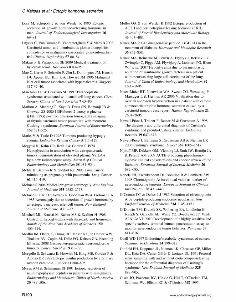
G Kaltsas et al.: Ectopic hormonal secretion
Losa M, Schopohl J & von Werder K 1993 Ectopic
secretion of growth hormone-releasing hormone in
man. Journal of Endocrinological Investigation 16
69–81.
Luyckx C, Van Damme B, Vanrenterghem Y & Maes B 2002
Carcinoid tumor and membranous glomerulonephritis:
coincidence or malignancy-associated glomerulonephri-
tis? Clinical Nephrology 57 80–84.
Makras P & Papapoulos SE 2009 Medical treatment of
hypercalcaemia. Hormones 8 83–95.
Mao C, Carter P, Schaefer P, Zhu L, Dominguez JM, Hanson
DJ, Appert HE, Kim K & Howard JM 1995 Malignant
islet cell tumor associated with hypercalcemia. Surgery
117 37–40.
Marchioli CC & Graziano SL 1997 Paraneoplastic
syndromes associated with small cell lung cancer. Chest
Surgery Clinics of North America 7 65–80.
Markou A, Manning P, Kaya B, Datta SN, Bomanji JB &
Conway GS 2005 [18F]fluoro-2-deoxy-D-glucose
([18F]FDG) positron emission tomography imaging
of thymic carcinoid tumor presenting with recurrent
Cushing’s syndrome. European Journal of Endocrinology
152 521–525.
Marks V & Teale D 1998 Tumours producing hypogly-
caemia. Endocrine-Related Cancer 5 111–129.
Megyesi K, Kahn CR, Roth J & Gorden P 1974
Hypoglycemia in association with extrapancreatic
tumors: demonstration of elevated plasma NSILA-s
by a new radioreceptor assay. Journal of Clinical
Endocrinology and Metabolism 38 931–934.
Mehta H, Bahuva R & Sadikot RT 2008 Lung cancer
mimicking as pregnancy with pneumonia. Lung Cancer
61 416–419.
Melmed S 2006 Medical progress: acromegaly. New England
Journal of Medicine 355 2558–2573.
Melmed S, Ezrin C, Kovacs K, Goodman RS & Frohman LA
1985 Acromegaly due to secretion of growth hormone by
an ectopic pancreatic islet-cell tumor. New England
Journal of Medicine 312 9–17.
Mitchell ML, Ernesti M, Raben MS & Scaliter H 1968
Control of hypoglycemia with diazoxide and hormones.
Annals of the New York Academy of Sciences 150
406–414.
Modlin IM, Oberg K, Chung DC, Jensen RT, de Herder WW,
Thakker RV, Caplin M, Delle FG, Kaltsas GA, Krenning
EP et al. 2008 Gastroenteropancreatic neuroendocrine
tumours. Lancet Oncology 9 61–72.
Morgello S, Schwartz E, Horwith M, King ME, Gorden P &
Alonso DR 1988 Ectopic insulin production by a primary
ovarian carcinoid. Cancer 61 800–805.
Moses AM & Scheinman SJ 1991 Ectopic secretion of
neurohypophyseal peptides in patients with malignancy.
Endocrinology and Metabolism Clinics of North America
20 489–506.
R190
Muller OA & von Werder K 1992 Ectopic production of
ACTH and corticotropin-releasing hormone (CRH).
Journal of Steroid Biochemistry and Molecular Biology
43 403–408.
Nauck MA 2004 Glucagon-like peptide 1 (GLP-1) in the
treatment of diabetes. Hormone and Metabolic Research
36 852–858.
Nauck MA, Reinecke M, Perren A, Frystyk J, Berishvili G,
Zwimpfer C, Figge AM, Flyvbjerg A, Lankisch PG, Blum
WF et al. 2007 Hypoglycemia due to paraneoplastic
secretion of insulin-like growth factor-I in a patient
with metastasizing large-cell carcinoma of the lung.
Journal of Clinical Endocrinology and Metabolism 92
1600–1605.
Netea-Maier RT, Nieuwlaat WA, Sweep CG, Wesseling P,
Massuger L & Hermus AR 2006 Virilization due to
ovarian androgen hypersecretion in a patient with ectopic
adrenocorticotrophic hormone secretion caused by a
carcinoid tumour: case report. Human Reproduction 21
2601–2605.
Newell-Price J, Trainer P, Besser M & Grossman A 1998
The diagnosis and differential diagnosis of Cushing’s
syndrome and pseudo-Cushing’s states. Endocrine
Reviews 19 647–672.
Newell-Price J, Bertagna X, Grossman AB & Nieman LK
2006 Cushing’s syndrome. Lancet 367 1605–1617.
Nijhoff MF, Dekkers OM, Vleming LJ, Smit JW, Romijn JA
& Pereira AM 2009 ACTH-producing pheochromo-
cytoma: clinical considerations and concise review of the
literature. European Journal of Internal Medicine 20
682–685.
Nobels FR, Kwekkeboom DJ, Bouillon R & Lamberts SW
1998 Chromogranin A: its clinical value as marker of
neuroendocrine tumours. European Journal of Clinical
Investigation 28 431–440.
O’Connor DT & Deftos LJ 1986 Secretion of chromogranin
A by peptide-producing endocrine neoplasms. New
England Journal of Medicine 314 1145–1151.
O’Dorisio TM, Krutzik SR, Woltering EA, Lindholm E,
Joseph S, Gandolfi AE, Wang YZ, Boudreaux JP, Vinik
AI & Go VL 2010 Development of a highly sensitive and
specific carboxy-terminal human pancreastatin assay to
monitor neuroendocrine tumor behavior. Pancreas 39
611–616.
Odell WD 1997 Endocrine/metabolic syndromes of cancer.
Seminars in Oncology 24 299–317.
Oldfield EH, Doppman JL, Nieman LK, Chrousos GP, Miller
DL, Katz DA, Cutler GB Jr & Loriaux DL 1991 Petrosal
sinus sampling with and without corticotropin-releasing
hormone for the differential diagnosis of Cushing’s
syndrome. New England Journal of Medicine 325
897–905.
Olsen JO, Pozderac RV, Hinkle G, Hill T, O’Dorisio TM,
Schirmer WJ, Ellison EC & O’Dorisio MS 1995
www.endocrinology-journals.org
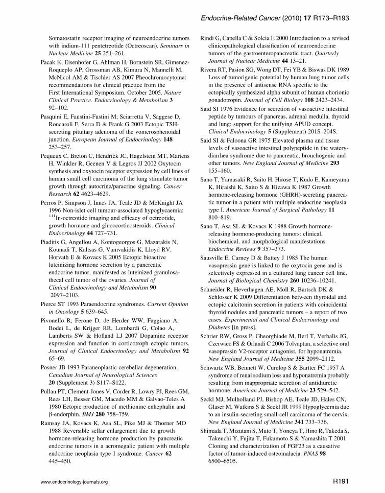
Endocrine-Related Cancer (2010) 17 R173–R193
Somatostatin receptor imaging of neuroendocrine tumors
with indium-111 pentetreotide (Octreoscan). Seminars in
Nuclear Medicine 25 251–261.
Pacak K, Eisenhofer G, Ahlman H, Bornstein SR, Gimenez-
Roqueplo AP, Grossman AB, Kimura N, Mannelli M,
McNicol AM & Tischler AS 2007 Pheochromocytoma:
recommendations for clinical practice from the
First International Symposium. October 2005. Nature
Clinical Practice. Endocrinology & Metabolism 3
92–102.
Pasquini E, Faustini-Fustini M, Sciarretta V, Saggese D,
Roncaroli F, Serra D & Frank G 2003 Ectopic TSH-
secreting pituitary adenoma of the vomerosphenoidal
junction. European Journal of Endocrinology 148
253–257.
Pequeux C, Breton C, Hendrick JC, Hagelstein MT, Martens
H, Winkler R, Geenen V & Legros JJ 2002 Oxytocin
synthesis and oxytocin receptor expression by cell lines of
human small cell carcinoma of the lung stimulate tumor
growth through autocrine/paracrine signaling. Cancer
Research 62 4623–4629.
Perros P, Simpson J, Innes JA, Teale JD & McKnight JA
1996 Non-islet cell tumour-associated hypoglycaemia:111In-octreotide imaging and efficacy of octreotide,
growth hormone and glucocorticosteroids. Clinical
Endocrinology 44 727–731.
Piaditis G, Angellou A, Kontogeorgos G, Mazarakis N,
Kounadi T, Kaltsas G, Vamvakidis K, Lloyd RV,
Horvath E & Kovacs K 2005 Ectopic bioactive
luteinizing hormone secretion by a pancreatic
endocrine tumor, manifested as luteinized granulosa-
thecal cell tumor of the ovaries. Journal of
Clinical Endocrinology and Metabolism 90
2097–2103.
Pierce ST 1993 Paraendocrine syndromes. Current Opinion
in Oncology 5 639–645.
Pivonello R, Ferone D, de Herder WW, Faggiano A,
Bodei L, de Krijger RR, Lombardi G, Colao A,
Lamberts SW & Hofland LJ 2007 Dopamine receptor
expression and function in corticotroph ectopic tumors.
Journal of Clinical Endocrinology and Metabolism 92
65–69.
Posner JB 1993 Paraneoplastic cerebellar degeneration.
Canadian Journal of Neurological Sciences
20 (Supplement 3) S117–S122.
Pullan PT, Clement-Jones V, Corder R, Lowry PJ, Rees GM,
Rees LH, Besser GM, Macedo MM & Galvao-Teles A
1980 Ectopic production of methionine enkephalin and
b-endorphin. BMJ 280 758–759.
Ramsay JA, Kovacs K, Asa SL, Pike MJ & Thorner MO
1988 Reversible sellar enlargement due to growth
hormone-releasing hormone production by pancreatic
endocrine tumors in a acromegalic patient with multiple
endocrine neoplasia type I syndrome. Cancer 62
445–450.
www.endocrinology-journals.org
Rindi G, Capella C & Solcia E 2000 Introduction to a revised
clinicopathological classification of neuroendocrine
tumors of the gastroenteropancreatic tract. Quarterly
Journal of Nuclear Medicine 44 13–21.
Rivera RT, Pasion SG, Wong DT, Fei YB & Biswas DK 1989
Loss of tumorigenic potential by human lung tumor cells
in the presence of antisense RNA specific to the
ectopically synthesized alpha subunit of human chorionic
gonadotropin. Journal of Cell Biology 108 2423–2434.
Said SI 1976 Evidence for secretion of vasoactive intestinal
peptide by tumours of pancreas, adrenal medulla, thyroid
and lung: support for the unifying APUD concept.
Clinical Endocrinology 5 (Supplement) 201S–204S.
Said SI & Faloona GR 1975 Elevated plasma and tissue
levels of vasoactive intestinal polypeptide in the watery-
diarrhea syndrome due to pancreatic, bronchogenic and
other tumors. New England Journal of Medicine 293
155–160.
Sano T, Yamasaki R, Saito H, Hirose T, Kudo E, Kameyama
K, Hiraishi K, Saito S & Hizawa K 1987 Growth
hormone-releasing hormone (GHRH)-secreting pancrea-
tic tumor in a patient with multiple endocrine neoplasia
type I. American Journal of Surgical Pathology 11
810–819.
Sano T, Asa SL & Kovacs K 1988 Growth hormone-
releasing hormone-producing tumors: clinical,
biochemical, and morphological manifestations.
Endocrine Reviews 9 357–373.
Sausville E, Carney D & Battey J 1985 The human
vasopressin gene is linked to the oxytocin gene and is
selectively expressed in a cultured lung cancer cell line.
Journal of Biological Chemistry 260 10236–10241.
Schneider R, Heverhagen AE, Moll R, Bartsch DK &
Schlosser K 2009 Differentiation between thyroidal and
ectopic calcitonin secretion in patients with coincidental
thyroid nodules and pancreatic tumors – a report of two
cases. Experimental and Clinical Endocrinology and
Diabetes [in press].
Schrier RW, Gross P, Gheorghiade M, Berl T, Verbalis JG,
Czerwiec FS & Orlandi C 2006 Tolvaptan, a selective oral
vasopressin V2-receptor antagonist, for hyponatremia.
New England Journal of Medicine 355 2099–2112.
Schwartz WB, Bennett W, Curelop S & Bartter FC 1957 A
syndrome of renal sodium loss and hyponatremia probably
resulting from inappropriate secretion of antidiuretic
hormone. American Journal of Medicine 23 529–542.
Seckl MJ, Mulholland PJ, Bishop AE, Teale JD, Hales CN,
Glaser M, Watkins S & Seckl JR 1999 Hypoglycemia due
to an insulin-secreting small-cell carcinoma of the cervix.
New England Journal of Medicine 341 733–736.
Shimada T, Mizutani S, Muto T, Yoneya T, Hino R, Takeda S,
Takeuchi Y, Fujita T, Fukumoto S & Yamashita T 2001
Cloning and characterization of FGF23 as a causative
factor of tumor-induced osteomalacia. PNAS 98
6500–6505.
R191

G Kaltsas et al.: Ectopic hormonal secretion
Shimizu K, Nakano S, Nakano Y, Ando M, Seki K &
Kameda N 1991 Ectopic atrial natriuretic peptide
production in small cell lung cancer with the syndrome of
inappropriate antidiuretic hormone secretion. Cancer 68
2284–2288.
Srirajaskanthan R, McStay M, Toumpanakis C, Meyer T &
Caplin ME 2009 Parathyroid hormone-related peptide-
secreting pancreatic neuroendocrine tumours: case
series and literature review. Neuroendocrinology 89
48–55.
Stewart PM, Gibson S, Crosby SR, Penn R, Holder R, Ferry
D, Thatcher N, Phillips P, London DR & White A 1994
ACTH precursors characterize the ectopic ACTH
syndrome. Clinical Endocrinology 40 199–204.
Strewler GJ 2000 The physiology of parathyroid
hormone-related protein. New England Journal of
Medicine 342 177–185.
Sundler F, Ekblad E, Grunditz T, Hakanson R & Uddman R
1988 Vasoactive intestinal peptide in the peripheral
nervous system. Annals of the New York Academy of
Sciences 527 143–167.
Suva LJ, Winslow GA, Wettenhall RE, Hammonds RG,
Moseley JM, Diefenbach-Jagger H, Rodda CP, Kemp BE,
Rodriguez H & Chen EY 1987 A parathyroid hormone-
related protein implicated in malignant hypercalcemia:
cloning and expression. Science 237 893–896.
Suzuki K, Miyashita A, Inoue Y, Iki S, Enomoto H,
Takahashi Y & Takemura T 1991 Interleukin-6-
producing pheochromocytoma. Acta Haematologica 85
217–219.
Szturmowicz M, Wiatr E, Sakowicz A, Slodkowska J,
Roszkowski K, Filipecki S & Rowinska-Zakrzewska ER
1995 The role of human chorionic gonadotropin beta
subunit elevation in small-cell lung cancer patients.
Journal of Cancer Research and Clinical Oncology 121
309–312.
Tabibzadeh SS, Poubouridis D, May LT & Sehgal PB 1989
Interleukin-6 immunoreactivity in human tumors.
American Journal of Pathology 135 427–433.
Takami H, Shikata J, Kakudo K & Ito K 1990 Calcitonin
gene-related peptide in patients with endocrine tumors.
Journal of Surgical Oncology 43 28–32.
Taupenot L, Harper KL & O’Connor DT 2003 The
chromogranin–secretogranin family. New England
Journal of Medicine 348 1134–1149.
Teale JD & Marks V 1998 Glucocorticoid therapy suppresses
abnormal secretion of big IGF-II by non-islet cell
tumours inducing hypoglycaemia (NICTH). Clinical
Endocrinology 49 491–498.
Teale JD, Blum WF & Marks V 1992 Alleviation of non-islet
cell tumour hypoglycaemia by growth hormone therapy is
associated with changes in IGF binding protein-3. Annals
of Clinical Biochemistry 29 314–323.
Terek RM & Nielsen GP 2001 Case records of the
Massachusetts General Hospital. Weekly
R192
clinicopathological exercises. Case 29-2001. A 14-year-
old with abnormal bones and a sacral mass. New England
Journal of Medicine 345 903–908.
Thirkill CE, Roth AM & Keltner JL 1993 Paraneoplastic
retinopathy syndrome. Ophthalmology 100 147.
Thorner MO, Frohman LA, Leong DA, Thominet J, Downs T,
Hellmann P, Chitwood J, Vaughan JM & Vale W 1984
Extrahypothalamic growth-hormone-releasing factor
(GRF) secretion is a rare cause of acromegaly: plasma
GRF levels in 177 acromegalic patients. Journal of
Clinical Endocrinology and Metabolism 59 846–849.
Tischler AS, Lee YC, Perlman RL, Costopoulos D,
Slayton VW & Bloom SR 1984 Production of
“ectopic” vasoactive intestinal peptide-like and
neurotensin-like immunoreactivity in human
pheochromocytoma cell cultures. Journal of Neuroscience
4 1398–1404.
Todd JF, Stanley SA, Roufosse CA, Bishop AE, Khoo B,
Bloom SR & Meeran K 2003 A tumour that secretes
glucagon-like peptide-1 and somatostatin in a patient
with reactive hypoglycaemia and diabetes. Lancet 361
228–230.
Tsagarakis S, Christoforaki M, Giannopoulou H, Rondo-
gianni F, Housianakou I, Malagari C, Rontogianni D,
Bellenis I & Thalassinos N 2003 A reappraisal of the
utility of somatostatin receptor scintigraphy in patients
with ectopic adrenocorticotropin Cushing’s syndrome.
Journal of Clinical Endocrinology and Metabolism 88
4754–4758.
Tsolakis AV, Portela-Gomes GM, Stridsberg M, Grimelius L,
Sundin A, Eriksson BK, Oberg KE & Janson ET 2004
Malignant gastric ghrelinoma with hyperghrelinemia.
Journal of Clinical Endocrinology and Metabolism 89
3739–3744.
Turkington RW 1971 Ectopic production of prolactin. New
England Journal of Medicine 285 1455–1458.
Upton GV & Amatruda TT Jr 1971 Evidence for the presence
of tumor peptides with corticotropin-releasing-factor-like
activity in the ectopic ACTH syndrome. New England
Journal of Medicine 285 419–424.
Uysal M, Temiz S, Gul N, Yarman S, Tanakol R &
Kapran Y 2007 Hypoglycemia due to ectopic release
of insulin from a paraganglioma. Hormone Research
67 292–295.
VanHouten JN, Yu N, Rimm D, Dotto J, Arnold A,
Wysolmerski JJ & Udelsman R 2006 Hypercalcemia
of malignancy due to ectopic transactivation of the
parathyroid hormone gene. Journal of Clinical
Endocrinology and Metabolism 91 580–583.
Vieira NL, Taboada GF, Correa LL, Polo J, Nascimento AF,
Chimelli L, Rumilla K & Gadelha MR 2007 Acromegaly
secondary to growth hormone-releasing hormone secreted
by an incidentally discovered pheochromocytoma.
Endocrine Pathology 18 46–52.
www.endocrinology-journals.org

Endocrine-Related Cancer (2010) 17 R173–R193
Vinik AI & Ziegler D 2007 Diabetic cardiovascular
autonomic neuropathy. Circulation 115 387–397.
Vinik AI, Silva MP, Woltering G, Go VL, Warner R &
Caplin M 2009 Biochemical testing for neuroendocrine
tumors. Pancreas 38 876–889.
Wajchenberg BL, Mendonca BB, Liberman B, Pereira MA,
Carneiro PC, Wakamatsu A & Kirschner MA 1994
Ectopic adrenocorticotropic hormone syndrome.
Endocrine Reviews 15 752–787.
Weidner N & Santa CD 1987 Phosphaturic mesenchymal
tumors. A polymorphous group causing osteomalacia or
rickets. Cancer 59 1442–1454.
Weintraub BD & Kadesky YM 1971 Fractionation of
antibodies to human chorionic somatomammotropin:
application to radioimmunoassay. Journal of
Clinical Endocrinology and Metabolism 33 432–435.
Weiss ES, Doty J, Brock MV, Halvorson L & Yang SC 2006
A case of ectopic parathyroid hormone production by
a pulmonary neoplasm. Journal of Thoracic and
Cardiovascular Surgery 131 923–924.
Woltering EA, Hilton RS, Zolfoghary CM, Thomson J, Zietz S,
Go VL, Vinik AI, Vinik E, O’Dorisio TM & Mamikunian G
2006 Validation of serum versus plasma measurements
of chromogranin a levels in patients with carcinoid
tumors: lack of correlation between absolute chromo-
granin a levels and symptom frequency. Pancreas 33
250–254.
www.endocrinology-journals.org
Wymenga AN, de Monchy JG & de Vries EG 1995 The
carcinoid syndrome and angioedema. Annals of Internal
Medicine 123 636.
Yaturu S, Harrara E, Nopajaroonsri C, Singal R & Gill S
2003 Gynecomastia attributable to human chorionic
gonadotropin-secreting giant cell carcinoma of lung.
Endocrine Practice 9 233–235.
Yoshizaki K, Matsuda T, Nishimoto N, Kuritani T,
Taeho L, Aozasa K, Nakahata T, Kawai H, Tagoh H
& Komori T 1989 Pathogenic significance of
interleukin-6 (IL-6/BSF-2) in Castleman’s disease.
Blood 74 1360–1367.
Young J, Deneux C, Grino M, Oliver C, Chanson P &
Schaison G 1998 Pitfall of petrosal sinus sampling in a
Cushing’s syndrome secondary to ectopic adrenocortico-
tropin–corticotropin releasing hormone (ACTH–CRH)
secretion. Journal of Clinical Endocrinology and
Metabolism 83 305–308.
Zangeneh F, Young WF Jr, Lloyd RV, Chiang M, Kurczynski
E & Zangeneh F 2003 Cushing’s syndrome due to ectopic
production of corticotropin-releasing hormone in an
infant with ganglioneuroblastoma. Endocrine Practice 9
394–399.
Zapf J, Futo E, Peter M & Froesch ER 1992 Can “big”
insulin-like growth factor II in serum of tumor patients
account for the development of extrapancreatic tumor
hypoglycemia? Journal of Clinical Investigation 90
2574–2584.
R193