Embed Size (px)
Citation preview


Guidelines for the detection and characterisation of
clinically relevant antibodies in allotransplantation
___________________________________________________________________________
Page 2 of 101
A collaborative publication by:
British Society for Histocompatibility & Immunogenetics
12 Coldbath Square, London, EC1 5HL
www.bshi.org.uk
and
British Transplantation Society
South Park Road, Macclesfield, Cheshire, SK11 6SH
www.bts.org.uk
Publication at www.bts.org.uk & www.bshi.org.uk, May 2010
Revision due May 2012 Based on search of publications at March 2010
The British Society for Histocompatibility & Immunogenetics is the professional body for healthcare and basic scientists supporting clinical transplantation and transplantation research. The British Transplantation Society is the professional body for Surgeons, Physicians, Specialist Nurses for Organ Donations, Nurses, Healthcare Scientists, Allied Health Professionals and Basic Scientists actively working in clinical transplantation and transplantation research.
Guidelines for the detection and characterisation of
clinically relevant antibodies in allotransplantation
___________________________________________________________________________
Page 3 of 101
Disclaimer:
These Guidelines are guides to best practice which inevitably change with the
passage of time. All practitioners need to undertake clinical care on an individual
basis and keep themselves up to date with changes in practice of clinical
medicine. The British Transplantation Society and The British Society for
Histocompatibility & Immunogenetics Guidelines ("the Guidelines") were
compiled by a joint working party of the Societies. The Guidelines represent the
collective opinions of a number of experts in the field and do not have the force
of law. The Guidelines contain information and guidance for use by practitioners
as a best practice tool; it follows that the Guidelines should be interpreted as
such rather than the letter of their contents. The opinions presented in
the Guidelines are subject to change and should not be considered to be a
treatment recommendation for any individual patient.
The British Transplantation Society and The British Society for
Histocompatibility & Immunogenetics cannot attest to the accuracy,
completeness or currency of the opinions contained herein and does not
accept any responsibility or liability for any loss or damage caused to
any practitioner or any third party as a result of any reliance being
placed on the Guidelines or as a result of any inaccurate or misleading
opinion contained in the Guidelines.

Guidelines for the detection and characterisation of
clinically relevant antibodies in allotransplantation
___________________________________________________________________________
Page 4 of 101
Contents: Page: 1 Process of Guidelines development 6 2 Evidence base 7 3 Writing committee 7 4 Overview, recommendations and best practice 10 4.1 Immunological risk 10 4.2 Recommendations 11 4.2.1 Kidney and pancreas 11 4.2.2 Islets 13 4.2.3 Thoracic organs 14 4.2.4 Liver 15 4.2.5 Intestinal and multi-visceral 15 4.2.6 HLA-specific antibody incompatible transplantation 16 5 Introduction 17 5.1 The immune system 18 5.1.1 T cell recognition of foreign antigens 18 5.1.2 Effector cell activation and functions 18 5.1.3 Control of allorecognition 20 5.2 Priming sources 21 5.3 Acute antibody mediated rejection mechanisms 22 5.3.1 Hyperacute rejection 22 5.3.2 Acute rejection 22 6 Defining risk 23 7 Identification of HLA specific antibodies 24 7.1 Cell based assays 24 7.1.1 Complement dependent cytotoxicity 24 7.1.2 Flow cytometry 26 7.2 Solid phase assays 27
7.2.1 Enzyme-linked immunosorbent assays 27 7.2.2 Flow cytometry 28
7.2.3 X-map (Luminex) 28 7.3 Interpretation of HLA specific antibody data in the clinical setting 31 7.4 Screening strategies 33 7.5 Collection & storage of samples for antibody screening & donor 35 crossmatching 7.5.1 pre-transplant 35 7.5.2 post-transplant 35 7.5.3 sample storage 36 7.6 Recommendations 36

Guidelines for the detection and characterisation of
clinically relevant antibodies in allotransplantation
___________________________________________________________________________
Page 5 of 101
8 Crossmatching 37 8.1 Crossmatch techniques 38 8.2 Pre-transplant virtual crossmatching 39 8.3 Sample selection 40 8.4 Results and interpretation 41 8.5 Recommendations 43 9 Kidney and pancreas transplantation 44 9.1 Pre-transplant antibody screening 45 9.2 Definition of unacceptable mismatches 47 9.3 Clinical relevance of crossmatching 48 9.3.1 The cytotoxic crossmatch 48 9.3.2 Flow cytometric crossmatching 50 9.3.3 Virtual crossmatching 52 9.3.4 Reporting crossmatch results 53 9.4 Development of HLA specific antibodies after kidney 53
transplantation 9.5 Recommendations 56 10 Islet transplantation 57 10.1 Pre-transplant antibody screening 57 10.2 Post first transplant and pre-second transplant antibodies 58 10.3 Crossmatching 59 10.4 Recommendations 59 11 Thoracic organ transplantation 60 11.1 Pre-transplant HLA specific antibodies 61 11.2 Post-transplant production of HLA specific antibodies 63 11.3 Non-HLA specific antibodies 64 11.4 Recommendations 65 12 Liver transplantation 66 12.1 Recommendations 69 13 Intestinal and multi-visceral transplantation 69 13.1 Recommendations 72 14 HLA-specific antibody incompatible transplantation (AiT) 72 14.1 Recommendations 76 15 Appendices 15.1 Laboratory resources and relationship 79 15.2 Glossary 80 15.3 References 90 15.4 Statements of potential conflicts of interests 101

Guidelines for the detection and characterisation of
clinically relevant antibodies in allotransplantation
___________________________________________________________________________
Page 6 of 101
1: Process of Guidelines development
The past decade witnessed a revolution in technology allowing the
histocompatibility laboratory to define the presence of HLA specific antibodies
with a high degree of sensitivity, resulting in ongoing re-definition of the
crossmatch boundary to allow successful transplantation of allo-sensitised
patients. These developments started early in the past decade leading to the
publication of the 2004 Guidelines. Subsequently technological developments
continued allowing relative quantitation of HLA specific antibody levels which,
combined with crossmatching results allowed a graded assessment of the
immunological risk should a transplant proceed, rather than a simple ‘positive’ or
‘negative’ crossmatch assessment. This facilitated the establishment and growth
of HLA antibody incompatible living donor kidney transplantation. Together,
these and other developments have driven a thorough revision of these
Guidelines, so that optimum approaches can be applied to maximise safe and
effective use of the donor organ pool and to help expand this pool. These revised
Guidelines accept ABO blood group compatibility as a pre-requisite and focus on
HLA specific antibodies in allotransplantation, although selective references to
non-HLA specific antibodies are included. ABO blood group incompatible kidney
transplantation is addressed as a separate issue. The role of HLA specific
antibodies in some forms of HLA mismatched haematopoietic progenitor cell
transplantation is also of interest, but for clarity is excluded from these
Guidelines. The Histocompatibility laboratory will also play a critical role in the
development of cell therapies and although the Writers discussed these
developments, it was decided to omit this exciting area.

Guidelines for the detection and characterisation of
clinically relevant antibodies in allotransplantation
___________________________________________________________________________
Page 7 of 101
2: THE EVIDENCE BASE:
The Writers drew the evidence to support their recommendations from peer-
review publications up to March 2010. The specialist nature of histocompatibility
testing in the context of clinical allotransplantation means that there are few
large or multicentre studies in this field and meta analyses are not possible.
Furthermore, recent changes in technology to detect and define HLA specific
sensitisation have made many historic publications less relevant or obsolete.
3: WRITING COMMITTEE:
Executive bodies from the BSHI and the BTS invited the persons below to
compile the original Guidelines which were published in 2004. A revision process,
involving the original authors and Martin Howell as Chair of the writing
committee, resulted in production of these Guidelines. One meeting, chaired by
Martin Howell, was held to complete this process and a final draft was sent to
selected clinical colleagues for review and comment. Finally, by email to all BSHI
and BTS Members, comments were invited by placing the draft Guidelines on
both Society websites for a four week period. Comments were reviewed and the
Guidelines amended by Phil Dyer, who also formatted this publication. There was
no specific request for non-professional comment.

Guidelines for the detection and characterisation of
clinically relevant antibodies in allotransplantation
___________________________________________________________________________
Page 8 of 101
Writing Committee: Chair: Martin Howell PhD FRCPath, Consultant Clinical Scientist Histocompatibility & Immunogenetics Laboratory, NHS Blood & Transplant Newcastle upon Tyne NE2 4NQ Chair of Writing Committee for 2004 Guidelines: Andrea Harmer PhD FRCPath, Consultant Clinical Scientist Histocompatibility & Immunogenetics Laboratory, NHS Blood & Transplant Sheffield S5 7JN Members: David Briggs PhD, Consultant Clinical Scientist Histocompatibility & Immunogenetics Laboratory, NHS Blood & Transplant Birmingham B15 2TT Philip Dyer PhD FRCPath, Consultant Clinical Scientist Histocompatibility & Immunogenetics, Scottish National Blood Transfusion Service Edinburgh EH16 4SA Susan Fuggle DPhil FRCPath, Consultant Clinical Scientist Transplant Immunology and Immunogenetics Laboratory, Churchill Hospital Oxford, OX3 7LJ Susan Martin PhD FRCPath, Consultant Clinical Scientist Transplantation Laboratory, Manchester Royal Infirmary Manchester, M13 9WL Paul Sinnott PhD FRCPath, Consultant Clinical Scientist The Royal London Hospital, London E1 1BB John Smith PhD, Consultant Head of Tissue Typing Service Tissue Typing Laboratory, Harefield Hospital Harefield UB9 6JH Craig Taylor PhD, FRCPath. Consultant Clinical Scientist, Tissue Typing Laboratory, Addenbrooke's Hospital Cambridge CB2 0QQ Robert Vaughan PhD FRCPath, Consultant Clinical Scientist Tissue Typing Laboratory, Guy’s Hospital London SE1 9RT

Guidelines for the detection and characterisation of
clinically relevant antibodies in allotransplantation
___________________________________________________________________________
Page 9 of 101
Clinical reviewers: Miss Lorna Marson, Consultant Transplant Surgeon and Senior Lecturer Royal Infirmary Edinburgh, Edinburgh EH16 4SA Dr Chas Newstead, Consultant Nephrologist St James’ University Hospital, Leeds LS9 7TF Dr Jayan Parameshwar, Consultant Cardiologist Papworth Hospital, Cambridge CB23 3RE Mr Keith Rigg, President – British Transplantation Society and Consultant Surgeon Nottingham University Hospitals NHS Trust, Nottingham NG5 1PB Dr Michael Sheaff, Consultant Histopathologist St Bartholomew’s Hospital, London EC1A 7BE Dr Raj Thuraisingham, Honorary Clinical Senior Lecturer and Consultant Nephrologist Royal London Hospital, London E1 1BB Dr Jane Tizard, Consultant Paediatric Nephrologist Bristol Royal Hospital for Children, Bristol BS32 8BJ Dr Nicholas Torpey, Consultant Transplant Nephrologist Addenbrooke’s Hospital, Cambridge CB2 0QQ Professor Anthony Warrens, Professor of Renal and Transplant Medicine and Honorary Consultant Physician Hammersmith Hospital, London W12 0NN Mr Christopher Watson, Reader in Surgery and Honorary Consultant Surgeon University of Cambridge Department of Surgery, Addenbrooke’s Hospital Cambridge CB2 0QQ Comments received from BSHI and BTS Members: Dr Peter ANDREWS, Consultant Nephrologist, Chair BTS Standards Committee SW Thames Renal & Transplantation Unit, St Helier Hospital Carshalton SM5 1AA Vaughan CARTER PhD FRCPath, Clinical Scientist Histocompatibility & Immunogenetics Laboratory, NHS Blood & Transplant Newcastle upon Tyne NE2 4NQ Brendan CLARKE PhD, Consultant Clinical Scientist Histocompatibility & Immunogenetics Laboratory, St James’ University Hospital, Leeds LS9 7TF Ms Sussie SHRESTHA, Clinical Research Fellow Royal Infirmary Edinburgh, Edinburgh EH16 4SA

Guidelines for the detection and characterisation of
clinically relevant antibodies in allotransplantation
___________________________________________________________________________
Page 10 of 101
4: OVERVIEW, RECOMMENDATIONS AND BEST PRACTICE
4.1 Immunological risk
Gebel et al [17] have suggested three broad risk categories for a given donor-
recipient combination (adapted below):
1) High immunological risk is indicated when there are high titre circulating
antibodies specific for mismatched donor HLA antigens present at the time of
transplantation. In most cases the high risk of hyperacute rejection would
constitute a veto to transplantation. Some centres may advocate carefully
planned pre-transplant desensitisation regimes together with close post-
transplant immunological monitoring.
2) Intermediate immunological risk is indicated by the presence of risk factors
such as prior donor reactive sensitisation (that is absent at the time of
transplantation) and in selected donor-recipient combinations with only weak
sensitisation to certain mismatched HLA specificities. In such cases, it may be
justified to consider augmented immunosuppression and post-transplant
immunological monitoring.
3) Low (or standard) immunological risk is indicated when non-sensitised or
sensitised patients receive a minimally HLA mismatched organ in the absence
of current or historical donor reactive antibodies.

Guidelines for the detection and characterisation of
clinically relevant antibodies in allotransplantation
___________________________________________________________________________
Page 11 of 101
4.2 Recommendations
4.2.1 Kidney and pancreas transplantation: • Laboratories must have a strategy in place for the detection and
characterisation of clinically relevant antibodies.
• The techniques adopted must have the capability of defining antibody
class and specificity.
• A combination of tests should be considered in order to fully resolve
complex antibody profiles.
• Laboratories should have a programme for investigating and
evaluating newly emerging technologies so that the
histocompatibility service supporting clinical transplant programmes
develops in line with current treatments.
• Patient serum samples must be sent to the histocompatibility
laboratory no less than three monthly for routine antibody
monitoring and also following transfusion of any blood products.
• Serum samples must be stored for potential use in future antibody
screening and crossmatch tests.
• Laboratory crossmatch tests should distinguish T cell and B cell
populations and between IgG and IgM antibodies.
• Serum samples used for crossmatching must include a current
sample and, where HLA specific antibodies have been detected,
samples that are representative of the patient’s antibody profile, over
time.

Guidelines for the detection and characterisation of
clinically relevant antibodies in allotransplantation
___________________________________________________________________________
Page 12 of 101
• The timing, duration, priming source, antibody titre and donor
specificity should be considered when interpreting the crossmatch
result.
• The reporting of results to clinical teams should include appropriate
advice on the clinical relevance of the result.
• Laboratories providing services for kidney transplant programmes
must have the capability of precisely defining HLA-A, -B, –Cw, -DR,
–DQ and -DP antibody specificities in their patients so that donors
who should be crossmatch negative can be identified.
• A patient’s HLA alloantibody profile must be assessed to delineate
the antigens regarded as unacceptable for transplant.
• A pre-transplant crossmatch should be performed for all patients
unless a programme exists for identifying those individuals who can
confidently be defined as unsensitised. Patients with no detectable
HLA specific antibodies can be transplanted on the basis of a negative
virtual crossmatch (vXM) without waiting for a crossmatch test to be
performed.
• Sensitised patients should be crossmatched using flow cytometric
techniques.
• Post-transplant antibody monitoring should be performed at agreed
regular intervals, at the time of biopsy and in cases of suspected
rejection. Samples should also be tested at times of declining graft
function when there is no other clinical cause.

Guidelines for the detection and characterisation of
clinically relevant antibodies in allotransplantation
___________________________________________________________________________
Page 13 of 101
• The clinical team must inform the laboratory of potential
sensitisation events, such as previous transplantation, skin grafting,
transfusion of blood products, pregnancy (including known
miscarriages) and recent infection or vaccination.
4.2.2 Islets
• Prior to listing a patient for transplant, it is recommended that
antibody screening and specificity analysis are performed on two
separate samples.
• Once the patient is listed, samples for antibody analysis should be
obtained no less than three monthly.
• Potential priming events should be notified promptly to the
laboratory and samples sent approximately 2-4 weeks after the
event.
• Antibody testing should be performed by two different assays,
including a highly sensitive technique to determine the specificity of
the antibodies.
• Both cytotoxic and flow cytometry crossmatching is recommended.
• Proceeding to transplant on the basis of a virtual crossmatch may be
acceptable if the laboratory and transplant centre have a validated
policy.
• It is recommended that samples are taken regularly following the
first and any subsequent transplants.

Guidelines for the detection and characterisation of
clinically relevant antibodies in allotransplantation
___________________________________________________________________________
Page 14 of 101
4.2.3 Thoracic organs:
• Laboratories must be able to precisely define antibodies specific
for HLA-A, -B, -Cw, -DR, -DQ and –DP using sensitive solid phase
assays.
• HLA antigens to which a patient has produced antibodies should
be listed as “unacceptable mismatches”
• Before transplantation, patients must be screened for HLA specific
antibodies on at least two occasions prior to listing, preferably
taken at least 24 hours apart, although this may not be possible
for urgent patients.
• Samples must also be collected following any sensitising events
such as transfusion of blood products or pregnancy, and every
three months whilst on the waiting list so that a complete
antibody profile is available prior to transplantation.
• Patients with no detectable HLA-specific antibodies can be
transplanted following virtual crossmatching in the
histocompatibility laboratory.
• Sensitised patients with fully defined HLA-specific antibodies and
with no residual reactivity can also be transplanted without a
prospective crossmatch test provided the virtual crossmatch is
negative i.e. the donor does not carry those HLA specificities to
which the patient is sensitised. Prospective crossmatching must
be performed for all other sensitised patients.

Guidelines for the detection and characterisation of
clinically relevant antibodies in allotransplantation
___________________________________________________________________________
Page 15 of 101
• For patients where a prospective crossmatch test is not performed
a retrospective crossmatch using serum collected within 48 hours
prior to transplantation must be performed.
• Crossmatch techniques should distinguish between IgG and IgM
as well as T and B cells as targets.
• Following transplantation patients should be screened for HLA
specific antibodies at regular intervals and must be tested when
clinically necessary.
4.2.4 Liver
• Prospective crossmatching is not indicated prior to liver
transplantation.
• Identification of donor specific antibody can be used to identify
patients at high risk of acute rejection and can aid post-transplant
management, such as changes in immunosuppression regimen or
antibody removal.
4.2.5 Intestinal and Multi-visceral
• Pre-transplant HLA-specific antibody screening and pre-transplant
donor lymphocyte crossmatching can identify those patients at
high risk of acute cellular or vascular rejection.

Guidelines for the detection and characterisation of
clinically relevant antibodies in allotransplantation
___________________________________________________________________________
Page 16 of 101
• A positive donor cytotoxic crossmatch caused by IgG HLA class I
specific antibodies is a relative contra-indication to isolated intestinal
transplantation (in the absence of a liver transplant from the same
donor) and the final decision to proceed with transplantation will
depend on a careful evaluation of the relative risk of proceeding
versus the risk of delayed transplantation.
4.2.6 HLA-specific antibody incompatible transplantation (AiT)
• The HLA specificity and level of donor specific antibodies must be
fully determined prior to antibody reduction.
• Antibody levels must be monitored regularly throughout the duration
of treatment to determine its effectiveness using a method that can
unequivocally distinguish between antibodies directed against donor
HLA mismatches, other HLA-specific antibodies and non-HLA
antibodies.
• The frequency of post-transplant donor specific antibody monitoring
should match the risk of adverse immunological events and this is
advised for all HLA AiT cases whether or not pretransplant removal
has been used.
• Antibody reduction should only be undertaken following
establishment of a clinical and laboratory protocol.

Guidelines for the detection and characterisation of
clinically relevant antibodies in allotransplantation
___________________________________________________________________________
Page 17 of 101
5 INTRODUCTION
The adaptive immune response to infection elicits antigen specific cells and
antibodies that bind with high affinity to foreign antigens, resulting in recovery
from infection and also protection against re-infection. An unwanted ‘side effect’
of this adaptive immune response is the response to non-infectious agents (e.g.
allografts, pollen, drugs) and even to an individual’s own body constituents
(autoimmunity).
Exposure of an individual’s immune system to tissue or cells from another
individual can result in immunological priming (sensitisation) to alloantigens.
Subsequent re-exposure to the same or structurally related, cross-reactive
antigens causes a vigorous humoral and/or cellular immune response. In the
context of organ transplantation, previous immunological priming to alloantigens
can cause hyperacute rejection due to circulating pre-formed donor reactive
antibodies, or accelerated acute rejection, which is difficult to control using
conventional immunosuppressive agents. Patient exposure to alloantigens of
another individual is a common occurrence and takes place through pregnancies,
blood transfusions or previous transplants. An audit of the UK’s national kidney
transplant waiting list in March 2009 showed that 41% of adult patients and
58% of paediatric patients were sensitised (calculated donor specific antibody
reaction frequency >10%). A critical function of the histocompatibility laboratory
is to identify sensitisation in patients to reduce the immunological risks of
allotransplantation.

Guidelines for the detection and characterisation of
clinically relevant antibodies in allotransplantation
___________________________________________________________________________
Page 18 of 101
5.1 The Immune System
5.1.1 T cell recognition of foreign antigens.
The essential feature guiding the evolution of the immune system of all
vertebrate species is the need to distinguish between ‘self’ and ‘non-self’. In
humans this is achieved through T cell recognition of self -human leukocyte
antigens (HLA) which bind and present antigens in the form of processed
peptides (Figure 1).
Antigen specific T cell clones with T cell receptors (TCR) that recognise
foreign peptide bound to self-HLA engage the antigen presenting cells (APC). In
the presence of co-stimulatory molecules present on mature APCs (e.g.
CD28/CD80 interaction), T cells receive the second signal that triggers their
activation. Activated T cells undergo clonal expansion and secrete cytokines that
initiate and control the inflammatory response and are involved in recruitment of
other effector cells such as B cells, cytotoxic T cells, macrophages and natural
killer cells. In addition, a sub-population of activated T cells express the CD45RO
molecule and become long-lived memory T cells that offer a rapid and vigorous
response on re-encounter with the same priming antigen.
5.1.2 Effector cell activation and functions.
The cytokines secreted by T helper cells direct the immune response by
regulating effector cell pathways towards a humoral and/or cellular response. T
cell secretion of the cytokines IL-2, IL-4, IL-5, IL-6 and IL-13 induces activation
and differentiation of antigen specific B cells. In the presence of these cytokines,
naïve B cells that express cell surface IgM undergo immunoglobulin class

Guidelines for the detection and characterisation of
clinically relevant antibodies in allotransplantation
___________________________________________________________________________
Page 19 of 101
switching so that high affinity IgG antibodies can be produced. Under T cell help,
B cells differentiate into antibody producing plasma cells with the initial
production of IgM antibodies and subsequently IgG antibodies due to class
switching and into memory B cells that respond rapidly upon repeat exposure to
the same antigenic stimulus (Figure 1). Antibody binding to its target antigen
facilitates opsonisation by phagocytes, and chemotaxis and lysis via the classical
complement pathway.
B cells can also function as antigen-presenting cells to T cells, providing
the second signal for T cell activation. Unlike T cells that recognise processed
antigen in the context of self-HLA, B cells express cell surface immunoglobulin
that can recognise and bind native antigen enabling the selection of antigen
specific B cell clones. B cells bind exogenous antigen through their cell surface
immunoglobulin which is internalised and broken down into peptide fragments.
These peptides are loaded into the antigen binding cleft of HLA class II
molecules for presentation at the cell surface to T cells. The interaction of the
TCR with HLA/peptide complex together with co-stimulatory molecules
(CD40/CD154) stimulates antigen-specific T cell activation for the provision of B
cell help.
T cell cooperation is needed for B cells to develop into alloantibody
producing plasma cells and the characterisation of the epitope(s) to which an
alloantibody is directed can be used as a measure of the extent of T cell
sensitisation. Although the original donor cell epitopes recognised by T and B
cells are not the same, this use of antibody to characterise the memory T cell
response is an important tool in the prevention of accelerated acute rejection,

Guidelines for the detection and characterisation of
clinically relevant antibodies in allotransplantation
___________________________________________________________________________
Page 20 of 101
the “second set” response. The dynamics of an antibody response may include
”epitope spreading”, whereby as the immune response develops, the specificity
of HLA directed antibodies may broaden from the original ‘dominant’ HLA
epitope. One way of investigating this is to test a serum in dilution; when
titrated, the main specificities should become apparent. It is possible that the
main specificity approximates more closely to the T cell epitope. It may
therefore be conjectured that the removal of the broad or secondary antibody
specificities may not engender an accelerated memory T cell response in a graft
expressing these HLA mismatches.
Helper T cells can initiate cellular immune responses by the activation of
antigen specific cytotoxic T cells and are also involved in the non-specific
activation of macrophages and natural killer cells causing cell-mediated
cytotoxicity. Currently there is no routine assay of T cell sensitisation.
5.1.3 Control of allorecognition.
In kidney transplantation, attempts are made to reduce the immunogenicity and
alloantigen load through avoiding HLA mismatching of donors to recipients. This
is effective at reducing the number and severity of acute rejection episodes and
is also translated into improved long-term graft survival [1-4]. In addition,
conventional calcineurin inhibitor based immunosuppressive regimens are potent
inhibitors of naïve T cells and effectively control the primary immune response to
HLA alloantigens expressed on transplanted tissue. In individuals who are
Guidelines for the detection and characterisation of
clinically relevant antibodies in allotransplantation
___________________________________________________________________________
Page 21 of 101
already primed to donor HLA antigens, both humoral and cellular secondary
responses are poorly controlled by current immunosuppressive agents.
5.2 Priming sources
Exposure of an individual’s immune system to alloantigens of another individual
by pregnancy, blood transfusion or transplantation can result in immunological
priming. In addition, HLA-specific antibodies have been observed in patients in
the absence of obvious priming events. Idiopathic HLA-specific antibodies are
usually IgM and may arise through cross-reactivity with infectious micro-
organisms. More recently, antibody screening by highly sensitive solid phase
binding assays using purified HLA molecules has identified IgG HLA-specific
antibodies in the sera of normal healthy individuals with no history of
allosensitisation events, although it is not yet known whether these antibodies
are clinically relevant to graft rejection [5-6].
It has been suggested that the use of leukodepleted blood negates the risk
of post transfusion allosensitisation but a randomised trial found that buffy coat
removal and additional white blood cell reduction by filtration resulted in similar
post-transfusion alloimmunisation frequencies after a single transfusion event
[7]. The risk of sensitisation after blood transfusion is highly variable and is
influenced by recipient factors such as genetic control of the immune response
and previous exposure to alloantigens. In addition the storage time of transfused
blood is relevant.
Guidelines for the detection and characterisation of
clinically relevant antibodies in allotransplantation
___________________________________________________________________________
Page 22 of 101
5.3 Acute Antibody Mediated Rejection Mechanisms
5.3.1 Hyperacute rejection.
Circulating antibodies which bind to donor ABO blood group or HLA antigens
expressed on endothelial cells of the transplanted organ cause activation of the
complement system which can lead to direct damage of the endothelial cells and
to cell lysis. There is an accumulation of granulocytes and platelets, endothelial
cell activation and loss of anti-thrombotic state with coagulation leading to
formation of microthrombi. The vessels become obstructed by thrombi leading to
ischaemia and infarction of the graft. Direct involvement of antibodies in this
process has been shown with deposition of IgG in the capillaries of hyperacutely
rejected kidneys due to antibodies to ABO blood group or HLA antigens [8].
Perfusion of kidneys with plasma containing antibodies directed against HLA
antigens present in the kidney has been shown to cause hyperacute rejection
[9].
5.3.2 Acute rejection.
Whilst acute rejection is regarded as primarily a cell mediated process, acute
antibody-mediated rejection is well defined. The onset of acute rejection may be
preceded by, or accompanied by the appearance of antibodies specific for donor
HLA antigens [10,11]. Recovery of both lymphocytes and donor HLA specific
antibodies from rejected grafts together with the identification of
immunoglobulin deposition in the vessel walls of some grafts [12] and the
demonstration of plasma cells amongst infiltrating cells recovered from failed
kidney grafts [13] indicate that both cellular and humoral responses may be

Guidelines for the detection and characterisation of
clinically relevant antibodies in allotransplantation
___________________________________________________________________________
Page 23 of 101
present in acute rejection. Further evidence comes from the identification of de
novo donor-specific antibody in patient sera and C4d in biopsies obtained from
kidney and heart transplants undergoing acute or chronic rejection [14,15]. C4d
is a product of complement indicating antibody dependent activation.
Antibodies may also initiate graft damage by the mechanism of antibody
dependent cell-mediated cytotoxicity (ADCC). Graft infiltrating cells have been
shown to mediate cellular lysis of antibody coated cells [16] and antibodies
eluted from rejected grafts have been found to mediate ADCC activity to donor
cells [12].
6: DEFINING RISK
Recent advances in immunosuppressive therapy have enabled successful
transplantation in some sensitised recipients and it is important for clinicians and
histocompatibility scientists to identify the immunological risk and understand its
clinical significance. Gebel et al [17] have suggested three broad risk categories
for a given donor-recipient combination (adapted below):
1) High immunological risk is attributed when there are high titre circulating
antibodies specific for mismatched donor antigens present at the time of
transplantation. In most cases the high risk of hyperacute rejection would
constitute a veto to transplantation. Nevertheless, some centres may
advocate carefully planned pre-transplant desensitisation regimes
together with close post-transplant immunological monitoring in certain
selected patients with a compelling reason to proceed in such
circumstances.

Guidelines for the detection and characterisation of
clinically relevant antibodies in allotransplantation
___________________________________________________________________________
Page 24 of 101
2) Intermediate immunological risk may be an appropriate categorisation in
the presence of risk factors such as prior donor reactive sensitisation (that
is absent at the time of transplantation) and in selected donor-recipient
combinations with only weak sensitisation to certain mismatched HLA
specificities. In such cases, it may be justified to consider augmented
immunosuppression and post-transplant immunological monitoring.
3) Low (or standard) immunological risk may be attributed to non-sensitised
patients or to sensitised patients receiving a minimally HLA mismatched
organ in absence of known current or historical donor reactive antibodies.
A summary of immunological pre-transplant risk assessment based on donor
crossmatch and antibody screening results [18] is given in Table 1.
7: IDENTIFICATION OF HLA SPECIFIC ANTIBODIES
There have been significant advances in the ability of the histocompatibility
laboratory to precisely detect and characterise HLA antibodies in recent years.
Methods in current use are outlined as follows:-
7.1 Cell based assays 7.1.1 Complement dependent cytotoxicity
The first established method for the detection and definition of HLA-specific
antibodies was the complement dependent cytotoxicity (CDC) test that employs
lymphocyte targets to detect complement-fixing IgG and IgM antibodies. The
CDC test is used widely in both antibody screening and crossmatching protocols.
Despite its widespread use, the CDC assay has several inherent problems [19];

Guidelines for the detection and characterisation of
clinically relevant antibodies in allotransplantation
___________________________________________________________________________
Page 25 of 101
an adequate cell panel can be difficult to obtain and test specificity and
sensitivity are influenced by cell viability and the rabbit complement used.
Furthermore, only complement-fixing antibodies are detected but these
may not be HLA-specific. Results from the CDC test are presented as the
percentage of the cell panel with which a serum has reacted (%PRA). The “%
PRA” is absolutely dependent on the composition of the cell panel and is
therefore of limited value. For example, a serum from a patient with a
monospecific antibody to HLA-A2, that is present at high frequency in the
population, may react with 50% of a random cell panel but with a much lower
percentage of a selected panel. PRA results cannot therefore be compared
between cell panels or between laboratories and do not necessarily reflect the
proportion of the donor pool to which a patient is sensitised. This problem is
compounded by false positive results due to the presence of autoreactive
lymphocytotoxic antibodies which could give “100% PRA” but are probably
irrelevant to transplant outcome [20]. Hence patients should not be defined as
sensitised simply on the basis of “%PRA”. Importantly, the HLA-specificities of
the antibodies must be defined. To facilitate this, autoantibodies can be removed
by absorption with autologous lymphocytes and/or treatment of the serum with
dithiothreitol (DTT) to remove IgM antibodies. The detection of IgM antibodies
in the CDC test is seen as a disadvantage and DTT is used to remove ‘false’
positive autoreactivity due to IgM. However, it will also remove reactivity due to
IgM alloantibodies as well as autoantibodies.
A number of developments have improved the detection and
characterisation of alloantibodies in sensitised patients. A method has been

Guidelines for the detection and characterisation of
clinically relevant antibodies in allotransplantation
___________________________________________________________________________
Page 26 of 101
described that employs the non-HLA expressing cell line K562, transfected with
cDNA encoding single HLA class I alleles [21,22]. These cells can be used in
cytotoxicity and flow cytometry assays and because they express only a single
HLA antigen their use facilitates the characterisation of HLA antibody specificity,
particularly in highly sensitised patients.
7.1.2 Flow Cytometry
As flow cytometry crossmatching (FCXM) became widely used, particularly for
sensitised and/or repeat transplant patients, the increased sensitivity of the
FCXM meant that antibody results derived from screening by CDC were not
necessarily predictive of the FCXM result. There was therefore a requirement for
increased sensitivity in the screening test.
Flow cytometry screening tests were originally developed using cell pools
designed to cover all major HLA specificities or serological cross reactive groups.
Cells from chronic lymphocytic leukaemia (CLL) patients [23], Epstein Barr Virus
(EBV) transformed lymphoblastoid cell line cells [24,25] and peripheral blood
lymphocytes (PBL) have all been used [26,27].
Screening sera using flow cytometry with individual cell panels is
cumbersome for large-scale use, so the option of pooled cells allows for a large
number of sera to be tested over a short period and is more sensitive than CDC.
Antibody positive sera can then be investigated to define specificity using flow
cytometry against individual cell panels, ELISA or CDC. More recently, the use of
a single HLA antigen expressing cell line in a flow cytometry assay has enabled

Guidelines for the detection and characterisation of
clinically relevant antibodies in allotransplantation
___________________________________________________________________________
Page 27 of 101
the identification of HLA antigens to which sensitised patients do and do not
have antibodies [22]. However, although cell based flow cytometry assays do
not detect IgM autoreactive antibodies, non HLA-specific IgG antibodies will be
detected and must be taken into account in the interpretation of results.
7.2 Solid phase assays
The development of solid phase ELISA and flow cytometry based assays using
purified or recombinant HLA class I and class II molecules has significantly
improved the detection and characterisation of alloantibodies in sensitised
patients. Solid phase assays offer a number of advantages over CDC:
• no requirement for viable lymphocytes and complement
• designed to detect only HLA-specific antibodies
• detect non-complement fixing antibodies
• objective and can be partially automated
• commercially available
7.2.1 Enzyme-linked immunosorbent assays
ELISA-based tests were the first solid phase assays to be introduced in the
1990s [28,29]. There are two types of ELISA based assays which are
commercially available. The first detects the presence or absence of HLA-specific
antibody and the second defines antibody specificity. These tests are
advantageous since positive reactions can be ranked according to optical density
in the test readout and antibody specificities can be clustered to aid

Guidelines for the detection and characterisation of
clinically relevant antibodies in allotransplantation
___________________________________________________________________________
Page 28 of 101
interpretation facilitating analysis of sera from sensitised patients with a high
reaction frequency [30,31]. It is important to note that discrepancies are
occasionally observed in the results obtained with kits from different
manufacturers due to differences in the HLA composition of the antigen pools
used in each kit. Sensitivity of ELISA testing is higher than CDC but lower than
flow cytometry [32].
7.2.2 Flow Cytometry
A flow cytometry solid phase assay using microparticles coated with soluble HLA
antigens to detect alloantibodies was developed in the mid 1990s [33]. The
presence of HLA class I specific and/or class II specific antibodies in serum can
be determined in a single test using this method [34]. Following the same
principles, microparticles are available that enable the separate definition of
antibodies to HLA-A, -B, -Cw and HLA-DR, -DQ, -DP. Studies have shown these
microparticles to be more sensitive and more specific than CDC for the detection
of HLA-specific antibodies [30]. A further advance was the development of
microparticles coated with a single antigen. These facilitate antibody specificity
definition and in particular enable the identification of HLA specificities to which a
highly sensitised patient is not sensitised, termed “acceptable mismatches”
[35,36].
7.2.3 X-Map (Luminex )
An assay using multiplexed beads (or microparticles) and flow cytometry [37]
has been applied to the detection and characterisation of HLA-specific
antibodies. X-Map (Luminex) technology uses pools of HLA class I or class II

Guidelines for the detection and characterisation of
clinically relevant antibodies in allotransplantation
___________________________________________________________________________
Page 29 of 101
antigen-coated microbeads. These are coloured with a combination of two dyes
and for each set of beads the dyes are in different proportions so that the bead
sets can be distinguished. Typically, 100 bead populations can be combined in a
single test and so this method allows simultaneous analysis of a far greater
number of HLA antigen coated microbeads than other current methods. HLA-
specific antibody binding to the microbeads is detected using R-phycoerythrin-
conjugated anti-human immunoglobulin and a flow analyzer. As for flow
cytometry solid phase assays, the construction of HLA class I and II recombinant
single antigens from transfected cell lines has allowed the production of
microparticles coated with single HLA antigens which enables identification of
antibody specificities in highly reactive sera [38]. This method can be applied to
monitoring patient antibody profiles for particular specificities, such as in
monitoring antibody removal pre-transplantation [39] and identification of
donor-specific antibodies post-transplantation [40].
Another advantage of this solid phase ‘single antigen bead’ technology is
that the data can be used to define epitopes accurately and hence understand
patterns of antibody reactivity [41]. HLA Matchmaker is a computer algorithm
that compares HLA compatibility at a structural level by considering triplet amino
acid sequences or amino acids clustered together structurally but not in
sequence (eplets) [42]. It has been demonstrated that triplet mismatches
correlate with antibody production as detected using Luminex single antigen
beads [43,44]. The combined use of these tools enables a patient sensitisation
profile to be established identifying not only those HLA epitopes to which the

Guidelines for the detection and characterisation of
clinically relevant antibodies in allotransplantation
___________________________________________________________________________
Page 30 of 101
patient has produced antibodies but also to which mismatches they are likely to
react.
The development and use of single antigen beads has enabled the
identification of antibodies to HLA-DQA1 and HLA-DPB1 epitopes [45,46] and
also allele-specific antibodies [47] that it had previously been impossible to
characterise. It is now crucial to establish the clinical relevance of these
antibodies in order to inform the selection of patients for transplantation and
their management post-transplant.
Since their introduction, there have been a number of modifications to the
commercially available bead array assays that have sought to facilitate analysis
of the results and their interpretation in the clinical setting. Recent reports have
shown that IgM HLA-specific antibodies present in some patient sera can block
binding of IgG HLA-specific antibodies to single antigen beads and give a
misleadingly low or negative assessment of alloantibody levels that may be
clinically important [48,49]. Pre-treatment of patient sera with DTT to destroy
inhibitory IgM HLA-specific antibodies has revealed previously undetected IgG
HLA-specific antibody binding that is masked during routine single antigen bead
screening [50]. These reports suggest that when using single antigen beads for
monitoring IgG HLA-specific antibodies in sensitised patients, it is important to
consider pre-treatment of sera with DTT to reveal HLA class I and class II
antibody specificities of potential clinical relevance that may otherwise be
masked by IgM and other blocking factors.
Although it has been widely believed that IgM HLA-specific antibodies are
not generally harmful to the graft, data from CDC testing have not always been

Guidelines for the detection and characterisation of
clinically relevant antibodies in allotransplantation
___________________________________________________________________________
Page 31 of 101
easy to interpret. In an attempt to address this, a modification to the flow
cytometry bead based assay was developed to detect IgM HLA-specific
antibodies [51] and a similar modification to a Luminex based bead array assay
has been reported [52].
Another reported modification to the bead array for the detection of HLA-
specific antibodies is designed to detect only those antibodies that fix
complement by detecting the complement fragment C4d fixed on to the Luminex
beads [53]. Again, the aim of this modification is to detect those antibodies that
help stratify the risk to determine clinical relevance.
7.3 Interpretation of HLA-specific antibody data in the clinical setting
One key question that is asked on behalf of sensitised patients is how their HLA-
specific antibody profile will influence their chance of being offered a transplant
from a deceased donor. Previously, the %PRA determined from screening
against cell panels has been used in this context; the higher the %PRA, the
lower the chance of a transplant. However, as discussed above, the %PRA will
reflect the composition of the cell panel rather than the potential donor pool. In
addition, %PRA is not applicable when single antigen bead assays have been
used.
For patients listed for a deceased donor transplant in the UK, a calculated
HLA antibody reaction frequency (cRF) is determined at NHS Blood & Transplant
- Organ Donation and Transplantation Directorate (NHSBT – ODT) from the
unacceptable HLA specificities reported for each patient. The unacceptable
specificities are compared with the HLA types of blood group identical donors

Guidelines for the detection and characterisation of
clinically relevant antibodies in allotransplantation
___________________________________________________________________________
Page 32 of 101
from a pool of 10,000 UK donors and the resulting HLA antibody reaction
frequency (cRF) is expressed as a % of HLA incompatible donors. The advantage
of this approach is that the figure can be calculated objectively and, as long as
the HLA antibodies in the patient’s antibody profile have been specified, it
represents an accurate reflection of the chance of a patient receiving an HLA
compatible deceased donor transplant in the UK.
The solid phase assays described above provide specific and sensitive
tools for the detection and characterisation of HLA-specific antibodies that have,
by and large, replaced cell based assays and become the gold standard.
However, there is still considerable debate as to the clinical significance of
antibodies detected by solid phase assays, particularly if they are not detected
by cell-based CDC or flow cytometry assays. Interpretation of the results is
further complicated by recent reports of naturally occurring HLA-specific
antibodies in sera from healthy males with no history of alloimmunisation [6].
Many of these antibodies reacted with dissociated antigens and have been
reported to be irrelevant to transplant outcome [54]. Nevertheless, the
possibility of their existence in patient sera must be considered when the results
of bead based assays are being interpreted.
One of the main purposes of characterising a patient’s HLA-specific
antibody profile is to define those HLA antigens that would be unacceptable in a
donor for that patient so that positive crossmatches and unnecessary shipping of
organs are avoided, whilst at the other end of the spectrum, transplantation is
allowed when it is safe to proceed prior to performing the crossmatching test (a
process recently termed a ‘virtual crossmatch (vXM)’) [55]. Critical to the

Guidelines for the detection and characterisation of
clinically relevant antibodies in allotransplantation
___________________________________________________________________________
Page 33 of 101
success of this approach is an understanding of the relationship between
antibodies detected in a solid phase assay and a crossmatch test result [55,38].
It is not possible to directly correlate the mean fluorescence intensity for the
bead to which antibody has bound and the crossmatch result for that antibody
because the amount of HLA protein bound to each bead varies between beads
within a batch and between batches [52]. Attempts have been made to correct
for this variation by testing each lot of beads with monoclonal antibodies to
determine the antigen density.
When using data from single antigen bead testing to define HLA antigens
that would be unacceptable in a donor for that patient, it is also important to
note that these assays now allow the identification of allele-specific antibodies
[47]. For example, a patient with HLA-A*68:01 genotype could have antibodies
to the antigen encoded by HLA-A*68:02. It is therefore now possible to identify
potentially graft damaging antibodies that might previously have been dismissed
as self (HLA-A68) reactive and therefore not graft damaging.
Consequently, it is crucial to have a full understanding of the results
generated from solid phase assays in order to assess the clinical significance of
the antibodies identified.
7.4 Screening strategies
A comprehensive programme for antibody detection and characterisation is an
essential component of histocompatibility laboratory support for solid organ
transplantation.

Guidelines for the detection and characterisation of
clinically relevant antibodies in allotransplantation
___________________________________________________________________________
Page 34 of 101
As only those transplant candidates who have previously been exposed to
allosensitisation are likely to be positive when screened for HLA-specific
antibodies, a rapid screening test is required to determine whether a serum
sample is antibody positive or negative. Effort can then be focused on antibody
definition in the positive samples.
A number of laboratory techniques are available for the definition of
recipient sensitisation. These tests have often been considered as alternatives,
but they each yield different information and have their individual advantages
and limitations. It is possible to devise a strategy that employs a combination of
assays to maximise the information obtained from minimal effort. Approaches
may differ between laboratories and each centre should evaluate which
combination of currently available technologies will most efficiently and
accurately define antibody specificities in their sensitised patients.
The aim of a laboratory’s screening strategy should be to support their
clinical transplantation service by:
• Identifying HLA-specific antibody positive sera that can be used in the
pre-transplant crossmatch test.
• Identifying HLA-specific antibodies in order to assess the risk associated
with transplantation from a given donor.
• Allowing a pre-transplant virtual crossmatch assessment (where
appropriate).
• Providing data to support a clinical antibody reduction protocol.
Guidelines for the detection and characterisation of
clinically relevant antibodies in allotransplantation
___________________________________________________________________________
Page 35 of 101
7.5 Sample collection and storage for antibody screening and
donor crossmatching
7.5.1 Pre-transplant.
The sensitisation status of a patient can vary over time, and therefore regular
monitoring of antibody levels is necessary until a time that transplantation is
permanently excluded as a treatment option. In order to define an individual’s
sensitisation status and interpret antibody screening results it is essential to
have accurate information about the timing and nature of potential priming
events including transfusions, pregnancies, transplantation and infections. It is
the responsibility of the clinical team to inform the histocompatibility laboratory
of potential allo-sensitisation events and ensure that samples for antibody
screening are sent to the laboratory at the agreed frequency. Patient serum
samples should be obtained following transfusion of any blood products in order
to detect any consequent sensitisation; this will optimally be between two and
four weeks after the transfusion. Ideally, samples will be obtained approximately
three-monthly for routine antibody monitoring.
7.5.2 Post-transplant.
De-novo synthesis of donor-specific antibodies after transplantation is associated
with acute rejection and chronic graft attrition. Following transplantation, it is
recommended that recipients are monitored for the presence of donor reactive
HLA-specific antibodies and that the histocompatibility laboratory continues to
receive serum samples for antibody screening. Failure to provide these samples
may jeopardise a patient's future chances of transplantation. Local policy should
stipulate the frequency of testing as well as testing following reduction or

Guidelines for the detection and characterisation of
clinically relevant antibodies in allotransplantation
___________________________________________________________________________
Page 36 of 101
cessation of immunosuppression and or transplant nephrectomy. It is
recommended that, post-transplant, antibody monitoring should be related to
the patient’s clinical course and the immunological risk associated with the
transplant. Serum samples should be obtained and tested at times of biopsy for
graft dysfunction to support the diagnosis of antibody-mediated rejection [56].
7.5.3 Sample storage
The consideration of historical patient serum samples for donor crossmatching is
essential to provide information on the clinical risk associated with a transplant
from a given donor because the use of only contemporary patient sera in the
donor crossmatch test has been associated with sub-optimal graft survival [31].
Serum samples must be stored indefinitely for potential use in future antibody
screening and crossmatch tests. For patients transferring to a different
transplant centre, samples of all archived serum specimens, records and test
results must be made available to the centre currently responsible for that
patient.
7.6 Recommendations
• Laboratories must have a strategy in place for the detection and
characterisation of clinically relevant antibodies.
• The techniques adopted must have the capability of defining antibody
class and specificity.
• A combination of tests should be considered in order to fully resolve
complex antibody profiles.

Guidelines for the detection and characterisation of
clinically relevant antibodies in allotransplantation
___________________________________________________________________________
Page 37 of 101
• Laboratories should have a programme for investigating and
evaluating newly emerging technologies so that the
histocompatibility service supporting clinical transplant programmes
develops in line with current treatments.
• Regular patient serum samples must be sent to the histocompatibility
laboratory as well as samples following transfusion of any blood
products
• Post-transplant serum samples should be obtained at times of graft
dysfunction
• Serum samples must be stored for potential use in future antibody
screening and crossmatch tests.
8: CROSSMATCHING
The purpose of the crossmatch test is to determine whether a patient has
antibodies which react with antigens expressed by a given donor and to inform
the immunological risk assessment for that patient/donor combination. Pre-
formed antibodies present in recipient serum at the time of kidney
transplantation and directed against donor ABO blood group and or HLA antigens
have been shown to cause hyperacute rejection [57]. A crossmatch between
donor and recipient is the definitive pre-transplantation test to avoid hyperacute
rejection although naturally occurring ABO blood group-specific antibodies must
be avoided in the same manner as for blood transfusion. The pre-transplant
crossmatch together with data on HLA-specific antibodies can also indicate
patients with an increased risk for graft loss.

Guidelines for the detection and characterisation of
clinically relevant antibodies in allotransplantation
___________________________________________________________________________
Page 38 of 101
8.1 Crossmatch Techniques
The first technique to be developed to detect donor-specific antibodies was
based on the complement dependent cytotoxicity test (CDC) [58]. A positive
donor-specific cytotoxic crossmatch test was shown to be predictive of
hyperacute rejection in 1969 [59]. The standard CDC technique detects both
HLA and non HLA-specific complement fixing antibodies. The flow cytometric
crossmatch was later developed [60] and is also recognised as a reliable and
highly sensitive method for the detection of donor HLA-specific antibodies. Like
the CDC test, this technique detects HLA and non HLA-specific antibodies. The
test can be adapted to detect different immunoglobulin classes although the
majority of methods in routine use detect both complement fixing and non-
complement fixing IgG subclasses. The flow cytometric crossmatch is therefore
able to detect antibody classes which are not identified by the standard CDC
test.
The target cells used for the crossmatch test are donor lymphocytes.
These are routinely isolated from peripheral blood, spleen or lymph node. T cells
are used for the detection of donor HLA class I specific antibodies and B cells for
donor HLA class I and II specific antibodies. Target cells isolated from peripheral
blood contain relatively low numbers of B cells and this can affect the reliability
of the test for the detection of HLA class II specific antibodies.
More recently solid phase crossmatch tests have been developed which
mirror the antibody screening techniques in routine use. However, the take up of
solid phase crossmatch tests has been low and there are few data to support
their use at present. This low take up may be due to the documented utility of

Guidelines for the detection and characterisation of
clinically relevant antibodies in allotransplantation
___________________________________________________________________________
Page 39 of 101
the highly specific and sensitive X-Map Luminex antibody screening to reliably
predict crossmatch outcome [38], leading laboratories to regard an additional
crossmatch test as unnecessary.
8.2 Pre-transplant Virtual Crossmatching The purpose of the pre-transplant crossmatch is the detection of pre-formed
donor HLA-specific antibodies. It should follow that if the recipient has never
experienced any potential sensitising events and/or has never produced HLA-
specific antibodies the crossmatch is superfluous to requirements. The difficulty
in translating this theoretical standpoint into practice has been the uncertainty
that sufficient information exists regarding potential sensitising events and the
ability to prove definitively that a patient has never, at any time, produced HLA-
specific antibodies. Initially some transplant units speculated on the possibility
of being able to define a sub-set of patients where the pre-transplant
crossmatch could be omitted [61,62] with the aim of reducing cold storage time.
It was first demonstrated that this works in practice with a study omitting the
pre-transplant crossmatch for a well defined group of patients [63].
Crossmatches which were performed retrospectively in this group were all
negative indicating that prediction of a negative crossmatch was reliable in this
carefully selected sub-set of patients. A more recent study by the same group
looked at the 10 year experience of omitting the pre-transplant crossmatch and
in all cases the retrospective crossmatch confirmed the prediction of a negative
crossmatch [64]. This large study also demonstrated that the omission of the

Guidelines for the detection and characterisation of
clinically relevant antibodies in allotransplantation
___________________________________________________________________________
Page 40 of 101
crossmatch did result in reduced cold storage time and additionally that there
may be a reduction in delayed graft function for these patients.
The term virtual crossmatch (vXM) is applied to the use of antibody data
to predict crossmatch outcome based on a comprehensive knowledge of the
specificity of any detected antibody and the potential reactivity with a donor of
given HLA type. As described above the virtual crossmatch can be used to
facilitate omission of the pre-transplant test. It has also been very effectively
used for many years in the UK by the listing of unacceptable antigens for all
patients on the waiting list which allows a virtual crossmatch to be performed
within the matching run so that offers are not made to potential recipients with a
predicted positive crossmatch [4].
8.3 Sample Selection
The selection of patient serum samples is of great importance and will affect the
interpretation of the crossmatch. A sample taken immediately prior to the
crossmatch test being performed is the most reliable means of determining the
current status of donor-specific sensitisation. In some cases a sample which has
been collected within the last 3 months may be accepted as a current sample
where it is known that the patient has had no potential sensitising events in the
intervening period. In addition it is advisable to crossmatch a selection of historic
serum samples which are representative of the patient’s sensitisation status over
time. This should include samples in which all the antibody specificities which
have been detected are represented. The laboratory must inform the clinical
team whenever more than three months has lapsed since a patient’s serum

Guidelines for the detection and characterisation of
clinically relevant antibodies in allotransplantation
___________________________________________________________________________
Page 41 of 101
sample has been received. The onus is on the referring centre to provide
appropriate screening samples so as to avoid an increasing risk of unknown
sensitisation and governance issues associated with failure to screen recipients
correctly.
8.4 Results and Interpretation
For each serum sample tested the result of a crossmatch is positive or negative.
A negative crossmatch indicates donor-specific antibodies are absent from the
recipient serum or are below the level detectable by the test. A positive result is
usually due to donor-specific antibodies but may also occur due to the presence
of non HLA-specific antibodies. The specificity and strength of the antibodies
causing the positive result is the most important factor in the interpretation of
the crossmatch and where it can be demonstrated that the antibodies are not
HLA-specific the positive result is not generally regarded as a veto to
transplantation. The demonstration of antibody binding to both T and B cells
suggests the antibody detected is likely to be directed at the HLA class I
antigens. B cell positive FCXMs may occur when the T cell FCXM is negative and
could be due to antibody directed at HLA class I or class II antigens or to
autoantibodies. The latter are distinguished by also giving positive results in
auto-flow crossmatches. A T cell positive FCXM where there is no antibody
binding with B cells suggests that the antibody may not be HLA-specific.
In addition the antibody class and the timing of samples giving a positive
result (i.e. historic versus current) are important in determining the clinical
relevance of the result.

Guidelines for the detection and characterisation of
clinically relevant antibodies in allotransplantation
___________________________________________________________________________
Page 42 of 101
The issue of clinical relevance is of paramount importance in interpreting
crossmatch results and in assigning the potential risk associated with any
transplant. As outlined in the following sections of these guidelines, whilst the
clinical relevance of a positive cytotoxic crossmatch due to donor-specific HLA
antibody is not generally questioned, in almost every other aspect of
crossmatching there is a lack of absolute correlation between results and clinical
outcome, although significant associations and trends are identifiable. This has
increasingly become the case as antibody detection techniques have become
more sensitive and at the same time immunosuppression and other
interventions have led to a greater ability to transplant in the face of donor-
specific HLA antibodies. The interpretation of crossmatch results by experienced
histocompatibility scientists in possession of all the details of the patient’s
allosensitisation history is essential if an appropriate risk assessment is to be
made (see Risk assessment Table 1). In certain urgent cases, a patient-specific
assessment may be necessary, involving dialogue between the histocompatibility
laboratory and the clinicians directly responsible for patient care.
A recent finding has been the increasing incidence of negative
crossmatches in patients where bead-based antibody testing has identified
donor-specific antibody. This phenomenon causes ‘false positive’ virtual
crossmatches i.e. cases where the crossmatch test does not confirm the
predicted result. Detailed analysis of the strength, specificity and, where known,
the patient’s exposure to potential sensitising events are important in the risk
analysis in such cases. Studies have shown associations between such antibodies
detected by bead-based assays only and acute rejection episodes [65] and with

Guidelines for the detection and characterisation of
clinically relevant antibodies in allotransplantation
___________________________________________________________________________
Page 43 of 101
long term but not short term graft outcome [66]. However it has also been
shown that antibodies reacting in bead-based assays are found in some non-
transfused, non-transplanted males indicating that these assays detect
antibodies in unsensitised individuals [5]. Such antibodies have been described
as ”natural” antibodies. The authors suggested that the antibodies may be
produced to pathogens or allergens and the reactivity with HLA coated beads is
due to cross-reactive epitopes. However, they also acknowledged the possibility
that the process of attaching the HLA molecules to the beads causes
denaturation of the molecules and that epitopes are exposed which would not be
present in naturally expressed HLA molecules on the cell surface. These factors
have given rise to the suggestion that whilst a negative virtual crossmatch is a
very reliable indictor for graft survival, the interpretation of a positive virtual
crossmatch is less straightforward [67].
It is essential that the interpretation of crossmatch results is undertaken
by experienced personnel who are able to determine and provide appropriate
advice on the clinical relevance of the result obtained.
8.5 Recommendations
• A prospective crossmatch must be performed (except for liver transplants) by
carrying out a pre-transplant crossmatch test or by performing a virtual
crossmatch in selected cases.
• Laboratory crossmatch tests should distinguish donor T cell and B cell
populations and between IgG and IgM antibodies.

Guidelines for the detection and characterisation of
clinically relevant antibodies in allotransplantation
___________________________________________________________________________
Page 44 of 101
• Serum samples used for crossmatching must include a current sample and,
where HLA-specific antibodies have been detected, samples that are
representative of the patient’s antibody profile over time.
• The timing, duration, priming source, antibody titre and donor specificity
should be considered when interpreting the clinical relevance of the
crossmatch result.
• The reporting of results to clinical teams should include appropriate advice on
the clinical relevance of the result.
9: KIDNEY AND PANCREAS TRANSPLANTATION.
Analysis of (as yet unpublished) UK pancreas transplant outcome by NHSBT-ODT
indicates that the two factors with greatest impact on pancreas graft function are
increasing donor age and cold ischaemia time. In this section, since there is little
other specific pancreas data, the kidney and pancreas are considered together in
terms of pre-transplant workup, crossmatching and follow up.
Particular care is required when a donor kidney or pancreas is
transplanted into a sensitised patient. Special consideration has to be given to
the donor HLA mismatch grade and to avoid HLA mismatched specificities to
which the patient is sensitised. The more stringent kidney allocation criteria with
respect to HLA match and negative pre-transplant crossmatch means that
sensitised patients can expect longer than average waiting times. In highly
sensitised patients who have an HLA antibody profile which excludes >85% of
potential donors, the increased immunological risk of transplant rejection may
have to be balanced against clinical risks of remaining on dialysis. In some

Guidelines for the detection and characterisation of
clinically relevant antibodies in allotransplantation
___________________________________________________________________________
Page 45 of 101
highly sensitised patients the likelihood of locating a suitable deceased donor is
remote and specific strategies such as antibody removal to facilitate living
donation may be considered.
Registry data from a large number of transplant centres has shown that
kidney transplant outcome in sensitised patients and regrafts is inferior to that in
non-sensitised patients [68]. Data from the UK are consistent with these
findings. Recent analysis shows that for first transplants and retransplants
performed between 2000-2008, five year graft survival was poorer in all groups
of sensitised patients compared with non-sensitised patients (first transplants
74-81% vs 84%; p=0.008; retransplants 75-78% vs 82%; p=0.002).
9.1 Pre-transplant Antibody Screening
Antibody characterisation aids the interpretation of crossmatch results and also
contributes to the success of organ sharing schemes set up to facilitate
successful transplantation of sensitised patients with well matched kidneys and
less well matched but compatible pancreata. Although the importance of HLA
matching in kidney transplantation is well accepted, it is not the only factor to
influence transplant outcome. Investigators have reported the detrimental effect
of prolonged cold storage times and delayed graft function on transplant
outcome [69,70]. It is important that transplant centres participating in organ
sharing programmes ensure that laboratory processes are in place to minimise
the chance of kidneys and/or pancreases being shipped and then being
crossmatch positive.
If sera are carefully screened during patient work-up for transplantation,
then HLA-specific antibodies can be defined and hence a patient’s crossmatch

Guidelines for the detection and characterisation of
clinically relevant antibodies in allotransplantation
___________________________________________________________________________
Page 46 of 101
reactivity against a particular donor of known HLA type predicted [71-73]. The
ability to accurately define the specificity allows the UK national allocation
scheme to perform a virtual crossmatch prior to allocation. This has been
successfully instituted for many years. In the case of zero HLA-A, -B, -DR
mismatched transplants, antibodies specific for these loci would not be expected
to have a role. However, the immunological loss of some HLA-A, -B, -DR
matched transplants suggests that antibodies specific for HLA-Cw or -DQ or -DP
antigens may have a role in transplant failure [74,75]. Definition of antibodies to
HLA-Cw and -DQ and –DP antigens in addition to -A, -B and -DR is therefore
necessary in order to predict whether a crossmatch with a particular donor will
be positive. This may also involve careful characterisation of any allele-specific
antibodies present. After initial sample testing, sera must be screened at three
monthly intervals and following each sensitising event such as blood transfusion
so that at the time of crossmatch against a potential organ donor each patient
has a comprehensive antibody profile available. This avoids unnecessary
crossmatching and facilitates interpretation of positive results. A significant
influence of matching for HLA-DP in repeat transplant patients also suggests a
possible role for HLA-DP specific antibodies in transplant failure [76]. Similarly,
the presence of IgM autoantibodies can be identified during patient work-up
through antibody screening and performing an autologous crossmatch. For
patients known to have IgM autoantibodies, the crossmatch can be carried out in
the presence of DTT and false positive crossmatch results can therefore be
avoided.

Guidelines for the detection and characterisation of
clinically relevant antibodies in allotransplantation
___________________________________________________________________________
Page 47 of 101
HLA-specific antibodies that are not apparently generated by exposure to
alloantigens have been detected with the latest sensitive screening techniques.
Their origin and clinical relevance is unclear [5].
9.2 Definition of Unacceptable Mismatches The results of antibody screening should be used to define unacceptable
mismatches. These will include HLA antigens for which the patient has been
shown to develop specific antibodies. Further unacceptable mismatches may be
identified, which can include mismatched antigens on previous failed transplants
to which specific antibody has not been demonstrated. This is because there
may be immunological memory of exposure even if there is no antibody
currently detectable. Mismatches which do not elicit an antibody response are
repeated with no apparent detriment but it is important that there are sufficient
screening data to determine that there has been no antibody response. This can
only be the case where regular post transplant serum samples have been
collected and analysed, in particular samples taken at the time of and
subsequent to graft loss. Where it is judged that the screening history is
incomplete, such as when mismatches from a past pregnancy are unknown, all
mismatched antigens should be regarded as representing a potentially increased
immunological risk.
For patients with a functioning transplant requiring transplantation of an
additional organ (e.g. thoracic organ or liver transplant patients requiring a
kidney transplant, or kidney transplant patients requiring a subsequent pancreas
transplant), previous mismatched antigens should not be listed as unacceptable

Guidelines for the detection and characterisation of
clinically relevant antibodies in allotransplantation
___________________________________________________________________________
Page 48 of 101
unless antibody specific for the mismatched antigens has been demonstrated.
This recommendation is based mostly on case reports. However, a limited UK
analysis of recipients of cardiothoracic organs who subsequently received a
sequential kidney transplant, did not show an adverse effect of a repeated
mismatch on kidney transplant outcome [77].
Other HLA antigens may be listed as unacceptable where it is desirable
to avoid sensitisation to these antigens. This could be the case for those patients
who may be considered for living donor transplantation at a future date,
especially if a particular donor was being considered.
9.3 The Clinical Relevance of Crossmatching
The crucial factors determining the clinical significance of any crossmatch are the
specificity and immunoglobulin class of the antibodies causing a positive result.
In addition the timing of the patient samples and the strength of the reaction are
of relevance.
9.3.1 The cytotoxic crossmatch.
It is generally accepted for kidney transplantation that IgG antibodies
directed against donor HLA-A or -B specificities and present at the time of
transplant will cause hyperacute rejection in the majority of cases [59,78].
Although less data are available, donor HLA-DR specific antibodies present in the
recipient may also result in rejection [48]. The outcome will differ between
individuals depending on antibody titre and level of expression of HLA-DR on the
donor organ. Hyperacute rejection has been described in cases of positive B cell
crossmatches due to HLA class II specific antibody and the elution of class II

Guidelines for the detection and characterisation of
clinically relevant antibodies in allotransplantation
___________________________________________________________________________
Page 49 of 101
specific antibody from the rejected kidney provides strong evidence of a role for
this antibody in the rejection process [79,80]. There is little information on the
role of antibodies to HLA-Cw, -DQ or –DP specificities in transplant failure.
Kidney transplant failure in a patient who had antibodies to HLA-Cw5 present in
the donor has been reported [74] and acute humoral rejection has been
associated with high titre IgG HLA-DQ specific antibodies [81]. If patients have
antibodies to HLA-Cw or HLA-DQ, many centres now consider it advisable to
avoid organ donors with those specificities just as they would when a patient has
antibodies to HLA-A, -B and -DR.
IgM autoreactive antibodies react with autologous as well as allogeneic
lymphocytes in the CDC crossmatch test and have been shown to be irrelevant
to transplant outcome [82]. They therefore give rise to false positive results. The
clinical relevance of IgM HLA-specific antibodies is not clear and whilst in many
cases they appear not to be detrimental, in some circumstances they may be an
association with rejection [83].
The clinical relevance of antibodies in non-current sera is also a point of
debate. There have been reports of successful kidney transplantation with a
“peak positive, current negative” crossmatch [84] but again it is the specificity of
the antibodies that may be the crucial factor. IgG HLA-A or -B specific antibodies
present in historic sera are associated with accelerated rejection and decreased
graft survival [78,85]. It has been suggested that the earlier antibody response
might be associated with T cell activation which then later mediates acute graft
rejection [86]. This hypothesis is supported by evidence of rejection associated

Guidelines for the detection and characterisation of
clinically relevant antibodies in allotransplantation
___________________________________________________________________________
Page 50 of 101
with the presence of ciclosporin-resistant cytotoxic T lymphocytes specific for
antigens to which antibody had previously been detected [87,88]. Rejection
could also be antibody mediated as there has been a report of a kidney
transplant following a “peak positive, current negative” crossmatch due to an
IgM alloantibody, which resulted in a secondary antibody response within 5 days
post-transplant, vascular rejection and transplant failure [89]. Decisions
regarding the transplantation of patients with antibodies in non-current sera
should take into account the requirement for effective post-transplant
management.
9.3.2 Flow cytometric crossmatching.
Early studies of FCXM showed the method to be more sensitive than
conventional CDC crossmatches for the detection of antibody [60]. The greater
sensitivity of flow cytometry and an association of a positive flow crossmatch
with graft rejection have been confirmed [90]. The technique has also been
shown to be more sensitive than the anti-human globulin (AHG) augmented CDC
crossmatch [91].
The application of FCXM to specific groups of potential recipients is a
matter on which evidence varies. The first clear association between a positive
flow crossmatch and graft failure in CDC crossmatch negative kidney allograft
recipients was shown in 1987 [92]. This association was significant only in
sensitised recipients (those with previous failed grafts or with panel reactive
antibodies). Other studies have demonstrated associations between positive
FCXMs and complications in both first and re-grafts [93,94]. In one of the
largest studies of FCXM and primary kidney transplantation, there was a

Guidelines for the detection and characterisation of
clinically relevant antibodies in allotransplantation
___________________________________________________________________________
Page 51 of 101
significant association between a positive T cell FCXM and reduced graft survival
at one year [95]. In contrast other studies have not found a significant
association between a positive FCXM and graft function [96,97], although the
latter study did show a non-significant trend towards poor graft survival in the
small number of regrafts studied.
The above studies focused on the T cell FCXM. B cell FCXMs are also
performed by increasing numbers of laboratories. A significant association
between positive T and B cell FCXMs and graft failure has been described [98].
The demonstration of antibody binding to both T and B cells suggests the
antibody detected is likely to be directed at the HLA class I antigens. A T cell
positive FCXM where there is no antibody binding with B cells suggests that the
antibody may not be HLA-specific. B cell positive FCXMs may occur when the
T cell FCXM is negative. A strongly positive B cell FCXM was significantly
associated with poorer graft survival at one year compared with those where
negative, or weakly positive, B cell FCXMs occurred [99]. This association was
found only in those patients receiving allografts mismatched for at least one
HLA-DR antigen, suggesting that the antibody detected may be specific for HLA
class II.
Stratification of outcome according to the FCXM results has been shown
with the highest survival in patients with T and B cell negative FCXM,
intermediate survival with a B cell positive FCXM and poorest survival with T and
B cell positive FCXM [100,101]. This stratification has also been shown in
relation to the development of chronic rejection, with the incidence highest in T
and B positive, intermediate in B positive and lowest in T and B negative FCXM

Guidelines for the detection and characterisation of
clinically relevant antibodies in allotransplantation
___________________________________________________________________________
Page 52 of 101
groups [102]. As with the CDC crossmatch the specificity of the antibody causing
the positive crossmatch is a critical factor.
Although some published studies have found no significant association
between a positive FCXM and graft outcome, the majority indicate that a positive
FCXM is predictive of early graft rejection and failure. In particular large multi-
centre studies do indicate a significant association between FCXM and graft
outcome [95,101].
9.3.3 Virtual crossmatching.
The purpose of the pre-transplant crossmatch is the detection of pre-formed
donor HLA-specific antibodies. It should follow that if the recipient has never
experienced any potential sensitising events and/or has never produced HLA-
specific antibodies the crossmatch is superfluous to requirements. The difficulty
in translating this theoretical standpoint into practice is the uncertainty that
sufficient information exists regarding potential sensitising events and the ability
to prove definitively that a patient has never, at any time, produced HLA-specific
antibodies. Some transplant units have speculated on the possibility of being
able to define a sub-set of patients where the pre-transplant crossmatch could
be omitted [61,62] with the aim of reducing cold storage time. It has been
demonstrated that this works in practice with a study omitting the pre-transplant
crossmatch for a well-defined group of patients [63]. Crossmatches which were
performed retrospectively in this group were all negative indicating that
prediction of a negative crossmatch was reliable in this carefully selected sub-set
of patients. More recently this approach has been shown to significantly lower
the cold ischaemic time. [64]. If a vXM strategy is to be implemented for

Guidelines for the detection and characterisation of
clinically relevant antibodies in allotransplantation
___________________________________________________________________________
Page 53 of 101
selected patients, close liaison between the transplant team and the
histocompatibility laboratory is essential.
9.3.4 Reporting crossmatch results. A positive crossmatch result is obtained when the patient’s serum sample
contains one or more antibodies which are assessed to bind to the potential
donor’s cells sufficiently. The evidence indicates that when this antibody is IgG
with specificity for HLA, there is a high risk of rejection and/or complications.
Usually the risk constitutes a veto to transplantation. Where the antibody is not
HLA-specific, the positive crossmatch is not a veto to transplantation. The
reporting of crossmatch results must clearly distinguish between postive
reactions thought to be clinically relevant and those thought not to be.
9.4 Development of HLA-specific antibodies after kidney
transplantation
Following transplantation, de novo HLA-specific antibodies have been identified
in kidney allograft recipients [89,102-116]. Antibodies have been identified
either by specifically crossmatching against donor cells or by demonstrating
HLA-specific antibody reactivity in conventional antibody screening assays. The
proportion of recipients reported to develop antibodies varies between 12 and
60% [117]. Clearly a number of factors could influence these figures, including
the type and sensitivity of assay used and clinical factors such as the degree of
mismatching between donor and recipient and immunosuppressive protocols.
Modification of immunosuppressive treatment can affect antibody production.

Guidelines for the detection and characterisation of
clinically relevant antibodies in allotransplantation
___________________________________________________________________________
Page 54 of 101
The development of HLA-specific antibodies following kidney
transplantation has been shown to be associated with a poorer transplant
outcome. Recipients developing HLA-specific antibodies have a higher incidence
of acute rejection and of chronic graft dysfunction than those patients without
antibodies. Many of the early reports demonstrated the presence of HLA-specific
antibodies using complement dependent cytotoxicity (CDC) assays, but with the
availability of new technology, donor-reactive antibodies have been more
accurately defined using sensitive and specific solid phase assays. In order to
identify de novo production of post transplant antibodies, it is important to
specify reactivity against mismatched donor antigens. In a recent report where
donor HLA-specificity was assigned to the antibody produced de novo following
kidney transplantation, antibodies directed against donor HLA-A, -B, -Cw, -DR, -
DQ and -DP mismatches were shown to be strongly predictive of transplant
failure [89]. Accordingly, samples should be taken from transplant recipients at
regular intervals, on an agreed basis (this may be determined on an individual
patient basis on the basis of perceived immunological risk), and at the time of
biopsy, suspected rejection and in cases of declining graft function where there
is no other clinical cause.
Whilst mismatched classical HLA antigens present targets for antibody
responses, other polymorphic antigens may also be important in this context.
Antibodies to the mismatched MICA antigens have recently been described in the
sera of transplant recipients [88, 89]. These antibodies may be of particular
interest because MICA expression has been described on kidney tubular epithelia
in rejecting allografts [119] and on endothelium in vitro, but not on lymphocytes
Guidelines for the detection and characterisation of
clinically relevant antibodies in allotransplantation
___________________________________________________________________________
Page 55 of 101
[119,120]. Therefore pre-existing MICA antibodies would not be detected by
current crossmatching tests.
While circulating donor-specific antibodies can easily be detected
following transplantation, the histological detection of immunoglobulins bound to
the endothelium in a transplant has proved difficult, because antibody is rapidly
removed from the endothelial surface. However, after antibody-mediated
activation of the classical complement pathway, the complement protein C4d is
covalently bound to the endothelial surface leaving a marker of antibody activity
that persists after complement activation. Following the initial report from
Feucht and colleagues [121], the presence of C4d on peritubular capillaries of
kidney transplant biopsies has been shown to be a reliable marker for humoral
rejection and as such an immunohistochemical marker of post-transplant donor
reactive antibody responses. However, the absence of detectable C4d positivity
does not preclude antibody-mediated rejection. Studies of biopsies obtained
during kidney allograft dysfunction have revealed that C4d deposition in the
peritubular capillaries is present in approximately 30% of acute rejection
biopsies [122-126] and analysis of early protocol biopsies has shown C4d to be a
specific marker of humoral rejection. [127]. There is now very strong evidence
that circulating donor reactive antibodies detected by post-transplant
crossmatching and screening are significantly associated with C4d deposition
[14,122,124-126,128,129].
The Banff 2007 classification of kidney allograft rejection recognises
negative, minimal, focal and diffuse C4d staining. However C4d deposition
without morphological evidence of active rejection has been added to the Banff

Guidelines for the detection and characterisation of
clinically relevant antibodies in allotransplantation
___________________________________________________________________________
Page 56 of 101
diagnoses under the antibody mediated category. This is largely to acknowledge
the diffuse C4d staining that is common in ABO incompatible transplantation but
is not associated with graft dysfunction [55]. Since the production of donor HLA-
specific antibodies following transplantation is associated with poor outcome,
there is a potential benefit to monitoring patients for production of post
transplant antibody. There is some evidence to suggest that the introduction of
agents such as mycophenolate mofetil into immunosuppressive regimens
decreases antibody production [130-132].
9.5 Recommendations
• Laboratories providing services for kidney transplant programmes must have
the capability of precisely defining HLA-A, -B, –Cw, -DR -DQ and –DP
antibody specificities in their patients so that donors who will be crossmatch
negative can be identified.
• A patient's HLA alloantibody profile must be assessed to delineate the
antigens regarded as unacceptable.
• Pre-transplant, samples should be obtained and tested at three monthly
intervals and after known sensitising events.
• A pre-transplant crossmatch test should be performed for all patients unless
a programme exists for identifying those individuals who can confidently be
defined as unsensitised. Patients with no detectable HLA-specific antibodies
can be transplanted on the basis of a negative virtual crossmatch (vXM)
without waiting for a crossmatch test to be performed.
• Sensitised patients should be crossmatched using flow cytometric techniques.

Guidelines for the detection and characterisation of
clinically relevant antibodies in allotransplantation
___________________________________________________________________________
Page 57 of 101
• Post-transplant antibody monitoring should be performed at agreed regular
intervals, at the time of biopsy and in cases of suspected rejection. Samples
should also be tested at times of declining graft function when there is no
other clinical cause.
• The clinical team must inform the laboratory of potential sensitisation events
such as previous transplantation, skin grafting, transfusion of blood products,
pregnancy (including known miscarriage) and recent infection or vaccination.
10: ISLET TRANSPLANTATION Islet transplantation is becoming established as a therapy for selected patients
with type 1 diabetes mellitus. Patients may require more than one allograft from
different donors in order to achieve metabolic success. The more limited pool of
donors suitable for islet isolation and the small number of patients on the
transplant list precludes significant HLA matching between donors and recipients
and therefore recipients may be exposed to multiple mismatched HLA
specificities during a course of treatment. Transplants may be performed as islet
alone, islet after kidney (IAK) or as simultaneous islet and kidney transplants.
10.1 Pre-transplant antibody screening
There is evidence that pre-existing sensitisation to donor HLA is detrimental to
survival of islet transplants [133, 204]. Prior to listing a patient for transplant, it
is recommended that antibody screening and specificity analysis are performed
on two separate samples. Once the patient is listed, samples for antibody
analysis should be obtained no less than three monthly. Potential priming

Guidelines for the detection and characterisation of
clinically relevant antibodies in allotransplantation
___________________________________________________________________________
Page 58 of 101
events should be notified promptly to the laboratory and samples sent
approximately 2-4 weeks after the event. It is recommended that antibody
testing is performed by two different assays, including a highly sensitive
technique to determine the specificity of the antibodies. Specificities detected
against a kidney graft in an IAK patient should be considered as unacceptable.
10.2 Post-first transplant and pre-second transplant antibodies
The appearance of donor HLA-specific antibodies has been reported following
successful islet transplantation and in a recent case study, humoral rejection
occurred in a graft that was subsequently rescued with rituximab and IVIg
therapy [134]. The incidence of donor specific antibodies following
transplantation is difficult to determine from the literature as studies have used
technologies that differ in sensitivity and some report data on relatively small
cohorts of patients. In islet alone transplant recipients, 23% patients developed
donor specific antibodies whilst on immunosuppression [135] and in combined
kidney and islet transplants the incidence of sensitisation has been reported to
be similar to that in kidney transplants alone [136]. The incidence of HLA-
specific antibodies has been reported to rise significantly after failure of islet
transplants and withdrawal of immunosuppression [137].
In order to monitor a patient’s antibody status after the first transplant it
is recommended that samples are obtained for antibody screening and specificity
analysis regularly until the next transplant. All donor-specific antibodies should
be reported to the clinical team and should be used to inform decisions about
selection of subsequent islet transplants. All HLA-specific antibodies detected in

Guidelines for the detection and characterisation of
clinically relevant antibodies in allotransplantation
___________________________________________________________________________
Page 59 of 101
the antibody screening programme may not necessarily be listed as
unacceptable specificities and a patient may be crossmatched against a donor
expressing such an antigen. If the crossmatches are negative and there is
appropriate discussion with the Consultant Clinician responsible for the
transplant programme, it is possible that a transplant may proceed in the
presence of donor specific antibodies detected only by Luminex technology. It is
recommended that samples are taken regularly following the first and any
subsequent transplants.
10.3 Crossmatching
Both CDC and FC crossmatching are recommended, but proceeding to transplant
on the basis of a virtual crossmatch may be acceptable if the laboratory and
transplant centre have a validated policy.
It is necessary to distinguish between auto- and alloreactivity, either by
performing autologous and allo-crossmatches in the acute on-call situation or by
performing the auto-antibody testing at an earlier stage in the work up of the
patient. A current sample should be included in the crossmatch and is usually
defined as a sample obtained within one month of the transplant, providing
there have been no sensitising events.
10.4 Recommendations
• Prior to listing a patient for transplant, it is recommended that antibody
screening and specificity analysis are performed on two separate samples.

Guidelines for the detection and characterisation of
clinically relevant antibodies in allotransplantation
___________________________________________________________________________
Page 60 of 101
• Once the patient is listed, samples for antibody analysis should be obtained
no less than three monthly.
• Potential priming events should be notified promptly to the laboratory and
samples sent approximately 2-4 weeks after the event.
• Antibody testing should be performed by two different assays, including a
highly sensitive technique to determine the specificity of the antibodies.
• Both cytotoxic and flow cytometry crossmatching are recommended.
• Proceeding to transplant on the basis of a virtual crossmatch may be
acceptable if the laboratory and transplant centre have a validated policy.
• It is recommended that samples are taken regularly following the first and
any subsequent transplants.
11: THORACIC ORGAN TRANSPLANTATION
The considerations of HLA-specific antibodies for cardiothoracic transplantation
are similar to those required for deceased donor kidney transplantation. In the
early era of cardiothoracic organ transplantation, patients were transplanted
without regard to their HLA-specific antibody status and without prospective
donor crossmatching. This led to a number of transplants being performed in
the face of a positive crossmatch, often leading to accelerated graft failure. In
most instances the donor-specific crossmatch was performed retrospectively,
using cells isolated from donor spleen.

Guidelines for the detection and characterisation of
clinically relevant antibodies in allotransplantation
___________________________________________________________________________
Page 61 of 101
11.1 Pre-transplant HLA-specific antibodies
For many years the CDC assay was the only method available for the detection
of HLA-specific antibodies and donor-specific crossmatching. Using CDC based
assays, it was demonstrated that pre-transplant donor HLA-specific antibodies
are strongly associated with hyperacute or accelerated rejection of thoracic
organ allografts, usually leading to death of the recipient [138-140]. An early
report showed that all 4 patients with a positive crossmatch had early graft
failure and death, contrasting with 7 of 28 (25%) recipients with a negative
crossmatch [139]. When T and B cell crossmatches are analysed separately it
can be seen that a positive IgG T cell crossmatch is associated with accelerated
graft failure for both heart and heart-lung transplant recipients [138]. Of 7
patients transplanted with a positive T cell crossmatch, 5 (71%) died within 2
weeks of transplantation, contrasting with 31 of 258 (12%) patients
transplanted with a negative T cell crossmatch [138].
In recent years, methods with increased sensitivity have been introduced
to detect and characterise HLA-specific antibodies. These assays utilise purified
or recombinant HLA molecules coated onto fluorescent labelled microbeads (X-
Map Luminex technology). The major problem facing cardiothoracic centres and
laboratories using these solid phase assays is that although low levels of
circulating antibodies can be detected, the clinical significance of these
antibodies is unclear.
Recent evidence suggests that HLA-specific antibodies detected by the
more sensitive solid phase assays are associated with increased acute rejection
and decreased graft survival. Stastny et al have demonstrated that microbead-

Guidelines for the detection and characterisation of
clinically relevant antibodies in allotransplantation
___________________________________________________________________________
Page 62 of 101
based assay detected donor HLA-specific antibodies are associated with
increased graft loss and increased acute rejection in cardiac transplant recipients
[141]. Furthermore, a recent study of 565 cardiac recipients showed that
patients with pre-formed donor HLA-specific antibodies had significantly
decreased graft survival compared to patients with no antibodies and those with
non donor-specific antibodies. In addition, a modification of the X-Map Luminex
assay which enabled detection of complement fixation on the microspheres
demonstrated that donor HLA-specific antibodies which activate the complement
cascade had the poorest graft survival [142].
Prior to listing, patients must be screened for HLA-specific antibodies on
at least two separate occasions preferably taken no less than 24 hours apart,
although this may not be possible for urgent patients. Samples must also be
collected following any sensitising events such as transfusion of blood products
or pregnancy, and every 3 months whilst on the waiting list so that a complete
antibody profile is available prior to transplantation. It is important therefore
that the histocompatibility laboratory is informed of any sensitising events and
that collection of blood samples is arranged.
FCXM is a more sensitive technique than conventional CDC crossmatching
and has demonstrated a correlation with increased early acute rejection episodes
in heart transplantation [143] and severe graft dysfunction in lung
transplantation [144].
If a potential recipient of a thoracic organ transplant is known to have
produced well defined HLA-specific antibodies with no undetermined reactivity, a
vXM should be performed. However, a lack of accurate information regarding

Guidelines for the detection and characterisation of
clinically relevant antibodies in allotransplantation
___________________________________________________________________________
Page 63 of 101
potential sensitising events in these patients would mean that there will always
be a degree of uncertainty as to whether some patients may have produced
HLA-specific antibodies at some point in their history (e.g. following pregnancy)
Evidence in lung transplantation suggests that the vXM is an acceptable
method for donor/recipient selection for sensitised patients, with comparable
outcomes to patients with no detectable HLA-specific antibodies [145]. If
however, the patient is highly sensitised, it may be necessary to perform a
prospective crossmatch with donor lymphocytes. This requires blood to be sent
from the donor hospital to the recipient’s histocompatibility laboratory. Given
that the acceptable ischaemia time for thoracic organs is less than five hours,
careful consideration should be given to the location of the donor hospital as to
when prospective crossmatching is feasible. For those cases where a vXM is
used, the crossmatch test should also be performed retrospectively using donor
lymphocytes. Using X-Map Luminex assays it is now possible to define
antibodies directed against HLA-DP molecules. At present deceased donors are
not routinely typed for HLA-DP and for patients with HLA-DP specific antibodies
prospective crossmatching should be performed.
The crossmatching techniques utilised should be able to determine the
presence of antibodies reactive with T and/or B cells as well as immunoglobulin
isotype which may have some relevance to graft outcome.
11.2 Post-transplant production of HLA-specific antibodies
HLA-specific antibodies produced following thoracic organ transplantation have
also been shown to have deleterious effects on graft outcome [146-152].

Guidelines for the detection and characterisation of
clinically relevant antibodies in allotransplantation
___________________________________________________________________________
Page 64 of 101
Recipients of cardiac allografts produce HLA-specific antibodies following
transplantation which, if specific for donor HLA antigens, have been shown to
convey increased risk of acute rejection [147], poor graft survival [146] and
chronic rejection [148,149,151]. A recent study utilising a solid phase assay
(FlowPRA) has shown that de novo detected HLA-specific antibodies are
associated with increased rejection, with HLA class II directed antibodies
strongly associated with graft vasculopathy and graft failure [151]. This is also
true for lung transplantation with patients producing HLA-specific antibodies at
increased risk of developing chronic rejection manifesting as bronchiolitis
obliterans syndrome (BOS). In one study, 66% of 15 patients with BOS
produced HLA-specific antibodies, whilst none of 12 patients free from BOS had
formed antibodies [149]. Similarly, over 60% of lung recipients without
demonstrable HLA-specific antibodies lacked signs of chronic rejection whereas
all patients with HLA-specific antibodies had developed BOS within 2 years of
transplantation [148].
It is recommended that post-transplant monitoring of patients for the
production of HLA-specific antibodies be performed at regular intervals following
transplantation, preferably every 3 months within the first year, annually
thereafter and if clinically necessary.
11.3 Non HLA-specific antibodies
There has been renewed interest in the production of antibodies to non-HLA
antigens in thoracic organ transplantation. Antibodies directed against
endothelial cell antigens have been associated with the development of

Guidelines for the detection and characterisation of
clinically relevant antibodies in allotransplantation
___________________________________________________________________________
Page 65 of 101
transplant associated vasculopathy after cardiac transplantation [153-156].
Although many antigens are likely to be involved, one of these is known to be
the intermediate filament vimentin [157]. A simple ELISA for the detection of
vimentin specific antibodies performed at regular intervals within the first year of
transplant can identify patients at risk for developing transplant vasculopathy
[158]. MICA is a functional gene of the MHC class I related chain family located
within the MHC region on chromosome 6. MICA molecules are structurally
similar to HLA class I molecules and are constitutively expressed on some
epithelial cells and has been shown to have inducible expression on endothelial
cells (25).
11.4 Recommendations
• Laboratories must be able to precisely define antibodies specific for HLA –
A, -B, -Cw, -DR, -DQ and –DP using sensitive solid phase assays.
• HLA antigens to which a patient has produced antibodies should be listed
as “unacceptable mismatches”.
• Before transplantation patients must be screened for HLA-specific
antibodies on at least two occasions prior to listing, preferably on sera
taken at least 24 hours apart, although this may not be possible for
urgent patients.
• Samples must also be collected following any sensitising events such as
transfusion of blood products or pregnancy, and every three months
whilst on the waiting list so that a complete antibody profile is available
prior to transplantation.

Guidelines for the detection and characterisation of
clinically relevant antibodies in allotransplantation
___________________________________________________________________________
Page 66 of 101
• Patients with no detectable HLA-specific antibodies can be transplanted
following virtual crossmatching by the histocompatibility laboratory.
• Sensitised patients with fully defined HLA-specific antibodies and with no
residual reactivity can also be transplanted without a prospective
crossmatch test provided the virtual crossmatch is negative i.e. the donor
does not carry those HLA specificities to which the patient is sensitised.
Prospective crossmatching must be performed for all other sensitised
patients.
• For patients where a prospective crossmatch test is not performed a
retrospective crossmatch using serum collected within 24-48 hours prior
to transplantation must be performed.
• Crossmatch techniques should distinguish between IgG and IgM as well as
T and B cells as targets.
• Following transplantation patients should be screened for HLA-specific
antibodies at regular intervals and must be tested when clinically
necessary.
12: LIVER TRANSPLANTATION.
Studies on HLA-specific antibodies in liver transplantation span transplants
performed over many years during which survival rates continued to improve.
This might reduce the value of comparisons between studies, which cover
different eras. There is no consensus regarding the clinical significance of HLA-

Guidelines for the detection and characterisation of
clinically relevant antibodies in allotransplantation
___________________________________________________________________________
Page 67 of 101
specific antibodies, but there is sufficient published information to indicate where
donor-specific antibodies constitute a risk and how this might be managed.
Hyperacute rejection in liver transplantation is unusual [159]. The fact
that HLA (and ABO)-specific antibodies can mediate immediate and irreversible
rejection of liver allografts demonstrates that the liver is not completely
protected from humoral rejection. It has been suggested that high titre
alloantibodies are necessary for hyperacute rejection, but without definition of
“high titre” in this context. However, it has been reported that in a multiply-
transfused male, (transfused within a few days of his first transplant), with
failure of two sequential hepatic allografts, HLA donor-specific antibodies had
titres between 1:16000 and 1:32000, which are high by any measure [159].
It is the practice of some units to perform a crossmatch, but often in
retrospect and not for recipient selection. Some studies from such centres show
no association between a positive crossmatch and reduced graft survival [160-
167], while in others a significant association has been reported [168-176].
Where a statistically significant association between a positive crossmatch and
reduced survival has been shown, the correlation is with graft loss within the
first 12 months. Furthermore, an increased rate of early rejection has been
found even in the absence of a high graft failure rate in crossmatch positive
cases [161,163,164].
In general, CDC T cell positive crossmatches have been shown to be a
better predictor of outcome contrasting with CDC B cell or FXCM. This implies
that clinically significant donor-specific antibodies may be limited to HLA class I.
The increased sensitivity of flow cytometry may detect antibodies at a level
Guidelines for the detection and characterisation of
clinically relevant antibodies in allotransplantation
___________________________________________________________________________
Page 68 of 101
below clinical significance. Where survival data have been analysed in relation to
flow cytometry crossmatches no association has been seen [165,166], although
it has been reported that high level HLA class I-specific antibodies were
associated with steroid-resistant rejections [166].
Evidence shows that donor HLA-specific antibodies represent a risk to liver
allografts, but in most cases this does not result in graft failure. There are
considerable differences between centres reporting the effects of a positive
crossmatch on outcome, but combining all cases shows one year graft survival is
reduced by about 12%. In most centres the rate of positive crossmatch
transplants is around 10% (range 7%-23%) and these were more likely for
female recipients.
The detrimental effects of pre-existing donor-specific antibodies are seen
during the early post transplant period. Graft loss may be prevented by
effective management [161,169] or be dependent on the HLA-specific antibody
titre. In addition, the size of the liver and tissue mass relative to the size of the
patient together with the strength of donor-specific antibody may also be
important factors that determine the resilience of a transplanted liver to
antibody mediated rejection. This may be particularly important in adult and
paediatric living donor liver transplantation and the use of ‘split’ livers that are
smaller in size and less able to resist immune mediated damage caused by pre-
transplant allosensitisation [177,178].
Persistence after transplantation has also been shown to be an important
factor in the pathogenicity of donor-specific antibodies [161,169]. Long term
outcome seems to be less dependent on a positive crossmatch at the time of

Guidelines for the detection and characterisation of
clinically relevant antibodies in allotransplantation
___________________________________________________________________________
Page 69 of 101
transplant. This could in part be due to selection for particularly resilient
transplants, as well as the collective effect of all other influences that Doyle et al
describe as “background noise” [164]. Overall, the effect of a positive
crossmatch is measurable and stands above the background of other pressures
on outcome.
12.1 Recommendations
• Prospective crossmatching is not indicated prior to liver transplantation.
• Identification of donor-specific antibody can be used to identify patients at
high risk of acute rejection and can aid post-transplant management, such as
changes in immunosuppression regimen or antibody removal.
13: INTESTINAL and MULTI-VISCERAL TRANSPLANTATION
Definitive evidence for a clinically important role of HLA allosensitisation and the
value of ensuring a negative pre-transplant donor-recipient crossmatch before
intestinal transplantation is lacking. The pool of potentially suitable deceased
multi-organ donors for a given patient is limited and any additional requirement
to avoid donor allosensitisation can be prohibitively difficult. For this reason,
some centres do not undertake any histocompatibility testing before
transplantation of intestinal organs and donor selection is often based solely on
ABO blood group compatibility, donor age, size and anatomy. Nevertheless,

Guidelines for the detection and characterisation of
clinically relevant antibodies in allotransplantation
___________________________________________________________________________
Page 70 of 101
there is no reason to suppose that intestinal transplants behave differently from
other solid organs (with the exception of the liver) and ignoring the presence of
donor-specific antibodies in modified multivisceral and small bowel alone
transplantation carries an inherent risk.
Rejection is the major cause of graft failure in intestinal transplantation. It
is accepted that when intestinal organs are transplanted together with a liver
obtained from the same donor, this will confer immediate protection from
hyperacute rejection caused by HLA class I specific antibodies. In contrast to
kidney transplantation, where a concomitant liver transplant from the same
donor is thought to reduce the incidence of acute rejection and improve kidney
graft survival, the risk of acute rejection of an intestinal allograft may not be
reduced with concomitant liver transplantation [179]. Furthermore, there is now
accumulating evidence, mainly in the form of individual case reports, of the
clinical importance of pre-transplant recipient allosensitisation, donor HLA-
specific antibodies and intestinal transplant rejection. Vascular rejection resulting
in reduced graft survival is seen following small bowel transplantation and this is
associated with a positive crossmatch [180-182]. A case of hyperacute rejection
following isolated intestinal transplantation has also been reported [183], along
with a case of ‘lethal hyperacute rejection’ following small bowel alone
transplantation [184]. Following this experience, the latter group went on to
investigate ‘second-set rejection’ following small bowel transplantation in rats
that were immunologically primed by a previous skin graft and described
mucosal necrosis, neutrophil infiltration and massive bleeding on day one,
similar to that of severe necrotizing haemorrhagic enteritis [185]. Wu et al

Guidelines for the detection and characterisation of
clinically relevant antibodies in allotransplantation
___________________________________________________________________________
Page 71 of 101
studied a series of five adult isolated intestinal allografts undertaken with a
strong positive crossmatch, all of which developed severe mucosal injury
immediately post-reperfusion, and three recipients had focal haemorrhage within
the first ten days, although this was successfully reversed with prompt
treatment using OKT3 [186]. More recently a case of immediate antibody
mediated hyperacute rejection in a small bowel transplant was recorded in the
presence of donor-specific antibodies causing severe ischaemic injury and
arteritis [187] but as noted above, after prompt intervention by intense
immunosuppression and plasmapheresis the transplant recovered and the
patient was clinically stable more than one year after transplant.
Post-transplant production of donor-specific antibodies has been
described in one case with acute vascular rejection [180] and the development
of HLA-specific antibodies after intestinal transplantation has been associated
with acute rejection episodes [188]. However, the incidence of exfoliative
rejection has been shown not to associate with a positive crossmatch [189].
Donor-specific antibodies are known to compromise all other forms of organ
transplantation to varying degrees so it reasonable to assume that intestinal
transplantation is no exception. However, the paucity of current data makes it
difficult to determine the magnitude of the effect of a positive crossmatch in
such transplants. In addition it may be that the general extreme immunogenicity
of intestinal tissue obscures the effect of such a single factor.
Guidelines for the detection and characterisation of
clinically relevant antibodies in allotransplantation
___________________________________________________________________________
Page 72 of 101
13.1 Recommendations
• Pre-transplant HLA-specific antibody screening and pre-transplant donor
lymphocyte crossmatch can identify those patients at high risk of acute
cellular or vascular rejection.
• A positive donor cytotoxic crossmatch caused by IgG HLA class I specific
antibodies is a relative contra-indication to isolated intestinal transplantation
(in the absence of a liver transplant from the same donor) and the final
decision to proceed with transplantation will depend on a careful evaluation of
the relative risk of proceeding versus the risk of delayed transplantation.
14: HLA-SPECIFIC ANTIBODY INCOMPATIBLE
TRANSPLANTATION (AiT)
Patients with HLA-specific antibodies, particularly those reacting with a broad
spectrum of HLA antigens (e.g. with specificity for common HLA epitopes) are
likely to wait significantly longer for a transplant. Although some of these
patients will modulate their antibodies naturally over time, for others the
antibodies will remain at high titre and of broad specificity, apparently without
any reduction over many years. The factors which govern the natural down-
regulation of antibody levels are not adequately understood [190]. Anti-idiotypic
antibodies probably have a role to play in the natural decline of an antibody
response, although in certain circumstances they may be stimulatory and act to
sustain a response [191].
The main rationale for removing antibody from patients awaiting
transplantation was provided by the observation that patients may be
Guidelines for the detection and characterisation of
clinically relevant antibodies in allotransplantation
___________________________________________________________________________
Page 73 of 101
successfully transplanted with a negative crossmatch with current sera, but a
positive T cell crossmatch using historical sera [192]. Kidney transplantation
with a current negative, historic positive crossmatch has become more widely
accepted, but is not always successful [83,84]. Nonetheless, the extended
waiting time for a transplant in highly sensitised patients and the less favourable
life expectancy on dialysis has encouraged the use of antibody removal
techniques to allow transplantation.
The recent enhanced assay sensitivities through technological
developments in HLA-specific antibody detection and characterisation have
shown that complete antibody removal is rarely achieved in desensitisation
protocols. Antibody reduction to a level considered clinically manageable is
recognised as the aim of antibody removal, and this generally means reduction
to give a negative crossmatch by conventional leukocyte crossmatches (CDC or
FXCM) [38, 193-197]. In most cases residual donor specific antibodies remains
readily detectable using a bead-based assay [38]. CDC crossmatch test
negative, bead positive (i.e. virtual crossmatch positive but CDC negative and
FCXM negative such that the donor specific antibody is only detected in the
solid-phase assay) transplants can give good outcome results for transplants
following antibody removal. This is consistent with outcome results shown for
CDC crossmatch negative cases without pre-transplant treatment where donor
specific antibody was detected using bead assays either prospectively [198] or
retrospectively [66].

Guidelines for the detection and characterisation of
clinically relevant antibodies in allotransplantation
___________________________________________________________________________
Page 74 of 101
There is currently no published methodology for precise quantitation of
HLA–specific antibodies, but traditionally cytotoxic titre or relative antibody
binding measured by flow cytometry have been used. More recently bead based
assays are being used to determine relative antibody levels and these seem to
correlate with clinical significance [199] and in HLA AiT rejection associates with
the higher pre-treatment or pre-transplant bead binding values (MFI – median
fluorescence intensity) [39,200]. Particularly where bead-based (or other solid-
phase purified antigen) assays are used, there must be local validation to
determine critical values [39,195,200].
Clearly the cytotoxic positive pre-treatment donor-specific antibody
levels carry the highest risk of poor outcome but all three assays can be used to
evaluate risk and if necessary guide or prescribe treatment. Measurement of
pre-treatment donor-specific antibody levels by CDC, FCXM and bead assay
allows prediction of how much antibody removal treatment is required as well as
the risk of subsequent rejection [39,201]. There are numerous approaches to
reducing humoral donor-specific reactivity, ranging from in vivo agents such as
IVIg, CD20 and other immunosuppressive agents to extracorporeal methods
such as plasma exchange. Guidance on antibody removal protocols is beyond
the scope of this document (although one of the factors used to choose which
method will be the level and type of donor-specific antibody in question). For
this the reader should consult the clinical guidelines for antibody removal
developed by The British Transplantation Society (www.bts.org.uk).

Guidelines for the detection and characterisation of
clinically relevant antibodies in allotransplantation
___________________________________________________________________________
Page 75 of 101
The optimum frequency of post-transplant monitoring is yet to be
established: the bead assays are relatively new and this is likely to vary across
different programmes because of case-mix variation. Bead-based assays, as
opposed to cell-based assays (i.e. CDC or FCXM), are also particularly useful for
post transplant monitoring to track changes in donor-specific antibody levels
[39,195,200]. These assays have been able to show early modulation or
persistence of post-transplant donor-specific antibody [39] and where daily
testing in the early post-transplant period has been used rapid and dramatic
rises (and falls) in donor-specific antibody have been revealed [202] with
significant variations seen within 24-hour periods. While such information is
relevant to the diagnosis of rejection, the clinical consequences of such changes
are yet to be fully understood. Cases of good graft function in the presence of
even rising levels of donor-specific antibody have been described, although in
general re-synthesis of donor-specific antibody and increasing donor specific
antibody levels associate with rejection [39,200]. As such, the treatment of
persisting or rising donor-specific antibody should depend on the developing
clinical situation, any histological information and other risk factors, including the
pre-transplant level of donor-specific antibody.
Bead-based, or other solid phase assays using purified HLA provide the
most practical method of donor-specific antibody monitoring at the intervals
required for antibody incompatible transplantation. Furthermore, these are
more standardised than donor leukocyte-based assays and if widely used, help
comparisons between centres using different transplant protocols. Cost may be
an important factor to be balanced against the significant benefits of avoiding

Guidelines for the detection and characterisation of
clinically relevant antibodies in allotransplantation
___________________________________________________________________________
Page 76 of 101
• Antibody reduction should only be undertaken following establishment of a
clinical and laboratory protocol.
• The frequency of post-transplant donor-specific antibody monitoring should
match the risk of adverse immunological events and this is advised for all HLA
AiT cases whether or not pretransplant removal has been used.
• Antibody levels must be monitored regularly throughout the duration of
treatment to determine its effectiveness using a method that can unequivocally
distinguish between antibodies directed against donor HLA mismatches, other
HLA-specific antibodies and non-HLA antibodies.
• The HLA-specificity and level of donor-specific antibodies must be fully
determined prior to antibody reduction.
14.1 Recommendations
unnecessary treatment and the detection of early immunological changes which
can guide treatment. Whatever the frequency of testing, donor-specific
antibodies must be discriminated from other third-party specificities, and for HLA
this means single antigen assays must be used [203], since as the confounding
effects of other antigens in the assay can give misleading results on both the
specificity and, crucially, the level of a specific antibody [202].

Guidelines for the detection and characterisation of
clinically relevant antibodies in allotransplantation
___________________________________________________________________________
Page 77 of 101
Table 1. Immunological pre-transplant risk assessment based on donor crossmatch and antibody screening results Donor crossmatch result
Crossmatch method
Current/ historical
Antibody screening results Interpretation
Positive T & B cell CDC (DTT) Current IgG HLA class I DSA Hyperacute rejection (veto to transplantation)
Positive B cell CDC (DTT) Current IgG HLA class II DSA High immunological risk*
Positive B cell CDC (DTT) Current Weak IgG HLA class I DSA Intermediate immunological risk^
Positive T & B cell FCXM (CDC neg) Current IgG HLA class I DSA Intermediate immunological risk^
Positive B cell FCXM (CDC neg) Current IgG HLA class II DSA Intermediate immunological risk^ Positive T & B cell CDC (DTT) Historical IgG HLA class I DSA High immunological risk$
Positive B cell CDC (DTT) Historical IgG HLA class II DSA High immunological risk$
Positive B cell CDC (DTT) Historical Weak IgG HLA class I DSA Intermediate immunological risk^
Positive T & B cell FCXM (CDC neg) Historical IgG HLA class I DSA Intermediate immunological risk^
Positive B cell FCXM (CDC neg) Historical IgG HLA class II DSA Intermediate immunological risk^
Positive T & B cell CDC (neg DTT) C or H IgM HLA class I DSA Low immunological risk
Positive B cell CDC (neg DTT) C or H IgM HLA class II DSA Low immunological risk
Positive T & B cell CDC (neg DTT) C or H IgM non-HLA (often autoreactive) Low immunological risk
Positive B cell CDC (neg DTT) C or H IgM non-HLA (often autoreactive) Low immunological risk Negative T & B cell FCXM C or H IgG HLA class I or II DSA (detected by Luminex
SAB alone) Low immunological risk
Positive T &/or B cell CDC and/or FCXM C or H Negative (Luminex Ab detection and/or SAB) Low immunological risk (IgM/IgG non-HLA, often showing in vitro autoreactivity)
Positive T; negative B cell
CDC and/or FCXM C or H Positive (Luminex SAB - not donor-specific) or negative
Low immunological risk (results suggest antibody is not HLA-specific)
Negative T & B cell FCXM C or H Positive (Luminex SAB) not donor HLA-specific Low immunological risk
Negative T & B cell CDC and/or FCXM C or H Negative (Luminex Ab detection and/or SAB) Low immunological risk
* high immunological risk: hyperacute rejection is unlikely (reported only in cases with very high titre HLA-DR antibodies) but donor-specific HLA class II antibodies are increasingly recognised as being associated with refractory humoral rejection and poor transplant prognosis. ^ intermediate immunological risk transplantation should be avoided if reasonably possible (i.e. short waiting time, easy to avoid unacceptable mismatches) but may be undertaken with appropriate clinical caution; consideration for enhanced immunosuppression, proactive use of clinical intervention strategies and post-transplant antibody monitoring. $ risk of anamnestic secondary T and/or B cell response; need to consider high risk immunosuppression strategy, the duration, titre and priming source of antibody and repeat mismatches (pregnancy or regraft). Historical positive crossmatches caused by cross-reactive alloantibodies (avoiding the main specificity and priming stimulus) constitute intermediate immunological risk and are less likely to be associated with refractory T or B cell responses.

Guidelines for the detection and characterisation of
clinically relevant antibodies in allotransplantation
___________________________________________________________________________
IL-1
Figure 1: Schematic representation of antigen presentation to T cells and the
activation of effector cells involved in the immune response.
Terms and abbreviations are explained in the glossary.
Page 78 of 101
APC
CD40 CD154
B-cell
bacterial peptides
B cell antigen receptor(surface immunoglobulin)
T helper cell
exogenous particle(e.g. bacteria)
Antigen specific antibody (IgG)
IL-2, IL-4, IL-5, IL-6, IL-13
T cell help (humoral response)
TcR
B7 (CD80/86)
CD28/CTLA4
antigenic epitope
HLA/peptide
Plasma cell
IL-2, IFN-γ
Tc MacrophageNK cell
B celldifferentiation
T cell help(cellular response)
CD4
CD4
Activation of effector cells

Guidelines for the detection and characterisation of
clinically relevant antibodies in allotransplantation
___________________________________________________________________________
Page 79 of 101
15: APPENDICES
15.1 Laboratory resources and relationship
Crucial to the provision of a quality service and the introduction of new
developments are the staffing structure and personnel qualifications within the
Histocompatibility & Immunogentics laboratory. A Consultant healthcare scientist or
Medical consultant who is in charge of the day-to-day laboratory activity and is available
for contact outside normal working hours must direct the laboratory. The director of the
laboratory must have experience of working in a Histocompatibility & Immunogenetics
laboratory and must have either Fellowship of the Royal College of Pathologists in
Histocompatibility & Immunogenetics or evidence of at least an equivalent level of training
in the subject.
Other healthcare scientist staff should have successfully completed a recognised
training scheme in Histocompatibility & Immunogenetics (for example British Society for
Histocompatibility & Immunogenetics Diploma) and have attained registration with the
Health Professions Council (HPC). Trainee healthcare scientists must participate in a
recognised training scheme so it is therefore essential that training opportunities be
provided within the laboratory for all personnel. The HPC has published guidance on the
expectations of trainees (see www.hpc-uk.org).
Staffing levels and laboratory resources should be sufficient to meet the demands of
the service, including staff training, annual leave, unforeseen absence and compliance with
the European Working Time Regulations. Work activity levels and provision for laboratory
resources required to meet this demand should be an integral part of the transplant centre
Business Plan. Recommendations for staffing numbers, skills and competencies required
are detailed in the British Renal Society Renal Workforce Planning document. All
Histocompatibility & Immunogentics laboratory staff should participate in appropriate CPD
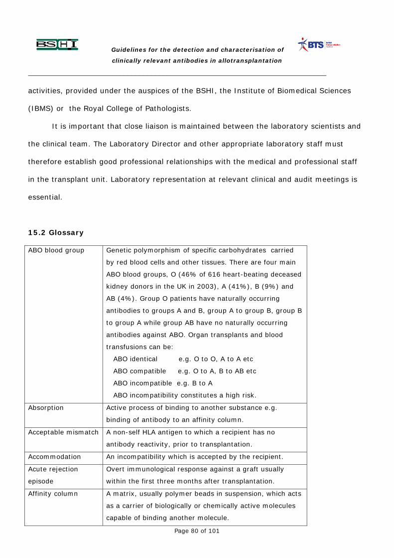
Guidelines for the detection and characterisation of
clinically relevant antibodies in allotransplantation
___________________________________________________________________________
Page 80 of 101
activities, provided under the auspices of the BSHI, the Institute of Biomedical Sciences
(IBMS) or the Royal College of Pathologists.
It is important that close liaison is maintained between the laboratory scientists and
the clinical team. The Laboratory Director and other appropriate laboratory staff must
therefore establish good professional relationships with the medical and professional staff
in the transplant unit. Laboratory representation at relevant clinical and audit meetings is
essential.
15.2 Glossary
ABO blood group Genetic polymorphism of specific carbohydrates carried
by red blood cells and other tissues. There are four main
ABO blood groups, O (46% of 616 heart-beating deceased
kidney donors in the UK in 2003), A (41%), B (9%) and
AB (4%). Group O patients have naturally occurring
antibodies to groups A and B, group A to group B, group B
to group A while group AB have no naturally occurring
antibodies against ABO. Organ transplants and blood
transfusions can be:
ABO identical e.g. O to O, A to A etc
ABO compatible e.g. O to A, B to AB etc
ABO incompatible e.g. B to A
ABO incompatibility constitutes a high risk.
Absorption Active process of binding to another substance e.g.
binding of antibody to an affinity column.
Acceptable mismatch A non-self HLA antigen to which a recipient has no
antibody reactivity, prior to transplantation.
Accommodation An incompatibility which is accepted by the recipient.
Acute rejection
episode
Overt immunological response against a graft usually
within the first three months after transplantation.
Affinity column A matrix, usually polymer beads in suspension, which acts
as a carrier of biologically or chemically active molecules
capable of binding another molecule.

Guidelines for the detection and characterisation of
clinically relevant antibodies in allotransplantation
___________________________________________________________________________
Page 81 of 101
Allele A genetic variant with a population frequency of more
than 1%. An allele can be defined at the DNA or protein
level.
Allograft or
allotransplant
A transplant between members of the same species e.g.
between humans.
Anamnestic response A response because of memory. The immune system
responds more aggressively when re-exposed to an
antigen in a secondary response.
Antibody Serum immunoglobulin expressed by B cells and secreted
by plasma cells that recognises a specific antigen.
Antibody dependent
cell mediated
cytotoxicity (ADCC)
Cytotoxic reaction whereby the effector activity is
provided by Fc receptor expressing cells (e.g.
macrophages, natural killer cells) that recognise antibody
coated targets.
Antibody detection A method which identifies the presence of an antibody in a
serum sample.
Immunoglobulin
isotype
Different structural, and therefore functionally different ,
forms due to the use of constant region alternatives of the
heavy chain. Designated IgM, IgD, IgG (subclasses IgG1,
IgG2, IgG3, and IgG4), IgE, and IgA.
Antibody removal Artificial reduction in circulating donor-specific antibody to
a level that allows transplantation (high immunological
risk).
Antigen presenting
cell (APC)
Specialised immune system cells which present degraded
antigen in the form of peptides complexed with MHC
molecules.
Antigen Any substance that is recognised by an immune system.
Anti-idiotypic
antibody
An antibody with specificity for that part of another
antibody which binds antigen.
Audit A cyclic process of review of a laboratory or clinical
systems with the aim of identifying improvements and
implementing change.
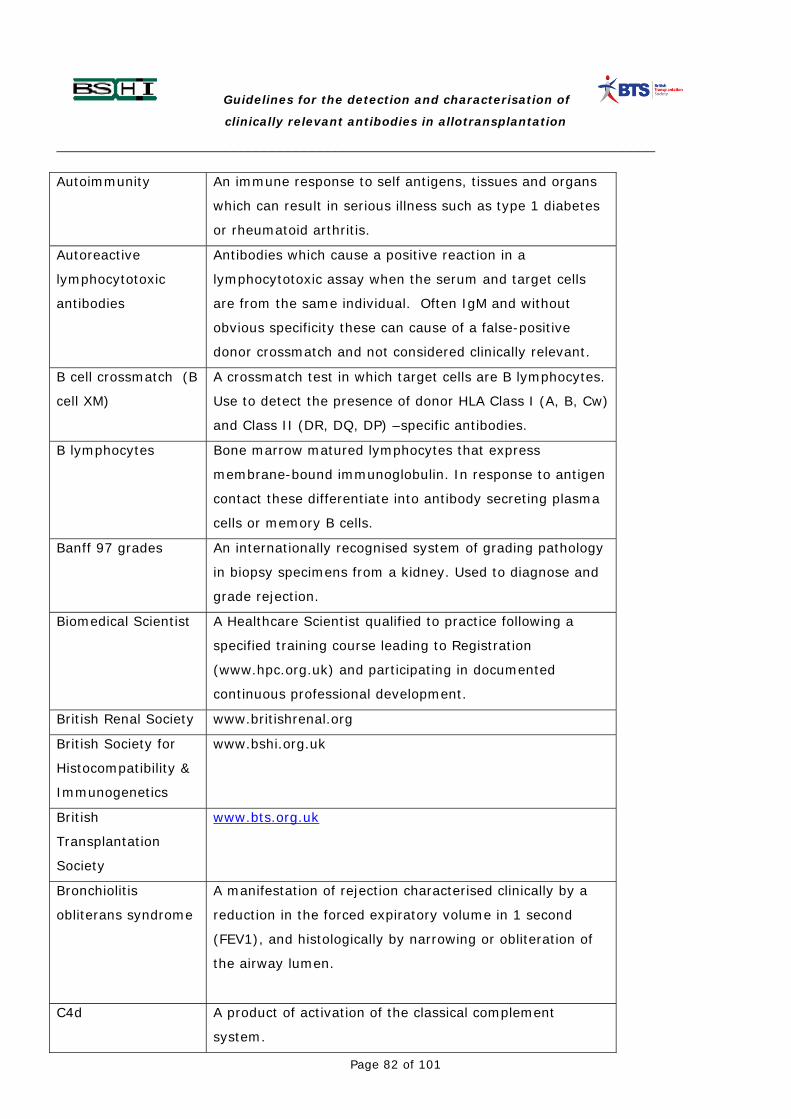
Guidelines for the detection and characterisation of
clinically relevant antibodies in allotransplantation
___________________________________________________________________________
Page 82 of 101
Autoimmunity An immune response to self antigens, tissues and organs
which can result in serious illness such as type 1 diabetes
or rheumatoid arthritis.
Autoreactive
lymphocytotoxic
antibodies
Antibodies which cause a positive reaction in a
lymphocytotoxic assay when the serum and target cells
are from the same individual. Often IgM and without
obvious specificity these can cause of a false-positive
donor crossmatch and not considered clinically relevant.
B cell crossmatch (B
cell XM)
A crossmatch test in which target cells are B lymphocytes.
Use to detect the presence of donor HLA Class I (A, B, Cw)
and Class II (DR, DQ, DP) –specific antibodies.
B lymphocytes Bone marrow matured lymphocytes that express
membrane-bound immunoglobulin. In response to antigen
contact these differentiate into antibody secreting plasma
cells or memory B cells.
Banff 97 grades An internationally recognised system of grading pathology
in biopsy specimens from a kidney. Used to diagnose and
grade rejection.
Biomedical Scientist A Healthcare Scientist qualified to practice following a
specified training course leading to Registration
(www.hpc.org.uk) and participating in documented
continuous professional development.
British Renal Society www.britishrenal.org
British Society for
Histocompatibility &
Immunogenetics
www.bshi.org.uk
British
Transplantation
Society
www.bts.org.uk
Bronchiolitis
obliterans syndrome
A manifestation of rejection characterised clinically by a
reduction in the forced expiratory volume in 1 second
(FEV1), and histologically by narrowing or obliteration of
the airway lumen.
C4d A product of activation of the classical complement
system.

Guidelines for the detection and characterisation of
clinically relevant antibodies in allotransplantation
___________________________________________________________________________
Page 83 of 101
Calcineurin inhibitor An immunosuppressive drug (ciclosporin, tacrolimus)
which acts by blocking immune cell activation by the
calcineurin pathway.
Calculated reaction
frequency
Calculated by NHSBT-ODT as the % incidence, among a
pool of 10,000 ABO compatible organ donors, of HLA
antigen incompatible donors with patient defined HLA-
specific antibody(s).
CD4, 8, 20, 28, 40,
80, 154, etc.
Cell surface molecules defined by specific monoclonal antibodies (Cluster of Differentiation) and recognised by an international standardisation body http://ca.expasy.org/cgi-bin/lists?cdlist.txt
Cell viability The proportion of target cells alive before or after a CDC
assay.
Characterisation of
antibodies
A process of testing for antibody(s) in serum in such a
way as to identify the specific antigen(s) with which the
antibody reacts.
Chemotaxis Modified movement of cells due to a concentration
gradient of a secreted substance.
Chronic rejection Process of graft failure occurring months or years post
transplantation. Progression is usually slow; e.g. chronic
transplant nephropathy (kidney), bronchiolitis obliterans
syndrome (lung) and coronary artery disease (heart).
Class switch Antigen driven process by which a B cell actively and
irreversibly changes the isotype but not the specificity of
the antibody it expresses.
Clinical Scientist A science graduate who has followed a recognised training
scheme to achieve Registration (www.hpc.org.uk) and
have documented continuous professional development
(CPD).
Cold ischaemia time The time during which an organ for transplant is stored
outside the body in the cold between cold perfusion in the
donor and removal for implantation in the recipient.
Complement A group of serum proteins which react in a regulated
enzymatic cascade. The classical pathway is initiated by
antibodies thus providing a cytotoxic effector mechanism.

Guidelines for the detection and characterisation of
clinically relevant antibodies in allotransplantation
___________________________________________________________________________
Page 84 of 101
Complement
dependent
cytotoxicity (CDC)
A laboratory test to identify presence of antibodies in a
serum sample using lymphocytes as targets and cell
viability as the read-out.
Coagulation Of red blood cells to form a thrombus (clot) mediated by
antibodies
Co-stimulatory
molecule
Cell surface ligand or receptor providing a non-specific
signal which is necessary for an antigen-specific response
by T or B cells.
CPD Continuous Professional Development schemes provide
independent assessment that an individual maintains
ongoing training.
Crossmatch test
(XM)
A test to identify antibody mediated reactivity to target
antigens in a potential organ donor. The test report must
be either positive or negative.
Cross-reactive An antibody which is able to bind to a series of structurally
closely related antigens.
Cytokine A chemical secreted by an immune cell which may either
enhance or suppress an immune response.
Desensitisation Removal of antibodies which are indicative of
sensitisation.
Differentiation A process of specialisation of cells and tissues to become a
functional organ or system. A one way step.
Dithiothreitol A chemical used in laboratory assays to dissociate the
pentameric IgM molecule and abrogate its activity.
Donor-Specific
Antibody
An antibody with specificity for a mismatched antigen
(HLA) present in an organ donor.
Endothelial cells Cells which line the blood vessels.
Enzyme linked
immunosorbent
assay (ELISA)
A laboratory assay in which specific antibody can be
detected. Known antigens are bound to a plastic plate and
reacted with a patient’s serum. If antibody is present it
will bind to the immobilised antigen and can be detected
by activation of an enzyme resulting in coloration of the
reaction. An instrument is used to measure the colour
change.
Epitope That part of the antigen structure to which antibody binds.
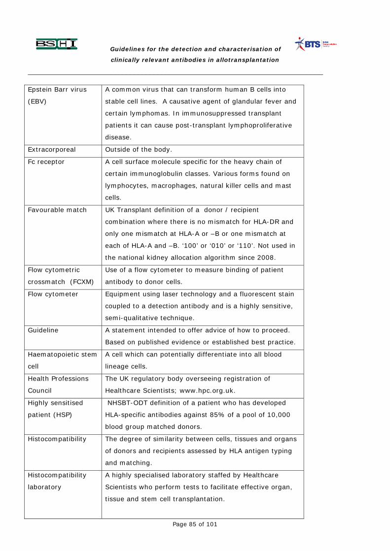
Guidelines for the detection and characterisation of
clinically relevant antibodies in allotransplantation
___________________________________________________________________________
Page 85 of 101
Epstein Barr virus
(EBV)
A common virus that can transform human B cells into
stable cell lines. A causative agent of glandular fever and
certain lymphomas. In immunosuppressed transplant
patients it can cause post-transplant lymphoproliferative
disease.
Extracorporeal Outside of the body.
Fc receptor A cell surface molecule specific for the heavy chain of
certain immunoglobulin classes. Various forms found on
lymphocytes, macrophages, natural killer cells and mast
cells.
Favourable match UK Transplant definition of a donor / recipient
combination where there is no mismatch for HLA-DR and
only one mismatch at HLA-A or –B or one mismatch at
each of HLA-A and –B. ‘100’ or ‘010’ or ‘110’. Not used in
the national kidney allocation algorithm since 2008.
Flow cytometric
crossmatch (FCXM)
Use of a flow cytometer to measure binding of patient
antibody to donor cells.
Flow cytometer Equipment using laser technology and a fluorescent stain
coupled to a detection antibody and is a highly sensitive,
semi-qualitative technique.
Guideline A statement intended to offer advice of how to proceed.
Based on published evidence or established best practice.
Haematopoietic stem
cell
A cell which can potentially differentiate into all blood
lineage cells.
Health Professions
Council
The UK regulatory body overseeing registration of
Healthcare Scientists; www.hpc.org.uk.
Highly sensitised
patient (HSP)
NHSBT-ODT definition of a patient who has developed
HLA-specific antibodies against 85% of a pool of 10,000
blood group matched donors.
Histocompatibility The degree of similarity between cells, tissues and organs
of donors and recipients assessed by HLA antigen typing
and matching.
Histocompatibility
laboratory
A highly specialised laboratory staffed by Healthcare
Scientists who perform tests to facilitate effective organ,
tissue and stem cell transplantation.

Guidelines for the detection and characterisation of
clinically relevant antibodies in allotransplantation
___________________________________________________________________________
Page 86 of 101
Historic serum
sample
A previously collected serum sample from a patient.
HLA Human Leukocyte Antigen. Cell surface molecules
determined by highly polymorphic linked genes on
chromosome 6 (HLA-A, -B, -Cw, -DR, -DQ, -DP: the
major histocompatibility complex or MHC). Biologically
these function to present protein fragments to T cells.
HLA class I
molecules
HLA-A, -B and –Cw molecules with structural and
functional similarity. Expressed by almost all nucleated
cells.
HLA class II
molecules
HLA-DR, -DQ and –DP molecules with structural and
functional similarity. Constitutively expressed only on
specialised antigen presenting cells but may be inducible.
HLA Matchmaker A computer software programme used to predict the
likelihood of HLA epitope (in)compatibility with a specified
HLA antigen
Human Tissue
Authority
www.hta.gov.uk
Humoral Of the blood. Usually used to indicate an antibody
mediated response (c.f. cellular response).
Hyperacute rejection Rejection of a transplant within a very short time of
transplantation (minutes) typically caused by pre-exisitng
donor-specific antibodies Usually results in irreversible
failure.
Immunoadsorption Binding of immunoglobulins to a solid phase. A process
which can remove immunoglobulin from the blood.
Immunogenicity The degree to which a substance can provoke the immune
system to respond e.g. high or low.
Institute of
Biomedical Scientists
The professional body for Biomedical Scientists
www.ibms.org
Interleukin (IL) Leukocyte secreted cytokine that affects the growth or
development immune system cells. E.g. IL-2, -4, -5, -6.
Islets Cluster of cells within the pancreas containing the beta
cells which secrete insulin

Guidelines for the detection and characterisation of
clinically relevant antibodies in allotransplantation
___________________________________________________________________________
Page 87 of 101
IVIg Intravenous immunoglobulin. A commercial preparation of
serum from a large number of blood donors which has
high levels of immunoglobulin with a wide range of
antibody specificities. Used to enhance, or suppress the
immune response.
Locus The position of a specified gene on a chromosome.
Lymphocyte Mononuclear leukocytes of various lineages (B cell, T cell,
NK cell).
Lymphoid cell line
(LCL) cells
Stable B lymphocyte line transformed with EBV in vitro.
Macrophage Mononuclear phagocytic leukocyte.
MICA – MHC class I -
related chain A
molecules
Molecules with close structural similarity to HLA molecules
but with a different function. MICA interact with natural
killer cells to regulate immune cell responses.
Microbeads Microscopic polystyrene beads to which antigens or DNA
probes can be bound. A vehicle for a solid-phase assay.
Median fluorescence
intensity
A semi-quantitative readout of the degree of binding of
antibody bound to (HLA) antigen on a microbead.
Mycophenolate
mofetil
An immunosuppressive drug with anti-proliferative
properties.
Monoclonal antibody An antibody secreted by a non-human cell line with
specificity for a single antigenic epitope. May be produced
commercially for in vivo therapeutic use. May be
“humanised” by engineering recombination of the
functional antibody binding domain with a major part of a
human immunoglobulin molecule to minimise
immunogenicity.
Natural killer (NK)
cell
Mononuclear leukocyte with innate ability to kill certain
tumours and virally infected cells.
Multivisceral
transplant
Transplantation of the liver and small bowel, possibly
including other organs such as the pancreas.
Modified
multivisceral
transplant
Transplantation of the small bowel and other organs such
as the pancreas, but excluding the liver.
Negative crossmatch Result of a laboratory test that has not detected donor-
specific antibody.

Guidelines for the detection and characterisation of
clinically relevant antibodies in allotransplantation
___________________________________________________________________________
Page 88 of 101
NHSBT-ODT NHS Blood and Transplant – Directorate of Organ
Donation and Transplant www.nhsbt.nhs.uk
www.organdonation.nhs.uk
Panel reactive
antibodies (PRA or
% PRA)
The calculated % of a panel of lymphocytes with which a
patient’s serum reacts.
Peak positive /
current negative
crossmatch (PPCN
XM)
A crossmatch result where reactivity to donor target cells
has been detected only in historic serum samples. There is
no reactivity to donor target cells in recent serum.
Peritubular
capillaries
Small blood vessels located in the kidney adjacent to the
structures (nephron) which filter the blood.
Plasma cell End stage cell of B lymphocyte lineage that secretes
immunoglobulin.
Plasma exchange or
plasmapheresis
Removal or dilution of plasma to lower the amount of
circulating antibody.
Platelets Small, irregular blood borne anucleate cells which are an
important component of a thrombus (clot).
Positive crossmatch Result of a laboratory test that has detected donor-
reactive antibody.
Post-transplant
immunological
monitoring
Laboratory tests to indicate the immune reactivity of a
recipient to transplanted tissues and organs.
Primary response The reaction of the immune system at the time of its first
exposure to a novel antigen..
Protein A A substance which non-specifically binds to
immunoglobulins.
Reaction frequency Originally the percentage of panel cells reacting with a
serum sample. Now defined as the proportion of a pool of
10,000 blood group matched donors against which a
recipient has HLA antibodies.
Recommendation A guideline which should usually be adhered to.
Rescue therapy A treatment aimed to prevent failure of a transplanted
organ in the face of an aggressive immune response.
Risk The degree to which a hazardous outcome is predicted.

Guidelines for the detection and characterisation of
clinically relevant antibodies in allotransplantation
___________________________________________________________________________
Page 89 of 101
Rituximab Brand of therapeutic chimeric CD20-specific monoclonal
antibody.
Royal College of
Pathologists
www.rcpath.org
Screening strategy A process to detect and define sensitisation.
Secondary response An enhanced immune response mounted on re-exposure
to a previously recognised antigen.
Sensitisation An immune response to an antigen resulting in T and/or B
cell memory.
Sensitivity (of a
patient)
The ability to mount an immune response to an antigen.
Sensitivity (of an
assay)
An evaluation of the accuracy of the results of a laboratory
test to predict an outcome. Usually quoted as a
percentage.
Single antigen beads A multiplex of microbeads each identifiable group being
loaded with a single HLA antigen
Solid phase assays A laboratory test to detect antibodies using antigen
targets immobilised to a plastic tray or microparticle.
These assays are performed as ELISA or fluid phase
assays using a flow cytometer. The target antigen can be
cell-free HLA molecules.
Specificity The defined reactivity of an antibody e.g. specific for an
HLA molecule.
T lymphocyte Mononuclear leukocyte having developed in the thymus.
Expresses a receptor specific for MHC plus processed
antigen.
T cell crossmatch
(T cell XM)
A crossmatch test in which the target cells are T
lymphocytes.
Titre Reciprocal of the last dilution of a serum giving a
detectable reaction.
UK Transplant The immediate predecessor of NHSBT-ODT.
Unacceptable
antigen
Antigen which due to prior exposure and specific
sensitisation (due to pregnancy or a transplant) excludes
a transplant if present in the donor’s HLA type.
Vascular rejection An aggressive antibody mediated immune response with
activity detected in the blood vessels of the transplant.
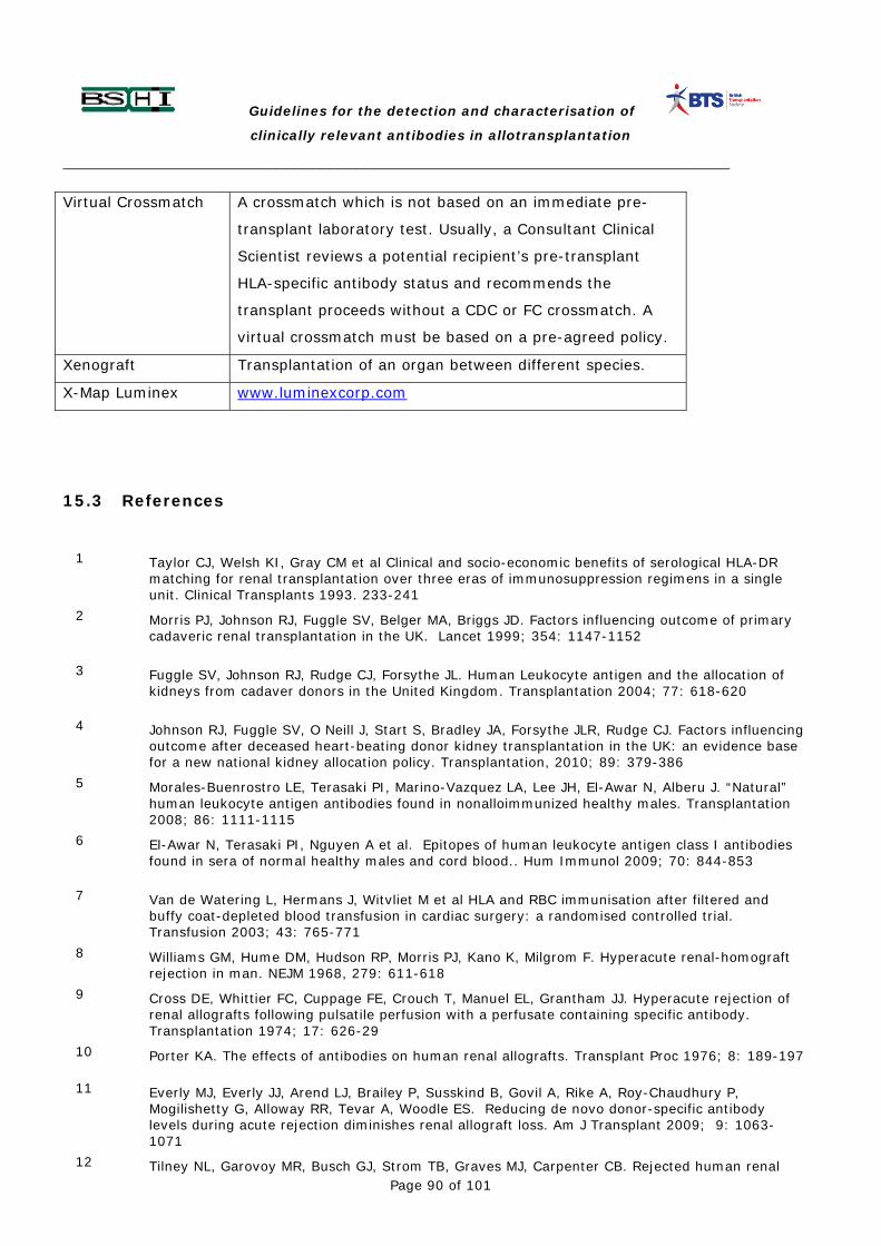
Guidelines for the detection and characterisation of
clinically relevant antibodies in allotransplantation
___________________________________________________________________________
Page 90 of 101
Virtual Crossmatch A crossmatch which is not based on an immediate pre-
transplant laboratory test. Usually, a Consultant Clinical
Scientist reviews a potential recipient’s pre-transplant
HLA-specific antibody status and recommends the
transplant proceeds without a CDC or FC crossmatch. A
virtual crossmatch must be based on a pre-agreed policy.
Xenograft Transplantation of an organ between different species.
X-Map Luminex www.luminexcorp.com
15.3 References
1 Taylor CJ, Welsh KI, Gray CM et al Clinical and socio-economic benefits of serological HLA-DR
matching for renal transplantation over three eras of immunosuppression regimens in a single unit. Clinical Transplants 1993. 233-241
2 Morris PJ, Johnson RJ, Fuggle SV, Belger MA, Briggs JD. Factors influencing outcome of primary cadaveric renal transplantation in the UK. Lancet 1999; 354: 1147-1152
3 Fuggle SV, Johnson RJ, Rudge CJ, Forsythe JL. Human Leukocyte antigen and the allocation of kidneys from cadaver donors in the United Kingdom. Transplantation 2004; 77: 618-620
4 Johnson RJ, Fuggle SV, O Neill J, Start S, Bradley JA, Forsythe JLR, Rudge CJ. Factors influencing outcome after deceased heart-beating donor kidney transplantation in the UK: an evidence base for a new national kidney allocation policy. Transplantation, 2010; 89: 379-386
5 Morales-Buenrostro LE, Terasaki PI, Marino-Vazquez LA, Lee JH, El-Awar N, Alberu J. “Natural” human leukocyte antigen antibodies found in nonalloimmunized healthy males. Transplantation 2008; 86: 1111-1115
6 El-Awar N, Terasaki PI, Nguyen A et al. Epitopes of human leukocyte antigen class I antibodies found in sera of normal healthy males and cord blood.. Hum Immunol 2009; 70: 844-853
7 Van de Watering L, Hermans J, Witvliet M et al HLA and RBC immunisation after filtered and buffy coat-depleted blood transfusion in cardiac surgery: a randomised controlled trial. Transfusion 2003; 43: 765-771
8 Williams GM, Hume DM, Hudson RP, Morris PJ, Kano K, Milgrom F. Hyperacute renal-homograft rejection in man. NEJM 1968, 279: 611-618
9 Cross DE, Whittier FC, Cuppage FE, Crouch T, Manuel EL, Grantham JJ. Hyperacute rejection of renal allografts following pulsatile perfusion with a perfusate containing specific antibody. Transplantation 1974; 17: 626-29
10 Porter KA. The effects of antibodies on human renal allografts. Transplant Proc 1976; 8: 189-197
11 Everly MJ, Everly JJ, Arend LJ, Brailey P, Susskind B, Govil A, Rike A, Roy-Chaudhury P, Mogilishetty G, Alloway RR, Tevar A, Woodle ES. Reducing de novo donor-specific antibody levels during acute rejection diminishes renal allograft loss. Am J Transplant 2009; 9: 1063-1071
12 Tilney NL, Garovoy MR, Busch GJ, Strom TB, Graves MJ, Carpenter CB. Rejected human renal

Guidelines for the detection and characterisation of
clinically relevant antibodies in allotransplantation
___________________________________________________________________________
Page 91 of 101
allografts. Recovery and characteristics if infiltrating cells and antibody. Transplantation 1979; 28: 421-426
13 Garovoy MR, Reddish MA, Busch GJ, Tilney NL. Immunoglobulin-secreting cells recovered from rejected human renal allografts. Transplantation 1982; 33: 109-111
14 Lederer SR, Kluth-Pepper B, Schneeberger H, Albert E, Land W, Feucht HE. Impact of humoral alloreactivity early after transplantation on the long term survival of renal allografts. Kidney Int 2001; 59: 334-341
15 Hidalgo LG, Campbell PM, Sis B, Einecke G, Mengel M, Chang J, Sellares J, Reeve J, Halloran PF. De novo donor-specific antibody at the time of kidney transplant biopsy associates with microvascular pathology and late graft failure. Am J Transplant. 2009; Nov; 9(11): 2532-2541
16 Strom TB, Tilney NL, Paradysz JM, Bancewicz J, Carpenter CB. Cellular components of allograft rejection: identity, specificity and cytotoxic function of cells infiltrating acutely rejection allografts. J Immunol 1977; 118: 2020-2026
17 Gebel HM, Bray RA, Nickerson P. Pre-transplant assessment of donor-reactive HLA-specific antibodies in renal transplantation: contraindication vs risk. Am J Transplant 2003; 3: 1488-1500
18 Taylor CJ, Kosmoliaptsis V, Summers DM, Bradley JA. Back to the future: Application of contemporary technology to long standing questions about the clinical relevance of HLA-specific alloantibodies in renal transplantation. Hum Immunol 2009; 70: 563-568
19 Zachary AA, Klingman L, Thorne N, et al: Variations of the lymphocytotoxicity test. An evaluation of sensitivity and specificity. Transplantation 1995, 60: 498-503
20 Ting A, Morris PJ. Renal transplantation and B-cell cross-matches with autoantibodies and alloantibodies. Lancet 1977; ii: 1095-1097
21 Zoet YM, Eijsink C, Kardol MJ et al . Hum Immunol 2005; 66: 519 – 525
22 Eijsink C, Zoet YM, Kardol MJ, Witvliet MD, Claas FH, Mulder A, Doxiadis II. Tissue Antigens 2007; 69: 354 – 357
23 Harmer AW, Sutton M, Bayne A, et al. A highly sensitive, rapid screening method for the detection of antibodies directed against HLA class I and class II antigens. Transpl Int 1993; 6: 277-280
24 Harmer AW, Heads AJ, Vaughan RW. Detection of HLA class I and class II specific antibodies by flow cytometry and PRA-STAT screening in renal transplant recipients. Transplantation 1997; 63: 1828-1832
25 Lederer SR, Schneeberger H, Albert E, et al. Early graft dysfunction: The role of preformed antibodies to DR-typed lymphoblastoid cell lines. Transplantation 1996; 61: 313-319
26 Cicciarelli J, Helstab K, Mendez R. Flow cytometry PRA, a new test that is highly correlated with graft survival. Clin Transplantation 1992; 6: 159-164
27 Shroyer TW, Deierhoi MH, Mink CA, et al A rapid flow cytometry assay for HLA antibody detection using a pooled cell panel covering 14 serological crossreacting groups. Transplantation 1995; 59: 626-630
28 Buelow R, Chiang T, Monteiro F, et al Soluble HLA antigens and ELISA - a new technology for crossmatch testing. Transplantation 1995; 60: 1594-1599
29 Kao KJ, Scornik JC, Small SJ. Enzyme-linked immunoassay for anti-HLA antibodies - an alternative to panel studies by lymphocytotoxicity. Transplantation 1993; 55: 192-196
30 Worthington JE, Robson AJ, Sheldon S, Langton A, Martin S. A comparison of enzyme-linked immunosorbent assays and flow cytometry techniques for the detection of HLA specific antibodies. Hum Immunol 2001; 62: 1178-1184
31 Zachary AA, Delaney NL, Lucas DP, Leffell MS. Characterisation of HLA class I specific antibodies by ELISA using solubilized antigen targets: evaluation of the GTI QuikID assay and analysis of

Guidelines for the detection and characterisation of
clinically relevant antibodies in allotransplantation
___________________________________________________________________________
Page 92 of 101
antibody patterns. Hum Immunol 2001; 62: 228-235
32 Monien S, Salama A, Schönemann C. ELISA methods detect HLA antibodies with varible sensitivity. Int J Immunogenet 2006; 33: 163-66
33 Karuppan SS, Moller E. The use of magnetic beads coated with soluble HLA class I or class II proteins in antibody screening and for specificity determination of donor-reactive antibodies. Transplantation 1996; 61: 1539-1545
34 Pei R, Wang C, Tarsitani C, et al: Simultanteous HLA class I and class II antibodies screening with flow cytometry. Human Immunol 1998; 59: 313-322
35 Ellis TM, Gebel HM, Pierce KL, Bray RA. Limitations to antibody detection using single antigen flow beads. Human Immunol 2003; 64(suppl1):S14
36 El-Awar N, Terasaki PI, Lee J-H et al. Analysis of mouse monoclonal antibodies with single antigen beads. Human Immunol 2003; 64(suppl1):S14
37 Fulton RJ, McDade RC, Smith PL, Kienker LJ, Kettman JR. Advanced multiplexed analysis with the FlowMetrix system. Clin Chem 1997; 43: 1749-56
38 Vaidya S, Partlow D, Susskind B, Noor M, Barnes T, Gugliuzza K. Prediction of crossmatch outcome of highly sensitized patients by single and/or multiple antigen bead luminex assay. Transplantation 2006; 82: 1524-1528
39 Higgins R, Hathaway M, Lowe D et al. Blood levels of donor-specific human leukocyte antigen antibodies after renal transplantation: resolution of rejection in the presence of circulating donor-specific antibody. Transplantation 2007; 84: 876-884
40 Zhang Q, Liang LW, Gjertson DW et al. Development of posttransplant antidonor HLA antibodies is associated with acute humoral rejection and early graft dysfunction. Transplantation 2005; 79: 591-598
41 El-Awar NR, Akaza T, Terasaki PI, et al. Human leukocyte antigen class I epitopes: update to 103 total epitopes, including the C locus. Transplantation 2007; 84: 532-540
42 Duquesnoy RJ, Marrari M. HLAMatchmaker-based definition of structural human leukocyte antigen epitopes detected by alloantibodies. Current Opinion in Organ Transplantation 2009; 14: 403-409.
43 Goodman RS, Taylor CJ, O’Rourke et al. Utility of HLAMatchmaker and single-antigen HLA-antibody detection beads for identification of acceptable mismatches in highly sensitized patients awaiting kidney transplantation. Transplantation 2006; 15: 1331-1336
44 Kosmoliaptsis V, Bradley JA, Sharples LD, Chaudhry A, Key T, Goodman RS, Taylor CJ. Predicting the immunogenicity of human leukocyte antigen class I alloantigens using structural epitope analysis determined by HLAMatchmaker. Transplantation 2008; 85: 1817-1825
45 Vaidya S, Hilson B, Sheldon S, Cano P, Fernandez Vina M. DP reactive antibody in a zero mismatch renal transplant pair. Hum Immunol 2007; 68: 947-949
46 Deng CT, El-Awar N, Ozawa M, Cai J, Lachmann N, Terasaki PI. Human leukocyte antigen class II DQ alpha and beta epitopes identified from sera of kidney allograft recipients. Transplantation 2008; 86: 452-459
47 Gebel HM, Moussa O, Eckels DD, Bray RA. Donor-reactive HLA antibodies in renal allograft recipients: considerations, complications, and conundrums. Hum Immunol 2009; 70: 610-617
48 Kosmoliaptsis V, Bradley JA, Peacock S, Chaudhry AN, Taylor CJ. Detection of IgG HLA specific alloantibodies in renal transplant patients using single antigen beads is compromised by the presence of IgM HLA specific alloantibodies. Transplantation 2009; 87: 813-820
49 Zachary AA, Lucas DP, Detrick B, Leffell MS. Naturally occurring interference in Luminex assays for HLA-specific antibodies: characteristics and resolution. Hum Immunol 2009; 70: 496-501

Guidelines for the detection and characterisation of
clinically relevant antibodies in allotransplantation
___________________________________________________________________________
Page 93 of 101
50 Kosmoliaptsis V, O’Rourke C, Bradley JA, Taylor CJ. Improved Luminex-based HLA-specific antibody screening using dithiothreitol treated sera. Hum Immunol 2010; 71: 45-49
51 Khan N, Robson AJ, Worthington JE, Martin S. The detection and definition of IgM alloantibodies in the presence of IgM autoantibodies using flow PRA beads. Hum Immunol 2003; 64: 593-599
52 Stastny P, Ring S, Lu C, Arenas J, Han M, Lavingia B. Role of immunoglobulin (Ig)-G and IgM antibodies against donor human leukocyte antigens in organ transplant recipients. Hum Immunol 2009; 70: 600-604
53 Smith JD, Hamour IM, Banner NR, Rose ML. C4d fixing, Luminex binding antibodies – a new tool for prediction of graft failure after heart transplantation. Am J Transplant 2007; 7: 2809 – 2815
54 Cai J, Terasaki PI, Anderson N, Lachmann N, Schenemann C. Intact HLA not beta 2 m free heavy chain specific HLA class I antibodies are predictive of graft failure. Transplantation 2009, 88: 226-230
55 Zachary AA, Sholander JT, Houp JA, Lefell MS. Using real data for a virtual crossmath. Hum Immunol 2009; 70: 574-579
56 Solez K. Colvin RB, Racusen LC et al. Banff 07 classification of renal allograft pathology: updates and future directions. Am J Transplant 2008; 8: 753-860
57 Kissmeyer-Nielsen F, Olsen S, Petersen V.P, Fjeldborg O. Hyperacute rejection of kidney allografts associates with pre-existing humoral antibodies against donor cells. Lancet 1966; 1: 662
58 Terasaki P.I, McClelland J.D. Microdroplet assay of human serum cytotoxins. Nature 1964; 204: 998-1000
59 Patel R, Terasaki PI. Significance of the positive crossmatch test in kidney transplantation. NEJM 1969; 280: 735-739
60 Garovoy MR, Rheinschmidt MA, Bigos M, Perkins H, Colombe B. Flow cytometry analysis: a high technology crossmatch technique facilitating transplantation. Transplant Proc 1983; 15: 1939-1944
61 Matas AJ, Sutherland DER. Kidney transplantation without a final crossmatch. Transplantation 1998; 66: 1835-1836
62 Kerman RH, Susskind B, Ruth J, Katz S, Van Buren CT, Kahan BD. Can an immunologically nonreactive potential allograft recipient undergo transplantation without a donor-specific crossmatch? Transplantation 1998; 66: 1833-1845
63 Taylor CJ, Smith SI, Morgan CH et al. Selective omission of the donor crossmatch before renal transplantation. Transplantation 2000; 69: 719-723
64 Taylor CJ, Kosmoliaptsis V, Sharples LD et al. Ten-year experience of selective omission of the pretransplant crossmatch test in deceased donor kidney transplantation. Transplantation 2010; 89:185-193
65 Van den Berg-Loonen EM, Billen EVA, Voorter CEM et al. Clinical relevance of pretransplant donor-directed antibodies detected by single antigen beads in highly sensitised renal transplant patients. Transplantation 2008; 85:1086-1090
66 Gupta A, Iverson V, Varagunam M, Bodger S, Sinnott P, Thuraisingham RC. Pretransplant donor-specific antibodies in cytotoxic negative crossmatch kidney transplants: are they relevant? Transplantation 2008; 85:1200-1204
67 Amico P, Hönger G, Steiger J, Schaub S. Utility of the virtual crossmatch in solid organ transplantation. Current Opinion in Organ Transplantation 2009;14:656-661
68 Cecka JM. The UNOS Transplant Registry. Clinical Transplants. 2001:1-18
69 Connolly JK, Dyer PA, Martin S, et al: Importance of minimising HLA-DR mismatch and cold preservation time in cadaveric renal transplantation. Transplantation 1996; 61: 709

Guidelines for the detection and characterisation of
clinically relevant antibodies in allotransplantation
___________________________________________________________________________
Page 94 of 101
70 Takemoto S, Carnahan E, Terasaki P: Report on 604 six-antigen-matched transplants, in Terasaki P (ed): Clinical Transplants 1990. Los Angeles, UCLA Tissue Typing Laboratory, 1990, 485-495
71 Braun WE: Laboratory and clinical management of the highly sensitised organ transplant recipient. Hum Immunol 1989; 26: 245-260
72 Duquesnoy RJ, White LT, Fierst JW, et al: Multiscreen serum analysis of highly sensitised renal dialysis patients for antibodies toward public and private class I HLA determinants. Transplantation 1990; 50: 427-437
73 Rodey GE, Revels K, Fuller TC: Epitope specificity of HLA class I alloantibodies: Stability of cross reactive group patterns over extended time periods. Transplantation 1997; 63: 885-893
74 Chapman, J. R., Taylor, C., Ting, A., Morris, P. J. (1986b). Hyperacute rejection of a renal allograft in the presence of anti-HLA-Cw5 antibody. Transplantation 1986; 42: 91-93.
75 Takemoto S, Cecka JM, Gjertson DW, et al: Six-antigen-matched transplants: causes of failure. Transplantation 1993; 55: 1005
76 Mytilineos J, Deufel A, Opelz G. Clinical relevance of HLA-DPB locus matching for cadaver kidney transplantation: a report of the Collaborative Transplant Study. Transplantation 1997; 63: 1351-1354
77 Caskey FJ, Johnson RJ, Fuggle SV. Renal after cardiothoracic transplant: the effect of repeat mismatches on outcome. Transplantation 2009; 87: 1727-1732
78 Chapman, J R, Taylor C, Ting A, Morris P J. (1986a). Immunoglobulin class and specificity of antibodies causing positive T cell crossmatches: relationship with renal transplant outcome. Transplantation 1986; 42: 608-613.
79 Mohanakumar T, Rhodes C, Mendez-Picon G, Goldman M, Moncure C, Lee H. Renal allograft rejection associated with presensitization to HLA-DR antigens. Transplantation 1981; 31: 93-95.
80 Scornik J.C, LeFor W.M, Cicciarelli J.C et al. Hyperacute and acute kidney graft rejection due to antibodies against B cells. Transplantation 1992; 54: 61-64
81 Taylor CJ, Chapman JR, Fuggle SV et al A positive B cell crossmatch due to IgG anti-HLA-DQ antibody present at the time of transplantation in a successful renal allograft. Tissue Antigens 1987; 30: 104-112
82 Ting A, Morris PJ: Successful transplantation with a positive T and B cell crossmatch due to autoreactive antibodies. Tissue Antigens 1983; 21: 219-226
83 Taylor CJ, Chapman JR, Ting A, Morris PJ: Characterization of lymphocytotoxic antibodies causing a positive crossmatch in renal transplantation. Relationship to primary and regraft outcome. Transplantation 1989; 48: 953-958
84 Cardella, C.J., The use of the kidney with an historical positive, and current negative crossmatch. Pediatr Nephrol, 1991; 5: 126-129.
85 Ten Hoor G.M, Coopmans M, Allebes W.A. Specificity and Ig class of preformed alloantibodies causing a positive crossmatch in renal transplantation. Transplantation 1993; 56: 298-304
86 Roelen DL, van Bree FPMJ, Witvliet MD. IgG antibodies against HLA antigen are associated with activated cytotoxic T cells against this antigen. IgM are not. Transplantation 1994; 57: 1388-1392.
87 Oostingh GJ, Davies HFS, Bradley JA , and Taylor CJ. Comparison of allogeneic and xenogeneic in vitro T cell proliferative responses of sensitised patients awaiting kidney transplantation. Xenotransplantation 2003; 10:545-551
88 Van Kampen CA, Roelen DL, Versteeg-van der Voort Maarschalk MFJ, Hoitsma AJ, Allebes WA, Claas FHJ. Activated HLA class I-reactive cytotoxic T lymphocytes associated with a positive

Guidelines for the detection and characterisation of
clinically relevant antibodies in allotransplantation
___________________________________________________________________________
Page 95 of 101
historical crossmatch predict early graft failure. Transplantation 2002; 74: 1114-1119
89 Worthington JE, Martin S, Al-Husseini DM, Dyer PA, Johnson RWG. Posttransplantation production of donor HLA-specific antibodies as a predictor of renal transplant outcome. Transplantation 2003; 75: 1034-1040
90 Chapman JR, Deierhoi MH, Carter NP, Ting A, Morris PJ. Analysis of flow cytometry and cytotoxicity crossmatches in renal transplantation. Transplant Proc 1985; 17: 2480-2481
91 Thistlethwaite JR, Buckingham MR, Stuart JK, Stuart FP. Detection of presensitisation in renal allograft recipients using a flow cytometric immunofluorescence crossmatch. Transplant Proc 1986; 18: 676-679
92 Cook DJ, Terasaki PI, Iwaki Y, Terashita GY, Lau M. An approach to reducing early kidney transplant failure by flow cytometry crossmatching. Clinical Transplantation 1987; 1: 253-256
93 Lazda VA, Pollak R, Mozes MF, Joasson O. The relationship between flow cytometer crossmatch results and subsequent rejection episodes in cadaver renal allograft recipients. Transplantation 1988; 45: 562-565
94 Talbot D, Givan A.L, Shenton B.K et al. The prospective value of the preoperative flow cytometric crossmatch assay in renal transplantation. Transplantation 1990; 49: 809-810
95 Ogura K, Terasaki P.I, Johnson C et al. The significance of a positive flow cytometric crossmatch test in primary kidney transplantation. Transplantation 1993; 56: 294-298
96 Horsburgh T, Mistry N, Weston S et al . Relevance of positive crossmatches in renal transplantation. Transplant Proc 1992; 24: 2508-2509
97 Evans PR, Lane AC, Lambert CM et al . Lack of correlation between IgG T-lymphocyte flow cytometric crossmatches with primary renal allograft outcome. Transplant Int 1992; 5: S609-S612
98 Martin S, Liggett H, Robson A, Connolly J, Johnson R.W.G. The association between a positive T and B cell flow cytometry crossmatch and renal transplant failure. Transplant Immunol 1993; 1: 270-276
99 Lazda VA. Identification of patients at risk for inferior renal allograft outcome by a strongly positive B cell flow cytometry crossmatch. Transplantation 1994; 57: 964-969
100 Kimball P, Rhodes C, King A, Fisher R, Ham J, Posner M. Flow crossmatching identifies patients at risk of post operative elaboration of cytotoxic antibodies. Transplantation 1998; 65: 444-446
101 Cho YW, Cecka JM. Crossmatch tests – an analysis of UNOS data from 1991-2000. Clinical Transplants 2001; 237-246
102 El Fettouh HA, Cook DJ, Bishay E et al. Association between a positive flow cytometry crossmatch and the development of chronic rejection in primary renal transplantation. Urology 2000; 56: 369-372
103 Suciu-Foca N, Reed E, D’Agati VD et al. Soluble HLA antigens anti-HLA antibodies and antidiotypic antibodies in the circulation of renal transplant recipients. Transplantation 1991; 51: 593-601
104 Halloran PF, Schlaut J, Solez K, Srinivasa NS The significance of the anti-class I response. II. Clinical and pathologic features of renal transplants with anti-class I-like antibody. Transplantation 1992; 53: 550-555
105 Al-Hussein KA, Shenton BK, Bell A et al Characterisation of donor-directed antibody class in the post-transplant period using flow cytometry. Transplant Int 1994; 7: 182-189
106 Harmer AW, Koffman CG, Heads AJ et al Sensitisation to HLA antigens occurs in 95% of primary renal transplant rejections. Transplant Proc 1995; 27: 666-667
107 Trpkov K, Campbell P, Pazderka F, Cockfield S, Solez K Halloran PF. Pathologic features of acute renal allograft rejection associated with donor-specific antibody. Analysis using the Banff grading

Guidelines for the detection and characterisation of
clinically relevant antibodies in allotransplantation
___________________________________________________________________________
Page 96 of 101
schema. Transplantation 1996; 61: 1586-1592
108 Martin S, Dyer PA, Mallick NP, Gokal R, Harris R, Johnson RW. Post transplant antidonor lymphocytotoxic antibody production in relation to graft outcome. Transplantation 1997; 44: 50-53
109 Monteiro F, Buelow R, Mineiro C, Rodrigues H Kalil J, Identification of patients at high rislk of graft loss by pre- and post-transplant monitoring of anti-HLA class I IgG antibodies by enzyme linked immunosorbent assay. Transplantation 1997; 63:542
110 Abe M, Kawai T, Futatsuyama K et al Postoperative production of anti-donor antibody and chronic rejection in renal transplantation. Transplantation 1997; 63: 1616-1619
111 Kerman RH, Orosz CG, Lober MI Clinical relevance of anti-HLA antibodies pre and post transplant. Am J Med Sci 1997; 313: 275-278
112 Scheonemann C, Groth J, Leveerenz S, May G. HLA class I and class II antibodies: monitoring before and after kidney transplantation and their clinical relevance. Transplantation 1998; 65: 1519-1523
113 Christiaans MH, Overhof-de Roos R, Nieman F, van Hoeff JP, van den Berg-Loonen EM.Donor-specific antibodies after transplantation by flow cytometry: relative change in fluorescence ratio most sensitive risk factor for graft survival. Transplantation 1998; 65; 427-433
114 Muller-Steinhardt M, Fricke L, Kirchner H, Hoyer J, Kluter H. Monitoring of anti-HLA class I and II antibodies by flow cytometry in patients after first cadaveric kidney transplantation. Clin Transplant 2000; 14: 85-89
115 Piazza A, Borrelli L, Monaco PI et al. Post-transplant donor-specific antibody characterisation and kidney graft survival. Transplant Int. 2000; 13: S439-S443
116 Piazza A, Poggi E, Borrelli L et al Impact of donor-specific antibodies on chronic rejection occurrence and graft loss in renal transplantation: posttransplant analysis using flow cytometric techniques. Transplantation 2001; 71: 1106-1112
117 McKenna RM, Takemoto SK, Terasaki PI HLA antibodies after solid organ transplantation. Transplantation 2000; 69: 319-326
118 Hankey KG Deachenberg CB, Papadimitriou JC et al MIC expression in renal and pancreatic allografts. Transplantation 2002; 73: 304-306
119 Zwirner NW, Fernandez-Vina MA, Stastny P. MICA, a new polymorphic HLA-related antigen, is expressed mainly by keratinocytes, endothelial cells, and monocytes. Immunogenetics 1998; 47: 139-148
120 Zwirner NW, Dole K, Stastny P. Differential surface expression of MICA by endothelial cells, fibroblasts, keratinocytes and monocytes. Hum Immunol 1999: 60: 323-330
121 Feucht HE, Schneeberger H, Hillebrand G, et al. Capillary deposition of C4d complement fragment and early renal graft loss. Kidney Int 1993; 43: 1333-1338
122 Crespo M, Pascula M, Tolkoff-Rubin N, et al. Acute humoral rejection in renal allograft recipients: I. Incidence, serology and clinical characteristics. Transplantation 2001; 71: 652-658
123 Herzenberg AM, Gill JS, Djurdjev O, Magil AB. C4d deposition in acute rejection: an independent long-term prognostic factor. J Am Soc Nephrol 2002; 13: 234-244
124 Mauiyyedi S, Crespo M, Collins AB, et al. Acute humoral rejection in kidney transplantation: II. Morphology, immunopathology, and pathologic classification. J Am Soc Nephrol 2002; 13: 779-787
125 Nickeleit V, Zeiler M, Gudat F, Thiel G, Mihatsch MJ. Detection of the complement degradation product C4d in renal allografts: diagnostic and therapeutic implications. J Am Soc Nephrol 2002; 13: 242-251
126 Bohmig GA, Exner M, Habicht A, et al. Capillary C4d deposition in kidney allografts: a specific marker of alloantibody-dependent graft injury. J Am Soc Nephrol 2002; 13: 1091-1099

Guidelines for the detection and characterisation of
clinically relevant antibodies in allotransplantation
___________________________________________________________________________
Page 97 of 101
127 Koo DH, Roberts ISD, Quiroga et al C4d deposition in early renal allograft protocol biopsies. Transplantation 2004; 78: 398-403
128 Collins AB, Schneeberger EE, Pascual MA, et al. Complement activation in acute humoral renal allograft rejection: diagnostic significance of C4d deposits in peritubular capillaries. J Am Soc Nephrol 1999; 10: 2208-2214
129 Worthington JE, McEwen A, Mc William LJ, Picton ML and Martin S. Association between C4d staining in renal transplant biopsies, production of donor specific HLA antibodies and graft outcome. Transplantation 2007; 83: 298-403
130 Theruvath TP, Saidman SL, Mauiyyedi S, et al. Control of antidonor antibody production with tacrolimus and mycophenolate mofetil in renal allograft recipients with chronic rejection. Transplantation 2001; 72: 77.
131 Rose ML, Smith J, Dureau G, Keogh A, Kobashigowa J. Mycophenolate mofetil decreases antibody production after cardiac transplantation. J Heart Lung Transplant 2002; 21: 282.
132 Ishida H, Tanabe K, Furusawa M, et al. Mycophenolate mofetil suppresses the production of anti-blood type antibodies after renal transplantation across the ABO blood barrier. Transplantation 2002; 74: 1187-1189.
133 Mohanakumar T, Narayanan K, Desai, N. A significant role for histocompatibility in human islet transplantation. Transplantation 2006; 82: 180-187
134 Kessler L, Parissiadis A, Bayle F et al. Evidence for humoral rejection of a pancreatic islet graft and resue with Rituximab and IV immunoglobulin therapy. Am J Transplant. 2009; 9: 1961-1966
135 Campbell PM, Senior PA, Salam A. et al. High risk of sensitisation after failed islet transplantation. Am J Transplant. 2007; 7: 2311-2317
136 Ferrari-Lacraz S, Berney T, Morel P. et al. Low risk of anti- human leukocyte antigen antibody sensitization after combined kidney and islet transplantation. Transplantation 2008; 86: 357-359
137 Cardini R, Pileggi A, Ricordi C. et al. Allosensitization of islet allograft recipients. Transplantation 2007; 84: 1413-1427
138 Smith JD, Danskine AJ, Laylor RM, Rose ML, Yacoub MH. The effect of panel reactive antibodies and the donor specific crossmatch on graft survival after heart and heart-lung transplantation. Transplant Immunol 1993; 1: 60-65.
139 Singh G, Thompson M, Griffith B. et al Histocompatibility in cardiac transplantation with particular reference to immunopathology of positive serologic crossmatch. Clin Immunol Immunopathol 1983; 28: 56-66
140 Itescu S, Tung TC, Burke EM et al. Preformed IgG antibodies against major histocompatibility complex class II antigens are major risk factors for high-grade cellular rejection in recipients of heart transplantation. Circulation 1998; 98: 786-793
141 Stastny P, Lavingia B, Fixler DE, Yancy CW, Ring WS. Antibodies against donor human leuckocyte antigens and the outcome of cardiac allografts in adults and children. Transplantation 2007; 84: 738-745
142 Smith JD, Hamour IM, Banner NR, Rose ML. C4d fixing, luminex binding antibodies - a new tool for prediction of graftfailure after heart transplantation. American Journal of Transplantation. 2007; 7: 2809-2815
143 Przybylowski P, Balogna M, Radovancevic B et al. The role of flow cytometry-detected IgG and IgM anti-donor antibodies in cardiac allograft recipients. Transplantation 1999; 67: 258-262
144 Scornik JC, Zander DS, Baz MA, Donnelly WH, Staples ED. Susceptibility of lung transplants to preformed donor-specific HLA antibodies as detected by flow cytometry. Transplantation 1999; 68: 1542-1546.
145 Appel JZ 3rd, Hartwig MG, Cantu E 3rd, Palmer SM, Reinsmoen NL, Davis RD. Role of flow cytometry to define unacceptable HLA antigens in lung transplant recipients with HLA-specific antibodies. Transplantation 2006; 81: 1049-1057

Guidelines for the detection and characterisation of
clinically relevant antibodies in allotransplantation
___________________________________________________________________________
Page 98 of 101
146 Suciu-Foca N, Reed E, Marboe C et al. The role of anti-HLA antibodies in heart transplantation. Transplantation 1991; 51: 716-724
147 Smith JD, Danskine AJ, Rose ML,Yacoub MH. Specificity of lymphocytotoxic antibodies formed after cardiac transplantation and correlation with rejection episodes. Transplantation 1992; 53: 1358-1362
148 Sundaresan S, Mohanakumar T, Smith MA. HLA-A locus mismatches and development of antibodies to HLA after lung transplantation correlate with the development of bronchiolitis obliterans syndrome. Transplantation 1998; 65: 648-653
149 Jaramillo A, Smith MA, Phelan D et al. Development of ELISA-detected anti-HLA antibodies precedes the development of bronchiolitis obliterans syndrome and correlates with progressive decline in pulmonary function after lung transplantation. Transplantation 1999; 67: 1155-1161
150 Rose ML. De novo production of antibodies after heart or lung transplantation should be regarded as an early warning system. Journal of Heart Lung Transplant 2004; 23: 385-395
151 Tambur AR, Pamboukian SV, Costanzo MR. The presence of HLA-directed antibodies after heart transplantation is associatedwith poor allograft outcome.Transplantation 2005; 80: 1019-1025
152 Kaczmarek I, Deutsch MA, Kauke T et al. Donor-specific HLA alloantibodies: long-term impact on cardiac allograft vasculopathy and mortality after heart transplant. Exp Clin Transplant 2008; 6: 229-235
153 Dunn MJ, Crisp SJ, Rose ML, Taylor PM, Yacoub MH. Anti-endothelial antibodies and coronary artery disease after cardiac transplantation. Lancet 1992; 339: 1566-1570
154 Faulk WP, Rose M, Meroni PL et al. Antibodies to endothelial cells identify myocardial damage and predict development of coronary artery disease in patients with transplanted hearts. Hum Immunol 1999; 60: 826-832
155 Ferry BL, Welsh KI, Dunn MJ et al. Anti-cell surface endothelial antibodies in sera from cardiac and kidney transplant recipients: association with chronic rejection. Transplant Immunol 1997; 5: 17-24
156 Fredrich R, Toyoda M, Czer LS et al. The clinical significance of antibodies to human vascular endothelial cells after cardiac transplantation. Transplantation 1999; 67: 385-391
157 Wheeler CH, Collins A, Dunn MJ et al. Characterization of endothelial antigens associated with transplant-associated coronary artery disease. Journal of Heart and Lung Transplantation 1995; 14 S188-197
158 Jurcevic S, Ainsworth ME, Pomerance A. Antivimentin antibodies are an independent predictor of transplant-associated coronary artery disease after cardiac transplantation. Transplantation 2001; 71: 886-892
159 Ratner LE, Phelan D, Brunt EM, Mohanakumar T, Hanto DW. Probable antibody-mediated failure of two sequential ABO-compatible hepatic allografts in a single recipient. Transplantation 1993; Apr;55(4): 814-819
160 Freese DK, Snover DC, Sharp HL, Gross CR, Savick SK, Payne WD. Chronic rejection after liver transplantation: a study of clinical, histopathological and immunological features. Hepatology 1991; 13: 882-891
161 Lobo PI, Spencer C, Douglas MT, Stevenson WC, Pruett TL. The lack of long-term detrimental effects on liver allografts caused by donor-specific anti-HLA antibodies. Transplantation 1993; 55: 1063-1066
162 Donaldson PT, Thomson LJ, Heads A et al. IgG donor-specific crossmatches are not associated with graft rejection or poor graft survival after liver transplantation. An assessment by cytotoxicity and flow cytometry. Transplantation. 1995 Nov 15;60(9):1016-1023
163 Goggins WC, Fisher RA, Kimball PM. The impact of a positive crossmatch upon outcome after liver transplantation. Transplantation 1996 Dec 27;62(12): 1794-1798

Guidelines for the detection and characterisation of
clinically relevant antibodies in allotransplantation
___________________________________________________________________________
Page 99 of 101
164 Doyle HR, Marino IR, Morelli F et al. Assessing risk in liver transplantation. Special reference to the significance of a positive cytotoxic crossmatch. Ann Surg. 1996; 224: 168-177
165 Fujita S, Rosen C, Reed A et al. Significance of preformed anti-donor antibodies in liver transplantation. Transplantation 1997; 15: 84-88
166 Scornik JC, Soldevilla-Pico C, Van der Werf WJ. Susceptibility of liver allografts to high or low concentrations of preformed antibodies as measured by flow cytometry. Am J Transplant 2001;1: 152-156
167 Gordon RD, Fung JJ, Markus B. The antibody crossmatch in liver transplantation. Surgery 1986; 100:705-715
168 Batts KP, Moore SB, Perkins JD, Wiesner RH, Grambsch PM, Krom RA. Influence of positive lymphocyte crossmatch and HLA mismatching on vanishing bile duct syndrome in human liver allografts. Transplantation 1988; 45:376-379
169 Takaya S, Bronsther O, Iwaki Y et al. The adverse impact on liver transplantation of using positive cytotoxic crossmatch donors. Transplantation 1992; 53: 400-406
170 Nikaein A, Backman L, Jennings L et al. HLA compatibility and liver transplant outcome. Improved patient survival by HLA and cross-matching. Transplantation 1994; 58: 786-792
171 Manez R, Kelly RH, Kobayashi M et al. Immunoglobulin G lymphocytotoxic antibodies in clinical liver transplantation: studies toward further defining their significance. Hepatology 1995; 21: 1345-1352
172 Charco R, Vargas V, Balsells J et al. Influence of anti-HLA antibodies and positive T-lymphocytotoxic crossmatch on survival and graft rejection in human liver transplantation. J Hepatol 1996; 24: 452-459
173 Hathaway M, Gunson BK, Keogh AC, Briggs D, McMaster P, Neuberger JM. A positive crossmatch in liver transplantation--no effect or inappropriate analysis? A prospective study. Transplantation 1997; 64:54-59
174 Doran TJ, Geczy AF, Painter D et al. A large, single center investigation of the immunogenetic factors affecting liver transplantation. Transplantation 2000; 69: 1491-1498
175 Bathgate AJ, McColl M, Garden OJ, Forsythe JL, Madhavan KK, Hayes PC. The effect of a positive T-lymphocytotoxic crossmatch on hepatic allograft survival and rejection. Liver Transpl Surg 1998; 4: 280-284
176 Bishara A, Brautbar C, Eid A, Sherman L, Ilan Y, Safadi R. Is presenstization relevant to liver transplantation outcome? Hum Immunol 2002; 63: 742-750
177 Hori T, Egawa H, Takada Y et al. Fatal impact of lymphocyte cross-matching upon humoral rejection after adult living related liver transplantation Transplant Int 2010; 23: 338-340
178 Hori T, Uemoto S, Takada Y et al. Does a positive lymphocyte cross-match contraindicate living-donor liver transplantation? Surgery 2010, in press
179 Grant D, on behalf of the Intestinal transplant registry. Intestinal transplantation: 1997 report of the international registry. Transplantation 1999; 67: 1061-1064.
180 Ruiz P, Garcia M, Berney T, et al. Mucosal vascula alterations in isolated small-bowel allografts: relationship to humoral sensitisation. Am J Transplant 2003; 3: 43-49
181 Takahashi H, Kato T, Mizutani K et al. Simultaneous antibody-mediated rejection of multiple allografts in modified multivisceral transplantation. Clinical Transplantation 2006 :529-34.
Guidelines for the detection and characterisation of
clinically relevant antibodies in allotransplantation
___________________________________________________________________________
Page 100 of 101
182 Ruiz P, Garcia M, Pappas P, Berney T, Esquenazi V, Kato T, Mittal N, Weppler D, Levi D, Nishida S, Nery J, Miller J, Tzakis A. Mucosal vascular alterations in isolated small-bowel allografts: relationship to humoral sensitization. Am J Transplant 2003; 3: 43-49
183 Sudan DL, Kaufman SS, Shaw BW Jr et al. Isolated intestinal transplantation for intestinal failure. Am J Gastroenterol 2000; 95: 1506-1515
184 Toyama N, Kobayashi E, Kiyozaki H, Mori Y, Miyata M. Lethal hyperacute rejection following small bowel transplantation. Transplant Proc 1994; 26: 1527
185 Toyama N, Kobayashi E, Yamada S, Enosawa S, Miyata M. Fulminant second-set allograft rejection and endoscopic findings following small bowel transplantation in the rat. J Gastroenterol 1995; 30: 465-471
186 Wu T, Abu-Elmagd K, Bond G, Demetris AJ. A clinicopathologic study of isolated intestinal allografts with preformed IgG lymphocytotoxic antibodies. Hum Pathol 2004; 35: 1332-1339
187 Ruiz P, Carreno M, Weppler D, Gomez C, Island E, Selvaggi G et al. Immediate antibody-mediated (hypeacute) rejection in small-bowel transplantation and relationship to cross-match status and donor-specific C4d-binding antibodies: case report. Transplant Proc 2010; 42: 95-99
188 Kato T, Mizutani K, Terasaki P et al. Association of emergence of HLA antibody and acute rejection in intestinal transplant recipients: a possible evidence of acute humoral sensitization. Transplant Proc 2006; 38: 1735-1737
189 Ishii T, Mazariegos GV, Bueno J, et al. Exfoliative rejection after intestinal transplantation in children. Pediatr Transplant 2003; 7: 185-191
190 Heyman B, Regulation of antibody responses via antibodies, complement and Fc receptors. Ann Rev Immunol 2000; 18: 709-737
191 Cardella, C.J., et al., Successful renal transplantation in patients with T-cell reactivity to donor. Lancet 1982; 2: 1240-1243
192 Hack N, Angra S, Friedman E, McKnight T, Cardella CJ. Anti-idiotypic antibodies from highly sensitized patients stimulate B cells to produce anti-HLA antibodies. Transplantation 2002; 73: 1853-1858
193 Taube D, Williams D, Cameron J Renal transplantation after removal and prevention of resynthesis of HLA antibodies. Lancet 1984; 1: 824
194 Montgomery RA, Zachary AA, Racusen LC et al. Plasmapheresis and intravenous immune globulin provides effective rescue therapy for refractory humoral rejection and allows kidneys to be successfully transplanted into cross-match-positive recipients.Transplantation 2000; 70: 887-895
195 Reinsmoen NL, Lai CH, Vo A et al. Acceptable donor-specific antibody levels allowing for successful deceased and living donor kidney transplantation after desensitization therapy. Transplantation 2008, 86: 820-825
196 Magee CC, Felgueiras J, Tinckam K, Malek S, Mah H, Tullius S. Renal transplantation in patients with positive lymphocytotoxicity crossmatches: one center's experience. Transplantation. 2008, 86: 96-103
197 Thielke JJ, West-Thielke PM, Herren et al. Living donor kindey transplantation across positive crossmatch: the University of Illinois at Chicago experience. Transplantation 2009; 87: 268-273
198 Bielmann D, Hönger G, Lutz D, Mihatsch MJ, Steiger J, Schaub S. Pretransplant risk assessment in renal allograft recipients using virtual crossmatching. Am J Transplant 2007; 7: 626-632
199 Mizutani K, Terasaki P, Hamdani E et al. The importance of anti-HLA-specific antibody strength in monitoring kidney transplant patients. Am J Transplant 2007; 7: 1027-1031
200 Burns JM, Cornell LD, Perry DK et al. Alloantibody levels and acute humoral rejection early after positive crossmatch kidney transplantation. Am J Transplant 2008; 8: 2684-2694
Guidelines for the detection and characterisation of
clinically relevant antibodies in allotransplantation
___________________________________________________________________________
Page 101 of 101
201 Zachary AA, Montgomery RA, Leffell MS. Factors associated with and predictive of persistence of donor-specific antibody after treatment with plasmapheresis and intravenous immunoglobulin. Hum Immunol 2005; 66: 364-370
202 Higgins R, Lowe D, Hathaway M et al. Rises and falls in donor-specific and third-party HLA antibody levels after antibody incompatible transplantation. Transplantation 2009; 27: 882-888
203 El-Awar N, Lee J, Terasaki PI: HLA antibody identification with single antigen beads compared to conventional methods. Hum Immunology 2005; 66: 989–997
204 Campbell PM, Salam A, Ryan EA. et al. Pretransplant HLA antibodies are associated with reduced graft survival after clinical islet transplantation. Am J Transplant. 2007; 7:1242-1248
15.4 Statements of potential conflicts of interests
Susan Fuggle has received support from Absorber for research and conference travel expenses.
Andrea Harmer holds a patent which is subject to licensing agreement with Invitrogen and has received support from Absorber towards conference attendance.
Martin Howell has received support from VH Bio and One Lambda to attend the 2008 One Lambda European Clinical Histocompatibility Conference.
Paul Sinnott has received support from One Lambda to attend conferences.
Robert Vaughan holds a patent which is subject to licensing agreement with Invitrogen.
David Briggs, Phil Dyer, Susan Martin, John Smith and Craig Taylor declared no conflicts of interest.