Embed Size (px)
Citation preview

Guidelines for management of Pediatric Hodgkin Lymphoma
Section of Leukemia/Lymphoma Department of Pediatric Hematology/Oncology
King Faisal Specialist Hospital and Research Center Riyadh

TABLE OF CONTENTS
1.0 Introduction and Background __________________________________________ 2
2.0 Diagnostic Workup ___________________________________________________ 9
3.0 Pathological Classification ____________________________________________ 10
4.0 Staging ___________________________________________________________ 11
4.1 Bulky disease ___________________________________________________________ 13
5.0 Risk Stratification ___________________________________________________ 13
6.0 Treatment Strategy _________________________________________________ 14
6.1 Low Stage ______________________________________________________________ 14
6.2 High Stage ______________________________________________________________ 15
7.0 Chemotherapy _____________________________________________________ 16
8.0 Management of Toxicity _____________________________________________ 16
8.1 Histological Toxicity ______________________________________________________ 16
8.2 Hepatic Toxicity _________________________________________________________ 17
8.3 Cardiac Toxicity _________________________________________________________ 17
8.4 Neurological Toxicity _____________________________________________________ 18
8.5 PulmonaaryToxicity ______________________________________________________ 19
8.6 Nausea and vomiting _____________________________________________________ 19
9.0 Off therapy evaluations _____________________________________________ 19
9.1 Imaging ________________________________________________________________ 19
9.2 Echocardiogram _________________________________________________________ 20
9.3 Blood tests _____________________________________________________________ 20
9.4 Pulmonary Function Test __________________________________________________ 20

2
1.0 Introduction and Background
Although a significant number of children are diagnosed with Hodgkin lymphoma, by
far the majority of cases occur in the adult age group. In the United States of
America the Seer Data reports the median age of diagnosis at 38 years and only 3.2%
of all Hodgkin lymphoma diagnoses occur in children under the age of 15 years. (1)
A further 15% of patients fall within the adolescent/young adult age group, while
the remaining 81.8% of the Hodgkin lymphoma diagnoses fall beyond 24 years of
age. Similar age related incidence rates are available in data from Europe. (2) The
National Cancer Registry for Saudi Arabia reports that in 2004 Hodgkin lymphoma
constituted 11.8% of all pediatric (<14 years old) malignancies. (3) This certainly
makes children a significant minority among the patients with HL.
Due to the much higher prevalence of HL in adults treatment strategy for childhood
HL has followed therapy utilized in adults. In the 1960s, the primary treatment for
localized disease was radiation therapy, and with the development of MOPP
chemotherapy, advanced stage disease patients were found to benefit with the
addition of MOPP to radiation therapy (XRT). By the 1970s most oncologists had
adopted the multi-modality therapy model and were treating pediatric patients with
MOPP plus XRT for all stages of disease. The 1980s saw a realization of the high rate
of long-term toxicity associated with the treatment of children with this strategy and
the ABVD protocol was adopted as the potentially less toxic replacement for MOPP.
From the 1990s onwards there has been an effort to determine subgroups where
there can be further reductions in therapy, either chemotherapy or XRT. While
numerous studies have been conducted which have allowed us over time to develop
HL treatment strategies, in the ensuing paragraphs I will present the results of a few
in order to demonstrate how this occurred.
In 2002 Donaldson et al reported the results of a study that utilized a novel
chemotherapy protocol for early stage (stages I and II) HL that had the potential for
reducing some of the long term toxicity associated with ABVD. (15) This protocol
replaced Bleomycin and Dacarbazine with Methotrexate and Prednisone, producing
the VAMP protocol (Vinblastine, Adriamycin, Methotrexate, and Prednisone). XRT
was used at much lower doses than previously and was administered on the basis of
response evaluation and to a treatment field that included only sites of initial
involvement (IF-XRT). Patients who achieved a complete response (CR) following
two cycles of VAMP chemotherapy received 1500 cGy, while those who had
achieved only a partial response (PR) were given 2550 cGy. All patients then
3
received the remaining two cycles of chemotherapy following XRT. With a median
follow-up of 5.6 years, the 5-year overall survival (OS) and event free survival (EFS)
were 99% and 93%, respectively. Early response to the chemotherapy was
predictive of outcome with EFS of 100% for those who had achieved a CR following
two cycles of chemotherapy as compared to 87% who had not. This study
demonstrated two important results; avoidance of alkylating agents, bleomycin,
etoposide and high-dose, extended-field XRT was feasible in patients with low-risk
disease, and a response-based, risk-stratification strategy could be successful in
achieving a high cure rate and reducing toxicity.
Around the same time the Children’s Cancer Group (CCG) in the US reported the
results of a clinical trial (CCG5942) that attempted to eliminate radiation therapy in
patients with low risk disease. (16) Patients with low risk disease who achieved a
complete response following either four or six courses of COPP/ABV hybrid
chemotherapy or patients with stage IV HL following intensive multi-agent
chemotherapy were randomized to receive either low-dose (2100cGy) involved-field
(LD IFXRT) XRT or no radiation therapy. While the OS and EFS for all patients was
good (95% and 87% at three years), there was a statistically different outcome for
patients who did or did not receive XRT (EFS 93+1.7%% v. 85+2.3%; p=0.0024). This
resulted in early closure of the study, with the conclusion that radiation therapy,
albeit LD IFXRT, even after complete response to chemotherapy was required. They
did, however, state that this addition of XRT did not confer a survival advantage at
this short follow-up time point.
In the 2008, at the 10th International Congress on Malignant Lymphoma in Lugano,
Switzerland, Dr. Nachman presented an update on these results. (17) At 10-years,
the EFS and OS for all patients remained acceptable at 83.4+1.3% and 92.5+1.0%,
respectively. Even at this time-point while there remained no difference in the OS
for patients who had received XRT compared to those who had not, the difference in
the EFS between the two groups continued to be significantly different favoring
those who had received XRT (91.2% v. 82.4%; p=0.004). The initial conclusion was
the same as that made six years earlier, supporting the use of XRT in patients who
achieve a complete response to chemotherapy. However, as there was no survival
advantage to XRT, it is clear that for every 100 patients irradiated nine relapses are
prevented. If XRT were a relatively non-toxic modality one could be safe in
recommending its use for all patients, however due to the potential long-term
effects more study has to be undertaken to identify smaller subsets of patients who
may benefit more from the XRT. Further analysis showed a significantly worse
4
remission rate for patients with nodular sclerosis subtype compared to mixed
cellularity and lymphocyte predominance disease. Also, there was a higher risk of
relapse in those patients with NS histology who received only chemotherapy.
Inclusion of any other clinical poor risk feature (B-symptoms, bulk disease or
ESR>20) to patients with NS histology identified a subgroup who benefited from XRT
even if they achieved CR to chemotherapy. Therefore, they recommended that XRT
would be beneficial to patients with any histology who did not achieve CR to
chemotherapy alone and to patients with NS histology who had additional clinical
risk factors.
Further evidence that a subset of HL patients could be treated without XRT came
from the Memorial Sloan Kettering Cancer Center. (18) They randomized low risk
patients (stages I, II and IIIA; non bulky) to receive six cycles of ABVD with or without
XRT (3600cGy), and showed equivalent survival rates (OS and EFS) for both groups of
patients.
Our own group at the King Faisal Specialist Hospital and Research Center (KFSHRC) in
Riyadh, Saudi Arabia, has also treated HL patients with ABVD with or without XRT.
The decision to irradiate was left to the primary treating physician, but generally was
administered to patients with clinical risk factors (bulky disease; mediastinal mass)
or those with a slow response to therapy (persistence of clinically evident disease
following two or three cycles of ABVD). All attempts were made to avoid XRT in
patients less than five years old at the time of diagnosis. 153 patients treated
between 1997 and 2004 had an OS at five years of 96.7% and an EFS of 86.7%
(Figure 1).

5
Figure 1: Overall survival (A: 96.7% at 5 years) and Event Free survival (B: 86.7%
at 5 years) for 153 pediatric patients treated for Hodgkin’s lymphoma at the King
Faisal Specialist Hospital & Research Center, Riyadh, Saudi Arabia, with ABVD +
XRT.
Of 118 low risk patients (stages I, II and IIIA), 40 were treated with chemotherapy
alone while 78 received additional XRT; 66 patients achieved a CR to chemotherapy
and received 1500cGy while 12 patients received between 1655 and 3500cGy for
residual disease at the end of scheduled chemotherapy (stages I-II received 3-4
cycles; stage III received 6 cycles). The OS for this group of low-risk patients was
99.1% at five years with an EFS of 89.9%. While the OS for patients who received
combined modality therapy (CMT) was similar to those patients who received only
chemotherapy (98.7% v. 100%; p=0.3; Figure 2A), the EFS was somewhat higher for

6
those who received only chemotherapy (87.3% v. 95%; p=0.3; Figure 2B), although
this was not statistically significant. This slightly higher rate of relapse for patients
who received CMT is likely related to the higher risk status for this group of patients.
Figure 2: Outcome of low risk patients (stages I, II and IIIA) treated with
chemotherapy alone or with additional radiation therapy. The 5-year OS and EFS
for all 118 patients is 99.1% and 89.9%. OS (A) for patients who were treated with
chemotherapy v. CMT is 100% and 98.7% (p=0.3), respectively. EFS (B) for these
patients is 95% and 87.3% (p=0.3), respectively.
Treatment for patients with advanced stage disease is complicated by the worse
outcomes in spite of the more intensive therapy generally administered to these
patients. The requirement for more multi-agent, intensive and consequently more
toxic chemotherapy has to be balanced against the need to use radiation therapy. In
addition the optimal dose of XRT and the radiation field also remains controversial.

7
While IF-XRT is practical and becoming standard of care for low-stage patients, even
IF-XRT in patients with disseminated disease could involve irradiation to extensive
areas. Using various chemotherapeutic regimens investigators have achieved
overall survival results in the range of 82% to 97%. (19; 20; 21; 22) Head-to-head
comparison of the regimens, however, did indicate a poorer outcome for the
Stanford V protocol when compared to two other chemotherapy regimens. (19) The
OS and FFS for patients treated with ABVD and MOPPEBVCAD in this study were
similar (OS: 90% and 89%; FFS: 78% and 81%, respectively), while there were
significantly more treatment failures in the Stanford V arm (FFS 54%; p<0.1).
Radiation therapy in this study was administered only to those patients who failed to
achieve an unequivocal CR or had bulky disease at presentation (59% of all patients).
In this context, it seems that the somewhat gentler Stanford V chemotherapeutic
regime requires the addition of XRT in order to maintain optimal efficacy. (20) On
the other hand, while failure from progression was best achieved with the intensive,
multi-agent MOPPEBVCAD protocol, this also resulted in the highest incidence of
hematological toxicity, deaths in CR and second malignancies. Two other studies
that tested the elimination of XRT in patients with advanced stage HL further
complicate the issue. While the POG 8725 study, which used an alternating
MOPP/ABVD chemotherapy regimen, failed to show an additional benefit of total
nodal XRT, (22) the GPOH-HD 95 study demonstrated a higher incidence of
treatment failure with their multi-agent (OEPA/OPPA + COPP) regimen when
radiation was withheld. (21) Clearly, the need for XRT should be determined by the
chemotherapeutic regimen used and generalized recommendations regarding the
requirement or not of XRT should be avoided. Intensification of chemotherapy
should also be considered judiciously as the toxicity of therapy may be different in
children as compared to adults. This was clearly suggested when the BEACOPP
regimen, which is gaining standard therapy status in adult advanced stage disease,
was tested in the pediatric population. (23) While early tumor control was excellent,
there was an increase in the incidence of typhlitis resulting in one death, suggesting
higher regimen-related toxicity in children as compared to adults.
At our institution we have opted to treat our patients primarily with chemotherapy
using the ABVD regimen. 35 patients were classified as advanced stage (stages IIB,
IIIB, IV). Twenty-nine patients received the target treatment of six cycles of ABVD;
one received eight cycles, four progressed on therapy following 2.5-4 cycles and one
suffered an early death following the first cycle. Fifteen patients received XRT; 13 to
a dose of 1500cGy and two with residual disease following chemotherapy received

8
1800cGy and 2550cGy. There were eight treatment failures with four relapses and
four had disease progression. The OS at five years was 88% with an EFS of 77%.
Special consideration has to be given to the very youngest group of children. Our
group has reported the clinical characteristics of these very young children, showing
a higher male preponderance, and lower incidence of poor risk features such as
bulky disease and mediastinal involvement when compared with older children. (14)
The United Kingdom Children’s Cancer Study Group (UKCCSG) reported on their
experience with treatment of these very young patients, reporting outcome for a
cohort of 81 patients treated on two consecutive UKCCSG protocols (HD1 and HD2).
(24) Twenty four patients (all limited stage disease) were treated with XRT alone and
slightly over one-third of the total patients received XRT. While the survival for this
group was acceptable, there was a worryingly high incidence of relapse in the
patients with stage I disease. Also, toxicity related to the XRT was common and
severe. Our own treatment strategy for these children has focused on the
avoidance of XRT. Only 20% of our 69 patients were treated with XRT and the
majority of these received a very low dose (1500 cGy). Treatment outcome for
these patients was no different from the older children and no XRT-related toxicity
has been documented so far.
The availability of newer modalities of functional imaging techniques that allow
determination of tumor viability following therapy has resulted in more emphasis on
treatment stratification based on response evaluation. FDG-PET examination of
chemotherapy response following two cycles of therapy has been shown to be
highly predictive of outcome. Patients who achieve a complete response at this
time-point can be expected to have a high chance of cure even with minimal therapy
and can avoid XRT.
Our experience with the ABVD chemotherapy protocol, administered either alone or
with minimal dose and field of radiation, provides us with the basis for our new
treatment strategy for patients with HL. In addition the availability of functional
scanning with the FDG-PET CT scan provides us with the necessary tool to risk
stratify these patients and use risk-based therapy. Such a strategy will allow us to
provide adequately intensive therapy to patients who need it, while avoiding
excessive treatment for those who can be treated with less. Certainly toxicity,
particularly long-term toxicity, of chemo- and radiation-therapy would be reduced
with this methodology.

9
2.0 Diagnostic Workup
The following studies need to be conducted on all patients prior to completion of
diagnostic workup and initiation of therapy:
Detailed history including past medical history, family history and immunization
history
Physical examination
Complete Blood Count with differential
Erythrocyte sedimentation rate (ESR)
Serum electrolytes and renal function studies including creatnine and blood urea
nitrogen (BUN)
Hepatic function studies including total and direct bilirubin, alanine
aminotranferase (ALT) and albumin.
Serum calcium, magnesium, phosphate
Serum uric acid
Serum lactate dehydrogenase (LDH)
Quantitative glucose 6 phosphate dehydrogenase (G6PD) levels
Varicella IgG
PT/PTT
EBV panel
Chest x-ray
Echocardiogram
Excisional biopsy of a representative lymph node. Patients who have already
undergone an excisional lymph node biopsy at the referring hospital do not
necessarily need to have a repeat biopsy. All effort needs to be made to procure
the biopsy sample (slides and paraffin blocks) from the referring hospital. These
should be submitted to the Department of Pathology and Laboratory Medicine
for review and diagnosis. If such a specimen is unavailable or the sample is
deemed non-representative or poor quality after pathologist’s review, a repeat
excisional biopsy should be undertaken. Fine needle aspiration (FNA) biopsy
should only be done under extenuating circumstances when the option of
excisional tissue does not exist.
CT scan of the head and neck, chest, abdomen and pelvis. Additional body sites
should be included in the CT scan if there is clinical suspicion of involvement with
the lymphoma.
FDG-PET CT scan should be done prior to initiation of therapy for all patients.
Results of the FDG-PET CT scan do not need to be available prior to starting

10
chemotherapy, however, these results must be reviewed as soon as available in
order to confirm stage and determine the extent of therapy.
Skeletal survey and bone scan should be performed in patients who have
suspicion of boney involvement, such as bone pain or tenderness, or elevated
alkaline phosphatase levels. This is particularly important in those patients who
have a negative FDG-PET scan.
Bone marrow aspirate and biopsy is not necessary for all patients. However, it is
essential for the following patients:
1) those who are stage IV by other criteria
2) all patients with B-symptoms
3) patients with extensive stage III disease (III2)
4) patients with cytopenias on peripheral blood examination
5) patients with WBC count >15 x 109/L
Patients who have a negative bone marrow result, but have evidence of
bone/bone marrow involvement on FDG-PET scan will be considered bone
marrow positive.
3.0 Pathological Classification
Pathological diagnosis of Hodgkin’s lymphoma should be made by review of tissue
derived from tissue rather than cytology. Diagnosis should be pathologically categorized
and recorded as below:
Hodgkin Lymphoma
Type Nodular Lymphocyte
Predominant Classical
Subtype
Nodular sclerosis
Mixed cellularity
Lymphocyte-rich
Lymphocyte-depleted
Table 1: Histopathological classification of Hodgkin Lymphoma
When Classical Hodgkin’s lymphoma cannot be subtyped, this should be clearly denoted
as “Unclassified”.

11
4.0 Staging
The disease should be staged using the modified Ann Arbor staging system as outlined
below.
Stage Definition
I Involvement of a single lymph node region (I) or of a single
extralymphatic organ or site (IE).
II Involvement of two or more lymph node regions on the same side
of the diaphragm (II) or localized involvement of an extralymphatic
organ or site and one or more lymph node regions on the same
side of the diaphragm (IIE).
III Involvement of lymph node regions on both sides of the
diaphragm (III), which may be accompanied by involvement of the
spleen (IIIS) or by localized involvement of an extralymphatic organ
or site (IIIE) or both (IIIES).
IV Diffuse or disseminated involvement of one or more
extralymphatic organs or tissues with or without associated lymph
node involvement.
Note: The absence or presence of unexplained fever higher than 38C for 3 consecutive
days, drenching night sweats, or unexplained loss of 10% or more of body weight in the
6 months preceding presentation are to the denoted in all cases by the suffix A or B,
respectively.
Table 2: Ann Arbor Staging Classification of Hodgkin’s Lymphoma.
Details of the lymph nodal regions are depicted in Figure 3. Laterality is utilized in
defining the stage when the following regions are involved and should be denoted
with “right” or “left” categorization:
Cervical, supraclavicular, occipital and pre-auricular.
Infraclavicular.
Axillary and pectoral.
Epitrochlear and brachial.
Hilar.
Iliac.
Inguinal and femoral.
Popliteal.

12
Figure 3
Further categorization of abdominal involvement to “upper” and “lower” abdomen is
depicted in Figure 4. This is denoted with a subscript 1 or 2, respectively, following the
major A or B categorization, e.g. IIIAs1 for a patient with stage III disease involving the
spleen and the upper abdominal lymph nodes.

13
Figure 4
4.1 Bulky Disease
Bulky disease is defined as:
A single lymph node greater than 5 cm in diameter
A conglomerate mass of nodes greater than 7 cm in diameter
Mediastinal widening as determined by an AP plain chest X-ray. Mediastinal
disease is considered bulky if the measurement of the widest segment of the
mediastinum is greater than one-third of the internal diameter of the chest
taken at its widest point.
5.0 Risk stratification
At initial evaluation, patients will be stratified into two risk groups:
Low risk: all patients with stages I, II and III1 or IIIs. These patients should
have no B symptoms.
High risk: all patients with B-symptoms and those with stages III2 and IV.
Bulky disease or mediastinal involvement will not be taken into consideration when
determining risk.

14
Further risk stratification will be determined by the response to chemotherapy as
defined by resolution of FDG activity following two cycles of chemotherapy.
6.0 Treatment strategy
All patients will receive two cycles of ABVD protocol therapy following completion of
diagnostic and staging workup. Response evaluation at this time will determine
subsequent therapy.
6.1 Low stage
Complete response
ABVD 1 ABVD2 CT-PET
ABVD X 2 CT-PET
ABVD x 2; no XRT
Incomplete response
Complete response Incomplete response
Involved field, low dose (1500 cGy) XRT to sites of slow response
Involved field, (2250 cGy) XRT to sites of slow response

15
6.2 High stage
ABVD 1 ABVD2 CT-PET Complete response ABVD x 4; No XRT
Incomplete response
Patients with evidence of disease progression at any time point, either clinically or
radiologically, will be considered as having failed therapy and should receive
alternative/second-line therapy.
ABVD x 4 cycles
Repeat CT-PET after completion of all 6 cycles
Complete response
Incomplete response
Involved field XRT 1500cGy
IFXRT 1500cGy to sites of delayed
response; 2250 cGy to sites of residual
activity

16
7.0 Chemotherapy
7.1 ABVD
Each cycle of ABVD lasts for 28 days, with chemotherapy administered on Days 0 and
14.
Adriamycin 25 mg/m2 IV administered on Days 0 and 14
Bleomycin 10 units/m2 IV administered on Days 0 and 14
Vinblastine 6 mg/m2 IV administered on Days 0 and 14
Dacarbazine 400 mg/m2 IV administered on Days 0 and 14
Prior to each dose of chemotherapy (Day 0 or Day 14) the absolute neutrophil count
(ANC) must be greater than 500 x 106/L and the platelet count must be greater than 75 x
109/L. Granulocyte colony stimulating factor (G-CSF) can be used at the treating
physician’s discretion in order to maintain the every two week timing for chemotherapy
administration.
8.0 Management of toxicity
8.1 Hematological toxicity
The most commonly encountered hematological toxicity on this protocol is
leucopenia and neutropenia. For most patients this is self resolving and does not
impact on the administration of subsequent doses of therapy. However on
occasion this may be more prolonged and could result in delay in administration
of the subsequent cycle of therapy. In such patients granulocyte colony
stimulating factor (GCSF) may be used at a dose of 5 mcg/kg/dose at a schedule
that is decided upon by the treating physician.
17
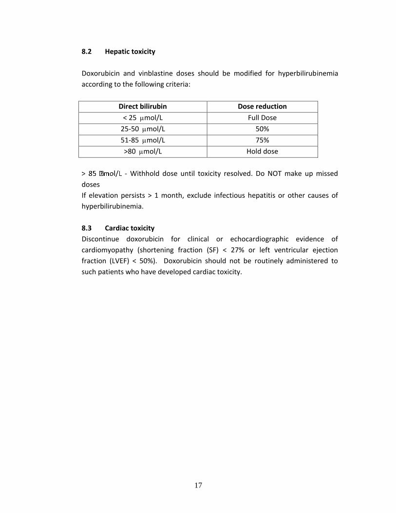
8.2 Hepatic toxicity
Doxorubicin and vinblastine doses should be modified for hyperbilirubinemia
according to the following criteria:
Direct bilirubin Dose reduction
< 25 mol/L Full Dose
25-50 mol/L 50%
51-85 mol/L 75%
>80 mol/L Hold dose
- Withhold dose until toxicity resolved. Do NOT make up missed
doses
If elevation persists > 1 month, exclude infectious hepatitis or other causes of
hyperbilirubinemia.
8.3 Cardiac toxicity
Discontinue doxorubicin for clinical or echocardiographic evidence of
cardiomyopathy (shortening fraction (SF) < 27% or left ventricular ejection
fraction (LVEF) < 50%). Doxorubicin should not be routinely administered to
such patients who have developed cardiac toxicity.

18
8.4 Neurological toxicity
PERIPHERAL NEUROPATHY: Neuropathy should be documented using the scale
provided below.
Modified “ Balis” Pediatric Scale of Peripheral Neuropathies
MOTOR NEUROPATHY
Grade 1 Subjective weakness, No deficit on neurological exam, other than
abnormal deep tendon reflexes
Grade 2 Weakness altering fine motor skills (buttoning shirt, coloring,
writing or drawing, eating) or gait abrogating ability to perform
tasks.
Grade 3 Unable to perform fine motor tasks or unable to ambulate without
assistance.
Grade 4 Paralysis
SENSORY NEUROPATHY
Grade 1 Paresthesia, pain, or numbness that do not require treatment or
interfere with extremity function.
Grade 2 Paresthesia, pain, or numbness that are controlled by non-narcotic
medications (without causing loss of function), or alteration of fine
motor skills or gait, without abrogating ability to perform tasks.
Grade 3 Paresthesias or pains that are controlled by narcotics, or interfere
with extremity function, or quality of life (loss of sleep, normal
activities severely impaired).
Grade 4 Complete loss of sensation, or pain that is not controlled by
narcotics.
SEVERE NEUROPATHIC PAIN (≥ GRADE 3): Hold dose(s). When symptoms
subside, resume at 4 mg/m2, and then escalate to full dose as tolerated. Drugs
such as gabapentin may be of value. If severe consider evaluation for Charcot
Marie Tooth Disease (CMT), type 1A or hereditary neuropathy with liability to
pressure palsies. Ask family history.
VOCAL CORD PARALYSIS: Hold dose(s). When symptoms subside, resume at 4
mg/m2, and then escalate to full dose as tolerated.
FOOT DROP, PARESIS: Should be grade 3 to consider holding or decreasing doses.
Physical therapy may be beneficial to maintain range of motion and provide
AFO’s and other forms of support. Gabapentin may be of value.
JAW PAIN – Treat with analgesics, DO NOT modify vinblastine dose.

19
8.5 Pulmonary toxicity
Pulmonary fibrosis is a serious risk with the use of Bleomycin. However, although
it may occur, pulmonary toxicity below a cumulative dose of 150 units/m2 is rare.
The cumulative dose on this protocol is 80 units/m2. Nonetheless, patients
experiencing dry cough, dyspnea, rales and pulmonary infiltrates should be
evaluated for potential pulmonary toxicity at any cumulative Bleomycin dose, as
a distinct hypersensitivity pneumonitis is recognized at any dose. Patients with
the above signs and symptoms should be evaluated by the pulmonology service
and should undergo pulmonary function studies. Patients with documented
pneumonitis should be treated with corticosteroids and should not receive any
further doses of Bleomycin.
8.6 Nausea and vomiting
Doxorubicin and Dacarbazine are both highly emetogenic agents. In addition
there is an incidence of delayed emesis with these agents. All patients should
receive either ondansetron or granisetron and dexamethasone intravenously
prior to the chemotherapy. Patients should also receive oral antiemetic agents
(ondansetron) for 24- to 48-hours following the chemotherapy. Longer duration
of antiemetic therapy may be required in a few patients.
9.0 Off therapy evaluations
9.1 Imaging studies
All patients should have CT scan evaluation of the sites of initial involvement at
the end of therapy. For patients who have received only chemotherapy this can
be performed within the first 2 months following the last dose of chemotherapy.
For patients who have received radiation therapy either for slow initial response
or for residual disease at the end of therapy should have CT-PET scan evaluation
of the sites of initial disease between 4 and 6 weeks following the end of the
course of radiation therapy.

20
Patients who have achieved a complete radiological response to therapy will be
followed by imaging studies according to the schema below:
Months
after
completion
of therapy
4 8 12 18 24 30 36 42 48 54 60 72 84 96 108
US abdomen x x x x x x x x x x x x x x x
Chest x-ray x x x x x x x x x x x x x x x
Any abnormalities on these imaging studies suspicious of recurrence will be
evaluated further by CT-PET.
9.2 Echocardiogram
All patients will undergo an echocardiogram at the end of therapy. Patients with
a normal cardiac function at this time point will henceforth be evaluated by an
echocardiogram once every year. Patients with abnormalities on any cardiac
evaluation may require more frequent or different evaluations, according to the
treating physician’s judgment.
9.3 Blood tests
Routine blood tests will be conducted at each follow-up clinic visit. These
routine tests include complete blood count with differential (CBCD), renal profile
(creatnine and electrolytes), hepatic profile (ALT, total bilirubin, albumin) and
erythrocyte sedimentation rate (ESR). In addition all patients should also be
tested annually for thyroid function with thyroid stimulating hormone (TSH) and
total T4.
9.4 Pulmonary Function Tests
Pulmonary function studies should be conducted on all patients within 6 months
off therapy and then once every other year. Frequency of testing would change
if there is any change in clinical status or alterations in the study results.