Embed Size (px)
Citation preview

GPR
GS
IsctcEdgpaotggtmt
o
D
T
A
1
rowth Factors and Signalingroteins in Craniofacial Development
obert Spears and Kathy K.H. Svoboda
Regulation of growth and development is controlled by the interactions of cells with eachother and the extracellular environment through signal transduction pathways that controlthe differentiation process by stimulating proliferation or causing cell death. This reviewwill define the common signaling molecules and provide an overview of the generalprinciples of signal transduction events. We also review the signal transduction pathwayscontrolling one specific mechanism found in craniofacial development termed epithelial-mesenchymal transformation (EMT) employed during gastrulation, cranial neural crestmigration, and secondary palate formation.Semin Orthod 11:184–198 © 2005 Elsevier Inc. All rights reserved.
rkmtt(llktaf(tthcv
so1ctagrTiim
rowth Factors andignal Transductionntracellular signaling is usually triggered by a cell surfaceevent such as a specific protein (ligand) binding to a cell
urface receptor to form a receptor-ligand interaction. Cellsontacting neighboring cells or their surrounding noncellularissue are termed cell-cell or cell-extracellular matrix (ECM)ontacts (Fig 1A).1 Interactions of cells with other cells or theCM can stimulate many reactions, including: increased cellivision, cell movement, differentiation, and even pro-rammed cell death (apoptosis). The cell surface bindingroteins (receptors) are classified by the protein structurend ligand characteristics. These proteins are usually locatedn the cell surface in the plasma membrane. Part of the pro-ein may be located outside the cell to interact with the li-and. This part of the proteins is called the extracellular li-and binding domain. Part of the protein will traversehrough the membrane, termed the membrane spanning do-ain, and a portion of the protein will be inside the cell and
ermed the cytoplasmic domain.Most modern cell biology textbooks list at least four types
f cell surface receptors, including the G protein-coupled
epartment of Biomedical Sciences, Baylor College of Dentistry, Texas A&MHealth Science Center, Dallas, TX.
his work was supported by grant sponsors: Baylor Oral Health Foundation;Tobacco Endowment Fund, Texas A&M University System; Grant Num-ber: 304-202850-4013.
ddress correspondence to Robert Spears, PhD, Texas A&M UniversitySystem, Baylor College of Dentistry, Department of Biomedical Sciences,3302 Gaston Ave, Dallas, TX 75266; Phone: 214-828-8297; Fax: 214-
t828-8951. E-mail: [email protected]
84 1073-8746/05/$-see front matter © 2005 Elsevier Inc. All rights reserved.doi:10.1053/j.sodo.2005.07.003
eceptors (GPCR, Fig 1A), ion-channel receptors, tyrosineinase-linked receptors, and receptors with intrinsic enzy-atic activity.2 The GPCRs are characterized by multiple
ransmembrane domains (usually seven) that wind the pro-ein in and out of the membrane in a serpentine conformationFig 1A, GPCR). The ion-channel receptors are closely re-ated and actually open a membrane channel when theigand binds. Many cytokine receptors are in the tyrosineinase-linked class as they lack intrinsic activity; but whenhe ligand binds, intracellular tyrosine kinases becomectivated to generate cellular changes. The classic growthactor receptors have kinase activity within the proteinintrinsic) and therefore make up the fourth class of recep-ors—the receptor tyrosine kinases, or receptor serine/hreonine kinases (Fig 1A and C). These receptors usuallyave one transmembrane domain, and at least two mole-ules must become closely associated (dimerize) to acti-ate the signal.In addition to these classic receptor classes, cells can re-
pond to their ECM environment through integrin receptorsr proteoglycan receptors such as the syndecan family (FigA and B). Single transmembrane domains with large extra-ellular and much smaller cytoplasmic domains characterizehese receptors. The syndecan molecules have long glycos-minoglycan chains that assist in sequestering the fibroblastrowth factors close to the cell membrane.3-6 The integrineceptors are heterodimers composed of � and � subunits.he family is very large with at least 25 integrin heterodimers,
ncluding 19 � subunits and 8 � subunits identified.7 Thentegrins do not have kinase activity, but on binding to ECM
olecules, some associated proteins become activated by au-
ophosphorylation and then phosphorylate (activate) sur-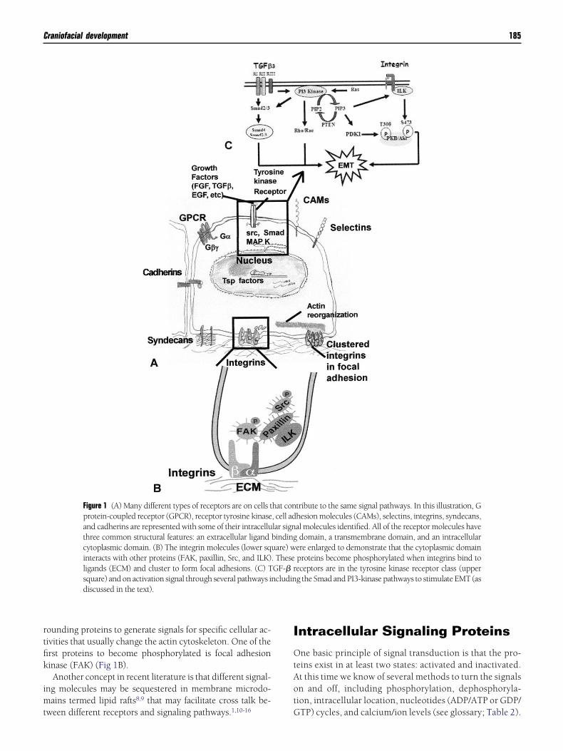
rtfik
imt
I
OtAot
Craniofacial development 185
ounding proteins to generate signals for specific cellular ac-ivities that usually change the actin cytoskeleton. One of therst proteins to become phosphorylated is focal adhesioninase (FAK) (Fig 1B).Another concept in recent literature is that different signal-
ng molecules may be sequestered in membrane microdo-ains termed lipid rafts8,9 that may facilitate cross talk be-
Figure 1 (A) Many different types of receptors are on cells tprotein-coupled receptor (GPCR), receptor tyrosine kinaseand cadherins are represented with some of their intracelluthree common structural features: an extracellular ligandcytoplasmic domain. (B) The integrin molecules (lower sqinteracts with other proteins (FAK, paxillin, Src, and ILK)ligands (ECM) and cluster to form focal adhesions. (C) Tsquare) and on activation signal through several pathways idiscussed in the text).
ween different receptors and signaling pathways.1,10-16 G
ntracellular Signaling Proteins
ne basic principle of signal transduction is that the pro-eins exist in at least two states: activated and inactivated.t this time we know of several methods to turn the signalsn and off, including phosphorylation, dephosphoryla-ion, intracellular location, nucleotides (ADP/ATP or GDP/
tribute to the same signal pathways. In this illustration, Ghesion molecules (CAMs), selectins, integrins, syndecans,al molecules identified. All of the receptor molecules havedomain, a transmembrane domain, and an intracellular
ere enlarged to demonstrate that the cytoplasmic domainproteins become phosphorylated when integrins bind toeceptors are in the tyrosine kinase receptor class (upperg the Smad and PI3-kinase pathways to stimulate EMT (as
hat con, cell adlar signbindinguare) w. TheseGF-� rncludin
TP) cycles, and calcium/ion levels (see glossary; Table 2).

Gnc(cbsgtblscissafsssnc
o(cisfmccwettco
Panotsmtfptotpcpa
afi
plcmbtret
NtwaRb(tTstthctaehd
Icctaacf
bmcpgahtRa
atkc
186 R. Spears and K.K.H. Svoboda
PCR cascade. One of the first signal transduction mecha-isms described was the GPCR cascade that generates thelassic second messengers cyclic AMP (cAMP), cyclic GMPcGMP), diacylglycerol (DAG), phosphoinositols, and cal-ium (Ca2�). When G protein-linked or hormone receptorsecome activated, they trigger a series of events at the cellurface that cause transient increases in these second messen-er molecules.2 As with other signaling events, there is aransient increase in the active form of the molecule followedy a rapid decrease to produce a “signal.” Briefly, when the
igand binds to the receptor it causes a change in the proteintructure that allows the G-protein � subunit to bind to theytoplasmic domain of the receptor (Fig 1A, GPCR). Thisnteraction causes the exchange of GDP for GTP on the �ubunit and the disassociation of the �� subunits from the �ubunit. The activated (GTP bound) � subunit interacts withdenylyl cyclase, the membrane-bound enzyme responsibleor producing cAMP. After activating adenylyl cyclase, the �ubunit reverts to the GDP state and reassociates with the ��ubunits. Not only is the number of GPCRs very large, but �ubunits are also numerous, providing a wide variety of sig-als to the cell. Once the G proteins are activated, the signalan be amplified by interactions with other proteins.
Classically, adenylyl cyclase produces cAMP that activatesther kinases termed the cyclic AMP-dependent kinasescAPKs), also called protein kinase A (PKA). These kinasesan phosphorylate (activate) a number of substrates depend-ng on the specific stimulus to amplify the signal from the cellurface.2 One effect of activation is the release of calciumrom intracellular storage areas, such as the rough endoplas-
ic reticulum and mitochondria. Since free calcium levels inells are maintained at very low levels, the rapid increase inalcium levels from these intracellular organelles has been aay to visualize signal events. Calcium can be labeled with
ither quantitative ratiometric dyes or single wavelength dyeso monitor these rapid changes in cells following stimula-ion.17 Rapid sequestering of the free calcium ions by mole-ules such as calmodulin that bind several calcium ions turnsff the calcium signal.
hosphorylation and dephosphorylation. Phosphorylationnd dephosphorylation provide another mechanism for sig-al on and off switches. The enzymes that phosphorylatether proteins are called kinases. The common amino acidshat become phosphorylated are tyrosine, threonine, anderine. As stated previously, the phosphorylated proteinsay be activated by adding phosphate molecules, and deac-
ivated by removing the phosphates, a function usually per-ormed by another enzyme class, the phosphatases. Someroteins can autophosphorylate themselves; once activated,hey can phosphorylate surrounding substrates. An examplef this type of protein was discussed previously in describinghe integrin molecules; focal adhesion kinase (FAK) becomeshosphorylated when integrins bind to their ligand, ECM (ie,ollagen, fibronectin, or laminin). Once FAK becomes phos-horylated it will activate or phosphorylate paxillin, an actin-
ssociated protein, and another kinase, Src (Fig 1B).18 Src can glso start activating surrounding proteins, creating an ampli-cation of the original signal.To complicate things, some proteins are inactive in the
hosphorylated state and become active after dephosphory-ation. Therefore, it is important to understand the possiblehanges in the proteins before starting an investigation. Cellsay contain a constant amount of a given protein in a pool,
ut the protein has to be in an active state to produce a signalhat will change the proteins around it.19 It is important toemember that just because an mRNA for a given protein isxpressed; it does not indicate that the protein is produced orhat it is activated.
ucleotide binding. Protein is generally in an inactive state ifhe ADP or GDP nucleotide is bound and becomes activatedhen the ATP or GTP is bound. An example of this type of
ctivation is the small G-protein families, Ras andho.1,12,20-23 These proteins alternate between the GTP-ound active form (on) and the GDP-bound inactive formoff) to regulate other downstream kinases. Many other pro-eins regulate the “on” and “off” state of the small G proteins.he guanine-nucleotide exchange factors (GEFs) are the “on”ignal as they add GTP to the protein. GTP-activating pro-eins (GAPs) are the “off” signal as they remove a phosphateo deactivate the protein. Guanine-nucleotide dissociation in-ibitor proteins (GDIs) sequester the inactive protein in theytoplasmic pool. We have shown that one of these regula-ory proteins (p190RhoGAP; 190 kDA protein that functionss a GAP for Rho) becomes phosphorylated very quickly inmbryonic epithelia in response to cells binding ECM.19 Weave also shown that decreasing Rho protein levels or activityecreased other integrin signaling molecules.24
ntracellular location. Some proteins move to specific intra-ellular structures, such as focal adhesions1,25 when they be-ome activated. Paxillin, �-actinin, and talin accumulate athe focal adhesions in both migratory and stationary cells. Inddition, integrin molecules become clustered at the focaldhesion of fibroblast cells (Fig 1A) and new evidence indi-ates that these proteins may act as sensors for mechanicalorces.14
Many proteins move to the plasma membrane when theyecome activated. The movement to the plasma membraneay take several steps, including release from a cytoplasmic
haperone protein and/or acquiring a lipid tail. The small Groteins (such as Rho) require both the release from theuanine-nucleotide dissociation inhibitor (GDI) protein andlipid tail to move to the membrane.26,27 So, in addition toaving a GTP bound to the protein, the protein itself moveso the site of action. Many of the small GTPase proteins (Ras,af, Rac) follow similar intracellular translocation patterns onctivation.
Other activated proteins move to the nucleus and may acts transcription factors. Examples of this type of intracellularranslocation are some of the MAP kinase proteins.28-30 MAPinases can respond to a variety of extracellular signals, in-luding osmotic stress, heat shock, cytokines, and mito-
ens.31 Two of the MAP kinases, the extracellular signal-reg-
utpvmlta
tfosSptn1dr
SIcbEmGespifmrmrmcRtrso
GIMCmvwbots
db
eaDcm
wstcddisodctnto
CBtlacJWptntecDiC
fpwgssctmt
s“
Craniofacial development 187
lated kinases (erk-1 and erk-2; also referred to as erk-1/2),ranslocate to the nucleus after activation to regulate the ex-ression of various transcription factors (Table 1).30,31 Acti-ation of the MAP kinase pathways has been identified as aechanism used by integrins to regulate gene expression
eading to cell shape changes during cell spreading or migra-ion,10,21,32,33 and as a cross-talk pathway between integrinsnd growth factors.1,10,34
Sometimes the signal protein needs to bind another pro-ein, a chaperone, before translocating to the nucleus. Trans-orming growth factor beta (TGF-�) proteins activate a classf proteins called Smads. These growth factors bind to cellurface receptors (T�RI and T�RII) and activate specificmad proteins (Smad 2 or 3) that then bind to a chaperonerotein, Smad 4, before translocating to the nucleus (Fig 1C)o act as transcription factors.35-38 Similarly, bone morphoge-etic proteins (BMPs) bind to BMP receptors, activate Smadsor 5, which bind Smad 4 to translocate to the nucleus. Moreetails concerning this pathway will be discussed later in thiseview (EMT regulation).
ummaryntracellular responses to cell surface receptors are compli-ated and the subject of many active investigations. A num-er of studies have established reciprocal linkages betweenCM-integrins, growth factor signaling, cell-cell adhesionolecules, specialized membrane domains (lipid rafts), andprotein-linked receptors.1 In addition, cross talk has been
stablished between the ECM and intracellular mitogen-timulated pathways, the small G proteins, and the phos-hoinositols.39,40 The cell’s microenvironment and the result-
ng tissue profoundly influence each of these linkages. Thus,or a cell to achieve a differentiated phenotype or respond to
icroenvironment changes, the ECM molecules and theireceptors must integrate both form and function. In contrast,utated genes and aberrant interactions with the microenvi-
onment may degrade this integration, possibly resulting inalignant transformation or abnormal development.41 Re-
ently, it has also become apparent that integrins regulateho GTPases and vice versa. Integrins and GTPases might
herefore be organized into complex signaling cascades thategulate cell behavior.1,10,21 In the next section of this review,pecific signaling pathways important for craniofacial devel-pment will be discussed in detail.
rowth Factorsmportant in Epithelial/esenchymal Interactions
ell phenotype transformations, from epithelial to mesenchy-al (epithelial-mesenchymal transformation, EMT) and vice
ersa have been well documented in embryonic development,ound healing, and tumor metastasis. Epithelia serve as theoundary between the external environment and the remainderf the organ while mesenchymal cells are found in the connec-ive tissue compartment. The epithelial barrier function is partly
upported by firm cell-cell junctions, such as tight junctions and mesmosomes. In addition, epithelial cells normally have apical-asal polarity and attach to basal lamina by hemidesmosomes.Mesenchymal cells are more mobile and surrounded by
xtracellular matrix. They have anterior-posterior polaritynd form only transient contacts with their neighboring cells.uring EMT cell phenotype transition, epithelial cells loseell-cell attachment, break through the basal lamina, becomeobile, and express mesenchymal proteins (Fig 2B).From the mid-1990s and more recently, several reviews
ere published on EMT in development and pathogene-is.42-48 EMT and the opposite, mesenchymal-epithelialransformation, occur during normal developmental pro-esses. EMT occurs during gastrulation, one of the earliestevelopmental events that change the two layered embryonicisc into a three layered embryo. This process involves the
nvagination of the top layer (epiblast) cells to form the me-oderm49 and endoderm germ layers.50,51 In addition severalther developmental processes such as sclerotome52 and car-iac cushion mesenchyme53-55 development require EMT. Inontrast, mesenchymal-epithelial transformations occur inhe formation of somites, kidneys, and caudal or secondaryeural tube.56 This review will concentrate on EMT duringhe development of craniofacial structures, and specificallyn cranial neural crest (CNC) and secondary palate formation.
ranial Neural Crestefore the closure of the neural folds in the mammalian head,he neural crest cells break away from an embryonic epithe-ial layer of the dorsal neural tube by changing their shapend properties from neuroepithelial cells to mesenchymalells. In a recent meeting celebrating the contributions ofames A. Weston to the understanding of neural crest,
eston proposed that the CNC were derived from an earlyopulation of non-neuronal ectoderm.57,58 While this con-roversial theory is certain to engage many investigators in theext few years, this review will concentrate on the currentheory that CNC are derived from neural epithelium. How-ver, as both ectoderm and neuroepithelium are epithelialell types, they still have to complete EMT. A recent issue ofevelopmental Dynamics is dedicated to this topic as a special
ssue subtitled “Special Focus on the Neural Crest and theontributions of James A. Weston.”59
A major difference between neural crest cells in the cranio-acial region and those of the trunk is that the CNC cells areatterned with level-specific instructions in the head,hereas those of the trunk do not appear to be prepro-rammed.60 In the cranial region, the CNC migrate in diffusetreams throughout the cranial mesenchyme, with a level-pecific instruction, to reach their final destinations. Theseells have also been referred to as ectomesenchyme in someextbooks.61 Extensive experiments demonstrated that theaintenance of this segmental characteristic is very impor-
ant in patterning of head development.62-64
The CNC cells are multipotent stem-like cells, which re-pond to temporal-spatially expressed signals and becomecommitted.” Candidate regulators include growth factors—
embers of the TGF-� family,65 fibroblast growth factors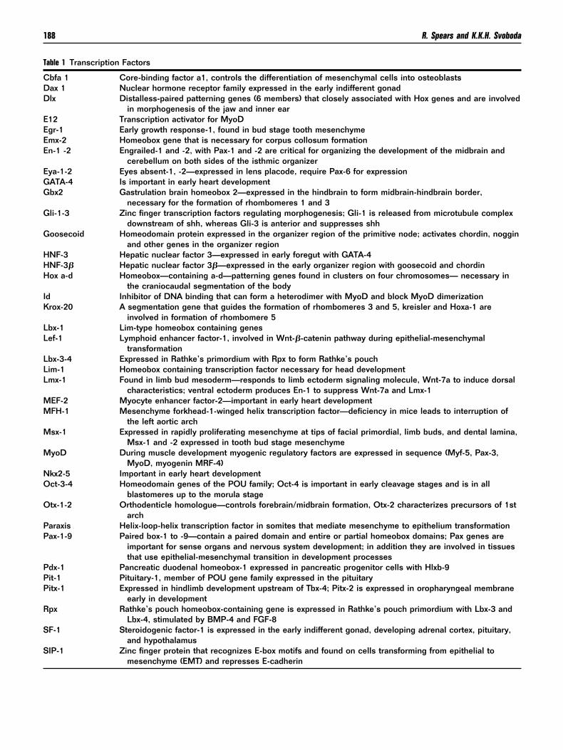
T
CDD
EEEE
EGG
G
G
HHH
IK
LL
LLL
MM
M
M
NO
O
PP
PPP
R
S
S
188 R. Spears and K.K.H. Svoboda
able 1 Transcription Factors
bfa 1 Core-binding factor a1, controls the differentiation of mesenchymal cells into osteoblastsax 1 Nuclear hormone receptor family expressed in the early indifferent gonadlx Distalless-paired patterning genes (6 members) that closely associated with Hox genes and are involved
in morphogenesis of the jaw and inner ear12 Transcription activator for MyoDgr-1 Early growth response-1, found in bud stage tooth mesenchymemx-2 Homeobox gene that is necessary for corpus collosum formationn-1 -2 Engrailed-1 and -2, with Pax-1 and -2 are critical for organizing the development of the midbrain and
cerebellum on both sides of the isthmic organizerya-1-2 Eyes absent-1, -2—expressed in lens placode, require Pax-6 for expressionATA-4 Is important in early heart developmentbx2 Gastrulation brain homeobox 2—expressed in the hindbrain to form midbrain-hindbrain border,
necessary for the formation of rhombomeres 1 and 3li-1-3 Zinc finger transcription factors regulating morphogenesis; Gli-1 is released from microtubule complex
downstream of shh, whereas Gli-3 is anterior and suppresses shhoosecoid Homeodomain protein expressed in the organizer region of the primitive node; activates chordin, noggin
and other genes in the organizer regionNF-3 Hepatic nuclear factor 3—expressed in early foregut with GATA-4NF-3� Hepatic nuclear factor 3�—expressed in the early organizer region with goosecoid and chordinox a-d Homeobox—containing a-d—patterning genes found in clusters on four chromosomes— necessary in
the craniocaudal segmentation of the bodyd Inhibitor of DNA binding that can form a heterodimer with MyoD and block MyoD dimerizationrox-20 A segmentation gene that guides the formation of rhombomeres 3 and 5, kreisler and Hoxa-1 are
involved in formation of rhombomere 5bx-1 Lim-type homeobox containing genesef-1 Lymphoid enhancer factor-1, involved in Wnt-�-catenin pathway during epithelial-mesenchymal
transformationbx-3-4 Expressed in Rathke’s primordium with Rpx to form Rathke’s pouchim-1 Homeobox containing transcription factor necessary for head developmentmx-1 Found in limb bud mesoderm—responds to limb ectoderm signaling molecule, Wnt-7a to induce dorsal
characteristics; ventral ectoderm produces En-1 to suppress Wnt-7a and Lmx-1EF-2 Myocyte enhancer factor-2—important in early heart developmentFH-1 Mesenchyme forkhead-1-winged helix transcription factor—deficiency in mice leads to interruption of
the left aortic archsx-1 Expressed in rapidly proliferating mesenchyme at tips of facial primordial, limb buds, and dental lamina,
Msx-1 and -2 expressed in tooth bud stage mesenchymeyoD During muscle development myogenic regulatory factors are expressed in sequence (Myf-5, Pax-3,
MyoD, myogenin MRF-4)kx2-5 Important in early heart developmentct-3-4 Homeodomain genes of the POU family; Oct-4 is important in early cleavage stages and is in all
blastomeres up to the morula stagetx-1-2 Orthodenticle homologue—controls forebrain/midbrain formation, Otx-2 characterizes precursors of 1st
archaraxis Helix-loop-helix transcription factor in somites that mediate mesenchyme to epithelium transformationax-1-9 Paired box-1 to -9—contain a paired domain and entire or partial homeobox domains; Pax genes are
important for sense organs and nervous system development; in addition they are involved in tissuesthat use epithelial-mesenchymal transition in development processes
dx-1 Pancreatic duodenal homeobox-1 expressed in pancreatic progenitor cells with Hlxb-9it-1 Pituitary-1, member of POU gene family expressed in the pituitaryitx-1 Expressed in hindlimb development upstream of Tbx-4; Pitx-2 is expressed in oropharyngeal membrane
early in developmentpx Rathke’s pouch homeobox-containing gene is expressed in Rathke’s pouch primordium with Lbx-3 and
Lbx-4, stimulated by BMP-4 and FGF-8F-1 Steroidogenic factor-1 is expressed in the early indifferent gonad, developing adrenal cortex, pituitary,
and hypothalamusIP-1 Zinc finger protein that recognizes E-box motifs and found on cells transforming from epithelial to
mesenchyme (EMT) and represses E-cadherin

(Wfrmp
nscmhcapsc(iptoai
RGEpgimc1im
dEhaoe(
nTmttie
ssTtsito
m1“ebb1RtH
T
S
S
S
S
T
T
VW
Z
*
Craniofacial development 189
FGFs),66,67 platelet-derived growth factor (PDGF),68 andnt gene products69,70 (Table 2). The role of the TGF-�
amily in CNC developmental processes has recently beeneviewed.71 The involvement of several signal transductionolecules and transcription factors have also been re-orted.44
The paired maxillary processes and mandibular promi-ences of the first branchial arch give rise mainly to thetructures of the upper and lower jaws. The neural crestomponent of the upper jaw derives from the forebrain andidbrain, while that of the lower jaw from the midbrain andindbrain (rhombomeres 1 and 2). These CNC cells mainlyontribute to the following structures in the first branchialrch: palate and maxilla, dermis and fat of skin, dental pa-illa, Schwann cells of peripheral nerves, melanocytes, andome connective tissue. A recent study demonstrated that theonditional removal of the transmembrane signaling receptorT�rII) gene in only the cranial neural crest lineage resultedn clefting of secondary palate and calvaria defects. Theathogenesis of cleft palate in these mice appears to be relatedo impairment of cell proliferation.72 The normal formationf some derivatives of the first branchial arch, such as palatend lip, needs epithelial-mesenchymal transformation dur-ng embryonic remodeling.
egulation of EMTrowth factors and signal transduction. The regulation ofMT is critical during dynamic developmental processes andostnatal homeostasis. Hay (1989) postulated that masterene(s) turned on in epithelia by changes in the environmentnitiate EMT (master gene theory) (Fig 2). Recently several
olecules have been identified as possible master genes in-luding the transcription factors Twist, Snail or SIP1 (Table). The changes in the environment that may initiate EMT
nclude growth factors, cell adhesion molecules, extracellular
able 1 Continued
lug Zinc finger protein expressed in epiblasplate stage of development; the cellsphenotypes
nail Zinc finger protein that recognizes E-bomesenchyme (EMT) and represses E-c
ox Large family (over 20) that have a commnucleotides on the minor groove of thin a large variety of tissues; Sox-9 con
ry Sex-determining region, a member of thdevelopment by inhibiting Dax-1
bx-4-5 T-box family, -5 expressed in forelimb; -dorsal retina
wist Basic helix-loop-helix protein that is a reincluding mesoderm development dur
ax-2 Ventral anterior homeobox-2—expressedt-1 Wilms’ tumor suppressor gene-1—expre
mesenchyme to epitheliumFY Zinc finger Y
Compiled from various sources including Carlson.138
atrix, the surface receptors and downstream signal trans- i
uction events (Table 2), and transcription factors (Table 1).pithelial cells are characterized by cell-cell adhesions (ad-erens junctions, desmosomes, and hemidesmosomes) andn intact basal lamina. During EMT the cells lose expressionf cell adhesion proteins, especially E-cadherin, and increasexpression of enzymes that break down the basal laminaFig 2B).
This review will focus on TGF-� and its downstream sig-aling molecules primarily during palate development. TheGF-� superfamily includes many small proteins that areultifunctional (controlling growth, migration, and differen-
iation) during both embryonic development and postnatalissue homeostasis.73-75 The cellular responses to TGF-� dur-ng craniofacial development and CNC regulation have beenxtensively studied and recently reviewed.71
Studies designed to investigate how cells interpret TGF-�ignals have identified three types of TGF-� transmembraneignaling receptors: type I, type II (T�RI and T�RII),76 and�RIII (Fig 1C). Types I and II TGF-� receptors have a cy-
oplasmic serine/threonine kinase domain. TGF-�s initiateignaling by assembling receptor complexes. The initial bind-ng of T�RII to ligand is recognized by T�RI and the forma-ion of ligand/T�RI/T�RII/complex results in the activationf T�RI by T�RII.77
Activated T�RI propagates the signal by phosphorylatingany downstream effectors, such as Smad proteins (Fig
C).78 The Smad proteins are homologues to the Drosophilamothers against dpp” (mad) proteins and the Caenorhabditislegans Sma proteins. In vertebrates at least nine genes haveeen found that comprise the Smad family. Different mem-ers of the Smad family have different signaling roles. Smads, 2, 3, and 5 interact with the receptors and are termed-Smads. These proteins become phosphorylated by the ac-
ivated receptors and are transported to the nucleus.71,79-81
owever, when there are no outside-in signals, R-Smads stay
during gastrulation and neural crest cells at the neuralpress slug transform from epithelial to mesenchymal
fs and found on cells transforming from epithelial toin; closely related to sluggh-mobility group (HMG) domain that binds to 7
helix; Sox proteins work with other transcription factorsdifferentiation of mesenchymal cells into precartilagegene family found on the Y-chromosome, triggers testis
essed in hind limb with Pitx-1, Tbx-5 also expressed in the
r of morphogenesis that plays an essential role in EMTstrulation by repressing E-cadherine ventral retinain mesonephric ducts—regulates transformation from
t cellsthat ex
x motiadheron hi
e DNAtrols
e Sox
4 expr
gulatoing ga
in thssed
n the cytoplasm partly by binding to the protein SARA
(pSR
St
190 R. Spears and K.K.H. Svoboda
Smad Anchor for Receptor Activation).82 Receptor phos-horylation of R-Smads weakens the affinity of R-Smads forARA and exposes their nuclear import signal. Activation of
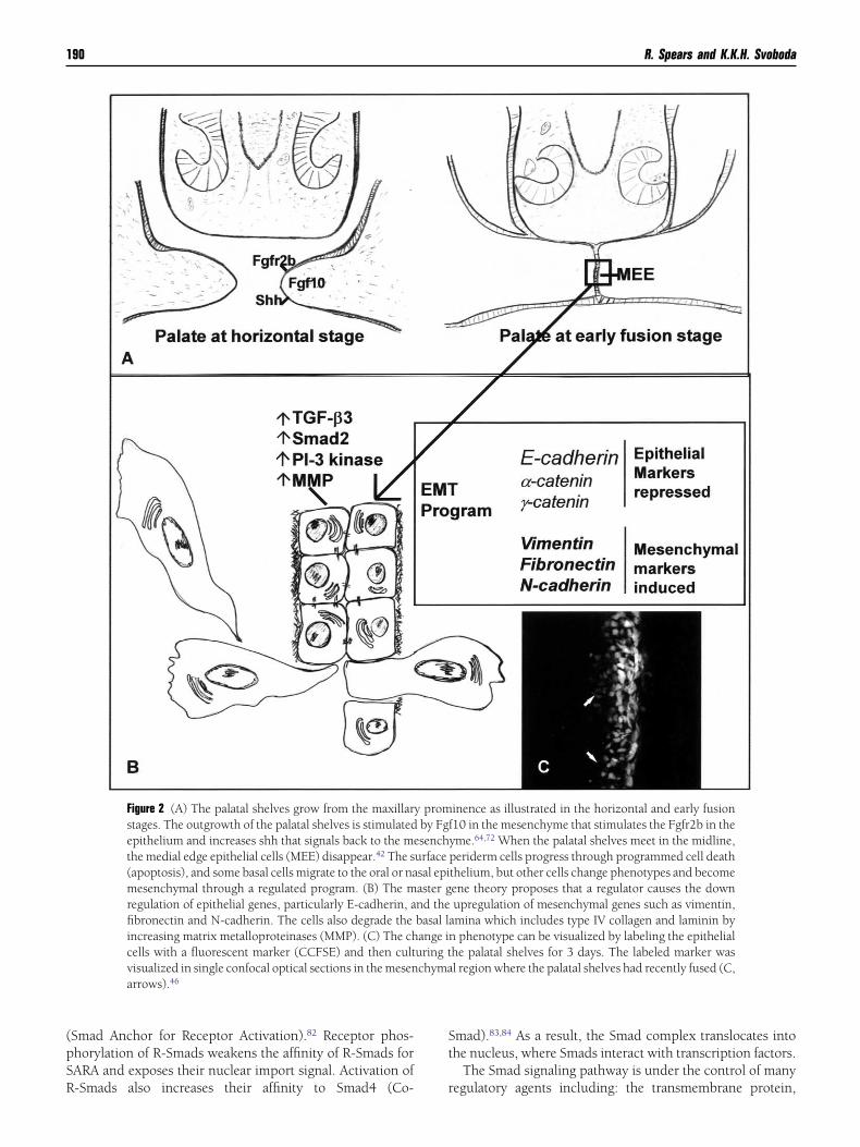
Figure 2 (A) The palatal shelves grow from the maxillarstages. The outgrowth of the palatal shelves is stimulatedepithelium and increases shh that signals back to the methe medial edge epithelial cells (MEE) disappear.42 The s(apoptosis), and some basal cells migrate to the oral or namesenchymal through a regulated program. (B) The mregulation of epithelial genes, particularly E-cadherin, afibronectin and N-cadherin. The cells also degrade theincreasing matrix metalloproteinases (MMP). (C) The chcells with a fluorescent marker (CCFSE) and then cultvisualized in single confocal optical sections in the mesenarrows).46
-Smads also increases their affinity to Smad4 (Co- r
mad).83,84 As a result, the Smad complex translocates intohe nucleus, where Smads interact with transcription factors.
The Smad signaling pathway is under the control of many
inence as illustrated in the horizontal and early fusion10 in the mesenchyme that stimulates the Fgfr2b in theyme.64,72 When the palatal shelves meet in the midline,periderm cells progress through programmed cell deaththelium, but other cells change phenotypes and becomeene theory proposes that a regulator causes the downupregulation of mesenchymal genes such as vimentin,
amina which includes type IV collagen and laminin byn phenotype can be visualized by labeling the epithelialhe palatal shelves for 3 days. The labeled marker wasl region where the palatal shelves had recently fused (C,
y promby Fgfsench
urfacesal epiaster gnd thebasal lange iuring tchyma
egulatory agents including: the transmembrane protein,

BsrtSobfcVtp
iappklpcp
vsTwcept
Pfiaepcgmccmc(f2E
1ehlteia
idhWtHpcad
ttiitdmpe
TrwgTinetpdf
pTmtivb5tRs(Tiottncpow
Craniofacial development 191
AMBI, that has an intracellular domain that mimics thetructure of a type I receptor and prevents the formation ofeceptor complexes85; the ubiquitin ligase, Smurf186; the an-agonistic Smad6 and Smad7,87,88 and the oncoproteins Ski/no.89 In addition, in the nucleus the cellular responses tother signal inputs determine which genes will be recognizedy the Smad complex.90 Recently the interferon regulatoryactor 6 (IRF6) gene was identified as the candidate for theause of an autosomal dominate form of cleft lip and palate,an der Woude’s syndrome.91 However, it is not clear how
he point mutation in this gene contributes to cleft lip oralate.Although the Smad pathway has received much attention
n the past 5years, it is now appreciated that the TGF-�-ctivated receptor complex can also signal through otherathways,92 such as those involving the mitogen-activatedrotein kinases (MAPKs),93,94 phosphoinositol-3 kinase (PI-3inase) (Fig 1C),95,96 and PP2A/p70s6K, though the molecu-
ar details of this coupling are still obscure. The relative im-ortance and interplay of these various pathways in thehanging responses of cells to TGF-� are just beginning to berobed.TGF-� family members are essential for EMT during de-
elopment. The embryonic cardiac cushion has been exten-ively studied in an organ culture model that demonstratedGF-�2-mediated initial endothelial cell-cell separationhile TGF-�3 was required for the cell morphological
hange that enabled the migration of cells into the underlyingxtracellular matrix.97 These conclusions were based on ex-eriments that used antisense oligodeoxynucleotides or neu-ralizing antibodies to TGF-�3 to inhibit EMT in vitro.97
alate development. As shown in Fig 2, the secondary palateorms as an outgrowth of the maxillary prominence. Interest-ngly, it was recently shown that sonic hedge hog (shh)nd the Fgfr2b were also expressed in the early palatalpithelium98 and appear to be induced by Fgf10. When thisathway was disrupted in transgenic animals, the palatal pro-esses failed to grow.98 The normal palatal shelves elevate androw toward the midline where they fuse and some of theedial edge epithelial (MEE) cells move into the mesen-
hyme through the EMT process (Fig 2). The migrating MEEells can be visualized if the epithelial cells are bathed in aarker that only penetrates the surface epithelium such as
arboxy 2,7= dichlorofluorescein diacetate succinimidyl esterCCFSE). After the palatal shelves have been organ culturedor 3 days, the label was found in the mesenchymal cells (FigC) demonstrating the labeled cells had progressed throughMT.99
During mammalian palate development TGF-� isoforms, 2, 3, T�RII, and T�RIII were detected in the medial edgepithelium (MEE) by in situ hybridization100 and immuno-istochemistry.101,102 Of particular interest was the highly
ocalized expression of TGF-�3 RNA, and to a lesser extenthat of TGF-�1 and TGF-�2 in the MEE and the nasal septumpithelium, which were destined to undergo EMT.100 Exper-ments using antisense oligodeoxynucleotides or neutralizing
ntibodies to TGF-�3, but not TGF-�1 or TGF-�2, resulted in the failure of palate fusion in vitro.103 Further experimentsemonstrated that Tgf-�3 transgenic and knockout miceave cleft palate as their only craniofacial birth defect.104,105
hen palatal shelves from Tgf-�3 knockout mice were cul-ured, the midline epithelia failed to go through EMT.106,107
owever, overexpressing Smad2 on the Tgf-�3 null animalartially rescued palatal fusion.108 In addition, although thehicken has naturally open palate, the cultured chicken pal-tal shelves fused when TGF-�3 was added into the me-ium.109
It is clear from these experiments that TGF-�3 is an essen-ial growth factor inducing EMT during palatal fusion. Inves-igators have proposed that possible mechanisms of TGF-�3-nduced palatal fusion include the regulation of fusion bynducing cell membrane filopodia on MEE before shelf con-act.107 Second, the regulation of extracellular matrix degra-ation by modulating the production of tissue inhibitor ofetalloproteinase-2 (TIMP-2), MMP13, and MMP2 has beenroposed.110 More recently, it was found that TGF-�3 is nec-ssary for inhibiting MEE proliferation during EMT.111
Several research groups have started to investigate theGF-�3-stimulated intracellular signaling molecules that areesponsible for EMT during palate fusion. Smad2 expressionas detected during palatal fusion,112 and it has been sug-ested that phosphorylation of Smad2 may be necessary forgf-�3 downregulation of MEE proliferation.111 Interest-
ngly, overexpression of Smad2 in the TGF-�3–/– mouse didot completely rescue secondary palate clefts. There is alsovidence from the studies of mammary epithelial cell culturehat downregulation of Smad signaling decreased Smad-de-endent growth and transcriptional responses; however, theownregulation did not affect TGF-�-mediated stress fiberormation and EMT.113
Understanding the signal transduction pathways can be com-licated. For example, we have recently investigated the role ofGF-� signaling during secondary palate formation in a mouseodel (Fig 3). We found that an alternative downstream effecter
o MEE signaling, PI-3 kinase, has been identified as an effectorn actin reorganization and TGF-�-mediated EMT.114-116 Acti-ated PI-3 kinase phosphorylates phosphatidylinositol 4,5-isphophate (PIP2) to generate phosphatidylinositol 3, 4,-trisphosphate (PIP3), which recruits its downstream effectorso the plasma membrane (Fig 1C). Along with the small GTPasesac and Rho, PIP3 activates several serine/threonine kinasesuch as 3-phosphoinostide-dependent protein kinasesPDKs).117,118 PDK1 activates PKC, p70S6K1,119 and targetshr308 on protein kinase B (PKB, also known as Akt),120 while
ntegrin-linked kinase (ILK), a newly found PDK activates Aktn its Ser473 site.121 On stimulation, Akt migrates and anchorso the membrane.122 Subsequently, activated Akt detaches fromhe plasma membrane and translocates into the cytoplasm anducleus, regulating cell survival, protein synthesis, and cell cy-le.123 It also appears that PI-3 kinase possesses both lipid androtein kinase activity124 and may directly control the activitiesf individual components of the RAS/RAF/ERK-mitogenic path-ay by forming a complex with signal proteins.The consequences of PI-3 kinase activation are numerous,
ncluding effects on cell cycle progression, suspension-medi-

T
AAB
b�
bB
CccCcC
CCC
DDdEEEeE
FF
F
GGGGGG
gHIIIIIIJKLLMMMMM
192 R. Spears and K.K.H. Svoboda
able 2 Signal Transduction Proteins, Abbreviations, and Definitions
ctivin Signaling protein, member of the TGF-� family, active in mesoderm inductionngiopoietin-1 Sprouting factor—interacts with Tie-2 (receptor) during angiogenesisAMBI Transmembrane protein, whose intracellular domain resembles the homodimerization interface of a
T�RI receptor and prevents the formation of TGF-� receptor complexesax bcl-associated x protein that accelerates cell death (apoptosis)-catenin Binds to E-cadherin in cell-cell junctions, but can be modulated by many signaling agents (Wnt, Ras,
and PI-3 kinase) to relocate to the nucleus and interacts with transcription factors (TCF/LEF-1);stabilized nuclear �-catenin has been shown to induce EMT in vitro133
cl-2 B cell lymphoma protein that blocks cell death (apoptosis)MP-1-9 Bone morphogenetic protein, members of the TGF-� family—induction of the neural plate, skeletal
differentiation, and other inductionsAM Cell adhesion molecules (neuronal CAM [N-CAM], L-CAM � E-cadherin)AMP Cyclic AMP, second messenger in GPCR signal transduction pathwaysAPKs Cyclic AMP-dependent kinases also called protein kinase A (PKA)ereberus Signaling factor, Lim-1 and Cereberus-related 1 null mice are headlessGMP Cyclic GMPhordin Signaling molecule active in very early development including primitive streak formation, expressed
with nodal, cripto, and Vgl; also part of the primitive node (organizer)ripto See chordin descriptionyclops Signaling molecule expressed in optic primordia to separate optic fieldsOL Collagen—extracellular matrix protein; over 25 members of the family have been identified; cell surface
receptors for collagen are integrinsAG Diacylglycerol, second messenger for GPCRsia p140 Diaphanous, substrate for RhoGTP, promotes actin polymerizationpp Decapentaplegic-TGF-� family, signaling in limb development-cadherin Cell adhesion molecule found in adherens junctionsCM Extracellular matrix includes collagen, fibronectin, proteoglycans, laminin, etcGF Epidermal growth factorrk Extracellular signal regulated protein found in the MAP kinase signaling cascadeT-1 Endothelin-1, signaling molecule secreted by coronary arteries to stimulate conduction system
developmentAK Focal adhesion kinase, associated with integrin receptors and signalingGF 1-10, Fibroblast growth factor 1-10, signaling molecules expressed throughout development; associated with
syndecan and FGF receptors I–IIIollistatin Signaling molecule that works with noggin and chordin (from notochord) to block BMP-4 to induce the
formation of the nervous systemAP GTPase activating protein in the Rho, Ras Cdc42 protein pathwaysdf-5 Growth/differentiation factor-5, member of BMP family, active in joint formation downstream of Wnt-14DI Guanine-nucleotide dissociation inhibitor, sequesters RhoGDP and keeps it in an inactive formDNF Glial cell-derived neurotrophic factor, stimulates ureteric bud outgrowth in metanephrogenic blastemaEF Guanine exchange factors, convert Rho family proteins from GDP to GTP statePCR G protein-coupled receptors, general description of receptors that require G proteins for propagation
of the signal—see glossary.rb2 Growth factor receptor-bound protein 2, adapter protein for growth factor receptorsGF Hepatic growth factor (also scatter factor)
CE Interleukin-1 Converting EnzymeGF Insulin growth factorhh Indian hedge hog, signaling molecule in the sonic hedgehog familynhibin Inhibition of gonadotropin secretion by hypophysisntegrins ECM receptors-� and � heterodimers—see glossaryLK Integrin-linked kinaseNK/SAPK Jun N-terminal kinase also known as SAPK, stress-activated protein kinaseinase Enzymes that phosphorylate other proteinsefty TGF-� family, determination of body asymmetryIF Leukemia inhibitory factorAP kinase Mitogen-activated protein kinase—see glossaryAPKK Mitogen-activated protein kinase kinase also known as MEKAPKKK Mitogen-activated kinase-kinase-kinase or MEKKIS Mullerian inhibitory substance, TGF-� family, regression of paramesonephric duct
MP Matrix metalloproteinase, large family of enzymes that digest ECM proteins
ahik
aitoctmfbptIoca
tdt
faws(DtEonoo
bdbEtpssT�
T
NNNNN
N
PPPPPPPPRrSRS
SSSTTTVW
* 138
Craniofacial development 193
ted apoptosis, cell migration, and alterations in cell-cell ad-esion.125 PI-3 kinase has key regulatory functions and is
nvolved in the development of cancer. The control of PI-3inase activity is, therefore, essential for homeostasis.Many of these cellular changes require the reorganization of
ctin cytoskeleton. As a downstream effecter of TGF-� signal-ng, PI-3 kinase is involved in actin reorganization, metallopro-einases (MMP) production, and cell mobility.114,115 It was dem-nstrated that LY294002, a specific inhibitor of PI-3 kinase,ompletely blocked TGF-�-mediated C-terminal phosphoryla-ion of Smad2, cell migration, and partially blocked EMT inammary epithelial cell culture.116 Since MMPs are necessary
or breaking down basement membrane, it was speculated thatlocking PI-3 kinase activity inhibited cell migration and MMProduction, which are essential processes during EMT. In addi-ion, the overexpression of a PI-3 kinase downstream effecterLK induced anchorage-independent epithelial cell growth, lossf E-cadherin expression, and EMT.126,127 ILK was also impli-ated in TGF-�-induced fibroblastic conversion of highly met-static cells.128
A recent investigation in our laboratory supported the theoryhat PI-3 kinase was necessary for EMT during secondary palatalevelopment in vitro (E13.5-E16.5).129 We demonstrated that
able 2 Continued
CAM Neural cell adhesion moleculeGF Nerve growth factorodal TGF-� family, formation of mesodermoggin Signaling moleculeotch Cell surface receptor activated by De
into the dominate phenotype; one mT-3 Neurotrophin 3, member of the nerve
especially in the cardiac outflow traDGF Platelet-derived growth factorDK 3-Phosphoinostide-dependent proteinKB Protein kinase B also known as AktKC Protein kinase Ctc Patched receptor for Shh that inhibitshosphatase Enzyme that dephosphorylates proteiI3kinase Phosphatidylinositol 3 kinaseIP2 Phosphatidylinositol (4, 5)-bisphosphA Retinoic acid
-Fng Radical fringe-expressed in the apicaARA Smad anchor for receptor activation,OCK Rho associated coiled-coil containinghh Sonic hedgehog, singling molecule th
inhibitory effect of Ptc on Smo; actzone of polarizing activity, hair andectoderm of 2nd arch, tips of epithe
mo Smoothened, integral membrane protmad sma and mad homology signal proteinrc A kinase that was first described in thGF-� 1-3 Transforming growth factor, large fam�RI, II, III Transforming growth factor receptorsIMP Tissue inhibitors or metalloproteinaseEG-f Vascular endothelial growth factornt-1 Homologous to Wingless in Drosoph
Various references including Lodish and colleagues2 and Carlson.
he PI-3 kinase inhibitor, LY294002, decreased the mean palatal t
usion score and blocked basal lamina degradation. Control pal-tes fused (Fig 3A, A’) and the MEE progressed through EMThile the basal lamina degraded (Fig 3 C, C’) in culture. Palatal
helves treated with LY294002 had MEE cells in the midlineFig 3B, B’) and the basal lamina still contained laminin (Fig 3 D,’) and therefore was not degraded. However, it was possible
hat inhibiting PI-3 kinase delayed but did not completely blockMT as some of the palates were partially fused129 (Fig 3, graphf mean fusion score). Although there is evidence that PI-3 ki-ase may be downstream of TGF-�, it is also downstream ofther growth factors and integrin receptors as mentioned previ-usly.
In addition to PI-3 kinase, other signaling pathways can alsoe activated by TGF-�. An investigation of TGF-�1-mediatedisassembly of epithelial cell-cell junctions demonstrated a linketween the TGF-�/Smad pathway and alterations of �-catenin/-cadherin phosphorylation.130 A later study by the same group
ransiently transfected epithelia with Smad2/4 or Smad3/4 ex-ression vectors but did not alter cell phenotype.131 These re-ults suggested that the Wnt pathway may be a further potentialignaling pathway mediating downstream events followingGF-� receptor binding. As part of the epithelial cytoskeleton,-catenin binds to E-cadherin. The activity of �-catenin is con-
rimitive streak, left-right axial fixation
Jagged that inhibits neighboring cell from differentiatingnism used to produce glial cells instead of neuronsth factor family, necessary for neural crest migration
es
unless bound to Shh—See Shh
ermal ridge during early limb developmentsignaling
in kinase also known as p160 Rho kinaseds to the receptor Ptc (patched) that releases theprimitive node, notochord, floor plate, intestinal portals,r buds, ectodermal tips of facial processes, apicalng buds, retina, genital tubercleat activates Gli, a transcription factoramed 1996, act in the TGF-� pathways sarcoma virus; however, it is also found in normal cellsgrowth factors
ound in neural ectoderm anterior to isthmic organizer
and p
lta orechagrowct
kinas
Smons
ate
l ectodTGF-�proteat bin
ive infeathelial lu
ein, ths rene Rouily of
s
ilae—f
rolled by a large number of binding partners that affect its

ssra
bv
194 R. Spears and K.K.H. Svoboda
tability and localization, which can be modulated by manyignaling agents such as Wnt, Ras, and PI-3 kinase.42,132,133 Onelease from the complex, �-catenin relocates into the nucleus
nd interacts with transcription factors such as TCF/LEF-1. Sta- eilized nuclear �-catenin has been shown to induce EMT initro.134
In addition to changes in growth factors, the modulation of
Figure 3 Individual sections of hematoxylinand eosin (H&E; A, A’, B, B’) and lamininstained (C, C’, D, D’) from palatal tissues after72 hours in control (A, C) or 1 �M LY294002-treated (B, D) medium. The palate completelyfused in control medium (A, A’). No epitheliawere observed in the midline. In the presenceof 1 �M LY294002, medial edge epithelia per-sisted (arrows in B, B’). Laminin was observedbeneath the oral surface epithelia in all groups(arrowheads in C, D) and in the mesenchymalareas that may be developing blood vessels.However, laminin was negative in the midlineof the control-cultured palate (arrows in C, C’),indicating that the basal lamina had completelydegraded in the fused palate. In contrast, lami-nin was detected in the midline of LY294002-treated groups (arrows in D, D’) lateral to twolayers of MEE. Scale bar � 80 �m in D (appliesto A–D), 40 �m in D’ (applies to A’–D’). Meanfusion scores of palates after 72 hour of culturein different treatment groups were calculatedas described in the study by Janji and col-leagues.127 There was no significant differencebetween controls and 100 �M LY294002-treated palates. However, the 1 and 10 �MLY294002-treated tissues were significantlydifferent than controls (*P � 0.01).129
xtracellular environment also includes the maintenance and

dt(asTdm
CCrtaudgttftaa
gieeoctamlagc
gcfvvsowad
GSRGrpt
Ei
Icc
TtddgTg
Tlt
Etmmgcewukppt
IMGAmAaaprmp
K(gs
Pg
Gsc
Craniofacial development 195
egradation of ECM, which is mediated in part by metallopro-einases (MMPs) and tissue inhibitors or metalloproteinasesTIMPs). Temporospatial expression of MMPs 2, 3, 7, 9, and 13,nd TIMPs 1 and 2 were observed during murine palatal fu-ion.135,136 In the palatal fusion zone of TGF-�3-deficient mice,IMP-2 was completely absent; MMP-2 and MMP-13 had re-uced levels.110 On exposure to MMP inhibitor (BB 3103), theurine palatal shelves failed to fuse in culture.137
onclusions and Future Directionsraniofacial growth and development is a complex and closely
egulated process. In recent years, it has become apparent thathe interaction between the numerous growth regulatory factorsnd the affected cells and tissues of the developing human arender tight temporal and spatial regulation.138 While a greateal of information has been gained since the first discoveries ofrowth factors, further studies are required to determine howhese factors can be manipulated within a clinical environmento augment treatment. Naturally occurring endogenous signalsor craniofacial development are potential therapeutic agents forreatment of various craniofacial anomalies. However, effectivend acceptable therapeutic agents with this property are lackingnd still remain in the initial stages of exploration.
The actions of growth factors are very complex, with eachrowth factor having both multiple and different effects on var-ous tissues, as well as the interactions each factor may have onach other. Yet to be determined are the complexities of theseffects and how they may be affected by concentration, durationf exposure to the factor, and the developmental stage of theells or tissue treated. Before growth factor therapy can be effec-ive in clinical settings, a variety of questions will need to benswered, including safety issues, dosage and treatment regi-ens, temporal concerns, and proper vehicles with which de-
ivery of growth factors to their intended site of action can bechieved. Many growth factors require a prolonged exposure toenerate a response, making the choice of delivery vehicle ofritical importance.
Despite the limits of current technology, the application ofrowth factors in craniofacial growth and development showsonsiderable promise. It is likely that combinations of growthactors may prove useful in some clinical circumstances. Theast potential for growth factors to enhance the treatment ofarious craniofacial anomalies should not be ignored but, in-tead, needs further investigation. We are clearly on the thresh-ld of a new era in developing treatment regimens, in which weill be able to regulate the processes governing both normal and
bnormal growth and development and perhaps ultimately un-erstand the complexities involved in craniofacial development.
lossary forignal Transductioneceptor Typesprotein-coupled receptors (GPCR): The largest family of
eceptor molecules. These receptors are characterized by multi-le transmembrane domains (usually seven) that wind the pro-
ein in and out of the membrane in a serpentine conformation. (xamples include: muscarinic acetylcholine receptors, nicotin-nc acetylcholine receptors, rhodopsin, �-adrenergic receptor.
on-channel receptors: The ion-channel receptors arelosely related to GPCRs and actually open a membranehannel when the ligand binds.
yrosine kinase receptors: These receptors usually have oneransmembrane domain and at least two molecules mustimerize to activate the signal. Examples include: EGFR, epi-ermal growth factor receptor; PDGFR, platelet-derivedrowth factor receptor; IGFR, insulin growth factor receptor;�RI and T�RII, TGF-� receptors I and II; FGF, fibroblastrowth factor.
yrosine kinase-linked class of receptors: These receptorsack intrinsic activity, but when the ligand binds, intracellularyrosine kinases become activated to generate cellular changes.
xtracellular matrix-associated receptors: Integrin recep-ors and the syndecan family have single transmembrane do-ain with a large extracellular and a much smaller cytoplas-ic domain. The syndecan molecules have long
lycosaminoglycan chains that assist in sequestering the FGFlose to the cell membrane.3-6 The integrin receptors are het-rodimers composed of � and � subunits. The family is largeith at least 25 integrin heterodimers, including 19 � sub-nits and 8 � subunits identified.7 The integrins do not haveinase activity, but on binding to ECM molecules, associatedroteins become activated by autophosphorylation and thenhosphorylate surrounding proteins to generate signals tohe MAP kinase, Rho, or PI-3 kinase pathways.
ntracellular Signalingolecules—2nd Messengers
eneral Definitionsdapter proteins: Proteins with specific protein-protein do-ains that hold large multiprotein complexes together.dapter proteins do not have catalytic activity and do notctivate other proteins. They contain different domains thatct as docking sites for other proteins such as domains forhosphotyrosine residues (SH2 and PTB domains), proline-ich (SH3 and WW domains), phosphoinositides (PH do-ains), and the unique sequence with the C-terminal hydro-hobic residue (PDZ domains).
inase: The enzyme class that phosphorylates amino acidsserine, threonine, and tyrosine) by transferring a phosphorylroup from ATP or GTP to the protein with a specific con-ensus sequence.
hosphatase: The enzyme that removes the phosphorylroup from a protein.
PCR-associated second messenger proteins: G��� disas-ociate and activate the adenylate cyclase enzyme to produceyclic AMP (cAMP), cyclic GMP (cGMP), diacylglycerol
DAG), phosphoinositols, and calcium (Ca2�).
RthltmavmstrRlTJ
Pk(tpclE
StfsctnGsDt
R
196 R. Spears and K.K.H. Svoboda
as-Raf-MAP kinase: The MAP kinase family can respondo a variety of extracellular signals including, osmotic stress,eat shock, cytokines, and mitogens. Upstream signaling
eads to an activation of tyrosine kinases that activate G pro-eins (Ras, Rac, cdc42) via adapter proteins such as grb2 orSOS1. The G proteins activate Raf, also known as mitogen-
ctivated kinase-kinase-kinase (MAPKKK) or MEKK. Acti-ated Raf stimulates (via serine/threonine phosphorylation)itogen-activated kinase-kinase (MAPKK) or MEK. MEK
timulates (via tyrosine and threonine phosphorylation) mi-ogen-activated protein kinase (MAPK) or extracellular signalegulated protein (erk). In the MEKK family, there are severalaf isoforms including raf-1, B-Raf, and A-Raf. There are at
east 5 MEKs with MEK 1/2 acting specifically on erk1/2.here are now several MAPK branches including erk1/2,NK/SAPK, p38, erk-3, and several p38-related homologues.
hosphoinositol signaling pathway via PI-3 kinase: PI-3inase phosphorylates PIP (PI(4)phosphate) or PIP2
PI(4,5)bisphosphate) at the D3 position to generate, respec-ively, PI(3,4)P2 or PI(3,4,5)P3.138 These phospholipid by-roducts have been implicated in downstream signaling ofytoskeletal reorganization through interactions with profi-in, gelsolin, and Rac. PI-3 kinase signaling is necessary forMT during palate development.
mall G proteins Rho, Rac, and Cdc42: Alternate betweenhe GTP-bound active form (on) and the GDP-bound inactiveorm (off). Many other proteins regulate the “on” and “off ”tate of the small G proteins. The guanine-nucleotide ex-hange factors (GEFs) are the “on” signal as they add GTP tohe protein. GTP-activating proteins (GAPs) are the “off ” sig-al as they remove a phosphate to deactivate the protein.uanine-nucleotide dissociation inhibitor proteins (GDIs)
equester the inactive protein in the cytoplasmic pool.1,12,20-23
ecreasing Rho protein levels or activity decreased other in-egrin signaling molecules.24
eferences1. Sastry SK, Burridge K: Focal adhesions: a nexus for intracellular sig-
naling and cytoskeletal dynamics. Exp Cell Res 261:25-36, 20002. Lodish H, Berk A, Matsudaira P, et al: Molecular Cell Biology. 4th ed.
New York, WH Freeman, 20043. Rapraeger AC: Heparan sulfate-growth factor interactions. Methods
Cell Biol 69:83-109, 20024. McQuade KJ, Rapraeger AC: Syndecan-1 transmembrane and extra-
cellular domains have unique and distinct roles in cell spreading.J Biol Chem 278:46607-46615, 2003
5. Rapraeger AC, Ott VL: Molecular interactions of the syndecan coreproteins [Review]. Curr Opin Cell Biol 10:620-628, 1998
6. Rapraeger AC: Molecular interactions of syndecans during develop-ment [Review]. Semin Cell Dev Biol 12:107-116, 2001
7. Humphries MJ: Integrin structure. Biochem Soc Trans 28:311-339,2000
8. Chapman HA, Wei Y, Simon DI, et al: Role of urokinase receptor andcaveolin in regulation of integrin signaling. Thromb Haemost 82:291-297, 1999
9. Giancotti FG, Ruoslahti E: Integrin signaling. Science 285:1028-1032, 1999
10. Schwartz MA, Baron V: Interactions between mitogenic stimuli, or, athousand and one connections. Curr Opin Cell Biol 11:197-202,
199911. O’Neill GM, Fashena SJ, Golemis EA: Integrin signaling: a new Cas(t)of characters enters the stage. Trends Cell Biol 10:111-119, 2000
12. Ridley A: Rho GTPases: integrating integrin signaling. Cell Biol 150:F107-F109, 2000
13. Giancotti FG: A structural view of integrin activation and signaling.Dev Cell 4:149-151, 2003
14. Katsumi A, Orr AW, Tzima E, et al: Integrins in mechanotransduction.J Biol Chem 279:12001-12004, 2004
15. Yamada KM, Pankov R, Cukierman E: Dimensions and dynamics inintegrin function. Braz J Med Biol Res 36:959-966, 2003
16. Vinogradova O, Vaynberg J, Kong X, et al: Membrane-mediated struc-tural transitions at the cytoplasmic face during integrin activation.Proc Natl Acad Sci USA 101:4094-4099, 2004
17. Nuccitelli R (ed): A Practical Guide to the Study of Calcium in LivingCells. Methods in Cell Biology, Vol 40. San Diego, Academic Press,1994
18. Cary LA, Guan JL: Focal adhesion kinase in integrin-mediated signal-ing. Front Biosci 4:D102-D113, 1999
19. Svoboda KKH, Orlow DL, Chu CL, et al: ECM stimulated actin bundleformation in embryonic corneal epithelia is tyrosine phosphorylationdependent. Anat Rec 254:348-359, 1999
20. Settleman J: Getting in shape with Rho. Nat Cell Biol 2:E7-E9, 200021. Schwartz MA, Shattil SJ: Signaling networks linking integrins and Rho
family GTPases. Trends Biochem Sci 25:388-391, 200022. Symons M, Settleman J: Rho family GTPases: more than simple
switches. Trends Cell Biol 10:415-419, 200023. Bokoch GM: Regulation of cell function by Rho family GTPases. Im-
munol Res 21:139-148, 200024. Reenstra WR, Orlow DL, Svoboda KK: ECM-stimulated signaling and
actin reorganization in embryonic corneal epithelia are Rho depen-dent. Invest Ophthalmol Vis Sci 43:3181-3189, 2002
25. Turner CE: Paxillin and focal adhesion signalling. Nat Cell Biol2:E231-E236, 2000
26. Read PW, Liu X, Longenecker K, et al: Human RhoA/RhoGDI com-plex expressed in yeast: GTP exchange is sufficient for translocation ofRhoA to liposomes. Protein Sci 9:376-386, 2000
27. Michaelson D, Silletti J, Murphy G, et al: Differential localization ofRho GTPases in live cells: regulation by hypervariable regions andRhoGDI binding. J Cell Biol 152:111-126, 2001
28. Roovers K, Assoian RK: Integrating the MAP kinase signal into the G1phase cell cycle machinery. Bioessays 22:818-826, 2000
29. Treisman R: Regulation of transcription by MAP kinase cascades. CurrOpin Cell Biol 8:205-215, 1996
30. Kortenjann M, Shaw PE: The growing family of MAP kinases: regula-tion and specificity. Crit Rev Oncog 6:99-115, 1995
31. Garrington TP, Johnson GL: Organization and regulation of mitogen-activated protein kinase signaling pathways. Curr Opin Cell Biol 11:211-218, 1999
32. Ridley AJ, Schwartz MA, Burridge K, et al: Cell migration: integratingsignals from front to back. Science 302:1704-1709, 2003
33. Robinson MJ, Cobb MH: Mitogen-activated protein kinase pathways.Curr Opin Cell Biol 9:180-186, 1997
34. Schwartz MA: Integrin signaling revisited. Trends Cell Biol 11:466-470, 2001
35. Giorgio L, Hemmati-Brivanlou A: A molecular basis for Smad speci-ficity. Dev Dynam 214:269-277, 1999
36. Schiffer M, von Gersdorff G, Bitzer M, et al: Smad proteins and trans-forming growth factor-beta signaling. Kidney Int Suppl 77:S45-S52,2000
37. Zimmerman CM, Padgett RW: Transforming growth factor beta sig-naling mediators and modulators. Gene 249:17-30, 2000
38. Massague J: How cells read TGF-beta signals. Nat Rev Mol Cell Biol1:169-178, 2000
39. Porter JC, Hogg N: Integrins take partners: cross-talk between inte-grins and other membrane receptors. Trends Cell Biol 8:390-396,1998
40. Ossowski L, Aguirre-Ghiso JA: Urokinase receptor and integrin part-nership: coordination of signaling for cell adhesion, migration and
growth. Curr Opin Cell Biol 12:613-620, 2000
Craniofacial development 197
41. Boudreau N, Bissell MJ: Extracellular matrix signaling: integration ofform and function in normal and malignant cells. Curr Opin Cell Biol10:640-646, 1998
42. Hay ED: The mesenchymal cell, its role in the embryo, and the re-markable signaling mechanisms that create it. Develop Dynam 233:706-720, 2005
43. Hay ED, Zuk A: Transformations between epithelium and mesen-chyme: normal, pathological, and experimentally induced. Am J Kid-ney Dis 26:678-690, 1995
44. Duband JL, Monier F, Delannet M, et al: Epithelium-mesenchyme tran-sition during neural crest development. Acta Anat 154:63-78, 1995
45. Guarino M, Micheli P, Pallotti F, et al: Pathological relevance of epi-thelial and mesenchymal phenotype plasticity. Pathol Res Pract 195:379-389, 1999
46. Kang P, Svoboda KKH: Epithelial-mesenchymal transformation dur-ing craniofacial development. J Dent Res 84:678-690, 2005
47. Nawshad A, LaGamba D, Hay ED: Transforming growth factor beta(TGFbeta) signalling in palatal growth, apoptosis and epithelial mes-enchymal transformation (EMT). Arch Oral Biol 49:675-689, 2004
48. Shuler CF: Programmed cell death and cell transformation in cranio-facial development. Crit Rev Oral Biol Med 6:202-217, 1995
49. Sanders EJ, Prasad S: Invasion of a basement membrane matrix bychick embryo primitive streak cells in vitro. J Cell Sci 92(Pt 3):497-504, 1989
50. Lawson A, Schoenwolf GC: Epiblast and primitive-streak origins ofthe endoderm in the gastrulating chick embryo. Development 130:3491-3501, 2003
51. Chapman SC, Schubert FR, Schoenwolf GC, et al: Anterior identity isestablished in chick epiblast by hypoblast and anterior definitiveendoderm. Development 130:5091-5101, 2003
52. Solursh M, Fisher M, Meier S, et al: The role of extracellular matrix in theformation of the sclerotome. J Embryol Exp Morphol 54:75-98, 1979
53. Runyan RB, Markwald RR: Invasion of mesenchyme into three-di-mensional collagen gels: a regional and temporal analysis of interac-tion in embryonic heart tissue. Dev Biol 95:108-114, 1983
54. Potts JD, Runyan RB: Epithelial-mesenchymal cell transformation inthe embryonic heart can be mediated, in part, by transforming growthfactor beta. Dev Biol 134:392-401, 1989
55. Boyer AS, Erickson CP, Runyan RB: Epithelial-mesenchymal transfor-mation in the embryonic heart is mediated through distinct pertussistoxin-sensitive and TGFbeta signal transduction mechanisms. DevDyn 214:81-91, 1999
56. Griffith CM, Wiley MJ, Sanders EJ: The vertebrate tail bud: three germlayers from one tissue. Anat Embryol (Berl) 185:101-113, 1992
57. Erickson CA: James A. Weston and the JAWsfest: a celebration of hiscontributions to our understanding of the neural crest. Dev Dyn 229:2-4,2004
58. Weston JA, Yoshida H, Robinson V, et al: Neural crest and the originof ectomesenchyme: neural fold heterogeneity suggests an alternativehypothesis. Dev Dyn 229:118-130, 2004
59. Schoenwolf GC (ed): Special focus on the neural crest and the contri-butions of James A. Weston, in Schoenwolf GC (ed): DevelopmentalDynamics, Vol 229. New York, Wiley-Liss, 2004
60. Graham A, Begbie J, McGonnell I: Significance of the cranial neuralcrest. Dev Dyn 229:5-13, 2004
61. Nanci A: Ten Cate’s Oral Histology: Development, Structure, andFunction. 6th ed. St Louis, Mosby, 2003
62. Noden DM: The role of the neural crest in patterning of avian cranialskeletal, connective, and muscle tissues. Dev Biol 96:144-165, 1983
63. Couly G, Grapin-Botton A, Coltey P, et al: Determination of the iden-tity of the derivatives of the cephalic neural crest: incompatibilitybetween Hox gene expression and lower jaw development. Develop-ment 125:3445-3459, 1998
64. Ferguson CA, Tucker AS, Sharpe PT: Temporospatial cell interactionsregulating mandibular and maxillary arch patterning. Development127:403-412, 2000
65. Delannet M, Duband JL: Transforming growth factor-beta control ofcell-substratum adhesion during avian neural crest cell emigration in
vitro. Development 116:275-287, 199266. Kinoshita Y, Kinoshita C, Heuer JG, et al: Basic fibroblast growthfactor promotes adhesive interactions of neuroepithelial cells fromchick neural tube with extracellular matrix proteins in culture. Devel-opment 119:943-956, 1993
67. Baird A: Fibroblast growth factors: activities and significance of non-neurotrophin neurotrophic growth factors. Curr Opin Neurobiol4:78-86, 1994
68. Morrison-Graham K, Schatteman GC, Bork T, et al: A PDGF receptormutation in the mouse (Patch) perturbs the development of a non-neu-ronal subset of neural crest-derived cells. Development 115:133-142,1992
69. Nusse R, Varmus HE: Wnt genes. Cell 69:1073-1087, 199270. Augustine K, Liu ET, Sadler TW: Antisense attenuation of Wnt-1 and
Wnt-3a expression in whole embryo culture reveals roles for thesegenes in craniofacial, spinal cord, and cardiac morphogenesis. DevGenet 14:500-520, 1993
71. Chai Y, Ito Y, Han J: TGF-beta signaling and its functional significancein regulating the fate of cranial neural crest cells. Crit Rev Oral BiolMed 14:78-88, 2003
72. Ito Y, Yeo JY, Chytil A, et al: Conditional inactivation of TGFbr2 incranial neural crest causes cleft palate and calvaria defects. Develop-ment 130:5269-5280, 2003
73. Massague J: TGF-beta signal transduction. Annu Rev Biochem 67:753-791, 1998
74. Massague J, Blain SW, Lo RS: TGFbeta signaling in growth control,cancer, and heritable disorders. Cell 103:295-309, 2000
75. Whitman M: Smads and early developmental signaling by the TGF-beta superfamily. Genes Dev 12:2445-2462, 1998
76. Sporn MB, Roberts AB: TGF-beta: problems and prospects. Cell Regul1:875-882, 1990
77. Wrana JL, Tran H, Attisano L, et al: Two distinct transmembraneserine/threonine kinases from Drosophila melanogaster form an ac-tivin receptor complex. Mol Cell Biol 14:944-950, 1994
78. Derynck R, Gelbart WM, Harland RM, et al: Nomenclature: vertebratemediators of TGFbeta family signals. Cell 87:173, 1996 (letter)
79. Heldin CH, Miyazono K, ten Dijke P: TGF-beta signalling from cell mem-brane to nucleus through SMAD proteins. Nature 390:465-471, 1997
80. Roberts AB: TGF-beta signaling from receptors to the nucleus. Mi-crobes Infect 1:1265-1273, 1999
81. Wrana JL, Attisano L: The Smad pathway. Cytokine Growth FactorRev 11:5-13, 2000
82. Tsukazaki T, Chiang TA, Davison AF, et al: SARA, a FYVE domain proteinthat recruits Smad2 to the TGFbeta receptor. Cell 95:779-791, 1998
83. Candia AF, Watabe T, Hawley SH, et al: Cellular interpretation ofmultiple TGF-beta signals: intracellular antagonism between activin/BVg1 and BMP-2/4 signaling mediated by Smads. Development 124:4467-4480, 1997
84. Engel ME, Datta PK, Moses HL: Signal transduction by transforminggrowth factor-beta: a cooperative paradigm with extensive negativeregulation. J Cell Biochem Suppl 31:111-122, 1998
85. Onichtchouk D, Chen YG, Dosch R, et al: Silencing of TGF-betasignalling by the pseudoreceptor BAMBI. Nature 401:480-485, 1999
86. Podos SD, Hanson KK, Wang YC, et al: The DSmurf ubiquitin-proteinligase restricts BMP signaling spatially and temporally during Dro-sophila embryogenesis. Dev Cell 1:567-578, 2001
87. Topper JN, Cai J, Qiu Y, et al: Vascular MADs: two novel MAD-relatedgenes selectively inducible by flow in human vascular endothelium.Proc Natl Acad Sci USA 94:9314-9319, 1997
88. Nakao A, Afrakhte M, Moren A, et al: Identification of Smad7, aTGFbeta-inducible antagonist of TGF-beta signalling. Nature 389:631-635, 1997
89. Stroschein SL, Wang W, Zhou S, et al: Negative feedback regulation ofTGF-beta signaling by the SnoN oncoprotein [see comments]. Science286:771-774, 1999
90. Watanabe M, Whitman M: The role of transcription factors involvedin TGFbeta superfamily signaling during development. Cell Mol Biol(Noisy-le-grand) 45:537-543, 1999
91. Zucchero TM, Cooper ME, Maher BS, et al: Interferon regulatory

1
1
1
1
1
1
1
1
1
1
1
1
1
1
1
1
1
1
1
1
1
1
1
1
1
1
1
1
1
1
1
1
1
1
1
1
1
1
1
198 R. Spears and K.K.H. Svoboda
factor 6 (IRF6) gene variants and the risk of isolated cleft lip or palate.N Engl J Med 351:769-780, 2004
92. Wakefield LM, Roberts AB: TGF-beta signaling: positive and negativeeffects on tumorigenesis. Curr Opin Genet Dev 12:22-29, 2002
93. Cucina A, Sapienza P, Borrelli V, et al: Nicotine reorganizes cytoskel-eton of vascular endothelial cell through platelet-derived growth fac-tor BB. J Surg Res 92:233-238, 2000
94. Engel ME, McDonnell MA, Law BK, et al: Interdependent SMAD andJNK signaling in transforming growth factor-beta-mediated transcrip-tion. J Biol Chem 274:37413-37420, 1999
95. Higaki M, Shimokado K: Phosphatidylinositol 3-kinase is required forgrowth factor-induced amino acid uptake by vascular smooth musclecells. Arterioscler Thromb Vasc Biol 19:2127-2132, 1999
96. Krymskaya VP, Hoffman R, Eszterhas A, et al: TGF-beta 1 modulatesEGF-stimulated phosphatidylinositol 3-kinase activity in human air-way smooth muscle cells. Am J Physiol 273:L1220-L1227, 1997
97. Boyer AS, Ayerinskas II, Vincent EB, et al: TGFbeta2 and TGFbeta3 haveseparate and sequential activities during epithelial-mesenchymal celltransformation in the embryonic heart. Dev Biol 208:530-545, 1999
98. Rice R, Spencer-Dene B, Connor EC, et al: Disruption of Fgf10/Fgfr2b-coordinated epithelial-mesenchymal interactions causes cleftpalate. J Clin Invest 113:1692-1700, 2004
99. Kang P, Svoboda KKH: Nicotine inhibits palatal fusion and modulatesnicotinic receptors and the PI-3 kinase pathway in medial edge epi-thelia. Orthod Craniofac Res 6:129-142, 2003
00. Fitzpatrick DR, Denhez F, Kondaiah P, et al: Differential expression ofTGF beta isoforms in murine palatogenesis. Development 109:585-595, 1990
01. Cui XM, Warburton D, Zhao J, et al: Immunohistochemical localiza-tion of TGF-beta type II receptor and TGF-beta3 during palatogenesisin vivo and in vitro. Int J Dev Biol 42:817-820, 1998
02. Cui XM, Shuler CF: The TGF-beta type III receptor is localized to themedial edge epithelium during palatal fusion. Int J Dev Biol 44:397-402, 2000
03. Brunet CL, Sharpe PM, Ferguson MW: Inhibition of TGF-beta 3 (butnot TGF-beta 1 or TGF-beta 2) activity prevents normal mouse em-bryonic palate fusion. Int J Dev Biol 39:345-355, 1995
04. Kaartinen V, Voncken JW, Shuler C, et al: Abnormal lung develop-ment and cleft palate in mice lacking TGF-beta 3 indicates defects ofepithelial-mesenchymal interaction. Nat Genet 11:415-421, 1995
05. Proetzel G, Pawlowski SA, Wiles MV, et al: Transforming growth factor-�3 is required for secondary palate fusion. Nat Genet 11:409-414, 1995
06. Kaartinen V, Cui XM, Heisterkamp N, et al: Transforming growthfactor-beta3 regulates transdifferentiation of medial edge epitheliumduring palatal fusion and associated degradation of the basementmembrane. Dev Dyn 209:255-260, 1997
07. Taya Y, O’Kane S, Ferguson MW: Pathogenesis of cleft palate in TGF-beta3 knockout mice. Develop 126:3869-3879, 1999
08. Cui XM, Shiomi N, Chen J, et al: Overexpression of Smad2 in Tgf-beta3-null mutant mice rescues cleft palate. Develop Biol 278:193-202, 2005
09. Sun D, Vanderburg CR, Odierna GS, et al: TGF-beta3 promotes trans-formation of chicken palate medical edge epithelium to mesenchymein vitro. Development 125:95-105, 1998
10. Blavier L, Lazaryev A, Groffen J, et al: Tgf-beta3-induced palatogenesisrequires matrix metalloproteinases. Mol Biol Cell 12:1457-1466, 2001
11. Cui XM, Chai Y, Chen J, et al: TGF-beta3-dependent SMAD2 phos-phorylation and inhibition of MEE proliferation during palatal fusion.Dev Dyn 227:387-394, 2003
12. Cui XM, Chai Y, Ito Y, et al: Expression of T�R-I and SMAD2 inembryonic palatal tissues. J Dent Res 79:416, 2000
13. Bhowmick NA, Ghiassi M, Bakin A, et al: Transforming growth factor-beta1 mediates epithelial to mesenchymal transdifferentiationthrough a RhoA-dependent mechanism. Mol Biol Cell 12:27-36, 2001
14. Sugiura T, Berditchevski F: Function of alpha3beta1-tetraspanin pro-tein complexes in tumor cell invasion. Evidence for the role of thecomplexes in production of matrix metalloproteinase 2 (MMP-2).J Cell Biol 146:1375-1389, 1999
15. Metzner B, Barbisch M, Bachmann F, et al: Evidence of the involve-
ment of phosphatidylinositol 3-kinase in the migration, actin stressfiber formation, and alpha v beta 3-integrin-mediated adherence ofhuman melanoma cells. J Invest Dermatol 107:597-602, 1996
16. Bakin AV, Tomlinson AK, Bhowmick NA, et al: Phosphatidylinositol3-kinase function is required for transforming growth factor beta-mediated epithelial to mesenchymal transition and cell migration.J Biol Chem 275:36803-36810, 2000
17. Alessi DR, Kozlowski MT, Weng QP, et al: 3-Phosphoinositide-de-pendent protein kinase 1 (PDK1) phosphorylates and activates thep70 S6 kinase in vivo and in vitro. Curr Biol 8:69-81, 1998
18. Le Good JA, Ziegler WH, Parekh DB, et al: Protein kinase C isotypescontrolled by phosphoinositide 3-kinase through the protein kinasePDK1. Science 281:2042-2045, 1998
19. Pullen N, Dennis PB, Andjelkovic M, et al: Phosphorylation and acti-vation of p70s6k by PDK1. Science 279:707-710, 1998
20. Burgering BM, Coffer PJ: Protein kinase B (c-Akt) in phosphatidylino-sitol-3-OH kinase signal transduction. Nature 376:599-602, 1995
21. Hannigan GE, Dedhar S: Protein kinase mediators of integrin signaltransduction. J Mol Med 75:35-44, 1997
22. Andjelkovic M, Alessi DR, Meier R, et al: Role of translocation in theactivation and function of protein kinase B. J Biol Chem 272:31515-31524, 1997
23. Kandel ES, Hay N: The regulation and activities of the multifunctionalserine/threonine kinase Akt/PKB. Exp Cell Res 253:210-229, 1999
24. Carpenter CL, Auger KR, Chanudhuri M, et al: Phosphoinositide 3-ki-nase is activated by phosphopeptides that bind to the SH2 domains ofthe 85-kDa subunit. J Biol Chem 268:9478-9483, 1993
25. Roymans D, Slegers H: Phosphatidylinositol 3-kinase in tumor pro-gression. Eur J Biochem 268:487-498, 2001
26. Wu C, Keightley SY, Leung-Hagesteijn C, et al: Integrin-linked pro-tein kinase regulates fibronectin matrix assembly, E- cadherin expres-sion, and tumorigenicity. J Biol Chem 273:528-536, 1998
27. Radeva G, Petrocelli T, Behrend E, et al: Overexpression of the inte-grin-linked kinase promotes anchorage-independent cell cycle pro-gression. J Biol Chem 272:13937-13944, 1997
28. Janji B, Melchior C, Gouon V, et al: Autocrine TGF-beta-regulatedexpression of adhesion receptors and integrin-linked kinase in HT-144 melanoma cells correlates with their metastatic phenotype. Int JCancer 83:255-262, 1999
29. Kang P, Svoboda KK: PI-3 kinase activity is required for epithelial-mes-enchymal transformation during palate fusion. Dev Dyn 225:316-321,2002
30. Tian YC, Phillips AO: Interaction between the transforming growthfactor-beta type II receptor/Smad pathway and beta-catenin duringtransforming growth factor-beta1-mediated adherens junction disas-sembly. Am J Pathol 160:1619-1628, 2002
31. Tian YC, Fraser D, Attisano L, et al: TGF-beta1-mediated alterations ofrenal proximal tubular epithelial cell phenotype. Am J Physiol RenalPhysiol 285:F130-F142, 2003
32. Espada J, Perez-Moreno M, Braga VM, et al: H-Ras activation pro-motes cytoplasmic accumulation and phosphoinositide 3-OH kinaseassociation of beta-catenin in epidermal keratinocytes. J Cell Biol 146:967-980, 1999
33. Willert K, Shibamoto S, Nusse R: Wnt-induced dephosphorylation ofaxin releases beta-catenin from the axin complex. Genes Dev 13:1768-1773, 1999
34. Kim K, Lu Z, Hay ED: Direct evidence for a role of beta-catenin/LEF-1signaling pathway in induction of EMT. Cell Biol Int 26:463-476, 2002
35. Morris-Wiman J, Du Y, Brinkley L: Occurrence and temporal variation inmatrix metalloproteinases and their inhibitors during murine secondarypalatal morphogenesis. J Craniofac Genet Dev Biol 19:201-212, 1999
36. Morris-Wiman J, Burch H, Basco E: Temporospatial distribution ofmatrix metalloproteinase and tissue inhibitors of matrix metallopro-teinases during murine secondary palate morphogenesis. Anat Em-bryol (Berl) 202:129-141, 2000
37. Brown NL, Yarram SJ, Mansell JP, et al: Matrix metalloproteinaseshave a role in palatogenesis. J Dent Res 81:826-830, 2002
38. Carlson BM: Human Embryology and Developmental Biology. 3rd ed.
Philadelphia, Mosby, 2004.