Embed Size (px)
Citation preview
N U S A N T A R A B I O S C I E N C E ISSN: 2087-3948Vol. 7, No. 1, pp. 15-19 E-ISSN: 2087-3956May 2015 DOI: 10.13057/nusbiosci/n070103
Green synthesis of silver nanoparticles from seed extract of Brassicanigra and its antibacterial activity
RAKSHA PANDITDepartment of Biotechnology, SGB Amravati University, Amravati-444602, Maharashtra, India. Tel: +91-721-2662207/8, Extension-267. Fax: +91-721-
2660949, 2662135, email: [email protected]
Manuscript received: 20 October 2014. Revision accepted: 29 November 2014.
Abstract. Pandit R. 2015. Green synthesis of silver nanoparticles from seed extract of Brassica nigra and its antibacterial activity.Nusantara Bioscience 7: 15-19. We report the green synthesis of silver nanoparticles using seed extract of Brassica nigra. UV-visiblespectroscopic analysis showed the absorbance peak at 432 nm which indicated the synthesis of silver nanoparticles. NanoparticlesTracking and Analysis (NTA) was used to determine the size of synthesized silver nanoparticles. Zeta potential analysis was carried outto study the stability of nanoparticles while FTIR analysis confirmed the presence of proteins as capping agents that provided stability tonanoparticles in colloid. Antibacterial activity of silver nanoparticles was evaluated against Propionibacterium acnes, Pseudomonasaeruginosa and Klebsiella pneumoniae. The activity of Vancomycin was significantly increased in combination with silvernanoparticles showing synergistic activity against all bacteria while the maximum activity was noted against P. acnes.
Keywords: Antibacterial, Brassica nigra, nanoparticles, phytosynthesis.
INTRODUCTION
Nanotechnology is a versatile subject, which deals withbiology, chemistry, physics and engineering. The term“Nano” is derived from the Greek word which meansdwarf and size of particle is around 1 to 100 nm (Singh etal. 2011). The word ‘nano’ is used to designate onebillionth of a meter (Brigger et al. 2002; Rai et al. 2009).Nanotechnology involves the synthesis of nanoparticleswhich exhibit different sizes, shapes and morphology(Singh et al. 2011). Nanoparticles being very small in sizepossess large surface area to volume ratio due to whichnanoparticles exhibit very different properties such aselectrical, magnetic and optical properties than its bulkmaterial (Kim et al. 2007). Biofabrication of nanoparticlesis the most flourishing area of interest in the field ofnanoscience and technology (Kalaiarasi et al. 2013).
Various chemical and physical methods are known forpreparation of silver and other metal nanoparticles. Thesemethods are very costly and toxic to the environment(Kalaiarasi et al. 2013). Silver nanoparticles are fabricatedby the reduction of silver ions to neutral silver atoms.Silver ions are reduced by the use of reducing agents(Kaushik et al. 2010). Biosynthesis of nanoparticles isnothing but the bottom up approach of nanoparticlessynthesis. Phytochemicals present in the plants possessanti-oxidant or reducing properties which are responsiblefor reduction of metal compounds. Methods used for thebiosynthesis of metal nanoparticles are eco-friendly,biocompatible, nontoxic and clean (Sharma and Yangard2009).
Phytosynthesis of silver nanoparticles has been reportedfrom actinomycetes (Ahmad et al. 2003), fungi (Gade et al.2008; Ingle et al. 2008; Gajbhiye et al. 2009; Gaikwad et
al. 2013) Carica papaya (Mude et al. 2009), Opuntia ficus-indica (Gade et al. 2010), Allium cepa (Saxena et al. 2010),Argemone mexicana (Singh et al. 2010), Ocimum(Mallikarjun et al. 2011), Foeniculum vulgare (Bonde2011), Iresine herbstii (Dipankar and Murugan 2012),Murraya koenigii (Bonde et al. 2012), Hydrilla verticillata(Sable et al. 2012), Rauvolfia tetraphylla (Kalaiarasi et al.2013), Lawsonia inermis (Gupta et al. 2014). Biologicalsynthesis of metal nanoparticles was also reported frombacteria (Drzewieck et al. 2014). Nanoparticles possessactivity against wide range of Gram positive and Gramnegative bacteria as well as it possesses antifungal (Guptaet al. 2013) and antiviral activity (Gaikwad et al. 2013).From the prior studies it was reported that silvernanoparticles can be used in several antimicrobialpreparations (Rai et al. 2009). As silver nanoparticlespossess antibacterial activity, it could be used forincreasing shelf life of fruits (Gudadhe et al. 2013), indental materials (Chladek et al. 2011), cosmetics (Kokuraet al. 2010), water treatment (Sheng and Liu 2011) and inthe coating of stainless steel which is used in medicaldevices (Knetsch and Koole 2011).
Brassica nigra is an important medicinal plant whichbelongs to Brassicaceae family. Seeds of B. nigra areblack, globular and near about 1mm in diameter. Theypossess pungent taste and nutty like odor. Seeds are used asa spice and also as a flavoring component. Due tomedicinal properties, the seeds of B. nigra are used in thetreatment of joint pains, tooth pain, throat tumors (Erdogrul2002). In the present study, we report, biological synthesisof silver nanoparticles from seeds of B. nigra and itsantibacterial efficacy against P. acnes, P. aeruginosa andK. pneumoniae.

N U S A N T A R A B I O S C I E N C E 7 (1): 15-19, May 201516
MATERIALS AND METHODS
Brassica nigra seeds were purchased from local marketof Amravati, Maharashtra, India.
Preparation of seed extract and synthesis of silvernanoparticles (Ag-NPs)
Five gram of B. nigra seeds were crushed in 100 mldistilled water with the help of mortar and pestle. It wasfiltered with Whatman filter paper no. 1 and centrifuged at4000 rpm for 20 min. Then it was again passed throughmembrane filter. The membrane filtered extract was treatedwith 1mM silver nitrate solution.
Detection and characterization of Ag-NPsVisual observation
The primary detection of silver nanoparticles synthesiswas observed by visual color change. The color changeindicates the formation of Ag-NPs.
UV-Vis SpectroscopySynthesis of silver nanoparticles was confirmed by UV-
Vis spectrophotometer (Shimadzu UV-1700 Japan) byanalyzing sample in the range of 200-800nm.
Nanoparticles tracking and Analysis system (NTA)The Ag-NPs synthesized by the extract were
characterized by NTA to find out the average size of theparticles. NTA is a laser based light scattering system inwhich particles are suspended in the liquid medium areinjected into LM viewing unit and viewed in closeproximity to the optical element. NTA depends upon theBrownian movement of the nanoparticles. For the analysis,samples were diluted with the nuclease free water and 0.5ml of diluted sample was injected into the sample chamberand observed through CCD camera attached to LM 20(Nanosight Ltd).
Measurement of zeta potential Zeta potential was measured by using a Zetasizer Nano
ZS 90 (Malvern Instrument ltd, UK). 1000 µl of samplewas transferred in the clear disposable zeta cell for themeasurement of zeta potential. Measurements were madeby means of Dynamic Light Scattering (DLS) in the rangeof 0.1-1000 µm.
Fourier Transform Infrared Spectroscopy (FTIR)FTIR reveals the biomolecules responsible for the
reduction of silver ions and stabilization of Ag-NPs in thesolution. The FTIR (Perkin-Elmer FTIR-1600, USA)analysis in the range of 500-4000 cm−1 was performed todetermine the presence of capping agent and role ofmolecules involved in the synthesis of Ag-NPs.
Assessment of antimicrobial activity of Ag-NPsTest pathogens
Antimicrobial activity of synthesized silvernanoparticles was tested against P. acnes (MTCC 1951), P.aeruginosa (MTCC 4676) and K. pneumoniae (MTCC4030).The pure cultures were procured from Microbial
Type Culture Collections (MTCC) centre, Institute ofMicrobial Technology (IMTECH), Chandigarh.
In vitro evaluation of antibacterial activityKirby-Bauer disc diffusion method was used to evaluate
the antibacterial potential of AgNPs alone and its combinedeffect along with antibiotic vancomycin against P. acnes,P. aeruginosa and K. pneumoniae. Standard antibiotic discsof vancomycin were purchased from Hi-Media, Mumbai.To evaluate the combined effects, each standard antibiotic discimpregnated with 20 µL solution of silver nanoparticles wasplaced on to the agar surface inoculated with test bacteria.The plates were then incubated at 370C for 24 hours. Afterincubation, the zones of inhibition were measured.
RESULTS AND DISCUSSION
Brassica nigra seed extract was used for the fabricationof silver nanoparticles. The color of the extract changesfrom yellowish to brick red color after addition of 1mMAgNO3 (Figure 1). The color change was because of thereduction of silver ions into silver nanoparticles. Theseresults corborates with the result obtained by researcherswho worked on the synthesis of silver nanoparticles fromplants (Bonde 2011; Mallikarjun et al. 2011; Gupta et al.2014).
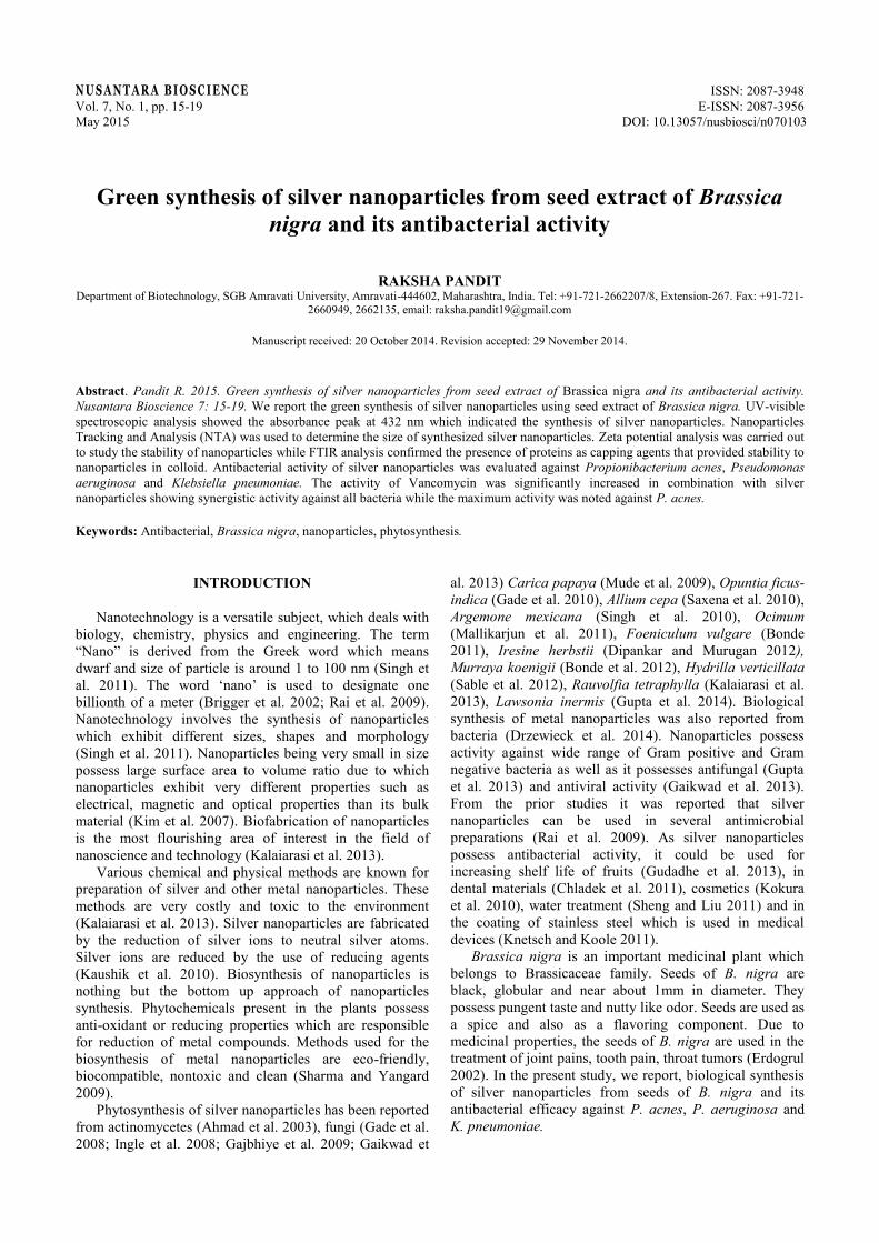
UV-Vis spectroscopy is the significant method whichgives the preliminary confirmation of silver nanoparticles.The absorption spectra of the fabricated silvernanoparticles showed absorbance spectra at 432 nm.(Figure 2). The synthesized silver nanoparticles showedabsorbance at specific wavelength because of the surfacePlasmon resonance phenomenon of silver nanoparticles(Gaikwad et al. 2013). The results showed resemblancewith the results of Gade et al. (2010) and Gupta et al.(2014) who reported the synthesis of silver nanoparticlesfrom Opuntia ficus-indica and Lawsonia inermis.
Figure 1. Synthesis of silver nanoparticles: A. B. nigra seedextract, B. Seed extract after treatment of 1mM AgNO3.
A B
PANDIT – Silver nanoparticles synthesis from Brassica nigra 17
Figure 2. UV-Vis spectra of the synthesized silver nanoparticlesshowing absorbance at 432 nm. A. Control (seed extract), B.Experimental (AgNPs)
Figure 3. Nanoparticle tracking analysis NTA (NanoSight-LM20) histogram showing particle size distribution and the averagesize of silver nanoparticles (41 nm).
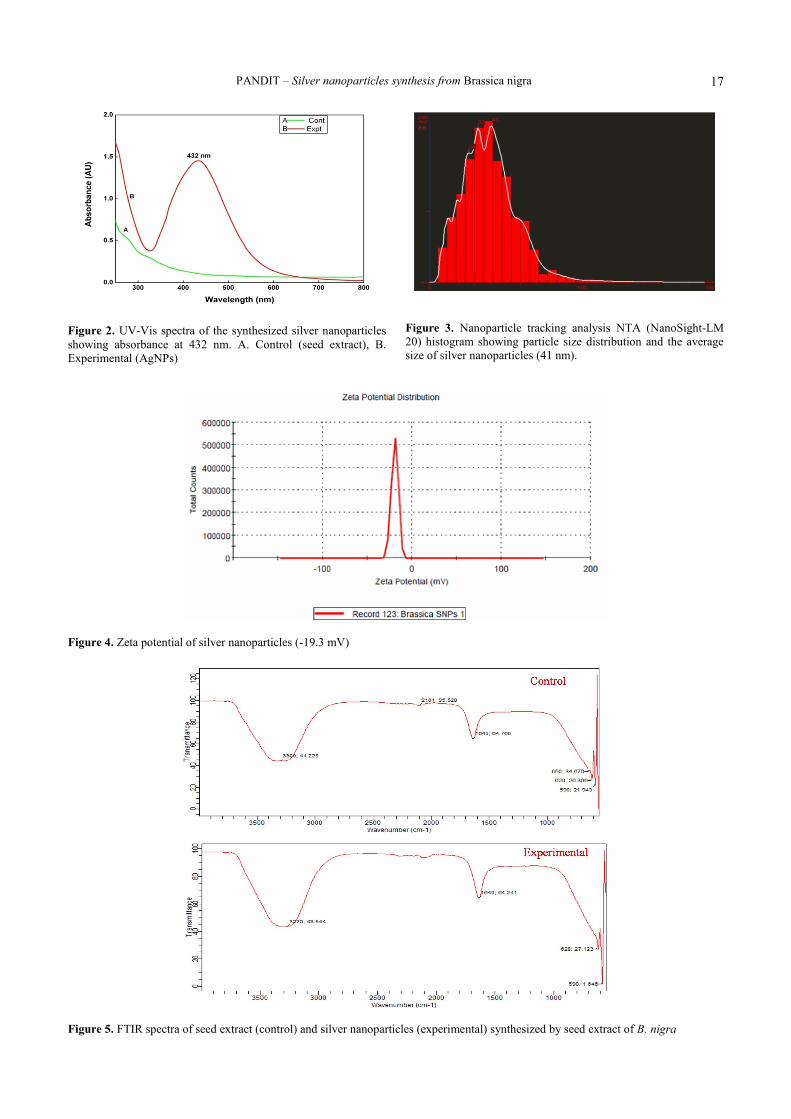
Figure 4. Zeta potential of silver nanoparticles (-19.3 mV)
Figure 5. FTIR spectra of seed extract (control) and silver nanoparticles (experimental) synthesized by seed extract of B. nigra

N U S A N T A R A B I O S C I E N C E 7 (1): 15-19, May 201518
Figure 6. Antibacterial activity of silver AgNPs and its synergistic activity against A. P. acnes, B. P. aeruginosa, C. K. pneumoniae,where: a. AgNPs, b. Control (Seed extract, c. antibiotic, d. Antibiotic + AgNPs.
Table 1. Antibacterial activity of silver nanoparticles and its synergistic activity against different microorganisms by disc diffusionmethod.
Zone of inhibition in mmName of test micro-organism AgNPs Seed extract (control) 1mM AgNO3 Vancomycin Vancomycin +AgNPsP. acnes 8±0.50 0 0 17±0.65 21±0.56P. aeruginosa 6±0.71 0 0 15±0.21 18±0.11K. pneumoniae 5±0.21 0 0 17±0.54 19±0.12Note: ± Standard deviation
Nanoparticle tracking analysis (NTA) of silvernanoparticles is based on tracking the Brownian motion ofeach particle. The average size of synthesized nanoparticleswas found to be 41 nm. The result of NTA correlates withresults of obtained by Gupta et al. (2013) for determiningthe size of Lawsonia-mediated synthesized AgNP. Stabilityof fabricated nanoparticles was measured by zeta potentialanalysis. Zeta potential was found to be-19.3mV (Figure4). It indicates that the nanoparticles synthesized weremoderately stable.
FTIR spectroscopy is performed to get the idearegarding various functional groups and their interactionswith silver, which may be accountable for fabrication andstabilization of silver nanoparticles. In FTIR spectrum ofcontrol (Figure 5.A) peaks were observed at 590, 620, 650,1640, 2101 cm-1 and in experimental (Figure 5.B) peakswere observed at 590, 628, 1640, 3270 cm-1. The bands at590, 620, 628, 650 cm-1 corresponds to the bondingvibrations of alkyl halide (Anarkali et al. 2012). The bandsobtained at 1640cm-1 is due to presence of C-N stretchingvibrations of amines (Raheman et al. 2011; Bharathi et al.2014).The bands obtained at 3270 corresponds to thebonding vibration of Secondary alcohol (Anarkali et al. 2012).
The in vitro activity of nanoparticles synthesized fromseed extract of B. nigra and combination of silvernanoparticles along with antibiotics were evaluated againstP. acnes, P. aeruginosa and K. pneumoniae (Figure 6). Theantibiotic Vancomycin was used as standard antibacterialagent against all microbes. 1 mM AgNO3 was also testedagainst all the above mentioned organisms but it does notshowed any zone of inhibition. Fabricated silvernanoparticles alone and in combination with antibiotics
showed significant activity against all tested bacteria.Aqueous seed extract and 1mM AgNO3 did not showactivity against any of the bacteria (Table 1). P. acnesshowed maximum antibacterial and synergistic activity ascompared to other two bacteria. The antibacterial activityof the synthesized nanoparticles showed similarity withAnarkali et al. (2012) and Raheman et al. (2011).They havereported that the efficiency of silver nanoparticlesenhanced in combination of standard antibiotics. Theresults corroborates with the finding of Gupta et al. (2014)who reported the antibacterial activity of silvernanoparticles against Candida albicans, Microsporumcanis, P. acne and Trichophyton mentagrophytes..
From the prior result it was considered that the freeradicals which are formed during synthesis of silvernanoparticles may be responsible for the bacterial death. Itwas suggested from electron spin resonance study silvernanoparticles may possess the ability to synthesise freeradicals when it comes in contact with the bacteria. Thesefree radicals may damage the cell membrane of bacteriaand it may lead to cell death (Danilcauk et al. 2006; Kim etal. 2007). Rajeskumar and Malarkodi (2014) proposed thatthe silver nanoparticles may have the ability to interactwith the bacterial cell wall and the silver ions may interferein the respiratory chain reaction. It may results in the damageof the bacteria. It was proposed that silver nanoparticlesmay inhibit the biofilm formation of the bacteria and it maybe responsible for the cell death (Percival et al. 2007).DNA is composed of sulphur and phosphorous and silvernanoparticles may react with DNA and it may cause theproblem in the replication process of bacteria and maycauses cell death (Prabhu and Poulose 2012).
A B C

PANDIT – Silver nanoparticles synthesis from Brassica nigra 19
In conclusion, rapid biosynthesis of silver nanoparticlesis possible with seed extract of B. nigra. The fabricatednanoparticles showed antimicrobial activity against all thetest pathogens P. acnes, P. aeruginosa and K. pneumoniae.It is clear from the synergistic activity that silvernanoparticles enhanced the activity of antibiotics.Phytosynthesis of silver nanoparticles is most suitable,easily scale up and eco-friendly method.
ACKNOWLEDGEMENTS
I am grateful to Prof. M. K. Rai and Dr. A.K. Gade fortheir insistent guidance and support. I wish to thank S.C.Gaikwad and G. Agarkar for their guidance. I am alsothankful to Funds for Infrastructure Science andTechnology (FIST).
REFERENCES
Ahmad A, Mukherjee P, Senapati S, Mandal D, Khan MI, Kumar R. 2003.Extracellular biosynthesis of silver nanoparticles using the fungusFusarium oxysporum. Coll Surf B 28: 313-318.
Anarkali J, Raj D, Rajathi K, Sridhar S. 2012. Biological synthesis ofsilver nanoparticles by using Mollugo nudicaulis extract and theirantibacterial activity. Arch Appl Sci Res 4 (3): 1436-1441.
Bharathi K, Thirumurugan V, Kavitha M, Muruganadam G, RavichandranK, Seturaman M. 2014. A comparative study on the greenbiosynthesis silver nanoparticles using dried leaves of Boerhaaviadiffusa L. and Cichorium intybus L. with reference to theirantimicrobial potential. World J Pharmaceut Sci 3 (5): 1415-1427.
Bonde SR, Rathod DP, Ingle AP, Ade RB, Gade AK, Rai MK. 2012.Murraya koenigii mediated synthesis of silver nanoparticles and itsactivity against three human pathogenic bacteria. Nanosci Methods 1:25-36.
Bonde SR. 2011. A biogenic approach for green synthesis of silvernanoparticles using extract of Foeniculum vulgare and its activityagainst Staphylococcus aureus and Escherichia coli. Nusantara Biosci3: 59-63.
Brigger I, Dubernet C, Couvreur P. 2002. Nanoparticles in cancer therapyand diagnosis. Adv Drug Deliv Rev 54 (5): 631-651.
Chladek G, Mertas A, Rybarek I. Nalewajek T, Zmudzki J, Krol W,Lukaszczyk J. 2011. Anti-fungal activity of denture softliningmaterial modified by silver nanoparticles: a pilot study. Int Mol Sci12 (7): 4735-4744.
Danilcauk M, Lund A, Saldo J, Yamada, H, Michalik J. 2006. Conductionelectron spin resonance of small silver particles. Spectrochimb ActaMol Biomol 63:189-191.
Dipankar C, Murugan S. 2012. Green synthesis, characterization andevaluation of the biological activities of silver nanoparticlessynthesized from Iresine herbstii leaf aqueous extracts. Colloids98:112-119.
Drzewiecka W, Gaikwad S, Laskowski D, Dahm H, Niedojadło J, GadeA, Rai M. 2014. Novel approach towards synthesis of silvernanoparticles from Myxococcus virescens and their lethality onpathogenic bacterial cells. Austin J Biotech Bioeng 1 (1): 1-7.
Erdogrul OT. 2002. Antibacterial activities of some plant extracts used inFolk medicine. Pharm Biol 40: 269-273.
Gade AK, Bonde PP, Ingle AP, Marcato PD, Duran N, Rai MK. 2008.Exploitation of Aspergillus niger for fabrication of silvernanoparticles. J Biobased Mater Bio 2: 243-247.
Gade AK, Gaikwad SC, Tiwari V, Yadav A, Ingle AP, Rai MK. 2010.Biofabrication of silver nanoparticles by Opuntia ficus-indica: In vitroantibacterial activity and study of the mechanism involved in thesynthesis. Curr Nanosci 6: 370-375.
Gaikwad S, Birla S, Ingle A, Gade A, Marcato P, Rai M, Duran N. 2013.Screening of different Fusarium species to select potential species forthe synthesis of silver nanoparticles. J Braz Chem Soc 24 (12): 1974-1982.
Gajbhiye M, Kesharwani J, Ingle A, Gade A, Rai M. 2009. Fungus-mediated synthesis of silver nanoparticles and their activity againstpathogenic fungi in combination with fluconazole. Nanomed 5: 382-386.
Gudadhe J, Yadav A, Gade A, Marcato PD, Duran N, Rai M. 2013.Preparation of an agar-silver nanoparticles (A-AgNp) film forincreasing the shelf-life of fruits. IET Nanobiotechnol Doi:10.1049/iet-nbt.2013.0010.
Gupta A, Bonde S, Gaikwad S, Ingle A, Gade A and Rai M.2014.Lawsonia inermis mediated synthesis of silver nanoparticles: activityagainst human pathogenic fungi and bacteria with special reference toformulation of antimicrobial nanogel. IET Nanobiotechnol 8 (3): 172-178.
Ingle A, Gade A, Pierrat S, Sonnichsen C, Rai M. 2008. Mycosynthesis ofsilver nanoparticles using the fungus Fusarium acuminatum and itsactivity against some human pathogenic bacteria. Curr Nanosci 4:141-144.
Kalaiarasi R, Prasannaraja G, Venkatachalama P. 2013. A rapid biologicalsynthesis of silver nanoparticles using leaf broth of Rauvolfiatetraphylla and their promising antibacterial. Indo Am J Pharm Res 3(10): 8052-8062.
Kaushik N, Thakkar S. Mhatre, Rasesh Y, Parikh MS. 2010. Biologicalsynthesis of metallic nanoparticles. Nanomed Nanotechnol Bio Med 6(2): 257-262.
Khanra K, Roy A, Bhattacharyya N. 2013. Evaluation of AntibacterialActivity and Cytotoxicity of Green Synthesized Silver NanoparticlesUsing Hemidesmus indicus R. Br. Am J Nanosci Nanotech Res 1:1-6.
Kim J, Yu k, Kim J, Park S, Lee H , Kim S, Park Y, Hwang C, Kim Y,Lee Y, Jeong D, Cho M. 2007. Antimicrobial effects of silvernanoparticle. Nanomed 3: 95-101.
Knetsch M, Koole L. 2011. New strategies in the development ofantimicrobial coatings: the example of increasing usage of silver andsilver nanoparticles. Polymer 1 (3): 340-366.
Kokura S, Handa O, Takagi T, Ishikawa T, Naito Y, Yoshikawa T. 2010.Silver nanoparticles as a safe preservative for use in cosmetics.Nanomed 6 (4): 570-574.
Mallikarjun K, Narsimha G, Dillip GR, Praveen B, Shreedhar B, LakshmiS, Reddy VS, Raju DP. 2011. Green synthesis of silver nanoparticlesusing Ocimum leaf extract and their characterization. Digest JNanomat Biostruct 6 (1):181-186.
Montes I, Walczyk D, Hole P, Smith J, and Lynch I, Dawson K. 2010.Characterisation of nanoparticles size and state prior tonanotoxicological studies. J Nanopart Res 12: 47-53.
Mude N, Ingle A, Gade A, Rai M. 2009. Synthesis of silver nanoparticlesusing callus extract of Carica papaya-a first report. J Plant BiochemBiotechnol 18: 83-86.
Percival SL, Bowler PG, Dolman J. 2007. Antimicrobial activity of silver-containing dressings on wound microorganisms using an in vitrobiofilm model. Int Wound J 4: 186-191.
Prabhu S, Poulose EK. 2012. Silver nanoparticles: mechanism ofantimicrobial action, synthesis, medical applications, and toxicityeffects. Int Nano Lett 2 (32): 1-10.
Raheman F, Deshmukh S, Ingle A, Gade A, Rai M. 2011. Silvernanoparticles: novel antimicrobial agent synthesized from anendophytic fungus Pestalotia sp. isolated from leaves of Syzygiumcumini (l) Nano Biomed Eng 3 (3): 174-178.
Rai M, Yadav A, Gade A. 2009. Silver nanoparticles as a new generationof antimicrobials. Biotec Adv 27 (1): 76-83.
Rajeshkumar S, Malarkodi C. 2014. In vitro antibacterial activity andmechanism of silver nanoparticles against foodborne pathogens.Bioinorg Chem Appl . DOI: 10.1155/2014/.
Sable N, Gaikwad S, Gade A, Rai M. 2012. Phytofabrication of silvernanoparticles by using aquatic plant Hydrilla verticillata. NusantaraBiosci 4 (2): 45-49.
Saxena A, Tripathy RM, Singh RP. 2010. Biological synthesis of silvernanoparticles by using onion (Allium cepa) extract and theirantibacterial activity. Digest J Nanomater Biostr 5: 427-432.
Sharma V, Yangard R. 2009. Green synthesis and their antimicrobialactivities. J Colloid Interface Sci 9: 83-96.
Sheng Z, Liu Y. 2011. Effects of silver nanoparticles on waste waterbiofilms. Water Res 45 (18): 6039-6050
Singh A, Jain D, Upadhyay MK, Khandelwal N, Verma HN. 2010. Greensynthesis of Silver nanoparticles using Argemone mexicana leafextract and evaluation of their antimicrobial activities. Digest JNanomat Biostruct 5 (2):483-489.
Singh C, Sharma V, Naik P, KhandelwaL V, Singh H. 2011. A greenbiogenic approach for synthesis of gold and silver nanoparticles usingZingiber officinale. Digest J Nanomat Biostruct 6 (2): 535-542.
A