Embed Size (px)
Citation preview

GREEN SYNTHESIS OF ZnO NANOPARTICLES
USING Abrus precatorius SEEDS EXTRACT AND
THEIR CHARACTERIZATION
Thesis submitted to Department of life science for the partial fulfillment of the
M.Sc. Degree in Life science
BY:
KAVITA VISHWAKARMA
ROLL NO: 411LS2069
UNDER THE SUPREME GUIDANCE OF
DR. SUMAN JHA
DEPARTMENT OF LIFE SCIENCE
NATIONAL INSTITUTE OF TECHNOLOGY
ROURKELA-769008
Dr. Suman Jha Ref. No.
Assistant Professor Date: ............................
CERTIFICATE
This is to certify that the thesis entitled “GREEN SYNTHESIS OF ZnO NANOPARTICLES
USING Abrus precatorius SEEDS EXTRACT AND THEIR CHARACTERIZATION”
which is being submitted by Mrs. Kavita Vishwakarma Roll No. 411LS2069, for the award of
the degree of Master of Science from National Institute of Technology, Rourkela, is absolutely
based upon his work carried out under my supervision. The results embodied in this thesis are
new and have not been submitted to any other university or institution for the award of any
degree/diploma.
Dr. Suman Jha
Assistant Professor,
Department of Life Sciences
National Institute of Technology
Rourkela – 769008, Odisha, India.
Phone no: 0661-2462687

DECLARATION
I do hereby declare that the Project Work entitled “GREEN SYNTHESIS OF ZnO
NANOPARTICLES USING Abrus precatorius SEEDS EXTRACT AND THEIR
CHARACTERIZATION”, submitted to the Department of Life Science, National Institute of
Technology, Rourkela is a faithful record of bonafide and original research work carried out
by me under the guidance and supervision of Dr. Suman Jha, Asst. Professor, Department
of Life Science, National Institute of Technology, Rourkela, Odisha.
Date:
Place: Kavita Vishwakarma
.

CONTENTS
Sl No Particulars Page no.
A Acknowledgement 5
B List of figure 6
C List of table 7
D Abbreviation 8
E Abstract 9
1 Introduction 10-11
2 Review of literature 12-29
3 Objectives 30
4 Materials and methods 31-32
5 Results and discussion 33-44
6 Conclusion 45
7 References
46-48

A. ACKNOWLEDGEMENT
Although theory is heard in many ways which seems to be very simple and effortless but when it
comes to real ground then it matters and competes to retrospect the system in each and every
way to realize the real retention process which leads to perfection. I feel myself speechless
before them to enumerate their help and guidance who are real pedagogues.
If words are considerable as symbols of approval and taken as acknowledgement then let the
words play a heralding role in expressing my gratitude.
First of all I express my deepest gratitude to Dr. Suman Jha, Assistant Professor of Department
of Life Science, National Institute of Technology, Rourkela for his suggestion to do this
innovative work. In fact he is a great visionary and researcher who have contributed immensely
towards this project work.
I would like to express my extreme sense of gratitude to Dr. S. K Patra, (HOD) and all faculty
members of Department of Life science, National Institute of Technology, Rourkela for giving
me permission to do this project work.
I am very much thankful to Mr. Manoranjan Aarkha, Junior research fellows, who helped me
and guided me in each and every step of my project work. Without his help I cannot complete
my project successfully.
I heartily thanks to all research scholars of Dept. of Life Science, National Institute of
Technology, Rourkela for their encouragement and necessary help during the project work.
I heartily thanks to Tamasa Panigrahi, Upasana Pattnaik, Lily Prava Das and all my friends
who helped me in each and every way that to complete this thesis successfully.
Finally I bow my head before God, my parents and my husband Mr.Ravikant Sharma, who
grew me up mentally and spiritually with prayers and Devine love.
B. LIST OF FIGURES
FIG NO. PARTICULARS PAGE
NO.
1. Flow chart for biosynthesis for NPs 18
2. Plant Abrus precatorius 21
3. ZnO crystal structure 24
4. Abrus precatorius seeds 31
5. UV-vis for 30:1 33
6 UV-vis for 60:1 34
7. UV- vis for 120:1 34
8. UV-vis for 240:1 35
9. SEM for 30:1 36
10. SEM for 60:1 36
11. SEM for 120:1 37
12. SEM for 240:1 37
13. FTIR for 30:1 38
14. FTIR for 120:1 49
15. DLS image for 60:1 40
16. Zeta potential image for 60:1 42
17. XRD image for 60:1 43

C. LIST OF TABLES
TABLE NO. PARTICULARS PAGE NO.
1 Zeta potential (surface potential) has direct
relation with stability of a form/structure
41
D. ABBREVIATION
NPs: Nanoparticles
Hrs : Hours
Mins : minutes
ZnO: zinc oxide
SEM: Scanning electron microscope
FTIR: Fourier transform infrared spectroscopy
DLS: Dynamic light scattering
XRD: X-ray diffraction
mV: millivolt

E. ABSTRACT
In modern science Nanotechnology is a ablaze field for the researchers.Nanoparticles having a
size of 1-100 nm in one dimension, is used significantly concerning medical chemistry, atomic
physics, and all other known fields. Nanoparticles are used immensely due to its small size,
orientation, physical properties,which are reportedly shown to change the performance of any
other material which is in contact with these tiny particles. These particles can be prepared easily
by different chemical, physical, and biological approaches. But the biological approach is the
most emerging approach of preparation, because, this method is easier than the other methods,
ecofriendly and less time consuming. The semiconductor ZnO has gained substantial interest in
the research community in part because of its large exciton binding energy 60 meV which could
lead to lasing action based on exciton recombination even above room temperature. The Green
synthesis was done by using the aqueous solution of Abrus precatorius seeds extract and zinc
acetate. A fixed ratio of plant extract to metal ion was prepared and the color change was
observed which proved the formation of nanoparticles. The nanoparticles were characterized by
UV-vis Spectrophotometer, FTIR, DLS, Zeta Analysis, XRD, and SEM. The particles
synthesized were of the size ranging from 90-500 nm.
1
INTRODUCTION
Nanotechnology emerges from the physical, chemical, biological and engineering sciences where
new techniques are being developed to probe and maneuver single atoms and molecules for
multiple applications in different field of scientific world. In nanotechnology, a nanoparticle is
defined as a small object that behaves as a whole unit in terms of its transport and properties. The
science and engineering technology of nanosystems is one of the most exigent and fastest
growing sectors of nanotechnology [1].
In the recent years, due to the advancement in Science and technology researchers have
attempted to synthesize nanoparticles within the size range of 100 nm and this extensive research
and concern on nanoparticles is widening due to their potential application in wide areas of
science and technology. ZnO belongs to the class of metal oxides, which is characterized by
photo catalytic and photo-oxidising capacity against chemical and biological species [2].
The progress of technology and quality of life of mankind has always been closely knit with the
progress in material science and material processing technology. Most techniques applied in
material processing are based on breaking up large chunk of a material into desired shapes and
sizes, inducing strain, lattice defects and other deformations in the processed material. Recent
developments and findings in nanotechnology and the demonstration based on various quantum
size effects in nanoscale particles, reveals that most of the novel work and devices of the future
will be based on properties of nanomaterials. Each nanoparticle contains only about 3-
107atoms/molecules. The traditional material processing techniques that induce lattice defects
and other imperfections will no longer be diluted for synthesis of nanoparticle by unmitigated
number of atoms. Furthermore, the application of traditional approach imparts difficulties for
synthesis of such small particles in a desirable size range.
Alternative synthetic technique for nanoparticles involves controlled precipitation of
nanoparticles from precursors mixed and dissolved in a solution. A micro suspension can also be
formed using surfactants between two immiscible liquids, with the reactionary isolated inside a
colloid, through hydrophobic versus hydrophilic forces. The resultant nanoparticles form a micro
colloidal suspension. Various factors such as thermodynamic determinant as well as van der
Waal‟s forces induce particle growth and accumulation, resulting in bigger particles that settle
down over time. A contingency in utilizing colloidal nanoparticles is that its stability is

2
maintained in colloidal suspension. The mechanism involved in stabilization of nanoparticles can
be categorized as a) electrostatic stabilization: involving the creation of a double layer of
adsorbed ions over the nanoparticles resulting in a coulombic repulsion between approaching
nanoparticles; or b) Steric hindrance: achieved by adsorption of polymer molecules over the
nanoparticles. Polymer molecules coated with nanoparticles feel osmotic repulsion when these
particle approach each other which occurs due to localized increase in their concentration and
hence keeps them (along with the nanoparticles) well separated.
Nature has devised various processes for the synthesis of nano and micro- length scaled
inorganic materials which have contributed to the development of relatively new and largely
unexplored area of research based on the biosynthesis of nanomaterials. Synthesis using bio-
organisms is congruent with the green chemistry principles. “Green synthesis” of nanoparticles
makes use of environmental friendly, non-toxic and safe reagents.
To the best of our knowledge, biological approach using milky latex of Calotropis procera has
been used for the first time as a reducing material as well as surface stabilizing agent for the
synthesis of spherical-shaped ZnO-NPs. The structure, phase, and morphology of synthesized
product were investigated by the standard characterization techniques. Spherical ZnO NPs has
been synthesized using milky latex of Calotropis procera and apart from this ZnO particle with
high stability and spherical shape have also been synthesized using Aloe vera extract.
The “green” route for nanoparticle synthesis is of great interest due to eco-friendliness, economic
prospects, feasibility and wide range of applications in nanomedicine, catalysis medicine, nano-
optoelectronics, etc. It is a new and emerging area of research in the scientific world, where day-
by-day developments is noted in warranting a bright future for this field. Zinc oxide is an
inorganic compound with the formula ZnO. It is usually insoluble in water and appears as a
white powder. The powder is widely used as an additive into numerous materials and products
including plastics, ceramics, glass, cement, rubber (e.g. car tyres), lubricants, paints, ointments,
adhesives, sealants, pigments, foods (source of Zn nutrient), batteries, ferrites, fire retardants, etc
ZnO is present in the Earth crust as a mineral zincite; however, commercially used ZnO is
produced synthetically. Zinc and oxygen belong to the 2nd
and 6th
groups of the periodic table
respectively and therefore in materials science, ZnO is often called a II-VI semiconductor. This
semiconductor has several favorable properties: good transparency, high electron mobility, wide
bandgap, strong roomtemperature luminescence, etc. ZnO with several favourable properties are

3
already used in emerging applications for transparent electrodes in liquid crystal displays and in
energy-saving or heat-protecting windows, and electronic applications of ZnO as thin-film
transistor and light-emitting diode are forthcoming as of 2009 [3].
4
REVIEW OF LITERATURE
Nanotechnology may be the next big thing in science, and before long we will probably find
ourselves immersed in it. It has attracted considerable attention in the scientific community ever
since its emergence as a powerful basic and applied science tool. Nanotechnology provides the
tools and technology platform for the investigation and transformation of biological systems, and
biology offers inspiration models and bio-assembled components to nanotechnology. Nano
biotechnology is defined as a field that applies the nanoscale principle and techniques to
understand and transform bio systems (living and non-living) and which uses biological
principles and materials to create new devices and systems integrated from the nanoscale [4].
Key advances have been made in the ability to make measurements at the sub-cellular level and
in understanding the cell as highly organized, self-repairing, self-replicating, information-rich
molecular machines [5] and [6] Smalley, classified nanotechnologies into wet and dry
nanotechnology, the first one describes the living bio systems and the second one deals with
man-made objects at nanoscale structures [7]. The presence of nanoparticles in commercially
available products is becoming more common.
Nanoparticles, according to the ASTM standard definition, are particles with lengths that range
from 1 to 100 nanometers in two or three dimensions [8]. A nanoparticle is defined as the
smallest unit that can still behave as a whole entity in terms of properties and transport.
Nanoparticles are used in bioapplications such as therapeutics, antimicrobial agents, drug
delivery agents, biosensors, imaging contrast agents, transfection vectors, and fluorescent labels.
Nanoparticles are of great scientific interest as they bridge the gap between bulk materials and
atomic or molecular structures. A bulk material has constant physical properties regardless of its
size, but at the nanoscale this is often not the case. Several well-characterized bulk materials
have been found to possess most interesting properties when studied in the nanoscale. There are
many reasons for this including the fact that nanoparticles possess a very high aspect ratio. In the
case of silver nanoparticles (AgNPs), this allows them to easily interact with other particles and
increases their antibacterial efficiency. This effect is extremely robust, and as little as 1 g of
AgNPs is known to impart antibacterial properties to hundreds of square meters of substrate
material.
5
METALS AND METAL-OXIDE NANOPARTIClES
Metallic nanoparticles have possible applications in diverse areas such as electronics, cosmetics,
coatings, packaging, and biotechnology. For example, nanoparticles can be induced to merge
into a solid at relatively lower temperatures, often without melting, leading to improved and
easy-to-create coatings for electronics applications (eg, capacitors). Typically, nanoparticles
possess a wavelength below the critical wavelength of light. This renders them transparent, a
property that makes them very useful for applications in cosmetics, coatings, and packaging.
Metallic nanoparticles can be attached to single strands of DNA nondestructively. This opens up
avenues for medical diagnostic applications. Nanoparticles can traverse through the vasculature
and localize any target organ. This potentially can lead to novel therapeutic, imaging, and
biomedical applications. Based on all of the above, the synthesis of metallic nanoparticles is an
active area of academic and, more importantly, “application research” in nanotechnology.
Metal oxides play a very significant role in material science for instance fabrication of
microelectronic circuits, sensors, piezoelectric devices, fuel cells, coatings for the passivation of
surfaces against corrosion and as catalyst. Metal oxides have also been employed as sorbents for
environmental pollutant. In the domain of nanotechnology, oxide nanoparticles can exhibit
unique chemical properties owing to their limited size and high density of corner or edge surface
sites. A number of physical and chemical preparative methods for accessing nanostructured
oxides are on records . Among the metal oxides, nano ZnO exhibits wide band gap (~3.4eV) and
large exciton binding energy (60meV) and thus is considered most promising candidate for
nanooptoelectronics, sensors, transistors, nanopiezoelectronics and UV-detection[9].
Various metal-oxide nanoparticles have been also prepared by Pulsed laser ablation in liquid media
( PLAL). The concept of fabricating oxide using laser irradiation of metal targets in water was
demonstrated in 1987, where iron and tantalum oxides were formed on target surfaces in water
using a Q-switched ruby pulsed laser [10] and [11]. Although the preparation of various kinds of
NPs by ablation of different targets in deionized water has been extensively studied in recent
days, papers reporting the fabrication of metal oxide-based nanomaterials from metal targets by
PLAL are still scarce compared with reports concerning the production of noble-metal
nanoparticles.

6
Of the inorganic materials, metal oxides such as TiO2, ZnO, MgO and CaO are of particular
interest as they are not only stable under harsh process conditions but also generally regarded as
safe materials to human beings and animals [12] and [13]. The use of nanoparticles of silver and
zinc oxide has been seen as a viable solution to stop infectious diseases due to the antimicrobial
properties of these nanoparticles. The intrinsic properties of a metal nanoparticle are mainly
determined by size, shape, composition, crystallinity and morphology [14].
The solution phase synthesis of metal oxide nanoparticles typically involves the reaction of a
metal salt with hydroxide ions[15]. The particle size is dependent on the kinetics of nucleation
and growth from a supersaturated solution as well as processes such as coarsening, [16, 17]
oriented attachment,[18-22] and aggregation. Synthesis of crystalline particles with diameters
less than 10 nm is often performed in nonaqueous solvents where nucleation and growth are
usually completed in a few minutes. Processes such as coarsening and oriented attachment occur
at longer times and can have a large influence on particle size. The ability to separate nucleation
and growth from supersaturation from processes such as coarsening and oriented attachment is
important for controlling the particle size.
Classification of nanoparticles
Nanoparticles can be broadly grouped into two: namely organic and inorganic nanoparticles.
Organic nanoparticles may include carbon nanoparticles (fullerenes) while some of the inorganic
nanoparticles may include magnetic nanoparticles, noble metal nanoparticles (like gold and
silver) and semiconductor nanoparticles (like titanium dioxideand zinc oxide).
There is a growing interest in inorganic nanoparticles as they provide superior material
properties with functional versatility. Due to their size features and advantages over available
chemical imaging drugs agents and drugs, inorganic nanoparticles have been examined as
potential tools for medical imaging as well as for treating diseases. Inorganic nanomaterials have
been widely used for cellular delivery due to their versatile features like wide availability, rich
functionality, good biocompatibility, capability of targeted drug delivery and controlled release
of drugs [23]. For example mesoporous silica when combined with molecular machines prove to
be excellent imaging and drug releasing systems. Gold nanoparticles have been used extensively
in imaging, as drug carriers and in thermo therapy of biological targets [24]. Inorganic

7
nanoparticles (such as metallic and semiconductor nanoparticles) exhibit intrinsic optical
properties which may enhance the transparency of polymer- particle composites. For such
reasons, inorganic nanoparticles have found special interest in studies devoted to optical
properties in composites. For instance, size dependant colour of gold nanoparticles has been used
to colour glass for centuries[25].
SYNTHESIS OF NANOPARTICLES
Nanoparticle having one or more dimensions of the order of 100nm or less- have attracted
considerable attraction due to their unusual and fascinating properties, with various applications,
over their bulk counterparts [26, 27]. Currently, a large number of physical, chemical, biological,
and hybrid methods are available to synthesize different types of nanoparticles[28-31]. Though
physical and chemical methods are more popular for nanoparticle synthesis, the use of toxic
compounds limits their applications. The development of safe eco-friendly methods for
biogenetic production is now of more interest due to simplicity of the procedures and
versatility[32, 33].
Traditionally nanoparticles were produced only by physical and chemical methods. Some of the
commonly used physical and chemical methods are ion sputtering, solvothermal synthesis,
reduction and sol gel technique. Basically there are two approaches for nanoparticle synthesis
namely the Bottom up approach and the Top down approach.
PHYSICAL AND CHEMICAL METHODS OF NANOPARTICLE SYNTHESIS
Some of the commonly used physical and chemical methods include:
Sol-gel technique, which is a wet chemical technique used for the fabrication of metal
oxides from a chemical solution which acts as a precursor for integrated network (gel) of
discrete particles or polymers. The precursor sol can be either deposited on the substrate
to form a film, cast into a suitable container with desired shape or used to synthesize
powders.
Solvothermal synthesis, which is a versatile low temperature route in which polar
solvents under pressure and at temperatures above their boiling points are used. Under
8
solvothermal conditions, the solubility of reactants increases significantly, enabling
reaction to take place at lower temperature.
Chemical reduction, which is the reduction of an ionic salt in an appropriate medium in
the presence of surfactant using reducing agents. Some of the commonly used reducing
agents are sodium borohydride, hydrazine hydrate and sodium citrate.
Laser ablation, which is the process of removing material from a solid surface by
irradiating with a laser beam. At low laser flux, the material is heated by absorbed laser
energy and evaporates or sublimates. At higher flux, the material is converted to plasma.
The depth over which laser energy is absorbed and the amount of material removed by
single laser pulse depends on the material‟s optical properties and the laser wavelength.
Carbon nanotubes can be produced by this method.
Inert gas condensation, where different metals are evaporated in separate crucibles inside
an ultra-high vacuum chamber filled with helium or argon gas at typical pressure of few
100 pascals. As a result of inter atomic collisions with gas atoms in chamber, the
evaporated metal atoms lose their kinetic energy and condense in the form of small
crystals which accumulate on liquid nitrogen filled cold finger. E.g. gold nanoparticles
have been synthesized from gold wires.
BIOSYNTHESIS OF NANOPARTICLES
The need for biosynthesis of nanoparticles rose as the physical and chemical processes were
costly. So in the search of for cheaper pathways for nanoparticle synthesis, scientists used
microorganisms and then plant extracts for synthesis. Nature has devised various processes for
the synthesis of nano- and micro- length scaled inorganic materials which have contributed to the
development of relatively new and largely unexplored area of research based on the biosynthesis
of nanomaterials [34].
Biosynthesis of nanoparticles is a kind of bottom up approach where the main reaction occurring
is reduction/oxidation. The microbial enzymes or the plant phytochemicals with anti oxidant or
reducing properties are usually responsible for reduction of metal compounds into their
respective nanoparticles.
9
The three main steps in the preparation of nanoparticles that should be evaluated from a green
chemistry perspective are the choice of the solvent medium used for the synthesis, the choice of
an environmentally benign reducing agent and the choice of a non toxic material for the
stabilization of the nanoparticles. Most of the synthetic methods reported to date rely heavily on
organic solvents. This is mainly due to the hydrophobicity of the capping agents used [35].
Synthesis using bio-organisms is compatible with the green chemistry principles: the bio-
organism is (i) eco-friendly as are (ii) the reducing agent employed and (iii) the capping agent in
the reaction [36]. Often chemical synthesis methods lead to the presence of some toxic chemical
species adsorbed on the surface that may have adverse effects in medical applications [37] . This
is not an issue when it comes to biosynthesized nanoparticles as they are eco friendly and
biocompatible for pharmaceutical applications.
Use of organisms to synthesize nanoparticles
Biomimetics refers to applying biological principles for materials formation. One of the primary
processes in biomimetics involves bioreduction. Initially bacteria were used to synthesize
nanoparticles and this was later succeeded with the use of fungi, actinomycetes and more
recently plants.
10
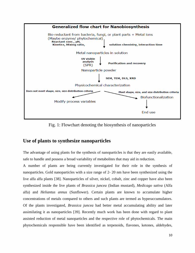
Fig. 1: Flowchart denoting the biosynthesis of nanoparticles
Use of plants to synthesize nanoparticles
The advantage of using plants for the synthesis of nanoparticles is that they are easily available,
safe to handle and possess a broad variability of metabolites that may aid in reduction.
A number of plants are being currently investigated for their role in the synthesis of
nanoparticles. Gold nanoparticles with a size range of 2- 20 nm have been synthesized using the
live alfa alfa plants [38]. Nanoparticles of silver, nickel, cobalt, zinc and copper have also been
synthesized inside the live plants of Brassica juncea (Indian mustard), Medicago sativa (Alfa
alfa) and Heliantus annus (Sunflower). Certain plants are known to accumulate higher
concentrations of metals compared to others and such plants are termed as hyperaccumulators.
Of the plants investigated, Brassica juncea had better metal accumulating ability and later
assimilating it as nanoparticles [39]. Recently much work has been done with regard to plant
assisted reduction of metal nanoparticles and the respective role of phytochemicals. The main
phytochemicals responsible have been identified as terpenoids, flavones, ketones, aldehydes,

11
amides and carboxylic acids in the light of IR spectroscopic studies. The main water soluble
phytochemicals are flavones, organic acids and quinones which are responsible for immediate
reduction. The phytochemicals present in Bryophyllum sp. (Xerophytes), Cyprus sp.
(Mesophytes) and Hydrilla sp. (Hydrophytes) were studied for their role in the synthesis of silver
nanoparticles. The Xerophytes were found to contain emodin, an anthraquinone which could
undergo redial tautomerization leading to the formation of silver nanoparticles.The Mesophyte
studied contained three types of benzoquinones, namely, cyperoquinone, dietchequinone and
remirin. It was suggested that gentle warming followed by subsequent incubation resulted in the
activation of quinones leading to particle size reduction. Catechol and protocatechaldehyde were
reported in the hydrophyte studied along with other phytochemicals. It was reported that catechol
under alkaline conditions gets transformed into protocatechaldehyde and finally into
protocatecheuic acid. Both these processes liberated hydrogen and it was suggested that it played
a role in the synthesis of the nanoparticles. The size of the nanoparticles synthesized using
xerophytes, mesophytes and hydrophytes were in the range of 2- 5nm [40].
Recently gold nanoparticles have been synthesized using the extracts of Magnolia kobus and
Diopyros kaki leaf extracts. The effect of temperature on nanoparticle formation was investigated
and it was reported that polydisperse particles with a size range of 5- 300nm was obtained at
lower temperature while a higher temperature supported the formation of smaller and spherical
particles [41].
While fungi and bacteria require a comparatively longer incubation time for the reduction of
metal ions, water soluble phytochemicals do it in a much lesser time. Therefore compared to
bacteria and fungi, plants are better candidates for the synthesis of nanoparticles. Taking use of
plant tissue culture techniques and downstream processing procedures, it is possible to
synthesize metallic as well as oxide nanoparticles on an industrial scale once issues like the
metabolic status of the plant etc. are properly addressed.
Abrus precatorius
Abrus precatorius L. is a member of the Papilionaceae family and known in various
communities with different names. The names include cat‟s eye, bead tree, rosary pea and
jeoquirity bean [42]. A climbing vine indigenous in India and Indonesia, leaves alternately
compound, flowers arranged in clusters, violet or pink. The seed pod curls back when it opens

12
and reveals the seeds. The seeds are flat and truncate shaped, 1.5. . 2cm long, with attractive
scarlet colour (Fig.2). They are highly poisonous. In leaves and roots sweet saponins are found,
comparable with liquorice.These are known to be under the most toxic plant parts worldwide
[43]. In the seeds the toxic principle is abrin, a mixture of at least five lectins, abrin A - D, and
abrus-agglutinin. The abrins consist of two peptide chains connected by a disulfide bridge. Abrin
A consists of an A-chain with N-glycosidase activity, which inhibits protein synthesis, and
lectin-like B-chain responsible for binding with cell-surface receptors and penetrating of abrin-A
molecule into the cell[44]. The toxicity of these abrins is variable, but they are the most potent
toxins of the world, comparable with the botulinus toxin. Abrus agglutinin, is not so very toxic
against cells, but it exhibits agglutination toward animal erythrocytes[45].
In the Ayurvedic medicine leaves of Abrus precatorius are laxative, expectorant and aphrodisiac
medicines. Seeds are said to be purgative, emetic, tonic, antiphlogistic, aphrodisiac and
antiopthalmic. For the indigenous people they are potent phytomedicines, many of them in
mixtures with other plants. Their toxicity is underestimated. The plant has haemopoeitic and
immunomodulatory activities[46]. Regular and wider applications of Abrus precatorius leaf in
the treatment of many diseases of human and animals in Nigeria have called for the need to
investigate the biochemical effects of the plant in a biosystem, especially at the time when people
are claiming the hepatoprotective property of the plant.
In this experiment we have taken the seed extract of these plant to synthesize zinc oxide (ZnO)
nanoparticles and characterize them using different instruments.

13
Fig. 2. Plant Abrus precatorius showing leaves and flower.
ZnO Nanoparticles Semiconductor nanoparticles are very important materials that have attracted attention of
researchers because of their wide applications in material science, chemical and electric
engineering during the past years. Nanoparticles have mainly two different excitations :i.exitonic
transitions and ii.deep-trap transitions. Whilst exitonic transitions come from the band-edge(from
conduction band to valance band), deep-trap transitions are caused by deep trap energy levels
and the surface defects that appear especially at higher surface to volume ratios. This is a result
of the physical properties of nanoparticles becoming size dependent when its radius becomes
comparable to the Bohr radius of the bulk material. It is observed that the emission spectra from
the exitonic level donot show any shift but if the emission spectra come from the deep trap level
they show some considerable shifts upon changing the excitation wavelength. The emission
spectra area direct function of the size of the nanoparticles and the optical properties of
semiconductor nanoparticles are strongly dependent on the effective particle size. Surface to
volume ratio is the most important point for the optical properties of the nanoparticles.As this
ratio increased the effects of the deep-trap transitions, becomes inefficient [47, 48].

14
ZnO is a kind of wide band gap (3.37 eV) semiconductor with large exciton binding energy (60
meV) [49]. ZnO is a biofriendly oxide semiconductor and an inexpensive luminescent material. It
has attracted intensive research efforts for its unique properties and versatile applications in
antireflection coatings, transparent electrodes in solar cells, ultraviolet (UV) light emitters, diode
lasers, varistors, piezoelectric devices, spin-electronics, surface acoustic wave propagator [50,
51], antibacterial agent [52], photonic material [53] and for gas sensing [54]. In general, ZnO is
considered “generally recognized as safe” (GRAS) [55] but ZnO nanoparticle system may be
toxic.
Among all the inorganic semiconducting nanoparticles, zinc oxide nanoparticles have attracted
increasing attention because ZnO nanoparticles can be easily synthesized and ZnO is a „„green‟‟
material that is biocompatible, biodegradable, and nontoxic for medical applica- tions and
environmental science [12].Recently, there are several physical or chemical synthetic methods of
preparing ZnO, such as thermal evaporation [56], pulsed laser deposition (PLD) [57], ion
implantation [58], reactive electron beam evaporation[59], thermal decomposition [60] and sol–
gel technique [61]. Recently,great effort has been made to the synthesis of size-controlled ZnO2
nanoparticles in order to explore their potentials.
PROPERTIES OF ZnO:- In this section crystal structures, inclusive of lattice parameters, electronic band structures,
mechanical properties, inclusive of elastic contants and piezoelectric constants, lattice dynamics
and vibrational processes, thermal properties, electrical properties, and low-field and high-field
carrier transports are treated.
STRUCTURE OF ZINC OXIDE (ZnO) Most of the group-II-VI binary compound semiconductors crystallize in either cubic zinc-blende
or hexagonal wurtzite structure where each anion is surrounded by four cations at the corners of
a tetrahedron, and vice versa. This tetrahedral coordination is typical of sp3 covalent bonding,
but these materials also have a substantial ionic character. ZnO is a II-VI compound
semiconductor whose ionicity resides at the borderline between covalent and ionic
semiconductor. The crystal structures shared by ZnO are wurtzite B4, zinc blende B3, and
rocksalt B1, as schematically shown in Fig. 3. At ambient conditions, the thermodynamically

15
stable phase is wurtzite. The zinc blende ZnO structure can be stabilized only by growth on cubic
substrates, and the rocksalt NaCl structure may be obtained at relatively high pressures.
The zinc blende form can be stabilized by growing ZnO on substrates with cubic lattice structure.
In both cases, the zinc and oxide centers are tetrahedral, the most characteristic geometry for
Zn(II).In addition to the wurtzite and zinc blende polymorphs, ZnO can be crystallized in the
rock salt motif at relatively high pressures about 10 GPa[62]. Hexagonal and zinc blende
polymorphs have no inversion symmetry (reflection of a crystal relative to any given point does
not transform it into itself). This and other lattice symmetry properties result in piezoelectricity
of the hexagonal and zincblende ZnO, and pyroelectricity of hexagonal ZnO.
The hexagonal structure has a point group 6 mm (Hermann-Mauguin notation) or C6v
(Schoenflies notation), and the space group is P63mc or C6v4. The lattice constants are a = 3.25 Å
and c = 5.2 Å; their ratio c/a ~ 1.60 is close to the ideal value for hexagonal cell c/a = 1.633. As
in most group II-VI materials, the bonding in ZnO is largely ionic (Zn2+
O2-
) with the
corresponding radii of 0.074 nm for Zn2+
and 0.140 nm for O2–
. This property accounts for the
preferential formation of wurtzite rather than zinc blende structure as well as the strong
piezoelectricity of ZnO. Because of the polar Zn-O bonds, zinc and oxygen planes are
electrically charged. To maintain electrical neutrality, those planes reconstruct at atomic level in
most relative materials, but not in ZnO its surfaces are atomically flat, stable and exhibit no
reconstruction [63].
a. b. c.
Fig. 3 Stick and ball representation of ZnO crystal structures: a) hexagonal wurtzite B4. b)
cubic zinc blende B3, and c) cubic rocksalt B1,

16
LATTICE PARAMETERS
The lattice parameters of a semiconductor usually depend on the following factors: i. free-
electron concentration acting via deformation potential of a conduction-band minimum occupied
by these electrons, ii. concentration of foreign atoms and defects and their difference of ionic
radii with respect to the substituted matrix ion, iii.external strains for example, those induced by
substrate, and iv. temperature. The lattice parameters of any crystalline material are commonly
and most accurately measured by high resolution x-ray diffraction HRXRD by using the Bond
method for a set of symmetrical and asymmetrical reflection [64].
For the wurtzite ZnO, lattice constants at room temperature determined by various experimental
measurements and theoretical calculations are in good agreement. The lattice constants mostly
range from 3.2475 to 3.2501 Å for the a parameter and from 5.2042 to 5.2075 Å for the c
parameter. The c/a ratio and u parameter vary in a slightly wider range, from 1.593 to 1.6035 and
from 0.383 to 0.3856, respectively. The deviation from that of the ideal wurtzite crystal is
probably due to lattice stability and ionicity. It has been reported that free charge is the dominant
factor responsible for expanding the lattice proportional to the deformation potential of the
conduction-band minimum and inversely proportional to the carrier density and bulk modulus.
The point defects such as zinc antisites, oxygen vacancies, and extended defects, such as
threading dislocations, also increase the lattice constant, albeit to a lesser extent in the
heteroepitaxial layers.
For the zinc-blende polytype of ZnO, the calculated lattice constants based on a modern ab initio
technique are predicted to be 4.60 and 4.619 Å. A high-pressure phase transition from the
wurtzite to the rocksalt structure decreases the lattice constant down to the range of 4.271–4.294
Å [65].
MECHANICAL PROPERTIES
The mechanical properties of materials involve various concepts such as hardness, stiffness, and
piezoelectric constants, Young‟s and bulk moduli, and yield strength. Although the wurtzite ZnO
crystal is acoustically anisotropic,there is only a very small difference between theshear sound
velocities vTA1 and vTA2 propagating along the(001} and (100) directions, respectively (vTA2/vTA1
) = 0.98.

17
Piezoelectricity is the electric charge that accumulates in certain solid materials in response to
applied mechanical stress. The word piezoelectricity means electricity resulting from pressure.
The piezoelectric effect is understood as the linear electromechanical interaction between the
mechanical and the electrical state in crystalline materials with no inversion symmetry.The
piezoelectric effect is a reversible process in that materials exhibiting the direct piezoelectric
effect (the internal generation of electrical charge resulting from an applied mechanical force)
also exhibit the reverse piezoelectric effect (the internal generation of a mechanical strain
resulting from an applied electrical field). The inverse piezoelectric effect is used in production
of ultrasonic sound waves. Among the tetrahedrally bonded semiconductors, it has been stated
that ZnO has the highest piezoelectric tensor. This property makes it a technologically important
material for many applications, which require a large electromechanical coupling. The
piezoelectric tensor has three independent components in hexagonal wurtzite phase and one in
the cubic zinc-blende phase, which characterize the full piezoelectric tensors of such crystals.108
Two of these components in wurtzite phase measure the polarization induced along the c axis, at
zero electric field, by a uniform strain either along the c axis or in the basal plane [66].
ELECTRONIC PROPERTIES
ZnO has a relatively large direct band gap of ~3.3 eV at room temperature; therefore, pure ZnO
is colorless and transparent. Advantages associated with a large band gap include higher
breakdown voltages, ability to sustain large electric fields, lower electronic noise, and high4
temperature and high-power operation. The bandgap of ZnO can further be tuned from ~3–4 eV
by its alloying with magnesium oxide or cadmium oxide.
Most ZnO has n-type character, even in the absence of intentional doping. Native defects such as
oxygen vacancies or zinc interstitials are often assumed to be the origin of this, but the subject
remains controversial. An alternative explanation has been proposed, based on theoretical
calculations, that unintentional substitutional hydrogen impurities are responsible. Controllable
n-type doping is easily achieved by substituting Zn with group-III elements Al, Ga, In or by
substituting oxygen with group-VII elements chlorine or iodine. Reliable p-type doping of ZnO
remains difficult. This problem originates from low solubility of p-type dopants and their
compensation by abundant n-type impurities, and it is pertinent not only to ZnO, but also to
18
similar compounds GaN and ZnSe. Measurement of p-type in "intrinsically" n-type material is
also not easy because inhomogeneity results in spurious signals.
Current absence of p-type ZnO does limit its electronic and optoelectronic applications which
usually require junctions of n-type and p-type material. Known p-type dopants include group-I
elements Li, Na, K; group-V elements N, P and As; as well as copper and silver. However, many
of these form deep acceptors and do not produce significant p-type conduction at room
temperature. Electron mobility of ZnO strongly varies with temperature and has a maximum of
~2000 cm2/(V·s) at ~80 Kelvin.[21] Data on hole mobility are scarce with values in the range 5-
30 cm2/(V·s).
OPTICAL PROPERTIES
ZnO is a wide band gap semiconductor that displays luminescent properties in the near ultra
violet and the visible regions. The emission properties of ZnO nanoparticles in the visible region
widely depend on their synthetic method as they are attributable to surface defects. Recently, we
have developed a novel organometallic synthetic method for the preparation at room temperature
of crystalline ZnO nanoparticles of controlled size and
shape. The studies on the emission properties of nanocrystalline ZnO nanoparticles and nanorods
was prepared following this organometallic synthetic method. They observed a clear influence of
the shape of the particles and of the ligands on the luminescence properties in the visible domain.
They observed two different emissions at 440 nm and at 580 nm that are associate with the
presence of surface defects on the particles. The first one corresponds to the well known yellow
emission located at 580 nm with a lifetime of 1850 ns for 4.0 nm size particles. The second
emission at 440 nm is observed when amine ligands are present. Based on the optical
measurements, they have proposed mechanism at the origin of the two emissions. They also
observed that the modification of the relative intensity between the two emissions is related to a
specific location of the amine ligands on the surface of the particles.
ZnO NANOSTRUCTURES
One-dimensional semiconductor nanowires and nanorods have attracted increasing attention due
to their physical properties arising from quantum confinement such as electronic quantum

19
transport and enhanced radiative recombination of carriers. Nanowires have promising potentials
in extensive applications and are the fundamental building blocks for fabricating short-
wavelength nanolasers, field-effect transistors, ultrasensitive nanosized gas sensors,
nanoresonators, transducers, actuators, nanocantilevers, and field emitters(Fes). These
nanostructures are the ideal systems for studying transport mechanisms in one-dimensional (1D)
systems, which are of benefit not only for understanding the fundamental phenomena in low-
dimensional systems but also for developing new generation nanodevices with high performance.
Many nanowires made of materials such as Si, C, InP, GaAs, CdS, SnO2, GaN, ZnO, and In2O3
have been fabricated for different applications using mostly a catalystassisted vapor-liquid-solid
(VLS) growth method (solid vapor process). Among these materials ZnO is considered to be the
most prospective one due to its large exciton binding energy (60 meV), high electromechanical
coupling constant, and resistivity to harsh environment. Therefore, 1D ZnO structures stimulated
so much attention,and a large number of publications have appeared lately reporting
nanostructures of various shapes such as nanowires , nanobelts, nanorings, nanotubes,
nanodonuts , nanopropellers ,etc. grown by different methods [65].
APPLICATIONS OF ZnO NPs
At present, the most widely revealed application for ZnO is an ITO replacement for displays and
photovoltaic panels, where ZnO could lower costs of transparent conductors. In addition to its
conductive nature, it also can be used as a semiconductor for making inexpensive transistors for
disposable electronics or even low-cost LEDs. ZnO is also finding applications in thin-film
batteries, and ZnO's ability to be engineered into interesting nanostructures hints at new
applications down the road. ZnO already is being tapped in spintronics.
Due to their excellent optical and electrical properties, ZnO nanoparticles have become
predominant semiconductor materials for nanoscale devices, such as nano-generators, gas
sensors, highly efficient solarcells, field-emission transistors , ultra violet photodetectors , and
biomedical systems ZnO is attracting considerable attention for its possible application to UV
light emitters, spin functional devices, gas sensors, transparent electronics and surface acoustic
wave devices [67].
20
Cytotoxicity of nanoparticles
The special physicochemimal properties of nanoparticles cause potential risk to human health
[68]. So considerable effort has been made to identify the potential toxicity of nanoparticles to
cells and organism . Exposure of cells to certain type nanoparticles can induces cytotoxicity and
many effects like oxidative injury, inflammation, fibrosis, and release of pro-inflammatory
mediators. The pathophysiological responses have also been found like generation of ROS
(Reactive oxygen species) in cells when cells are exposed to nanoparticles. This ROS generation
is the initiating factor for toxicity of nanoparticles exposed to cells. Another initiating factor was
lysosomal destabilization. loss of mitochondrial membrane integrity cause cell death.
Nanoparticles cause toxicity because: a) nanostructures have electronic, optical and magnetic
properties that tells about the physical dimensions and breakage in the nanostructures can lead to
a unique toxic effect that is unpredictable . b) The surfaces of nanostructures are involved in
many catalytic and oxidative reaction .These reactions may induce cytotoxicity .cytoxicity can be
greater in the nanomaterial than a bulk material because surface area to volume ratio for
nanoparticle is more . c) some nanostructures cantain metals or compound with toxicity when
breakdown can educe toxic responses to the components themselves [69].
Common assumption about the nanostructures is that smaller size of nanostructures can easily
enter into tissues, cells,organelles and functional biomolecular structures, as the actual size of an
engineered nanostructures is similar to many biological molecules and structures. The entry of
the nanostructures into biological systems can cause damage , which can cause harm to human
health. Nanostructures can enter biological system via six principle routes: intra venous, dermal,
subcutaneous, inhalation, intraperitoneal, and oral. After entery absorbtion takes place where
nanostructures first interact with biological component like proteins, cells. Then they are
distributed to various organs in body and may be structurally same, may be modified or
metabolized [67].

21
OBJECTIVES
Synthesis of Zinc Oxide (ZnO) nanoparticle
Physical characterization using SEM, UV-visible spectroscopy,
FTIR, XRD, Particle Size Analysis (DLS) and Zeta Potential
measureme

22
MATERIALS AND METHOD
1) Preparation of Abrus precatorius seed extract
Seeds shown in fig.4 were grinded after removal of outer coating. Then the powdered
form of seed was used to synthesize ZnO nanoparticles.
Fig.4 Abrus precatorius seeds
1) Synthesis of Zinc Oxide Nanoparticles:
Four concentration ratios of seed extract and metal ions were prepared (30:1, 60:1,120:1
& 240:1) by increasing the concentration of seed extract in deionized water. 1% plant extract
solution was prepared with deionized water. The prepared solution was set for incubation for 30
min. at 37o C. then the solution was set for centrifugation for 30 mins. at 25
o C in 10000 rpm and
the supernatant was collected. The supernatant was then filtered and 10.97 mg was added in the
solution.
23
2) Characterization of Silver Nanoparticles:
UV-Visible Analysis:
The optical property of zinc oxide was determined by the result of UV-Vis spectrophotometer.
After the addition of zinc acetate to all the ratio i.e 240:1, 120:1, 60:1, 30:1, the spectra was
taken in time interval up to 4hrs between 300 nm to 500 nm. Then the spectra was taken after 24
hrs. of zinc acetate addition.
FTIR analysis:
Supernatant of ratio 30:1 and 120:1 that we collected was dried at 75o C and the dried powder
was taken for FTIR analysis. FTIR was obtained in the range 4000–400 Cm−1 using KBR pellet
method.
SEM Analysis:
After 24Hrs. of the addition of zinc acetate the SEM slide was prepared by making a smear of the
solution on a slide. Then the slide was set for SEM analysis after coating the slide with platinum
and the SEM image was taken.
DLS & Zeta-Potential Analysis:
The supernatant of ratio 60:1 was filtered and then sonicated for 5 minutes. The the solution was
centrifuged for 15 min.at 25o C with 5000 rpm and the supernatant was collected. Then the
supernatant was diluted for 4 to 5 times and then set for DLS and Zeta-Potential analysis.
XRD Analysis:
ZnO nanoparticles were examined by X-ray diffractometer. The powdered metal was sticked in
the cubes of XRD and then the result was taken in the XRD equipment.

24
RESULTS AND DISCUSSION:
UV-visible spectrophotometer
It refers to absorption spectroscopy or reflectance spectroscopy in the ultraviolet-visible
spectral region. This means it uses light in the visible and adjacent ranges. The absorption or
reflectance in the visible range directly affects the perceived color of the chemicals involved. In
this region of the electromagnetic spectrum, molecules undergo electronic transitions. It is based
on the principle that molecules containing π-electrons or non-bonding electrons (n-electrons) can
absorb the energy in the form of ultraviolet or visible light to excite these electrons to higher
anti-bonding molecular orbitals. The more easily excited the electrons (i.e. lower energy gap
between the HOMO and the LUMO), the longer the wavelength of light it can absorb.
The figures showing the graph of uv-visible spectrophotometer for four ratios are as follows
(Fig5-8):
Figure 5: UV image of ZnO NPs 30:1 in different time interval
25
Figure 6: UV image of ZnO NPs 60:1 in different time interval
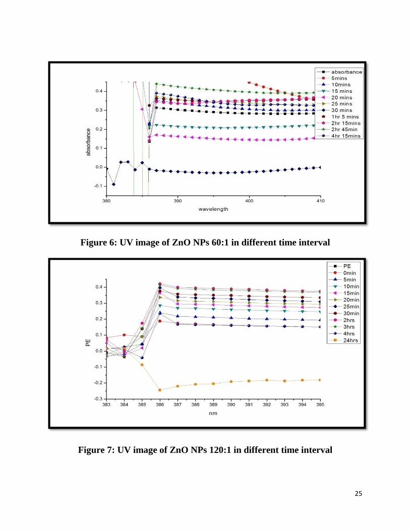
Figure 7: UV image of ZnO NPs 120:1 in different time interval

26
Figure 8: UV image of ZnO NPs 240:1 in different time interval
The colour change i.e light white to dark white colour was oberserved in the UV- vis
spectrophotometer is due to the Plasmon resonance phenomenon which is the collection of
oscillation of electrons. The reduction zinc acetate to zinc oxide is monitored by UV- vis
spectrum. The band was observed around 386 – 450 nm which was identified as “surface
Plasmon resonance band” and this band is ascribed to excitation of valence electrons of ZnO
arranged in the nanoparticles (nanocrystal/ nanosphere). The shape of the band was symmetrical,
suggesting uniform scattering of spherical shape nanoparticles.
SEM RESULT
It is a type of electron microscope that produces images of a sample by scanning it with a
focused beam ofelectrons. The electrons interact with electrons in the sample, producing various
signals that can be detected and that contain information about the sample's surface topography
and composition. The electron beam is generally scanned in a raster scan pattern, and the
beam's position is combined with the detected signal to produce an image. SEM can achieve
resolution better than 1 nanometer.
The SEM images of four different ratios are as follows (Fig.9 -12).

27
Fig. 9: SEM image of ZnO NPs synthesized from Abrus with 30:1 ratio
Fig. 10: SEM image of ZnO NPs synthesized from Abrus with 60:1 ratio

28
Fig. 11: SEM image of ZnO NPs synthesized from Abrus with 120:1 ratio
Fig. 12: SEM image of ZnO NPs synthesized from Abrus with 240:1 ratio
29
SEM provided further insight into the morphology and size details of the ZnO nanoparticle. The
size of the particles was from nano to micron range and morphology of particles was nearly
spherical in all ratio except in 240:1 ratio. In 240:1 ratio different morphology was observed i.e
spherical , rod shape particles was observed , this is because in this ratio the population of seed
extract was more. The size of the prepared nanoparticles was more than the size of nanoparticle
i.e.; between 1-100 nm. This was because the proteins were bound to the surface of the
nanoparticles. As the ratio differs size also differs, this is because of the concentration varies.
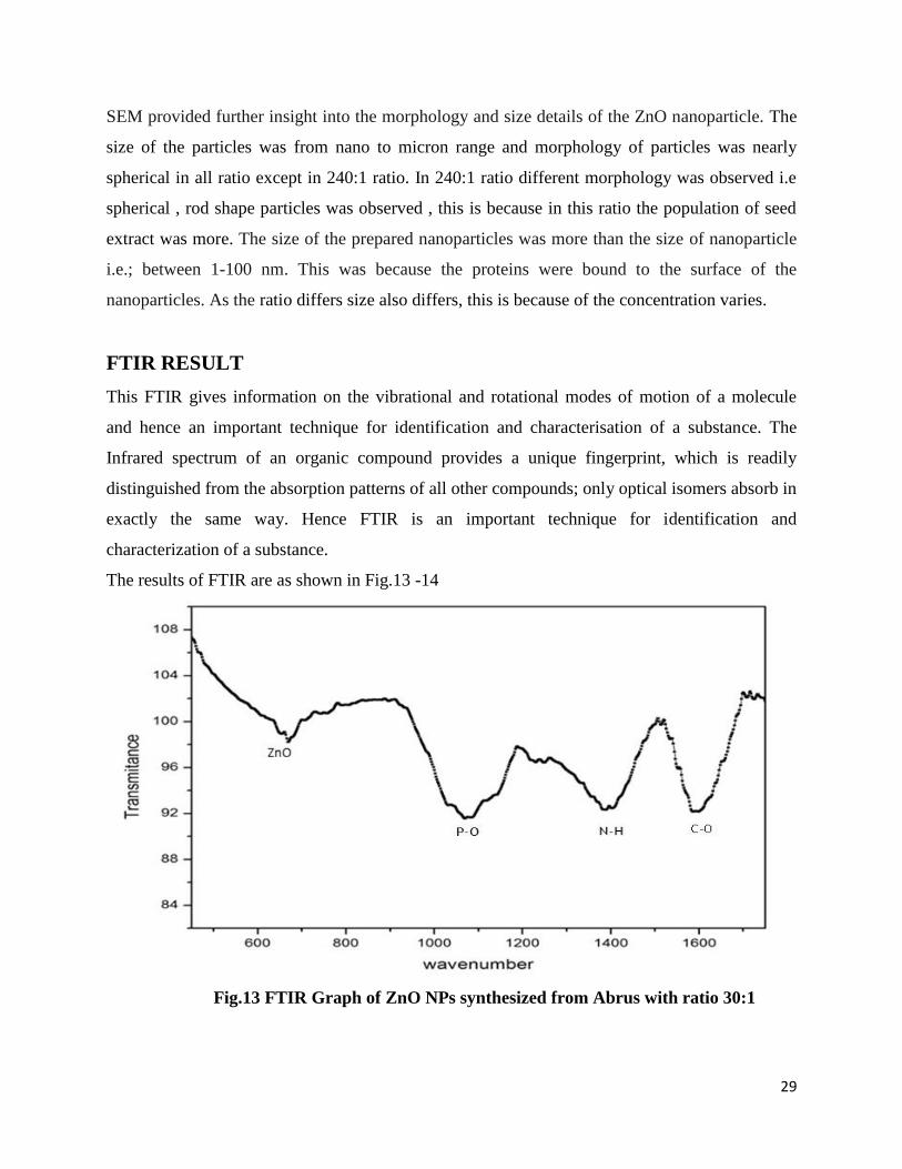
FTIR RESULT
This FTIR gives information on the vibrational and rotational modes of motion of a molecule
and hence an important technique for identification and characterisation of a substance. The
Infrared spectrum of an organic compound provides a unique fingerprint, which is readily
distinguished from the absorption patterns of all other compounds; only optical isomers absorb in
exactly the same way. Hence FTIR is an important technique for identification and
characterization of a substance.
The results of FTIR are as shown in Fig.13 -14
Fig.13 FTIR Graph of ZnO NPs synthesized from Abrus with ratio 30:1
30
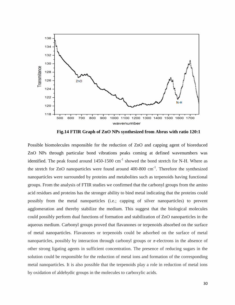
Fig.14 FTIR Graph of ZnO NPs synthesized from Abrus with ratio 120:1
Possible biomolecules responsible for the reduction of ZnO and capping agent of bioreduced
ZnO NPs through particular bond vibrations peaks coming at defined wavenumbers was
identified. The peak found around 1450-1500 cm-1
showed the bond stretch for N-H. Where as
the stretch for ZnO nanoparticles were found around 400-800 cm-1
. Therefore the synthesized
nanoparticles were surrounded by proteins and metabolites such as terpenoids having functional
groups. From the analysis of FTIR studies we confirmed that the carbonyl groups from the amino
acid residues and proteins has the stronger ability to bind metal indicating that the proteins could
possibly from the metal nanoparticles (i.e.; capping of silver nanoparticles) to prevent
agglomeration and thereby stabilize the medium. This suggest that the biological molecules
could possibly perform dual functions of formation and stabilization of ZnO nanoparticles in the
aqueous medium. Carbonyl groups proved that flavanones or terpenoids absorbed on the surface
of metal nanoparticles. Flavanones or terpenoids could be adsorbed on the surface of metal
nanoparticles, possibly by interaction through carbonyl groups or π-electrons in the absence of
other strong ligating agents in sufficient concentration. The presence of reducing sugars in the
solution could be responsible for the reduction of metal ions and formation of the corresponding
metal nanoparticles. It is also possible that the terpenoids play a role in reduction of metal ions
by oxidation of aldehydic groups in the molecules to carboxylic acids.
31
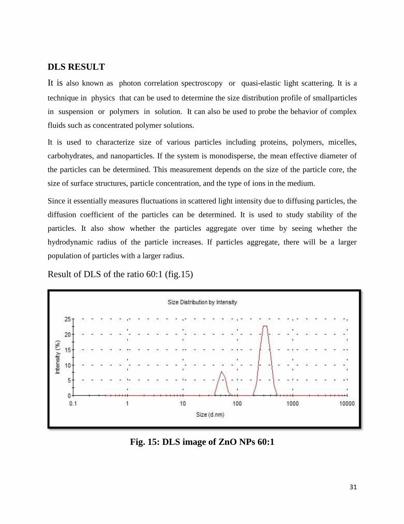
DLS RESULT
It is also known as photon correlation spectroscopy or quasi-elastic light scattering. It is a
technique in physics that can be used to determine the size distribution profile of smallparticles
in suspension or polymers in solution. It can also be used to probe the behavior of complex
fluids such as concentrated polymer solutions.
It is used to characterize size of various particles including proteins, polymers, micelles,
carbohydrates, and nanoparticles. If the system is monodisperse, the mean effective diameter of
the particles can be determined. This measurement depends on the size of the particle core, the
size of surface structures, particle concentration, and the type of ions in the medium.
Since it essentially measures fluctuations in scattered light intensity due to diffusing particles, the
diffusion coefficient of the particles can be determined. It is used to study stability of the
particles. It also show whether the particles aggregate over time by seeing whether the
hydrodynamic radius of the particle increases. If particles aggregate, there will be a larger
population of particles with a larger radius.
Result of DLS of the ratio 60:1 (fig.15)
Fig. 15: DLS image of ZnO NPs 60:1

32
DLS result showed that particles were polydispersed means different size of particles were
formed i.e the population of particle size 500nm was more than 90nm particles.
ZETA POTENTIAL RESULt
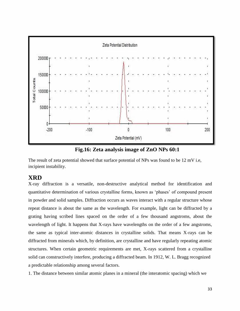
Zeta potential is the electric potential in the interfacial double layer (DL) at the location of the slipping
plane versus a point in the bulk fluid away from the interface. In other words, it is the potential difference
between the dispersion medium and the stationary layer of fluid attached to the dispersed particle.A
value of 25 mV (positive or negative) can be taken as the arbitrary value that separates low-charged
surfaces from highly-charged surfaces. The significance of zeta potential is that its value can be
related to the stability of colloidal dispersions (e.g., a multivitamin syrup). The zeta potential
indicates the degree of repulsion between adjacent, similarly charged particles (the vitamins) in a
dispersion. For molecules and particles that are small enough, a high zeta potential will confer
stability, i.e., the solution or dispersion will resist aggregation. When the potential is low,
attraction exceeds repulsion and the dispersion will break and flocculate. Zeta potential (Surface
potential) has direct relation with the stability of a form/structure as mentioned in table1.
Table.1. Zeta potential (Surface potential) has diret relation with the stability of a
form/structure as mentioned below:
Zeta potential [mV]
Stability behavior of the colloid
From 0 to ±5
Rapid coagulation or flocculation
From ±10 to ±30
Incipient instability
From ±30 to ±40
Moderate stability
From ±40 to ±60
Good stability
More than ±61
Excellent stability
Result of zeta potential of ratio 60:1 (fig. 16).
33
Fig.16: Zeta analysis image of ZnO NPs 60:1
The result of zeta potential showed that surface potential of NPs was found to be 12 mV i.e,
incipient instability.
XRD X-ray diffraction is a versatile, non-destructive analytical method for identification and
quantitative determination of various crystalline forms, known as „phases‟ of compound present
in powder and solid samples. Diffraction occurs as waves interact with a regular structure whose
repeat distance is about the same as the wavelength. For example, light can be diffracted by a
grating having scribed lines spaced on the order of a few thousand angstroms, about the
wavelength of light. It happens that X-rays have wavelengths on the order of a few angstroms,
the same as typical inter-atomic distances in crystalline solids. That means X-rays can be
diffracted from minerals which, by definition, are crystalline and have regularly repeating atomic
structures. When certain geometric requirements are met, X-rays scattered from a crystalline
solid can constructively interfere, producing a diffracted beam. In 1912, W. L. Bragg recognized
a predictable relationship among several factors.
1. The distance between similar atomic planes in a mineral (the interatomic spacing) which we
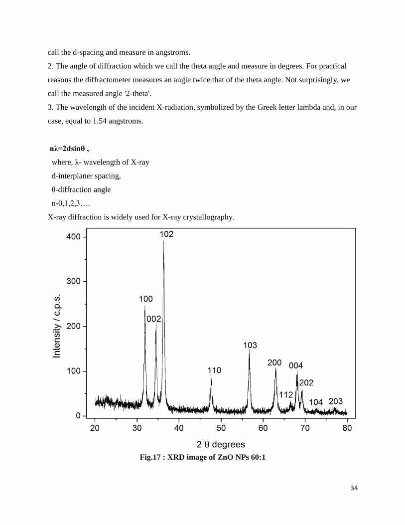
34
call the d-spacing and measure in angstroms.
2. The angle of diffraction which we call the theta angle and measure in degrees. For practical
reasons the diffractometer measures an angle twice that of the theta angle. Not surprisingly, we
call the measured angle '2-theta'.
3. The wavelength of the incident X-radiation, symbolized by the Greek letter lambda and, in our
case, equal to 1.54 angstroms.
nλ=2dsinθ ,
where, λ- wavelength of X-ray
d-interplaner spacing,
θ-diffraction angle
n-0,1,2,3….
X-ray diffraction is widely used for X-ray crystallography.
Fig.17 : XRD image of ZnO NPs 60:1
35
XRD spectrum showed diffraction peaks arround 37Ο, which are indexed the (102) of the
wurtzite crystal structure of ZnO. These sharp Bragg peaks might have resulted due to capping
agent stabilizing the nanoparticle. Intense Bragg reflections suggest that strong X-ray scattering
centres in the crystalline phase and could be due to capping agents. Independent crystallization
of the capping agents was ruled out due to the process of centrifugation and redispersion of the
pellet in millipore water after nanoparticles formation as a part of purification process.
Therefore, XRD results also suggested that the crystallization of the bio-organic phase occurs on
the surface of the ZnO nanoparticles or vice versa. Generally, the broadening of peaks in the
XRD patterns of solids is attributed to particle size effects. Broader peaks signify smaller particle
size and reflect the effects due to experimental conditions on the nucleation and growth of the
crystal nuclei.

36
CONCLUSION
The ZnO nanoparticles were prepared by using seed extract of Abrus precatorius called as green
synthesis. The phytochemicals present in the plants reduced zinc acetate (). By UV- visible
spectrophotometer we found that the band was observed around 386 – 450 nm which was
identified as “surface Plasmon resonance band” and this band is ascribed to excitation of valence
electrons of ZnO arranged in the nanoparticles (nanocrystal/ nanosphere) .The size of particles
formed were 90 nm and 500 nm studied by the particle size analyzer. XRD anylsis put forward
that the particles was crystalline in nature and has 102 atomic arrangement i.e the particles are
arranged in Wurtzite crystal. SEM of the ZnO sample shows that agglomeration has been taken
place. The particle size is irregular to some extend spherical. zeta potential showed that surface
potential of NPs was found to be 12 mV i.e, incipient instability. FTIR Identified the possible
biomolecules responsible for the reduction of ZnO and capping agent of bioreduced ZnO NPs through
particular bond vibrations peaks coming at defined wavenumbers .

37
REFERENCE
1. Prathna T.C. , L.M., N. Chandrasekaran, Ashok M. Raichur and Amitava Mukherjee, Biomimetic
Synthesis of Nanoparticles:Science, Technology & Applicability, .
2. Deepali Sharma , J.R., B.S. Kaith , Mohinder Kaur , Sapna Sharma Synthesis of ZnO nanoparticles
and study of their antibacterial and antifungal properties. Thin Solid Films 2010. 519: p. 1224-
1229.
3. Hasna Abdul Salam, R.P., Kamaraj M., Jagadeeswaran P., Sangeetha Gunalan and Rajeshwari
Sivaraj, Plants: Green Route for Nanoparticle Synthesis. International Research Journal of
Biological Sciences, 2012. 1(5): p. 85-90.
4. Mihail, C.R., Curr. Opin.ss, 2003. 14: p. p. 337.
5. A. Ishijima, T.Y., Trends Biochem. Sci., 2001. 26: p. 438.
6. D.J. Muller, H.J., T. Lehto, L. Kuerschner, K. Anderson, Prog. Biophys. Mol. Biol., 2002. 79: p. p. 1.
7. D. MubarakAli, N.T., M. Gunasekaran, Plant extract mediated synthesis of silver and gold
nanoparticles and its antibacterial activity against clinically isolated pathogens. colloids and
surfaces B: Biointerfaces, 2011. 85: p. 360 - 365.
8. al., R.D.e., Cytotoxicity of Nanoparticles. nano small micro, 2008. 1: p. 26 – 49.
9. Chen, Y.B., D.M.; Koh, H.J.; Park, K.T.; Hiraga,K.; Zhu, Z.; Yao, T., Plasma assisted molecular beam
epitaxy of ZnO on c-plane sapphire :Growth and characterization. J. Appl. Phys., 1998. 34: p.
3912-3918.
10. S.V. Ghaisas, A.P.M., P.P. Patil, S.M. Kanetkar, S.B. Ogale, V.G. Bhide, Pulsed ruby laser induced
aqueous oxidation of tantalum: X‐ray diffraction and x‐ray photoelectron spectroscopic study J.
Appl. Phys, 1987. 62: p. 2799.
11. P.P. Patil, D.M.P., S.A. Kulkarni, S.V. Ghaisas, S.K. Kulkarni, S.M. Kanetkar, S.B. Ogale, V.G. Bhide,
Phys. Rev. Lett, 1987. 58(Pulsed-laser–induced reactive quenching at liquid-solid interface:
Aqueous oxidation of iron): p. 238.
12. Stoimenov P.K., K.R.L., Marchin G.L. and Klabunde K.J., Metal oxide nanoparticles as bactericidal
agents. Langmuir, 2002. 18: p. 6679–6686.
13. Fu L., L.Z., Liu Y., Han B., Hu P., Cao L. and Zhu D., Beaded Cobalt oxide nanoparticles along
carbon nanotubes: towards more highly integrated electronic devices. Advanced Materials,
2005. 17: p. 217-221.
14. L.A., D.R.M.a.L., Unidirectional plasmon propagation in metallic nanowires. Journal of Physical
Chemistry, 2000. 104: p. 6095 -6098.
38
15. Sugimoto, T., AdV. Colloid Interface Sci, 1987. 28: p. 65.
16. Lifshitz, I.M.S., V. V. J, Phys. Chem. Solids. 1961. 19: p. 35.
17. Wagner, C.Z., Elektrochem, 1961. 65: p. 581.
18. Penn, R.L.B., J. F., Science, 1998. 281: p. 969.
19. Banfield, J.F.W., S. A.; Zhang, H.; Thomsen Ebert, T.; Penn, and R. L., Science, 2000. 289: p. 751.
20. Alivisatos, A.P., Science, 2000. 289: p. 736.
21. Huang, F.Z., H. Z.; Banfield, J. F. J., Phys. Chem. B, 2003. 107: p. 10470.
22. He, T.C., D. R.; Jiao, X. L., Chem. Mater, 2004. 16: p. 737.
23. Xu, Z.P.Z., Q.H.; Lu, G.Q. & Yu, A.B., Inorganic nanoparticles as carriers for efficient cellular
delivery. Chemical Engineering Science, 2006. 61: p. 1027- 1040.
24. Cheon, J.H., G. , Inorganic nanoparticles for biological sensing, imaging and therapeutics. J.
Mater. Chem, 2009. 19: p. 6249- 6250.
25. Caseri, W., Inorganic nanoparticles as optically effective additives for polymers. Chemical
Engineering Communications 2009. 196(5): p. 549- 572.
26. D., D.M.C.a.A., Gold nanoparticles: assembly,supramolecular chemistry, quantum-size-related
properties, and applications toward biology, catalysis, and nanotechnology. Chem Rev., 2004.
104: p. 293–346.
27. H., K., In vitro assays: tracking nanoparticles inside scells. Nat Nanotechnol, 2011. 6: p. 139–140.
28. Liu J., Q.S.Z., H, Q.H. and Lu G.Q., Magnetic nanocomposites with mesoporous structures:
synthesis and applications. Small., 2011. 7: p. 425–443.
29. Grass L.R.N., A.E.K.a.S.W.J., Bottomup fabrication of metal/metal nanocomposites from
nanoparticles of immiscible metals. Chem Mater, 2010. 22: p. 155-160.
30. Tiwari D.K., B.J.a.S.P., Time and dosedependent antimicrobial potential of Ag nanoparticles
synthesized by top-down approach. Curr Sc.i, 2008. 95: p. 647–655.
31. Mohanpuria P., R.N.K.a.Y.S.K., Biosynthesis of nanoparticles: technological concepts and future
applications,. J Nanopart Res, 2008. 10: p. 507–517.
32. Li X., X.H., Chen Z., Chen G., Biosynthesis of nanoparticles by microorganisms and their
applications. J Nanomater, 2011. 2011: p. 1-16.
33. Popescu M., V.A.a.L.A., Biogenic production of nanoparticles. Dig J Nanomater Bios, 2010. 5: p.
1035 – 1040.
34. Mohanpuria, P.R., K.N. & Yadav, S.K, Biosynthesis of nanoparticles:technological concepts and
future applications. Journal of Nanoparticle Research. 10: p. 507- 517.

39
35. Raveendran, P.F., J. & Wallen., S.L., Completely “Green” Synthesis and Stabilization of metal
nanoparticles. Journal of American Chemical Society, 2003. 125(46): p. 13940-13941.
36. Li, S.S., Y.; Xie,A.; Yu, X.; Qui, L.; Zhang, L. & Zhang, Q., Green synthesis of silver nanoparticles
using Capsicum annum L. extract. Green Chemistry, 2007. 9: p. 852-858.
37. Parashar, U.K.S., S.P. & Srivastava, A., Bioinspired synthesis of silver nanoparticles. Digest journal
of nanomaterials and biostructures, 2009. 4(1): p. 159- 166.
38. Torresday, J.L.G.P., J.G.; Gomez, E.; Videa, J.P.; Troiani, H.E.; Santiago, P. & Yacaman, M.J. (2002).
Formation and growth of Au nanoparticles inside live alfa alfa plants. Nanoletters, 2002. 2(4): p.
397- 401.
39. Bali, R.R., N.; Lumb, A. & Harris, A.T. (2006). The synthesis of metal nanoparticles inside live
plants. IEEE Xplore, 2006.
40. Jha, A.K.P., K. & Prasad, K., A green low- cost biosynthesis of Sb2O3 nanoparticles. Biochemical
engineering journal, 2009. 43: p. 303-306.
41. Song, J.Y.J., H.K. & Kim, B.S. (2009). , Biological synthesis of gold nanoparticles using Magnolia
kobus and Diopyros kaki leaf extracts. Process Biochemistry. 44: p. 1133-1138.
42. Mann A, G.M., Nda Umar A., Medicinal and economic plants of Nupeland. Bida, Jube, 2003. 275.
43. EM., A., Antiinflammatory activity of compounds isolated from the aerial parts of Abrus
precatorius (Fabaceae) Phytomedicine. 2001. 8(l): p. 24-7.
44. Ohba H, T.T.e.a., Spectroscopic analysis of the cytoagglutinating activity of abrin-B isolated from
Abrus precatorius seeds against leukemic cells. Biosci Biotechnol Biochem, 1997. 61(4): p. 737-9.
45. Herrmann MS, B.W., A characterisation of Abrin A from the seeds of the Abrus precatorius plant
Biochem Biophys Acta, 1981. 667: p. 397-410.
46. Saganuwan AS, O.P., Etuk EU., Acute toxicity and haematological studies of Aqueous extract of
Abrus Precatorious in Mus musculus. In: Book of Abstract of 2009 Afr Educ Initiative Int Sci Conf,
2009. 65: p. 22-25.
47. R.Rossetti, S.N., L.E.Brus,s., J.Chem.Phys., 1983. 79: p. 1086.
48. G.Ma, W.S., S.Tang,H.Zhang,Z.Shen,S.Qian,, Opt.Lett., 2002. 27: p. 1043.
49. Dutta S, C.S., Sarkar A, et al, Role of defects in tailoring structural, electrical and optical
properties of ZnO. Prog Mater Sci, 2009. 54: p. 89–136.
50. C.R.Gorla, N.W.E., S.Liang,W.E.Mayo,Y.Lu,M.Wraback,H.Shen,, J. Appl.Phys., 1999. 85: p. 2595.
51. U¨ . O¨ zgu¨ r, Y.I.A., C.Liu,A.Teke,M.A.Reshchikov,S.Dog˘an, V.Avrutin,S.-J. Cho,H.Morkoc-d,
J.Appl.Phys, 2005. 98.

40
52. L.Zhang, Y.D., M.Povey,D.York, Prog.Nat.Sci., 2008. 18: p. 939.
53. J.Xie, H.D., Z.Q.Xu,Y.Li,J.Huang,J., Cryst.Growth, 2006. 292: p. 227.
54. C.Liewhiran, S.P., Sensors., 2007. 7: p. 650.
55. Rasmussen JW, M.E., Louka P, Wingett DG, Zinc Oxide Nanoparticles for Selective Destruction of
Tumor cells and Potential for Drug delivery Applications. Expt Opin Drug Deliv, 2010. 7: p. 1063–
1077.
56. Masaki T, K.S., Watanabe H, et al., Synthesis of Nano-Sized ZnO Powders Prepared by Precursor
Process. J Ceram Process Res., 2003. 4: p. 135–139.
57. Chen L, C.Z., Shang XZ, et al:, Effect of annealing temperature on density of ZnO quantum dots.
Solid State Commun, 2006. 137: p. 561–565.
58. ZL, W., Zinc oxide nanostructures: growth, properties and applications. J Phys Condens Matter,
2004. 16: p. R829–R858.
59. Wu HZ, Q.D., Cai YJ, et al, Optical studies of ZnO quantum dots grown on Si(0 0 1). J Cryst
Growth, 2002. 245: p. 50–55.
60. Yang LL, Y.J., Liu XY, et al, Low-temperature synthesis and characterization of ZnO quantum dots.
J Alloys Compd, 2008. 463: p. 92–95.
61. Spanhel L, A.M., Semiconductor Clusters in the Sol–gel Process: Quantized Aggregation, Gelation,
and Crystal Growth in Concentrated ZnO Colloids. J Am Chem Soc, 1991. 113: p. 2826–2833.
62. Özgür, Ü.A., Ya. I.; Liu, C.; Teke, A.; Reshchikov, M. A.; Doğan, S.; Avrutin, V.; Cho, S.-J. et al. , A
comprehensive review of ZnO materials and devices. Journal of Applied Physics 2005. 98.
63. Baruah, S.a.D., J. s, Hydrothermal growth of ZnO nanostructures. Sci. Technol. Adv. Mater, 2009.
64. W. L. Bond, Acta Crystallogr, 1960. 13: p. 814.
65. Ü. Özgür, Y.I.A., C. Liu, A. Teke, M. A. Reshchikov, A comprehensive review of ZnO materials and
devices. J. Appl. Phys, 2005. 98.
66. J. F. Nye, Physical Properties of Crystals,. Clarendon, Oxford, 1975.
67. Mahdie Rahban, A.D., Ali A. Saboury,and A. Golestani, Nanotoxicity and Spectroscopy Studies of
Silver Nanoparticle: Calf Thymus DNA and K562 as Targets J. Phys. Chem., 2010. 114: p. 5798-
5803.
68. Nel A, X.T., Madler L, Li N, Toxic potential of materials at the nanolevel. Science 2006. 311: p.
622-627.
69. Nastassja Lewinski, V.C., and Rebekah Drezek, Cytotoxicity of Nanoparticles. nano small micro.

41

![Index [] fileIndex a Abrus precatorius L. (jequirity bean) 804 absinth 516 ... – GC–MS 109, 112, 116 – HPLC 109, 112, 116, 193 – immunoassay 203 – intoxication 144](https://img.dokumen.tips/doc/110x75/5ca57a3088c993101e8c4670/index-a-abrus-precatorius-l-jequirity-bean-804-absinth-516-gcms.jpg)


![Index [] · Index a Abrus precatorius L. (jequirity bean) 804 absinth 516 absorptionspectrophotometry(AS) 652–656, 668–671 ACE (angiotensin converting enzyme)](https://img.dokumen.tips/doc/110x75/5f0c7a317e708231d4359952/index-index-a-abrus-precatorius-l-jequirity-bean-804-absinth-516-absorptionspectrophotometryas.jpg)


