Embed Size (px)
Citation preview
Advances in Nanoparticles, 2013, 2, 78-86 http://dx.doi.org/10.4236/anp.2013.22014 Published Online May 2013 (http://www.scirp.org/journal/anp)
Green Synthesis of Gold Nanoparticles Using Glycerol as a Reducing Agent
Pradnya Nalawade, Tulsi Mukherjee, Sudhir Kapoor* Radiation & Photochemistry Division, Bhabha Atomic Research Centre, Mumbai, India
Email: *[email protected]
Received December 27, 2012; revised January 29, 2013; accepted February 5, 2013
Copyright © 2013 Pradnya Nalawade et al. This is an open access article distributed under the Creative Commons Attribution Li-cense, which permits unrestricted use, distribution, and reproduction in any medium, provided the original work is properly cited.
ABSTRACT
We report one pot synthesis of uniform and stable polyvinyl pyrolidone (PVP) protected gold nanoparticles (Au NPs) using environmental friendly, glycerol as reducing agent. The effect of the presence of a capping agent (PVP) and the concentration of reactants (glycerol, tetra chloroauric acid, and NaOH) on the size and homogeneity of the Au NPs formed were investigated. Highly stable and well-distributed Au NPs were obtained at higher concentration of NaOH in the presence of PVP with a clear dependence of the size and the concentration of glycerol, NaOH and the presence of capping agent, whereas, large heterogeneous Au NPs were obtained in absence of PVP. The particle morphology, size and crystallinity were characterized using UV-Vis spectroscopy, transmission electron microscopy and X-ray diffrac-tion techniques. The catalytic performance of as synthesized Au NPs for the reduction of o-nitro aniline was investi-gated in aqueous solution. The pseudo-first-order rate constants were also calculated for the catalytic reaction. Keywords: Gold Nanoparticles; Glycerol; Synthesis; Catalytic Reaction; Characterization
1. Introduction
In recent decades there in growing interest in the syn-thesis of metal nanoparticles due to their unique proper-ties, which are significantly different from the behavior of the respective bulk material [1,2]. Gold nanoparticles (Au NPs) have wide attraction because of their elec-tronic, biosensing, plasmonic, photonics, catalysis, bio- medical and surface-enhanced Raman scattering (SERS) properties [2-7]. Most of the chemical methods reported in the literature for the synthesis of Au NPs often in-volve use of toxic reducing agents (such as sodium borohydride, hydrazine, etc.) and harsh reaction pa-rameters like high temperatures in the polyol method [8-10]. Glycerol, also known as glycerin, is commonly used in the pharmacological application and its deriva-tive used in the synthesis of drugs [11]. It is also used as a sweetener in food industry and due to its hygroscopic nature it is widely used as dehydrating and moistening agent [12]. Besides that it is easily biodegradable in aerobic conditions thus can be replaced by traditional reducing agent.
Few studies have been reported in the literature dem-onstrating the formation of silver and Au NPs using glyc-
erol as reducing agent at low temperature. Genc et al. have shown low temperature method to obtain mo- no-dispersed Au NPs using glycerol-incorporated nano- sized liposome, where glycerol, is incorporated on both the external and internal polar surfaces of liposome en- capsulating chloroauric acid, facilitates the reduction of Au(III) to form Au(0) atoms and subsequently nanopar- ticles [13]. Singh et al. have reported the formation of nickel nanoparticles in glycerol at 100˚C by using hydra-zine hydrate in alkaline medium [14]. Nisaratanaporn and Wongsuwan prepared silver powders of par- ticle size more than 63 nm using silver alkoxide as sil- ver ion precursor and glycerol as a reducing agent at high tem-perature [15]. Grace and Pandian reported the synthesis of spherical and prism shaped Au NPs in gly- cerol at both reflux and microwave conditions [16].
As water is the most beneficial solvent it will be of interest to study the effect of water in glycerol on the formation of NPs. To the best of our knowledge, there is no report demonstrating the effect of concentration of glycerol, NaOH and capping agent on the formation of Au NPs at room temperature without requiring any ad- ditional reactants. In this paper we report environmen- tally friendly, low temperature method to prepare Au NPs without using any external reducing agent. The *Corresponding author.
Copyright © 2013 SciRes. ANP

P. NALAWADE ET AL. 79
formed particles were further studied for their catalytic application in reduction of o-nitro aniline.
2. Materials and Methods
2.1. Materials
Tetra chloroauric acid, glycerol, PVP (Mol. wt. 40000), sodium hydroxide, o-nitro aniline and sodium borohy- dride were purchased from Sigma-Aldrich and used as received. Millipore purified water having 18.2 MΩ elec- trical resistivity was used for making solutions.
2.2. Characterization
Absorption measurements were carried out on a Jasco V- 650 spectrophotometer. The spectra were recorded at room temperature using 1 cm quartz cuvette. Samples for transmission electron microscopy (TEM) were prepared by putting a drop of the colloidal solution on a copper grid coated with a thin amorphous carbon film placed on a filter paper. The excess solvent was removed using a filter paper, and letting the solvent evaporate at room temperature. TEM characterization was carried out using a Phillips CM 200 electron microscope with working voltage 200 kV having magnification 34,000 to 78,000. Particle size was measured by TEM photograph and cal-culated the size by considering at least 100 particles. XRD measurement was carried out on precipitated nanoparticles using Philips X’pert Pro machine with monochromatised CuKα X-ray source operated at 20 kV and 30 mA.
2.3. Method
2.3.1. Synthesis of Au NPs in Glycerol or Water: Glycerol System
All the solutions were prepared freshly in order to avoid any photochemical reactions and experiments were car-ried out in an aerated condition. Gold sol was prepared
by mixing the required concentration of NaOH (5 × 10⁻⁴ M - 1 × 10⁻³ M) and chloroauric acid solution (1 × 10⁻⁴ M - 1 × 10⁻³ M) in either neat glycerol or glycerol-water mixtures [80 to 20% (v/v) glycerol in water] at room temperature in the absence or presence of PVP [0.05% - 0.1% (w/v)]. Gradual formation of different color with time indicates the formation of Au NPs (Scheme 1). The experiments were repeated at least three times and found to be within the experimental error of 5%.
2.3.2. Catalytic Reduction of o-Nitro Aniline The catalytic reduction reaction was carried out in 5 ml volumetric flask. In a typical reaction excess of ice cold NaBH4 (3 × 10⁻² M) was mixed with o-nitro aniline (2.3 × 10⁻⁴ M) solution in water. This was followed by the addition of Au NPs. The absorption spectra were re-corded immediately after mixing with a time interval of 2 min in a scanning range of 200 - 600 nm at 25˚C till the yellow colored o-nitro aniline solution became colorless (from 4 - 25 min depending upon the concentration of catalyst used). The experiments were repeated at least three times and found to be within the experimental error of 10%.
3. Results and Discussion
3.1. Formation Au NPs in Neat Glycerol
Metal nanoparticles (especially Ag, Au and Cu) exhibit a unique UV-Vis absorption band derived from collective oscillation of conduction electrons upon interaction with electromagnetic radiation, known as surface plasmon resonance (SPR) [2]. The absorption maximum of SPR depends on the shape and size of the particles [2]. This optical property of noble metal NPs is exploited for many applications [2]. In the present work Au NPs were pre- pared by simply adding NaOH to the tetra chloroauric acid solution in neat glycerol. Formation of nanoparticles
Scheme 1. Synthesis of gold nanoparticles.
Copyright © 2013 SciRes. ANP
P. NALAWADE ET AL. 80
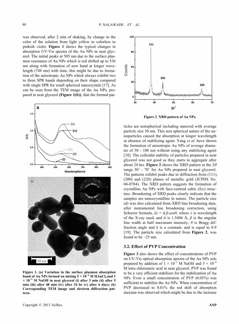
was observed, after 2 min of shaking, by change in the color of the solution from light yellow to colorless to pinkish violet. Figure 1 shows the typical changes in absorption UV-Vis spectra of the Au NPs in neat glyc-erol. The initial peaks at 505 nm due to the surface plas-mon resonance of Au NPs which is red shifted up to 536 nm along with formation of new band at longer wave-length (748 nm) with time, this might be due to forma-tion of the anisotropic Au NPs which always exhibit two to three SPR bands depending on their shape compared with single SPR for small spherical nanocrystals [17]. As can be seen from the TEM image of the Au NPs, pre-pared in neat glycerol (Figure 1(b)), that the formed par-
400 500 600 700 800 9000.0
0.5
1.0
1.5
a
(v)
(iv)
(iii)
(ii)
(i)
O.D
.
Wavelength(nm)
Figure 1. (a) Variation in the surface plasmon absorption band of Au NPs formed on mixing 5 × 10⁻⁴ M HAuCl4 and 1 × 10⁻³ M NaOH in neat glycerol (i) after 3 min (ii) after 5 min (iii) after 48 min (iv) after 24 hr (v) after 6 days; (b) Corresponding TEM image and electron diffraction pat-tern.
30 40 50 60 70
0
30
60
90
120
220
200
111
Cou
nts
20
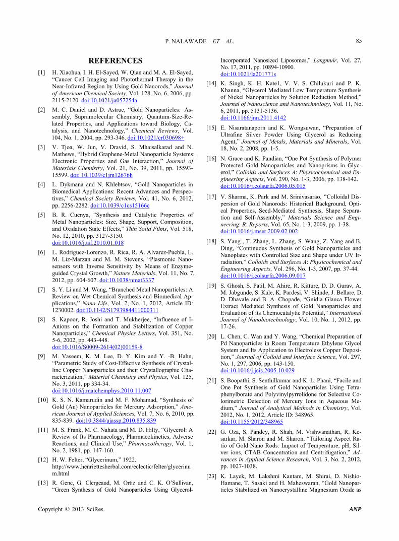
Figure 2. XRD pattern of Au NPs. ticles are nonspherical including nanorod with average particle size 30 nm. This non spherical nature of the na- noparticles caused the absorption at longer wavelength in absence of stabilizing agent. Yang et al. have shown the formation of anisotropic Au NPs of average diame-ter of 50 - 100 nm without using any stabilizing agent [18]. The colloidal stability of particles prepared in neat glycerol was not good as they starts to aggregate after about 24 hrs. Figure 2 shows the XRD pattern in the 2 range 30˚ - 70˚ for Au NPs prepared in neat glycerol. The patterns exhibit peaks due to diffraction from (111), (200) and (220) planes of metallic gold (JCPDS No. 04-0784). The XRD pattern suggests the formation of crystalline Au NPs with face-centred cubic (fcc) struc-ture. Broadening of XRD peaks clearly indicate that the samples are nanocrystalline in nature. The particle size (d) was also calculated from XRD line broadening data, after instrumental line broadening correction, using Scherrer formula, kλ = d.β.cosθ, where λ is wavelength of the X-ray used, and it is 1.5406 Å, β is the angular line width at half maximum intensity, θ is Bragg dif-fraction angle and k is a constant, and is equal to 0.9 [19]. The particle size calculated from Figure 2, was found to be ~25 nm.
3.2. Effect of PVP Concentration
Figure 3 also shows the effect of concentrations of PVP on UV-Vis optical absorption spectra of the Au NPs sols prepared by addition of 1 × 10⁻³ M NaOH and 5 × 10⁻⁴ M tetra chloroauric acid in neat glycerol. PVP was found to be a very efficient stabilizer for the stabilization of Au NPs. Even a small concentration of PVP (0.05%) was sufficient to stabilize the Au NPs. When concentration of PVP decreased to 0.01% the red shift of absorption maxima was observed which might be due to the increase
Copyright © 2013 SciRes. ANP

P. NALAWADE ET AL. 81
in particle size of the Au NPs. However, concentration of PVP (0.05%) has no significant effect on stability as well as size, as Au NPs prepared with different concen- trations are equally stable for more than a year. Since the particles were stable in the sol form XRD could not be carried out, nevertheless, TEM and selective area elec- tron diffraction (SAED) confirmed the presence of fcc Au NPs (results not shown).
3.3. Effect of NaOH Concentration
The above results showed that concentration of PVP ( 0.05%) had no significant effect on the stability of the particles; hence, for the preparation of particles we had selected 0.1% PVP to make sure that the particles do not aggregate. Figure 4 shows the effect of NaOH concen- tration (5 × 10⁻⁴ M - 1 × 10⁻³ M) on the UV-Vis absorp- tion spectrum of Au NPs in presence of 0.1% PVP in neat glycerol. The blue shift of SPR band with increas-ing concentration of NaOH indicates the decrease in particle size of Au NPs. The corresponding TEM image of Au NPs formed in presence of 5 × 10⁻⁴ M - 1 × 10⁻³ M NaOH (Figures 5(a) and (b)) shows that the average particle size falls from 20 nm to 5 - 7 nm when the NaOH concentration was increased from 5 × 10⁻⁴ M - 1 × 10⁻³ M NaOH. Since the particles were stable in the sol form XRD could not be carried out. Chen et al. have observed similar results where palladium nanoparticles in the size range of 8.6 to 2.4 nm were prepared by changing the concentration of NaOH in presence of PVP [20]. However, in our case, no particle formation was observed when concentration of NaOH was below 5 × 10⁻⁴ M.
400 500 600 700 8000.0
0.5
1.0
1.5
2.0
0.01 % PVP 0.05 % PVP 0.075 % PVP 0.1 % PVP
Ab
sorb
ance
Wavelength, nm
Figure 3. Effect of PVP concentration (wt %) on the optical absorption spectrum of Au NPs prepared by addition of 1 × 10⁻³ M NaOH and of 5 × 10⁻⁴ M tetra chloroauric acid. (i) 0.01 (ii) 0.05 (iii) 0.075 and (iv) 0.1. Spectra were recorded after 48 hr.
400 500 600 700 8000.0
0.5
1.0
1.5
2.0
0.5 0.6 0.7 0.8 0.9 1.0 1.1
525
530
535
540
545
550
max
[NaOH], (mM)
(iii)(ii)
(i)
Ab
so
rba
nc
e
Wavelength(nm)
Figure 4. Effect of NaOH concentration on Au NPs sol syn-thesized by the reaction of (i) 5 × 10⁻⁴, (ii) 7.5 × 10⁻⁴ and (iii) 1 × 10⁻³ M NaOH with 5 × 10⁻⁴ M auric acid in the presence of 0.1% PVP in glycerol.
Figure 5. TEM image of Au NPs obtained with different concentration of NaOH other conditions are same as Figure 4. (a) 7.5 × 10⁻⁴ M (b) 1 × 10⁻³ M.
3.4. Effect of Gold Ion Concentration
On addition of NaOH (1 × 10⁻³ M) to different concen-trations of the tetra chloroauric acid solution in absence
Copyright © 2013 SciRes. ANP

P. NALAWADE ET AL. 82
of PVP, the color of the solution changed from light yel-low to colorless to shades of blue and violet. However, in presence of PVP the color of the gold sol range from wine red to pink to magenta to violet for gold ion con-centration of 1 × 10⁻⁴ M, 2 × 10⁻⁴ M, 5 × 10⁻⁴ M and 7.5 × 10⁻⁴ M, respectively. Figure 6 shows the UV-Vis spec-tra of the Au NPs obtained for various concentrations of gold ions in the absence and presence of 0.1% PVP, at room temperature. The peak at ~530 nm corresponds to the surface plasmon absorption of Au NPs. The absorp-tion peak intensity of Au NPs in both absence and pres-ence of PVP was found to increase and the max gradually shifted towards higher wavelength with the increasing concentration of gold ions, this is possibly due to the formation of large size spherical nanoparticles at higher concentrations of gold ions. However, for the gold ions concentration of 5 × 10⁻⁴ M and above in absence of PVP,
400 500 600 700 800 9000.0
0.3
0.6
0.9
1.2
(iv)(iii)
(ii)
(i)
a
Ab
sorb
ance
Wavelength(nm)
400 500 600 700 800 9000.0
0.5
1.0
1.5
2.0(iv)
(iii)
(ii)
(i)
b
O.D
.
Wavelength(nm)
Figure 6. UV-Vis spectra showing effect of tetra chloroauric acid concentration on Au NPs sol synthesized by the reac-tion of (i) 1 × 10⁻⁴, (ii) 2 × 10⁻⁴, (iii) 5 × 10⁻⁴and (iii) 7.5 × 10⁻⁴ M auric acid with 5 × 10⁻⁴ M NaOH in glycerol. (a) without PVP (b) with 0.1 % PVP. All spectra were taken after 24 hr.
the shoulder at 635 nm in the case of 5 × 10⁻⁴ M or new peak at 735 nm in the case of 7.5 × 10⁻⁴ M was observed which is possibly due to formation of nonsherical Au NPs as explained in Figure 1. Similar results were obtained by S. Boopathi et al. in the synthesis of Au NPs using tetra-phenylborate and PVP for colorimetric detection of mer-cury ions [21]. Oza et al. studied the effect of chloroauric acid concentration on the formation of gold nanorods in presence of cetyl trimethylammonium bromide (CTAB) and it was found that high yield of nanorods formation occurred when the concentration of gold ions was ≥1 × 10⁻³ M, whereas, yield of spherical Au NPs was high at lower concentration of gold ions [22]. However, in our study it was found that, on addition of stabilizing agent PVP during the synthesis, the band at longer wavelength was not observed indicating formation of almost spheri-cal nanoparticles. This might be due to the combine ef-fect of viscosity of medium and adsorption of PVP on the surface of nanoparticles during growth which result in to the formation of spherical nanoparticles. The increased absorbance may be correlated to the production of metal nanoparticles in larger quantities in the presence of higher concentration of gold ion precursor. It was also seen that at concentration of 5 × 10⁻⁴ M and 7 × 10⁻⁴ M tetra chloroauric acid more stable, spherical and small size Au NPs were formed. Also, the yield of the Au NPs was rea-sonably good. At both the concentrations similar size of particles (~10 nm) were observed (results not shown). As mentioned above due to the good stability of the particles XRD could not be carried out.
Based on above results to get the good dispersion as well as stable Au NPs in neat glycerol the optimum con-centrations of tetra chloroauric acid, NaOH and PVP are 5 × 10⁻⁴ M, 1 × 10⁻³ M and 0.1 % (w/v), respectively.
400 500 600 700 800 9000.0
0.6
1.2
1.8
2.4
3.0
Ab
sorb
ance
Wavelength(nm)
80% Glycerol50% Glycerol20% Glycerol
Figure 7. Effect of glycerol concentration on the UV-Vis spectra of Au NPs in presence of 1 × 10⁻³ M NaOH and 7 × 10⁻⁴ M HAuCl4 without using PVP.
Copyright © 2013 SciRes. ANP

P. NALAWADE ET AL.
Copyright © 2013 SciRes. ANP
83
3.5. Synthesis of Au NPs in Glycerol: Water Mixture
To see the effect of concentration of glycerol on the par-ticle size and shape of nanoparticles, synthesis of Au NPs were carried out at different concentrations of glycerol in water with fixed concentration of NaOH without using PVP. Figure 7 shows the UV-Vis spectra of the Au NPs obtained with different concentration (v/v) of glycerol. In 80% glycerol two SPR band centered at 543 and 694 due of Au NPs was observed. This new band formation at longer wavelength indicates the presence of nonspherical nanoparticles along with spherical NPs. However when concentration of glycerol decreased up to 20%, almost spherical and monodispersed nanoparticles were obtained. Figure 8 shows the TEM images of Au NPs obtained at 80%, 50% and 20 % of glycerol. The shape and average particle size obtained by TEM measurements at different concentration of glycerol is summarized in Table 1.
The above results show that at low concentrations of glycerol it is possible to stabilize the particles without using stabilizer. It seems that the stability of the particles depend on the viscosity of the medium though not in direct proportionality. High viscosity means that formation of nuclei in a smaller zone and thus the interaction of parti-cles in a smaller confined volume results in aggregation of
the particles. At appropriate viscosity the interaction be-tween the particles decreases and this leads to the stability of the particles. Thus, it can be concluded that for the preparation of small and stable Au NPs the mixture of glycerol:water is better as compared to neat glycerol.
3.6. Catalytic activity of Au NPs
Aromatic amino compounds are widely used in industry as an intermediate in the preparation of polymers, azo dyes, etc. However, the chemical reduction of aromatic nitro compounds with sodium borohydride is extremely slow, and the use of a catalyst is essential. Of late, metal nanoparticles have been explored for this reaction. In this the reduction of nitro aromatics to its corresponding amino derivatives with an excess amount of NaBH4 has frequently been used as a model reaction to examine the catalytic performance of metal nanoparticles [23,24].
It is often discussed in the literature that the presence of stabilizer can inhibit the catalytic activity of the metal nanoparticles [2]. In the present work it was possible to stabilize small Au NPs in the absence of PVP in 20:80 (v/v) glycerol:water mixture. Hence, for catalytic reac-tion Au NPs prepared in 20:80 (v/v) glycerol:water mix-ture were used; however, the catalytic reaction was car-ied out in an aqueous solution. The strong UV-Vis peaks r
Figure 8. TEM images of Au NPs obtained in presence of different concentration of glycerol (a) 80% (b) 60% and (c) 20%.

P. NALAWADE ET AL. 84
Table 1. Summary of particle size and shape of Au NPs at different glycerol:water ratios (v/v) in the absence of PVP.
% conc. of glycerol SPR peak (nm) Shape and size distribution Particle size by TEM (nm) Colloidal stability
100% 554 748
Non spherical, Polygonal and polydispersed
50 Turbid after 1 day
80% 543 694
Non spherical, polygonal and polydispersed
25 Stable at least up to 8 months
50% 533 Spherical and nearly monodispersed
20 Stable at least up to 8 months
20% 522 Spherical and monodispersed
8 Stable at least up to 8 months
Table 2. Variation in the rate of reduction of o-nitro aniline with increasing amount of Au NPs.
Sr. No. Concentration of Au NPs (µg/mL) kobs, s−1
1 14 2.5 × 10⁻³
2 27 3.2 × 10⁻³
3 54 7.5 × 10⁻³ 4 82 3 × 10⁻²
300 400 5000.0
0.5
1.0
1.5
2...14
2 min time iterval
Ab
sorb
ance
Wavelength (nm)
Figure 9. Time-dependent variation in the absorption spec-trum of o-nitro aniline reduced by 5 µL/mL of Au NPs pre-pared in 20% glycerol. Inset: Plot of ln(Ct/C0) versus time.
characteristic of o-nitro aniline appears at 412 nm and 283 nm [25]. It was found that there was a very slow decrease of absorbance during the chemical reaction without catalyst. However, after the addition of Au na- noparticles a decrease in the absorbance at 412 nm and shift of the peak from 283 nm to 289 nm with reaction time usually indicate steady reduction of o-nitro aniline to benzenediamine (Figure 9). The progress of reduction reaction was monitored by measuring UV-Vis absorption recorded at different times; t, at 298 K at different con-centration of Au NPs. The ratio of Ct and C0, where Ct and C0 refer to the concentration of o-nitro aniline at times t and 0, is measured from the relative intensity ratio
of the respective absorbance, at 412 nm. The linear cor-relation of ln(Ct/C0) versus time is shown in inset of Fig-ure 9 indicating the reaction follows pseudo first-order kinetics. The rate constant (k) for the reaction, directly calculated from the slope of the straight line, for different concentration of Au NPs are tabulated in Table 2. It was found that when the gold content of the dispersion (ran- ging from 14 to 82 g/mL) was varied keeping other parameters constant, to determine the effect of the amount of catalyst on the rate of the reaction, the rate constant showed an increase of factor ca. 10 times.
0 200 400 600 800 1000-1.5
-1.0
-0.5
0.0
Ct/
C0
Time(s)
K= 3.2x10⁻³ s-1
4. Conclusion
Green synthesis of Au NPs was carried out by simply adding NaOH to the tetra chloroauric acid solution pre-pared in glycerol. Glycerol is used both as a reducing and stabilizing agent. The different size and shape of Au NPs can be prepared by varying the concentration of glycerol, concentration of NaOH and concentration of gold ions. Generally, stabilizers are added in the synthesis of the nanoparticles to prevent the aggregation of particles. The results of the present study show that the viscosity of the medium can play a similar role as of stabilizer by de-creasing the interaction of the particles. However, there is no direct relationship between the viscosity and the stability of the particles. Thus the present work highlights that by exploiting the intrinsic properties of the solution it is possible to synthesize small and stable nanoparticles without using stabilizer. The Au NPs as synthesized in 20:80 (v/v) glycerol:water mixture were found to be catalytically active and had shown good catalytic activity for reduction of o-nitro aniline.
Copyright © 2013 SciRes. ANP
P. NALAWADE ET AL. 85
REFERENCES [1] H. Xiaohua, I. H. El-Sayed, W. Qian and M. A. El-Sayed,
“Cancer Cell Imaging and Photothermal Therapy in the Near-Infrared Region by Using Gold Nanorods,” Journal of American Chemical Society, Vol. 128, No. 6, 2006, pp. 2115-2120. doi:10.1021/ja057254a
[2] M. C. Daniel and D. Astruc, “Gold Nanoparticles: As- sembly, Supramolecular Chemistry, Quantum-Size-Re- lated Properties, and Applications toward Biology, Ca- talysis, and Nanotechnology,” Chemical Reviews, Vol. 104, No. 1, 2004, pp. 293-346. doi:10.1021/cr030698+
[3] V. Tjoa, W. Jun, V. Dravid, S. Mhaisalkarad and N. Mathews, “Hybrid Graphene-Metal Nanoparticle Systems: Electronic Properties and Gas Interaction,” Journal of Materials Chemistry, Vol. 21, No. 39, 2011, pp. 15593- 15599. doi: 10.1039/c1jm12676h
[4] L. Dykmana and N. Khlebtsov, “Gold Nanoparticles in Biomedical Applications: Recent Advances and Perspec-tives,” Chemical Society Reviews, Vol. 41, No. 6, 2012, pp. 2256-2282. doi:10.1039/c1cs15166e
[5] B. R. Cuenya, “Synthesis and Catalytic Properties of Metal Nanoparticles: Size, Shape, Support, Composition, and Oxidation State Effects,” Thin Solid Films, Vol. 518, No. 12, 2010, pp. 3127-3150. doi:10.1016/j.tsf.2010.01.018
[6] L. Rodriguez-Lorenzo, R. Rica, R. A. Alvarez-Puebla, L. M. Liz-Marzan and M. M. Stevens, “Plasmonic Nano- sensors with Inverse Sensitivity by Means of Enzyme- guided Crystal Growth,” Nature Materials, Vol. 11, No. 7, 2012, pp. 604-607. doi:10.1038/nmat3337
[7] S. Y. Li and M. Wang, “Branched Metal Nanoparticles: A Review on Wet-Chemical Synthesis and Biomedical Ap-plications,” Nano Life, Vol. 2, No. 1, 2012, Article ID: 1230002. doi:10.1142/S1793984411000311
[8] S. Kapoor, R. Joshi and T. Mukherjee, “Influence of I- Anions on the Formation and Stabilization of Copper Nanoparticles,” Chemical Physics Letters, Vol. 351, No. 5-6, 2002, pp. 443-448. doi:10.1016/S0009-2614(02)00159-8
[9] M. Vaseem, K. M. Lee, D. Y. Kim and Y. -B. Hahn, “Parametric Study of Cost-Effective Synthesis of Crystal-line Copper Nanoparticles and their Crystallographic Cha- racterization,” Material Chemistry and Physics, Vol. 125, No. 3, 2011, pp 334-34. doi:10.1016/j.matchemphys.2010.11.007
[10] K. S. N. Kamarudin and M. F. Mohamad, “Synthesis of Gold (Au) Nanoparticles for Mercury Adsorption,” Ame- rican Journal of Applied Sciences, Vol. 7, No. 6, 2010, pp. 835-839. doi:10.3844/ajassp.2010.835.839
[11] M. S. Frank, M. C. Nahata and M. D. Hilty, “Glycerol: A Review of Its Pharmacology, Pharmacokinetics, Adverse Reactions, and Clinical Use,” Pharmacotherapy, Vol. 1, No. 2, 1981, pp. 147-160.
[12] H. W. Felter, “Glycerinum,” 1922. http://www.henriettesherbal.com/eclectic/felter/glycerinum.html
[13] R. Genc, G. Clergeaud, M. Ortiz and C. K. O’Sullivan, “Green Synthesis of Gold Nanoparticles Using Glycerol-
Incorporated Nanosized Liposomes,” Langmuir, Vol. 27, No. 17, 2011, pp. 10894-10900. doi:10.1021/la201771s
[14] K. Singh, K. H. Kate1, V. V. S. Chilukuri and P. K. Khanna, “Glycerol Mediated Low Temperature Synthesis of Nickel Nanoparticles by Solution Reduction Method,” Journal of Nanoscience and Nanotechnology, Vol. 11, No. 6, 2011, pp. 5131-5136. doi:10.1166/jnn.2011.4142
[15] E. Nisaratanaporn and K. Wongsuwan, “Preparation of Ultrafine Silver Powder Using Glycerol as Reducing Agent,” Journal of Metals, Materials and Minerals, Vol. 18, No. 2, 2008, pp. 1-5.
[16] N. Grace and K. Pandian, “One Pot Synthesis of Polymer Protected Gold Nanoparticles and Nanoprisms in Glyc-erol,” Colloids and Surfaces A: Physicochemical and En-gineering Aspects, Vol. 290, No. 1-3, 2006, pp. 138-142. doi:10.1016/j.colsurfa.2006.05.015
[17] V. Sharma, K. Park and M. Srinivasarao, “Colloidal Dis-persion of Gold Nanorods: Historical Background, Opti-cal Properties, Seed-Mediated Synthesis, Shape Separa-tion and Self-Assembly,” Materials Science and Engi-neering: R: Reports, Vol. 65, No. 1-3, 2009, pp. 1-38. doi:10.1016/j.mser.2009.02.002
[18] S. Yang , T. Zhang, L. Zhang, S. Wang, Z. Yang and B. Ding, “Continuous Synthesis of Gold Nanoparticles and Nanoplates with Controlled Size and Shape under UV Ir-radiation,” Colloids and Surfaces A: Physicochemical and Engineering Aspects, Vol. 296, No. 1-3, 2007, pp. 37-44. doi:10.1016/j.colsurfa.2006.09.017
[19] S. Ghosh, S. Patil, M. Ahire, R. Kitture, D. D. Gurav, A. M. Jabgunde, S. Kale, K. Pardesi, V. Shinde, J. Bellare, D. D. Dhavale and B. A. Chopade, “Gnidia Glauca Flower Extract Mediated Synthesis of Gold Nanoparticles and Evaluation of its Chemocatalytic Potential,” International Journal of Nanobiotechnology, Vol. 10, No. 1, 2012, pp. 17-26.
[20] L. Chen, C. Wan and Y. Wang, “Chemical Preparation of Pd Nanoparticles in Room Temperature Ethylene Glycol System and Its Application to Electroless Copper Deposi-tion,” Journal of Colloid and Interface Science, Vol. 297, No. 1, 297, 2006, pp. 143-150. doi:10.1016/j.jcis.2005.10.029
[21] S. Boopathi, S. Senthilkumar and K. L. Phani, “Facile and One Pot Synthesis of Gold Nanoparticles Using Tetra-phenylborate and Polyvinylpyrrolidone for Selective Co- lorimetric Detection of Mercury Ions in Aqueous Me-dium,” Journal of Analytical Methods in Chemistry, Vol. 2012, No. 1, 2012, Article ID: 348965. doi:10.1155/2012/348965
[22] G. Oza, S. Pandey, R. Shah, M. Vishwanathan, R. Ke-sarkar, M. Sharon and M. Sharon, “Tailoring Aspect Ra-tio of Gold Nano Rods: Impact of Temperature, pH, Sil-ver ions, CTAB Concentration and Centrifugation,” Ad-vances in Applied Science Research, Vol. 3, No. 2, 2012, pp. 1027-1038.
[23] K. Layek, M. Lakshmi Kantam, M. Shirai, D. Nishio- Hamane, T. Sasaki and H. Maheswaran, “Gold Nanopar-ticles Stabilized on Nanocrystalline Magnesium Oxide as
Copyright © 2013 SciRes. ANP

P. NALAWADE ET AL.
Copyright © 2013 SciRes. ANP
86
an Active Catalyst for Reduction of Nitroarenes in Aqu- eous Medium at Room Temperature,” Green Chemistry, Vol. 14, No. 11, 2012, pp. 3164-3174. doi:10.1039/c2gc35917k
[24] A. Leelavathi, T. U Bhaskara Rao and T. Pradeep, “Sup-ported Quantum Clusters of Silver as Enhanced Catalysts
for Reduction,” Nanoscale Research Letters, Vol. 6, No. 2, 2011, pp. 123-132. doi:10.1186/1556-276X-6-123
[25] N. Pradhan, A. Pal and T. Pal, “Catalytic Reduction of Aromatic Nitro Compounds by Coinage Metal Nanopar-ticles,” Langmuir, Vol. 17, No. 5, 2001, pp 1800-1802. doi:10.1021/la000862d