Embed Size (px)
Citation preview

Inactivation of G-protein coupled receptor 48 (Gpr48/Lgr4) impairs definitive erythropoiesis at midgestation through downregulation of ATF4 Signaling
pathway Huiping Song1,3,4, Jian Luo2, Weijia Luo1, Jinsheng Weng1, Zhiqing Wang3, Baoxing Li4, Dali Li2,
and Mingyao Liu1,2,* 1 Institute of Biosciences and Technology, and Department of Molecular and Cellular Medicine, Texas
A&M University Health Science Center, 2121 W. Holcombe Blvd., Houston, Texas 77030, USA 2 The Institute of Biomedical Sciences and School of Life Sciences, East China Normal University, 500
Dongchuan Road, Shanghai 200241, China 3 The Second Hospital of Tangshan, Tangshan 063000, China
4 Shanxi Provincial Tissue Bank, Southern Medical University, Taiyuan 030006, China Running head: Deletion of Gpr48 impairs definitive erythropoiesis
* Address correspondence to: Dr. Mingyao Liu, Institute of Biosciences and Technology, Texas A&M Health Science Center, 2121 W. Holcombe Blvd., Houston, Texas 77030, Tel: 713-677-7505, Fax: 713-
677-7512, Email: [email protected]
G protein-coupled receptors (GPCRs), one of the most versatile groups of cell surface receptors, can recognize specific ligands from neural, hormonal, and paracrine organs and regulate cell growth, proliferation and differentiation. Gpr48/LGR4 is a recently identified orphan GPCR with unknown functions. To reveal the functions of Gpr48 in vivo, we generated Gpr48-/- mice and found that Gpr48-/- fetuses displayed transient anemia during midgestation and abnormal definitive erythropoiesis. The dramatic decrease of definitive erythroid precursors (Ter119pos population) in Gpr48-/- fetal liver at E13.5 was confirmed by histological analysis and blood smear assays. Real-time PCR analyses showed that in Gpr48-/- mice both adult hemoglobin � and � chains were decreased while embryonic hemoglobin chains (�, �H1 and �y) were increased, providing another evidence for the impairment of definitive erythropoiesis. Furthermore, Proliferation was suppressed in Gpr48-/- fetal liver with decreased c-Myc and cyclin D1 expression, whereas aptotosis was unaffected. ATF4, a key transcription factor in erythropoiesis, was downregulated in Gpr48-/- fetal livers during midgestation stage through cAMP-PKA-CREB pathway, suggesting that Gpr48 regulated definitive erthropoiesis through ATF4-mediated definitive erythropoiesis.
Erythyopoiesis occurs sequentially in
distinct anatomical locations in two different phases during embryogenesis. The earlier phase is defined as primitive erythropoiesis which, in the mouse, originates from yolk sac at embryonic day 7.5 (E7.5) and the later phase is definitive erythropoiesis which was carried out in the fetal liver during midgestation after E12.5. Nonnucleated adult-type red blood cells are first generated in the fetal liver, the primary organ for erythropoiesis during midgestation from E12-E16(1). Erythropoiesis then transfers to bone marrow and spleen in the adult(2;3). However, the molecular mechanism of regulating erythropoiesis has not been completely delineated. Most of the studies were focused on the transcription factors to explore the mechanisms of erythropoiesis (4;5). In recent years, several transcription factors were also identified to regulate definitive erythropoiesis in fetal liver during midgestation(6-11). One of the transcription factors is ATF4, which has been shown to regulate cell proliferation in response to a broad spectrum of cell stresses and can be either an activator or a repressor in response to different extracellular signals (12). ATF4-/- mice have been reported to cause defective definitive erythropoiesis, and severe anemia at midgestation (13). Although receptors, such as c-Kit and EPOR, have been well studied in erythropoiesis (14), little is known on the function of G-protein-coupled receptors (GPCRs) in erythropoiesis during development (14;15).
The GPCR family represents the largest and
1
http://www.jbc.org/cgi/doi/10.1074/jbc.M800721200The latest version is at JBC Papers in Press. Published on October 27, 2008 as Manuscript M800721200
Copyright 2008 by The American Society for Biochemistry and Molecular Biology, Inc.
by guest on January 5, 2019http://w
ww
.jbc.org/D
ownloaded from

most versatile group of cell surface receptors (16-18). GPCRs can recognize their ligands, a diverse array of extracellular signals, then transmit these signals to intracellular responses by the ligand-receptor interaction. For its versatile roles, the GPCR family is one of the most promising and attractive targets to develop pharmaceutical drugs for human diseases ranging from allergic rhinitis to pain, hypertension and schizophrenia (16). The glycoprotein hormone receptors represent a subgroup of GPCRs that have a large N-terminal extracellular (ecto-) domain containing leucine-rich repeats (LRRs), a versatile structural domain that is important for glycoprotein hormone ligands recognition(19-21). Based on the comparison of peptide hormones and glycoprotein hormone receptors, a sub-group of GPCRs, leucine-rich repeat-containing GPCRs (LGRs), was identified(22). Many studies suggest that the expanding family of LGRs, including three known glycoprotein hormone receptors (LH, FSH, and TSH), the orphan receptor LGR4 (also termed Gpr48)-6, and LGR7-8, are homologous and conservative (22-24). The mammalian glycoprotein hormone receptors have diverse structural features and mainly couple through the cAMP-dependent pathway for signal transduction (25).
Gpr48 (LGR4) has a putative horseshoe-like structure composed of 17 LRRs which is proposed to be the ligand-binding site for this family of receptors (21;26). The molecular structure and evolutionary features of Gpr48 have been well studied; however, the ligands and physiological functions remain unclear (19;20;25;27;28). Gpr48 is widely expressed in multiple organs at both embryonic and adult stage (19;27;29), which suggests that Gpr48 might play a vital role in development and adult physiological functions. Recent studies from our and other labs have indicated that Gpr48 plays an important role in renal, eye, and reproductive system development (30-33). However little is known thus far about the function and molecular mechanism of Gpr48 in erythropoiesis. In this study, we demonstrated that Gpr48 is expressed in both embryonic and adult liver, and that the deletion of Gpr48 in mouse impairs definitive erythropoiesis at midgestation through downregulation of c-Myc, cyclin D1, and ATF4 pathways.
EXPERIMENTAL PROCEDURES
Generation of GPR48 knocked out mice Gpr48 gene trap ES clone (LST020) was obtained from Bay Genomics (34;35). The Gpr48 ES clones were injected into C57BL/6 blastocysts and transferred to ICR females. Male chimera mice were mated with C57BL/6 females, resulting in transmission of the inserted allele to the germ line. Positive mice were interbred and maintained on a mixed 129×C57BL/6 background.
Gpr48 knocked out mice were generated based on the secretory-trap approach as previously described (33;36) by disrupting the endogenous Gpr48 gene. This vector (Pgt0,1,2tm-pfs, 11.98 kb) includes the mouse En-2 SA sequence, a fragment of CD4 containing the transmembrane domain inserted in frame with �-geo together with a downstream IRES, the PLAP gene, and the simian virus 40 polyadenylation signal in the Pgt1tm vector. The vector was inserted into intron 1 of Gpr48 to produce a transcript containing exon 1 fused to the �geo-PLAP cassette. A male chimeric mouse line was intercrossed to B6/129 wild type mice so as to obtain heterozygous mice with Gpr48 mutant. Homozygous were obscured by intercrossing these heterozygous mice. Mice were housed under controlled humidity, temperature, and light regimen and fed ad libitum. Animal care was consistent with institutional and NIH guidelines. DNA-based genotyping Genotype of mice was determined by using four primers, for wild-type , forward (5’-TGT TTC AAC CTT TTA AAG ACT GTA GCG AGA -3’), reverse (5’-TAA AGG ACT TAA TGC CAA ATG TGA TTG GTT -3’); for mutant, forward (5’-CCA ATC ACC ACT CTT ACA CAA TGG CTA AC-3’), reverse (5’-GGT CTT TGA GCA CCA GAG GAC-3’). PCR was carried out by using standard techniques. PCR conditions were 35 cycles at 94ºC for 40 seconds, 53 ºC for 40 seconds, and 72 ºC for 1 minute. DNA was prepared from 0.5 cm of clipped tail of embryos and extracted by using DirecPCR kit (VIAGEN, CA) protocols. Genomic DNA was extracted by ethanol precipitation after proteinase K digestion in a tissue lysis buffer (50 mom Trish (pH 7.5), 50 mM EDTA (pH 8.0), 100
2
by guest on January 5, 2019http://w
ww
.jbc.org/D
ownloaded from

mM NaCl, 0.5 mM spermidine, 1% SDS, 5 mM DTT). Reaction products were electrophoresed on 1% agarose gels stained with ethidium bromide. Two expected products were 450bp for wild-type and 750bp for mutant. Due to the insertion of the large 11.98kb trap vector between primer A and primer B, the A/B PCR product could not be amplified in the mutant allele under the present PCR conditions. RNA extraction and RT-PCR amplification Total RNA was extracted from fetal livers by using Trizol RNA extraction kit (Invitrogen). First-strand cDNA synthesis was performed using Moloney murine leukemia virus reverse transcriptase and oligo(dT) (Promega) according to the manufacturer’s protocol. The primers used for Gpr48 wildtype and mutant transcripts analysis were: forward (5’-GGA ATT CGC CGC CAT GCC GGG CCC G-3’); reverse (5’-AGC TCT TTA AGG CTG GGA AGG GCT TT-3’). The conditions for reaction were 30 cycles at 94ºC for 40 seconds, 60ºC for 40 seconds, and 72ºC for 1 minute. The primers used for ATF4 were: forward (5’-CCC ACA ACA TGA CCG AGA T-3’), reverse (5’-CTC ATC TGG CAT GGT TTC C-3’) under the amplified reaction conditions of 35 cycles at 94ºC for 30 seconds, 53 ºC for 30 seconds, 72 ºC for 1 minute, GAPDH primers were used as controls. The primers used for GAPDH: forward (5’-GGT GAA GGT CGG TGT GAA CG-3’), reverse (5’-CTT CTG GGT GGC AGT GAT GG-3’). Sample preparation and LacZ histochemistry Sample preparation and LacZ staining followed the protocol from Dr. James Martin’s lab (Texas A&M Health Science Center, Institute of Bioscience and Technology, Houston). The embryos were fixed for 1 hr at room temperature in the fixation buffer containing 25% glutataldehyde,100mM EGTA (pH 8.0), 500mM NaH2PO4 (pH 7.3), 1M MgCl2, and 37% formaldehyde. Samples were then washed three times with rinse buffer containing Deoxycholic Acid, NP40, and 1M MgCl2. Then these samples were stained overnight at room temperature with LacZ staining buffer containing 1mg/ml X-gal, 5mM K3Fe(CN)6, 5mM K4Fe(CN)6. After LacZ staining, the samples were stored in 70% alcohol
overnight, then dehydrated sequentially by 95% Alcohol, 100% Alcohol, 100% isopropyl alcohol and embedded in paraffin to be sectioned at 6μm and 12 μm. Subsequently these slides were counter stained with eosin using standard procedures. Quantitative Real-time PCR Total RNA extraction and cDNA synthesize from E13.5 blood cells collected by centrifuge of whole blood and fetal livers were performed as previously described. Primers were used to detect embryonic hemoglobin chains for blood cells: �y, forward (5’- CAA GCT ACA TGT GGA TCC TGA GAA-3’), reverse (5’- TGCCGAAGTGACTAGCCAAA-3’); � ( 5’- GCGAGCTGCATGCCTACAT -3’), reverse (5’- GCCATTGTGACCAGCAGACA -3’); and �h1, forward ( 5’- AGGCAGCTATCACAAGCATCTG - 3’), revise (5’- AACTTGTCAAAGAATCTCTGAGTCC -3’); GPA, forward ( 5’- GCC GAA TGA CAA AGA AAA GTT CA-3’), reverse ( 5’-TCA ATA GAA CTC AAA GGC ACA CTG T -3’). GPA mRNAs were used as internal controls. Amplification reactions (25μl) were prepared using SuperArray Real-time SYBR Green/ROX PCR master mix (SuperArray) according to the manufacture’s protocol. Quantitative real-time PCR were carried out using Mx3000PTM PCR System (Stratagene, CA) and analysed with Mx3000P software. The reaction conditions were performed at 40 cycles (95 ºC for 30 seconds, 53 ºC for 1 minute, and 72ºC for 30 seconds) with GPA as an internal control. For fetal livers, following primers were used: adult �-globin forward (5’- AAT ATG GAG CTG AAG CCC TGG-3’), reverse (5’-AAC ATC AAA GTG AGG GAA GTA GGT CT -3’); adult-�, forward (5’-GTG AGC TCC ACT GTG ACA AGC T -3’), reverse (5’-GGT GGC CCA GCA CAA TCA CGA TC -3’); �-Actin, forward (5’- GCA CCA GGG TGT GAT GGT G -3’), reverse (TGG ATG GCT ACG TAC ATG GC -3’). The conditions of amplification reactions (25μl) were performed at 40 cycles (95 ºC for 30 seconds, 52 ºC for 1 minute, 72 ºC for 30 seconds), and �-actin was used as an internal control. ATF4 primers used for Real-time PCR were the same as performed in RT-PCR with �-Actin used as an internal control. The
3
by guest on January 5, 2019http://w
ww
.jbc.org/D
ownloaded from

conditions of amplification reactions (25μl) were performed at 37 cycles (95 ºC for 30 seconds, 54 ºC for 1 minute, and 72ºC for 30 seconds).
Western Immunoblot Analysis Fetal liver lysates were harvested as the method described by Mitchell et al (37). The protein concentration was determined using BCATM Protein Assay Kit (Pierce, Rockford, IL) following the manufacturer’s procedure. Samples were then diluted into loading buffer at 1 mg/ml. Following heat denaturation, samples containing 8μg of protein were loaded onto and separated on 12% SDS-PAGE gels as needed. Proteins were then transferred electrophoretically to 140μm nitrocellulose membrane (Pall Corp., Ann Arbor, MI). After incubating the membranes in blocking solution, primary antibody and second antibody were used at appropriate dilution. Proteins were detected using the SuperSignal West Pico Chemiluminescent substrate (Pierce) according to the manufacturer’s instructions. The following antibodies from Santa Cruz were used: rabbit polyclonal anti-ATF4 (1:300), rabbit polyclonal anti-C-myc (1:400), rabbit polyclonal anti-Cyclin D1 (1:400). Goat polycolonal anti-Actin (1:500) was used as internal control. Histology and Immunohistochemistry Embryos dissected from mice were fixed in 10% formalin and embedded in paraffin according to the standard techniques. Five micrometer-thick sections were stained with hematoxylin and eosin. For proliferation assays, the sections were stained with anti-PCNA using the Zymed mouse PCNA kit (Zymed, South San Francisco, CA) and then counterstained with hematoxylin. For BrdU incorporation, mice were injected with 100mg/kg BrdU (Sigma) intraperitoneally 2 h before liver tissue harvesting. BrdU incorporation studies were performed using BrdU labeling kit (Zymed) on paraffined slides. The ApopTaq peroxidase in situ apoptosis detection kit (Chemicon, Temecula, CA) was used for apoptosis assay according to the manufacture’s instructions with methyl green as counterstain. Immunohistochemical staining was performed by using ABC staining kit (Santa Cruz). The following antibodies from Santa Cruz were used: rabbit polyclonal anti-ATF4 (C-20) (1:100), rabbit polyclonal anti-C-myc (1:200), rabbit
polyclonal anti-Cyclin D1(1:200). Hematoxylin was carried out as counterstain. Blood smear Blood was harvested from the carotid arteries of decapitated embryos. Blood smears were prepared by using the wedge technique followed by air drying and Wright-Giemsa staining (Sigma-Aldrich, St. Louis, MO) following the manufacture’s staining protocol. The number of nucleated erythrocytes and enucleated erythrocytes were counted in high power views.Flow cytometry Single-cell suspensions were obtained from E13.5 wild-type and homozygous fetal livers. Briefly, fetal livers were dissected from embryos, then placed in Dulbecco’s Modified Eagle’s Medium (DMEM, HyClone, Logan, Utah) with 5% Fetal Bovine Serum (FBS). The cells were dissociated by repeated flushing with a 23-gauge needle to obtain the single cell suspension and the cell number was counted. Cell suspensions with the same concentration were first incubated on ice with rat anti-mouse CD16/CD32 (PharMingen) to block nonspecific binding to Fc-receptors. Subsenquently, cells were incubated with rat anti-mouse PE-conjugated anti-c-Kit and anti-CD44, FITC-conjugated anti-CD34 and anti-Ter-119 (all from PharMingen). Appropriate isotype control antibodies were used respectively. Then the cells were washed and transferred to PBS 1% PFA. Cell surface expression of different markers was analyzed in a Becton Dickinson FACScan using CellQuest software. To measure cell size of erythrocyte in fetal livers, liver cells from E13.5 GPR48-/- fetus and their wild type littermates was harvested by flushing the femoral medullary cavity with DMEM. Cells are then centrifuged at 4oC 1500 rpm for 5min followed by resuspending in PBS (0.1M, pH7.2) and staining with anti-Ter119 (FITC conjugated) as a marker of mature erythrocyte for Flow cytometry assay. Generation of constitutive activated Gpr48 PCR-based point mutagenesis was performed to generate mutant Gpr48 using cDNA encoding human Gpr48 receptors. To introduce the amino acid mutation at T755I into the wild type cDNA of human Gpr48, overlapping primers containing mutated sequences (Forward: 5’- CGC TTG GCT AAT CTT CAT CAA TTG C -3’; Reverse: 5’-
4
by guest on January 5, 2019http://w
ww
.jbc.org/D
ownloaded from

GCA ATT GAT GAA GAT TAG CCA AGC G -3’) were used to replace the wild-type amino acid in the region. All cDNAs were subcloned into the expression vector pcDNA3.1 (-) (Invitrogen Corp., Carlsbad, CA), and plasmids were purified using a Maxi plasmid preparation kit (Qiagen, Inc., Valencia, CA). Fidelity of the PCR was confirmed by sequencing on both strands of the final constructs before use in expression studies. Cell transfection and luciferase assays Human embryonic kidney 293T was maintained in modified Eagle's medium/Ham's F-12 (DMEM/F12) supplemented with 10% fetal bovine serum, 100 μg/ml penicillin, 100 μg/ml streptomycin, and 2 mM L-glutamine. Before transfection, cells (5 × 10⁴/culture) were seeded in 24-well plate (Nalge Nunc International, Naperville, IL). When cells were 70-80% confluent, transient transfection was performed with 0.25 μg of plasmid using the lipofectmine method following replacement of culture media. After 6 -12 h of incubation with the lipofectmine, media were replaced with DMEM containing 10% fetal bovine serum. 48 hours after transfection, cells were washed twice with Dulbecco's phosphate-buffered saline (D-PBS), harvested from the plate. For luciferase transfection experiments, 48 hour after transcfection, cell lysate was collected and luminescence assays were performed. Within a given assay, plate wells were set up in triplicate for each transfected construct or control vector. To allow for normalization of firefly luciferase values based on transfection efficiency, a co-reporter vector expressing �-galactosidase (pcDNA3-LacZ) was included at a ratio of 1:20 of co-reporter plasmid to experimental promoter construct (or control vector) in the transfection mixture. Statistical analysis Data are presented as mean ± standard deviation (m ± SD) and analyzed by One-way ANOVA or Student t-test. For all analyses, P < 0.05 was considered statistically significant.
RESULTS
Targeted inactivation of Gpr48 gene and the expression of Gpr48 in fetal liver — The murine Gpr48/LGR4 gene was disrupted by inserting a large secretory trap vector (11.98kb) to the intron
1 (S-Fig. 1A). The Gpr48 ES clones were injected into C57BL/6 blastocysts to generate chimeric mice. Heterozygous Gpr48 mutant offspring were obtained by intercross between male chimera mice and C57BL/6 female mice. Heterozygous mice were intercrossed to obtain homozygous mice. The genotypes of offspring of mutant mice were confirmed by PCR analysis(S-Fig. 1B). The mRNA level of Gpr48 in the liver of homozygous, heterozygous, and wild type mice was detected by RT-PCR analysis. The results showed that Gpr48 is expressed in the fetal liver of wild type fetus, and that there was no expression of Gpr48 in homozygous mutant (Fig. 1A), confirming the successful deletion of Gpr48 in homozygous mutant. Since the mutant Gpr48 gene generates a chimeric protein containing the N-terminal leucine-rich repeat (LRRNT) domain and �-galactosidase, the expression patterns of Gpr48 in both heterozygous and homozygous mutant mice can be examined with the activity of �-galactosidase (LacZ staining) in fetal liver. As shown in Fig. 1B, LacZ staining was performed to measure �-galactosidase activity and the expression of Gpr48 in E15.5 fetal liver tissue sections. The expression of Gpr48 was found not only in hepatocytes but also in erythroid precursor cells (Fig. 1C). During midgestation, the erythroid precursors are mainly derived from liver cells, homologous to the hepatocytes. The expression of Gpr48 in erythroid precursor cells suggests a potential role of the receptor in erythropoiesis. Deletion of Gpr48 affects fetal liver size and definitive erythropoiesis at midgestation in Gpr48-
/- embryos To understand the in vivo function of Gpr48 in fetal liver growth and erythropoiesis, we examined fetal liver weight and body weight in Gpr48-/- mice compared to wt mice. As shown in Fig. 2A, at E14.5, inactivation of Gpr48 gene affected liver growth with a 41% weight reduction (Fig. 2A(a)) and 25% reduction in body weight (data not shown). Furthermore, the ratio of liver to body weight showed significant decrease (Fig. 2A (b)) and the fetal livers of Gpr48 null embryos showed marked smaller size, indicating hypoplastic development during fetal liver development from E12.5-E16.5 (Fig. 2B). Since fetal liver is the predominant site for definitive erythropoiesis during embryonic stages, we further examined the morphological features of fetal liver
5
by guest on January 5, 2019http://w
ww
.jbc.org/D
ownloaded from

from E12.5 to E16.5. As shown in Fig. 2B, Gpr48-/- embryos and fetal livers from E12.5 to E15.5 appeared considerably paler than their control wt littermates, suggesting less number of erythrocytes with low hemoglobin. However, little difference was found at E16.5 between wt and Gpr48-/- mice (Fig. 2B). In fetal liver, erythropoietic precursor cells can be distinguished from hepatocytes based on their smaller cell size, more condensed and deeply stained nucleus. Histological examination and hematoxylin and eosin (H&E) staining show that the gross morphologic features of homozygous fetal livers (green arrows) were normal in cellular architecture (Fig. 3). However, fewer erythroid precursor or definitive progenitors (yellow arrows) and erythroid foci (orange arrows) were visible in the homozygous fetal livers from E12.5-E15.5 embryos compared with that in wild-type (Fig. 3). The fetal livers of Gpr48-/- embryos showed marked smaller size, indicating hypoplastic development during E12.5-E16.5.
Primitive erythrocyte is nucleated red blood cell in peripheral blood. From blood smear, the nucleated erythrocytes showed the same size and shape in homozygous compared with that in wild-type, but significant less enucleated erythrocytes appeared in homozygous from E13.5 to E15.5 days (Fig. 4A). In order to quantify the nucleated erythrocytes, the number of both nucleated and enucleated erythrocyte was counted in the same high powder views. In Gpr48-/- embryos from E13.5 to E15.5, the percentage of nucleated erythrocytes was elevated 2.5, 1.8 and 1.5 fold compared with that in wild-type (Fig. 4B, P<0.05, 0.01, 0.01), respectively. However, no marked difference between homozygous and wild-type controls was found at E12.5 and E16.5 (P>0.05). These nucleated erythrocytes in homozygous appeared the same morphologic characteristics as from wild-type controls, suggesting that nucleated erythrocytes in homozygous blood of these early embryos represented primitive erythrocytes produced by blood islands in yolk sac rather than the premature definitive erythrocytes releasing to blood stream from fetal liver.
The discrepancy of hemoglobin chain provides another way to identify these two kinds of erythrocytes. Primitive erythrocytes express
embryonic globins (�, �H1, �y) in contrast to that definitive erythrocytes express adult forms of hemoglobins (� and �)(2;38-40). Real-Time PCR analyses were performed to examine the mRNA levels of different kinds of globins. In E13.5 embryos, the mRNA expression levels of embryonic globins �, �H1 and �y from blood samples showed marked increase in Gpr48-/- mice compared with their wt littermates (p<0.05) (Fig 4C). This result was consistent with the elevated ratio of primitive erythrocytes in the blood of homozygous mice (Fig. 4A and 4B). On the other hand, the fetal liver is the primary site to produce adult forms of hemoglobins, the expression levels of � and � globin mRNAs in the liver of Gpr48-/- mice at E13.5 showed significantly decreased (p<0.01 and p<0.05, respectively) (Fig. 4D), indicating that deletion of Gpr48 affected definitive erythropoiesis in fatal liver rather than primitive erythropoiesis. The decreased � and � globin expression also implies a smaller erythrocytes size in Gpr48-/- fetal livers. Indeed, the red blood cell size of Ter-119 positive population, representing committed erythroid cells in definitive erythropoiesis (5; 10), was decreased in E13.5 Gpr48-/- fetal livers (Figure 4E, x axis represents cell size), indicating deletion of Gpr48 reduced red blood cell size by regulating the expression levels of mature hemoglobins. Furthermore, the less amounts of adult forms of globins lead to the paler appearance of the homozygous Gpr48 livers compared to the wt livers as shown in Fig. 2B.
Analysis of hematopoietic cell populations
and their differentiation in Gpr48-/- fetal liverHematopoietic cell populations and differentiation stages can be distinguished by the expression of characteristic markers such as CD34, CD44, c-Kit, and Ter-119 (5;10). Flow cytometry was performed to verify the expression of these cell surface markers (Fig. 5). Single-cell suspensions of fetal livers at E13.5 isolated from embryos were prepared for cell sorting. Ter-119pos cells were significantly reduced in Gpr48-/- fetal liver. In addition, the population harboring the long-term reconstitution hematopoietic stem cells (CD34posc-Kitpos) was also at similar level in both wild-type and homozygous fetal liver (Fig. 5). On the other hand, Gpr48-/- fetal liver contained a relative
6
by guest on January 5, 2019http://w
ww
.jbc.org/D
ownloaded from
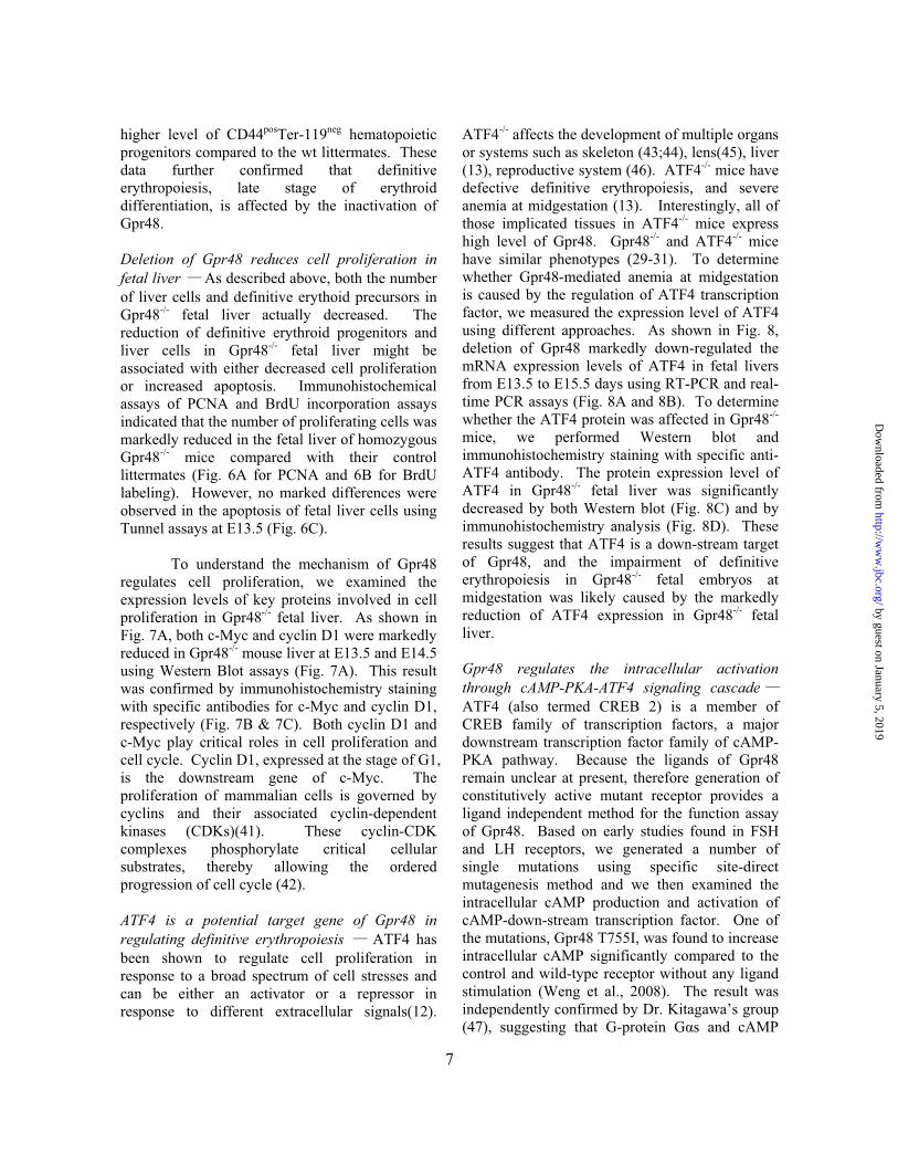
higher level of CD44posTer-119neg hematopoietic progenitors compared to the wt littermates. These data further confirmed that definitive erythropoiesis, late stage of erythroid differentiation, is affected by the inactivation of Gpr48. Deletion of Gpr48 reduces cell proliferation in fetal liver As described above, both the number of liver cells and definitive erythoid precursors in Gpr48-/- fetal liver actually decreased. The reduction of definitive erythroid progenitors and liver cells in Gpr48-/- fetal liver might be associated with either decreased cell proliferation or increased apoptosis. Immunohistochemical assays of PCNA and BrdU incorporation assays indicated that the number of proliferating cells was markedly reduced in the fetal liver of homozygous Gpr48-/- mice compared with their control littermates (Fig. 6A for PCNA and 6B for BrdU labeling). However, no marked differences were observed in the apoptosis of fetal liver cells using Tunnel assays at E13.5 (Fig. 6C).
To understand the mechanism of Gpr48 regulates cell proliferation, we examined the expression levels of key proteins involved in cell proliferation in Gpr48-/- fetal liver. As shown in Fig. 7A, both c-Myc and cyclin D1 were markedly reduced in Gpr48-/- mouse liver at E13.5 and E14.5 using Western Blot assays (Fig. 7A). This result was confirmed by immunohistochemistry staining with specific antibodies for c-Myc and cyclin D1, respectively (Fig. 7B & 7C). Both cyclin D1 and c-Myc play critical roles in cell proliferation and cell cycle. Cyclin D1, expressed at the stage of G1, is the downstream gene of c-Myc. The proliferation of mammalian cells is governed by cyclins and their associated cyclin-dependent kinases (CDKs)(41). These cyclin-CDK complexes phosphorylate critical cellular substrates, thereby allowing the ordered progression of cell cycle (42). ATF4 is a potential target gene of Gpr48 in regulating definitive erythropoiesis ATF4 has been shown to regulate cell proliferation in response to a broad spectrum of cell stresses and can be either an activator or a repressor in response to different extracellular signals(12).
ATF4-/- affects the development of multiple organs or systems such as skeleton (43;44), lens(45), liver (13), reproductive system (46). ATF4-/- mice have defective definitive erythropoiesis, and severe anemia at midgestation (13). Interestingly, all of those implicated tissues in ATF4-/- mice express high level of Gpr48. Gpr48-/- and ATF4-/- mice have similar phenotypes (29-31). To determine whether Gpr48-mediated anemia at midgestation is caused by the regulation of ATF4 transcription factor, we measured the expression level of ATF4 using different approaches. As shown in Fig. 8, deletion of Gpr48 markedly down-regulated the mRNA expression levels of ATF4 in fetal livers from E13.5 to E15.5 days using RT-PCR and real-time PCR assays (Fig. 8A and 8B). To determine whether the ATF4 protein was affected in Gpr48-/- mice, we performed Western blot and immunohistochemistry staining with specific anti-ATF4 antibody. The protein expression level of ATF4 in Gpr48-/- fetal liver was significantly decreased by both Western blot (Fig. 8C) and by immunohistochemistry analysis (Fig. 8D). These results suggest that ATF4 is a down-stream target of Gpr48, and the impairment of definitive erythropoiesis in Gpr48-/- fetal embryos at midgestation was likely caused by the markedly reduction of ATF4 expression in Gpr48-/- fetal liver. Gpr48 regulates the intracellular activation through cAMP-PKA-ATF4 signaling cascadeATF4 (also termed CREB 2) is a member of CREB family of transcription factors, a major downstream transcription factor family of cAMP-PKA pathway. Because the ligands of Gpr48 remain unclear at present, therefore generation of constitutively active mutant receptor provides a ligand independent method for the function assay of Gpr48. Based on early studies found in FSH and LH receptors, we generated a number of single mutations using specific site-direct mutagenesis method and we then examined the intracellular cAMP production and activation of cAMP-down-stream transcription factor. One of the mutations, Gpr48 T755I, was found to increase intracellular cAMP significantly compared to the control and wild-type receptor without any ligand stimulation (Weng et al., 2008). The result was independently confirmed by Dr. Kitagawa’s group (47), suggesting that G-protein G�s and cAMP
7
by guest on January 5, 2019http://w
ww
.jbc.org/D
ownloaded from

pathways are coupled to Gpr48. To further explore the molecular mechanism how Gpr48 regulates ATF4, we examined the proximal promoter region of ATF4 promoter and found a semi-CRE binding site CGTCA (at position -921 relative to the transcription start site) for potential binding and activation of the ATF4 gene by the CREB transcription factor and cAMP-PKA signaling pathway. To confirm that Gpr48-mediated cAMP-CREB pathways regulate the promoter activity of ATF4, we cotransfected the constitutively active mutant Gpr48 (T755I) and a 1.2 kb ATF4 promoter-luciferase reporter gene into the cells, and then examined the direct activation of ATF4 by Gpr48 and cAMP pathways. As shown in Fig. 8E, Gpr48 active mutant receptor (T755I) significantly increased the intracellular cAMP production. The activation of cAMP level by Gpr48 active receptor was significantly inhibited by adding PKI, a competitive PKA inhibitor (Fig. 8E). In order to further determine whether Gpr48 regulates ATF4 through PKA, we also used another PKA inhibitor, H89, a chemical PKA inhibitor, to measure ATF4 promoter activation mediated by the Gpr48-cAMP-PKA pathway. As shown in Fig. 8E, H89 successfully inhibited the activation of ATF4 promoter activated by Gpr48 active mutant receptor (T755I) (Fig. 8E). These data indicate that ATF4 was the direct downstream target gene of Gpr48-mediated cAMP-PKA-CREB signaling pathway.
DISCUSSION
The present study demonstrated that inactivation of Gpr48 (LGR4) lead to impairment of definitive erythropoiesis at midgestation in fetal liver. Using gene trapped approach described previously (36), we constructed the Gpr48 inactivation model by inserting a secretary-trap vector (11.98kb) at intron 1 of wild-type Gpr48. Ablation of Gpr48 resulted in the impairment of definitive erythropoiesis at midgestation, which is associated with the down-regulation in the expression levels of c-Myc, cyclin D1, and ATF4 through cAMP-PKA-CREB signaling pathway.
Gpr48 is expressed in a wide range of
tissues, including skeleton, cartilage, ovary, testis, adrenal, thyroid, kidney, heart, and liver
(19;27;29), suggesting that Gpr48 is involved in the developmental regulation of multiple organs including fetal liver. The facts that Gpr48 is highly expressed in the fetal liver and in the membrane of premature erythroblast at midgestation indicate that Gpr48 is involved in the development of definitive erythropoiesis. Not only the body size was affected in Gpr48-/- mice, fetal livers at midgestation during E13.5-E15.5 also displayed much smaller and paler phenotype in Gpr48 mutant mice than those in normal control mice. Fetal liver is the predominant site for development of definitive erythropoiesis during midgestation after E12.5, so the deficient fetal liver in Gpr48-/- mice suggests that Gpr48 play a key role for the development of definitive erythropoiesis. Erythropoiesis includes two sequential process termed primitive erythropoiesis and definitive erythropoiesis respectively. Our date indicated that the silencing of Gpr48 had no evident effects on primitive erythropoiesis, but dramatically reduced definitive erythropoiesis. Based on different characteristics between definitive erythropoiesis and primitive erythropoiesis (2;16), we counted the nucleated erythrocytes (primitive erythrocyte) and enucleated erythrocytes (definitive erythrocytes) from blood smear and distinguish these two populations by real-time PCR arrays for different globin chains. The elevated level of nucleated erythrocytes indicated that more primitive erythrocytes in the circulation of Gpr48-/- mice, which was consistent with the result of real-time PCR analysis for globin chains (�, �H1, and �y). However, adult � and � globin chains was decreased markedly, indicating reduced definitive erythrocytes in Gpr48-/- mice. Examinations of the hemoglobin chain suggest that the relative increased number of nucleated erythrocytes in homozygous mutants resulted from persistence of primitive yolk sac-derived erythrocytes rather than the premature release of nucleated definitive erythrocytes into circulation due to stress erythropoiesis.
ATF4, also termed CREB2, is a member of the ATF/CREB ubiquitous basic leucine zipper (bZip) transcription factors. ATF4 is essential for cell proliferation (13;43), especially for the processes that require high-level proliferation, such as fetal liver hematopoiesis (13), bone
8
by guest on January 5, 2019http://w
ww
.jbc.org/D
ownloaded from

9
development (45), lens development (44), and human cancer cells (48). ATF4-/- mice showed severe anemia due to impairment of definitive erythropoiesis during midgestation, indicating that ATF4 play a pivotal role for definitive fetal-liver erythropoiesis under stress response (13). Similar to ATF4-/- mice, GPR48-/- fetal livers also have elevated nucleated primitive erythrocytes relative to enucleated definitive erythrocytes without morphologically changes of primitive erythrocytes. Similarities between ATF4-/- mice and GPR48-/- mice are also observed in bone and lens developments (unpublished data), suggesting a correlation between GPR48 and ATF4. Here we provide evidences that Gpr48 regulates the expression level of ATF4 through cAMP-PKA-CREB signaling pathway. As a critical transcription factor in response to hypoxia, ATF4 activates its downstream gene expression including CHOP and TRB3 (49;50). These genes are stress-induced factors and collaborate to modulate a complex regulation network for fulfilling a fine regulation under conditions of Integrated Stress Response (IRS) or erythropoiesis (50-52). Furthermore, the pathway of signaling transduction for ATF4-CHOP-TRB3 might be independent of HIF-1� (49), a critical factor to mediate hematopoiesis through inducing EPO pathway.
One of the most important changes in erythroid development is the ongoing maturation and the loss of proliferative capacity in favor of the differentiation program. The balance between erythropoietin (Epo)-mediated erythroid differentiation and stem cell factor (SCF)-mediated proliferation are strictly regulated during erythropoiesis. In the presence of Epo, Signal transducer and activator of transcription 5 (STAT5) is recruited to the Epo receptor and phosphorylated by the Janus kinase 2 (JAK2). Once phosphorylated, STAT5 is released from EpoR, homodimerizes in the cytosol and translocates to the nucleus where it activates its target gene expression including Bcl-X and cyclin D1(53-58). Phosphorylation of CREB by PKA enhances Epo-stimulated STAT5 transactivation
by inducing recruitment of CREB/CBP/p300 to the STAT5 transactivational complex (58;59). Inactivation of Gpr48 caused dramatic decrease in the proliferation of definitive erythroid progenitors and erythroblast islands in fetal liver. Moreover, analyses of key proliferation genes demonstrate that both c-Myc and cyclin D1 were markedly downregulated in Gpr48-/- fetal liver. This data suggest that Gpr48 also affect definitive erythropoiesis by regulating Epo-mediated erythroid differentiation through cAMP-PKA-CREB pathway.
The erythropoiesis of Gpr48 -/- embryos was mainly affected during E12.5-E15.5 with E13.5 as the most severe stage. After E15.5, the anemia phenotype in blood smear of Gpr48-/- mice looked gradually normal, suggesting that the defect of definitive erythropoiesis in Gpr48 homozygous mice is a transient process at midgestation. Other factors might serve as compensatory regulators for Gpr48-/- embryos surviving through midgestation. These compensatory genes might be expressed a bit later than ATF4 during the onset of fetal-liver hematopoiesis to coordinate with ATF4 and to regulate hematopoiesis under Gpr48 ablation.
In summary, Gpr48 ablation resulted in impaired fetal-liver definitive erythropoiesis at midgestation. Reduced population of definitive erythrocytes was associated with decreased proliferation and down regulation of c-Myc and cyclin D1 in Gpr48-/- fetal liver. These results suggest that Gpr48 is a critical G-protein coupled receptor to regulate fetal-liver erythropoiesis during midgestation stage through intracellular signaling pathways coupled to the regulation of ATF4. Acknowledgement
This work is partially supported by the NIH grants (5R01HL064792 and 1R01CA106479) to Mingyao Liu. Gpr48 gene trap ES cell clone (LST020) was obtained from Bay Genomics at MMRRC of University of California, Davis.
by guest on January 5, 2019http://w
ww
.jbc.org/D
ownloaded from

REFERENCES 1. Socolovsky, M., Fallon, A. E., Wang, S., Brugnara, C., and Lodish, H. F. (1999) Cell 98, 181-191 2. Godin, I. and Cumano, A. (2002) Nat. Rev. Immunol. 2, 593-604 3. Palis, J., Robertson, S., Kennedy, M., Wall, C., and Keller, G. (1999) Development 126, 5073-5084 4. Perry, C. and Soreq, H. (2002) Eur. J. Biochem. 269, 3607-3618 5. Cantor, A. B. and Orkin, S. H. (2002) Oncogene 21, 3368-3376 6. Kawane, K., Fukuyama, H., Kondoh, G., Takeda, J., Ohsawa, Y., Uchiyama, Y., and Nagata, S. (2001) Science
292, 1546-1549 7. Dumitriu, B., Patrick, M. R., Petschek, J. P., Cherukuri, S., Klingmuller, U., Fox, P. L., and Lefebvre, V. (2006)
Blood 108, 1198-1207 8. Hodge, D., Coghill, E., Keys, J., Maguire, T., Hartmann, B., McDowall, A., Weiss, M., Grimmond, S., and
Perkins, A. (2006) Blood 107, 3359-3370 9. Iavarone, A., King, E. R., Dai, X. M., Leone, G., Stanley, E. R., and Lasorella, A. (2004) Nature 432, 1040-1045 10. Neubauer, H., Cumano, A., Muller, M., Wu, H., Huffstadt, U., and Pfeffer, K. (1998) Cell 93, 397-409 11. Socolovsky, M., Nam, H., Fleming, M. D., Haase, V. H., Brugnara, C., and Lodish, H. F. (2001) Blood 98,
3261-3273 12. Hai, T. and Hartman, M. G. (2001) Gene 273, 1-11 13. Masuoka, H. C. and Townes, T. M. (2002) Blood 99, 736-745 14. Munugalavadla, V. and Kapur, R. (2005) Crit Rev. Oncol. Hematol. 54, 63-75 15. Marchese, A., George, S. R., Kolakowski, L. F., Jr., Lynch, K. R., and O'Dowd, B. F. (1999) Trends Pharmacol.
Sci. 20, 370-375 16. Hill, S. J. (2006) Br. J. Pharmacol. 147 Suppl 1, S27-S37 17. Palczewski, K., Kumasaka, T., Hori, T., Behnke, C. A., Motoshima, H., Fox, B. A., Le, T., I, Teller, D. C.,
Okada, T., Stenkamp, R. E., Yamamoto, M., and Miyano, M. (2000) Science 289, 739-745 18. Iiri, T., Farfel, Z., and Bourne, H. R. (1998) Nature 394, 35-38 19. Hsu, S. Y., Liang, S. G., and Hsueh, A. J. (1998) Mol. Endocrinol. 12, 1830-1845 20. Osuga, Y., Kudo, M., Kaipia, A., Kobilka, B., and Hsueh, A. J. (1997) Mol. Endocrinol. 11, 1659-1668 21. Kobe, B. and Kajava, A. V. (2001) Curr. Opin. Struct. Biol. 11, 725-732 22. Hsu, S. Y. (2003) Trends Endocrinol. Metab 14, 303-309 23. Fredriksson, R., Lagerstrom, M. C., Lundin, L. G., and Schioth, H. B. (2003) Mol. Pharmacol. 63, 1256-1272 24. Hsu, S. Y., Nakabayashi, K., Nishi, S., Kumagai, J., Kudo, M., Sherwood, O. D., and Hsueh, A. J. (2002)
Science 295, 671-674 25. Hsu, S. Y., Kudo, M., Chen, T., Nakabayashi, K., Bhalla, A., van der Spek, P. J., van, D. M., and Hsueh, A. J.
(2000) Mol. Endocrinol. 14, 1257-1271 26. Kajava, A. V. (1998) J. Mol. Biol. 277, 519-527 27. Loh, E. D., Broussard, S. R., and Kolakowski, L. F. (2001) Biochem. Biophys. Res. Commun. 282, 757-764 28. Loh, E. D., Broussard, S. R., Liu, Q., Copeland, N. G., Gilbert, D. J., Jenkins, N. A., and Kolakowski, L. F., Jr.
(2000) Cytogenet. Cell Genet. 89, 2-5 29. Van, S. G., Mendive, F., Pochet, R., and Vassart, G. (2005) Histochem. Cell Biol. 124, 35-50 30. Hoshii, T., Takeo, T., Nakagata, N., Takeya, M., Araki, K., and Yamamura, K. (2007) Biol. Reprod. 76, 303-313 31. Kato, S., Matsubara, M., Matsuo, T., Mohri, Y., Kazama, I., Hatano, R., Umezawa, A., and Nishimori, K. (2006)
Nephron Exp. Nephrol. 104, e63-e75 32. Mendive, F., Laurent, P., Van, S. G., Skarnes, W., Pochet, R., and Vassart, G. (2006) Dev. Biol. 290, 421-434 33. Mazerbourg, S., Bouley, D. M., Sudo, S., Klein, C. A., Zhang, J. V., Kawamura, K., Goodrich, L. V., Rayburn,
H., Tessier-Lavigne, M., and Hsueh, A. J. (2004) Mol. Endocrinol. 18, 2241-2254 34. Stryke, D., Kawamoto, M., Huang, C. C., Johns, S. J., King, L. A., Harper, C. A., Meng, E. C., Lee, R. E., Yee,
A., L'Italien, L., Chuang, P. T., Young, S. G., Skarnes, W. C., Babbitt, P. C., and Ferrin, T. E. (2003) Nucleic Acids Res. 31, 278-281
35. Skarnes, W. C., von, M. H., Wurst, W., Hicks, G., Nord, A. S., Cox, T., Young, S. G., Ruiz, P., Soriano, P., Tessier-Lavigne, M., Conklin, B. R., Stanford, W. L., and Rossant, J. (2004) Nat. Genet. 36, 543-544
36. Leighton, P. A., Mitchell, K. J., Goodrich, L. V., Lu, X., Pinson, K., Scherz, P., Skarnes, W. C., and Tessier-Lavigne, M. (2001) Nature 410, 174-179
37. Mitchell, D. C., Abdelrahim, M., Weng, J., Stafford, L. J., Safe, S., Bar-Eli, M., and Liu, M. (2006) J. Biol. Chem. 281, 51-58
38. McGrath, K. E. and Palis, J. (2005) Exp. Hematol. 33, 1021-1028
10
by guest on January 5, 2019http://w
ww
.jbc.org/D
ownloaded from

39. Samakoglu, S., Fattori, E., Lamartina, S., Toniatti, C., Stockholm, D., Heard, J. M., and Bohl, D. (2001) Blood 97, 2213-2220
40. Basu, P., Morris, P. E., Haar, J. L., Wani, M. A., Lingrel, J. B., Gaensler, K. M., and Lloyd, J. A. (2005) Blood 106, 2566-2571
41. Kozar, K., Ciemerych, M. A., Rebel, V. I., Shigematsu, H., Zagozdzon, A., Sicinska, E., Geng, Y., Yu, Q., Bhattacharya, S., Bronson, R. T., Akashi, K., and Sicinski, P. (2004) Cell 118, 477-491
42. Bjorklund, M., Taipale, M., Varjosalo, M., Saharinen, J., Lahdenpera, J., and Taipale, J. (2006) Nature 439, 1009-1013
43. Elefteriou, F., Benson, M. D., Sowa, H., Starbuck, M., Liu, X., Ron, D., Parada, L. F., and Karsenty, G. (2006) Cell Metab 4, 441-451
44. Yang, X., Matsuda, K., Bialek, P., Jacquot, S., Masuoka, H. C., Schinke, T., Li, L., Brancorsini, S., Sassone-Corsi, P., Townes, T. M., Hanauer, A., and Karsenty, G. (2004) Cell 117, 387-398
45. Tanaka, T., Tsujimura, T., Takeda, K., Sugihara, A., Maekawa, A., Terada, N., Yoshida, N., and Akira, S. (1998) Genes Cells 3, 801-810
46. Fischer, C., Johnson, J., Stillwell, B., Conner, J., Cerovac, Z., Wilson-Rawls, J., and Rawls, A. (2004) Biol.Reprod. 70, 371-378
47. Gao, Y., Kitagawa, K., Shimada, M., Uchida, C., Hattori, T., Oda, T., and Kitagawa, M. (2006) Hokkaido Igaku Zasshi 81, 101-5, 107, 109
48. Ameri, K., Lewis, C. E., Raida, M., Sowter, H., Hai, T., and Harris, A. L. (2004) Blood 103, 1876-1882 49. Blais, J. D., Filipenko, V., Bi, M., Harding, H. P., Ron, D., Koumenis, C., Wouters, B. G., and Bell, J. C. (2004)
Mol. Cell Biol. 24, 7469-7482 50. Ohoka, N., Yoshii, S., Hattori, T., Onozaki, K., and Hayashi, H. (2005) EMBO J. 24, 1243-1255 51. Jousse, C., Deval, C., Maurin, A. C., Parry, L., Cherasse, Y., Chaveroux, C., Lefloch, R., Lenormand, P., Bruhat,
A., and Fafournoux, P. (2007) J. Biol. Chem. 282, 15851-15861 52. Cui, K., Coutts, M., Stahl, J., and Sytkowski, A. J. (2000) J. Biol. Chem. 275, 7591-7596 53. Grimley, P. M., Dong, F., and Rui, H. (1999) Cytokine Growth Factor Rev. 10, 131-157 54. Damen, J. E., Wakao, H., Miyajima, A., Krosl, J., Humphries, R. K., Cutler, R. L., and Krystal, G. (1995)
EMBO J. 14, 5557-5568 55. Gobert, S., Chretien, S., Gouilleux, F., Muller, O., Pallard, C., Dusanter-Fourt, I., Groner, B., Lacombe, C.,
Gisselbrecht, S., and Mayeux, P. (1996) EMBO J. 15, 2434-2441 56. Quelle, F. W., Wang, D., Nosaka, T., Thierfelder, W. E., Stravopodis, D., Weinstein, Y., and Ihle, J. N. (1996)
Mol. Cell Biol. 16, 1622-1631 57. Klingmuller, U., Bergelson, S., Hsiao, J. G., and Lodish, H. F. (1996) Proc. Natl. Acad. Sci. U. S. A 93, 8324-
8328 58. Boer, A. K., Drayer, A. L., Rui, H., and Vellenga, E. (2002) Blood 100, 467-473 59. Boer, A. K., Drayer, A. L., and Vellenga, E. (2003) Exp. Hematol. 31, 512-520 60. Weng J, Luo J, Cheng X, Jin C, Zhou X, Qu J, Tu L, Ai D, Li D, Wang J, Martin JF, Amendt BA, Liu M.
(2008) Proc Natl Acad Sci U S A. 105, 6081-6. Epub 2008 Apr 18.
11
by guest on January 5, 2019http://w
ww
.jbc.org/D
ownloaded from

FIGURE LEGENDS Figure 1. Expression of Gpr48 in fetal liver. A. RT-PCR analysis of Gpr48 mRNA levels from E13.5 Gpr48 wild-type (+/+), heterozygous (+/–), and homozygous (–/–) fetal livers, showing no Gpr48 expression in Gpr48-/- fetal liver. B-C. LacZ staining of E15.5 Gpr48 wild-type (+/+), heterozygous (+/-) and homozygous (–/–) fetal liver sections. Gpr48+/- and Gpr48-/- fetal liver showed positive staining in liver (upper right panel, B, magnification 100×) and at cell surface membrane (lower right panel, C, magnification 1000×). Sections were counterstained with eosin. Figure 2. Defects of Gpr48–/– mice in body size and fetal liver. A. Gpr48-/- fetal mice at E14.5 showed significantly decreased liver weight and the ratio of liver weight / body weight. (a) Gpr48-/- fetal liver weight was decreased about 1.68 fold compared with their wild type littermates (p<0.001). (b) The ratio of liver weight/body weight was decreased about 1.25 fold in Gpr48-/- mice compared with wild-type littermates (p<0.02). B. Gpr48-/- mice embryos and livers (right) during E12.5-15.5 are paler and smaller than wild-type. E16.5 Gpr48-/- embryos show smaller in size but similar in color compared with wild-type fetuses. Figure 3. Significant decrease of erythroid precursor cells and erythroblast islands in Gpr48-/- fetal liver by histological analysis. Gpr48–/– fetal liver showed significant decrease of definitive erythroid precursor cells and erythroblast islands from E12.5 to E15.5 (A-D). Erythropoietic precursor cells are indicated by yellow arrows, hepatocytes by green arrows, and erythroblast islands by orange arrows (Magnification 1000 ×). Figure 4. Increased nucleated erythrocytes at midgestation of Gpr48-/- mice. A. Blood smears from E13.5-16.5 embryos were stained with Wright-Giemsa stain. Gpr48–/–embryos (right) of E13.5-15.5 showed increased percentage of nucleated erythrocytes compared with the samples from their wild-type littermates (left), but no significant difference from E16.5 samples between Gpr48-/- and their wild-type littermates (magnification 1000×). B. Relative number in percentage of nucleated erythrocytes from blood smear slides. E13.5 and E14.5 Gpr48–/– embryos showed about 2.5-fold and1.8-fold increase in nucleated erythrocytes percentage compared to wild-type respectively(p<0.03). E12.5 and E16.5 littermates showed little differences in percentage of nucleated erythrocytes between wild-type and null embryos (p>0.05). C-D. Real-time PCR of embryonic hemoglobin chains (�, �h1, and �y) in E13.5 blood cell samples (C) and adult hemoglobin chains (� and �) in E13.5 livers (D). E. The cell size of erythrocytes was reduced by Gpr48 inactivation in E13.5 fetal livers.� Ter119pos cells in Gpr48-/- mice bone marrow displayed a reduction in cell size compared to those in their WT littermates (x-axis represents the cell size). Green in the histogram represents Ter119 positive cells (mature red blood cells). Similar reduction of red blood cell size was observed from adult Gpr48-/- cells. However, the cell number did not show significant decrease in Gpr48-/- null mice.
Figure 5. Flow cytometry analysis of fetal liver hematopoietic cells from E13.5 Gpr48+/+ and Gpr48–/– embryos. Cell suspensions from E13.5 fetal livers were labeled with anti-c-kit, anti-CD34, anti-CD44, or anti-Ter119 specific antibody. The populations of Ter119 positive cells which represent definitive erythrocytes in Gpr48–/– embryos were markedly decreased. Figure 6. Reduced cell proliferation in Gpr48-/- fetal liver. A. E13.5 Gpr48-/- fetal liver showed dramatically decreased number of positive cells (dark brown) compared with wild-type sections using PCNA by immunohistochemistry. Hematoxylin stain was performed as counterstain (magnification 1000×). B. Proliferation assays with BrdU labeling demonstrated that Gpr48-/- E13.5 fatal liver have marked decrease in the number of proliferating cells (BrdU positive sells, brown color). Sections were counterstained by methylene green (magnification 1000X). C. Tunnel assays of E13.5 fetal liver showed
12
by guest on January 5, 2019http://w
ww
.jbc.org/D
ownloaded from

13
no significant differences in the number of positive cells (brown) between Gpr48-/- (left pannel) and Gpr48+/+ fetal livers. Sections were counterstained by methylene green (magnification 1000X). Figure 7. Down-regulation of c-Myc and Cyclin D1 in Gpr48-/- fetal liver. A. Western blot analysis showed that protein levels of c-Myc and Cyclin D1 were both decreased in Gpr48-/- fetal livers compared with that in the wild-type. Actin was used as an internal control. B. Fetal liver sections from E13.5 Gpr48+/+ and Gpr48-/- embryos were stained with anti-c-Myc antibody. Significant decrease of c-Myc protein level was observed in Gpr48-/- fetal liver (brown color, magnification 1000×). C. Decreased protein levels of Cyclin D1 in Gpr48-/- fetal liver compared to wild-type. Fetal liver sections from E13.5 Gpr48+/+ and Gpr48-/- embryos were stained with anti- Cyclin D1 antibody (brown color, magnification 1000×).
Figure 8. Down-regulation of ATF4 in Gpr48-/- fetal liver. A. RT-PCR showed that ATF4 mRNA was dramatically decreased in Gpr48-/- livers during E13.5-E15.5, GAPDH was used as an internal control. B. Real-time PCR showed significantly reduced mRNA levels of ATF4 in Gpr48-/- livers during E13.5-E15.5, �-Actin used as an internal control. C. The protein expression level of ATF4 was significantly decreased at E13.5, E14.5 and E15.5 in Gpr48-/- livers by Western blot. Actin was used as an internal control. D. The expression level of ATF4 was markedly reduced in E13.5 Gpr48-/- fetal livers by immunocytochemistry staining. Sections were stained with anti-ATF4 specific antibody. The number of positive cells (brown) was markedly reduced compared to wild-type. Hematoxylin was used as counterstain (1000×). E. Gpr48 regulates ATF4 promoter through cAMP-PKA-CREB pathway. PKA inhibitor PKI strongly inactivated the ATF4 1.2k promoter activity induced by a constitutively activated mutant Gpr48 (T755I). The activation of ATF4 by the active Gpr48 mutant was also markedly inhibited by PKA specific inhibitor H89, suggesting Gpr48 activates ATF4 through activation of PKA pathway.
by guest on January 5, 2019http://w
ww
.jbc.org/D
ownloaded from

Song et al. FIG.1.
+/+ +/- -/-
Gpr48
A
B
GAPDH
Gpr48
+/-+/+ -/-B
C+/+
-/-
by guest on January 5, 2019http://w
ww
.jbc.org/D
ownloaded from

Song et al. FIG. 2
A
ght
(a) (b)
r wig
ht (m
g)
liver
/bod
y w
eig
40
60
80
100
10
15
20
25
(a) (b)
**
Live
r
Rat
io o
f
+/+ +/- -/-(n=11)(n=23)(n=11)
+/+ +/- -/-(n=11)(n=23)(n=11)
0
20
0
5
+/+ -/-
+/+ -/-
B
E12.5
E15.5
E13.5
E14.5
E16.5
by guest on January 5, 2019http://w
ww
.jbc.org/D
ownloaded from

+/+ -/-
Song et al. FIG. 3
A(E12 5)(E12.5)
B(E13.5)
CC(E14.5)
D(E15 5)(E15.5)
by guest on January 5, 2019http://w
ww
.jbc.org/D
ownloaded from

+/+ -/-
E13.5
Song et al. FIG. 4A
B
E14.5
ucle
ated
ery
thro
cyte
40
60
80
100
120WT HO
E15.5
Rel
ativ
e N
u
0
20
40
E16.5
RN
A le
vel
C
80
100
120
140WT HO
E WT
r119
� �h1 �y
Rel
ativ
e m
0
20
40
60
Gpr48-/-
Te
ve m
RN
A le
vel
D
Ter1
19
� �
Rel
ativ
by guest on January 5, 2019http://w
ww
.jbc.org/D
ownloaded from

8.1 2.3 6.6 2.2
Song et al. FIG. 5
89.3 0.2 0.390.8
+/+
11.6 3.0 9.9 2.1
-/-
0.285.2
CD34
CD
44
1.186.8
CD34
C-K
it
CD34 CD34
2.5 7.0 8.1 0.6
69.521.0 71.819.5
+/+
3.6 7.0
C
1.315.9
-/-
47.042.4
Ter119
C-kit
27.4 55.3
Ter119
CD
44
by guest on January 5, 2019http://w
ww
.jbc.org/D
ownloaded from

A
Song et al. FIG. 6
+/+ -/-
ositi
ve c
ells PCNA staining
**
No.
of p
o
WT HO
B
ositi
ve c
ells BrdU labeling
**
+/+ -/-
C
No.
of p
o
WT HO
CTunnel assays
ositi
ve c
ells
+/+ -/-
WT HO
No.
of p
o
by guest on January 5, 2019http://w
ww
.jbc.org/D
ownloaded from

Song et al. FIG. 7
A+/+ / +/+ /
C-Myc
Cyclin D1
+/+ -/- +/+ -/-
Actin
E13.5 E14.5
B Gpr48+/+ Gpr48-/-
c-Myc
CCyclin D1
by guest on January 5, 2019http://w
ww
.jbc.org/D
ownloaded from

B250
A
Song et al. FIG. 8
ativ
e m
RN
A le
vel
50
100
150
200
250WTHO
+/+ -/- +/+ -/- +/+
E13.5 E14.5 E15.5
-/-
ATF4
E13.5 E14.5 E15.5CR
ela
E13.5 E14.5 E15.50GAPDH
ATF4
A ti
+/+ -/- +/+ -/- +/+ -/-
Actin
DE
Gpr48+/+
eac
tivity
3
4
51.2 k ATF4-Luc
ange
)
Gpr48-/-
pcDNA31 Gpr48 T755I PKI
Luci
fera
se
T755I
0
1
2
(Fol
d ch
pcDNA3.1 Gpr48 T755I PKIT755I
+H89
by guest on January 5, 2019http://w
ww
.jbc.org/D
ownloaded from

and Mingyao LiuHuiping Song, Jian Luo, Weijia Luo, Jinsheng Weng, Zhiqing Wang, Baoxing Li, Dali Lierythropoiesis at midgestation through downregulation of ATF4 signaling pathway
Inactivation of G-protein coupled receptor 48 (Gpr48/Lgr4) impairs definitive
published online October 27, 2008J. Biol. Chem.
10.1074/jbc.M800721200Access the most updated version of this article at doi:
Alerts:
When a correction for this article is posted•
When this article is cited•
to choose from all of JBC's e-mail alertsClick here
Supplemental material:
http://www.jbc.org/content/suppl/2008/10/27/M800721200.DC1
by guest on January 5, 2019http://w
ww
.jbc.org/D
ownloaded from