Embed Size (px)
Citation preview
City University of New York (CUNY) City University of New York (CUNY)
CUNY Academic Works CUNY Academic Works
Publications and Research CUNY Graduate Center
2015
Glycine and GABAA receptors mediate tonic and phasic inhibitory Glycine and GABAA receptors mediate tonic and phasic inhibitory
processes that contribute to prepulse inhibition in the goldfish processes that contribute to prepulse inhibition in the goldfish
startle network startle network
Paul C.P. Curtin CUNY Graduate Center
Thomas Preuss CUNY Hunter College
How does access to this work benefit you? Let us know!
More information about this work at: https://academicworks.cuny.edu/gc_pubs/330
Discover additional works at: https://academicworks.cuny.edu
This work is made publicly available by the City University of New York (CUNY). Contact: [email protected]
ORIGINAL RESEARCHpublished: 24 March 2015
doi: 10.3389/fncir.2015.00012
Glycine and GABAA receptorsmediate tonic and phasic inhibitoryprocesses that contribute to prepulseinhibition in the goldfish startlenetworkPaul C. P. Curtin 1* and Thomas Preuss 2*
1 Graduate Center, City University of New York, New York, NY, USA, 2 Hunter College, City University of New York,New York, NY, USA
Edited by:Takao K. Hensch,
Harvard University, USA
Reviewed by:Alan Roberts,
University of Bristol, UKSusanne Schmid,
University of Western Ontario,Canada
*Correspondence:Paul C. P. Curtin, Graduate Center,
City University of New York, 365 5thAve, New York, NY 10016, USA
[email protected];Thomas Preuss, Hunter College, CityUniversity of New York, 695 Park Ave,
New York, NY 10065, [email protected]
Received: 16 November 2014Accepted: 04 March 2015Published: 24 March 2015
Citation:Curtin PCP and Preuss T (2015)
Glycine and GABAA receptorsmediate tonic and phasic inhibitory
processes that contribute to prepulseinhibition in the goldfish
startle network.Front. Neural Circuits 9:12.
doi: 10.3389/fncir.2015.00012
Prepulse inhibition (PPI) is understood as a sensorimotor gating process that attenuatessensory flow to the startle pathway during early stages (20--1000 ms) of informationprocessing. Here, we applied in vivo electrophysiology and pharmacology to determineif PPI is mediated by glycine receptors (GlyRs) and/or GABAA receptors (GABAARs)in the goldfish auditory startle circuit. Specifically, we used selective antagonists todissect the contributions of target receptors on sound-evoked postsynaptic potentials(PSPs) recorded in the neurons that initiate startle, the Mauthner-cells (M-cell). Wefound that strychnine, a GlyR antagonist, disrupted a fast-activated (5 ms) and rapidly(<50 ms) decaying (feed-forward) inhibitory process that contributes to PPI at 20 msprepulse/pulse inter-stimulus intervals (ISI). Additionally we observed increases of theevoked postsynaptic potential (PSP) peak amplitude (+87.43 ± 21.53%, N = 9) andduration (+204 ± 48.91%, N = 9). In contrast, treatment with bicuculline, a GABAARantagonist, caused a general reduction in PPI across all tested interstimulus intervals(ISIs) (20--500 ms). Bicuculline also increased PSP peak amplitude (+133.8 ± 10.3%,N = 5) and PSP duration (+284.95 ± 65.64%, N = 5). Treatment with either antagonistalso tonically increased post-synaptic excitability in the M-cells, reflected by an increasein the magnitude of antidromically-evoked action potentials (APs) by 15.07 ± 3.21%,N = 7 and 16.23 ± 7.08%, N = 5 for strychnine and bicuculline, respectively. Theseresults suggest that GABAARs and GlyRs are functionally segregated to short- andlonger-lasting sound-evoked (phasic) inhibitory processes that contribute to PPI, withthe mediation of tonic inhibition by both receptor systems being critical for gain controlwithin the M-cell startle circuit.
Keywords: sensorimotor integration, Mauthner cell, tonic inhibition, phasic inhibition, sensory processing,prepulse inhibition, auditory startle circuit
Startle is a rapid, massive contraction of facial and skeletal muscles that is triggered by theonset of intense and/or abrupt visual, auditory, or tactile stimuli. Startle is thought to functionas a protective mechanism that minimizes impacts to vulnerable areas (e.g., the eyes) and/orfacilitates collision avoidance or escape (Eaton et al., 1981; Bennett, 1984; Koch, 1999; Yeomanset al., 2002, 2006). The startle response is relatively stereotyped and predictably elicited, but is also
Frontiers in Neural Circuits | www.frontiersin.org 1 March 2015 | Volume 9 | Article 12
Curtin and Preuss GlyRs and GABAARs mediate PPI
the target of multiple modulatory mechanisms that enabledynamic adjustments to stimulus-response parameters invarying sensory and behavioral contexts. PPI (PPI) is a centralinhibitory process that contributes to startle plasticity bybriefly (20--1000 ms) reducing startle excitability while non-startling stimuli (prepulses) are processed (Graham, 1975;Hoffman and Ison, 1980; Koch, 1999). This sensorimotorgating mechanism is thought to preserve sensory processingand action selection by midbrain and forebrain processes thatare activated by prepulses and would be disrupted by thesubsequent initiation of startle (Graham, 1975). Consistentwith this notion, information-processing disorders, includingschizophrenia, Tourette’s syndrome, and obsessive-compulsivedisorder are associated with diminished or disordered PPI(Swerdlow et al., 1992; Parwani et al., 2000; Braff et al.,2001). Consequently, identifying the neural processes underlyingPPI presents an important goal for basic and translationalneuroscience.
Anatomical and pharmacological studies indicate that PPIis mediated by multiple midbrain and forebrain circuits thatmodulate the time-course of inhibition in the startle circuitvia multiple neurotransmitter systems. In mammals, startleis initiated by the firing of a population of giant hindbrainneurons in the ventrocaudal pontine reticular formation(PnC) (Lingenhöhl and Friauf, 1994; Fendt, 1999; Koch,1999; Yeomans et al., 2002, 2006; Geis and Schmid, 2011).Anatomical studies indicate that PPI is produced by theexcitation of midbrain circuits that project inhibitory terminalsto PnC neurons; these include nuclei in the inferior colliculus,pedunculopontine tegmental nucleus, superior colliculus, andlaterodorsal tegmental nucleus (Koch and Schnitzler, 1997;Fendt, 1999; Yeomans et al., 2010). Pharmacological studiesin rodents emphasize that these inputs mediate PPI bymultiple neurotransmitters that contribute discrete componentstoward the time-course of inhibition. Muscarinic receptors,for example, contribute to inhibition mediated at longerintervals, i.e., 100--1000 ms from prepulse onsets (Jones andShannon, 2000; Ukai et al., 2004). GABA receptors are alsocritical mediators of PPI, with GABAA receptors (GABAARs)contributing during the peak inhibitory response, and GABABreceptors adding to the longer lasting inhibition mediatedby muscarinic receptors (Yeomans et al., 2010). These linesof evidence derive from behavioral pharmacology studies inadult animals, or ex vivo slice preparations derived fromimmature tissue (e.g., Yeomans et al., 2010; Geis and Schmid,2011).
The Mauthner-cell (M-cell) circuit in teleost fish presents analternative model system for studying PPI and startle plasticitythat is accessible to in vivo electrophysiology. The M-cells area pair of large reticulospinal neurons, bilaterally opposed, thatintegrate excitatory and inhibitory inputs elicited by visual,auditory, and/or tactile stimulation (reviewed in Eaton et al.,2001; Korn and Faber, 2005). A single action-potential (AP)in either M-cell is sufficient to trigger a startle response(the C-start), and inhibition of APs is sufficient to preventstartle; thus, the M-cells are the decision-making sensorimotorinterface for startle (Eaton et al., 1981). The M-cells are the
focus of two well-characterized inhibitory networks that controlstartle excitability; these being, a collateral (feedback) inhibitorynetwork that is bilaterally activated by cranial relay neurons whenthe M-cell fires, and a commissural (feed-forward) inhibitorynetwork activated by parallel VIIIth nerve afferents to countersound-evoked excitation in the M-cell and thereby regulatestartle response properties (Eaton et al., 2001; Korn and Faber,2005). Glycine receptor (GlyR) antagonists disrupt feedforwardand feedback inhibition (Faber and Korn, 1978, 1987; Korn andFaber, 2005), but GABAARs also mediate M-cell excitability andare thought to be involved in auditory processing (Diamondet al., 1973). These inhibitory networks mediate two distincttypes of processes: phasic inhibition, that includes transientlyactivated or stimulus-dependent inhibitory inputs, including thefeed-forward and feedback inhibitory processes described, andtonic inhibition, that is, persistent inhibitory synaptic noise thatarises from spontaneous quantal neurotransmitter release andintermittent firing at inhibitory synaptic terminals on the M-cell(Faber et al., 1989; Hatta et al., 2001; Marti et al., 2008).
A growing number of studies indicate that PPI in theM-cell system is modulated by multiple pre- and post-synapticmechanisms. Neumeister et al. (2008) showed that PPI ingoldfish is mediated by post-synaptic conductance changesactivated in the M-cells. Burgess and Granato (2007) showed thatdopaminergic agonists disrupt behavioral PPI in zebrafish, whileMedan and Preuss (2011) showed dopaminergic mechanismsmodulating time-specific components of PPI in the M-cellmembrane, likely reflecting control of upstream networksinvolved in PPI. Furthermore, Curtin et al. (2013) showedthat 5-HT5A receptors modulate the excitability of goldfishM-cells, and linked these effects to changes in startle plasticity.Given these advances in our understanding of neuromodulatoryprocesses contributing to startle plasticity, here we investigatedthe signaling mechanisms directly mediating PPI at the level ofthe M-cell.
This study focused on the roles of GlyRs and GABAARsin the mediation of PPI in the M-cells, the decision-makingneurons of the goldfish auditory startle circuit. We targeted thesereceptor systems because they are densely expressed in theM-cellmembrane (Triller et al., 1985; Seitanidou et al., 1988; Petrovet al., 1991; Lee et al., 1993; Sur et al., 1995) and their involvementin a diverse array of tonic and phasic inhibitory processes is wellcharacterized (discussed above; see also Korn and Faber, 2005).We thus sought to identify the effector mechanisms for auditoryPPI in the context of co-activated tonic and phasic inhibitoryprocesses that are typically inaccessible in other model systems.Our findings indicate that GABAARs directly mediate the peakinhibitory components of PPI, while GlyRs indirectly contributeto the onset of PPI by the mediation of a feed-forward inhibitoryprocess that overlaps with the earliest components of PPI.
Materials and Methods
SubjectsSixteen common goldfish (Carassius auratus) of either sexwere used in these experiments. Adult fish 7--13 cm instandard body length were purchased from Hunting Creek
Frontiers in Neural Circuits | www.frontiersin.org 2 March 2015 | Volume 9 | Article 12
Curtin and Preuss GlyRs and GABAARs mediate PPI
Fisheries (Thurmont, MD). Fish were socially housed, with5--6 fish per 60L aquaria, in recirculating conditioned water(7.5 pH; 335 µS; 18C) with a 12:12 light/dark photoperiod.Animals were housed and treated in accord with protocolsestablished by the Institutional Animal Care and UseCommittee (IACUC) of Hunter College, City University ofNew York.
ElectrophysiologyThe surgical techniques and methods used forelectrophysiological recordings were described previously(Medan and Preuss, 2011; Curtin et al., 2013). Fish wereimmersed in icewater for 10--15 min to induce immobility,then placed in a recording chamber. Two steel pins were placedon each side of the head to stabilize the fish and a tube wasplaced in the mouth to provide recirculating aerated watercontaining the general anesthetic, MS-222 (20 mg/l). Thisanesthetic dosage was chosen because prior studies have shownit does not interfere with auditory processing (Palmer andMensinger, 2004; Cordova and Braun, 2007). Recirculatingwater in the recording chamber was initially near 0C (whenfish were fish removed from ice to begin procedures) but wasgradually heated to 18C before recordings were taken. Waterconditions in all other measures were consistent with conditionsin holding tanks.
A small lateral incision was made to expose the spinal cordat the caudal midbody, and a bipolar electrode was placed onthe unopened spine to transmit low intensity (5--10 V) electricalstimulation generated by an isolated stimulator (Digitimer,Ltd, Wewyn Garden City, UK). A visible muscular contraction(twitch) was elicited with spinal stimulation to confirm properplacement of the spinal electrode, then d-tubocurarine (1 µg/gb.w.; Abbot, Chicago, IL) was administered intramuscularly.When the twitch response was abolished, typically within0--3 min of injecting the tubocurarine, a craniotomy wasperformed to expose the medulla for microelectrode placementand recordings. The water level in the recording chamberwas maintained throughout these procedures (and subsequentrecordings) at the height of the mid-body, below the spinalincision.
The M-cell was localized by a characteristic negativeextracellular potential (15--20 mV) generated in the axon capduring antidromic stimulation, which provides an unambiguousindicator of electrodes’ placement relative to the soma andaxon cap (Faber and Korn, 1978). In these experiments, M-cellswere impaled somatically with sharp electrodes (3--8 MΩ) filledwith 5 M potassium acetate (KAc). An Axoprobe-1A amplifier(Molecular Devices, Foster City, CA) in current-clamp modemeasured intracellular potentials, and a data acquisition card(PCI-E, National Instruments, Austin, TX) sampling at 25 KHzin aMacintoshG5 collected and recorded data. Recordings whereresting membrane potential (RMP) varied by more than 10%from initial measure were excluded from analysis.
PharmacologyOur experimental design required the use of GlyR and GABAARantagonists. We chose to use strychnine (Sigma-Aldritch), the
classical GlyR antagonist, and bicuculline (Tocris Biosciences),the classical GABAAR antagonist, because both drugs havelong and well-documented histories of use in the M-cellsystem. Drugs were dissolved in a 500 µL solution ofphysiological saline (in mM: 124.0 NaCl, 5.1 KCl, 2.8 NaH2PO4,0.9 MgSO4, 1.6 CaCl2, 5.6 glucose, and 20.0 HEPES, bufferedto pH 7.2) and superfused directly to the exposed medulla,as in past studies (Diamond et al., 1973; Faber et al., 1991;Pereda et al., 1992, 1994; Hatta et al., 2001). This routeof administration was chosen over others because it alloweddirect reference to past studies and published dose-responsecurves. Given those studies, strychnine solutions (5 mM)were prepared as per Diamond et al. (1973), and bicucullinesolutions (10 mM) were made as per Hatta et al. (2001).Physiological measures confirmed these concentrations weresufficient to achieve clear experimental effects (see SectionResults). Nonetheless, given the 1.5 mm depth of the M-cellfrom the brain surface, and the diffusion volume, we expectthat effective concentrations of strychnine and bicuculline were1--2 orders of magnitude less at the site of the M-cell, followingestimations offered in prior in vivo pharmacological studieswith the M-cell system (Pereda et al., 1992, 1994; Hatta et al.,2001). All solutions were prepared on the day of experimentsand were warmed to the temperature of the fish beforeapplication.
Stimulus ProtocolsSound (pulse) stimuli were used to activate orthodromic inputsto the M-cells with or without a preceding sound stimulus(prepulse), the latter at multiple interstimulus intervals (ISIs)ranging between 20--500 ms. Sound stimuli in all experimentalconditions were 200 Hz single-cycle ‘‘pips’’ produced at80 dB re: 20 µPa in air. This stimulus intensity was chosento elicit subthreshold responses in the M-cell because PPIis by definition elicited by subthreshold sounds, and theuse of an identical conditioning (prepulse) and test (pulse)stimuli allows a within-subjects comparison of prepulse-pulserelationships on a trial-by-trial basis that is less sensitive tochanges in baseline excitability. Stimuli were generated by afunction generator (Agilent 33210A, Santa Clara, CA), andoutput to a shielded subwoofer (SA-WN250, Sony) placed30 cm from the recording chamber. A microphone placed10 cm above the fish’s head recorded acoustic waveformsand encoded these in parallel with intracellular recordings. Ahydrophone (SQ01, Sensor, Collingwood, ON, Canada) wasalso used for sound calibration but was removed duringexperiments. In testing conditions that measured PPI, soundstimuli were produced at 6 inter-stimulus intervals (ISIs: 20,50, 75, 150, 300, 500 ms) measured from the onset of eachstimulus.
Waveform Analysis of Evoked SynapticResponsesIntracellular recordings were analyzed offline with customand commercial software (Igor Pro; Wavemetrics, LakeOswego, OR). To analyze the contribution of distinctinhibitory networks on sensory processing, we measured
Frontiers in Neural Circuits | www.frontiersin.org 3 March 2015 | Volume 9 | Article 12
Curtin and Preuss GlyRs and GABAARs mediate PPI
the peak depolarization and duration of sound-evokedpost-synaptic potentials (PSPs) for comparison across treatmentconditions. PSP duration was defined as the time betweenpeak depolarization and the decline of excitation to 37% ofpeak, as per the calculation of tau (τ). We also comparedpeak depolarizations of early and later components of sound-evoked PSPs in order to compare how consistently and/orselectively each treatment condition acted on discrete temporalcomponents of the sound response (see Section Results fordetails).
PPI was measured by comparing the peak depolarizationevoked by an initial sound stimulus (prepulse) presented20--500 ms prior to an identical stimulus (pulse), as inLingenhöhl and Friauf (1994), Neumeister et al. (2008),Medan and Preuss (2011), Curtin et al. (2013). This methodallows synaptic PPI effects to be quantified according toa commonly used formula 100 − (PSPPULSE/PSPPREPULSE∗
100), i.e., as the normalized percentage change of the pulseresponse by a prepulse. Thus, higher PPI values reflectgreater inhibition. Importantly, this relative measure allowsthe consistency of prepulse-pulse relationships to be testedacross treatment conditions independently of possible changesin M-cell excitability (Medan and Preuss, 2011; Curtin et al.,2013). Both drugs increased spontaneous activity in the M-cellcharacterized by rhythmic bursts of sub-thresholdmembranedepolarizations and action potentials (APs), as previouslydescribed by Furukawa et al. (1964). For our analysis ofthe sound-evoked PSPs we excluded traces that involvedsuch activity bouts. Spontaneous activity was identified intraces without sound stimuli as any deviation of membranepotential exceeding 10% of RMP. In traces with sound stimuli,spontaneous activity was identified as any depolarizationinitiated more than 10 ms from the onset of the soundstimuli.
Statistical AnalysesData were analyzed with JMP 10.0.0 (SAS) or Graphpad v5.0(Prism). All data reported in figures and text reflect meanvalues and error bars illustrate SEM. All datasets were testedwith the D’Agostino and Pearson Omnibus or K-S tests toconfirm assumptions of normality were met for parametricstatistical tests. In almost all cases, simple paired-t analyses wereappropriate for the within-subjects design of these experiments.The exception to this was the analysis of the pharmacologicaleffects on PPI, which by design considered three potentialmain effects (drug, ISI, and drug X ISI interaction), includetwo dimensions of repeated measures (ISI and drug treatment),and two axes that are better measured on continuous ratherthan categorical scales. Given those parameters, general linearmixed-models (GLMM) were used for those analyses. Inthese models subjects were treated as random effects (i.e.,repeated measures) while ISI and drug-treatment were treatedas fixed effects, and the dependent variable was the magnitudeof PPI. Planned post hoc comparisons were made betweencomparable ISIs in drug and control conditions (e.g., %PPI at20 ms ISIs before and after drug) with the Holmes-Bonferronicorrection.
Results
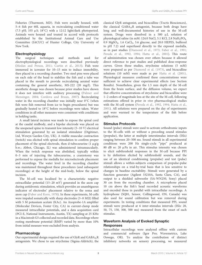
Glycine Receptors Mediate InhibitionContributing to the Onset of PPIThese experiments tested if treatment with strychnine, aGlyR antagonist, affects auditory PPI in the Mauthner cells(M-cells), the decision-making neurons of the goldfish startlecircuit. Figures 1A--C shows somatically recorded PSPs inresponse to prepulse/pulse sound stimuli (identical subthreshold200 Hz ‘‘pips’’ at 80 dB; see methods) for ISIs rangingfrom 20--75 ms in control (black traces) and strychnine (redtraces) treatment conditions. In each experiment (N = 9),5--10 responses in a single M-cell were measured at ISIsranging from 20--500 ms in control and drug treatmentconditions. The results show an overall attenuation of thePSP to the secondary stimulus (pulse) when compared tothe PSP evoked by the lead stimulus (prepulse) in controlconditions; in short, synaptic PPI (Figures 1A--C). Afterapplication of strychnine synaptic PPI magnitude remainedlargely unchanged for all but the shortest ISI, despite the factthat the drug changed the overall PSP waveform (Figures 1A--C;black vs. red traces, see also below). Figure 1D plots thequantification of PPI across control (black line) and strychnine(red line) treatment conditions. Although PPI remained robustlyintact after treatment with the GlyR antagonist for allISIs >20 ms (Figures 1B,C, black vs. red traces; Figure 1D,black vs. red lines), we observed an ISI-specific reductionof PPI at the shortest ISI tested (20 ms; Figure 1A, blackvs. red traces). Supporting these results, we found thatstrychnine had no significant main effect on the magnitudeof PPI (F(1,86.87) = 2.98, P = 0.088, N = 9), but ouranalysis identified a significant ISI X strychnine interaction(F(6,83.68) = 5.7276, P < 0.0001, N = 9). Post hoc tests (Holmes-Bonferonni) confirmed this effect was due to an ISI-specificreduction in the PPI effect at the 20 ms ISI (P < 0.001).These findings indicate that GlyRs mediate inhibition thatcontributes to PPI for as long as 20 ms, but this glycinergiccomponent decays within 50 ms of the onset of prepulsestimuli.
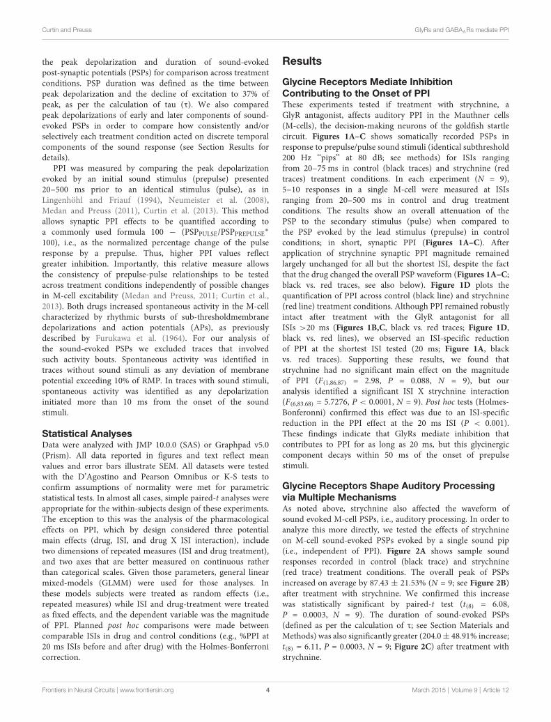
Glycine Receptors Shape Auditory Processingvia Multiple MechanismsAs noted above, strychnine also affected the waveform ofsound evoked M-cell PSPs, i.e., auditory processing. In order toanalyze this more directly, we tested the effects of strychnineon M-cell sound-evoked PSPs evoked by a single sound pip(i.e., independent of PPI). Figure 2A shows sample soundresponses recorded in control (black trace) and strychnine(red trace) treatment conditions. The overall peak of PSPsincreased on average by 87.43 ± 21.53% (N = 9; see Figure 2B)after treatment with strychnine. We confirmed this increasewas statistically significant by paired-t test (t(8) = 6.08,P = 0.0003, N = 9). The duration of sound-evoked PSPs(defined as per the calculation of τ; see Section Materials andMethods) was also significantly greater (204.0± 48.91% increase;t(8) = 6.11, P = 0.0003, N = 9; Figure 2C) after treatment withstrychnine.
Frontiers in Neural Circuits | www.frontiersin.org 4 March 2015 | Volume 9 | Article 12
Curtin and Preuss GlyRs and GABAARs mediate PPI
FIGURE 1 | Glycinergic inhibition contributes to the earliestcomponents of PPI. (A--C). Sample intracellular recordings from theMauthner-cell (M-cell) soma in response to paired (prepulse/pulse) soundpips at ISIs of 20 ms (A1,A2), 50 ms (B1,B2), and 75 ms (C1,C2) incontrol (black) and after application of the GlyR antagonist strychnine(red). Bottom traces show sound stimuli (200 Hz single-cycle “pips” at
80 dB re: 20 µPa). Dashed lines and brackets indicate how PPI wasquantified by comparing peak depolarization between the two evokedpost-synaptic potentials (PSPs). (D) Plots of the mean % PPI effect(± SEM, N = 9) across the full range of ISIs tested in control (black line)and strychnine (red line) conditions; asterisks indicate an ISI-specificreduction in PPI at the 20 ms ISI (see text).
We next analyzed how the GlyR antagonist acted on differentcomponents of the overall sound response. The M-cell PSPreflects the integration of multiple excitatory and inhibitoryinputs activated by primary auditory afferences. These includeelectrotonic and chemical excitation via mixed VIIIth nervesynapses at the M-cell lateral dendrite (Furshpan, 1964; Faberand Korn, 1975; Lin and Faber, 1988a; Curti and Pereda, 2004).This excitation is counteracted by chemical inhibition (onset ofabout 5 ms relative to stimulus onset; see Preuss and Faber, 2003;Medan and Preuss, 2011) that peaks at 10--12 ms, mediated bya feed-forward network that is also activated by VIIIth nerveafferents (Korn and Faber, 2005; Szabo et al., 2006; Weiss et al.,2008; see also Introduction). In other words, the monosynapticexcitatory pathway and disynaptic inhibitory pathway allow abrief interval within the first 5 ms of the postsynaptic responsewhen sound-evoked depolarization reflects largely excitatoryinputs (i.e., an EPSP), whereas later components of the sound-response represent the integration of excitatory and inhibitoryinputs (i.e., a mixed PSP). Figure 2A (inset) shows the onsetof the sound-response in an expanded time scale to emphasizethe effects of strychnine (compare black vs. red traces) on theEPSP (light gray area, 0--5 ms from stimulus onset) and the PSP(dark gray area, >5 ms from stimulus onset). We found thatstrychnine caused a relatively mild enhancement (14.33 ± 7.2%increase, N = 9) of peak depolarization during the initial EPSP,but a significantly greater enhancement of excitation duringthe mixed-PSP component of the response(116.5 ± 18.73%increase; t(8) = 8.54, P = 0.0034; see Figure 2D). This time coursesuggests a minor effect of strychnine on presynaptic excitatorypathways and/or M-cell tonic excitability, but is consistent witha drug-induced disruption of feed-forward inhibition in theM-cell.
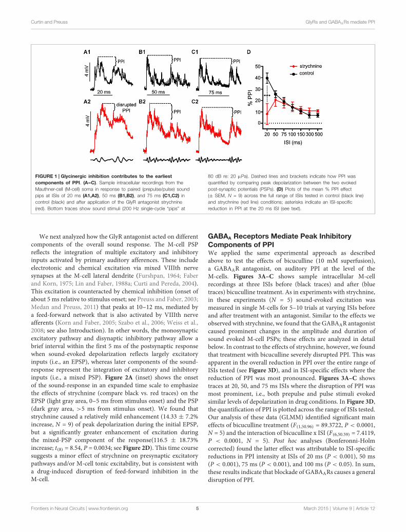
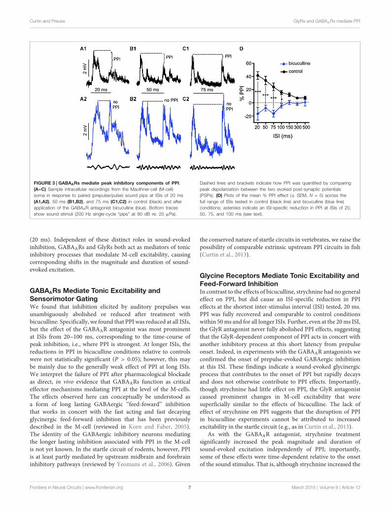
GABAA Receptors Mediate Peak InhibitoryComponents of PPIWe applied the same experimental approach as describedabove to test the effects of bicuculline (10 mM superfusion),a GABAAR antagonist, on auditory PPI at the level of theM-cells. Figures 3A--C shows sample intracellular M-cellrecordings at three ISIs before (black traces) and after (bluetraces) bicuculline treatment. As in experiments with strychnine,in these experiments (N = 5) sound-evoked excitation wasmeasured in single M-cells for 5--10 trials at varying ISIs beforeand after treatment with an antagonist. Similar to the effects weobserved with strychnine, we found that the GABAAR antagonistcaused prominent changes in the amplitude and duration ofsound evoked M-cell PSPs; these effects are analyzed in detailbelow. In contrast to the effects of strychnine, however, we foundthat treatment with bicuculline severely disrupted PPI. This wasapparent in the overall reduction in PPI over the entire range ofISIs tested (see Figure 3D), and in ISI-specific effects where thereduction of PPI was most pronounced. Figures 3A--C showstraces at 20, 50, and 75 ms ISIs where the disruption of PPI wasmost prominent, i.e., both prepulse and pulse stimuli evokedsimilar levels of depolarization in drug conditions. In Figure 3D,the quantification of PPI is plotted across the range of ISIs tested.Our analysis of these data (GLMM) identified significant maineffects of bicuculline treatment (F(1,50.96) = 89.3722, P < 0.0001,N = 5) and the interaction of bicuculline x ISI (F(6,50.59) = 7.4119,P < 0.0001, N = 5). Post hoc analyses (Bonferonni-Holmcorrected) found the latter effect was attributable to ISI-specificreductions in PPI intensity at ISIs of 20 ms (P < 0.001), 50 ms(P < 0.001), 75 ms (P < 0.001), and 100 ms (P < 0.05). In sum,these results indicate that blockade of GABAARs causes a generaldisruption of PPI.
Frontiers in Neural Circuits | www.frontiersin.org 5 March 2015 | Volume 9 | Article 12
Curtin and Preuss GlyRs and GABAARs mediate PPI
FIGURE 2 | Glycinergic inhibition mediates auditory processing in theM-cell. (A) Sample intracellular recordings from the Mauthner-cell (M-cell)soma in response to an individual sound pip before (black trace) and after (redtrace) treatment with strychnine. The insetshows the initial part of the evokedresponse at an expanded time scale. Light and dark shaded areas distinguishthe initial (EPSP; 0--5 ms) and later components (mixed PSP; >5 ms) of theresponse, respectively. Bottom traces show sound stimulus (200 Hzsingle-cycle “pip” at 80 dB re: 20 µPa). (B) Plots of mean (± SEM, N = 9)overall peak amplitude of sound-evoked depolarization in control (black bar)and strychnine (red bar) conditions. (C) Plots of mean (± SEM, N = 9) PSPduration (Tau) before (black) and after (red) treatment with strychnine. (D) Plotsof the mean (± SEM, N = 9) relative change in sound-evoked depolarizationfor the initial (EPSP) and later parts (mixed PSP) of the sound response aftertreatment with strychnine. Asterisks indicate p values in statisticalcomparisons that were < 0.01 (**), < 0.001 (***).
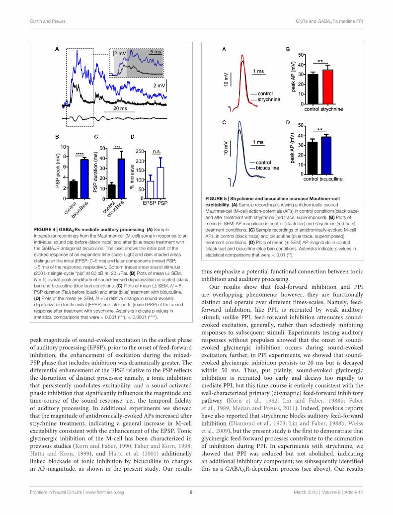
GABAA Receptors Mediate a Tonic Increase inSound-Evoked ExcitationIn these experiments, we tested the effects of the GABAARantagonist, bicuculline on sound-evoked depolarization inseparate trials using only a single sound pip (without prepulses)to determine how GABAARs contribute to auditory processingindependently of PPI. We again approached our analysisby examining the effect of the GABAAR antagonist on theoverall peak depolarization and duration of sound-evokedexcitation. Figure 4A shows sound-evoked PSPs recorded incontrol (black trace) and bicuculline (blue trace) treatmentconditions. We found that bicuculline significantly increasedthe mean overall peak of sound-evoked PSPs by 133.8 ±
10.3% (Figure 4B; t(4) = 28.12, P < 0.0001, N = 5). Similarly,the duration of sound-evoked PSPs increased by 284.95 ±
65.64% in drug conditions (Figure 4C; t(4) = 3.07, P = 0.037,N = 5).
As in our analysis of GlyR-mediated components of sound-evoked excitation, we also measured the potentially differential
effects of the GABAAR antagonist on initial (EPSP) andsubsequent components (PSP) of the sound-response (Figure 4Ainset, light gray shading vs. dark gray shading). In contrastto strychnine, bicuculline produced large enhancement in bothcomponents of the response (EPSP: 90.95% increase; PSP:164.32% increase); however, bicuculline’s effect on the EPSP wasnot significantly different from the PSP (t(4) = 1.116, P = 0.327,N = 5). The latter result is consistent with a drug induced increasein presynaptic excitation and/or by a decrease in inhibitory tonein the M-cell system which increases the neurons excitability (seebelow).
Strychnine and Bicuculline Disrupt TonicInhibition Contributing to M-cell ExcitabilityIn prior experiments, we reported that GlyR and GABAARantagonists enhance sound-evoked excitation in the M-cell(Figures 2, 4). Namely, strychnine treatment predominatelyenhanced later parts of the PSP, i.e., demonstrating a time-dependent effect, whereas bicuculline produced an enhancementof the entire PSP consistent with a tonic change in M-cellexcitability. Accordingly, we next tested whether theseantagonists affect electrotonic excitability in M-cell. Sincethe M-cells’ soma-dendritic membrane is non-regenerative,changes in the magnitude of somatic APs provide a measureof corresponding changes in tonic membrane conductivity(excitability; reviewed in Korn and Faber, 2005; Curtin et al.,2013). Figure 5A shows sample traces of APs elicited incontrol (black trace) and strychnine (red trace) treatmentconditions. On average, treatment with strychnine increasedthe peak magnitude of APs by 15.07 ± 3.21% (Figure 5B;paired-t, t(6) = 4.314, P = 0.005) consistent with a decreasein conductance. Importantly, RMP (RMPcontrol = −80.6 ±
0.82 mV; RMPstrychnine = −80.9 ± 0.86 mV) was not affected bytreatment with strychnine (t(9) = 0.4104, P = 0.69), indicatingthe disruption of a shunting inhibition rather than a persistenthyperpolarization.
Similarly, bicuculline treatment increased the magnitude ofAPs by 16.23 ± 7.08% (Figure 5C, black vs. blue traces;Figure 5D, t(4) = 3.09, P = 0.036, N = 5), but had nosignificant effect on RMP (RMPcontrol = −78.6 ± 1.55 mV;RMPbicuculline = −77.22 ± 1.35 mV), consistent with a shuntinginhibitory process.
In sum, these results are consistent with the notion thattonic inhibitory processes regulate M-cell excitability, and theseprocesses are mediated by GlyRs and GABAARs.
Discussion
The aim of this study was to determine if GlyRs and/orGABAARs mediate auditory PPI in the decision-making neuronsthat initiate startle, the Mauthner-cells (M-cells). Our primaryfindings indicate that GABAARs function as effectormechanismsmediating the onset and peak effect of PPI, corresponding tointerstimulus-intervals (ISIs) ranging from 20--100 ms. GlyRs,in contrast, are primarily involved in the mediation of fast-onset feed-forward (sound-evoked) inhibitory processes thatrapidly decay but overlap and contribute to the onset of PPI
Frontiers in Neural Circuits | www.frontiersin.org 6 March 2015 | Volume 9 | Article 12
Curtin and Preuss GlyRs and GABAARs mediate PPI
FIGURE 3 | GABAARs mediate peak inhibitory components of PPI.(A--C) Sample intracellular recordings from the Mauthner-cell (M-cell)soma in response to paired (prepulse/pulse) sound pips at ISIs of 20 ms(A1,A2), 50 ms (B1,B2), and 75 ms (C1,C2) in control (black) and afterapplication of the GABAAR antagonist bicuculline (blue). Bottom tracesshow sound stimuli (200 Hz single-cycle “pips” at 80 dB re: 20 µPa).
Dashed lines and brackets indicate how PPI was quantified by comparingpeak depolarization between the two evoked post-synaptic potentials(PSPs). (D) Plots of the mean % PPI effect (± SEM, N = 5) across thefull range of ISIs tested in control (black line) and bicuculline (blue line)conditions; asterisks indicate an ISI-specific reduction in PPI at ISIs of 20,50, 75, and 100 ms (see text).
(20 ms). Independent of these distinct roles in sound-evokedinhibition, GABAARs and GlyRs both act as mediators of tonicinhibitory processes that modulate M-cell excitability, causingcorresponding shifts in the magnitude and duration of sound-evoked excitation.
GABAARs Mediate Tonic Excitability andSensorimotor GatingWe found that inhibition elicited by auditory prepulses wasunambiguously abolished or reduced after treatment withbicuculline. Specifically, we found that PPI was reduced at all ISIs,but the effect of the GABAAR antagonist was most prominentat ISIs from 20--100 ms, corresponding to the time-course ofpeak inhibition, i.e., where PPI is strongest. At longer ISIs, thereductions in PPI in bicuculline conditions relative to controlswere not statistically significant (P > 0.05); however, this maybe mainly due to the generally weak effect of PPI at long ISIs.We interpret the failure of PPI after pharmacological blockadeas direct, in vivo evidence that GABAARs function as criticaleffector mechanisms mediating PPI at the level of the M-cells.The effects observed here can conceptually be understood asa form of long lasting GABAergic ‘‘feed-foward’’ inhibitionthat works in concert with the fast acting and fast decayingglycinergic feed-forward inhibition that has been previouslydescribed in the M-cell (reviewed in Korn and Faber, 2005).The identity of the GABAergic inhibitory neurons mediatingthe longer lasting inhibition associated with PPI in the M-cellis not yet known. In the startle circuit of rodents, however, PPIis at least partly mediated by upstream midbrain and forebraininhibitory pathways (reviewed by Yeomans et al., 2006). Given
the conserved nature of startle circuits in vertebrates, we raise thepossibility of comparable extrinsic upstream PPI circuits in fish(Curtin et al., 2013).
Glycine Receptors Mediate Tonic Excitability andFeed-Forward InhibitionIn contrast to the effects of bicuculline, strychnine had no generaleffect on PPI, but did cause an ISI-specific reduction in PPIeffects at the shortest inter-stimulus interval (ISI) tested, 20 ms.PPI was fully recovered and comparable to control conditionswithin 50ms and for all longer ISIs. Further, even at the 20ms ISI,the GlyR antagonist never fully abolished PPI effects, suggestingthat the GlyR-dependent component of PPI acts in concert withanother inhibitory process at this short latency from prepulseonset. Indeed, in experiments with the GABAAR antagonists weconfirmed the onset of prepulse-evoked GABAergic inhibitionat this ISI. These findings indicate a sound-evoked glycinergicprocess that contributes to the onset of PPI but rapidly decaysand does not otherwise contribute to PPI effects. Importantly,though strychnine had little effect on PPI, the GlyR antagonistcaused prominent changes in M-cell excitability that weresuperficially similar to the effects of bicuculline. The lack ofeffect of strychnine on PPI suggests that the disruption of PPIin bicuculline experiments cannot be attributed to increasedexcitability in the startle circuit (e.g., as in Curtin et al., 2013).
As with the GABAAR antagonist, strychnine treatmentsignificantly increased the peak magnitude and duration ofsound-evoked excitation independently of PPI; importantly,some of these effects were time-dependent relative to the onsetof the sound stimulus. That is, although strychnine increased the
Frontiers in Neural Circuits | www.frontiersin.org 7 March 2015 | Volume 9 | Article 12
Curtin and Preuss GlyRs and GABAARs mediate PPI
FIGURE 4 | GABAARs mediate auditory processing. (A) Sampleintracellular recordings from the Mauthner-cell (M-cell) soma in response to anindividual sound pip before (black trace) and after (blue trace) treatment withthe GABAAR antagonist bicuculline. The inset shows the initial part of theevoked response at an expanded time scale. Light and dark shaded areasdistinguish the initial (EPSP; 0--5 ms) and later components (mixed PSP;>5 ms) of the response, respectively. Bottom traces show sound stimulus(200 Hz single-cycle “pip” at 80 dB re: 20 µPa). (B) Plots of mean (± SEM,N = 5) overall peak amplitude of sound-evoked depolarization in control (blackbar) and bicuculline (blue bar) conditions. (C) Plots of mean (± SEM, N = 5)PSP duration (Tau) before (black) and after (blue) treatment with bicuculline.(D) Plots of the mean (± SEM, N = 5) relative change in sound-evokeddepolarization for the initial (EPSP) and later parts (mixed PSP) of the soundresponse after treatment with strychnine. Asterisks indicate p values instatistical comparisons that were < 0.001 (***), < 0.0001 (****).
peak magnitude of sound-evoked excitation in the earliest phaseof auditory processing (EPSP), prior to the onset of feed-forwardinhibition, the enhancement of excitation during the mixed-PSP phase that includes inhibition was dramatically greater. Thedifferential enhancement of the EPSP relative to the PSP reflectsthe disruption of distinct processes; namely, a tonic inhibitionthat persistently modulates excitability, and a sound-activatedphasic inhibition that significantly influences the magnitude andtime-course of the sound response, i.e., the temporal fidelityof auditory processing. In additional experiments we showedthat the magnitude of antidromically-evoked APs increased afterstrychnine treatment, indicating a general increase in M-cellexcitability consistent with the enhancement of the EPSP. Tonicglycinergic inhibition of the M-cell has been characterized inprevious studies (Korn and Faber, 1990; Faber and Korn, 1998;Hatta and Korn, 1999), and Hatta et al. (2001) additionallylinked blockade of tonic inhibition by bicuculline to changesin AP-magnitude, as shown in the present study. Our results
FIGURE 5 | Strychnine and bicuculline increase Mauthner-cellexcitability. (A) Sample recordings showing antidromically-evokedMauthner-cell (M-cell) action-potentials (APs) in control conditions(black trace)and after treatment with strychnine (red trace, superimposed). (B) Plots ofmean (± SEM) AP magnitude in control (black bar) and strychnine (red bars)treatment conditions. (C) Sample recordings of antidromically-evoked M-cellAPs, in control (black trace) and bicuculline (blue trace, superimposed)treatment conditions. (D) Plots of mean (± SEM) AP magnitude in control(black bar) and bicucilline (blue bar) conditions. Asterisks indicate p values instatistical comparisons that were < 0.01 (**).
thus emphasize a potential functional connection between tonicinhibition and auditory processing.
Our results show that feed-forward inhibition and PPIare overlapping phenomena; however, they are functionallydistinct and operate over different times-scales. Namely, feed-forward inhibition, like PPI, is recruited by weak auditorystimuli; unlike PPI, feed-forward inhibition attenuates sound-evoked excitation, generally, rather than selectively inhibitingresponses to subsequent stimuli. Experiments testing auditoryresponses without prepulses showed that the onset of sound-evoked glycinergic inhibition occurs during sound-evokedexcitation; further, in PPI experiments, we showed that sound-evoked glycinergic inhibition persists to 20 ms but is decayedwithin 50 ms. Thus, put plainly, sound-evoked glycinergicinhibition is recruited too early and decays too rapidly tomediate PPI, but this time-course is entirely consistent with thewell-characterized primary (disynaptic) feed-forward inhibitorypathway (Korn et al., 1982; Lin and Faber, 1988b; Faberet al., 1989; Medan and Preuss, 2011). Indeed, previous reportshave also reported that strychnine blocks auditory feed-forwardinhibition (Diamond et al., 1973; Lin and Faber, 1988b; Weisset al., 2009), but the present study is the first to demonstrate thatglycinergic feed-forward processes contribute to the summationof inhibition during PPI. In experiments with strychnine, weshowed that PPI was reduced but not abolished, indicatingan additional inhibitory component; we subsequently identifiedthis as a GABAAR-dependent process (see above). Our results
Frontiers in Neural Circuits | www.frontiersin.org 8 March 2015 | Volume 9 | Article 12
Curtin and Preuss GlyRs and GABAARs mediate PPI
thus emphasize the likely contribution of multiple inhibitorypathways in the mediation of PPI. These findings should beinterpretated cautiously, however, as the selective effects of theantagonists used are dependent on their effective concentrations,which could not be confirmed in the intact, in vivo preparationsused in these experiments.
Mediators, Modulators, and Model SystemsThese results highlight some striking similarities with advancesin rodent model systems. Yeomans et al. (2010) showedthat bicuculline disrupts the peak inhibitory componentsof behavioral PPI in rodents, and that PnC neurons (thesensorimotor interface equivalents of the M-cell in themammalian startle circuit) are inhibited by GABA in anex vivo brain-slice preparation. Moreover, the time-course of PPImediated by GABAARs reported in rodents is similar to the ISI-specific effects we report here in the fish startle system.
Our findings are somewhat in contrast with studies ofglycinergic inhibition in rodent preparations. Koch and Friauf(1995) showed that local and systemic applications of strychninehad no effect on phasic inhibitory processes including short-term habituation of startle and PPI. Geis and Schmid (2011)used in vitro patch-clamp recordings to demonstrate that glycinedirectly inhibits PnC neurons in a rat brain slice preparation;however, they found no evidence that GlyRs were involved inphasic inhibitory processes, including feed-forward inhibitionand short-term synaptic depression.
In contrast, our experiments identified GlyR-dependentphasic inhibitory processes that attenuate sound-evokedexcitation in multiple contexts (Figures 1, 2). These contrastingfindings may reflect underlying differences in goldfish androdents, or in experimental preparations or stimulus protocols.
Whereas the present study measured in vivo synaptic processesin mature, awake goldfish, in vitro slice preparations used torecord from PnC neurons were derived from embryonic ratbrains (Yeomans et al., 2010; Geis and Schmid, 2011). Given theprofound structural and functional shifts attributed to GlyRs andGABAARs during development, functional differences betweenmature and embryonic circuits may be expected (Ehrlich et al.,1999; Nabekura et al., 2004).
In sum, this study characterized in vivo synaptic signalingmechanisms that directly mediate the balance of excitationand inhibition at the sensorimotor interface of the startlecircuit. Prior studies in the M-cell and other model systemshave examined the role of neuromodulators, particularlymonoaminergic transmitters (Medan and Preuss, 2011; Curtinet al., 2013), involved in PPI. Our results emphasize thatin vivo electrophysiological methods can be applied to dissectoverlapping inhibitory processes and effector mechanisms todirectly test predictions drawn from advances in other modelsystems. Thus the M-cell presents an appropriate tool fordissecting the functional roles of synaptic processes as well as theeffector mechanisms mediating their effects.
Acknowledgments
This work was supported by National Science Foundationgrants IOS 0946637 and IOS 11471172 and by a grant fromthe Professional Staff Congress City University of New York(CUNY) Research Award Program, with additional support fromHunter College and the Graduate Center, CUNY. The authorsadditionally thank Dr. V. Medan, A.Curtin, and members of thePreuss laboratory for discussion and constructive feedback thatcontributed to the improvement of the manuscript.
References
Bennett, M. V. L. (1984). ‘‘Escapism: some startling revelations,’’ in NeuralMechanisms of Startle Behavior, ed R. C. Eaton (New York, NY: Plenum Press),353--363.
Braff, D. L., Geyer, M. A., and Swerdlow, N. R. (2001). Human studies of prepulseinhibition of startle: normal subjects, patient groups and pharmacologicalstudies. Psychopharmacology (Berl) 156, 234--258. doi: 10.1007/s002130100810
Burgess, H. A., and Granato, M. (2007). Sensorimotor gating in larval zebrafish.J. Neurosci. 27, 4984--4994. doi: 10.1523/jneurosci.0615-07.2007
Cordova, M. S., and Braun, C. B. (2007). The use of anesthesia during evokedpotential audiometry in goldfish (Carassius auratus). Brain Res. 1153, 78--83.doi: 10.1016/j.brainres.2007.03.055
Curti, S., and Pereda, A. (2004). Voltage-dependent enhancement of electricalcoupling by a subthreshold sodium current. J. Neurosci. 24, 3999--4010. doi: 10.1523/jneurosci.0077-04.2004
Curtin, P. C. P., Medan, V., Neumeister, H., Bronson, D. R., and Preuss, T. (2013).The 5-HT5A receptor regulates excitability in the auditory startle circuit:functional implications for sensorimotor gating. J. Neurosci. 33, 10011--10020.doi: 10.1523/jneurosci.4733-12.2013
Diamond, J., Roper, S., and Yasargil, G. M. (1973). The membrane effects andsensitivity to strychnine, of neural inhibition of the Mauthner cell and itsinhibition by glycine and GABA. J. Physiol. 232, 87--111. doi: 10.1113/jphysiol.1973.sp010258
Eaton, R. C., Lavender, W. A., and Wieland, C. M. (1981). Identification ofMauthner-initiated response patterns in goldfish: evidence from simultaneous
cinematography and electrophysiology. J. Comp. Physiol. 144, 521--531. doi: 10.1007/bf01326837
Eaton, R., Lee, R., and Foreman, M. (2001). TheMauthner cell and other identifiedneurons of the brainstem escape network of fish. Prog. Neurobiol. 63, 467--485.doi: 10.1016/s0301-0082(00)00047-2
Ehrlich, I., Lohrke, S., and Friauf, E. (1999). Shift from depolarizing tohyperpolarizing glycine action in rat auditory neurones is due to age-dependentCl- regulation. J. Physiol. 520, 121--137. doi: 10.1111/j.1469-7793.1999.00121.x
Faber, D. S., Fetcho, J. R., and Korn, H. (1989). Neuronal networks underlying theescape response in goldfish. General implications for motor control. Ann. N YAcad. Sci. 563, 11--33. doi: 10.1111/j.1749-6632.1989.tb42187.x
Faber, D. S., and Korn, H. (1975). Inputs from the posterior lateral line nervesupon the goldfish Mauthner cell. II. Evidence that the inhibitory componentsare mediated by interneurons of the recurrent collateral netowrk. Brain Res. 96,349--356. doi: 10.1016/0006-8993(75)90746-5
Faber, D. S., and Korn, H. (1978). ‘‘Electrophysiology of the Mauthner cell: basicproperties, synaptic mechanisms and associated networks,’’ in Neurobiology ofthe Mauthner Cell, eds D. S. Faber and H. Korn (New York, NY: Raven Press),47--131.
Faber, D. S., and Korn, H. (1987). Voltage-dependence of glycine-activated Cl-channels: a potentiometer for Inhibition? J. Neurosci. 7, 807--811.
Faber, D. S., and Korn, H. (1998). Unitary conductance changes at teleostMauthner cell glycinergic snapses: a voltage-clamp and pharmacologic analysis.J. Neurophysiol. 60, 1982--1999.
Faber, D. S., Korn, H., and Lin, J. W. (1991). Role of medullary networks andpostsynaptic membrane properties in regulating Mauthner cell responsivenessto sensory excitation. Brain Behav. Evol. 37, 286--297. doi: 10.1159/000114366
Frontiers in Neural Circuits | www.frontiersin.org 9 March 2015 | Volume 9 | Article 12
Curtin and Preuss GlyRs and GABAARs mediate PPI
Fendt, M. (1999). Enhancement of prepulse inhibition after blockade of GABAactivity within the superior colliculus. Brain Res. 833, 81--85. doi: 10.1016/s0006-8993(99)01525-5
Furshpan, E. J. (1964). ‘‘Electrical transmission’’ at an excitatory synapsein a vertebrate brain. Science 144, 878--880. doi: 10.1126/science.144.3620.878
Furukawa, T., Fukama, Y., and Asada, Y. (1964). Effects of strychnine and procaineon collateral inhibition of the Mauthner cell of goldfish. Jpn. J. Physiol. 14,386--399. doi: 10.2170/jjphysiol.14.386
Geis, H. R., and Schmid, S. (2011). Glycine inhibits startle-mediating neuronsin the caudal pontine reticular formation but is not involved in synapticdepression underlying short-term habituation of startle. Neurosci. Res. 71,114--123. doi: 10.1016/j.neures.2011.06.007
Graham, F. K. (1975). Presidential address, 1974: the more or less startling effectsof weak prestimulation. Psychophysiology 12, 238--248. doi: 10.1111/j.1469-8986.1975.tb01284.x
Hatta, K., Ankri, N., Faber, D. S., and Korn, H. (2001). Slow inhibitory potentialsin the teleost Mauthner cell. Neuroscience 103, 561--579. doi: 10.1016/s0306-4522(00)00570-4
Hatta, K., and Korn, H. (1999). Tonic inhibition alternates in paired neuronsthat set direction of fish escape reaction. Proc. Natl. Acad. Sci. U S A 96,12090--12095. doi: 10.1073/pnas.96.21.12090
Hoffman, H. S., and Ison, J. R. (1980). Reflex modification in the domain of startle:I. Some empirical findings and their implications for how the nervous systemprocesses sensory input. Psychol. Rev. 87, 175--189. doi: 10.1037//0033-295x.87.2.175
Jones, C. K., and Shannon, H. E. (2000). Muscarinic cholinergic modulation ofprepulse inhibition of the acoustic startle reflex. J. Pharmacol. Exp. Ther. 294,1017--1023.
Koch,M. (1999). The neurobiology of startle. Prog. Neurobiol. 59, 107--128. doi: 10.1016/s0301-0082(98)00098-7
Koch, M., and Friauf, E. (1995). Glycine receptors in the caudal pontine reticularformation: are they important for the inhibition of the acoustic startle response?Brain Res. 671, 63--72. doi: 10.1016/0006-8993(94)01309-6
Koch, M., and Schnitzler, H. U. (1997). The acoustic startle response in rats--circuits mediating evocation, inhibition and potentiation. Behav. Brain Res. 89,35--49. doi: 10.1016/s0166-4328(97)02296-1
Korn, H., and Faber, D. S. (1990). Transmission at a central inhibitory synapse. IV.Quantal structure of synaptic noise. J. Neurophysiol. 63, 198--222.
Korn, H., and Faber, D. S. (2005). The Mauthner cell half a century later: aneurobiological model for decision-making? Neuron 47, 13--28. doi: 10.1016/j.neuron.2005.05.019
Korn, H., Mallet, A., Triller, A., and Faber, D. S. (1982). Transmission at a centralinhibitory synapse. II. Quantal description of release, with a physical correlatefor binomial n. J. Neurophysiol. 48, 679--707.
Lee, R., Finger, T., and Eaton, R. C. (1993). GABAergic innervation of theMauthner cell and other reticulospinal neurons in the goldfish. J. Comp. Neurol.338, 601--611. doi: 10.1002/cne.903380409
Lin, J. W., and Faber, D. S. (1988a). Synaptic transmission mediated by single clubendings on the godlifhs Mauthner cell. I. Characteristics of electrotonic andchemical postsynaptic potentials. J. Neurosci. 8, 1302--1312.
Lin, J. W., and Faber, D. S. (1988b). Synaptic transmission mediated by single clubendings on the goldfish Mauthner cell. II. Plasticity of excitatory postsynapticpotentials. J. Neurosci. 8, 1313--1325.
Lingenhöhl, K., and Friauf, E. (1994). Giant neurons in the rat reticular formation:a sensorimotor interface in the elementary startle circuit? J. Neurosci. 14,1176--1194.
Marti, F., Korn, H., and Faure, P. (2008). Interplay between subthresholdpotentials and gamma oscillations in Mauthner cells’ presynaptic inhibitoryinterneurons. Neuroscience 151, 983--994. doi: 10.1016/j.neuroscience.2007.11.054
Medan, V., and Preuss, T. (2011). Dopaminergic-induced changes in Mauthnercell excitability disrupt prepulse inhibition in the startle circuit of goldfish.J. Neurophysiol. 106, 3195--3204. doi: 10.1152/jn.00644.2011
Nabekura, J., Katsurabayashi, S., Kakazu, Y., Shibata, S., Matsubara, A., Jinno, S.,et al. (2004). Developmental switch from GABA to glycine release in singlecentral synaptic terminals. Nat. Neurosci. 7, 17--23. doi: 10.1038/nn1170
Neumeister, H., Szabo, T. M., and Preuss, T. (2008). Behavioral and physiologicalcharacterization of sensorimotor gating in the goldfish startle response.J. Neurophysiol. 99, 1493--1502. doi: 10.1152/jn.00959.2007
Palmer, L. M., and Mensinger, A. F. (2004). Effect of the anesthetic tricaine(MS-222) on nerve activity in the anterioir lateral line of the oyster toadfish,Optsanus tau. J. Neurophysiol. 92, 1034--1041. doi: 10.1152/jn.01151.2003
Parwani, A., Duncan, E. J., Bartlett, E., Madonick, S. H., Efferen, T. R., Rajan, R.,et al. (2000). Impaired prepulse inhibition of acoustic startle in schizophrenia.Biol. Psychiatry 47, 662--669. doi: 10.1016/S0006-3223(99)00148-1
Pereda, A. E., Nairn, A. C., Wolszon, L. R., and Faber, D. S. (1994). Postsynapticmodulation of synaptic efficacy at mixed synapses on the Mauthner cell.J. Neurosci. 14, 3704--3712.
Pereda, A. E., Triller, A., Korn, H., and Faber, D. S. (1992). Dopamine enhancesboth electrotonic coupling and chemical excitatory postsynaptic potentialsat mixed synapses. Proc. Natl. Acad. Sci. U S A 89, 12088--12092. doi: 10.1073/pnas.89.24.12088
Petrov, T., Seitanidou, T., Triller, A., and Korn, H. (1991). Differential distributionof GABA- and seroton-containing afferents on an identified central neuron.Brain Res. 559, 75--81. doi: 10.1016/0006-8993(91)90288-7
Preuss, T., and Faber, D. S. (2003). Central cellular mechanisms underlyingtemperature-dependent changes in the goldfish startle-escape behavior. J.Neurosci. 23, 5617--5626.
Seitanidou, T., Triller, A., and Korn, H. (1988). Distribution of glycine receptorson the membrane of a central neuron: an immunoelectron microscopy study.J. Neurosci. 438, 4319--4333.
Sur, C., McKernan, R., and Triller, A. (1995). GABAA receptor-likeimmunoreactivity in the goldfish brainstem with emphasis on the Mauthnercell. Neuroscience 66, 697--706. doi: 10.1016/0306-4522(94)00579-t
Swerdlow, N., Caine, S., Braff, D. L., and Geyer, M. A. (1992). The neuralsubstrates of sensorimotor gating of the startle reflex: a review of recentfindings and their implications. J. Psychopharmacol. 6, 176--190. doi: 10.1177/026988119200600210
Szabo, T. M., Weiss, S. A., Faber, D. S., and Preuss, T. (2006). Representation ofauditory signals in the M-cell: role of electrical synapses. J. Neurophysiol. 95,2617--2629. doi: 10.1152/jn.01287.2005
Triller, A., Cluzeaud, F., Pfeiffer, F., Betz, H., and Korn, H. (1985). Distributionof glycine receptors at central synapses: an immunoelectron microscopy study.J. Cell Biol. 101, 683--688. doi: 10.1083/jcb.101.2.683
Ukai, M., Okuda, A., and Mamiya, T. (2004). Effects of anticholinergic drugsselective for muscarinic receptor subtypes on prepulse inhibition in mice. Eur.J. Pharmacol. 492, 183--187. doi: 10.1016/j.ejphar.2004.03.066
Weiss, S. A., Preuss, T., and Faber, D. S. (2008). A role of electrical inhibitionin sensorimotor integration. Proc. Natl. Acad. Sci. U S A 105, 18047--18052.doi: 10.1073/pnas.0806145105
Weiss, S. A., Preuss, T., and Faber, D. S. (2009). Phase encoding in the Mauthnersystem: implications in left-right sound source discrimination. J. Neurosci. 29,3431--3441. doi: 10.1523/jneurosci.3383-08.2009
Yeomans, J. S., Bosch, D., Alves, N., Daros, A., Ure, R. J., and Schmid, S. (2010).GABA receptors and prepulse inhibition of acoustic startle in mice and rats.Eur. J. Neurosci. 31, 2053--2061. doi: 10.1111/j.1460-9568.2010.07236.x
Yeomans, J. S., Lee, J., Yeomans, M. H., Steidl, S., and Li, L. (2006). Midbrainpathways for prepulse inhibition and startle activation in rat. Neuroscience 142,921--929. doi: 10.1016/j.neuroscience.2006.06.025
Yeomans, J. S., Liang, L., Scott, B., and Frankland, P. W. (2002). Tactile, acousticand vestibular systems sum to elicit the startle reflex. Neurosci. Biobehav. Rev.26, 1--11. doi: 10.1016/s0149-7634(01)00057-4
Conflict of Interest Statement: The authors declare that the research wasconducted in the absence of any commercial or financial relationships that couldbe construed as a potential conflict of interest.
Copyright © 2015 Curtin and Preuss. This is an open-access article distributedunder the terms of the Creative Commons Attribution License (CC BY). The use,distribution and reproduction in other forums is permitted, provided the originalauthor(s) or licensor are credited and that the original publication in this journalis cited, in accordance with accepted academic practice. No use, distribution orreproduction is permitted which does not comply with these terms.
Frontiers in Neural Circuits | www.frontiersin.org 10 March 2015 | Volume 9 | Article 12


![A - Benzodiazepine-Chloride Receptor-Targeted Therapy for ......nisms through GABAA and GABAB receptors [12]. GABA is classified into two main categories: GABAA and GABAB. GABAA and](https://img.dokumen.tips/doc/110x75/60f82a0e0bab2d34196b5ccd/a-benzodiazepine-chloride-receptor-targeted-therapy-for-nisms-through.jpg)