Embed Size (px)
Citation preview

Glucose-dependent insulinotropic polypeptidesignaling in pancreatic b-cells and adipocytesChristopher HS McIntosh*, Scott Widenmaier†, Su-Jin Kim
ABSTRACT
Glucose-dependent insulinotropic polypeptide (GIP) was the first incretin to be identified. In addition to stimulating insulin secretion,GIP plays regulatory roles in the maintenance, growth and survival of pancreatic islets, as well as impacting on adipocyte function.The current review focuses on the intracellular signaling pathways by which GIP contributes to the regulation of b-cell secretion andsurvival, and adipocyte differentiation and lipogenesis. Studies on signaling underlying the insulinotropic actions of the incretinhormones have largely been carried out with glucagon-like peptide-1. They have provided evidence for contributions by bothprotein kinase A (PKA) and exchange protein directly activated by cyclic adenosine monophosphate (EPAC2), and their probable rolein GIP signaling is discussed. Recent studies have shown that inhibition of the kinase apoptosis signal-regulating kinase 1 (ASK1) byGIP plays a key role in reducing mitochondria-induced apoptosis in b-cells through protein kinase B (PKB)-mediated pathways, andthat GIP-induced post-translational modification of voltage- dependent K+ (Kv) channels also contributes to its prosurvival role.Through regulation of gene expression, GIP tips the balance between pro- and anti-apoptotic members of the B-cell lymphoma-2(Bcl-2) protein family towards b-cell survival. GIP also plays important roles in the differentiation of pre-adipocytes to adipocytes, andin the regulation of lipoprotein lipase expression and lipogenesis. These events involve interactions between GIP, insulin and resistinsignaling pathways. (J Diabetes Invest, doi: 10.1111/j.2040-1124.2012.00196.x, 2012)
KEY WORDS: Apoptosis, Glucose-dependent insulinotropic polypeptide, Incretin
INTRODUCTIONThe hormones, glucose-dependent insulinotropic polypeptide(GIP) and glucagon-like peptide-1 (GLP-1), are released fromthe intestine in response to nutrient ingestion and stimulateinsulin secretion in a glucose-dependent manner, as a result ofwhich they have been classified as ‘incretins’1–5. Additionally,both hormones play regulatory roles in the maintenance, growthand survival of pancreatic islets, as well as acting in an integra-tive manner on processes involved in nutrient metabolism,including events underlying the passage of chyme through thegastrointestinal tract, and nutrient digestion, absorption andstorage1–4. The biological actions of GIP and GLP-1 are termi-nated by the prolyl endopeptidase, dipeptidyl peptidase-4 (DPP-4)2,6. DPP-4 resistant analogs of GLP-1 and DPP-4 inhibitorsare two recently introduced classes of type 2 diabetes therapeu-tics that mimic or potentiate, respectively, actions of the incre-tins2,6. Although these agents have proven to be extremelyeffective in improving glucose tolerance, it is unlikely that theirtherapeutic potential has been fully realized, and it is importantto elucidate the mechanistic basis underlying incretin actions. Inthe present review, we focus on the current understanding of
the signaling events involved in GIP actions on b-cell functionand the adipocyte, with an emphasis on studies carried out inthe authors’ laboratory.
GIP STIMULATION OF INSULIN SECRETIONThe GIP receptor (GIPR) is a member of the class B G protein-coupled receptor family that has now been identified in chor-dates ranging from fish to mammals3,7, although little is knownabout cellular signaling events in lower species. b-Cell responsesto GIP are glucose-dependent in rodents8,9 and humans10. Thereis extensive literature on the mode of action of GLP-1 on theb-cell that has been expertly reviewed11–15, and information thatis relevant to GIP action will be integrated into the currentreview. Uptake and metabolism of glucose in the b-cell increasesthe intracellular adenosine triphosphate (ATP)/adenosinediphosphate (ADP) ratio, resulting in closure of ATP-sensitiveK+ (KATP) channels and membrane depolarization, with conse-quent activation of voltage-dependent Ca2+ channels (VDCC),increases in intracellular Ca2+ (iCa2+) and triggering of insulingranule exocytosis16–18. Membrane repolarization is mediated byvoltage-dependent K+ (Kv) channels19 and Ca2+-sensitive K+
(KCa) channels. The incretin hormones act at multiple levelswithin this complex series of events.
Both GIP and GLP-1 have been shown to stimulate adenylylcyclase (AC) through a stimulatory G protein (Gs) coupled pro-cess resulting in increased cyclic adenosine monophosphate(cAMP)20,21, and this is considered to be the major signaling
Department of Cellular and Physiological Sciences and the Diabetes Research Group,Life Sciences Institute University of British Columbia, Vancouver, BC, Canada*Corresponding author. Christopher HS McIntosh Tel.: 1-604-822-3088Fax: 1-604-822-2316 E-mail address: [email protected]†Present address: Department of Genetics and Complex Diseases, Harvard School ofPublic Health, Boston, MA, USA.Received 8 December 2011; accepted 14 December 2011
R E V I E W A R T I C L E
96 Journal of Diabetes Investigation Volume 3 Issue 2 April 2012 ª 2012 Asian Association for the Study of Diabetes and Blackwell Publishing Asia Pty Ltd

pathway involved in their potentiation of glucose-induced insulinsecretion. Although GIP-stimulated insulin secretion is glucose-dependent, cAMP production and subsequent activation ofdownstream signaling modules are not22,23. As discussed furtherbelow, synergistic interaction between glucose and incretin actionoccurs when Ca2+ fluxes are increased through modulation of
KATP channels and VDCC, as well as through release from theendoplasmic reticulum (Figure 1)15,17,24. Type VIII AC has beenproposed to act as a ‘coincidence detector’ of signals from glu-cose and cAMP17, as it is activated by both Ca2+-calmodulin andGas25. Ca2+-calmodulin also modulates the activity of phospho-diesterase (PDE) 1C, and synergistic interactions between Gas
VDCCCa2+
VDCCCa2+
SUR1 Kir6.2
GIP
Gαs AC AC
CAM
Ca2+
Ca2+ Ca2+
cAMP
cAMP
cAMP cAMP
cAMP
PKA
Epac2
Rap1
PLCεIP3
DAG
PIP2K+
P
SUR1 Kir6.2
GIP
Gαs AC AC
CAM
Ca2+Epac2
Rap1
PLCε
K+
cAMP
P
cAMP cAMP
PKAPKA
Ca2+Ca2+
Ca2+
PIP2
ER Ca2+
Ca2+
SERCA
DAG
IP3PKCε
RyRIP3R
cAMP cAMP
(a)
(b)
Figure 1 | Signaling pathways proposed to be involved in proximal events in glucose-dependent insulinotropic polypeptide (GIP)-mediatedpotentiation of glucose-induced insulin secretion. (a) Evidence has been presented supporting roles for both protein kinase A (PKA) and cyclicadenosine monophosphate (cAMP)-activated guanine nucleotide exchange factor (cAMP-GEF)/exchange protein directly activated by cAMP (Epac) inthe modulation of adenosine triphosphate (ATP)-sensitive K+ (KATP) channels. Dissociation of cAMP-Epac2 from sulfonulurea receptor 1 (SUR1) bind-ing has been proposed to activate phospholipase C-e (PLCe) through Ras-related protein 1 (Rap1), resulting in phosphatidylinositol 4,5 bisphosphate(PIP2) metabolism and inhibition of adenosine triphosphate-sensitive channel subunit, Kir6.2, membrane depolarization and activation of voltage-dependent Ca2+ channels (VDCC). (b) Diacylglycerol-activated PKCe potentiates calcium-induced calcium release through ryanodine receptors andphosphatidylinositol trisphosphate (IP3) stimulates Ca2+ release from IP3 sensitive endoplasmic reticulum (ER) Ca2+ stores. PKA might act to sensitizethe Ca2+ release channels (based on references 15, 20, 31, 36). AC, adenylyl cyclase; CAM, calmodulin; Gas, stimulatory G protein a-subunit, IP3R,inositol trisphosphate receptor; P, phosphate; RyR, ryanodine receptor; SERCA, sarco(endo)plasmic reticulum Ca2+-ATPase.
ª 2012 Asian Association for the Study of Diabetes and Blackwell Publishing Asia Pty Ltd Journal of Diabetes Investigation Volume 3 Issue 2 April 2012 97
GIP signaling in b-cells and adipocytes

and Ca2+-calmodulin have been suggested to drive synchronous,in-phase oscillations of cAMP and Ca2+ 24,26.
Incretin-induced increases in b-cell cAMP result in the activa-tion of both protein kinase A (PKA) and cAMP-activated guan-ine nucleotide exchange factor (cAMP-GEF)/exchange proteindirectly activated by cAMP (Epac) (Figure 1)15,20,21,27–32. Epac2Ahas been identified as the major splice-variant involved in b-cellincretin signaling32, but there are multiple PKA isoforms presentin b-cells and it is unclear as to which contribute to the modula-tion of insulin secretion. There is also uncertainty over the rela-tive roles played by Epac2 and PKA in events leading toincreases in insulin secretion, with KATP channel closure, Ca2+
influx and intracellular mobilization, and processes underlyinggranule movement and exocytosis, all being potential targets. Anumber of investigators have carried out sophisticated electro-physiological and imaging studies to identify the pathways tar-geted by GLP-1 and/or GIP and, as these studies have beencomprehensively reviewed15,21,30,32, only a summary of the mainfindings will be presented.
GLP-1 was first suggested to modulate b-cell KATP channelactivity through a PKA-mediated pathway33, and it was laterreported that phosphorylation of the b-cell sulfonylurea 1(SUR1) subunit resulted in KATP channel closure (Figure 1a)34.There is now controversy over the contribution of PKA to KATP
channel regulation15,32,35, as compelling evidence points toEpac2 as the major factor linking cAMP to KATP channel clo-sure and increasing Ca2+ influx15,20,31,32. Under non-stimulatedconditions, Epac2 interacts with the nucleotide-binding fold-1 ofSUR1 (Figure 1a)30,36–39. On binding cAMP, Epac2 dissociatesfrom SUR138,39, and activates the GTPase, Ras-related protein 1(Rap1). Holz et al. have proposed that Rap1 stimulates phos-pholipase C-e (PLCe)40, resulting in localized metabolism ofplasma membrane phosphatidylinositol 4,5 bisphosphate (PIP2)to phosphatidylinositol trisphosphate (IP3) and diacylglycerol(DAG; Figure 1)34. As earlier studies showed that PIP2 reducesthe sensitivity of KATP channels to ATP41,42, its depletion wouldbe expected to result in potentiated ATP-dependent channel clo-sure and membrane depolarization.
b-Cell stimulation by both GLP-1 and GIP in the presence ofelevated glucose has been shown to result in increased Ca2+
uptake through VDCC and non-selective ion channels33,43,44, aswell as stimulation of Ca2+ release from intracellular stores that,in the case of GLP-1, has been shown to involve Epac2 activa-tion15,31. GIP probably activates identical pathways (Figure 1b),although studies on KATP channel-deficient mice showed thatGIP actions on insulin secretion showed a greater dependencyon KATP channels than GLP-145. Phospholipase Ce has beenproposed to link Epac and iCa2+ fluxes through the activation ofendoplasmic reticulum (ER) inositol trisphosphate (IP3) channelsand protein kinase Ce (PKCe)-mediated potentiation ofcalcium-induced calcium release (CICR) through ryanodinereceptors (Figure 1b)15,46, possibly through activation ofcalcium-calmodulin kinase II15. In addition, PKA is capable ofsensitizing the intracellular Ca2+ release channels to the effects
of IP3 and Ca2+ 15. GIP also activates an islet group VIA Ca2+-independent phospholipase A2 (iPLA2), resulting in increasedarachidonic acid (AA) production from membrane lipids47, andAA has been shown to increase release of Ca2+ from intracellularstores, suggesting that it might be coupled to insulin secretion.
b-Cell repolarization involves closure of VDCC, as well asopening of delayed rectifier and A-type Kv channels18,48. GIPand GLP-1 both reduce Kv channel currents, prolonging b-cellaction potentials and potentiating Ca2+ signals19,48,49. Kv2.1 isthe major delayed rectifier channel in rodent b-cells, playing adominant role in GLP-119, and probably GIP, action. Post-trans-lational modification of Kv2.1 in response to GIP and GLP-1can modulate channel gating, promote inactivation and increasechannel internalization through processes that involve phos-phorylation by PKA and PKCf19,50,51, and acetylation bycAMP-response element binding protein (CREB) binding pro-tein (CBP; see b-Cell Prosurvival Effects of GIP section)51. GIPalso stimulates endocytosis of Kv1.4 channels through PKA-dependent phosphorylation49. As AA was also recently shownto increase the rate of inactivation of Kv2.1 channels52, GIP-acti-vation of iPLA2 might also be linked to b-cell repolarization.
In addition to the ‘up-stream’ events involved in insulin secre-tion, both incretins exert distal effects on secretory granuleexocytosis through PKA-53 and Epac-28,30,32,36,46 dependent path-ways. Protein kinase A phosphorylates proteins that are com-ponents of the exocytotic machinery21, including a-solubleN-ethylmaleimide-sensitive fusion protein-attachment protein(a-SNAP) and mammalian uncoordinated homology 13-1(Munc 13-1)4. A number of models have been proposed toexplain the role of Epac2 in increasing the probability of granuleexocytosis15,28,54. Eliasson et al.54 proposed that Epac2 interactswith SUR1 associated with both the secretory granule and theplasma membrane, resulting in activation of a secretory granulechloride channel CIC-315,30, granule acidification and primingthrough a v-type H+-ATPase54. In a series of elegant experi-ments, Seino et al.32 showed that cAMP increases both readilyreleasable and reserve pools of secretory granules. Epac2Aappears to be more important for regulating the readily-releas-able pool during the first phase of insulin secretion, whereasPKA might be critical for second phase release32. In addition toSUR1 and Rap1, Epac2 also interacts with Ras-related in brain 3(Rab3)-interactive molecule 2 (Rim2), Piccolo and SNAP-25,and a cAMP–Epac-2–Rim2 complex was shown to play a centralrole in secretory granule dynamics20,21. Although the mecha-nisms involved in complex formation are still being clarified,dissociation of cAMP–Epac2A from Sur1 promotes Ca2+-depen-dent heterodimerization of Rim2a and Piccolo, followed byinteraction with Rab3A and Munc13-1, core components of theexocytotic apparatus21,55. GIP-induced insulin secretion wasgreatly impaired in Rim2a knockout mice and their isolated islets,establishing its importance in the secretory pathway55. Addition-ally, phosphorylation of snapin by PKA has been shown to beessential for incretin-stimulated assembly of collectrin, SNAP-25and Epac256. Finally, we recently found that a selective Epac
98 Journal of Diabetes Investigation Volume 3 Issue 2 April 2012 ª 2012 Asian Association for the Study of Diabetes and Blackwell Publishing Asia Pty Ltd
McIntosh et al.

agonist was capable of activating protein kinase B (PKB; Akt)57,emphasizing the importance of interaction between kinasepathways. It is currently unclear as to whether this interaction isrelated to insulin secretion or restricted to b-cell mitogenic andprosurvival effects of GIP (see next section).
b-CELL PROSURVIVAL EFFECTS OF GIPb-Cell dysfunction and reduced b-cell mass are major factors inthe etiology of type 1 diabetes and type 2 diabetes. Whereasautoimmune reactions are responsible for apoptotic loss ofb-cells in type 1 diabetes58, chronic hyperglycemia and hyperlip-idemia, elevated cytokines, amyloid deposits, ER stress and otherfactors contribute to type 2 diabetes59. A number of proceduresare being investigated for replenishing b-cell mass in diabetespatients, including the production of surrogate b-cells for trans-plantation and stimulation of residual b-cell proliferation. How-ever, it appears that human b-cell regenerative capacity islimited to around the first three decades of life60,61. Therefore,prevention of b-cell loss by inhibiting apoptotic processes is anattractive alternative target.
A role for incretin hormones in maintaining the normalintegrity of pancreatic islets was first shown by the observationthat GLP-1R)/) mice have elevated levels of b-cell apoptosis62.Both GIP and GLP-1 have been shown to exert strong prosur-vival effects on b-cells in vitro3,4,11,12. Additionally, studies on anumber of rodent models have shown that GIPR or GLP-1Ragonists exert marked anti-apoptotic effects in vivo63–67. Ourlaboratory has focused on identifying mechanisms underlyingthe prosurvival actions of GIP and the following overview sum-marizes recent findings. GIP shows protective effects on cellsthat have been subjected to a number of apoptosis-inducingstressors, including high glucose ± fatty acids (glucolipotoxicity),serum depletion and a low glucose environment or treat-ment with agents that induce genotoxic, mitochondrial or ERstress57,65,68,69. In studies on staurosporine-induced apoptosis ininsulinoma-1 (INS-1) b-cells, GIP was shown to exert effects atmultiple levels in the apoptotic pathway, with reduced mito-chondrial translocation of B-cell lymphoma-2 (Bcl-2) associateddeath promoter (Bad) and Bcl-2 interacting mediator of celldeathEL (BimEL) and oligomerization of Bcl-2-associated X pro-tein (Bax), release of cytochrome C and caspase 3 activation68.Ultimately, GIP reduces the influence of mitochondria-associ-ated pro-apoptotic Bcl-2 family members, thus restraining theeffects of stressors on the b-cell.
Although the pathways linking incretin-induced productionof cAMP and activation of PKA and Epac2A with the stimula-tion of insulin secretion have been extensively studied, the roleof cAMP in prosurvival pathways has not been clearly defined.GIP-mediated anti-apoptotic signaling in INS-1 b-cells appearsto be strongly dependent on cAMP production, as low concen-trations of the adenylyl cyclase inhibitor MDL-12,300A (SantaCruz Biotechnology, Santa Cruz, CA, USA) ablated the pro-tective effects of GIP on staurosporine-induced cell death68.One major pathway by which GIPR- and GLP1-R-mediated
increases in cAMP production promote survival is by increasingexpression of anti-apoptotic genes through PKA phosphoryla-tion of CREB at Serine (Ser)13370–72. In the case of GIP, bcl-2gene expression in INS-1 cells was also found to involve PKA-stimulated dephosphorylation of AMP activated protein kinase(AMPK) and increased nuclear entry of cAMP-responsiveCREB coactivator 2 (TORC2)70. Incretins activate a number ofother genes involved in prosurvival pathways; for example,expression of insulin receptor substrate 2 (IRS2) is stimulatedby GLP-1-activated PKA phosphorylation of CREB. Recently,CREB was reported to be responsible for an acute phase ofcAMP-dependent gene expression in b-cells, whereas a delayedphase involved induction of IRS2/PKB pathways, activation ofmammalian target of rapamycin (mTOR) and increasinghypoxia-inducible factor (HIF) activity73. This increase wasshown to be associated with altered INS-1 b-cell metabolic activ-ity and improved cell viability73. An additional effect of sus-tained b-cell stimulation by GIP-induced activation of PKB isthe phosphorylation and nuclear exclusion of forkhead box pro-tein O1 (Foxo1), that also promotes cell survival as a result ofthe requirement of nuclear Foxo1 for expression of pro-apopto-tic proteins, such as Bax65. It remains to be determined whetherthe HIF pathway also modulates expression of prosurvival bcl-2family proteins.
Activation of PKB plays a central role in additional anti-apop-totic effects of GIP65,67,68,74,75. PKB phosphorylation on Threo-nine (Thr)308 and Ser473 has been shown to be essential forenzyme activation in many cell types. However, in b-cells, GIPwas found to produce rapid increases in PKB activity through anon-PI3kinase-activated pathway in the absence of detectableThr308 phosphorylation. As 8-(4-chlorophenylthio)-2¢-O-methylcAMP (8¢-CPT cAMP), an EPAC-selective agonist, mimickedthe effects of GIP, activation appears to be mediated by EPAC2,although it is currently unclear as to whether Rap1 isinvolved57,68.
Apoptosis signal-regulating kinase 1 (ASK1) has been shownto operate as a redox sensor that, on exposure to excessive levelsof reactive oxygen species (ROS), initiates the mitochondria-mediated apoptotic pathway through activation of p38 mitogen-activated protein kinase (p38 MAPK) and jun N-terminal kinase(JNK). Among the stressors shown to activate ASK1 are oxida-tive and ER stress, Ca2+ overload and receptor mediated inflam-matory signals, all of which could contribute to b-cell death.Exposure of INS-1 b-cells transfected with human ASK1 tothapsigargin was found to increase phosphorylation of Thr845and reduce phosphorylation of Ser83 in ASK1, changes thatincrease its enzyme activity76,77, resulting in phosphorylationand activation of p38 MAPK and JNK, through MAPK/extra-cellular signal-regulated kinase (ERK) kinase (Mek) 3/6 andMek 4/7, respectively. Activation of PKB with GIP treatmentprevented these changes in ASK1 phosphorylation and down-stream targets4,68. Suppressing ASK1 activation with GIP treat-ment inhibited apoptosis induced by all apoptosis-inducingagents tested. As a result of the complexity of the system, the
ª 2012 Asian Association for the Study of Diabetes and Blackwell Publishing Asia Pty Ltd Journal of Diabetes Investigation Volume 3 Issue 2 April 2012 99
GIP signaling in b-cells and adipocytes
mechanism of GIP action is currently only open to speculation.In non-stressed cells, ASK1 forms a high molecular weight com-plex that has been termed the ‘ASK1 signalosome’77, and, innon-stressed cells when ASK1 activity is inhibited, the anti-oxi-dative protein, thioredoxin (Trx), is a component of the signalo-some. If the oxidative state of the cell is greatly increased, theoxidized form of Trx dissociates from ASK1 and tumor necrosisfactor (TNF) receptor-associated factor 2 (TRAF2) and TRAF6binding activates ASK1 signaling77. Although GIP-stimulatedphosphorylation of Ser83 by Akt was clearly involved in inhibit-ing ASK1 activity, it is also possible that the binding of thiore-doxin and/or TRAF2/6 to ASK1 were impacted on. Furtherdetails remain to be elucidated.
Additional gene regulatory actions of GIP and GLP-1 involvemodification of chromatin structure, thus altering the accessibil-ity of transcription factors to target genes. A number of differentpost-translational modifications of histone N-termini have beenidentified, and we showed that both incretin hormones modu-late b-cell chromatin structure by increasing acetylation andphosphorylation of core H3 histones78, with both elevated his-tone H3 acetyltransferase and reduced histone deacetylase activi-ties contributing to the former78. Responses to both incretinsinvolved activation of PKA, and downstream Erk1/2 (p44/42MAPK) and p38 MAPK signaling modules, ultimately resultingin the activation of mitogen- and stress-activated kinase-1(MSK-1) and CREB. Histone acetyltransferase (HAT) inhibition
resulted in significant reductions in incretin-stimulated, CREB-activated, Bcl-2 gene transcription, showing that histone H3modification plays an important role in the regulation of apop-tosis-related proteins78. Both GIP and GLP-1 also inhibit ERstress-induced apoptosis68,79,80 through complex mechanisms4
that involve both reductions in the apoptotic pathway, leading tocaspase activation, and altered expression or activity of a numberof proteins involved in the unfolded protein response (UPR),including activating transcription factor 4 (ATF4), bindingimmunoglobin protein (Bip), CCAAT-enhancer-binding proteinhomologous protein (CHOP) and growth arrest and deoxyribo-nucleic acid damage-inducible protein 34 (GADD34)79,80.
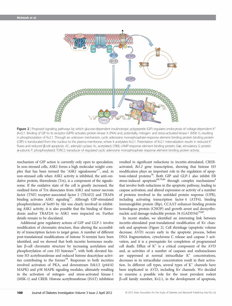
In recent studies, we identified an interesting link betweenincretin-stimulated post-translational modification of Kv chan-nels and apoptosis (Figure 2). Cell shrinkage (apoptotic volumedecrease; AVD) occurs early in the apoptotic process, beforeDNA fragmentation, cytochrome C release and caspase 3 acti-vation, and it is a prerequisite for completion of programmedcell death. Efflux of K+ is a critical component of the AVDand, as activities of a number of caspases and endonucleasesare suppressed at normal intracellular K+ concentrations,decreases in its intracellular concentration result in their activa-tion. In different cell types, several types of K+ channels havebeen implicated in AVD, including Kv channels. We decidedto examine a possible role for the most prevalent rodentb-cell family member, Kv2.1, in the development of apoptosis,
H2A H2B
H4 H3
Gαs GαsAC AC
PKA CBP
CBP
CREB CREB CBP
TORC2
P P
MSK PA
c PA
c
PAc
GIPKv2.1
P Ac
Figure 2 | Proposed signaling pathways by which glucose-dependent insulinotropic polypeptide (GIP) regulates endocytosis of voltage-dependent K+
(Kv)2.1. Binding of GIP to its receptor (GIPR) activates protein kinase A (PKA) and, potentially, mitogen- and stress-activated kinase-1 (MSK-1), resultingin phosphorylation of Kv2.1. Through an unknown mechanism, cyclic adenosine monophosphate-response element binding protein binding protein(CBP) is translocated from the nucleus to the plasma membrane, where it acetylates Kv2.1. Potentiation of Kv2.1 internalization results in reduced K+
fluxes and reduced b-cell apoptosis. AC, adenylyl cyclase; Ac, acetylated; CREB, cAMP-response element binding protein; Gas, stimulatory G proteina-subunit; P, phosphorylated; TORC2, transducer of regulated cyclic adenosine monophosphate response element binding protein activity.
100 Journal of Diabetes Investigation Volume 3 Issue 2 April 2012 ª 2012 Asian Association for the Study of Diabetes and Blackwell Publishing Asia Pty Ltd
McIntosh et al.

and determine whether regulation of this process by GIP andGLP-1 contributes to their prosurvival effects51. INS-1 b-cells, inwhich Kv2.1 was overexpressed, showed potentiated apoptoticresponses to mitochondrial and ER stress, whereas GIP orGLP-1 reduced the potentiation. In studies designed to identifytheir mode of action, both GIP and GLP-1 promoted phos-phorylation and acetylation of Kv2.1 through pathways involv-ing PKA, and/or MSK-1 and HAT (Figure 2). This wasassociated with reduced cell surface expression of Kv2.1. Wesubsequently found that GIP and GLP-1 promoted nuclear/cytoplasmic shuttling of CBP, resulting in its interaction withKv2.1 (Figure 2). Downregulation of CBP ablated incretin-induced acetylation of Kv2.1, suggesting that this HAT is pri-marily responsible for the acetylation51. As ASK1 has beenshown to play a key role in the pro-apoptotic modulation of Kvchannels81, it is likely that the GIP-activated pathway leading tophosphorylation of serine 83 in ASK1 interacts with eventsleading to Kv protein modification.
EFFECTS OF GIP ON ADIPOSE TISSUEFat ingestion is a major stimulus for GIP secretion in humans,dogs and rodents3,82,83, and there is increasing evidence support-ing a physiological role for GIP in promoting fat storage. Thereare two pathways by which GIP impacts on adipocyte metabo-lism: directly through interaction with GIP receptors on the adi-pocyte and through stimulation of insulin secretion. GIPinfusion has been shown to promote the clearance of chylomi-cron-associated triglyceride (TG) in dogs84, and to lower plasmaTG responses to intraduodenal fat in rats85. However, GIP hadno major effect on the rate of removal of intravenously adminis-tered TG86, suggesting that GIP stimulates release of TG fromchylomicrons and uptake into adipose tissue. Support for a rolefor GIP in regulating adipose tissue mass came from rodentstudies. Miyawaki et al.87 first showed that GIPR)/) mice wereresistant to obesity when fed a high-fat diet. Mice that were trea-ted with GIPR peptide antagonists88, vaccinated against GIP89
or subjected to selective K-cell ablation90 all showed increasedresistance to high-fat feeding-induced obesity3, showing thatGIP normally promotes lipid storage.
Early studies on direct adipocyte actions of GIP showed stim-ulatory effects on fatty acid (FA) synthesis from acetate in adi-pose tissue explants91, increased uptake and incorporation ofglucose into lipids92, as well as enhanced free FA (FFA) incorpo-ration into adipose tissue93. Adipocyte lipoprotein lipase (LPL)is responsible for the hydrolysis of TG in circulating chylomi-crons, TG-rich lipoproteins and very low-density lipoproteins,resulting in adipocyte uptake of FFA and monoacylglycerol, andthe promotion of lipogenesis. GIP was shown to increase LPLenzyme activity, in an insulin-dependent manner, in cultured3T3-L1 adipocytes, rodent adipocytes and subcutaneous humanadipocytes94–96. In view of the close correlation between humanGIP responses and plasma post-heparin LPL levels97, it has beensuggested that GIP acts on adipocyte storage by matching adi-pose tissue uptake of FA with the triglyceride load98.
Suboptimal levels of circulating FFA result in greatly reducedb-cell responsiveness to subsequent glucose stimulation99.In vitro studies showed that, in the absence of insulin, GIPstimulates adipocyte TG hydrolysis through PKA activa-tion100,101, and we suggested that GIP primes b-cells duringfasting by releasing adipocyte FFA into the circulation3,100.Getty-Kaushik et al.102 showed that GIP-stimulated increases inglycerol production were accompanied by decreased FFA inperifused adipocytes. They interpreted these responses as reflect-ing GIP-induced re-esterification from excess FFA102,103. A simi-lar response was recently reported in humans, with GIPinfusion resulting in small increases in adipose tissue FFAre-esterification104. Although, that study was carried out underhyperglycemic conditions. GIP might also have long-term effectson lipid metabolism, as synthesis of pancreatic lipase and coli-pase were both stimulated by GIP105, an effect that shouldincrease efficiency of lipid uptake.
In the presence of insulin, GIP signaling appears to playimportant roles in both the differentiation of preadipocytes andlipogenesis. A complex set of events is involved in preadipocyteto adipocyte development, including growth arrest, increasedtranscription factor and lipogenic enzyme expression, accumula-tion of lipid, and the development of sensitivity to regulatoryhormones106. Expression of the GIPR is extremely low in prea-dipocytes103,107,108, but both messenger ribonucleic acid (mRNA)and protein expression increase during differentiation of 3T3-L1cell103,108 and human107 preadipocytes. GIP acted synergisticallywith insulin to increase neutral lipid accumulation during pro-gression of 3T3-L1 preadipocytes to the adipocyte phenotype108.However, it was unclear as to whether synergistic effects of GIPand insulin on GIPR mRNA levels were a result of direct effectson gene transcription or secondary to the progression of differ-entiation. 3T3-L1 cell differentiation was associated with upregu-lation of nuclear levels of peroxisome proliferator-activatedreceptor (PPAR)c. Treatment with the PPARc receptor agonists,LY171883 and rosiglitazone, increased GIPR expression in fullydifferentiated 3T3-L1 adipocytes, whereas the antagonist,GW9662, ablated expression108. Acetylation of histone H3/H4was also increased during differentiation, and both PPARc andacetylated histone H3/H4 bound to a region of the GIPR pro-moter containing the peroxisome proliferator response element(PPRE). As RNA interference (RNAi) knockdown of PPARc indifferentiated 3T3-L1 adipocytes greatly reduced GIPR levels,PPARc appears to be a critical transcription factor in regulatingadipocyte receptor expression, but it is unclear as to the roleplayed by insulin.
Evidence has been presented for the involvement of bothPPARa109,110 and PPARc111 in the regulation of rodent b-cellGIPR expression, and further studies are required to clarifywhether there are cell-selective differences in regulation. Addi-tionally, in earlier studies, it was shown that the GIPR is down-regulated in b-cells of obese rodent models of diabetes109,110,112,but in studies on Vancouver Diabetic Fatty (VDF) Zucker rats,we recently found that, compared with lean controls, GIPR and
ª 2012 Asian Association for the Study of Diabetes and Blackwell Publishing Asia Pty Ltd Journal of Diabetes Investigation Volume 3 Issue 2 April 2012 101
GIP signaling in b-cells and adipocytes

PPARc protein levels were increased in epididymal and retro-peritoneal fat pads, decreased in the perirenal fat depot andunchanged in other fat deposits (Figure 3). In contrast, GIPRexpression in subcutaneous adipose tissue from human obesefemales was reported to be lower than in lean control sub-jects113. However, these results are difficult to compare, becauseof the different fat depots studied. Additionally, the sensitivity ofGIPR expression to the prevailing insulin concentration, thelevel of adipose tissue insulin resistance and the glycemic statusof the subjects/animals could all contribute significantly to thelevel of GIPR expression.
The pathways involved in GIP-stimulated lipogenesis areproving difficult to define, as a result of interactions between
GIP, insulin and adipokine signaling. GIP stimulation of glucoseuptake was shown to involve increasing plasma membrane glu-cose transporter-4 (GLUT-4) levels through a PKB-mediatedpathway103. Human LPL gene expression is also stimulated byGIP activation of PKB114, resulting in downstream reductions inLKB1 and AMPK phosphorylation, and increased translocationof TORC2 (cAMP-responsive CREB coactivator 2 [CRTC2])into the nucleus. Interaction between TORC2 and phospho-CREB results in increased LPL gene expression114. Regulation ofthe phosphorylation state of TORC2 is complex and othermembers of the AMPK family (salt-inducible kinases [SIK]and MARK2)115 are also capable of TORC2 phosphorylation,whereas calcineurin and a cAMP-activated pathway inducedephosphorylation116. It has not been established as to which ofthese contribute to GIP-mediated effects. GIP also enhancesLPL enzyme activity in cultured 3T3-L1 cells and subcutaneoushuman adipocytes by non-transcriptional mechanisms95,96. Inthe 3T3-L1 cell line, GIP induces transient activation of p38MAPK and sustained activation of stress-activated proteinkinase (SAPK)/JNK, resulting in the release of resistin that, inturn activates PKB95. Somewhat surprisingly, subsequent eventsmimic those downstream of GIP in the human adipocyte, withdecreases in LKB1 and AMPK phosphorylation linked toincreased LPL secretion95. Human resistin (FIZZ3) shares onlymoderate sequence homology (�53%) with the mouse pep-tide117. Additionally, FIZZ3/resistin is only weakly expressed inhuman adipocytes, and monocytes/macrophages are the majorsites of FIZZ3/resistin production in adipose tissue118. It iscurrently unknown whether FIZZ3/resistin serves a paracrinefunction in adipocyte regulation or whether there is an entero–adipokine axis involving GIP and FIZZ3/resistin in humans.However, FIZZ3/resistin has been reported to increase FFAre-esterification119.
In the majority of in vitro studies to date, the effects of GIPon adipogenesis and lipogenesis have been shown to involvesynergistic actions with insulin, and there has been significantinterest in GIPR antagonists as potential therapies for obesity88.However, adipose tissue expansion has been suggested to be animportant adaptive response to increased food intake, as it pro-tects against excess fat deposition in other sites, such as liverand muscle120,121. GIP could be an important contributor to thisresponse and reducing its effect might not result in the antici-pated benefits. Additionally, studies on both transgenic mice122
and pigs123 expressing a dominant-negative GIPR showedgreatly reduced b-cell mass, with the mice becoming severelydiabetic, supporting a critical role for GIP signaling in b-celldevelopment and proliferation.
ACKNOWLEDGEMENTSStudies from the authors’ laboratory described in the reviewwere generously supported by funding to CHSMc from theCanadian Institutes of Health Research, Canadian DiabetesAssociation and the Canadian Foundation for Innovation. Theauthors declare no conflict of interest.
0
1
2
3
4
5
6
7
GIP
R pr
otei
n ex
pres
sion
(fold
diff
eren
ce v
s co
ntro
l)
Epididymal
Perirenal
Retro-p
eritoneal
Inguinal
Mesenteric
IBAT
Intraca
rdial
LeanFatty
**
**
**
**
0
1
2
3
4
5
6
PPA
Rγ2
prot
ein
expr
essi
on(fo
ld d
iffer
ence
vs
cont
rol)
Epididymal
Perirenal
Retro-p
eritoneal
Inguinal
Mesenteric IBAT
Intraca
rdial
**
**
**
**
(a)
(b)
Figure 3 | Glucose-dependent insulinotropic polypeptide receptor(GIPR) and peroxisome proliferator-activated receptor (PPAR)c proteinexpression levels in adipose tissue depots from lean and obese Vancou-ver Diabetic Fatty (VDF) Zucker rats. Tissue was collected from 18-week-old rats and western blot analyses were carried out, with quantificationby densitometry (n = 4–6 rats). Significance was tested using analysis ofvariance (ANOVA) with Newman–Keuls post-hoc test. **P < 0.05 vs leancontrol group. IBAT, interscapular brown adipose tissue.
102 Journal of Diabetes Investigation Volume 3 Issue 2 April 2012 ª 2012 Asian Association for the Study of Diabetes and Blackwell Publishing Asia Pty Ltd
McIntosh et al.

REFERENCES1. Brubaker PL. The glucagon-like peptides: pleiotropic regula-
tors of nutrient homeostasis. Ann N Y Acad Sci 2006; 1070:10–26.
2. Drucker DJ, Nauck MA. The incretin system: glucagon-likepeptide-1 receptor agonists and dipeptidyl peptidase-4inhibitors in type 2 diabetes. Lancet 2006; 368: 1696–1705.
3. McIntosh CHS, Widenmaier S, Kim S-J. Glucose-dependentinsulinotropic polypeptide (Gastric Inhibitory Polypeptide;GIP). Vitam Horm 2009; 80: 409–471.
4. McIntosh CHS, Widenmaier S, Kim S-J. Pleiotropic actions ofthe incretin hormones. Vitam Horm 2010; 84: 21–79.
5. Holst JJ, Vilsbøll T, Deacon CF. The incretin system and itsrole in type 2 diabetes mellitus. Mol Cell Endocrinol 2009;297: 127–136.
6. McIntosh CHS. Dipeptidyl peptidase IV inhibitors and diabe-tes therapy. Front Biosci 2008; 13: 1753–1773.
7. Irwin D. Molecular evolution of mammalian incretinhormone genes. Regul Pept 2009; 155: 121–130.
8. Jia X, Brown JC, Ma P, et al. Effects of glucose-dependentinsulinotropic polypeptide and glucagon-like peptide-I-(7-36) on insulin secretion. Am J Physiol 1995; 268: E645–E651.
9. Pederson RA, Brown JC. The insulinotropic action of gastricinhibitory polypeptide in the perfused isolated rat pancreas.Endocrinology 1976; 99: 780–785.
10. Vilsbøll T, Krarup T, Madsbad S, et al. Both GLP-1 and GIPare insulinotropic at basal and postprandial glucose levelsand contribute nearly equally to the incretin effect of ameal in healthy subjects. Regul Pept 2003; 114: 115–121.
11. Drucker DJ. The biology of incretin hormones. Cell Metab2006; 3: 153–165.
12. Baggio LL, Drucker DJ. Biology of incretins: GLP-1 and GIP.Gastroenterology 2007; 132: 2131–2157.
13. Doyle ME, Egan JM. Mechanisms of action of glucagon-likepeptide 1 in the pancreas. Pharmacol Ther 2007; 113: 546–593.
14. Yu Z, Jin T. New insights into the role of cAMP in the pro-duction and function of the incretin hormone glucagon-likepeptide-1 (GLP-1). Cell Signal 2010; 22: 1–8.
15. Leech CA, Chepurny OG, Holz GG. Epac2-dependent rap1activation and the control of islet insulin secretion by gluca-gon-like peptide-1. Vitam Horm 2010; 84: 279–302.
16. Ashcroft F, Rorsman P. Molecular defects in insulin secretionin type-2 diabetes. Rev Endocr Metab Disord 2004; 4: 135–142.
17. Hinke SA, Hellemans K, Schuit FC. Plasticity of the beta cellinsulin secretory competence: preparing the pancreatic betacell for the next meal. J Physiol (Lond) 2004; 558: 369–380.
18. Hiriart M, Aguilar-Bryan L. Channel regulation of glucosesensing in the pancreatic beta-cell. Am J Physiol EndocrinolMetab 2008; 295: 1298–1306.
19. MacDonald P, Wheeler MB. Voltage-dependent K+ channelsin pancreatic beta cells: role, regulation and potential astherapeutic targets. Diabetologia 2003; 46: 1046–1062.
20. Kashima Y, Miki T, Shibasaki T, et al. Critical role of cAMP-GEFII–Rim2 complex in incretin-potentiated insulinsecretion. J Biol Chem 2001; 276: 46046–46053.
21. Seino S, Shibasaki T. PKA-dependent and PKA-independentpathways for cAMP-regulated exocytosis. Physiol Rev 2005;85: 1303–1342.
22. Hinke SA, Pauly RP, Ehses J, et al. Role of glucose in chronicdesensitization of isolated rat islets and mouse insulinoma(betaTC-3) cells to glucose-dependent insulinotropic poly-peptide. J Endocrinol 2000; 165: 281–291.
23. Ehses JA, Pelech SL, Pederson RA, et al. Glucose-dependentinsulinotropic polypeptide activates the Raf-Mek1/2-ERK1/2module via a cyclic AMP/cAMP-dependent protein kinase/Rap1-mediated pathway. J Biol Chem 2002; 277: 37088–37097.
24. Holz GG, Heart E, Leech CA. Synchronizing Ca2+ and cAMPoscillations in pancreatic beta-cells: a role for glucosemetabolism and GLP-1 receptors? Am J Physiol Cell Physiol2008; 294: C4–C6.
25. Willoughby D, Cooper D. Organization and Ca2+ regulationof adenylyl cyclase in cAMP microdomains. Physiol Rev2007; 87: 965–1010.
26. Fridlyand LE, Harbeck MC, Roe MW, et al. Regulation ofcAMP dynamics by Ca2+ and G protein-coupled receptorsin the pancreatic beta-cell: a computational approach. Am JPhysiol Cell Physiol 2007; 294: C1924–C1933.
27. Holz GG. New insights concerning the glucose-depen-dent insulin secretagogue action of glucagon-like pep-tide-1 in pancreatic beta-cells. Horm Metab Res 2004; 36:787–794.
28. Seino S, Takahashi H, Fujimoto W, et al. Roles of cAMPsignaling in insulin granule exocytosis. Diabetes Obes Metab2009; 11(Suppl 4): 180–188.
29. Holz GG. Epac: A new cAMP-binding protein in support ofglucagon-like peptide-1 receptor-mediated signal transduc-tion in the pancreatic beta-cell. Diabetes 2004; 53: 5–13.
30. Holz GG, Kang G, Harbeck M, et al. Cell physiology of cAMPsensor Epac. J Physiol (Lond) 2006; 577: 5–15.
31. Chepurny OG, Kelley GG, Dzhura I, et al. PKA-dependentpotentiation of glucose-stimulated insulin secretion by Epacactivator 8-pCPT-2¢-O-Me-cAMP-AM in human islets ofLangerhans. Am J Physiol Endocrinol Metab 2010; 298: E622–E633.
32. Seino S, Shibasaki T, Minami K. Dynamics of insulin secre-tion and the clinical implications for obesity and diabetes.J Clin Invest 2011; 121: 2118–2125.
33. Gromada J, Bokvist K, Ding WG, et al. Glucagon-like peptide1 (7-36) amide stimulates exocytosis in human pancreaticbeta-cells by both proximal and distal regulatory steps instimulus-secretion coupling. Diabetes 1998; 47: 57–65.
34. Light PE, Manning Fox JE, Riedel MJ, et al. Glucagon-likepeptide-1 inhibits pancreatic ATP-sensitive potassium chan-nels via a protein kinase A- and ADP-dependent mecha-nism. Mol Endocrinol 2002; 16: 2135–2144.
ª 2012 Asian Association for the Study of Diabetes and Blackwell Publishing Asia Pty Ltd Journal of Diabetes Investigation Volume 3 Issue 2 April 2012 103
GIP signaling in b-cells and adipocytes

35. Kasai H, Hatakeyama H, Ohno M, et al. Exocytosis in isletbeta-cells. Adv Exp Med Biol 2010; 654: 305–338.
36. Niimura M, Miki T, Shibasaki T, et al. Critical role of theN-terminal cyclic AMP-binding domain of Epac2 in itssubcellular localization and function. J Cell Physiol 2009; 219:652–658.
37. Kang G, Chepurny OG, Malester B, et al. cAMP sensor Epacas a determinant of ATP-sensitive potassium channel activ-ity in human pancreatic beta cells and rat INS-1 cells. J Phys-iol (Lond) 2006; 573: 595–609.
38. Shibasaki T, Sunaga Y, Fujimoto K, et al. Interaction of ATPsensor, cAMP sensor, Ca2+ sensor, and voltage-dependentCa2+ channel in insulin granule exocytosis. J Biol Chem2004; 279: 7956–7961.
39. Shibasaki T, Takahashi H, Miki T, et al. Essential role of Epac2/Rap1 signaling in regulation of insulin granule dynamics bycAMP. Proc Natl Acad Sci USA 2007; 104: 19333–19338.
40. Dzhura I, Chepurny OG, Leech CA, et al. PhospholipaseC-epsilon links Epac2 activation to the potentiation ofglucose-stimulated insulin secretion from mouse islets ofLangerhans. Islets 2011; 3: 121–128.
41. Baukrowitz T, Schulte U, Oliver D, et al. PIP2 and PIP asdeterminants for ATP inhibition of KATP channels. Science1998; 282: 1141–1144.
42. Shyng S-L, Nichols C. Membrane phospholipid control ofnucleotide sensitivity of KATP channels. Science 1998; 282:1138–1141.
43. Lu M, Wheeler MB, Leng XH, et al. Stimulation of insulinsecretion and insulin gene expression by gastric inhibitorypolypeptide. Trans Assoc Am Phys 1993; 106: 42–53.
44. Wheeler MB, Gelling RW, McIntosh CHS, et al. Functionalexpression of the rat pancreatic islet glucose-dependentinsulinotropic polypeptide receptor: ligand binding andintracellular signaling properties. Endocrinology 1995; 136:4629–4639.
45. Miki T, Minami K, Shinozaki H, et al. Distinct effects of glu-cose-dependent insulinotropic polypeptide and glucagon-like peptide-1 on insulin secretion and gut motility. Diabetes2005; 54: 1056–1063.
46. Gloerich M, Bos JL. Epac: defining a new mechanism forcAMP action. Annu Rev Pharmacol Toxicol 2010; 50: 355–375.
47. Ehses JA, Lee SS, Pederson RA, et al. A new pathway forglucose-dependent insulinotropic polypeptide (GIP) recep-tor signaling: evidence for the involvement of phospholi-pase A2 in GIP-stimulated insulin secretion. J Biol Chem2001; 276: 23667–23673.
48. MacDonald PE, El-Kholy W, Riedel MJ, et al. The multipleactions of GLP-1 on the process of glucose-stimulated insu-lin secretion. Diabetes 2002; 51(Suppl 3): S434–S442.
49. Kim S-J, Choi WS, Han JSM, et al. A novel mechanism forthe suppression of a voltage-gated potassium channel byglucose-dependent insulinotropic polypeptide: proteinkinase A-dependent endocytosis. J Biol Chem 2005; 280:28692–28700.
50. MacDonald PE, Salapatek AMF, Wheeler MB. Glucagon-likepeptide-1 receptor activation antagonizes voltage-depen-dent repolarizing K+ currents in beta-cells: a possible glu-cose-dependent insulinotropic mechanism. Diabetes 2002;51(Suppl 3): S443–S447.
51. Kim S-J, Widenmaier SB, Choi WS, et al. Pancreatic b-cellprosurvival effects of the incretin hormones involve post-translational modification of Kv2.1 delayed rectifier chan-nels. Cell Death Differ 2012; 19: 333–344.
52. Jacobson DA, Weber CR, Bao S, et al. Modulation of thepancreatic islet b-cell-delayed rectifier potassium channelKv2.1 by the polyunsaturated fatty acid arachidonate. J BiolChem 2007; 282: 7442–7449.
53. Ding WG, Gromada J. Protein kinase A-dependent stimula-tion of exocytosis in mouse pancreatic beta-cells by glu-cose-dependent insulinotropic polypeptide. Diabetes 1997;46: 615–621.
54. Eliasson L, Ma X, Renstrom E, et al. SUR1 regulates PKA-independent cAMP-induced granule priming in mousepancreatic B-cells. J Gen Physiol 2003; 121: 181–197.
55. Yasuda T, Shibasaki T, Minami K, et al. Rim2alpha deter-mines docking and priming states in insulin granuleexocytosis. Cell Metab 2010; 12: 117–129.
56. Song W-J, Seshadri M, Ashraf U, et al. Snapin mediatesincretin action and augments glucose-dependent insulinsecretion. Cell Metab 2011; 13: 308–319.
57. Widenmaier SB, Sampaio AV, Underhill TM, et al. Noncanon-ical activation of Akt/protein kinase B in b-cells by the incre-tin hormone glucose-dependent insulinotropic polypeptide.J Biol Chem 2009; 284: 10764–10773.
58. Mandrup-Poulsen T. Apoptotic signal transduction path-ways in diabetes. Biochem Pharmacol 2003; 66: 1433–1440.
59. Rhodes CJ. Type 2 diabetes-a matter of beta-cell life anddeath? Science 2005; 307: 380–384.
60. Perl S, Kushner JA, Buchholz BA, et al. Significant humanbeta-cell turnover is limited to the first three decades of lifeas determined by in vivo thymidine analog incorporationand radiocarbon dating. J Clin Endocrinol Metab 2010; 95:E234–E249.
61. Cnop M, Igoillo-Esteve M, Hughes SJ, et al. Longevity ofhuman islet alpha- and beta-cells. Diabetes Obes Metab2011; 13(Suppl 1): 39–46.
62. Li Y, Hansotia T, Yusta B, et al. Glucagon-like peptide-1receptor signaling modulates beta cell apoptosis. J BiolChem 2003; 278: 471–478.
63. Wang Q, Brubaker PL. Glucagon-like peptide-1 treatmentdelays the onset of diabetes in 8 week-old db/db mice.Diabetologia 2002; 45: 1263–1273.
64. Farilla L, Hui H, Bertolotto C, et al. Glucagon-like peptide-1promotes islet cell growth and inhibits apoptosis in Zuckerdiabetic rats. Endocrinology 2002; 143: 4397–4408.
65. Kim S-J, Winter K, Nian C, et al. Glucose-dependent insulino-tropic polypeptide (GIP) stimulation of pancreatic beta-cell
104 Journal of Diabetes Investigation Volume 3 Issue 2 April 2012 ª 2012 Asian Association for the Study of Diabetes and Blackwell Publishing Asia Pty Ltd
McIntosh et al.

survival is dependent upon phosphatidylinositol 3-kinase(PI3K)/protein kinase B (PKB) signaling, inactivation of theforkhead transcription factor Foxo1, and down-regulation ofbax expression. J Biol Chem 2005; 280: 22297–22307.
66. Maida A, Hansotia T, Longuet C, et al. Differential impor-tance of glucose-dependent insulinotropic polypeptide vsglucagon-like peptide 1 receptor signaling for beta cellsurvival in mice. Gastroenterology 2009; 137: 2146–2157.
67. Widenmaier SB, Kim S-J, Yang GK, et al. A GIP receptor ago-nist exhibits beta-cell anti-apoptotic actions in rat modelsof diabetes resulting in improved beta-cell function andglycemic control. PLoS One 2010; 5: e9590.
68. Widenmaier SB, Ao Z, Kim S-J, et al. Suppression of p38MAPK and JNK via Akt-mediated inhibition of apoptosis sig-nal-regulating kinase 1 constitutes a core component ofthe beta-cell pro-survival effects of glucose-dependentinsulinotropic polypeptide. J Biol Chem 2009; 284: 30372–30382.
69. Ehses JA, Casilla VR, Doty T, et al. Glucose-dependent insuli-notropic polypeptide promotes beta-(INS-1) cell survival viacyclic adenosine monophosphate-mediated caspase-3 inhi-bition and regulation of p38 mitogen-activated proteinkinase. Endocrinology 2003; 144: 4433–4445.
70. Kim S-J, Nian C, Widenmaier S, et al. Glucose-dependentinsulinotropic polypeptide-mediated up-regulation of beta-cell antiapoptotic Bcl-2 gene expression is coordinated bycyclic AMP (cAMP) response element binding protein(CREB) and cAMP-responsive CREB coactivator 2. Mol CellBiol 2008; 28: 1644–1656.
71. Hui H, Nourparvar A, Zhao X, et al. Glucagon-like peptide-1inhibits apoptosis of insulin-secreting cells via a cyclic5¢-adenosine monophosphate-dependent protein kinaseA- and a phosphatidylinositol 3-kinase-dependent pathway.Endocrinology 2003; 144: 1444–1455.
72. Jhala US, Canettieri G, Screaton RA, et al. cAMP promotespancreatic beta-cell survival via CREB-mediated induction ofIRS2. Genes Dev 2003; 17: 1575–1580.
73. Van de Velde S, Hogan MF, Montminy M. mTOR links incre-tin signaling to HIF induction in pancreatic beta cells. ProcNatl Acad Sci USA 2011; 108: 16876–16882.
74. Trumper K, Trumper A, Trusheim H, et al. Integrative mito-genic role of protein kinase B/Akt in beta-cells. Ann N YAcad Sci 2000; 921: 242–250.
75. Trumper A, Trumper K, Trusheim H, et al. Glucose-depen-dent insulinotropic polypeptide is a growth factor for beta(INS-1) cells by pleiotropic signaling. Mol Endocrinol 2001;15: 1559–1570.
76. Tobiume T, Saitoh M, Ichijo H. Activation of ApoptosisSignal-Regulating Kinase 1 by the stress-induced activatingphosphorylation of pre-formed oligomer. J Cell Physiol 2002;191: 95–104.
77. Takeda K, Noguchi T, Naguro I, et al. Apoptosis signal-regu-lating kinase 1 in stress and immune response. Annu RevPharmacol Toxicol 2008; 48: 199–225.
78. Kim S-J, Nian C, McIntosh CHS. Glucose-dependent insulino-tropic polypeptide and glucagon-like peptide-1 modulatebeta-cell chromatin structure. J Biol Chem 2009; 284: 12896–12904.
79. Cunha DA, Ladriere L, Ortis F, et al. Glucagon-like peptide-1agonists protect pancreatic beta-cells from lipotoxic endo-plasmic reticulum stress through upregulation of BiP andJunB. Diabetes 2009; 58: 2851–2862.
80. Yusta B, Baggio LL, Estall JL, et al. GLP-1 receptor activationimproves beta cell function and survival following induc-tion of endoplasmic reticulum stress. Cell Metab 2006; 4:391–406.
81. Aras M, Aizenman E. Obligatory role of ASK1 in the apopto-tic surge of K+ currents. Neurosci Lett 2005; 387: 136–140.
82. Lardinois CK, Starich GH, Mazzaferri EL. The postprandialresponse of gastric inhibitory polypeptide to various dietaryfats in man. J Am Coll Nutr 1988; 7: 241–247.
83. Pederson RA. Gastric inhibitory polypeptide. In: Walsh JH,Dockray GJ (eds). Gut Peptides: Biochemistry and Physiology.Raven Press, New York, 1994; 217–259.
84. Wasada T, McCorkle K, Harris V, et al. Effect of gastric inhibi-tory polypeptide on plasma levels of chylomicron triglyce-rides in dogs. J Clin Invest 1981; 68: 1106–1107.
85. Ebert R, Nauck M, Creutzfeldt W. Effect of exogenous orendogenous gastric inhibitory polypeptide (GIP) on plasmatriglyceride responses in rats. Horm Metab Res 1991; 23:517–521.
86. Jorde R, Pettersen JE, Burhol PG. Lack of effect of exo-genous or endogenous gastric inhibitory polypeptide onthe elimination rate of Intralipid in man. Acta Med Scand1984; 216: 19–23.
87. Miyawaki K, Yamada Y, Ban N, et al. Inhibition of gastricinhibitory polypeptide signaling prevents obesity. Nat Med2002; 8: 738–742.
88. Irwin N, Flatt PR. Therapeutic potential for GIP receptoragonists and antagonists. Baillieres Best Pract Res Clin Endo-crinol Metab 2009; 23: 499–512.
89. Fulurija A, Lutz TA, Sladko K, et al. Vaccination against GIPfor the treatment of obesity. PLoS One 2008; 3: e3163.
90. Althage MC, Ford EL, Wang S, et al. Targeted ablation ofglucose-dependent insulinotropic polypeptide-producingcells in transgenic mice reduces obesity and insulin resis-tance induced by a high fat diet. J Biol Chem 2008; 283:18365–18376.
91. Oben J, Morgan L, Fletcher J, et al. Effect of the entero-pancreatic hormones, gastric inhibitory polypeptide andglucagon-like polypeptide-1(7-36) amide, on fatty acidsynthesis in explants of rat adipose tissue. J Endocrinol 1991;130: 267–272.
92. Hauner H, Glatting G, Kaminska D, et al. Effects of gastricinhibitory polypeptide on glucose and lipid metabolism ofisolated rat adipocytes. Ann Nutr Metab 1988; 32: 282–288.
93. Beck B, Max JP. Direct metabolic effects of gastric inhibitorypolypeptide (GIP): dissociation at physiological levels of
ª 2012 Asian Association for the Study of Diabetes and Blackwell Publishing Asia Pty Ltd Journal of Diabetes Investigation Volume 3 Issue 2 April 2012 105
GIP signaling in b-cells and adipocytes

effects on insulin-stimulated fatty acid and glucose incorpo-ration in rat adipose tissue. Diabetologia 1986; 29: 68.
94. Eckel RH, Fujimoto WY, Brunzell JD. Gastric inhibitory poly-peptide enhanced lipoprotein lipase activity in culturedpreadipocytes. Diabetes 1979; 28: 1141–1142.
95. Kim S-J, Nian C, McIntosh CHS. Activation of lipoproteinlipase by glucose-dependent insulinotropic polypeptide inadipocytes. A role for a protein kinase B, LKB1, and AMP-activated protein kinase cascade. J Biol Chem 2007; 282:8557–8567.
96. Kim S-J, Nian C, McIntosh CHS. Resistin is a key mediator ofglucose-dependent insulinotropic polypeptide (GIP) stimula-tion of lipoprotein lipase (LPL) activity in adipocytes. J BiolChem 2007; 282: 34139–34147.
97. Murphy MC, Isherwood SG, Sethi S, et al. Postprandial lipidand hormone responses to meals of varying fat contents:modulatory role of lipoprotein lipase? Eur J Clin Nutr 1995;49: 578–588.
98. Morgan LM. The metabolic role of GIP: physiology andpathology. Biochem Soc Trans 1996; 24: 585–591.
99. McGarry J. Dysregulation of fatty acid metabolism in theetiology of type 2 diabetes. Diabetes 2002; 51: 7–18.
100. McIntosh CHS, Bremsak I, Lynn FC, et al. Glucose-depen-dent insulinotropic polypeptide stimulation of lipolysis indifferentiated 3T3-L1 cells: wortmannin-sensitive inhibitionby insulin. Endocrinology 1999; 140: 398–404.
101. Yip RG, Boylan MO, Kieffer TJ, et al. Functional GIP receptorsare present on adipocytes. Endocrinology 1998; 139: 4004–4007.
102. Getty-Kaushik L, Song DH, Boylan MO, et al. Glucose-dependent insulinotropic polypeptide modulates adipocytelipolysis and reesterification. Obesity (Silver Spring) 2006; 14:1124–1131.
103. Song DH, Getty-Kaushik L, Tseng E, et al. Glucose-depen-dent insulinotropic polypeptide enhances adipocyte devel-opment and glucose uptake in part through Akt activation.Gastroenterology 2007; 133: 1796–1805.
104. Asmar M, Simonsen L, Madsbad S, et al. Glucose-depen-dent insulinotropic polypeptide may enhance fatty acidre-esterification in subcutaneous abdominal adipose tissuein lean humans. Diabetes 2010; 59: 2160–2163.
105. Duan RD, Erlanson-Albertsson C. Gastric inhibitory polypep-tide stimulates pancreatic lipase and colipase synthesis inrats. Am J Physiol 1992; 262: G779–G784.
106. Rosen E, Spiegelman B. Molecular regulation of adipogene-sis. Annu Rev Cell Dev Biol 2000; 16: 145–171.
107. Weaver RE, Donnelly D, Wabitsch M, et al. Functionalexpression of glucose-dependent insulinotropic polypeptidereceptors is coupled to differentiation in a human adipo-cyte model. Int J Obes (Lond) 2008; 32: 1705–1711.
108. Kim S-J, Nian C, McIntosh CHS. Adipocyte expression of theglucose-dependent insulinotropic polypeptide receptorinvolves gene regulation by PPARgamma and histoneacetylation. J Lipid Res 2011; 52: 759–770.
109. Lynn FC, Pamir N, Ng EH, et al. Defective glucose-depen-dent insulinotropic polypeptide receptor expression indiabetic fatty Zucker rats. Diabetes 2001; 50: 1004–1011.
110. Lynn FC, Thompson SA, Pospisilik JA, et al. A novel pathwayfor regulation of glucose-dependent insulinotropic polypep-tide (GIP) receptor expression in beta cells. FASEB J 2003;17: 91–93.
111. Gupta D, Peshavaria M, Monga N, et al. Physiologic andpharmacologic modulation of glucose-dependent insulino-tropic polypeptide (GIP) receptor expression in beta-cells byperoxisome proliferator-activated receptor (PPAR)-gammasignaling: possible mechanism for the GIP resistance intype 2 diabetes. Diabetes 2010; 59: 1445–1450.
112. Xu G, Kaneto H, Laybutt DR, et al. Downregulation of GLP-1and GIP receptor expression by hyperglycemia: possiblecontribution to impaired incretin effects in diabetes. Diabe-tes 2007; 56: 1551–1558.
113. Rudovich N, Kaiser S, Engeli S, et al. GIP receptor mRNAexpression in different fat tissue depots in postmenopausalnon-diabetic women. Regul Pept 2007; 142: 138–145.
114. Kim S-J, Nian C, McIntosh CHS. GIP increases human adipo-cyte LPL expression through CREB and TORC2-mediatedtrans-activation of the LPL gene. J Lipid Res 2010; 51: 3145–3157.
115. Jansson D, Ng AC-H, Fu A, et al. Glucose controls CREBactivity in islet cells via regulated phosphorylation of TORC2.Proc Natl Acad Sci USA 2008; 105: 10161–10166.
116. Fu A, Screaton RA. Using kinomics to delineate signalingpathways: control of CRTC2/TORC2 by the AMPK family.Cell Cycle 2008; 7: 3823–3828.
117. Yang R-Z, Huang Q, Xu A, et al. Comparative studies ofresistin expression and phylogenomics in human andmouse. Biochem Biophys Res Commun 2003; 310: 927–935.
118. McTernan PG, Kusminski CM, Kumar S. Resistin. Curr OpinLipidol 2006; 17: 170–175.
119. Ort T, Arjona AA, MacDougall JR, et al. Recombinant humanFIZZ3/resistin stimulates lipolysis in cultured human adipo-cytes, mouse adipose explants, and normal mice. Endocri-nology 2005; 146: 2200–2209.
120. Sethi J, Vudal-Puig A. Adipose tissue function and plasticityorchestrate nutritional adaptation. J Lipid Res 2007; 48:1253–1262.
121. Kim J-Y, van de Wall E, Laplante M, et al. Obesity-associatedimprovements in metabolic profile through expansion ofadipose tissue. J Clin Invest 2007; 117: 2621–2637.
122. Herbach N, Goeke B, Schneider M, et al. Overexpression ofa dominant negative GIP receptor in transgenic mice resultsin disturbed postnatal pancreatic islet and beta-cell devel-opment. Regul Pept 2005; 125: 103–117.
123. Renner S, Fehlings C, Herbach N, et al. Glucose intoler-ance and reduced proliferation of pancreatic beta-cells intransgenic pigs with impaired glucose-dependent insulino-tropic polypeptide function. Diabetes 2010; 59: 1228–1238.
106 Journal of Diabetes Investigation Volume 3 Issue 2 April 2012 ª 2012 Asian Association for the Study of Diabetes and Blackwell Publishing Asia Pty Ltd
McIntosh et al.


![Apache2 Ubuntu Default Page: It works · Web viewJournal of Parkinson's disease 2014; 4: 337-344. [35]Faivre E, Gault VA, Thorens B, Holscher C. Glucose-dependent insulinotropic polypeptide](https://img.dokumen.tips/doc/110x75/60f6f730c345001eb01b5231/apache2-ubuntu-default-page-it-works-web-view-journal-of-parkinsons-disease-2014.jpg)