Embed Size (px)
Citation preview

University of Massachusetts AmherstScholarWorks@UMass Amherst
Masters Theses 1911 - February 2014
2011
Effect of the Flavonoid Quercetin on AdipocytesJennifer C. SwickUniversity of Massachusetts Amherst
Follow this and additional works at: https://scholarworks.umass.edu/theses
Part of the Alternative and Complementary Medicine Commons, Biochemical Phenomena,Metabolism, and Nutrition Commons, and the Nutritional and Metabolic Diseases Commons
This thesis is brought to you for free and open access by ScholarWorks@UMass Amherst. It has been accepted for inclusion in Masters Theses 1911 -February 2014 by an authorized administrator of ScholarWorks@UMass Amherst. For more information, please [email protected].
Swick, Jennifer C., "Effect of the Flavonoid Quercetin on Adipocytes" (2011). Masters Theses 1911 - February 2014. 724.Retrieved from https://scholarworks.umass.edu/theses/724

EFFECT OF THE FLAVONOID QUERCETIN ON ADIPOCYTES
A Thesis Presented
By:
JENNIFER C SWICK
Submitted to the Graduate School of the University of Massachusetts Amherst in partial fulfillment
of the requirements for the degree of
MASTER OF SCIENCE
SEPTEMBER 2011
Department of Nutrition
EFFECT OF THE FLAVONOID QUERCETIN ON ADIPOCYTES
A Thesis Presented
By:
JENNIFER C SWICK Approved as to style and content by: ________________________________________ Young-Cheul Kim, Chair ________________________________________ Richard Wood, Member ________________________________________ Jerusha Peterman, Member
__________________________________ Nancy Cohen, Department Head
Department of Nutrition

DEDICATION
To those not eating fruits and vegetables; your mother was right.

iv
ACKNOWLEDGEMENTS Words do not express the gratitude I have for my advisor, Dr. Young-Cheul
Kim, and the opportunity he provided me with to complete this project. He opened a
door for me, and along the way has generously offered his hand and his heart in
support and guidance for me as a developing young researcher. To him, I am forever
grateful.
I would like to thank committee member, Dr. Richard Wood, for his
encouraging words, contagious attitude, and brilliant insight into the scientific pursuit.
I would like to thank committee member, Dr. Jerusha Peterman, for her
thoughtful comments and suggestions, and inspiring support and interest in my
professional development.
I would like to thank Dr. Oh-kwan Lee, Dr. Yong-Ook Kim, and Dr.
Jeongsook Noh for their generous investment of time and energy put into the progress
of my research. Without them I would not have been able to have accomplished all
that I have. And more, I would not know the value of honesty, hard work,
perseverance, and friendship in the lab. I will carry with me what each of you taught
me.
I would like to extend a special thank you to all my fellow colleagues in the
Department of Nutrition, especially Brianna Gray and Eunjee Ahn who have worked
alongside me in the lab. Thank you all for your friendship and support over the last
two years, especially for your open ears and kind words which have sustained me
along the way. Finally I would like to extend my deepest gratitude to my family and
friends for your unwavering love and support, and for a place I can always call home.

v
ABSTRACT
EFFECT OF THE FLAVONOID QUERCETIN ON ADIPOCYTES
SEPTEMBER 2011
JENNIFER C SWICK, B.S., UNIVERSITY OF MASSACHUSETTS, AMHERST
M.S. UNIVERSITY OF MASSACHUSETTS, AMHERST
DIRECTED BY: PROFESSOR YOUNG-CHEUL KIM
Obesity is an urgent global public health concern as prevalence rates continue
to increase, especially among children. Obesity is defined at the cellular level as an
increase in adipocyte number (hyperplasia) and size (hypertrophy). Both lead to the
dysfunction of adipose tissue, which has been identified as the link between obesity
and chronic disease. Bioactive compounds, naturally occurring in fruits and vegetables,
hold enormous potential in regulating adipocyte biology. Quercetin, the most
commonly consumed dietary flavonoid, is a strong potential anti-obesity agent that has
been implicated as an AMP-activated protein kinase (AMPK) activator and shown to
ameliorate symptoms of metabolic syndrome in vivo. Here we investigated quercetin’s
effect on (1) adipogenesis, the process of increasing adipocyte number, and (2)
metabolism of mature adipocytes. In 3T3-L1 preadipocytes, quercetin dose-
dependently inhibited adipogenesis, as evidenced by decreased lipid accumulation and
expression of adipogenic markers such as peroxisome proliferator-activated receptor
(PPAR) γ, CCAAT/ enhancer binding protein (C/EBP) α, adipocyte fatty acid binding
protein 2 (aP2), and acetyl-CoA carboxylase (ACC) on mRNA and protein levels. This
inhibitory effect was limited to the early stages of adipogenesis (0-36 hours), and

vi
quercetin treatment altered the normal expression pattern of cell cycle related genes
Cyclin A and p27, indicating quercetin may inhibit adipogenesis through cell cycle
events. We next investigated quercetin’s ability to activate AMPK and the metabolic
pathways related to AMPK activation: lipolysis and β-oxidation. Quercetin increased
phosphorylation of AMPK and its downstream target ACC. Further, quercetin
treatment (100μM) increased free fatty acid content in the media through an AMPK-
dependent mechanism. Quercetin up-regulated mRNA expression of uncoupling
proteins 3 (UCP3) and peroxisome proliferator-activated receptor-gamma co-activator
1 alpha (PGC-1α), indicating that quercetin may induce mitochondrial oxidative
pathways, also through an AMPK-dependent pathway. These findings suggest (1)
quercetin inhibits adipogenesis through the regulation of early cell cycle events
required for adipogenic differentiation, and (2) quercetin’s activation of AMPK
induces lipolytic and oxidative pathways. Taken together, quercetin could be further
developed as an anti-obesity agent because of its potential to inhibit both hyperplasia
and hypertrophy in vitro.

vii
TABLE OF CONTENTS
Page ACKNOWLEDGEMENTS....................................................................................................................iv
ABSTRACT............................................................................................................................................v
LIST OF TABLES..................................................................................................................................x
LIST OF FIGURES................................................................................................................................xi
CHAPTER
1 BACKGROUND....................................................................................................................................1
1.1Overview................................................................................................................................1
1.2 The Extent of the Problem of Obesity..................................................................................2
1.3 The Role of Adipose Tissue in the Development of Obesity...............................................4
1.3.1 Adipogenesis.......................................................................................................4
1.3.2 Adipocyte Metabolism........................................................................................8
1.3.2.1 De Novo Lipogenesis.........................................................................9
1.3.2.2 Lipolysis............................................................................................9
1.3.2.3 β-oxidation........................................................................................10
1.4 The Role of AMP activated protein Kinase (AMPK) in Adipocyte Biology .....................12
1.4.1 Overview............................................................................................................12
1.4.2 Inhibitors and Activators....................................................................................13
1.4.3 Role of AMPK in Adipogenesis.........................................................................13
1.4.4 Role of AMPK in Mature Adipocytes................................................................14
1.5 The Potential Health Benefits of Bioactive Compounds in the Development of Obesity..16
1.6 The Dietary Flavonoid Quercetin.........................................................................................17
1.6.1 Overview.............................................................................................................17
1.6.2 Bioavailability.....................................................................................................19
1.6.3 Gut Metabolism...................................................................................................19
1.6.4 Absorption and Transport ...................................................................................20
1.7 Quercetin’s Effect on Metabolic Syndrome: In Vivo Studies...............................................21

viii
2 EFFECT OF QUERCETIN ON ADIPOGENESIS..............................................................................24
2.1 Literature Review................................................................................................................24
2.2 Purpose of Study..................................................................................................................26
2.3 Materials and Methods.........................................................................................................27
2.3.1 Cell Culture Model and Treatments....................................................................27
2.3.2 Oil Red O Lipid Staining....................................................................................28
2.3.3 Cell Viability and Counting................................................................................28
2.3.4 Protein Isolation and Western Blotting...............................................................29
2.3.5 RNA Isolation and Analysis................................................................................29
2.3.6 Stastical Analysis.................................................................................................30
2.4 Results...................................................................................................................................31
2.4.1 Quercetin inhibits lipid accumulation and adipogenic transcriptional factors....31
2.4.2 Quercetin’s inhibitory effect is limited to early time points................................31
2.4.3 Quercetin may inhibit adipogenesis through regulation of early cell cycle
Events.............................................................................................................32
2.5 Discussion.............................................................................................................................40
3 EFFECT OF QUERCETIN ON ADIPOCYTE METABOLISM..........................................................44
3.1 Literature Review..................................................................................................................44
3.2 Purpose of Study...................................................................................................................45
3.3 Materials and Methods..........................................................................................................46
3.3.1 Cell Culture and Treatments................................................................................46
3.3.2 Oil Red O Lipid Staining.....................................................................................47
3.3.3 Protein Isolation and Western Blotting...............................................................47
3.3.4 RNA isolation and Analysis................................................................................48
3.3.5 Free Fatty Acid and Glycerol Assays..................................................................49
3.3.6 Statistical Analysis..............................................................................................49
3.4 Results...................................................................................................................................49
3.4.1 Quercetin phosphorylates ACC, a downstream target of AMPK.......................49

ix
3.4.2 Quercetin induces partial lipolysis......................................................................50
3.4.3 Quercetin induces oxidative pathways................................................................50
3.5 Discussion.............................................................................................................................56
4 CONCLUSION & FUTURE DIRECTIONS.........................................................................................60
4.1 Summary...............................................................................................................................60
4.2 Limitations............................................................................................................................61
4.3 Significance of Findings in the Context of Obesity and Metabolic Syndrome....................62
4.3.1 Weight Management...........................................................................................63
4.3.2 Insulin Resistance................................................................................................64
4.4. Application of Quercetin from a Public Health Perspective................................................65
4.5 Future Research Directions...................................................................................................66
4.5.1 Quercetin’s effect on adipogenesis......................................................................66
4.5.2. Quercetin’s effect on mature adipocytes............................................................66
APPENDICES
1 QUERCETIN MAY INHIBIT NOX4 EXPRESSION DURING EARLY STAGES OF
ADIPOGENESIS.........................................................................................................68
2 QUERCETIN INHIBITS LIPID ACCUMULATION OF 10T1/2 MESENCHYMAL STEM
CELLS..........................................................................................................................69
3 QUERCETIN’S INHIBITORY EFFECT IS NOT LIMITED TO EARLY TIME POINTS IN
10T1/2 MESENCHYMAL STEM CELLS..................................................................70
4 COMPOUND C DID NOT REVERSE THE ANTI-LIPOGENIC EFFECT OF
QUERCETIN...............................................................................................................71
5 QUERCETIN TREATMENT IN MATURE ADIPOCYTES RESULTS IN DECREASED
GLYCEROL CONTENT IN MEDIA..........................................................................72
6 QUERCETIN TREATMENT IN MAUTRE ADIPOCYTES FOR 48 HOURS DID NOT
ALTER INTRACELLULAR TRIGLYCERIDE LEVELS.........................................73
7 QUERCETIN UPREGULATES MRNA EXPRESSION OF ADIPONECTIN....................74
REFERENCES..........................................................................................................................................75

x
LIST OF TABLES
Table Page
1. Sources of Quercetin.........................................................................................18
2. Primer Information............................................................................................30
xi
LIST OF FIGURES
Figure Page
1. Structure of quercetin.............................................................................................17
2. Quercetin dose-dependently inhibits lipid accumulation.......................................33
3. Quercetin inhibits adipogenic factors at the mRNA level......................................34
4. Quercetin inhibits adipogenic factors at the protein level......................................35
5. Quercetin’s inhibitory effect on lipid accumulation is limited to early time
point..................................................................................................................36
6. Quercetin does not affect cell viability but may inhibit cell number.....................37
7. Quercetin delays mRNA expression of Cyclin A during the early stages of
adipogenesis......................................................................................................38
8. Quercetin sustains protein expression of P27 during the early stages of
adipogenesis......................................................................................................39
9. Quercetin increases protein expression of pAMPK and pACC.............................51
10. Quercetin 100μM increases free fatty acid content in media.................................52
11. Quercetin up-regulates mRNA expression of PGC-1α mRNA..............................53
12. Quercetin up-regulates mRNA expression of Uncoupling Protein 1.....................54
13. Quercetin up-regulates mRNA expression of Uncoupling Protein 3.....................55
1
CHAPTER 1
BACKGROUND
1.1 Overview
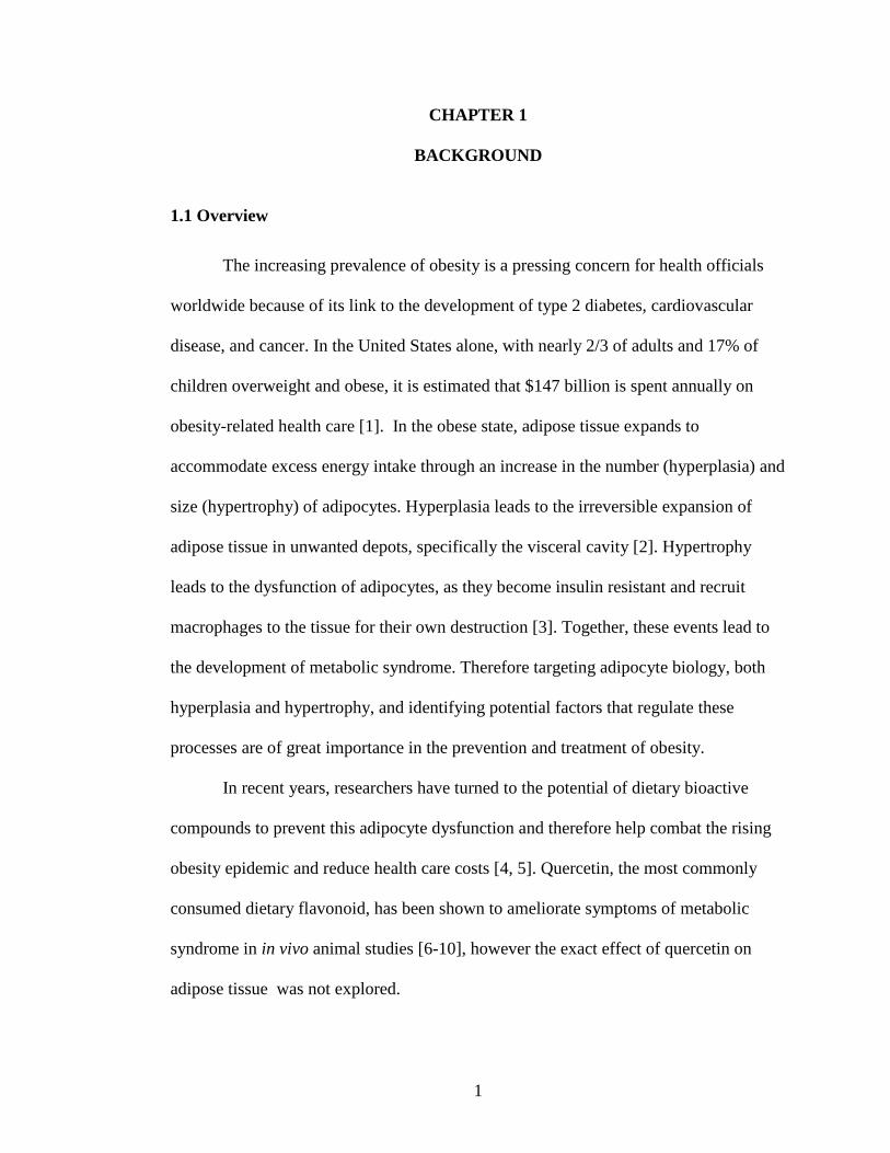
The increasing prevalence of obesity is a pressing concern for health officials
worldwide because of its link to the development of type 2 diabetes, cardiovascular
disease, and cancer. In the United States alone, with nearly 2/3 of adults and 17% of
children overweight and obese, it is estimated that $147 billion is spent annually on
obesity-related health care [1]. In the obese state, adipose tissue expands to
accommodate excess energy intake through an increase in the number (hyperplasia) and
size (hypertrophy) of adipocytes. Hyperplasia leads to the irreversible expansion of
adipose tissue in unwanted depots, specifically the visceral cavity [2]. Hypertrophy
leads to the dysfunction of adipocytes, as they become insulin resistant and recruit
macrophages to the tissue for their own destruction [3]. Together, these events lead to
the development of metabolic syndrome. Therefore targeting adipocyte biology, both
hyperplasia and hypertrophy, and identifying potential factors that regulate these
processes are of great importance in the prevention and treatment of obesity.
In recent years, researchers have turned to the potential of dietary bioactive
compounds to prevent this adipocyte dysfunction and therefore help combat the rising
obesity epidemic and reduce health care costs [4, 5]. Quercetin, the most commonly
consumed dietary flavonoid, has been shown to ameliorate symptoms of metabolic
syndrome in in vivo animal studies [6-10], however the exact effect of quercetin on
adipose tissue was not explored.

2
Research has shown that quercetin inhibits adipogenesis in both human and
mice cell models [11-13], and induces lipolysis [14, 15] in mature mice adipocytes;
however the underlying mechanisms are unknown. Interestingly, quercetin has recently
been implicated as an AMP-activated protein kinase (AMPK) activator [13]. AMPK is
the major metabolic regulator of the cell, and a major target for obesity-related
conditions because of its ability to induce fatty acid oxidation [16]. However,
quercetin’s ability to alter the metabolic state of mature adipocytes has not been
explored. Therefore, our focus was to investigate (1) mechanisms of quercetin’s anti-
adipogenic effect, and (2) metabolic effects of quercetin in mature adipocytes through
AMPK activation. These findings will largely improve our understanding of quercetin’s
ability to ameliorate hyperplasia and hypertrophy, and thus inform its development as
an anti-obesity agent.
1.2 The Extent of the Problem of Obesity
Obesity has become one of the most urgent concerns of health care officials
over the past two decades, as dramatic increases in prevalence rates have been seen not
only in the United States but worldwide. In the U.S. alone, with nearly 2/3 of adults and
17% of children overweight or obese, it is estimated that $147 billion is spent annually
on obesity-related health care [1]. The World Health Organization outlines the severity
of the problem with the following: (1) worldwide obesity has more than doubled since
1980, (2) overweight and obesity are the 5th leading cause of death globally, (3) in 2008
more than 10% of the population was obese, (3) in 2010 nearly 43 million children

3
under the age of five were overweight, and (4) obesity accounts for the incidence of
44% of diabetes, 23% of ischemic attacks, and up to 41% of certain cancers [17]. This
places an enormous burden on health care systems and impacts health care costs
worldwide.
Obesity has been linked to all of the following: coronary heart disease, type 2
diabetes, cancer (specifically breast and colon), hypertension, dyslipidemia, stroke,
liver and gallbladder disease, sleep apnea, osteoarthritis, and gynecological
complications [18]. In an effort to classify the most common conditions related to
obesity, the term “metabolic syndrome” has been used to include the development of
hypertension, dyslipidemia, hyperglycemia, and insulin resistance. This term was
coined because of the surprising combination of conditions seen repeatedly in
overweight and obese patients. Currently 8.3% of the US population is affected by
diabetes [19], 12% by cardiovascular disease [20], and 34% of adults are classified as
having metabolic syndrome [21]. If no action is taken, the prevalence of metabolic
syndrome will likely only increase in the population, as childhood obesity has tripled
over the last 25 years, and continues to grow [22].
At the cellular level, adipose tissue expands its capacity to store lipid by
increasing adipocyte size (hypertrophy) and number (hyperplasia). Hyperplasia leads
to the expansion of adipose tissue in unwanted depots, specifically the visceral cavity,
through the process of adipogenesis. Adipogenesis occurs in two stages: (1) the
commitment of precursor mesenchymal stem cells to preadipocytes, and (2) the
differentiation of preadipocytes to mature adipocytes [3]. Because adipocytes are
4
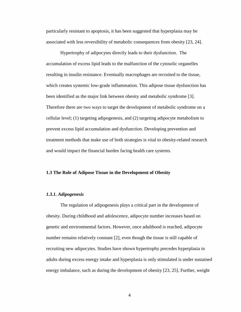
particularly resistant to apoptosis, it has been suggested that hyperplasia may be
associated with less reversibility of metabolic consequences from obesity [23, 24].
Hypertrophy of adipocytes directly leads to their dysfunction. The
accumulation of excess lipid leads to the malfunction of the cytosolic organelles
resulting in insulin resistance. Eventually macrophages are recruited to the tissue,
which creates systemic low-grade inflammation. This adipose tissue dysfunction has
been identified as the major link between obesity and metabolic syndrome [3].
Therefore there are two ways to target the development of metabolic syndrome on a
cellular level; (1) targeting adipogenesis, and (2) targeting adipocyte metabolism to
prevent excess lipid accumulation and dysfunction. Developing prevention and
treatment methods that make use of both strategies is vital to obesity-related research
and would impact the financial burden facing health care systems.
1.3 The Role of Adipose Tissue in the Development of Obesity
1.3.1. Adipogenesis
The regulation of adipogenesis plays a critical part in the development of
obesity. During childhood and adolescence, adipocyte number increases based on
genetic and environmental factors. However, once adulthood is reached, adipocyte
number remains relatively constant [2], even though the tissue is still capable of
recruiting new adipocytes. Studies have shown hypertrophy precedes hyperplasia in
adults during excess energy intake and hyperplasia is only stimulated is under sustained
energy imbalance, such as during the development of obesity [23, 25]. Further, weight

5
loss results in a reduction in adipocyte volume but not necessarily adipocyte number [2].
Adipocytes are extremely resistant to apoptosis; therefore adipose tissue that has been
expanded by hyperplasia it will be maintained, making it harder for an individual to
sustain weight loss and worsening the prognosis for treatment [26, 27]. Targeting
hyperplasia is thus crucial for preventing the progression of childhood and adult obesity.
The development of new adipocytes through the adipogenic process, occurs in
two phases: the proliferation of preadipocytes from stromal vascular cells, and the
terminal differentiation of preadipocytes to lipid-laden mature adipocytes. This
transition is tightly regulated by cell cycle events and the induction of adipogenic
factors.
Nearly all the progress made in understanding the regulation of adipogenesis at
the molecular level has come from in vitro studies. Researchers have found that
behavior of cell line models for adipogenesis, whether from mouse or human, can vary
significantly based on depot origin, in vivo imprinting, and pluripotency stage [28]. As
previously discussed, in vivo adipogenesis proceeds in two stages; therefore, cell
models may reflect one or the other depending on their source of origin. 3T3-L1 murine
preadipocytes are pre-committed to terminally differentiate into adipocytes, in response
to hormonal stimulation. This cell line has been the most widely used cell model for
studying adipogenic program. The 3T3-L1 cell line was derived from disaggregated
Swiss 3T3 mouse embryos [29] and consists of unipotent preadipocytes, that have been
shown to require mitotic clonal expansion (MCE) for terminal differentiation [30, 31]
although it is still debated [32]. The research done in this model system thus reflects
what might occur in vivo to pre-committed preadipocytes. Other cells lines commonly
6
used are 10T1/2 mesenchymal stem cells, and various human vascular stem cells
isolated from individual patients.
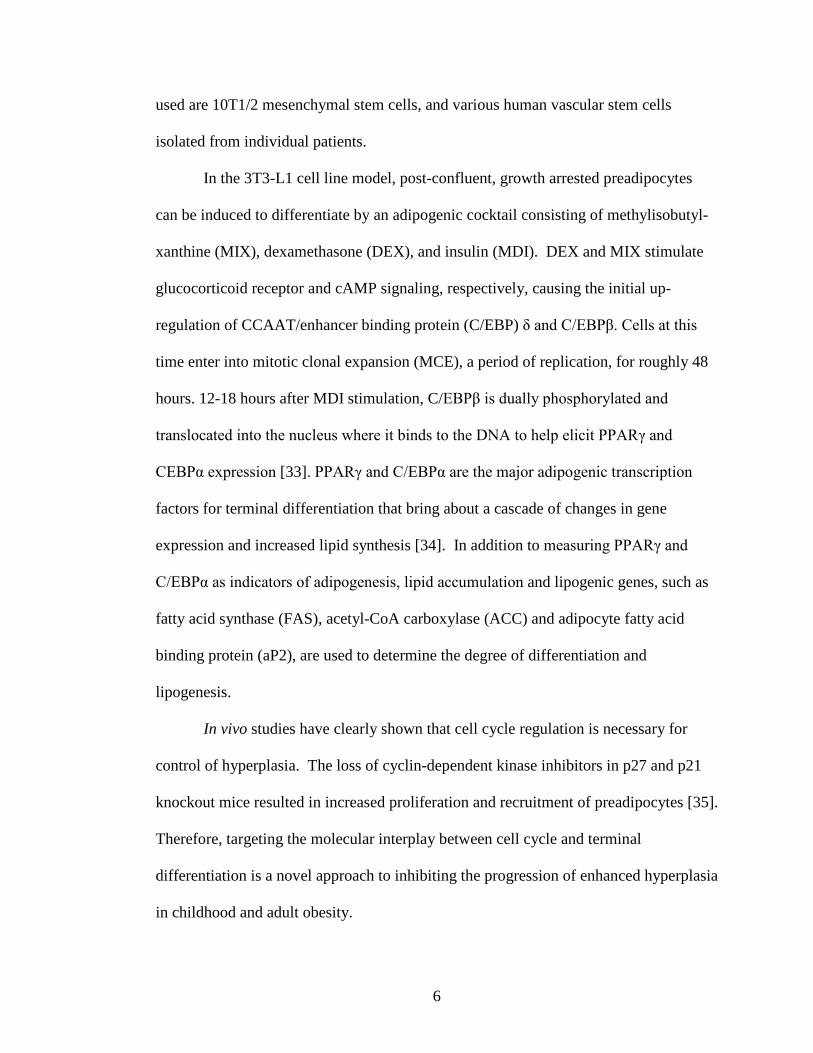
In the 3T3-L1 cell line model, post-confluent, growth arrested preadipocytes
can be induced to differentiate by an adipogenic cocktail consisting of methylisobutyl-
xanthine (MIX), dexamethasone (DEX), and insulin (MDI). DEX and MIX stimulate
glucocorticoid receptor and cAMP signaling, respectively, causing the initial up-
regulation of CCAAT/enhancer binding protein (C/EBP) δ and C/EBPβ. Cells at this
time enter into mitotic clonal expansion (MCE), a period of replication, for roughly 48
hours. 12-18 hours after MDI stimulation, C/EBPβ is dually phosphorylated and
translocated into the nucleus where it binds to the DNA to help elicit PPARγ and
CEBPα expression [33]. PPARγ and C/EBPα are the major adipogenic transcription
factors for terminal differentiation that bring about a cascade of changes in gene
expression and increased lipid synthesis [34]. In addition to measuring PPARγ and
C/EBPα as indicators of adipogenesis, lipid accumulation and lipogenic genes, such as
fatty acid synthase (FAS), acetyl-CoA carboxylase (ACC) and adipocyte fatty acid
binding protein (aP2), are used to determine the degree of differentiation and
lipogenesis.
In vivo studies have clearly shown that cell cycle regulation is necessary for
control of hyperplasia. The loss of cyclin-dependent kinase inhibitors in p27 and p21
knockout mice resulted in increased proliferation and recruitment of preadipocytes [35].
Therefore, targeting the molecular interplay between cell cycle and terminal
differentiation is a novel approach to inhibiting the progression of enhanced hyperplasia
in childhood and adult obesity.

7
In vitro, MCE is induced by DEX and MIX stimulation from the media.
Growth-arrested cells, held in G0 phase, are stimulated to transition into G1 phase,
typically within the first 12 hours, which prepares the cell for replication. In order for
the cell to transition into S phase, when DNA replication occurs, it must first pass
through a checkpoint to ensure the cell is in proper condition. This checkpoint between
G1 to S phase requires the accumulation of Cyclin A within the cytosol. Cyclin A is
part of the cyclin family, which regulates the cell cycle by binding and activating cyclin
dependent kinases (CDKs). It is the accumulation of these complexes and individual
proteins that allow a cell to pass through checkpoints. P27, a CDK inhibitor, binds
cyclin and CDKs, and inhibits cell cycle progression. Therefore, if the cell is
functioning properly, expression of p27 will be transiently down-regulated during
checkpoint transitions, allowing cyclin-CDK complexes to accumulate. When the cell
enters S phase, typically between 18-36 hours, it begins DNA replication. Between 36-
72 hours, the cell enters G2 phase when it prepares for final mitosis, the cleaving of two
daughter cells. It has been shown that, during MCE events, induction of PPARγ
increases the expression of CDK inhibitors p18 and p21, and thus shifts the cell toward
cell cycle exit [36] which is seen typically 48-72 hours post MDI induction.
Reactive oxygen species (ROS) signaling is required for MCE and the addition
of ROS enhances terminal differentiation in 3T3-L1 preadipocytes [37]. Hypertrophied
adipocytes, incapable of managing excess ROS production, secrete excess ROS to
surrounding tissue, stimulating precursor cells residing within the tissue to differentiate.
This is likely the mechanism by which hyperplasia is accelerated in conditions of
excess caloric intake, and thus explains why hypertrophy precedes hyperplasia in

8
adulthood. Therefore anti-oxidant treatment, capable of quenching excess ROS, may be
one potential way of inhibiting hyperplasia in vivo.
1.3.2 Adipocyte Metabolism
In a state of excess energy intake, severe hypertrophy leads to adipocyte
dysfunction. Adipocytes can expand upwards of 100μm in diameter, nearly 10 times
that of a typical mammalian cell. Visceral adipocytes greater than 75μm in diameter
have been linked to the development of metabolic syndrome [38]. Therefore,
preventing or alleviating excess lipid storage is a strategic approach in the fight against
obesity that involves understanding how adipocytes regulate the ebb and flow of lipid
synthesis and breakdown.
The primary role of adipocytes is to store energy, in the form of triglyceride, for
times of need, such as during exercise and fasting. Circulating free fatty acids are
readily taken up and stored as triglyceride, while excess energy in the form of glucose
is synthesized into triglyceride through the process of de novo lipogenesis. When
needed, adipocytes release this stored lipid into the blood stream through the process of
lipolysis, in which triglyceride is broken down into free fatty acids (FFA) and glycerol
that are released into the bloodstream [39]. Another fate of stored lipid is β-oxidation,
in which the adipocyte itself uses free fatty acids to generate energy. Investigating ways
to manipulate lipolysis and β-oxidation is an approach to reduce the amount of lipid
stored in hypertrophied adipocytes and thus alleviate the metabolic demand they face.

9
1.3.2.1 De Novo Lipogenesis
Glucose is the primary source of energy for most cells. Therefore, it is readily
metabolized through glycolysis to produce pyruvate. Pyruvate is further converted into
acetyl-CoA, which along with citrate is a substrate for aerobic respiration. If excess
glucose is present in the cell, acetyl-CoA and citrate will accumulate in the cytoplasm.
This buildup metabolically shifts the cell from an energy-burning to an energy-storing
state. Citrate buildup allosterically enhances the function of Acetyl-CoA Carboxylase
(ACC), which converts acetyl-CoA to malonyl CoA, the substrate for lipid synthesis.
Subsequently, the buildup of malonyl CoA inhibits the function of carnitine
palmitoyltransferase1 (CPT1), which transports lipids across the mitochondrial
membrane for β-oxidation. Therefore, the buildup of malonyl CoA promotes
lipogenesis while blunting oxidation [40]. This response is one of the major ways
adipocytes switch between energy storage and energy expenditure.
1.3.2.2 Lipolysis
Adipocytes purge stored triglyceride through the process known as lipolysis.
Triglyceride is stored in a lipid droplet surrounded by perilipin protein that serves as a
protective coating. When lipolysis is activated by β-adrenergic signaling, the second
messenger cAMP activates protein kinase A (PKA). PKA, in turn, phosphorylates
perilipin and hormone sensitive lipase (HSL) [41]. Phosphorylated perilipin detaches
from the lipid droplet giving phosphorylated HSL access to begin breaking down the
stored lipid. Two other constitutively expressed lipases involved in lipolysis,
acyltriglyceride lipase (ATGL) and monoacylglycerol lipase (MAGL), have been

10
recently discovered [39]. However, both of their activities are not as tightly regulated
during lipolysis as that of HSL. ATGL is primarily responsible for releasing the first
free fatty acid; HSL the second, and MAGL, the third [42]. The resulting glycerol and
free fatty acids are transported out of the cell and into the bloodstream for other tissue
to take up.
Lipolysis is of interest to anti-obesity researchers because of its ability to reduce
lipid storage; however, the resulting increase in circulating FFA and thus potential harm
to the circulatory system is a source of much concern. Therefore, another approach
involves investigating how to block the release of lipolyzed FFA from and redirect FFA
to β-oxidation within the cell.
1.3.2.3 β-oxidation
Lipid is the most energy dense storage unit the body has. When energy is
needed, the body taps into this store through the process of β-oxidation, which yields
significantly more ATPs than carbohydrate and protein metabolism. To carry out this
process, the cell must transport FFA across the mitochondrial lipid bilayer. It does so
through the tight regulation of carnitine palmitoyltransferase 1 (CPT1) [40], which is
the enzyme responsible for trafficking FFA through the mitochondrial membrane. As
mentioned previously, the build up of malonyl CoA, the substrate for de novo
lipogenesis, allosterically inhibits CPT1. Therefore adipocytes can easily shift between
lipid synthesis and breakdown.
Once in the mitochondria, fatty acid carbon chains are oxidized by niacin
adenine dinucleotide (NAD+) and flavin adenine dinucleotide (FAD), and the

11
remaining acetyl-CoA is further degraded by the citric acid cycle. NADH and FADH2
transport H atoms to the electron transport chain, where the build up of H atoms across
a membrane supplies the energy for ATP synthesis. NAD and FAD are then recycled
back for further oxidation [40].
A transcriptional regulator of β-oxidation is the coactivator peroxisome
proliferator-activated receptor (PPAR)-γ coactivator (PGC)-1α. PGC-1α interacts with
a broad range of transcription factors to regulate expression of genes related to
metabolic processes in the cell, such as thermogenesis, mitochondrial biogenesis, and
glucose/fatty acid metabolism. Specifically, PGC-1α is known to enhance transcription
of Uncoupling Protein 1, which is the major thermogenic gene found in muscle cells
and brown fat and a suggested therapeutic target for obesity treatment and prevention
[43]. AMP- activated protein kinase (AMPK), the major metabolic regulator of the
cell, is also known to stimulate β-oxidation and increase expression of PGC-1α to
improve ATP/AMP ratios [44].
Of great interest to obesity research is the ability of mitochondria to uncouple β-
oxidation from ATP synthesis, by the use of uncoupling proteins (UCP1, 2, and 3).
These proteins dissipate the buildup of H atoms across the inner mitochondrial
membrane, resulting in thermogenesis instead of ATP synthesis. It has been suggested
that adipocytes carry out a basal level of uncoupled β-oxidation that protects the body
from fatty acid leakage, and, in the hypertrophied state, mitochondrial dysfunction
impairs the cell’s ability to keep up with the increased flux of free fatty acids [45].
Therefore, finding factors that stimulate or enhance β-oxidation, and specifically

12
uncoupled β-oxidation, is a promising approach to preventing and alleviating adipocyte
hypertrophy.
1.4 The Role of AMP- activated protein kinase (AMPK) in Adipocytes
1.4.1 Overview
When targeting obesity and the development of metabolic syndrome through
hyperplasia and hypertrophy, it is important to consider a major metabolic regulator of
the cell, AMP-activated protein kinase (AMPK). The function of this protein affects
both differentiation and metabolic pathways in adipocytes.
AMPK is a well-known sensor of cellular ATP/AMP ratios and activation of
AMPK shifts the cell toward an energy-producing state when ATP levels are too low.
AMPK is comprised of 3 subunits (α, β, and γ) and is active in its phosphorylated state
(α-subunit p-172) [46]. The γ- subunit binds AMP causing a conformational change in
the protein that protects the α-subunit p-127 from cytosolic phosphatases [47].
Although AMPK activation is nearly 100-fold greater with AMP bound, AMP-
independent pathways also lead to AMPK activation. These pathways include
calmodulin-dependent kinase kinase (CaMKK), protein kinase A (PKA), and liver
kinase B1 (LKB1) [47]. There are several downstream targets of pAMPK, but of
interest in adipocyte research is the phosphorylation of acetyl coenzyme A carboxylase
(ACC), a lipogenic enzyme that is inhibited by its phosphorylation.
13
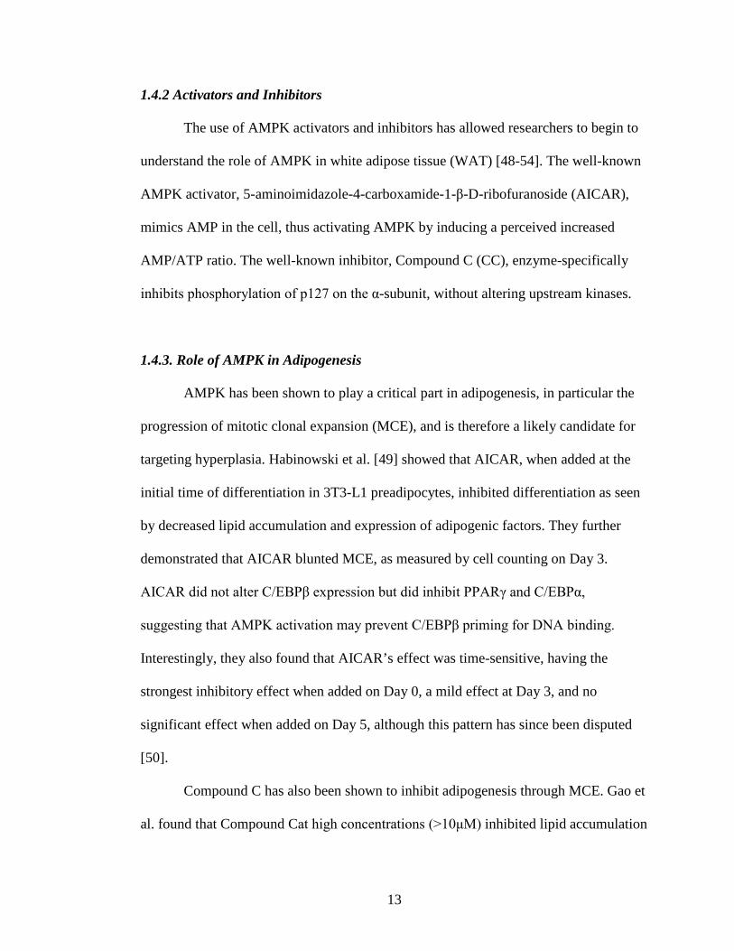
1.4.2 Activators and Inhibitors
The use of AMPK activators and inhibitors has allowed researchers to begin to
understand the role of AMPK in white adipose tissue (WAT) [48-54]. The well-known
AMPK activator, 5-aminoimidazole-4-carboxamide-1-β-D-ribofuranoside (AICAR),
mimics AMP in the cell, thus activating AMPK by inducing a perceived increased
AMP/ATP ratio. The well-known inhibitor, Compound C (CC), enzyme-specifically
inhibits phosphorylation of p127 on the α-subunit, without altering upstream kinases.
1.4.3. Role of AMPK in Adipogenesis
AMPK has been shown to play a critical part in adipogenesis, in particular the
progression of mitotic clonal expansion (MCE), and is therefore a likely candidate for
targeting hyperplasia. Habinowski et al. [49] showed that AICAR, when added at the
initial time of differentiation in 3T3-L1 preadipocytes, inhibited differentiation as seen
by decreased lipid accumulation and expression of adipogenic factors. They further
demonstrated that AICAR blunted MCE, as measured by cell counting on Day 3.
AICAR did not alter C/EBPβ expression but did inhibit PPARγ and C/EBPα,
suggesting that AMPK activation may prevent C/EBPβ priming for DNA binding.
Interestingly, they also found that AICAR’s effect was time-sensitive, having the
strongest inhibitory effect when added on Day 0, a mild effect at Day 3, and no
significant effect when added on Day 5, although this pattern has since been disputed
[50].
Compound C has also been shown to inhibit adipogenesis through MCE. Gao et
al. found that Compound Cat high concentrations (>10μM) inhibited lipid accumulation

14
and expression of PPAR γ, C/EBP α, FAS, and aP2 [55].This effect was limited to early
time points as treatment after the first two days of differentiation had no impact on lipid
accumulation or adipogenic factors. Further, Compound C treatment decreased cell
number from Day 0 to Day 3 of differentiation. Nam et al. also found Compound C
inhibited adipogenesis through MCE, as Compound C increased levels of p21, a cyclin
kinase dependent inhibitor, levels [53].
Taken together, the general consensus in the literature is that either activation or
inhibition of AMPK alters MCE events and inhibit adipogenesis. This effect is
understandable, as the cell’s energy needs must be tightly regulated during such a
demanding transition. Therefore, identifying factors that target AMPK are of interest in
preventing hyperplasia. Interestingly several researchers have shown that certain
bioactive compounds exert their anti-adipogenic effect through AMPK activation [47,
56].
1.4.4 Role of AMPK in Mature Adipocytes
Much interest surrounds the role of AMPK in the metabolism of mature
adipocytes. Specifically, activation of AMPK to induce lipolysis and increase
mitochondrial oxidation of fatty acids would lead to a possible approach to alleviate
hypertrophy.
In general, activated AMPK turns on catabolic pathways that produce ATP and
turns off anabolic pathways that consume ATP. Likewise, it has been shown that
AMPK increases β-oxidation in muscle and liver cells [57], but the effect on adipocytes
remains unclear. In theory, AMPK activation would activate oxidative pathways while

15
inhibiting lipid synthesis. It is known that AMPK phosphorylates and deactivates the
lipid synthesizing enzyme ACC, resulting in a cytosolic decrease in malonyl CoA. This
decrease subsequently results in the availability of CPT1 to transfer fatty acids into the
mitochondria, thus shifting the cell toward β-oxidation. Gaidhu et al. [58] found these
effects in isolated epididymal rat adipocytes. Their findings showed that chronic
AICAR exposure (15hours) increased pACC, fatty acid oxidation, and PGC-1α, CPT1,
acetyl-CoA oxidase mRNA expression, while UCP1 and UCP2 were unaltered by
AICAR treatment. The authors concluded that AMPK activation could remodel
adipocytes toward energy dissipation.
Also of particular interest is AMPK’s effect on lipolysis. Upon normal β-
adrenergic stimulation of lipolysis, cAMP activates PKA, which in turn phosphorylates
and activates HSL for lipolysis. Djouder et al. found that PKA concurrently inactivates
AMPK in order to promote efficient lipolysis [59]. However other researchers have
found that AMPK activation through cAMP was necessary for induced lipolysis, as
lipolysis was no longer stimulated in the presence of mutant AMPK alpha subunits [60].
When AMPK was therapeutically activated by the use of AICAR in isolated rat
adipocytes, AICAR inhibited HSL activation and glycerol release, but over the course
of 15 hrs increased FFA release [52]. The authors here concluded that the length of
AMPK activation determined the lipolytic response; acute treatment resulted in an
inhibition of lipolysis, while chronic activation resulted in an increase of fatty acid
release. This difference may explain some of the controversy observed among
researchers. Because glycerol release did not also increase with time, the authors
suggested that AMPK activation may prevent the complete hydrolysis of triglyceride
16
molecules through the inhibition of HSL phosphorylation.. If triglyceride hydrolysis is
incomplete triglycerides are broken down to diacylglycerides by ATGL but not further,
resulting in the release of some FFA and the shunting of diacylglycerides to other
metabolic pathways in the cell. This provides a potential mechanism and indicates that
AMPK may play a critical role in the cells ability to undergo lipolysis.
AMPK regulation provides a promising new avenue toward alleviating
adipocyte hypertrophy as it has been implicated in β-oxidation and lipolysis. Recently,
several bioactive compounds have been implicated as AMPK activators [16], which
could provide a natural approach toward targeting hyperplasia and hypertrophy in the
progression of obesity.
1.5 Potential Health Effects of Bioactive Compounds in the Development of
Obesity
To prevent chronic disease, increasing fruit and vegetable consumption has been
the longstanding advice. Consistently, fruit and vegetable intake is negatively
correlated with the occurrence of obesity and metabolic syndrome [61, 62], and further,
increasing fruit and vegetable consumption may decreases risk for heart disease and
diabetes [63, 64]. Not only do fruits and vegetables provide a low-calorie, high-fiber
option, but researchers within the past several decades have also identified more than
thousands of phytochemicals that naturally occur in the produce we eat [65]. Discovery
of bioactive compounds has invited researchers to explore the vast array of beneficial
effects these compounds possess. Much interest surrounds the use of bioactive

17
compounds, through increased consumption and supplementation, to prevent or
alleviate symptoms of metabolic syndrome as drug therapy is not only costly, but often
elicits negative side-effects [4, 66].
Currently, it is estimated that humans consume about 1-3g of bioactive
compounds daily, depending on their level of fruit and vegetable intake [67]. Nearly all
bioactive compounds have been shown to possess health-promoting properties,
typically in a dose-dependent manner. The American Dietetic Association (ADA)
acknowledges the significance of consumption of bioactive compounds and therefore
supports the marketing of functional foods, either conventional or modified, when there
is substantial scientific evidence of benefit [68]. Flavonoids, a sub group of
polyphenols, are currently a top priority of research efforts to The National Institute of
Health’s National Center for Complimentary and Alternative Medicine (NCCAM)
because the health benefits shown. Specifically, the flavonoid quercetin is of great
interest because of its suggested ability to alleviate obesity-related conditions [67].
1.6 The Dietary Flavonoid Quercetin
1.6.1 Overview
Flavonoids are a class of phytochemicals,
discovered in the 1930s, that generate pigment in
plants and play a biological role in other cellular
processes. It is estimated that the average dietary
intake of flavonoids in humans is several hundred Figure 1. Structure of Quercetin

18
milligrams per day [65].
Quercetin, the most abundant dietary flavonoid, has been the focus of 30 years
of research for its strong antioxidant capabilities. It is a flavonol, one of the five
subclasses of flavonoids, and it’s ring structure and aglycone configuration of hydroxyl
groups, make it one of the most potent flavonoids in terms of antioxidant capabilities
[69, 70]. Quercetin is found in numerous fruits and vegetables but the major dietary
sources include onion, red apples, red wine, tea,
cranberry, kale, hot peppers and broccoli [71].
Determining exact amounts of flavonoid content
in any plant is very challenging because flavonoid
production is affected by sunlight, environmental
factors, degree of ripeness, and species genetic
variability [72]. Further quercetin is stored in the
skin of most fruits and vegetables, therefore content can very significantly depending
on skin to overall weight ratio. Consequently, estimating quercetin intakes through
dietary analysis is particularly limited, making it difficult to complete epidemiological
studies relating quercetin intake to chronic disease [65]. Estimated quercetin intakes
range from 10-100mg per day depending on an individual’s consumption of these fruits
and vegetables [67]. It could be speculated that 6-8 servings of fruits and vegetables
daily would provide an average daily consumption of 100mg quercetin, however as
discussed, exact quercetin content data is limited. Higher levels of intake between
500mg-1200mg can be safely achieved by the use of supplements (this amount would
be likely be comparable to consumption of 5-10 times the recommended servings, 6-8,
Table 1. Sources of Quercetin
Food Source Amount ( mg/kg) Onions 284-486 Kale 110 Asparagus 142 Apple 21-72 Green Beans 39 Cherry Tomatoes 17-200 Broccoli 30 Tea 10-25 Red Wine 4-16
Modified from Manach, 2005 [71]

19
of fruits and vegetable, and could only be achieved through supplementation).
Researchers are thus trying to utilize the potential of this readily available flavonoid
through increased consumption and supplementation for its antioxidant, anti-
inflammatory, and anti-proliferative properties [67, 73].
1.6.2 Bioavailability
As with any bioactive compound, bioavailability is of great interest and
importance. Quercetin is predominantly found in nature in variations of its glycoside
form: either quercetin 3-O-β-D-glucoside (Q3G) or quercetin 4’-O-β-D-glucoside
(Q4G) [74]. These forms are useful to the plant because of their water-soluble
properties, but do not have the same antioxidant capabilities of the aglycone form, and
are thought to be metabolized by the gut microflora before absorption. Three factors
that determine the bioavailability of quercetin: (1) metabolism by gut microflora, (2)
transport through small intestinal cells, and (3) final circulating metabolites. Although
the question of which metabolites are absorbed and circulate remains controversial,
advancements in detection methods have come a long way since 1975, when Gugler et
al. [75] first gloomily reported the quercetin doses did not alter plasma levels in humans.
1.6.3 Gut Metabolism
It is estimated that 9 out of 10 cells in the body are bacterial [76], which puts in
perspective the influence of an individual’s gut microflora on digestion and gut
metabolism. Specifically, it has been well demonstrated that gut bacteria are capable of
degrading quercetin into phenolic acids and CO2, methylating aglycone compounds,
20
and hydrolyzing quercetin glucosides [77]. Chen et al. [78] determined in rats that
93.3% of absorbed quercetin had been metabolized by the gut and only 3% by the liver
after absorption. They also showed that 60% of total ingested quercetin was absorbed.
Earlier studies done by Hollman et al. [79, 80] indicated that a dose of quercetin in
glucoside form was better absorbed than the aglycone, as measured by ileostomy
effluent content of glucosides. In stark contrast, Walgren et al. [81] showed, using a
Caco-2 cell monolayer, that aglycones are more readily transported across the intestinal
layer than glucosides. Walle et al. [82] aimed to address this conflict and discovered
that glucosides in an onion meal are effectively hydrolyzed to the aglycone form, which
can be absorbed. These findings suggest that nearly all excreted quercetin is aglycone
and further that we rely on gut bacteria to hydrolyze dietary quercetin for absorption.
This explains why significant variation may exist among individuals in their ability to
absorb quercetin based on the make up of their gut microflora.
1.6.4 Absorption and Transport
Piskula and Terao [83] were among the first, along with Manach [84], to
demonstrate that quercetin doses in rats result in plasma metabolite concentrations of 1-
2µM. Their findings further suggested that absorption was affected by the solubility of
quercetin in the vehicle of administration (quercetin dissolved in propylene glycol
resulted in plasma concentration of 50µM). Human supplementation studies typically
result in a total plasma quercetin metabolite range of 10-200nM [77]. The most
common detection method for plasma quercetin digests all the quercetin metabolites
and provides an overall concentration. With more sensitive techniques, researchers are

21
trying to determine which metabolites, specifically aglycone verses glucosides,
circulate at higher levels, as this would likely be the metabolite that exerts an effect on
tissues. The pertinent body of research remains inconclusive, according to a recent
review, as some researchers indicated that quercetin glucoside forms were detected in
plasma [85, 86], and others reported that aglycone forms but not glucosides were
present [87, 88]. Other studies have shown a wide range of quercetin metabolites
detected in the plasma [89]. Because of the significant in vitro findings of quercetin
aglycone, work is currently underway to develop nanoparticle vehicles that protect
quercetin from gut and liver metabolism and therefore deliver it in its potent aglycone
form to tissues [73].
Because it is reasonable to postulate that both aglycone and glucoside forms
circulate in the plasma, it is of interest to study the effect of both metabolites on adipose
tissue. However the scope of research in this study focuses on the aglycone form.
Although concentration ranges used in this study are higher than physiological
concentrations, as compared to what is observed in vivo after supplementation (10-
200nM), it is relevant for our work in less sensitive cell culture systems to observe the
molecular changes that occur in response to quercetin.
1.7 Quercetin’s Effect on Metabolic Syndrome: In Vivo Studies
Quercetin is currently of great interest to researchers because of significant in
vivo animal studies showing its ability to ameliorate metabolic syndrome. Vessal et al.
[6] found that in streptozocin (STZ)-induced diabetic rats (a model that is comparable

22
to type 1 diabetics), doses of 10mg/kg and 15mg/kg per day, began to lower blood
glucose levels after only 3 days and had completely normalized blood sugar levels after
10 days. The authors concluded that quercetin may be able to stimulate pancreatic islet
cell regeneration. In 2008, Rivera et al. [7], found that quercetin doses (2 or 10 mg/kg)
ameliorated high plasma concentrations of triglycerides, total cholesterol, FFA, and
insulin in Zucker rats, and further that the higher dose increased plasma concentrations
of adiponectin, reduced NOx levels in plasma, and lowered VAT TNF-alpha production.
Kobori et al. [8] fed mice a diet high in fat, cholesterol, and sugar for 20 weeks, and
supplemented one group with 0.05% quercetin. After the 20-week period,
supplemented mice had plasma quercetin levels of 14.16μM, and had significantly less
fat accumulation in the liver, reduced circulating triglyceride levels, and improved
blood glucose and insulin levels. The same group had previously found quercetin-fed
(0.1% or 0.5%) streptozotocin (STZ)-induced diabetic mice had improved blood
glucose and plasma insulin levels [9]. Bansal et al. [10] showed similar results feeding
STZ-induced mice a high-fat diet with Pileamicrophylla (PM1), a plant containing
quercetin, among other flavonoids. PM1 at a dose of 100 mg/kg/day, for 28 days
produced significant (p<0.05) reduction in body weight, plasma glucose, triglycerides,
and total cholesterol content. It is important to note, due to the complexity of quercetin
bioavailability, significant results are not always found with varying quercetin doses
[90]. However, the majority of animal studies to date strongly suggest quercetin as an
anti-obesity, insulin-sensitizing agent, warranting further research.
Human trials show promising preliminary results. Egert et al., in a randomized
double-blinded, placebo-controlled cross-over trial gave overweight and obese subjects
23
150mg dose of quercetin daily for 6 weeks (resulting in plasma quercetin levels of 71 to
269 nM/l). Quercetin treatment decreased systolic blood pressure (-2.6 mmHg,
p<0.001), oxidized LDL levels, and, interestingly, HDL levels, when compared to the
placebo group [91]. Further, this group retrospectively characterized the effect of
quercetin on serum blood pressure and lipid profile to be linked to variations in
apolipoproteinepsilon genotype [92], suggesting an explanation for varied responses to
quercetin treatment. Although some supplementation studies have shown no significant
effect [93, 94], this is likely due to dosage, study duration, poor bioavailability, and
variable gut microflora of each individual. Therefore, optimizing study design is
essential in human supplementation trials.
These significant in vivo findings command further mechanistic research of
quercetin’s effect at the cellular level, specifically in the areas of hyperplasia and
hypertrophy as both lead to the development of metabolic syndrome. Current research
detailing what is known about both quercetin’s effect on adipogenesis and adipocyte
metabolism, as well as our recent findings, are described below in Chapters 2 and 3,
respectively.

24
CHAPTER 2
EFFECT OF QUERCETIN ON ADIPOGENESIS
2.1 Literature Review
The rise in obesity will significantly decrease life expectancy and continue to
burden health care systems because of increased risk for the development of chronic
diseases [95]. On a cellular level, obesity is defined as the increase in size
(hypertrophy) and number (hyperplasia) of adipocytes. Studies have shown that
adipocyte number is determined during childhood and maintained through adulthood,
emphasizing the importance of preventing childhood obesity. In adulthood, hypertrophy
precedes hyperplasia to accommodate initial excess energy intake, however as excess
intake persists, such as in the development of obesity, hyperplasia is accelerated [23,
25]. Further, weight loss results in a reduction in adipocyte volume but not necessarily
adipocyte number [2]. Adipocytes are extremely resistant to apoptosis; therefore
adipose tissue that has been expanded by hyperplasia will be maintained, making it
harder for an individual to sustain weight loss and worsening the prognosis for
treatment [26, 27]. Targeting hyperplasia is thus essential for preventing the
progression of childhood and adult obesity.
Several bioactive compounds have been shown to inhibit adipogenesis, the
process of developing new adipocytes. Quercetin, the most commonly consumed
flavonoid, is one such compound. Because quercetin is known to have anti-proliferative
and apoptotic properties, it was first hypothesized to inhibit preadipocyte growth. Hsu

25
et al. [96] ,while comparing the effects of various flavonoids on undifferentiated 3T3-
L1 pre-adipocytes, found that quercetin most strongly inhibited cell population growth
and induced cell apoptosis through caspase-3 in a dose-dependent manner (50-250μM).
Quercetin was also found to be anti-proliferative in human stromal cells [97]. Park et al.
[11] showed that quercetin, alone or in combination with genistein and resveratrol
inhibited lipid accumulation in differentiating 3T3-L1 and human adipocytes. Similar
results were also found by Yang et al. [12] who determined in 3T3-L1 cells that
quercetin (25μM) alone inhibited lipid accumulation by 15.9+/-2.5% (p<0.001), and in
combination with resveratrol (25μM) inhibited lipid accumulation by 68.6+/-0.7%
(p<0.001). Although it is generally agreed that quercetin is anti-adipogenic, Morikawa
[98] interestingly showed that quercetin induced apoptotic genes without inhibiting
PPARγ and fatty acid synthase (FAS) during differentiation of human preadipocytes,
suggesting quercetin may work through PPARγ-independent pathways.
One potential mechanism for quercetin’s anti-adipogenic effect was recently
proposed by Ahn et al. in 2008 [13]. They reported that differentiating 3T3-L1 cells in
the presence of quercetin (10-100μM) decreased lipid accumulation and expression of
adipogenic factors (CEBPα, PPARγ, SREBP-1, FAS). They further looked at the
effects of quercetin during differentiation on AMP-activated protein kinase (AMPK),
the metabolic regulator of the cell, and its down stream target acetyl-CoA carboxylase
(ACC), and found that phosphorylation of both of these proteins by Day 8 was
increased in the presence of higher concentrations of quercetin (50μM and 100μM).
The authors suggested AMPK activation as a potential mechanism for quercetin’s anti-

26
adipogenic effect. The role of AMPK activation during differentiation has been
considered anti-adipogenic [49] and potentially regulates mitotic clonal expansion.
Mitotic clonal expansion (MCE) has been implicated as a critical regulator of
adipogenesis in vivo and is a current targeted pharmacological approach in the fight
against obesity [25]. Cell cycle is tightly regulated by transitory cyclin dependent
kinases and kinase inhibitors, two of which are Cyclin A and p27, respectively. The
abolishment of p27 in vivo lead to increased proliferation and recruitment of
preadipocytes [35]. .MCE has been shown to be necessary for terminal differentiation
in vitro in 3T3-L1 preadipocytes [99]. Given quercetin’s anti-proliferative properties,
and ability to activate AMPK, it is logical to hypothesize that quercetin may inhibit
adipogenesis through altering cell cycle events; however this effect has never been
explored.
Understanding this mechanism will help develop quercetin as an anti-obesity
agent because of its ability to target hyperplasia, a critical process in the development
of obesity and metabolic syndrome.
2.2 Purpose of Study
Given the current gap in the literature surrounding quercetin’s ability to inhibit
adipogenesis through mitotic clonal expansion, and the significance these findings
would have on the development of quercetin as an anti-obesity agent, we aimed to fill
this gap by determining (1) quercetin’s effect on adipogenesis using 3T3-L1

27
preadipocytes, and (2) quercetin’s effect on cell number and cell cycle related genes
Cyclin A and p27, key regulators for cell cycle progression from G1 phase to S phase.
Our hypothesis was that quercetin will (1) inhibit lipid accumulation, as
measured by Oil Red O staining, and expression of adipogenic factors PPARγ, C/EBPα,
aP2, ACC on the mRNA and protein levels, and (2) inhibit cell number, a measure of
mitotic clonal expansion, and alter mRNA expression of Cyclin A and p27 when
analyzed at 0-48 hours.
2.3 Materials and Methods
2.3.1 Cell Culture Model and Treatments
For the differentiation of 3T3-L1 preadipocytes, cells were grown to 100% confluence
in Growth Media [DMEM (high glucose), 10% Calf Serum, 1% Penicilin/
Streptamycin] replaced every two days. At two days post-confluence, growth media
was changed to Differentiation Media [DMEM (high glucose), 10% Fetal Bovine
Serum, 1% P/S, 1% Insulin, 1% 3-Isobutyl-1-methylxanthine 11.5mg/ml, 0.01%
Dexamethasone 3.9mg/ml] in the presence or absence of quercetin aglycone (Sigma-
Aldrich Catalog # 6151-25-3). The combination of 3-Isobutyl-1-methylxanthine (MIX),
dexamethasone, and insulin is known as the standard MDI adipogenic cocktail. At two
days post-MDI induction, media was replaced every two days with post-differentiation
media [DMEM (high glucose), 10% Fetal Bovine Serum, 1% P/S, 1% Insulin, +/-
quercetin]. 3T3-L1 preadipocytes generally reach 80% differentiation 4 to 5 days post-
differentiation.

28
2.3.2 Oil Red O Lipid Staining
Cells were harvested on desired days and underwent Oil Red O Staining to quantify
lipid accumulation, as it is an indirect determiner of cell differentiation. Cells, grown on
24-well and 6-well plates, were treated with 10% formaldehyde in phosphate buffer
solution (PBS) for 1 hour, washed with 60% isopropanol, and completely dried. Then,
cells were stained with 0.5% Oil Red O solution in 60:40 (v/v) isopropanol: H2O, for
30 minutes at room temperature. Finally, wells were rinsed with distilled water and
dried. Optical density was then measured at 490nm, after eluting with isopropanol, to
quantify lipid accumulation.
2.3.3 Cell Viability and Counting
Cells were grown to confluence in 24-well plates. At two-days post-confluence cells
were differentiated with MDI in the presence or absence of quercetin (25 and 50μM).
Media was replaced, two days later, with post-differentiation media (treatment
continued). On day 3, cells were prepared for Trypan Blue staining. Media was
removed and wells were washed with PBS. Trypsin was added to each well (0.25ml)
and let sit for 10 minutes to ensure cell detachment. PBS (0.75ml) was then added to
bring cell suspension volume to 1ml. Trypan Blue (modified) 0.4% solution 1N PBS
(from MP Biomedical Inc, cat# 1691049) was added to cell suspension (1:1) in a fresh
centrifuge tube for 3 minutes. A hemocytometer was used to count cells from each well
twice. Each treatment had 6 replicate wells. Both stained and unstained cells were
counted (stained cells are non-viable). Cells were counted in the 4 corners of the
hemocytometer, and total cell number was calculated as (total of 4 squares) x (2500) x

29
(2 dilution factor). Percent viable equaled the number of viable cells divided by total
cell number (viable plus non viable counts). Total viable cell number was the average
number of unstained cells from each treatment replicate.
2.3.4 Protein Isolation and Western Blotting
Cells were harvested at desired times with RIPA Buffer containing protease inhibitor
(500ul per p-100 dish). Samples were stored at -80° C until protein quantification.
Samples were thawed on ice, sonicated, and centrifuged. Supernatant, containing whole
cell protein, was transferred to a fresh tube for protein quantification using the
Bicinchoninic Acid (BCA) assay. A BCA standard curve was established using bovine
serum albumin (BSA)[2mg/ml] protein and measured at 570nm. Sample concentrations
were then measured against the standard, and prepared with sample buffer to allow for
15μg to be loaded to the gel. Samples were run on acrylamide (varying percentages)
gels via electrophoresis, and then proteins were transferred to a PVDF membrane
through wet transfer. Upon successful transfer, membranes were blocked with 5% non-
fat dry milk, and treated with primary and secondary antibodies (purchased from Santa
Cruz Biological). HRP detection was done through ECL solution. Chemiluminescent
bands were captured on x-ray and developed.
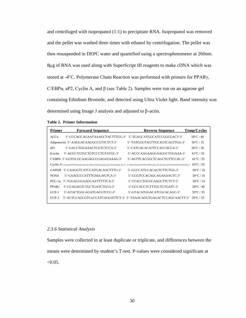
2.3.5 RNA Isolation and Analysis
Cells washed with phosphate buffer solution (PBS), were harvested with Trizol
Reagant and stored at -80° C until mRNA isolation. For isolation, samples were thawed
and centrifuged with 200µL chloroform. Supernatant was transferred to a fresh tube
30
and centrifuged with isopropanol (1:1) to precipitate RNA. Isopropanol was removed
and the pellet was washed three times with ethanol by centrifugation. The pellet was
then resuspended in DEPC water and quantified using a spectrophotometer at 260nm.
8µg of RNA was used along with SuperScript III reagents to make cDNA which was
stored at -4°C. Polymerase Chain Reaction was performed with primers for PPARγ,
C/EBPα, aP2, Cyclin A, and β (see Table 2). Samples were run on an agarose gel
containing Ethidium Bromide, and detected using Ultra Violet light. Band intensity was
determined using Image J analysis and adjusted to β-actin.
Table 2. Primer Information
2.3.6 Statistical Analysis
Samples were collected in at least duplicate or triplicate, and differences between the
means were determined by student’s T-test. P-values were considered significant at
<0.05.
Primer Forward Sequence Reverse Sequence Temp/Cycles ACCα 5’-CCCAGCAGAATAAAGCTACTTTGG-3’ 5’-TGAGCATGGCATCCGGCGACT-3’ 58°C / 40
Adiponectin 5’-AAGGACAAGGCCGTTCTCT-3’ 5’-TATGGGTAGTTGCAGTCAGTTGG-3’ 56°C / 35
aP2 5’-GACCTGGAAACTCGTCTCCA-3’ 5’-CATGACACATTCCACCACCA-3’ 56°C / 30
β-actin 5’-AGGCTGTGCTGTCCCTGTATGC-3’ 5’-ACCCAAGAAGGAAGGCTGGAAA-3’ 61°C / 35
C/EBPα 5’-GGTGCGCAAGAGCCGAGATAAAG-3’ 5’-AGTTCACGGCTCAGCTGTTCCAC-3’ 61°C / 35
Cyclin A 5’GCAGTTTTGAATCACCACATGCATGCCGGGCACCTCGAGGCAT-3’ 5’-TGGCTGCCTCTTCATGTAACCTCACACACTTAGTGTCTCTGGTG-3’ 63°C / 35
GAPDH 5’-CAAGGTCATCCATGACAACTTTG-3’ 5’-GGCCATCCACAGTCTTCTGG-3’ 60°C / 35
NOX4 5’-GAAGCCCATTTGAGGAGTCA-3’ 5’-GGGTCCACAGCAGAAAACTC-3’ 56°C / 35
PGC-1α 5’-TGGACGGAAGCAATTTTTCA-3’ 5’-TTACCTGCGCAAGCTTCTCT-3’ 56°C / 35
PPARγ 5’-CCAGAGTCTGCTGATCTGCG-3’ 5’-GCCACCTCTTTGCTCTGATC-3’ 58°C / 40
UCP-1 5’-ATACTGGCAGATGACGTCCC-3’ 5’-GTACATGGACATCGCACAGC-3’ 59°C / 35
UCP-3 5’-ACTCCAGCGTCGCCATCAGGATTCT-3’ 5’-TAAACAGGTGAGACTCCAGCAACTT-3’ 59°C / 35

31
2.4 Results
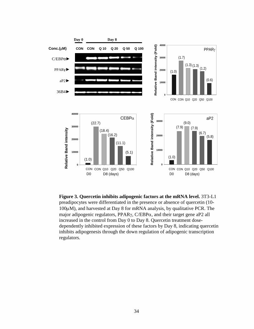
2.4.1 Quercetin inhibits lipid accumulation and adipogenic transcriptional factors
3T3-L1 pre-adipocytes were stimulated to differentiate at Day 0 in the presence of
increasing concentrations of quercetin (0-100μM). By Day 8 of differentiation
quercetin dose-dependently inhibited lipid accumulation (Figure 2). (Troglitazone, a
PPAR γ ligand, was used as a positive control to demonstrate the ability of our model
system to be stimulated toward differentiation.) Further we determined quercetin
inhibited mRNA expression of PPAR γ (1.1 fold), C/EBP α (17.6 fold), aP2 (2.1 fold)
(Figure 3) and protein expression of PPAR γ, and ACC (Figures 4). These results
indicate that quercetin inhibits adipogenesis in our model system through the inhibition
of critical transcriptional adipogenic factors.
2.4.2 Quercetin’s inhibitory effect is limited to early time points
Mitotic clonal expansion (MCE) occurs in 3T3-L1 preadipocytes between Day 0 and
Day 3 after MDI stimulation. Because quercetin is known to be a strong anti-
proliferative agent and AMPK activator, we tested the hypothesis that quercetin’s effect
on lipid accumulation is limited to treatment during early time points of differentiation.
Quercetin treatment was staggered, with treatments starting at 0, 6, 12, 18, 24, 36, 48,
72 hours after MDI stimulation at time 0 hour. All treatments were continued until Day
6 of differentiation when cells were harvested for Oil Red O staining. Quercetin’s
strongest inhibitory effect was observed when added between 0-18hours (Figure 5).
Quercetin treatment starting at 24-36 hours still significantly inhibited lipid

32
accumulation as compared to the control, however after 48 hours quercetin had no
significant effect. This finding indicates quercetin’s effect is limited to early time points
of adipogenesis.
2.4.3 Quercetin may inhibit adipogenesis through regulation of early cell cycle events
We further investigated quercetin’s effect on mitotic clonal expansion by looking at cell
number and cell cycle related genes. Figure 6a shows that between Day 0 and 3, cell
number dramatically increases. Quercetin significantly inhibited total cell number in
this trial. We next repeated this experiment using Trypan Blue staining to detect viable
and non-viable cells, and only looked at Day 3 counts since they reflect the end of MCE.
Quercetin treatment did not alter the % of viable cells indicating quercetin’s effect is
not cytotoxic. Although not significant, there was a clear trend indicating quercetin may
decrease viable cell number at Day 3. This suggests quercetin may inhibit MCE. We
next looked at cell cycle genes Cyclin A, a positive regulator of cell cycle, and p27, an
inhibitory regulatory of cell cycle. In preadipocytes stimulated to differentiate at time 0
hour, Cyclin A mRNA rose at 12 hours and fell by 24 hours in the control. However
quercetin (50µM) treatment inhibited Cyclin A expression at 12 and 24 hours, delaying
Cyclin A induction until 36 and 48 hours (Figure 7). Quercetin treatment also altered
the expression of p27. Preadipocytes stimulated to differentiate at time 0 hour
transiently decreased protein expression of p27 between 18-24 hours. Quercetin
(50µM) sustained p27 expression through the mitotic clonal expansion period (Figure
8).

33
Figure 2. Quercetin dose-dependently inhibits lipid accumulation. 3T3-L1 preadipocytes were differentiated in the presence or absence of quercetin (10-100µM), and harvested at Day 0, 4, and 8 for Oil Red O staining to measure lipid accumulation. Troglitazone (Tro), a PPARγ ligand, was used as a positive control. Compared to the control (CON) with just MDI differentiation media, quercetin treatment 50 and 100µM significantly decreased lipid accumulation dose-dependently at Day 4 and Day 8 of adipogenesis.
CON(MDI)
Abs
orba
nce
at 4
90 n
m
0.0
0.2
0.4
0.6
Day 0 Day 4 Day 8
TRO(10 µM)
10 20 50 100 SQ (µM)
b a b b c c
ab b b
c
d
Q (μM)
34
Conc.(μM) CON CON Q 10 Q 20 Q 50 Q 100
36B4►
Day 0 Day 8
C/EBPα►
PPARγ►
aP2►
0
10000
20000
30000
40000
CEBPα
CON D0
CON Q10 Q20 Q50 Q100 D8 (days)
(1.0)
(22.7)
(18.4)(16.2)
(11.1)
(5.1)
Rel
ativ
e B
and
inte
nsity
(F
ld)
Rel
ativ
e B
and
inte
nsi
ty (
Fo
ld)
0
10000
20000
30000
40000
(1.0)
CON D0
CON Q10 Q20 Q50 Q100 D8 (days)
PPARγ
(1.7)
(1.3) (1.3)(1.2)
(0.6)
Rel
ativ
e B
and
inte
nsity
(Fol
d)
0
10000
20000
30000
CON D0
CON Q10 Q20 Q50 Q100 D8 (days)
aP2
(1.0)
(7.9)(9.0)
(7.9)(6.7)
(5.8)
Figure 3. Quercetin inhibits adipogenic factors at the mRNA level. 3T3-L1 preadipocytes were differentiated in the presence or absence of quercetin (10-100µM), and harvested at Day 8 for mRNA analysis, by qualitative PCR. The major adipogenic regulators, PPARγ, C/EBPα, and their target gene aP2 all increased in the control from Day 0 to Day 8. Quercetin treatment dose-dependently inhibited expression of these factors by Day 8, indicating quercetin inhibits adipogenesis through the down regulation of adipogenic transcription regulators.

35
CON +Q50μM 0h 12 24 48 72 D6 12h 24 48 72 D6
ACC ►
PPARγ►
β-actin►
Figure 4. Quercetin inhibits adipogenic factors at the protein level. 3T3-L1 preadipocytes were differentiated in the presence or absence of quercetin (50µM), and harvested at sequential time points (0, 12, 24, 48, 72 hours, and Day 6) for protein analysis. Acetyl-CoA Carboxylase (ACC), an enzyme responsible for lipid synthesis, and PPARγ, the transcriptional regulator of adipogenesis both increased in the control over the course of adipogenesis. Quercetin treatment (50µM) inhibited the increase of expression in both these adipogenic factors, especially the induction around 24 and 48 hours.

36
Time of Quercetin (50uM) Treatment
Con 0hr 6 12 18 24 36 48 72
Abs
490n
m
0.00
0.25
0.50
0.75
1.00
1.25a
a
ab
bb
bcbc
cc
Figure 5. Quercetin’s inhibitory effect on lipid accumulation is limited to early time points. 3T3-L1 preadipocytes were stimulated to differentiate at time 0 hour. Quercetin was added to differentiating media at subsequent times (0, 6, 12, 18, 24, 36, 48, and 72 hours) and continued through the duration of the experiment until Day 6 when all cells were harvested for Oil Red O staining to measure lipid accumulation. Quercetin inhibited lipid accumulation strongest when added between 0-18 hours, less but still significant at 24-36 hours, and no effect if added after 48 hours. These results indicate quercetin’s anti-adipogenic effect is limited to early events of adipogenesis.

37
Cell Viability
Treatment
Con Q10 Q20 Q50
% o
f Con
trol
0
20
40
60
80
100
120
Total Viable Cells
Treatment
Con Q10 Q20 Q50
Cel
l Num
ber
0
20000
40000
60000
80000
Figure 6. Quercetin does not affect cell viability but may inhibit cell number. Mitotic clonal expansion (MCE) occurs between Day 0-3 of differentiation in 3T3-L1 preadipocytes, dramatically increasing the number of cells in a well 2 or 3 fold by Day 3. (A) Trial 1: Cells were differentiated and harvested at 0, 24, 48, and 72 hours for cell counting by hemocytometer (n=3). The control clearly increased over 72hours. Quercetin significantly inhibited cell number at 24 and 72 hours. (B and C) Trial 2: Cells were differentiated in the presence or absence of quercetin (50µM) and harvested at Day 3 for Trypan Blue staining and counting by hemocytometer (n=6). Viable and non-viable cells were counted and % viable was determined by # of viable cells/ #of total cell, indicating the percent of living cells at Day 3. Quercetin treatment did not alter the % viable, indicating quercetin’s effect is not cytotoxic. Although not significant, there was a clear trend indicating quercetin may decrease cell number at Day 3. Trypan Blue staining allows for the detection between viable and non viable and was thus employed during the second trial.
Total Cell Number
0 hour 24 hours 48 hours 72 hours
Cel
l Num
ber
0.0
5.0e+5
1.0e+6
1.5e+6
2.0e+6
2.5e+6ControlQ20µMQ50µM
*
**
A
B C

38
0hr 12hr 24hr 36hr 48hr Q 50μM - - + - + - + - +
Cyclin A ►
β-actin ►
Figure 7. Quercetin delays mRNA expression of Cyclin A during early stages of adipogenesis. During mitosis, Cyclin A accumulates to regulate the transition from G0 to S phase. In 3T3-L1 preadipocytes stimulated to differentiate at time 0 hour, Cyclin A mRNA rose at 12 hours and fell by 24 hours. However quercetin (50µM) treatment, at time 0 hour, inhibited Cyclin A expression at 12 and 24 hours, delaying Cyclin A induction until 36 and 48 hours. A possible explanation is that quercetin is able to blunt reactive oxygen species signaling that is required for the stimulation of cell cycle during the first 24 hours but losses its anti-oxidant capacity by 36 hours allowing Cyclin A to accumulate. Thus, this data suggests quercetin is able to alter normal cell cycle events.
Cyclin A%
β-a
ctin
ban
d in
tens
ity
0
20
40
60
80
100
0hr 12hr 24hr 36hr 48hr - - + - + - + - + Qtn 50µMQ50μM

39
P 27
Q50μM - +
0h 6hr 12hr 18hr 24hr 36hr 48hr D4
Figure 8. Quercetin sustains protein expression of P27 during the early stages of adipogenesis. P27 is a cyclin dependent kinase inhibitor that inhibits cell cycle progression. Cells undergoing mitosis transiently down regulate this protein to allow the transition from G0 to S phase. Preadipocytes stimulated to differentiate at time 0 hour transiently decreased protein expression of p27 between 18-24 hours. Quercetin (50µM) sustained p27 expression through the mitotic clonal expansion period. This data suggests quercetin inhibits the progression of cell cycle.
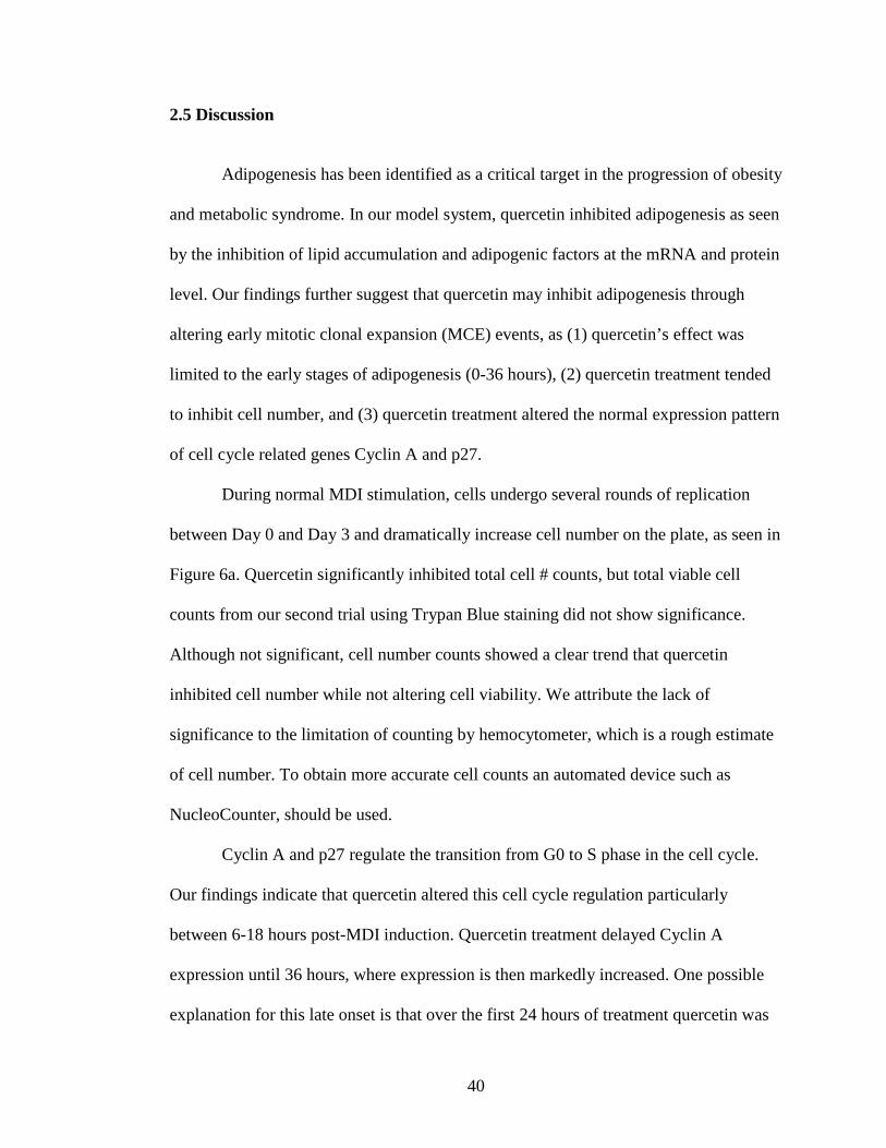
40
2.5 Discussion
Adipogenesis has been identified as a critical target in the progression of obesity
and metabolic syndrome. In our model system, quercetin inhibited adipogenesis as seen
by the inhibition of lipid accumulation and adipogenic factors at the mRNA and protein
level. Our findings further suggest that quercetin may inhibit adipogenesis through
altering early mitotic clonal expansion (MCE) events, as (1) quercetin’s effect was
limited to the early stages of adipogenesis (0-36 hours), (2) quercetin treatment tended
to inhibit cell number, and (3) quercetin treatment altered the normal expression pattern
of cell cycle related genes Cyclin A and p27.
During normal MDI stimulation, cells undergo several rounds of replication
between Day 0 and Day 3 and dramatically increase cell number on the plate, as seen in
Figure 6a. Quercetin significantly inhibited total cell # counts, but total viable cell
counts from our second trial using Trypan Blue staining did not show significance.
Although not significant, cell number counts showed a clear trend that quercetin
inhibited cell number while not altering cell viability. We attribute the lack of
significance to the limitation of counting by hemocytometer, which is a rough estimate
of cell number. To obtain more accurate cell counts an automated device such as
NucleoCounter, should be used.
Cyclin A and p27 regulate the transition from G0 to S phase in the cell cycle.
Our findings indicate that quercetin altered this cell cycle regulation particularly
between 6-18 hours post-MDI induction. Quercetin treatment delayed Cyclin A
expression until 36 hours, where expression is then markedly increased. One possible
explanation for this late onset is that over the first 24 hours of treatment quercetin was

41
able to quench reactive oxygen species (ROS) signaling necessary for the induction of
cell cycle events [37], however, by the end of 24 hours quercetin’s antioxidant capacity
was depleted allowing the cell to mount one final attempt at cell cycle progression. We
have further shown in preliminary data (see Appendix 1) that quercetin inhibits the
mRNA expression of NADPH oxidase 4 (NOX4), a mitochondrial protein responsible
for producing ROS required for differentiation [100]. Nonetheless, it is clear that
quercetin prevented early induction of Cyclin A which facilitates the transition from S
phase into G2 phase. To further determine quercetin’s effect on cell cycle, Flow
Cytometry should be employed to provide a quantitative profile of the number cells in
each cell cycle stage.
MCE has been implicated to be required for terminal differentiation in 3T3-L1
adipocytes. 3T3-L1 pre-adipocytes in the presence of a mitotic inhibitor fail to
differentiate [99]. However, other adipogenic models (10T1/2 MSCs, and Human
MSCs) have been shown to differentiate independent of MCE, and reflect in vivo
stimulation of precursors cells one step prior to preadipocyte commitment. This raises
the question as to whether or not compounds previously characterized anti-adipogenic
in 3T3-L1 systems are able to inhibit adipogenesis in models less dependent on MCE
events. To pursue this question we tested quercetin in 10T1/2 cells, and found that
quercetin (50µM) significantly inhibited lipid accumulation and that this effect was not
time dependent (see Appendices 2 and 3). Although further work needs to be done in
this model system, our preliminary data suggest that quercetin’s effect is not limited to
the 3T3-L1 model system, affirming that quercetin may inhibit adipogenesis in vivo.

42
Because AMPK has been implicated to inhibit differentiation and cell cycle
progression, we investigated whether Compound C, an AMPK inhibitor, mediates
quercetin’s effect on adipogenesis, testing the hypothesis that quercetin may inhibit
adipogenesis via AMPK activation. Both strong activation and strong inhibition of
AMPK has been shown to inhibit adipogenesis suggesting AMPK is tightly regulated
during these early stages. Compound C inhibits adipogenesis at high concentrations
(>1μM), therefore lower concentrations were used (0.1-1μM) in combination with
quercetin (25 and 50μM). Preadipocytes were stimulated to differentiate in the presence
of both of these compounds. Lipid accumulation was measured at Day 6 (see Appendix
4). Compound C did not mediate quercetin’s effect at any concentration, suggesting
that quercetin may inhibit adipogenesis through AMPK-independent pathways. Protein
analysis confirming Compound C’s ability to block AMPK activation is needed to
further demonstrate that Compound C is still effective at lower concentrations.
The physiological impact of inhibiting cell cycle and thus adipogenesis could be
viewed positively or negatively: (1) it would be beneficial to mediate the accelerated
rate of adipogenesis seen in childhood obesity, early stages of obesity, or in patients
taking TZD drugs used for type 2 diabetes, or (2) it would be harmful to alter the
body’s natural response to increase fat storage, and potentially accelerate the
hypertrophy of mature adipocytes leading to their dysfunction. Both views are valid at
this point because of our limited knowledge of in vivo regulation of adipogenesis,
however quercetin should not be thought to completely block adipogenesis but rather
mediate an accelerated, irreversible, over-stimulation of adipogenesis. It is also

43
important to consider quercetin’s effect on mature adipocytes, to gain a holistic view of
quercetin’s potential as an anti-obesity agent.

44
CHAPTER 3
EFFECT OF QUERCETIN ON ADIPOCYTE METABOLSIM
3.1 Literature Review
The rise in obesity will significantly decrease life expectancy and continue to
burden health care systems because of increased risk for the development of chronic
disease [95]. Therefore, feasible, large-scale prevention and treatment options must be
made available to the public in order to reverse this growing epidemic. At the cellular
level, obesity is defined as the increase in size (hypertrophy) and number (hyperplasia)
of adipocytes. Hypertrophy of adipocytes leads to their dysfunction as the endoplasmic
reticulum and mitochondria are unable to keep up with increased metabolic demands.
Dysfunctional adipocytes become insulin resistant and elicit macrophage induction to
the adipose tissue further promoting the development of chronic disease [3]. Therefore
understanding adipocyte metabolism and preventing adipocyte hypertrophy is a critical
approach in the fight against obesity.
Adipocytes handle lipid storage through the synthesis (lipogenesis) and
breakdown (lipolysis) of triglyceride. Naturally, there is much interest in factors that
inhibit lipogenesis and promote lipolysis. However, adipocytes are also capable of
burning stored fatty acids through β-oxidation, providing an exciting approach against
hypertrophy. AMP-activated protein kinase (AMPK), the major metabolic regulator of
all cells, has been shown to induce partial lipolysis and β-oxidation [57]. Therefore
AMPK activators are one such way to metabolically alter adipocytes.

45
Several bioactive compounds have been identified as AMPK activators [16].
One such compound is quercetin, the most commonly consumed dietary flavonoid in
the western diet. Not only has quercetin been implicated as an AMPK activator [13], in
vivo studies have shown quercetin has the ability to improve hyperglycemia,
dyslipidemia, and blunt excess weight gain in animal models of obesity and diabetes [7-
10]. However the role of quercetin on adipocyte metabolism remains largely unknown.
Several reports have identified quercetin’s effect on mature adipocytes to be
lipolytic [101, 102], apoptotic [11, 13, 96, 98], and anti-inflammatory [103]. In the
early 1990s, Kuppusamy et al. carried out experiments with several flavonoids and
showed that quercetin, among others, inhibited phosphodiesterase break down of cAMP
and increased lipolysis in the presence or absence of a lipolytic agent [102]. They
further found that this effect was mediated through activation of β-adrenergic signaling,
as the effect was inhibited by an β-adrenergic antagonist [101]. Furthermore, Ohkoshi
et al. [104] found extract from Nelumbonecifera containing quercetin metabolites to
also stimulate lipolysis in white adipose tissue. Thus, quercetin is generally recognized
as lipolytic, however the mechanism remains unknown.
Currently, no studies have characterized quercetin’s ability to activate β-
oxidation, which is a likely hypothesis given quercetin’s ability to activate AMPK.
3.2 Purpose of Study
Quercetin, a widely consumed dietary flavonoid, holds enormous potential as an
anti-obesity agent. Recently quercetin has been implicated as an AMPK activator,
bringing into question its ability to induce lipolysis and β-oxidation in adipocytes. We

46
therefore aimed to fill this gap of knowledge by testing quercetin’s effect on lipolysis
and fatty acid oxidation, specifically through the activation of AMPK. To test this, in
mature 3T3-L1 adipocytes we investigated (1) quercetin’s action on AMPK and
downstream target ACC at the protein level, (2) quercetin’s action on free fatty acid
content in the media, and (3) quercetin’s effect on mRNA expression of PGC-1α, UCP-
1 and UCP-3.
We hypothesized that (1) quercetin would increase protein levels of
phosphorylated AMPK and the downstream target ACC, and that Compound C, an
AMPK inhibitor, would inhibit this effect, (2) quercetin would increase free fatty acid
(a lipolytic effect) and that Compound C would block this effect, and (3) quercetin
would increase mRNA expression of PGC-1α, UCP-1 and UCP-3 (genes involved in
fatty acid oxidation) and that Compound C would block this effect.
3.3 Materials and Methods
3.3.1 Cell Culture and Treatments
For the differentiation of 3T3-L1 preadipocytes, cells were grown to 100% confluence
in Growth Media [DMEM (high glucose), 10% Calf Serum, 1% Penicilin/
Streptamycin] replaced every two days. Two days post-confluence, growth media was
changed to Differentiation Media [DMEM (high glucose), 10% Fetal Bovine Serum,
1% P/S, 1% Insulin, 1% 3-Isobutyl-1-methylxanthine 11.5mg/ml, 0.01%
Dexamethasone 3.9mg/ml] in the presence or absence of quercetin aglycone (Sigma-
Aldrich Catalog # 6151-25-3). Three days post-differentiation, media was replaced with

47
Post-Differentiation media [DMEM (high glucose), 10% Fetal Bovine Serum, 1% P/S,
1% Insulin, +/- quercetin]. 3T3 L1 preadipocytes generally reach 80 -90%
differentiation 8 to 9 days post MDI induction. At this point cells were washed with
PBS to remove residual serum and insulin, and media was replaced with serum and
insulin free DMEM supplemented with 0.5% bovine serum albumin (BSA) in the
presence or absence of treatment. (Quercetin : 25, 50 , 100 μM, AICAR: 0.5, 1 mM,
and Compound C: 10 μM). Treatment lasted 15-48 hrs at which point cells and media
were harvested for analysis.
3.3.2 Oil Red O Lipid Staining
Cells were harvested on desired days and underwent Oil Red O Staining to quantify
lipid accumulation, as it is an indirect determiner of cell differentiation. Cells, grown on
24-well and 6-well plates, were treated with 10% formaldehyde in PBS for 1 hour,
washed with 60% isopropanol, and completely dried. Then, cells were stained with
0.5% Oil Red O solution in 60:40 (v/v) isopropanol: H2O, for 30 minutes at room
temperature. Finally wells were washed with distilled water and dried. Optical density
was then measured at 490nm, after eluting with isopropanol, to quantify lipid
accumulation.
3.3.3 Protein Isolation and Western Blotting
Cells were harvested at desired times with RIPA Buffer containing protease inhibitor
(500ul per p-100 dish). Samples were stored at -80° C until protein quantification.
Samples were thawed on ice, sonicated, and centrifuged. Supernatant, containing whole
48
cell protein, was transferred to a fresh tube for protein quantification using the
Bicinchoninic Acid (BCA) assay. A BCA standard curve was established using bovine
serum albumin (BSA) [2mg/ml] protein and measured at 570nm. Sample
concentrations were then measured against the standard, and prepared with sample
buffer to allow for 15μg to be loaded to the gel. Samples were run on acrylamide
(varying percentages) gels via electrophoresis, and then proteins were transferred to a
PVDF membrane through wet transfer. Upon successful transfer, membranes were
blocked with 5% non-fat dry milk, and treated with primary and secondary antibodies
(purchased from Santa Cruz Biological). HRP detection was done through ECL
solution. Chemiluminescent bands were captured on x-ray and developed.
3.3.4 RNA isolation and Analysis
Cells washed with phosphate buffer solution were harvested with Trizol Reagant and
stored at -80° C until mRNA isolation. For isolation, samples were thawed and
centrifuged with 200µL chloroform. Supernatant was transferred to a fresh tube and
centrifuged with isopropanol (1:1) to precipitate RNA. Isopropanol was removed and
the pellet was washed three times with ethanol by centrifugation. Pellet was
resuspended in DEPC water and quantified using a spectrophotometer at 260nm.
8µg of RNA was used along with SuperScript III reagents to make cDNA which was
stored at -4°C. Polymerase Chain Reaction was performed with the primers for PGC-1α,
UCP-1, UCP-3, and adiponectin (see Table 2). Samples were run on an agarose gel
containing Ethidium Bromide and detected using UV light. Band intensity was
determined using Image J analysis and adjusted to β-actin.

49
3.3.5 Free Fatty Acid and Glycerol Assays
Media was collected from wells and stored at 4°C. Free Glycerol Assay (K630-100)
and Free Fatty Acid Quantification Kits (K612-100) were purchased from BIOVISION.
Product protocols were followed. In brief, reagents were warmed to room temperature,
standard curves and samples (undiluted) were added to a 96-well plate. Reagent mix
was added and plates were incubated for the appropriate time. Absorbance was taken at
570nm.
3.3.6 Statistical Analysis
Samples were collected in at least duplicate or triplicate, and differences between the
means were determined by student T-test analysis. P-values were considered significant
at <0.05.
3.4 Results
3.4.1 Quercetin phosphorylates ACC, a downstream target of AMPK
Quercetin has been implicated as an AMPK activator; therefore we tested in our model
system whether or not quercetin activates AMPK and its downstream target, acetyl-
CoA Carboxylase (ACC). Quercetin treatment (50μM) increased activated AMPK as
compared to standard MDI treatment over the course of differentiation (Figure 9A).
Further, quercetin treatment (25-100μM) increased phosphorylated ACC, and this
effect was blocked by the AMPK inhibitor Compound C (Figure 9B). This confirms
that we have a working model to study quercetin’s action dependent on AMPK
activation.
50
3.4.2 Quercetin induces partial lipolysis
We next looked at quercetin’s ability to induce lipolysis by measuring release of free
fatty acids in the media of mature adipocytes treated with serum and insulin-free media.
Our data pooled from 3 separate trials (total n = 7) show that quercetin (100μM)
significantly increased the release of free fatty acids significantly and Compound C
(10μM) blocked this effect (Figure 10).
3.4.3 Quercetin induces oxidative pathways
Because AMPK is known to induce β-oxidation, we looked at quercetin’s ability to
induce the expression of PGC-1α, a transcriptional regulator of oxidative genes,
Uncoupling Protein 1 (UCP-1) and Uncoupling Protein 3 (UCP-3), two of the
uncoupling proteins expressed in the mitochondria of muscle and brown adipose tissue
to uncouple oxidation from ATP synthesis. Mature adipocytes were treated with
quercetin (25-100μM) for 24 hours in serum-free insulin-free media. Quercetin
increased mRNA expression of PGC-1α, and this effect appeared to be attenuated by
Compound C treatment (Figure 11). Further, quercetin treatment induced mRNA
expression of UCP-1 and UCP-3 (Figures 12 and 13). Upregulation of UCP-3 by
quercetin appeared to be attenuated by Compound C treatment. These results suggest
quercetin is able to induce uncoupled fatty acid oxidation through the activation of
AMPK.

51
Con CC A CC/A Q25 Q50 Q100 Q25 Q50 Q100
+Compound C
pACC ►
β-actin ►
B
0hr 12 24 48 Day 3 Day 6
pAMPK ►
Q50μM _ +
A
Figure 9. Quercetin increases protein expression of pAMPK and pACC. Protein analysis of pAMPK and downstream target pACC. (A) 3T3-L1 preadipocytes were differentiated in the presence or absence of quercetin (50μM) and harvested at subsequent time points throughout the course of adipogenesis to determine AMPK activation. In the control pAMPK increased at 12 hour but then decreased to a basal level from 24 hour to Day 6. Quercetin treatment sustained pAMPK levels through the course of adipogenesis. (B) Mature adipocytes were treated for 48 hours in serum-free insulin-free %0.5 BSA DMEM in the presence or absence of Compound C (CC) 10 μM, AICAR (A) 1mM, or quercetin (Q) 25-100μM. AICAR treatment increased pACC protein levels and CC attenuated this effect. Quercetin treatment dose-dependently increased pACC expression and CC also attenuated this effect. These results indicate quercetin activates AMPK pathways and CC, and AMPK inhibitor successfully blocked AMPK’s downstream action.
pAMPK ►

52
Free Fatty Acid Release
Con Aicar CC Aicar/CC Q25 Q50 Q100 Q25 Q50 Q100
% C
ontro
l
0
50
100
150
200
250
300
350
+Compound C [10uM]
*
*
Figure 10. Quercetin 100μM increases free fatty acid content in media. Mature adipocytes were treated with Compound C (CC) 10µM, Aicar (A) 0.5 and 1mM, and quercetin (Q) 25-100µM for either 24 or 48hrs and media was collected and analyzed for free fatty acid content. Data presented here reflects pooled data from 3 separate trials each slightly varied by length of treatment or AICAR concentration (n=7). AICAR treatment significantly increased FFA content in the media while CC attenuated this effect. Likewise quercetin (100 µM) significantly increased FFA content and CC attenuated this effect suggesting quercetin is able to stimulate FFA release through AMPK activation.

53
Con A CC A/CC Q25 Q50 Q100 Q25 Q50 Q100
PGC-1α ►
+ Compound C
β-actin ►
Figure 11. Quercetin up-regulates mRNA expression of PGC-1α. PGC-1α is a transcriptional regulator of β-oxidation among other metabolic pathways. 3T3-L1 mature adipocytes were treated with AICAR (A) 1mM, Compound C (CC) 10µM, and quercetin (Q) 25-100µM for 24 hours and harvested for mRNA analysis by qualitative PCR. AICAR increased PGC-1α expression and CC blunted this effect. Quercetin also increased PGC-1α expression and CC attenuated this effect. This data suggests quercetin is able to increase PGC1α mRNA through an AMPK dependent pathway.
PGC-1α
Treatment
Con Aicar CC A/CC Q25 Q50 Q100 Q25 Q50 Q100
Rel
ativ
e to
β-a
ctin
0
1
+Compound C

54
Con Con Tro Q10 Q50 Q100
Day 8 Day 9
UCP-1 ►
β-actin►
Figure 12. Quercetin up-regulated mRNA expression of Uncoupling Protein 1. Uncoupling Protein 1 (UCP-1) is the major thermogenic gene expressed predominantly in muscle and brown fat. UCP1 uncouples ATP synthesis from mitochondrial oxidation. 3T3-L1 mature adipocytes were treated with quercetin (10-100μM) for 24 hours. Slight UCP-1 expression was induced in the control between Day 8 and 9, however quercetin 50μM and 100μM significantly increased UCP-1 expression. Troglitazone (Tro 10μM), a diabetic drug and PPARγ ligand had no effect on UCP-1 expression.

55
Figure 13. Quercetin increased mRNA expression of Uncoupling Protein 3. Uncoupling Protein 3 (UCP-3) is part of the group of uncoupling proteins that uncouples ATP synthesis from oxidation in the mitochondria and is a transcriptional regulator of β-oxidation among other metabolic pathways. 3T3-L1 mature adipocytes were treated with AICAR (A) 1mM, Compound C (CC) 10µM, and quercetin (Q) 25-100µM for 24 hours and harvested for mRNA analysis by qualitative PCR. AICAR increased PGC-1α expression and CC blunted this effect. Quercetin also increased PGC-1α expression and CC attenuated this effect. This data suggests quercetin increases PGC-1α mRNA through an AMPK dependent pathway.
Con A CC A/CC Q25 Q50 Q100 Q25 Q50 Q100
UCP-3 ►
+ Compound C
B-actin ►
UCP-3 mRNA
Treatment
Con Aicar CC A/CC Q25 Q50 Q100 Q25 Q50 Q100
Exp
ress
ion
Rel
ativ
e to
B-a
ctin
0
1
+Compound C

56
3.4 Discussion
There is great potential in the use of bioactive compounds to alter adipocyte
metabolism and thus thwart the progression of adipocyte dysfunction and metabolic
syndrome. Our preliminary findings suggest quercetin is one such compound.
In our model system we clearly showed quercetin’s ability to activate AMPK, a target
in the treatment of metabolic syndrome.
AMPK is the major metabolic regulator of the cell, regulated by the level of
AMP/ATP. As AMP levels rise, more AMP binds to the gamma subunit of AMPK
causing a conformational change protecting AMPK from being dephosphorylated. Thus
a rise in AMP results in increased phosphorylated AMPK. It has been determined that
AMPK is activated by low glucose, hypoxia, ischemia, adiponectin, leptin, and alpha
adrenergic receptor. It would be of interest to further investigate the mechanism by
which quercetin activates AMPK. It is possible that quercetin creates a hypoxic
environment because of its antioxidant capacity. However, it has also been suggested
that quercetin activates adrenergic receptors to mediate lipolysis, thus it is plausible that
quercetin may interact with membrane bound receptors [101]. It has been also been
shown that quercetin is able to penetrates the membrane through passive diffusion and
significantly accumulates in the mitochondria [105]. In fact, Gledhill et al. determined
that quercetin binds and inhibits the rotary mechanism of the F1-ATPase, required for
ATP synthesis in the mitochondria [106]. This action may suggest quercetin activates
AMPK by lowering the ATP/AMPK ratio in the cell; however, this mechanism would
need to be further tested.

57
AMPK has been implicated to induce partial lipolysis and β-oxidation. We
therefore investigated quercetin’s effect on both of these pathways. As shown,
quercetin stimulated free fatty acid release into the media, and this effect was blunted
by the AMPK inhibitor, Compound C. Figure 10 consists of pooled data from 3
separate trials, each with a slightly different experimental design. Therefore these trials
should be repeated with a consistent design. From our experience, the best design
would be to treat mature adipocytes (80% differentiated) in serum-free, insulin-free
DMEM supplemented with 0.5% Bovine Serum Albumin (BSA) for 48hrs using
AICAR (1mM) and Compound C (10μM) concentrations.
Interestingly, quercetin significantly reduced glycerol release in the media, and
Compound C did not consistently reverse this effect in all treatments (see Appendix 5).
This finding suggests quercetin induces partial lipolysis (the release of free fatty acids
but not glycerol), and this effect may be through AMPK-independent pathways, as
AICAR treatment not alter glycerol release. It has been implicated that AMPK
deactivates HSL the major lipolytic enzyme, however ATGL is still available in the
cytoplasm to begin TG breakdown by releasing the first free fatty acid (FFA) [52].
Therefore, if HSL is inactivated by AMPK incomplete lipolysis occurs, which explains
why free fatty acid is released but not glycerol. However, this explanation does not
account for the significant decrease in glycerol release seen with quercetin treatment;
therefore quercetin may act independently of AMPK on glycerol metabolism. It would
be of great interest to explore the mechanism behind this observation, specifically by
looking at the activity of glycerol-3-phosphate dehydrogenase in the mitochondria, and

58
aquaporin 3 and 9, which are membrane bound glycerol transporters, all of which
regulate glycerol release during lipolysis.
Quercetin increased PGC-1α, UCP-1 and UCP-3 mRNA expression, suggesting
quercetin induces uncoupled oxidation in the cell. These preliminary findings should be
repeated, as results here only reflect one trial and qualitative RT-PCR is not as precise
as real-time PCR analysis. Interestingly, it appears that Compound C blunted
quercetin’s ability to induce PGC-1α, and UCP-3, suggesting that AMPK activation
may be the mechanism by which quercetin induces oxidation. Quercetin’s effect on
UCP-1 expression in the presence of Compound C was not investigated here and is
worth exploring. Other researchers found that chronic AMPK stimulation through
AICAR induced mitochondrial biogenesis and fatty acid oxidation , but did not
stimulate UCP-1 expression [48, 107].Therefore it is likely that quercetin may stimulate
UCP-1 through an AMPK-independent pathway. These exciting preliminary findings
showing quercetin ability to stimulate uncoupled β-oxidation would have far reaching
effects if translated into human cells lines and further into in vivo models. Potentially
quercetin could enhance β-oxidation in hypertrophied adipocytes enabling them to
prevent or recover from metabolic dysfunction, from the burden of excess lipid.
Although mRNA expression seen in this research suggests quercetin induces
uncoupled fatty acid oxidation, further research needs to be done to confirm this effect.
In particular it would be of interest to determine whether or not quercetin increases
oxygen consumption, which would be an indication of increased mitochondrial function.
Quercetin did not significantly decrease triglyceride content in mature adipocytes over
a 48hr period in our model system (see Appendix 6). In animal studies, quercetin doses

59
did result in weight loss [7] suggesting prolonged treatment of quercetin might be
necessary to see dramatic changes in adipocyte lipid content. Increased uncoupled
oxidation of free fatty acids in adipose tissue provides a possible mechanism for
quercetin’s effect.
Quercetin’s ability to stimulate partial lipolysis and uncoupled fatty acid
oxidation in vivo could potentially prevent and/or alleviate the progression of
hypertrophied adipocytes. It is carefully noted that releasing free fatty acids into
circulation is not ideal given cardiovascular risk; however partial lipolysis would evoke
a mild effect in vivo, because essentially only a small fraction of the broken down
triglyceride would be released. The remaining diglyceride would be further metabolized
in the cell, likely through uncoupled β-oxidation. Therefore, these findings suggest that
quercetin may improve signs of metabolic syndrome, specifically hyperglycemia and
insulin sensitivity, in vivo by preventing or ameliorating the lipid burden in
hypertrophied adipocyte through AMPK mediated metabolic changes.

60
CHAPTER 4
CONCLUSION & FUTURE DIRECTIONS
4.1 Summary
Obesity is one of the most pressing concerns of health care officials because of
its increasing prevalence and significant link to the development of type 2 diabetes,
cardiovascular disease and cancer. At the cellular level obesity is defined as the
increase in cell number (hyperplasia) and cell size (hypertrophy), resulting in the
irreversible expansion of adipose tissue in unwanted depots and the dysfunction of
adipocytes leading to the development of metabolic system. Our findings here
demonstrate that quercetin, the most widely consumed dietary flavonoid, shows
promising potential as an anti-obesity agent because of its ability to target both
hyperplasia and hypertrophy in our in vitro model.
We found that quercetin inhibited adipogenesis in 3T3-L1 preadipocytes
through altering cell cycle events, specifically cell number, and expression of cell cycle
regulators Cyclin A and p27. These findings are significant as in vivo hyperplasia has
been implicated to be tightly controlled by cell cycle [35]. In 10T1/2 mesenchymal
stem cells, a cell line one step removed from preadipocyte commitment, quercetin
significantly inhibited adipogenesis, further supporting quercetin’s potential as an anti-
adipogenic agent in vivo. Quercetin has been implicated as an AMPK activator [13],
and further either strong inhibition or activation of AMPK inhibits adipogenesis
through MCE events. However our preliminary findings did not indicate that
quercetin’s anti-adipogenic effect is AMPK dependent.

61
In mature adipocytes, quercetin induced lipolytic and oxidative pathways.
Quercetin significantly increased free fatty acid content in the media, and up-regulated
expression of PGC-1α, Uncoupling Protein 1 (UCP-1) and 3 (UCP-3). Compound C,
an AMPK inhibitor, blunted these effects suggesting quercetin’s action is AMPK
dependent. These initial findings indicate quercetin may be able to shift adipocytes
toward energy burning pathways, which could alleviate adipocyte hypertrophy
in vivo.
4.2 Limitations
There are a few limitations of the work presented here that are important to
discuss. First is the concentration of quercetin used in our in vitro model. Physiological
levels generally approach the 200nM range, however the concentration used in our
experiments were 25, 50, and 100µM. These in vitro levels have never been reached in
humans, and would likely only result from enhanced supplementation such as
nanoparticle delivery of quercetin. Nonetheless, in vitro studies allow researchers to
understand mechanistic aspects of quercetin’s action.
A second limitation we encountered was the use of Compound C to investigate
whether quercetin’s anti-adipogenic effect was AMPK dependent. Compound C at high
doses (5-10µM) inhibited adipogenesis (data not included), therefore lower
concentrations (0.1-1µM) were used in combination with quercetin. However, this
brings into consideration Compound C’s effectiveness to inhibit AMPK at such a low
62
dose. Therefore, other strategies for determining whether quercetin anti-adipogenic
effect is through AMPK activation need to be developed.
A third limitation was sample size, due to time constraints. Most of the work
presented here is in the preliminary stage and needs to be repeated in order to gain
statistical significance. However we were able to gain valuable insight from these initial
findings.
Finally a major limitation not encountered here but commonly encountered with
human supplementation trials of quercetin is bioavailability. To date, human studies
have only shown quercetin supplementation to reduce systolic blood pressure [91].
Quercetin has been shown to be safe at high doses [108] therefore pharmacological
doses can be used. However absorption is still variable from individual to individual
because of the gut microflora’s ability to metabolize quercetin. One possible way to
overcome this limitation is through the use of nanoparticle quercetin delivery, which is
currently being developed [73, 109].
4.3 Significance of findings in the context of obesity and metabolic syndrome
In vivo animal studies have shown quercetin to ameliorate metabolic syndrome,
specifically insulin resistance, hyperglycemia, and weight gain. Our findings here of
quercetin’s effect on adipocytes help speculate on the mechanisms behind quercetin’s
in vivo effects.

63
4.3.1 Weight Management
Quercetin’s ability to stimulate oxidative pathways, specifically thermogenesis,
through AMPK activation is an exciting new finding that suggests quercetin may be
able to attenuate weight gain and help promote weight loss in obese individuals.
Currently a major focus of anti-obesity drug designers is targeting thermogenesis to
increase resting metabolic rate [110]. It is known that those with lower daily energy
expenditure are more susceptible to obesity [111]. As an individual’s body mass
increases, their resting metabolic rate will increase to maintain the excess tissue,
however it has been shown that after weight loss, post-obese individuals have a reduced
metabolic rate [112]. Further fatty acid oxidation is impaired in obese individuals, even
after weight loss [110, 113]. The only uncoupling drug used in humans, dinitrophenol,
resulted in weight loss up to 3kg per week, but caused sweating and worse hypoxia
[114]. Therefore milder thermogenic agents are currently being pursued to obtain
similar weighty loss effects at a safer level.
Quercetin’s ability to increase metabolic rate has been determined in animals
but not yet in humans. Stewart et al. [115] looked at energy expenditure, as measured
by indirect calorimetry, in C57BL/6J mice fed a high-fat diet with quercetin (0.8g/kg)
for 8 weeks. At week 3, energy expenditure was significantly higher (p<0.05) in the
quercetin group over the control, however at week 8 there was no difference observed
between the two groups. Based on this finding in animals, and quercetin’s proposed
ability to stimulate oxygen consumption in muscle cells [116], Egert et al. conducted a
pilot study in humans to test quercetin’s ability to increase metabolic rate. Six healthy
normal-weight women were given 150mg doses of quercetin and respiratory
64
consumption of oxygen was measured 5 min – 3 hours post-ingestion, however no
significant difference was found with quercetin treatment. This result is likely due to
the small sample size (n=6) and lower dosage of quercetin (300 times lower than
Stewart et al. dosage in rats), therefore plasma quercetin concentrations may have been
below the threshold for any observed metabolic effect. Our findings that quercetin
increases mRNA expression of UCP-1, UCP-3 and PGC-1α in 3T3-L1 adipocytes
strongly supports continued efforts to determine quercetin’s metabolic effects in vivo.
4.3.2 Insulin Sensitivity
Quercetin has repeatedly been shown to improve hyperglycemia and insulin
sensitivity in animal models. Our findings that quercetin stimulates AMPK, induces β-
oxidation, and increases adiponectin mRNA expression all suggest potential
mechanisms for quercetin’s in vivo effect. AMPK is a major target for drug companies
AMPK has been shown to improve glucose uptake in vitro and improve hyperglycemia
in vivo. Furthermore, the commonly prescribed class of diabetic drugs,
Thiazolidinediones (TZDs), have been shown to activate AMPK [117]. Chronic use of
these drugs has been associated with negative side-effects therefore milder activators of
AMPK are wanted to allow for long-term use [66, 118]. We clearly showed quercetin’s
ability to activate AMPK in 3T3-L1 preadipocytes. Therefore, quercetin holds great
potential as an anti-diabetic agent, and AMPK activation is likely the mechanism
behind quercetin’s in vivo effect. So far quercetin has been shown to improve glucose
uptake in adipocytes [119] and in muscle cells [120], and AMPK has been implicated in
the mechanism.

65
One area that warrants future research is quercetin’s ability to increase
adiponectin levels and the anti-diabetic effects associated with that. Adiponectin levels
are negatively correlated with obesity and type 2 diabetes [121], and further
administration of adiponectin in rats improves glucose regulation [122]. Quercetin
treatment has been shown to increase circulating adiponectin levels in animal models [7,
8, 123]. To our knowledge no human studies have explored quercetin’s ability to
increase circulating adiponectin levels and our preliminary findings that show
quercetin’s ability to increases mRNA expression of adiponectin in mature adipocytes
certainly warrant this exploration.
4.4 Application of Quercetin from a Public Health Perspective
Estimating quercetin content in plants is extremely limited due to the variability
in flavonoid production based on environmental as well as genetic factors [65],
therefore limited data exists on the correlation between quercetin intake and chronic
disease. Because of this missing link, it is too soon for public health officials to develop
feasible recommendations to the public for the use of quercetin as an anti-obesity agent.
Our working knowledge, based largely on supplementation studies done by
Egert et al., is that 150mg supplemental doses (roughly the equivalent of 12-15 servings
of fruits and vegetables) may not be sufficient to elicit the powerful health benefits
found in animal models using nearly 200 to 300 times the dosage amount. Therefore it
can be speculated that the future use of quercetin as an anti-obesity agent will be

66
through supplementation or product enhancement, from which a quercetin intake of
around 1000mg could be reached.
This gap in our current knowledge should not however detract from the
recommendation of health officials to increase consumption of quercetin through intake
of fruits and vegetables, specifically those high in quercetin (onions, apples, kale, etc.).
It is likely that sustained lower doses (50-100mg, the equivalent of roughly 5-8 servings
of fruits and vegetables) would have a preventative effect, while higher doses would be
required to use quercetin therapeutically to treat conditions related to obesity.
4.5 Direction of Future Research
4.5.1 Quercetin’s effect on adipogenesis
Although our findings here suggest quercetin inhibits adipogenesis through
altering cell cycle events, this effect should be further confirmed in vitro through the
use of (1) a more accurate cell counting technique, NucleoCounter, (2) Flow Cytometry,
to quantify the number of cells in each cell cycle stage, and (3) human cell lines that
may behave differently than 3T3-L1 preadipocyte because of pluripotency stage. Once
quercetin’s effect has been confirmed in vitro, animal studies should be employed in
order to determine quercetin’s ability to inhibit hyperplasia in various adipose depots.
4.5.2 Quercetin’s effect on mature adipocytes Our preliminary findings suggest quercetin may induce partial lipolysis and
shift the cell toward uncoupled β-oxidation. To further confirm this effect in vitro (1)
our experiments should be repeated at least two times to gain validity, and (2) oxygen
67
consumption should be analyzed to determine quercetin’s ability to increase
mitochondrial oxidation. Next it would be of interest to determine whether quercetin
decreases adipocyte size in an obese animal model. This would confirm quercetin’s
ability to ameliorate hypertrophied cells and the consequences associated with them. It
should also be determined whether quercetin improves adiponectin levels in vivo as our
findings indicate quercetin increases mRNA expression of adiponectin.

68
APPENDIX 1
QUERCETIN MAY INHIBIT NOX4 EXPRESSION DURING THE EARLY
STAGES OF ADIPOGENESIS
Q50μM - - + - + - + - + - + 0hr 6hr 12hr 24hr 36hr 48hr
Nox 4 ►
β-actin►
Appendix 1. NADPH oxidase 4 (NOX4) is a major producer of reactive oxygen species (ROS) in the cell. During mitotic clonal expansion NOX4 has shown to be required for ROS signaling that promotes cell cycle events. Here we investigated whether quercetin treatment alters NOX4 mRNA expression. Our hypothesis was that quercetin would inhibit or alter NOX4 expression, thus inhibiting mitotic clonal expansion (MCE), a process required for terminal differentiation. 3T3-L1 preadipocytes were differentiated in the presence or absence of quercetin (50µM) and harvested at subsequent time points (0,6,12,24,36,48 hours) for mRNA analysis to characterize NOX4 mRNA expression during MCE. In the absence of quercetin Nox 4 expression rose steadily from 6 hours to 36 hours and then was down-regulated by 48 hours. However in the presence of quercetin NOX4 mRNA expression was completely blunted until slight stimulation came on a 36 and 48 hours. Similar to Cyclin A (see Figure 9) results, it is possible that quercetin’s anti-oxidant capacity is responsible for blunting the expression of NOX4 and that by 36 hours quercetin’s anti-oxidant capacity is quenched, allowing the cells to signal a delayed response to the adipogenic cocktail (MDI). The implications of these findings would be that quercetin may inhibit cell cycle progression by blunting the signaling pathways involved in initiating and sustaining cell cycle events. It has also been shown that strong antioxidant treatment inhibit cell cycle, therefore the mechanism by which quercetin inhibits cell cycle is likely through it’s anti-oxidant capacity. It would be of interest to determine ROS levels during early time points to confirm quercetin ability to quench ROS.

69
Quercetin Treatment (uM)
Con 1 5 10 20 50
Abs
490n
m
0.0
0.1
0.2
0.3
0.4
*
Appendix 2. 10T1/2 mesenchymal stem cells are an adipogenic cell line model one-step removed from preadipocytes commitment, they therefore more closely model adipogenesis as it occurs from the initial recruitment of stromal vascular stem cells to preadipocytes to mature adipocytes. This cell line has been determined to be less reliant on mitotic clonal expansion (MCE) for terminal differentiation therefore it was of interest to test quercetin’s effect on their differentiation. Our hypothesis was that if quercetin inhibits adipogenesis through MCE events then quercetin would have no effect on 10T1/2 cell differentiation. 10T1/2 cells were differentiated in the presence or absence of quercetin (1, 5, 10, 20, 50µM) and harvested at Day 8 to measure lipid accumulation, an indirect measure of adipogenesis, by Oil Red O staining. Quercetin (50µM) significantly inhibited lipid accumulation by Day 8, indicating that quercetin is able to inhibit adipogenesis in adipogenic models less depending on MCE, further affirming quercetin potential to inhibit adipogenesis in vivo. It should be noted that lower concentrations of quercetin did not significantly inhibit lipid accumulation. 10T1/2 cells are thought to be more sensitive than 3T3-L1 preadipocytes to treatments, therefore the fact that lower concentrations had no effect may suggest that quercetin’s effect in 10T1/2 cells is indeed weaker and only very strong doses has an effect.
APPENDIX 2
QUERCETIN INHIBITS LIPID ACCUMULATION OF 10T1/2
MESENCHYMAL STEM CELLS
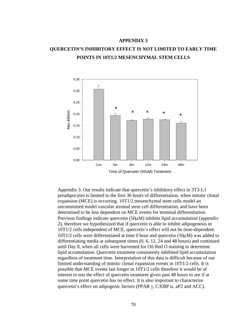
70
APPENDIX 3
QUERCETIN’S INHIBITORY EFFECT IS NOT LIMITED TO EARLY TIME
POINTS IN 10T1/2 MESENCHYMAL STEM CELLS
Time of Quercetin (50uM) Treatment
Con 0hr 6hr 12hr 24hr 48hr
Abs
490n
m
0.00
0.05
0.10
0.15
0.20
0.25
0.30
0.35
* * * * *
Appendix 3. Our results indicate that quercetin’s inhibitory effect in 3T3-L1 preadipocytes is limited to the first 36 hours of differentiation, when mitotic clonal expansion (MCE) is occurring. 10T1/2 mesenchymal stem cells model an uncommitted model vascular stromal stem cell differentiation, and have been determined to be less dependent on MCE events for terminal differentiation. Previous findings indicate quercetin (50µM) inhibits lipid accumulation (appendix 2), therefore we hypothesized that if quercetin is able to inhibit adipogenesis in 10T1/2 cells independent of MCE, quercetin’s effect will not be time-dependent. 10T1/2 cells were differentiated at time 0 hour and quercetin (50µM) was added to differentiating media at subsequent times (0, 6, 12, 24 and 48 hours) and continued until Day 8, when all cells were harvested for Oil Red O staining to determine lipid accumulation. Quercetin treatment consistently inhibited lipid accumulation regardless of treatment time. Interpretation of this data is difficult because of our limited understanding of mitotic clonal expansion events in 10T1/2 cells. It is possible that MCE events last longer in 10T1/2 cells therefore it would be of interest to test the effect of quercetin treatment given past 48 hours to see if at some time point quercetin has no effect. It is also important to characterize quercetin’s effect on adipogenic factors (PPAR γ, C/EBP α, aP2 and ACC).
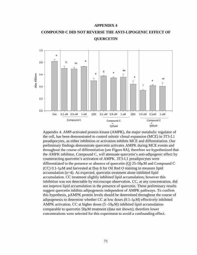
71
APPENDIX 4
COMPOUND C DID NOT REVERSE THE ANTI-LIPOGENIC EFFECT OF
QUERCETIN
Appendix 4. AMP-activated protein kinase (AMPK), the major metabolic regulator of the cell, has been demonstrated to control mitotic clonal expansion (MCE) in 3T3-L1 preadipocytes, as either inhibition or activation inhibits MCE and differentiation. Our preliminary findings demonstrate quercetin activates AMPK during MCE events and throughout the course of differentiation (see Figure 8A), therefore we hypothesized that the AMPK inhibitor, Compound C, will attenuate quercetin’s anti-adipogenic effect by counteracting quercetin’s activation of AMPK. 3T3-L1 preadipocytes were differentiated in the presence or absence of quercetin (Q) 25-50µM and Compound C (CC) 0.1-1µM and harvested at Day 6 for Oil Red O staining to measure lipid accumulation (n=4). As expected, quercetin treatment alone inhibited lipid accumulation. CC treatment slightly inhibited lipid accumulation; however this inhibition was not detectable by microscope observation. CC, at any concentration, did not improve lipid accumulation in the presence of quercetin. These preliminary results suggest quercetin inhibits adipogenesis independent of AMPK pathways. To confirm this hypothesis, pAMPK protein levels should be determined throughout the course of adipogenesis to determine whether CC at low doses (0.1-1µM) effectively inhibited AMPK activation. CC at higher doses (5 -10µM) inhibited lipid accumulation comparable to quercetin 50µM treatment (data not shown); therefore lower concentrations were selected for this experiment to avoid a confounding effect.
Con 0.1 uM 0.5 uM 1 uM Q25 0.1 uM 0.5 uM 1 uM Q50 0.5 uM 0.1uM 1 uM
Abs
490
nm
0.0
0.2
0.4
0.6
0.8
1.0
Compound C Compound C + Q25uM
Compound C + Q50uM
a
b bcb
ee
ede
f
eef
d

72
Con Aicar CC Aicar/CC Q25 Q50 Q100 Q25 Q50 Q100
%C
ontro
l
0
20
40
60
80
100
120
+Compound C [10uM]
* ** *
* *
APPENDIX 5
QUERCETIN TREATMENT IN MATURE ADIPOCYTES RESULTS IN
DECREASED GLYCEROL CONTENT IN THE MEDIA
Appendix 5. Lipolysis is the breakdown of triglyceride into free fatty acids and glycerol, which are released from the cell. Researchers have previously determined quercetin enhances lipolysis however the mechanism remains unknown. Our findings and others indicate quercetin activates AMP-activated protein kinase (AMPK), the major metabolic regulator of the cell. AMPK has been implicated to induce partial lipolysis, the release of free fatty acid, but not glycerol. We therefore tested quercetin’s effect on glycerol release in the presence or absence of an AMPK inhibitor, Compound C (CC) to determine whether quercetin’s effect on glycerol release was dependent on AMPK activation. Our hypothesis was two fold (1) if quercetin induces lipolysis, then quercetin treatment will increase glycerol release independent of CC treatment, and (2) if quercetin induces partial lipolysis, as seen with AMPK activation, then quercetin treatment will not alter glycerol release. Mature adipocytes were treated with AICAR (0.5-1mM), CC (10 µM) and quercetin (Q) (25-100µM) for 15-48hrs in serum free, insulin free media. Media was collected and analyzed for glycerol content. Data from 3 trials (n=7) was pooled and shown here. Each trial had a slightly different experiment design, either varied by AICAR treatment (0.5 or 1mM) or length of treatment time (15, 24, or 48 hours). The pooled data showed quercetin treatment significantly decreased glycerol content in the media, indicating quercetin’s effect is neither lipolytic nor partially lipolytic through AMPK activation. Interestingly CC only blunted the effect of the strong quercetin treatment (100µM). These findings suggest quercetin treatment alters the normal metabolism of glycerol, and therefore future studies should look at quercetin’s effect on glycerol metabolism, specifically glycerol-3 phosphate dehydrogenase (GPDH) in the mitochondria. Insight here will illuminate quercetin’s overall metabolic effect on mature adipocytes.

73
APPENDIX 6
QUERCETIN TREATMENT IN MATURE ADIPOCYTES FOR 48 HOURS DID
NOT ALTER INTRACELLULAR TRIGLYCERIDE LEVELS
Appendix 6. Adipocytes undergo lipolysis to breakdown stored triglyceride for use in the body. Quercetin has been implicated to stimulate lipolysis. Further quercetin has also been implicated to activate AMPK, which is known to stimulate partial lipolysis. We therefore tested whether quercetin treatment significantly reduced the amount of store triglyceride content and whether the AMPK inhibitor, Compound C mediated this effect. Mature adipocytes were treated with Compound C (CC) 10µM, Aicar (A) 0.5 and 1mM, and quercetin (Q) 25-100µM for either 24 or 48hrs and media was collected and analyzed for free fatty acid content. Data presented here reflects 1 trial (n=3). No treatment significantly altered triglyceride content in the cell. This is likely because any quantifiable change in triglyceride content would take a longer treatment period, since mature adipocytes are resistant to purging lipid.
Intracellular Triglyceride Content
Treatment
Con Aicar CC Aicar/CC Q25 Q50 Q100 Q25 Q50 Q100
TG [n
mol
]
0
5
10
15
20
25
+Compound C [10uM]

74
APPENDIX 7
QUERCETIN UPREGULATES MRNA EXPRESSION OF ADIPONECTIN
Con A CC A/CC Q25 Q50 Q100 Q25 Q50 Q100
adiponectin ►
+ Compound C
β-actin ►
Appendix 7. Adiponectin, an adipokine known to improve insulin sensitivity, is negatively associated with obesity. Therefore increasing adiponectin levels in obese individuals may be beneficial. Mature adipocytes were treated with AICAR (A) 1mM, Compound C (CC) 10µM, and quercetin (Q) 25-100µM for 24 hours and harvested for mRNA analysis by qualitative PCR. AICAR and CC treatment equally stimulated adiponectin mRNA expression, however quercetin treatment dramatically up-regulated expression. CC attenuated this effect suggesting quercetin may increase adiponectin mRNA expression through an AMPK dependent mechanism.
Adiponectin
Treatment
Con Aicar CC A/CC Q25 Q50 Q100 Q25 Q50 Q100
Rel
ativ
e to
β-a
ctin
0.0
0.5
1.0
1.5
+ Compound C
75
REFERENCES
1. Finkelstein, E.A., et al., Annual medical spending attributable to obesity: payer-
and service-specific estimates. Health affairs (Project Hope), 2009. 28(5): p. w822-31.
2. Spalding, K.L., et al., Dynamics of fat cell turnover in humans. Nature, 2008. 453(7196): p. 783-787.
3. de Ferranti, S. and D. Mozaffarian, The perfect storm: obesity, adipocyte dysfunction, and metabolic consequences. Clinical chemistry, 2008. 54(6): p. 945-955.
4. Santos, A.P., M.M. Rogero, and D.H. Bastos, Edible plants, their secondary metabolites and antiobesogenic potential. Recent patents on food, nutrition & agriculture. 2(3): p. 195-212.
5. Gonzalez-Castejon, M. and A. Rodriguez-Casado, Dietary phytochemicals and their potential effects on obesity: A review. Pharmacological research : the official journal of the Italian Pharmacological Society.
6. Vessal, M., M. Hemmati, and M. Vasei, Antidiabetic effects of quercetin in streptozocin-induced diabetic rats. Comparative biochemistry and physiology.Toxicology & pharmacology : CBP, 2003. 135C(3): p. 357-364.
7. Rivera, L., et al., Quercetin ameliorates metabolic syndrome and improves the inflammatory status in obese Zucker rats. Obesity (Silver Spring, Md.), 2008. 16(9): p. 2081-2087.
8. Kobori, M., et al., Chronic dietary intake of quercetin alleviates hepatic fat accumulation associated with consumption of a Western-style diet in C57/BL6J mice. Molecular nutrition & food research.
9. Kobori, M., et al., Dietary quercetin alleviates diabetic symptoms and reduces streptozotocin-induced disturbance of hepatic gene expression in mice. Molecular nutrition & food research, 2009. 53(7): p. 859-868.
10. Bansal, P., et al., Antidiabetic, antihyperlipidemic and antioxidant effects of the flavonoid rich fraction of Pilea microphylla (L.) in high fat diet/streptozotocin-induced diabetes in mice. Experimental and toxicologic pathology : official journal of the Gesellschaft fur Toxikologische Pathologie.
11. Park, H.J., et al., Combined effects of genistein, quercetin, and resveratrol in human and 3T3-L1 adipocytes. Journal of medicinal food, 2008. 11(4): p. 773-783.
12. Yang, J.Y., et al., Enhanced inhibition of adipogenesis and induction of apoptosis in 3T3-L1 adipocytes with combinations of resveratrol and quercetin. Life Sciences, 2008. 82(19-20): p. 1032-1039.
13. Ahn, J., et al., The anti-obesity effect of quercetin is mediated by the AMPK and MAPK signaling pathways. Biochemical and biophysical research communications, 2008. 373(4): p. 545-549.
14. Harmon, A.W. and J.B. Harp, Differential effects of flavonoids on 3T3-L1 adipogenesis and lipolysis. American journal of physiology.Cell physiology, 2001. 280(4): p. C807-13.
76
15. Rayalam, S., M.A. Della-Fera, and C.A. Baile, Phytochemicals and regulation of the adipocyte life cycle. The Journal of nutritional biochemistry, 2008. 19(11): p. 717-726.
16. Hwang, J.T., D.Y. Kwon, and S.H. Yoon, AMP-activated protein kinase: a potential target for the diseases prevention by natural occurring polyphenols. New biotechnology, 2009. 26(1-2): p. 17-22.
17. WorldHealthOrganization. Fact Sheet. Obesity and Overweight [website http://www.who.int/mediacentre/factsheets/fs311/en/index.html] March 2011 [cited August 10, 2011].
18. NIH. Clinical Guidelines on the Indentification, Evaluation, and Treatment of Overweight and Obesity in Adults. NHLBI Obesity Education Initiative 1998 [cited August 10,2011].
19. CDC, National diabetes fact sheet: national estimates and general information on diabetes and prediabetes in the United States. 2011, U.S. Department of Health and Human Services, Centers for Disease Control and Prevention: Atlanta, GA.
20. Pleis, J., B. Ward, and J. Lucas, Summary health statistics for U.S. adults: National Health Interview Survey, 2009. National Center for Health Statistics., 2010. Vital Health Stat10(249).
21. Ervin, R.B., Prevalence of metabolic syndrome among adults 20 years of age and over, by sex, age, race and ethnicity, and body mass index: United States, 2003-2006. National health statistics reports, 2009. (13)(13): p. 1-7.
22. Skelton, J.A., et al., Prevalence and trends of severe obesity among US children and adolescents. Academic pediatrics, 2009. 9(5): p. 322-329.
23. Bjorntorp, P., M. Karlsson, and P. Pettersson, Expansion of adipose tissue storage capacity at different ages in rats. Metabolism: clinical and experimental, 1982. 31(4): p. 366-373.
24. Hirsch, J., et al., The fat cell. The Medical clinics of North America, 1989. 73(1): p. 83-96.
25. Suwa, A., T. Kurama, and T. Shimokawa, Adipocyte hyperplasia and RMI1 in the treatment of obesity. The FEBS journal. 278(4): p. 565-569.
26. Daquinag, A.C., Y. Zhang, and M.G. Kolonin, Vascular targeting of adipose tissue as an anti-obesity approach. Trends in pharmacological sciences. 32(5): p. 300-307.
27. Wing, R.R. and S. Phelan, Long-term weight loss maintenance. The American Journal of Clinical Nutrition, 2005. 82(1 Suppl): p. 222S-225S.
28. Poulos, S.P., M.V. Dodson, and G.J. Hausman, Cell line models for differentiation: preadipocytes and adipocytes. Experimental biology and medicine (Maywood, N.J.). 235(10): p. 1185-1193.
29. Green, H. and M. Meuth, An established pre-adipose cell line and its differentiation in culture. Cell, 1974. 3(2): p. 127-133.
30. Font de Mora, J., et al., Mitogen-activated protein kinase activation is not necessary for, but antagonizes, 3T3-L1 adipocytic differentiation. Molecular and cellular biology, 1997. 17(10): p. 6068-6075.

77
31. Kim, S.W., et al., Regulation of adipogenesis by a transcriptional repressor that modulates MAPK activation. The Journal of biological chemistry, 2001. 276(13): p. 10199-10206.
32. Qiu, Z., et al., DNA synthesis and mitotic clonal expansion is not a required step for 3T3-L1 preadipocyte differentiation into adipocytes. The Journal of biological chemistry, 2001. 276(15): p. 11988-11995.
33. Kim, J.W., et al., Effect of phosphorylation and S-S bond-induced dimerization on DNA binding and transcriptional activation by C/EBPbeta. Proceedings of the National Academy of Sciences of the United States of America, 2007. 104(6): p. 1800-1804.
34. Rosen, E.D., et al., Transcriptional regulation of adipogenesis. Genes & development, 2000. 14(11): p. 1293-1307.
35. Naaz, A., et al., Loss of cyclin-dependent kinase inhibitors produces adipocyte hyperplasia and obesity. The FASEB journal : official publication of the Federation of American Societies for Experimental Biology, 2004. 18(15): p. 1925-1927.
36. Morrison, R.F. and S.R. Farmer, Role of PPARgamma in regulating a cascade expression of cyclin-dependent kinase inhibitors, p18(INK4c) and p21(Waf1/Cip1), during adipogenesis. The Journal of biological chemistry, 1999. 274(24): p. 17088-17097.
37. Lee, H., et al., Reactive oxygen species facilitate adipocyte differentiation by accelerating mitotic clonal expansion. The Journal of biological chemistry, 2009. 284(16): p. 10601-10609.
38. Kabir, M., et al., Large Size Cells in the Visceral Adipose Depot Predict Insulin Resistance in the Canine Model. Obesity (Silver Spring, Md.).
39. Langin, D., Adipose tissue lipolysis as a metabolic pathway to define pharmacological strategies against obesity and the metabolic syndrome. Pharmacological research : the official journal of the Italian Pharmacological Society, 2006. 53(6): p. 482-491.
40. Gropper, S., J. Smith, and J. Groff, Advanced Nutrition and Human Metabolism. 4th ed. 2005, Belmont: Thomas Wadsworth. 128-164.
41. Yin, W., J. Mu, and M.J. Birnbaum, Role of AMP-activated protein kinase in cyclic AMP-dependent lipolysis In 3T3-L1 adipocytes. The Journal of biological chemistry, 2003. 278(44): p. 43074-43080.
42. Bezaire, V. and D. Langin, Regulation of adipose tissue lipolysis revisited. The Proceedings of the Nutrition Society, 2009. 68(4): p. 350-360.
43. Puigserver, P. and B.M. Spiegelman, Peroxisome proliferator-activated receptor-gamma coactivator 1 alpha (PGC-1 alpha): transcriptional coactivator and metabolic regulator. Endocrine reviews, 2003. 24(1): p. 78-90.
44. Ruderman, N.B., A.K. Saha, and E.W. Kraegen, Minireview: malonyl CoA, AMP-activated protein kinase, and adiposity. Endocrinology, 2003. 144(12): p. 5166-5171.
45. Maassen, J.A., J.A. Romijn, and R.J. Heine, Fatty acid-induced mitochondrial uncoupling in adipocytes as a key protective factor against insulin resistance and beta cell dysfunction: a new concept in the pathogenesis of obesity-associated type 2 diabetes mellitus. Diabetologia, 2007. 50(10): p. 2036-2041.

78
46. Hawley, S.A., et al., Characterization of the AMP-activated protein kinase kinase from rat liver and identification of threonine 172 as the major site at which it phosphorylates AMP-activated protein kinase. The Journal of biological chemistry, 1996. 271(44): p. 27879-27887.
47. Hardie, D.G., Sensing of energy and nutrients by AMP-activated protein kinase. The American Journal of Clinical Nutrition.
48. Gaidhu, M.P., et al., Chronic AMP-kinase activation with AICAR reduces adiposity by remodeling adipocyte metabolism and increasing leptin sensitivity. Journal of lipid research. 52(9): p. 1702-1711.
49. Habinowski, S.A. and L.A. Witters, The effects of AICAR on adipocyte differentiation of 3T3-L1 cells. Biochemical and biophysical research communications, 2001. 286(5): p. 852-856.
50. Giri, S., et al., AICAR inhibits adipocyte differentiation in 3T3L1 and restores metabolic alterations in diet-induced obesity mice model. Nutrition & metabolism, 2006. 3: p. 31.
51. Anthony, N.M., M.P. Gaidhu, and R.B. Ceddia, Regulation of visceral and subcutaneous adipocyte lipolysis by acute AICAR-induced AMPK activation. Obesity (Silver Spring, Md.), 2009. 17(7): p. 1312-1317.
52. Gaidhu, M.P., et al., Prolonged AICAR-induced AMP-kinase activation promotes energy dissipation in white adipocytes: novel mechanisms integrating HSL and ATGL. Journal of lipid research, 2009. 50(4): p. 704-715.
53. Nam, M., et al., Compound C inhibits clonal expansion of preadipocytes by increasing p21 level irrespectively of AMPK inhibition. Archives of Biochemistry and Biophysics, 2008. 479(1): p. 74-81.
54. Dagon, Y., Y. Avraham, and E.M. Berry, AMPK activation regulates apoptosis, adipogenesis, and lipolysis by eIF2alpha in adipocytes. Biochemical and biophysical research communications, 2006. 340(1): p. 43-47.
55. Gao, Y., et al., Effects of an AMP-activated protein kinase inhibitor, compound C, on adipogenic differentiation of 3T3-L1 cells. Biological & pharmaceutical bulletin, 2008. 31(9): p. 1716-1722.
56. Yin, J., H. Zhang, and J. Ye, Traditional chinese medicine in treatment of metabolic syndrome. Endocrine, metabolic & immune disorders drug targets, 2008. 8(2): p. 99-111.
57. Carling, D., The role of the AMP-activated protein kinase in the regulation of energy homeostasis. Novartis Foundation symposium, 2007. 286: p. 72-81; discussion 81-5, 162-3, 196-203.
58. Gaidhu, M.P., S. Fediuc, and R.B. Ceddia, 5-Aminoimidazole-4-carboxamide-1-beta-D-ribofuranoside-induced AMP-activated protein kinase phosphorylation inhibits basal and insulin-stimulated glucose uptake, lipid synthesis, and fatty acid oxidation in isolated rat adipocytes. The Journal of biological chemistry, 2006. 281(36): p. 25956-25964.
59. Djouder, N., et al., PKA phosphorylates and inactivates AMPKalpha to promote efficient lipolysis. The EMBO journal. 29(2): p. 469-481.
60. Dagon, Y., et al., Nutritional status, cognition, and survival: a new role for leptin and AMP kinase. The Journal of biological chemistry, 2005. 280(51): p. 42142-42148.

79
61. Hermsdorff, H.H., et al., Fruit and vegetable consumption and proinflammatory gene expression from peripheral blood mononuclear cells in young adults: a translational study. Nutrition & metabolism. 7: p. 42.
62. Kouki, R., et al., Food consumption, nutrient intake and the risk of having metabolic syndrome: the DR's EXTRA Study. European journal of clinical nutrition. 65(3): p. 368-377.
63. Bogaert, Y.E. and R.W. Schrier, Into the future: prevention of diabetes. Contributions to nephrology. 170: p. 256-263.
64. Ovesen, L.F., Increased consumption of fruits and vegetables reduces the risk of ischemic heart disease. Ugeskrift for laeger, 2005. 167(25-31): p. 2742-2747.
65. Ross, J.A. and C.M. Kasum, Dietary flavonoids: bioavailability, metabolic effects, and safety. Annual Review of Nutrition, 2002. 22: p. 19-34.
66. Tolman, K.G., The safety of thiazolidinediones. Expert opinion on drug safety. 10(3): p. 419-428.
67. Bischoff, S.C., Quercetin: potentials in the prevention and therapy of disease. Current opinion in clinical nutrition and metabolic care, 2008. 11(6): p. 733-740.
68. Hasler, C.M., A.C. Brown, and A. American Dietetic, Position of the American Dietetic Association: functional foods. Journal of the American Dietetic Association, 2009. 109(4): p. 735-746.
69. Bors, W., et al., Radical chemistry of flavonoid antioxidants. Advances in Experimental Medicine and Biology, 1990. 264: p. 165-170.
70. Rice-Evans, C.A., N.J. Miller, and G. Paganga, Structure-antioxidant activity relationships of flavonoids and phenolic acids. Free radical biology & medicine, 1996. 20(7): p. 933-956.
71. Manach, C., et al., Polyphenols: food sources and bioavailability. The American Journal of Clinical Nutrition, 2004. 79(5): p. 727-747.
72. Bravo, L., Polyphenols: chemistry, dietary sources, metabolism, and nutritional significance. Nutrition reviews, 1998. 56(11): p. 317-333.
73. Leonarduzzi, G., et al., Design and development of nanovehicle-based delivery systems for preventive or therapeutic supplementation with flavonoids. Current medicinal chemistry. 17(1): p. 74-95.
74. Herrmann, K., On the occurrence of flavonol and flavone glycosides in vegetables. Zeitschrift für Lebensmitteluntersuchung und -Forschung A, 1988. 186(1): p. 1-5.
75. Gugler, R., M. Leschik, and H.J. Dengler, Disposition of quercetin in man after single oral and intravenous doses. European journal of clinical pharmacology, 1975. 9(2-3): p. 229-234.
76. O'Keefe, S.J., Nutrition and colonic health: the critical role of the microbiota. Current opinion in gastroenterology, 2008. 24(1): p. 51-58.
77. Harwood, M., et al., A critical review of the data related to the safety of quercetin and lack of evidence of in vivo toxicity, including lack of genotoxic/carcinogenic properties. Food and chemical toxicology : an international journal published for the British Industrial Biological Research Association, 2007. 45(11): p. 2179-2205.
78. Chen, X., et al., Pharmacokinetics and modeling of quercetin and metabolites. Pharmaceutical research, 2005. 22(6): p. 892-901.

80
79. Hollman, P.C., et al., Absorption of dietary quercetin glycosides and quercetin in healthy ileostomy volunteers. The American Journal of Clinical Nutrition, 1995. 62(6): p. 1276-1282.
80. Hollman, P.C., et al., Relative bioavailability of the antioxidant flavonoid quercetin from various foods in man. FEBS letters, 1997. 418(1-2): p. 152-156.
81. Walgren, R.A., U.K. Walle, and T. Walle, Transport of quercetin and its glucosides across human intestinal epithelial Caco-2 cells. Biochemical pharmacology, 1998. 55(10): p. 1721-1727.
82. Walle, T., et al., Quercetin glucosides are completely hydrolyzed in ileostomy patients before absorption. The Journal of nutrition, 2000. 130(11): p. 2658-2661.
83. Piskula, M.K. and J. Terao, Quercetin's solubility affects its accumulation in rat plasma after oral administration. Journal of Agricultural and Food Chemistry, 1998(46): p. 4313-7.
84. Manach, C., et al., Bioavailability of rutin and quercetin in rats. FEBS letters, 1997. 409(1): p. 12-16.
85. Aziz, A.A., et al., Absorption and excretion of conjugated flavonols, including quercetin-4'-O-beta-glucoside and isorhamnetin-4'-O-beta-glucoside by human volunteers after the consumption of onions. Free radical research, 1998. 29(3): p. 257-269.
86. Paganga, G. and C.A. Rice-Evans, The identification of flavonoids as glycosides in human plasma. FEBS letters, 1997. 401(1): p. 78-82.
87. Moon, J.H., et al., Accumulation of quercetin conjugates in blood plasma after the short-term ingestion of onion by women. American journal of physiology.Regulatory, integrative and comparative physiology, 2000. 279(2): p. R461-7.
88. Manach, C., et al., Bioavailability and bioefficacy of polyphenols in humans. I. Review of 97 bioavailability studies. The American Journal of Clinical Nutrition, 2005. 81(1 Suppl): p. 230S-242S.
89. Day, A.J. and G. Williamson, Biomarkers for exposure to dietary flavonoids: a review of the current evidence for identification of quercetin glycosides in plasma. The British journal of nutrition, 2001. 86 Suppl 1: p. S105-10.
90. Stewart, L.K., et al., Failure of dietary quercetin to alter the temporal progression of insulin resistance among tissues of C57BL/6J mice during the development of diet-induced obesity. Diabetologia, 2009. 52(3): p. 514-523.
91. Egert, S., et al., Quercetin reduces systolic blood pressure and plasma oxidised low-density lipoprotein concentrations in overweight subjects with a high-cardiovascular disease risk phenotype: a double-blinded, placebo-controlled cross-over study. The British journal of nutrition, 2009. 102(7): p. 1065-1074.
92. Egert, S., et al., Serum lipid and blood pressure responses to quercetin vary in overweight patients by apolipoprotein E genotype. The Journal of nutrition. 140(2): p. 278-284.
93. Egert, S., G. Rimbach, and M.J. Muller, No evidence for a thermic effect of the dietary flavonol quercetin: a pilot study in healthy normal-weight women. European journal of applied physiology. 111(5): p. 869-873.

81
94. Knab, A.M., et al., Influence of quercetin supplementation on disease risk factors in community-dwelling adults. Journal of the American Dietetic Association. 111(4): p. 542-549.
95. Gustafson, B., Adipose tissue, inflammation and atherosclerosis. Journal of Atherosclerosis and Thrombosis. 17(4): p. 332-341.
96. Hsu, C.L. and G.C. Yen, Induction of cell apoptosis in 3T3-L1 pre-adipocytes by flavonoids is associated with their antioxidant activity. Molecular nutrition & food research, 2006. 50(11): p. 1072-1079.
97. Kim, Y.J., et al., Quercetin, a flavonoid, inhibits proliferation and increases osteogenic differentiation in human adipose stromal cells. Biochemical pharmacology, 2006. 72(10): p. 1268-1278.
98. Morikawa, K., et al., Growth arrest and apoptosis induced by quercetin is not linked to adipogenic conversion of human preadipocytes. Metabolism: clinical and experimental, 2007. 56(12): p. 1656-1665.
99. Tang, Q.Q., T.C. Otto, and M.D. Lane, Mitotic clonal expansion: a synchronous process required for adipogenesis. Proceedings of the National Academy of Sciences of the United States of America, 2003. 100(1): p. 44-49.
100. Schroder, K., et al., Nox4 acts as a switch between differentiation and proliferation in preadipocytes. Arteriosclerosis, Thrombosis, and Vascular Biology, 2009. 29(2): p. 239-245.
101. Kuppusamy, U.R. and N.P. Das, Potentiation of beta-adrenoceptor agonist-mediated lipolysis by quercetin and fisetin in isolated rat adipocytes. Biochemical pharmacology, 1994. 47(3): p. 521-529.
102. Kuppusamy, U.R. and N.P. Das, Effects of flavonoids on cyclic AMP phosphodiesterase and lipid mobilization in rat adipocytes. Biochemical pharmacology, 1992(44): p. 1307-15.
103. Chuang, C.C., et al., Quercetin is equally or more effective than resveratrol in attenuating tumor necrosis factor-{alpha}-mediated inflammation and insulin resistance in primary human adipocytes. The American Journal of Clinical Nutrition.
104. Ohkoshi, E., et al., Constituents from the leaves of Nelumbo nucifera stimulate lipolysis in the white adipose tissue of mice. Planta Medica, 2007. 73(12): p. 1255-1259.
105. Fiorani, M., et al., Mitochondria accumulate large amounts of quercetin: prevention of mitochondrial damage and release upon oxidation of the extramitochondrial fraction of the flavonoid. The Journal of nutritional biochemistry. 21(5): p. 397-404.
106. Gledhill, J.R., et al., Mechanism of inhibition of bovine F1-ATPase by resveratrol and related polyphenols. Proceedings of the National Academy of Sciences of the United States of America, 2007. 104(34): p. 13632-13637.
107. Gaidhu, M.P. and R.B. Ceddia, The role of adenosine monophosphate kinase in remodeling white adipose tissue metabolism. Exercise and sport sciences reviews. 39(2): p. 102-108.
108. Okamoto, T., Safety of quercetin for clinical application (Review). International journal of molecular medicine, 2005. 16(2): p. 275-278.

82
109. Fasolo, D., V.L. Bassani, and H.F. Teixeira, Development of topical nanoemulsions containing quercetin and 3-O-methylquercetin. Die Pharmazie, 2009. 64(11): p. 726-730.
110. Clapham, J.C. and J.R. Arch, Thermogenic and metabolic antiobesity drugs: rationale and opportunities. Diabetes, obesity & metabolism, 2007. 9(3): p. 259-275.
111. Ravussin, E., et al., Reduced rate of energy expenditure as a risk factor for body-weight gain. The New England journal of medicine, 1988. 318(8): p. 467-472.
112. Astrup, A., et al., Meta-analysis of resting metabolic rate in formerly obese subjects. The American Journal of Clinical Nutrition, 1999. 69(6): p. 1117-1122.
113. Blaak, E.E., et al., Weight reduction and the impaired plasma-derived free fatty acid oxidation in type 2 diabetic subjects. The Journal of clinical endocrinology and metabolism, 2001. 86(4): p. 1638-1644.
114. Clapham, J.C., Treating obesity: pharmacology of energy expenditure. Current Drug Targets, 2004. 5(3): p. 309-323.
115. Stewart, L.K., et al., Quercetin transiently increases energy expenditure but persistently decreases circulating markers of inflammation in C57BL/6J mice fed a high-fat diet. Metabolism: clinical and experimental, 2008. 57(7 Suppl 1): p. S39-46.
116. da-Silva, W.S., et al., The small polyphenolic molecule kaempferol increases cellular energy expenditure and thyroid hormone activation. Diabetes, 2007. 56(3): p. 767-776.
117. LeBrasseur, N.K., et al., Thiazolidinediones can rapidly activate AMP-activated protein kinase in mammalian tissues. American journal of physiology.Endocrinology and metabolism, 2006. 291(1): p. E175-81.
118. Lipscombe, L.L., et al., Thiazolidinediones and cardiovascular outcomes in older patients with diabetes. JAMA : the journal of the American Medical Association, 2007. 298(22): p. 2634-2643.
119. Fang, X.K., J. Gao, and D.N. Zhu, Kaempferol and quercetin isolated from Euonymus alatus improve glucose uptake of 3T3-L1 cells without adipogenesis activity. Life Sciences, 2008. 82(11-12): p. 615-622.
120. Eid, H.M., Stimulation of AMP-activated protein kinase and enhancement of basal glucose uptake in muscle cells by quercetin and quercetin glycosides, active principles of the antidiabetic medicinal plant Vaccinium vitis-idaea. Mol. Nutr. Food Res., 2010. 54: p. 991-1003.
121. Ryu, H.K., et al., Hypoadiponectinemia is strongly associated with metabolic syndrome in Korean type 2 diabetes patients. Journal of the American College of Nutrition. 29(3): p. 171-178.
122. Park, S., et al., Long-term central infusion of adiponectin improves energy and glucose homeostasis by decreasing fat storage and suppressing hepatic gluconeogenesis without changing food intake. Journal of neuroendocrinology. 23(8): p. 687-698.
123. Wein, S., et al., Quercetin enhances adiponectin secretion by a PPAR-gamma independent mechanism. European journal of pharmaceutical sciences : official journal of the European Federation for Pharmaceutical Sciences. 41(1): p. 16-22.