Embed Size (px)
Citation preview

Giant Intracranial Aneurysms

Naci Kocer
Giant Intracranial Aneurysms A Case-Based Atlas of Imaging and Treatment
In collaboration with
Civan Islak Osman Kizilkilic Sedat Giray Kandemirli Prabath Kumar Mondel
Naci Kocer Department of Neuroradiology Istanbul University Cerrahpasa Medical School Istanbul Turkey
ISBN 978-3-319-41786-8 ISBN 978-3-319-41788-2 (eBook) DOI 10.1007/978-3-319-41788-2
Library of Congress Control Number: 2016954637
© Springer International Publishing Switzerland 2016 This work is subject to copyright. All rights are reserved by the Publisher, whether the whole or part of the material is concerned, specifi cally the rights of translation, reprinting, reuse of illustrations, recitation, broadcasting, reproduction on microfi lms or in any other physical way, and transmission or information storage and retrieval, electronic adaptation, computer software, or by similar or dissimilar methodology now known or hereafter developed. The use of general descriptive names, registered names, trademarks, service marks, etc. in this publication does not imply, even in the absence of a specifi c statement, that such names are exempt from the relevant protective laws and regulations and therefore free for general use. The publisher, the authors and the editors are safe to assume that the advice and information in this book are believed to be true and accurate at the date of publication. Neither the publisher nor the authors or the editors give a warranty, express or implied, with respect to the material contained herein or for any errors or omissions that may have been made.
Printed on acid-free paper
This Springer imprint is published by Springer Nature The registered company is Springer International Publishing AG Switzerland The registered company address is Gewerbestrasse 11, 6330 Cham, Switzerland
Collaborators
Civan Islak Department of Neuroradiology Istanbul University Cerrahpasa Medical School Istanbul Turkey
Osman Kizilkilic Department of Neuroradiology Istanbul University Cerrahpasa Medical School Istanbul Turkey
Sedat Giray Kandemirli Department of Neuroradiology Istanbul University Cerrahpasa Medical School Istanbul Turkey
Prabath Kumar Mondel Consultant in Interventional Neuroradiology Fortis Hospital Mumbai India

To my wife Oya, my children, Batuhan and Defne, and my parents

VII
Foreword
Giant Intracranial Aneurysms. A Case-Based Atlas of Imaging and Treatment is a superb atlas on the state of the art in the treatment of giant intracra-nial aneurysms in general and the endovascular treatment in particular. Th e author and his col-laborators are well recognized as experts and pio-neers in the fi eld of endovascular surgery, with a long-term experience in the ever-expanding tech-nical improvements in the minimally invasive treatment of giant intracranial aneurysms, where we have evolved from endovascular coiling, which was seen as ineff ective, with a very high index of recanalization and compaction, diffi cult surgical techniques including bypass techniques, or the requirement of cardiac standstill that presently belongs to the history of the treatment of giant intracranial aneurysms, thanks to innovation and creativity so well illustrated in this atlas as case presentations, studying the clinical presentation, the imaging, and the potential techniques needed for management. In addition, each case is accom-panied with pertinent bibliography, illustrations, relevant anatomy, and embryology.
Th e atlas is very well illustrated, permitting the reader to learn not only the techniques but the
proper analysis of the pathology, rationale of treatment, and with many cases the long-term results. Th e case presentations, illustration of technical detail, and the methods of dealing with some of the complications add to the use of this excellent work for individual practitioners.
I believe that this work will prove to be of tre-mendous value also to those that are in the early stage of their career, as those of us with experi-ence, through its illustrations, discussions, and reviews of the complex approach to giant intra-cranial aneurysms.
I congratulate Dr. Kocer and his collaborators for an excellent book, written in a simple yet use-ful format, and for demonstrating their skill and dedication to the management of giant intracra-nial aneurysms.
Alejandro Berenstein, MD Professor of Neurosurgery, Radiology and Pediatrics Icahn School of Medicine at Mount Sinai Health Systems New York, NY, USA
IX
As a reference center in our institution – Istanbul University, Cerrahpasa Medical Faculty, Neurora-diology Section – we are regularly treating patients with large, giant, and complex aneurysms from the beginning of the modern endovascular era in the early 1990s.
At Symposium Neuroradiologicum 2014, Springer-Verlag approached us with the idea of writing a textbook on giant intracranial aneu-rysms. I decided to write a case-based atlas instead of a didactic textbook on the subject. Th e aim was to illustrate neuroendovascular techniques and their evolution over the last two decades in the management of this challenging disease.
We wanted to provoke the readers to deliberate on the individual cases and their management strategies in that period and compare them with the present-day treatment. Th e time line for the cases is given in individual fi gure legends and also in a pullout excel sheet.
Th e book refl ects our experience for the past 25 years in treating giant aneurysms in a tertiary care center in Turkey and various other institu-tions throughout the world. Th e same group of authors treated all the cases selected in this book. Th e selection and arrangement of the cases follow a pattern that we believe will stimulate and engage the minds of our readers. Th ough we have tried to move away from a theoretically intensive text-book, readers looking for salient features and top-ics of interest related to giant aneurysms will fi nd them in purposely built boxes. Th ese boxes describe either our thought process on a particular aspect of the case under review sometimes sup-ported by illustrations (blue background) or more frequently a synopsis of the current literature per-taining to the topic or a sprinkling of anatomy and relevant embryology (gray background).
Th e book is divided into two main sections: Introduction —containing description, relevant
history, diagnosis, classifi cation, and manage-ment options for giant intracranial aneurysms.
Cases —arranged to refl ect the changing manage-ment strategies based on the hemodynamic or morphological characteristics of the giant aneu-rysms. Additionally, a few relevant open surgi-cal options are described. However, it is not our aim to discuss in detail the open neurosurgical techniques involved in giant aneurysms.
All the cases are described with their dates of treatment and their follow-up. Cases with rela-tively long follow-ups were given priority over those with short- duration follow-ups in their selection for the book. Furthermore, a selection of a variety of techniques and materials was done to invoke the interest of readers. Figure legends are intentionally kept long and detailed with description including dates, techniques, and our underlying reasoning. A few animations and illustrations have also been included in the boxes and fi gures to help the reader better understand the technical complexity of the cases. Addition-ally, a few videos are available online for inquisi-tive readers to better understand the 3D anatomy and pre- and postprocedural angiograms in selected cases.
I have tried to include and cover all the diff er-ent aspects that are relevant to giant aneurysms and their management. However, the aim of an atlas is to illustrate by fi gures rather than words and stimulate thinking on the subject and indi-vidual cases among readers rather than a com-prehensive textbook on the subject. Finally, though there have been a few technical innova-tions and breakthroughs in the fi eld of giant aneurysms and their management, we still have a long way to go before we make the management of giant aneurysms completely safe for all our patients.
Naci Kocer Istanbul, Turkey
Pref ace

XI
Commonly Mentioned Endovascular Devices and Their Manufacturers
FRED® Microvention, Tustin, CA, USA
LEO® Balt Extrusion, Montmorency, France
Onyx® and Onyx HD-500 ® Onyx Liquid Embolic System, MTI, Irvine, CA, USA
PED® ev3, Plymouth, Minnesota, USA
SILK® and SILK + ® SFD; Balt Extrusion, Montmorency, France
VasoCT® Philips Healthcare, The Netherlands
XIII
ACA Anterior cerebral artery
ACoM Anterior communicating artery
AICA Anterior inferior cerebellar artery
BTO Balloon test occlusion
CCA Common carotid artery
CE MRA Contrast-enhanced magnetic resonance angiogram
CECT Contrast-enhanced computed tomography
CTA Computed tomography angiography
DSA Digital subtraction angiography
FD Flow diverter
FDCT Flat panel detector computed tomography
FDCTA Flat panel detector computed tomography angiography
GFAs Giant fusiform aneurysms
GIA Giant intracranial aneurysm
GSaAs Giant saccular aneurysms
GSeAs Giant serpentine aneurysms
IAFDCTA Intra-arterial fl at panel detector computed tomography angiography
ICA Internal carotid artery
IEL Internal elastic lamina
ISS In-stent stenosis
IVFDCTA Intravenous fl at panel detector computed tomography angiography
MCA Middle cerebral artery
MDCT Multi-detector computed tomography
MRA Magnetic resonance angiography
MRI Magnetic resonance imaging
NECT Non-enhanced computed tomography
PCA Posterior cerebral artery
PCoM Posterior communicating artery
PICA Posterior inferior cerebellar artery
PTA Persistent trigeminal artery
PVO Parent vessel occlusion
SAH Subarachnoid hemorrhage
SCA Superior cerebellar artery
TOF MRA Time-of-fl ight magnetic resonance angiogram
Abbreviations
XV
Contents
Introduction ...................................................................................................................................................... xviiHistory, Evolution, Defi nition and Epidemiology ................................................................................ xviiWhat’s Diff erent About Giant Intracranial Aneurysms? .................................................................... xviiTherapeutic Options ....................................................................................................................................... xviiiGenetics ............................................................................................................................................................... xviiiTypes of Giant Intracranial Aneurysms .................................................................................................... xixPathophysiology of Giant Intracranial Aneurysms ............................................................................. xixImaging Features of Giant Intracranial Aneurysms ............................................................................ xxReferences .......................................................................................................................................................... xxii
Case 1 ................................................................................................................................................................... 1Flow Computational Studies in Basilar Fusiform GIA Treated with Scaff olding Technique ............................................................................................................................................................ 2
Case 2 ................................................................................................................................................................... 7In-Stent Stenosis (ISS) ..................................................................................................................................... 8
Case 3 ................................................................................................................................................................... 13 Side-Branch and Perforator Occlusion Post-FD Therapy ................................................................ 14Scaff olding Technique ................................................................................................................................ 15
Case 4 ................................................................................................................................................................... 21 Cavernous ICA GIAs ..................................................................................................................................... 22
Case 5 ................................................................................................................................................................... 27 Antiplatelet Regimen for FD Therapy of Ruptured and Unruptured Aneurysms ................ 28
Case 6 ................................................................................................................................................................... 33 Looping and Anchoring Technique ....................................................................................................... 34
Case 7 ................................................................................................................................................................... 39 Telescopic Multiple FD Technique ......................................................................................................... 40
Case 8 ................................................................................................................................................................... 45 Troubleshooting FDs .................................................................................................................................. 46
Case 9 ................................................................................................................................................................... 51 Cerebral Edema in Cavernous GIAs ....................................................................................................... 52
Case 10 ................................................................................................................................................................ 57 Late Rupture After FD Therapy ............................................................................................................... 58
Case 11 ................................................................................................................................................................ 61 Theories of Late Rupture and Flow Studies ....................................................................................... 62
Case 12 ................................................................................................................................................................ 67 Evolution in Serpentine Aneurysms ..................................................................................................... 68
Case 13 ................................................................................................................................................................ 71 Posterior Cerebral Artery and Parent Vessel Occlusion................................................................. 72
Case 14 ................................................................................................................................................................ 77 Onyx® (Liquid Embolic Agents) in Intracranial Aneurysms .......................................................... 78
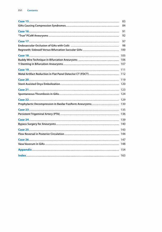
Case 15 ........................................................................................................................................................ 83 GIAs Causing Compression Syndromes ....................................................................................... 84
Case 16 ........................................................................................................................................................ 91 “True” PCoM Aneurysms ................................................................................................................... 92
Case 17 ........................................................................................................................................................ 97 Endosaccular Occlusion of GIAs with Coils ............................................................................... 98
Regrowth: Sidewall Versus Bifurcation Saccular GIAs ............................................................. 100
Case 18 ........................................................................................................................................................ 105 Buddy Wire Technique in Bifurcation Aneurysms .................................................................. 106Y-Stenting in Bifurcation Aneurysms .............................................................................................. 107
Case 19 ........................................................................................................................................................ 111 Metal Artifact Reduction in Flat Panel Detector CT (FDCT) ................................................. 112
Case 20 ........................................................................................................................................................ 119 Stent-Assisted Onyx Embolization ................................................................................................ 120
Case 21 ........................................................................................................................................................ 123 Spontaneous Thrombosis in GIAs .................................................................................................. 124
Case 22 ........................................................................................................................................................ 129 Prophylactic Decompression in Basilar Fusiform Aneurysms............................................. 130
Case 23 ........................................................................................................................................................ 135 Persistent Trigeminal Artery (PTA) ................................................................................................ 136
Case 24 ........................................................................................................................................................ 139 Bypass Surgery for Aneurysms ....................................................................................................... 140
Case 25 ........................................................................................................................................................ 143 Flow Reversal in Posterior Circulation ......................................................................................... 144
Case 26 ........................................................................................................................................................ 147 Vasa Vasorum in GIAs ......................................................................................................................... 148
Appendix ................................................................................................................................... 154
Index ............................................................................................................................................ 163
XVI Contents
XVII
Introd uction
History, Evolution, Defi nition, and Epidemiology
Dandy in his paper entitled “Intracranial aneu-rysms” stated that Morgagni was probably the fi rst to recognize intracranial aneurysms at nec-ropsy in 1761. Virchow in 1851 was the fi rst to describe the regular “berry” aneurysm [1]. Bonet and Wiseman are believed to be the fi rst to sug-gest intracranial aneurysms as a cause of sub-arachnoid hemorrhage. In 1875, Hutchison diagnosed a giant internal carotid aneurysm in a patient with III and VI cranial nerve palsy and bruit. Th is is the fi rst described instance of giant intracranial aneurysm (GIA) in a patient. In 1890, Keen stated that Horsley had diagnosed a GIA prior to craniotomy. Th ough the site was not spec-ifi ed, this is the fi rst instance where therapy for GIA was considered. Prior to angiography, intra-cranial aneurysms were diagnosed by clinical signs and/or surgical or autopsy observation. When Egaz Moniz demonstrated cerebral vascu-lature by angiography, Jeff erson had commented referring to aneurysm that “In no case can Moniz’s method of angiography be of greater benefi t” [2].
Fearnsides et al. published the fi rst clinical description of GIAs in 1916. He described a 67-year-old lady who died due to a ruptured par-tially thrombosed left middle cerebral artery aneurysm. Sarwar et al., in 1976, suggested that in most angiograms a size of 2 cm or more should be used to defi ne GIAs [1]. Gabor and Potondi also used 2 cm as a cut-off to describe GIAs. Th is was based on a hypothesis that the real diameter of these aneurysms would be larger due to nonvisu-alized clot within the aneurysms on cerebral angi-ography [2].
Th e international cooperative study of intra-cranial aneurysms and subarachnoid hemorrhage collected data from a central registry over a 7-year period from 1963 to 1970 and classifi ed aneu-rysms into various groups and assigned 25 mm as a cut-off to defi ne GIAs. Th is size criterion has since been adopted universally to defi ne GIAs [3, 4]. However, as GIAs are a very heterogeneous group, this classifi cation based wholly on size as a criterion is considered inadequate by many authors. Th is is because it does not adequately
consider the heterogeneous and varied subtypes that have distinct clinical, morphological, angio-graphic, and pathological features such as saccu-lar versus fusiform versus serpentine, thrombosed versus nonthrombosed, etc. Furthermore, it is recognized that intracranial aneurysms are a spectrum extending from small to large to very large to giant and complex aneurysms and hence cannot have fi xed discriminating sizes [3, 5].
Intracranial aneurysms are localized patho-logical dilatation of cerebral arteries. Th e preva-lence of intracranial aneurysms is estimated to be around 2 % [6]. GIAs represent 5 % of the total aneurysms and commonly become symptomatic between 40 and 70 years. Giant aneurysms are arbitrarily defi ned as intracranial aneurysms with a fundus diameter of 25 mm or more. Th ere is a female predominance with a ratio of 3:1 to 1:1. Approximately 5 to 10 % of these giant aneurysms present in the pediatric population [7]. Th e pro-portion of giant aneurysms in the pediatric popu-lation is higher and ranges from 7 to 14 % although there is some recruitment bias in the studies [6].
What’s Diff erent About Giant Intracranial Aneurysms?
Th e clinical presentation, natural history, loca-tion, therapeutic challenges, and treatment strat-egy are considerably diff erent from routine intracranial aneurysms to warrant special atten-tion to this subgroup. GIAs have three common clinical presentations. Th e commonest presenta-tion is with mass eff ect and is seen in 50–75 % of all reported series. Th e other common presenta-tions include subarachnoid hemorrhage and isch-emic symptoms. Mass eff ect may result in optic tract and hypothalamic, hemispheric, or posterior fossa compression [6, 7]. In addition, cavernous GIAs with their particular compression syn-dromes are suffi ciently unique so as to warrant a separate category. “True” cavernous GIAs consti-tute a larger proportion of GIAs, approximately 13–23 %, have a benign natural history in com-parison to other GIAs and rarely rupture. Th ey are also more common in women (up to 80 %) and
XVIII
their natural history is characterized by a very low mortality rate [8–10].
Giant aneurysms have a very poor natural his-tory with a mortality rate around 68–100 % at 2 years that is much worse than regular aneurysms. More than 50 % of untreated giant aneurysms rup-ture [11]. According to the International Study of Unruptured Intracranial Aneurysms (ISUIA), giant aneurysms have an 8 % annual risk of rupture in anterior circulation and 10 % annual rupture risk in posterior circulation. In contrast, small aneurysms have a 0–3 % annual rupture risk in anterior circulation and 0.5–3.7 % annual rupture risk in posterior circulation [12].
Around two-thirds of GIAs are located in the anterior circulation and one-third in posterior circulation [6]. Giant anterior circulation aneu-rysms have increased involvement of the cavern-ous and ophthalmic segments followed by the middle cerebral artery (MCA). In the posterior circulation, there is a predominant involvement of the BA bifurcation, P1 segment of posterior cerebral arteries (PCAs) and the superior cerebel-lar arteries (SCAs) [11].
Th erapeutic management of GIAs is challeng-ing. Patients with GIAs are generally older and have multiple medical comorbidities and a higher risk of general anesthesia complications. Addi-tionally, GIAs have very wide necks, intraluminal thrombus, higher rate of wall thickening, calcifi -cations, and atherosclerotic plaque with a higher frequency of multiple perforating arteries and major branches arising from the aneurysm [6, 7].
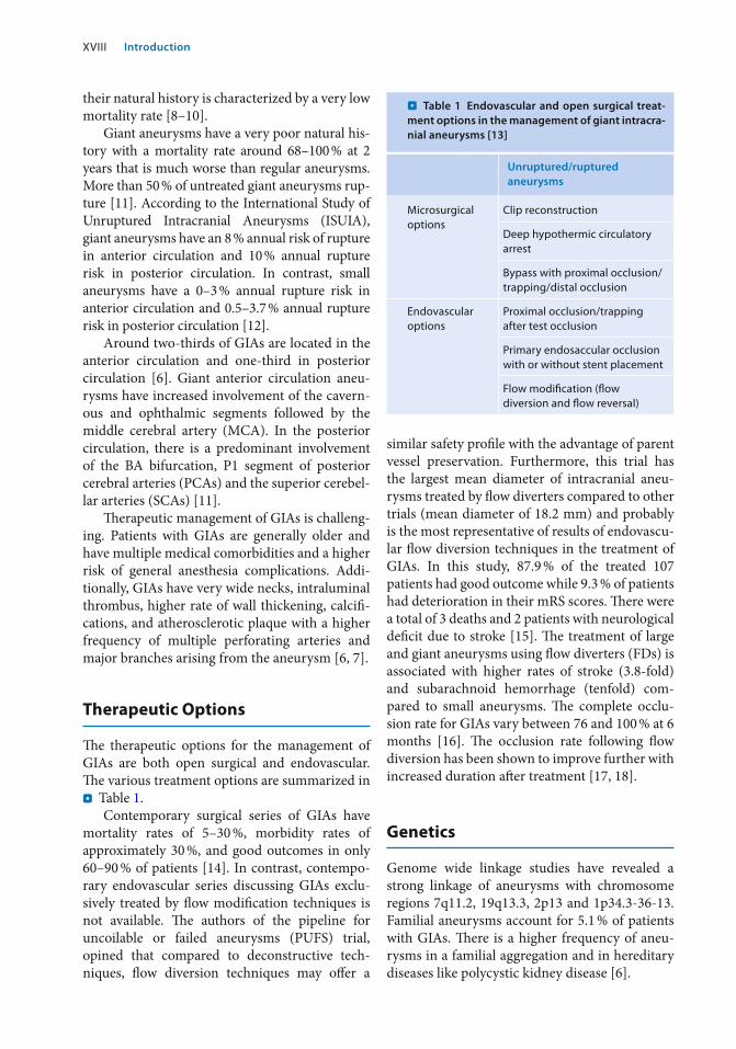
Therapeutic Options
Th e therapeutic options for the management of GIAs are both open surgical and endovascular. Th e various treatment options are summarized in . Table 1 .
Contemporary surgical series of GIAs have mortality rates of 5–30 %, morbidity rates of approximately 30 %, and good outcomes in only 60–90 % of patients [14]. In contrast, contempo-rary endovascular series discussing GIAs exclu-sively treated by fl ow modifi cation techniques is not available. Th e authors of the pipeline for uncoilable or failed aneurysms (PUFS) trial, opined that compared to deconstructive tech-niques, fl ow diversion techniques may off er a
similar safety profi le with the advantage of parent vessel preservation. Furthermore, this trial has the largest mean diameter of intracranial aneu-rysms treated by fl ow diverters compared to other trials (mean diameter of 18.2 mm) and probably is the most representative of results of endovascu-lar fl ow diversion techniques in the treatment of GIAs. In this study, 87.9 % of the treated 107 patients had good outcome while 9.3 % of patients had deterioration in their mRS scores. Th ere were a total of 3 deaths and 2 patients with neurological defi cit due to stroke [15]. Th e treatment of large and giant aneurysms using fl ow diverters (FDs) is associated with higher rates of stroke (3.8-fold) and subarachnoid hemorrhage (tenfold) com-pared to small aneurysms. Th e complete occlu-sion rate for GIAs vary between 76 and 100 % at 6 months [16]. Th e occlusion rate following fl ow diversion has been shown to improve further with increased duration aft er treatment [17, 18].
Genetics
Genome wide linkage studies have revealed a strong linkage of aneurysms with chromosome regions 7q11.2, 19q13.3, 2p13 and 1p34.3-36-13. Familial aneurysms account for 5.1 % of patients with GIAs. Th ere is a higher frequency of aneu-rysms in a familial aggregation and in hereditary diseases like polycystic kidney disease [6].
. Table 1 Endovascular and open surgical treat-ment options in the management of giant intracra-nial aneurysms [13]
Unruptured/ruptured aneurysms
Microsurgical options
Clip reconstruction
Deep hypothermic circulatory arrest
Bypass with proximal occlusion/trapping/distal occlusion
Endovascular options
Proximal occlusion/trapping after test occlusion
Primary endosaccular occlusion with or without stent placement
Flow modifi cation (fl ow diversion and fl ow reversal)
Introduction
XIX
Types of Giant Intracranial Aneurysms
Giant aneurysms are classifi ed into three mor-phological types namely: saccular, fusiform, and serpentine.
Giant Saccular Aneurysms (GSaAs)
Th ese aneurysms have a demonstrable neck with a sac-like dilatation arising from the parent artery [6, 7].
Giant Fusiform Aneurysms (GFAs)
Th ese aneurysms are characterized by circumfer-ential dilatation of the parent artery [6, 7].
Giant Serpentine Aneurysms (GSeAs)
Th ese aneurysms are partially thrombosed with residual serpiginous channel on angiography. GSeAs have an irregular eccentric channel through intraluminal thrombus with a wavy sinu-soidal course [6, 7, 19].
Pathophysiology of Giant Intracranial Aneurysms
Giant Saccular Aneurysm
Th ese aneurysms are believed to be the giant counterparts of small “berry” aneurysms at the circle of Willis. As the diameter of small berry aneurysm increases the transmural pressure increases as per the law of Laplace that favors fur-ther aneurysmal expansion [7].
Th ere are two subtypes of saccular aneu-rysms namely: partially thrombosed and those without any thrombosis. Aneurysmal location (side-wall or bifurcation), 3D geometry, and intra-aneurysmal fl ow pattern are believed to infl uence the presence or absence of intra-aneu-rysmal thrombus.
Gradual growth of saccular aneurysms due to cyclical periods of endothelial damage by turbu-
lent fl ow followed by periodic aberrant healing contributes to aneurysmal growth. Fresh hemor-rhages have been demonstrated within the aneu-rysmal wall or between an existing thrombus and aneurysmal wall. Th is has been demonstrated in the majority of giant aneurysms (90–100 %) both on cross-sectional imaging and histopathological studies [20, 21].
Partially thrombosed aneurysms have lami-nated organized thrombus in their periphery. It is believed that an increase in the number and size of vasa vasorum coupled with fresh hemor-rhage and thrombus formation in the infl ow zone contribute to their growth over a period of time [6, 7].
It is believed that hemorrhagic enlargement of GIAs occur due to subadventitial rupture of this vasa vasorum. Th ere are an increased number of vessels in the vasa vasorum in the proximal part of the aneurysm and in its wall that may contrib-ute to its angiogenic potential. Th ese aneurysms undergo continuous remodeling with weakening of wall in the zone of smooth muscle cell prolif-eration due to proteolysis. Th is mechanism can explain growth and expansion of completely thrombosed giant aneurysms either following treatment or spontaneously [20].
However, the second subgroup does not con-tain any thrombus. Th ese remain stable for long periods of time but may exhibit sudden growth spurt with rapid increase in size to reach giant proportions and may also rupture [6, 7].
Th us saccular GIAs adapt well to prolonged periods of increasing wall tension and local hemodynamic factors and do not rupture ini-tially. Th ey continue to undergo progressive remodeling; however this does not prevent pro-gressive aneurysmal growth or subsequent rup-ture [6, 7].
However, cavernous GIAs do not follow this well-established pattern. Th ey appear the least likely to rupture amongst the GIAs and are able to adapt well to local hemodynamic changes by pro-gressive increase in size [10].
Giant Fusiform Aneurysms
Fusiform aneurysms commonly occur following atherosclerotic and rarely nonatherosclerotic degeneration of vessel wall connective tissue.
Introduction
XX
Fusiform GIAs are seen in patients with connec-tive tissue diseases like Marfan’s syndrome, Ehlers Danlos syndrome, and pseudoxanthoma elasti-cum. Fragmentation of the internal elastic lamina and thickening of the intima are seen on histopa-thology. Th ere are extensive defects in both the muscularis and internal elastic lamina in GFAs in contrast to focal defects of saccular aneurysms. Irregular thickness of tunica media, thickened fi brous tissue, hypertrophic and swollen connec-tive tissue, and absence of intima have been described in GFAs [22].
It is believed that initially there is lipid depo-sition within and below the intima that disrupts the internal elastic lamina (IEL) and subse-quently infi ltrates the muscularis. Th is results in atrophy of both the elastic and muscular layers leading to tortuosity of the blood vessels. Th e blood fl ow becomes sluggish in certain portions of the tortuous artery and leads to thrombosis. Th ere is usually an associated patent peripher-ally located vascular channel through the thrombosed region. Th e other proposed alterna-tive mechanisms for GFA formation include congenital anomaly, proximal stenosis leading to “jet phenomenon” causing mechanical injury, initial intimal disruption due to dissection, and severe reticular fi ber defi ciency [23, 24]. Th ese events are followed by intramural hemorrhage from the newly formed vessels within the thrombus and progressive aneurysmal growth or rupture [6, 7, 25].
Giant Serpentine Aneurysms
Giant serpentine aneurysms (GSeAs) grow when the main blood fl ow is diverted away from aneu-rysmal wall resulting in intraluminal thrombosis. Th ere is progressive increase in the thrombosed portion with subsequent intramural hemorrhage and further development of new vascular chan-nels within the thrombus that becomes cyclical. Th e eccentric vascular channel forms due to Coanda eff ect. Fodstad et al. fi rst described this eff ect in a patient. As per this hemodynamic principle, there is a tendency of a jet of fl owing blood to be defl ected and preferentially fl ow towards one wall of the blood vessel rather than fl ow through its central portion. Subsequently,
pressure changes along the jet stream reinforce its eccentric path.
On histopathology of GSeAs, a laminated clot is seen in the thrombosed portion. In addition to the irregular serpentine portion, multiple small irregular channels that end in blind pouches are seen in the wall. Arterial vessels similar to vasa vasorum along with hemosiderin deposits and cal-cifi cation have been demonstrated in the aneurys-mal wall. Th e aneurysmal wall is thicker compared to other aneurysms, around 1–3 mm and is pri-marily composed of fi brous tissue with no internal elastic lamina or endothelial lining [6, 7, 19].
Imaging Features of Giant Intracranial Aneurysms
Cross Sectional Imaging
Nonenhanced CT (NECT) GIAs are well-circumscribed, mass lesions in the region of circle of Willis that are mild to moder-ately hyperdense on NECT [3]. Th in-walled aneu-rysms appear slightly hyperdense on NECT [26]. Th ey are seen in close association with the circle of Willis. Partially thrombosed GIAs on NECT show the following components: (a) A dense region composed of acutely throm-
bosed portion (b) A lower density region containing the
circulating channel (c) A lowest density composed of chronically
thrombosed portion
Th ere is mass eff ect with frequent vasogenic edema in the adjacent cerebral parenchyma. Mass eff ect may cause parenchymal displacement across the midline, gyral, and sulcal eff acement and acute/chronic obstructive hydrocephalus [27].
In patients with recent acute symptoms with partially thrombosed GIAs, a peripheral cresentic or convex-shaped region of hyperdensity in a laminated (variegated) thrombus is suggestive of recent hemorrhage [5, 28]. A peripheral rim of calcifi cation may also be seen. Th e rim calcifi ca-tion is fi ne, linear, or crescentic. Rim calcifi cation longer than 3 cm is almost invariably due to GIAs.
In long standing cases with GIA, NECT on bone window may show erosion and scalloping of adjacent osseous structures [27].
Introduction
XXI
In ruptured GIAs, NECT may show hemor-rhage as subarachnoid hemorrhage (sulci/ cister-nal hyperdensity), intraventricular hemorrhage or intra-parenchymal hematoma [27].
Contrast Enhanced CT (CECT) On CECT, non-thrombosed GIAs generally show homogenous enhancement.
Partially thrombosed GIAs show homoge-nous intense enhancement of the central circu-lating channel that is brighter than the acutely thrombosed portion in the periphery of the aneurysm. Although the thrombus does not enhance, there is enhancement of the aneurys-mal wall. Th is combination of densities is known to produce the “Target sign” [26]. Th is sign was fi rst described by Kricheff as a central hyperden-sity of circulating channel, intermediate hypodensity of thrombus and peripheral hyper-density of the wall [27].
In completely thrombosed GIAs there is absence of luminal enhancement [26]. A rim enhancement may be seen along the aneurysmal wall that is suggestive of vasa vasorum [29].
CT Angiography (CTA) CTA is useful in analyzing the circulating chan-nel of giant aneurysm. When the fl ow is fast and homogenous, there is complete opacifi cation of the circulating channel and it is easier to analyze its size and relationship to the parent artery, adja-cent vessels, and parenchyma. However, when the fl ow is slow and the channel is wide, there is partial fi lling of the circulating channel and problems in assessing the above-mentioned parameters [27].
Magnetic Resonance Imaging (MRI) On T1W and T2W images, the fast fl owing por-tions of the aneurysm are seen as fl ow voids. Th e “fl ow void” sign has a sensitivity of 88 % on both T1W and T2W sequences. Th e persistence of “fl ow void” on postcontrast T1W images is highly specifi c for the diagnosis of GIA. Th e combination of “fl ow void” sign and postcon-trast T1W luminal enhancement of GIA has increased sensitivity, specifi city, and higher pos-itive and negative predictive value in the diagno-sis of GIAs. Flow artifacts and ghost images extending on either side of the aneurysmal
lumen along the phase encoding direction are motion artifacts. Th eir presence indicates patent aneurysmal lumen [30].
Th e slow fl owing and thrombosed portions of aneurysms are heterogeneous due to varying signal characteristics of blood products. Gradient recalled echo (GRE) and susceptibility weighted imaging (SWI) (T2*W images) are very sensitive to blood products and calcifi cations. Th is appears as a strong hypointense signal that spills over to adja-cent normal parenchyma. Th is is artifactual and causes overestimation of lesion size [27]. Addition-ally, the aneurysmal wall shows hemorrhage of diff erent ages and postcontrast T1W enhancement of the aneurysmal wall suggestive of neovascular-ization due to vasa vasorum [30].
Circulating high blood fl ow can be demon-strated by three sequences namely: 1. Time-of-fl ight (TOF) MR angiogram (MRA):
3D preferred over 2D techniques 2. Contrast enhanced MR angiogram (CE
MRA): 3D sequence
3D TOF MRA: Th e fl ow through GIAs is oft en heterogeneous because of a wide circulating chan-nel. Th erefore, it underestimates the circulating portion of the GIA and may miss some arterial eff erent branches.
3D CE MRA: Provides reliable information about the size of the circulating portion of the aneurysm and patency of collateral branches. It also depicts aneurysmal enhancement [27].
Th e brain parenchyma may show edema (hypointense on T1W and hyperintense on T2W). It is, however, diffi cult to diff erentiate the infl ammatory component from the component that is due to aneurysmal mass eff ect only. Edema that is seen distal to the aneurysmal location is commonly cytotoxic in nature and is due to isch-emia. Subarachnoid hemorrhage (SAH) appears hyperintense on FLAIR and hypointense on T2*W images. Intraparenchymal hemorrhage is heterogeneous on T1W and T2W images and strongly hypointense on T2*W images. It also shows mass eff ect, compression and trophy of cranial nerves [27].
Partially thrombosed GIAs show an onionskin appearance of the thrombosed portion of aneu-rysm. Th is is due to the diff erent ages of the hem-orrhages. Postcontrast T1W images do not show enhancement of the thrombosed portion (sensi-tivity 100 %) [30].
Introduction
XXII
Flat Panel Detector Computed Tomography (FDCT) FDCT combines 2D radiography/fl uoroscopy with 3D CT imaging. FDCT has a higher spatial resolu-tion. Th e distinct advantage of FDCT is the imme-diate availability of CT imaging in the angiography suite. FDCT has a number of disadvantages, namely, lower dose effi ciency, smaller fi eld of view and lower temporal resolution [31]. Th e isotropic spatial resolution of FDCT potentially approaches 150x150x150 μm 3 . Th is is superior in resolution in terms of both the voxel size and isotropic nature compared to multidetector computed tomography (MDCT). Th is allows manipulation of recon-structed images and interactive viewing of FDCT volumes more fl exible and effi cient [32].
Applications of FDCT [32] (a) Evaluation of intracranial stents in the treat-
ment of stenosis and aneurysms. It allows visualization of stent struts as small as 50−70 μm. Th e images can be rotated and reformatted in any plane, allowing superior stent visibility. It can also show kinking, pro-lapse, and fl attening of stents.
(b) CT-like soft tissue imaging. Allows detection of intracerebral hemorrhage, SAH, assessment of ventricles, and immediate postprocedural patient management.
(c) FDCTA (FDCT angiography). Gives better depiction of vascular morphology compared to 3D DSA.
(d) Follow-up imaging. Detects in-stent stenosis (ISS). IVFDCTA is a less invasive option and safer compared to IAFDCTA.
(e) FDCT perfusion is still under development.
Digital Subtraction Angiography (DSA)
DSA remains the “gold standard” in the imaging of GIAs as it has excellent spatial and time resolution. Th ough dynamic, it cannot demonstrate noncircu-lating components of GIAs, namely, thrombosed portion, aneurysmal wall, and surrounding paren-chyma. Th e size and morphology of aneurysms are assessed on 2D angiography and 3D rotational angiography. It enables dynamic study of arteries distal to aneurysm and collateral network. GIAs with slow fl ow may show recruitment of pial
collaterals and transdural collaterals from menin-geal arteries.
A balloon test occlusion (BTO) can be done to assess the adequacy of collaterals to enable a safe parent vessel occlusion (PVO) [27].
References
1. Sarwar M, Batnitzky S, Schechter MM Tumorous aneu-rysms. Neuroradiology 12(2):79–97. doi: 10.1007/bf00333123
2. Fox JL (1983) Intracranial aneurysms, vol 1. Springer Science & Business Media
3. Pia HW, Zierski J (1982) Giant cerebral aneurysms. Neu-rosurg Rev 5(4):117–148
4. Sahs AL (1974) Cooperative study of intracranial aneu-rysms and subarachnoid hemorrhage. Report on a ran-domized treatment study. I. Introduction. Stroke 5(4):550–551
5. Krings T, Alvarez H, Reinacher P, Ozanne A, Baccin CE, Gandolfo C, Zhao WY, Reinges MH, Lasjaunias P (2007) Growth and rupture mechanism of partially thrombosed aneurysms. Intervent Neuroradiol 13(2):117–126
6. Lonjon M, Pennes F, Sedat J, Bataille B (2015) Epidemi-ology, genetic, natural history and clinical presentation of giant cerebral aneurysms. Neuro-Chirurgie 61(6):361–365. doi: 10.1016/j.neuchi.2015.08.003
7. Parkinson RJ, Eddleman CS, Batjer HH, Bendok BR (2006) Giant intracranial aneurysms: endovascular chal-lenges. Neurosurgery 59(5 Suppl 3):S103–S112; discussion S103–S113. doi: 10.1227/01.neu.0000237410.32115.c9
8. Hahn CD, Nicolle DA, Lownie SP, Drake CG (2000) Giant cavernous carotid aneurysms: clinical presenta-tion in fi ft y-seven cases. J Neuroophthalmol 20(4):253–258
9. Kupersmith MJ, Hurst R, Berenstein A, Choi IS, Jafar J, Ransohoff J (1992) Th e benign course of cavernous carotid artery aneurysms. J Neurosurg 7(5):690–693. doi: 10.3171/jns.1992.77.5.0690
10. Penchet G, Mourier K (2015) Collaborative retrospec-tive multicentre series of giant intracavernous carotid aneurysms. Neuro-Chirurgie 61(6):366–370. doi: 10.1016/j.neuchi.2013.12.004
11. Choi IS, David C (2003) Giant intracranial aneurysms: development, clinical presentation and treatment. Eur J Radiol 46(3):178–194. doi: 10.1016/s0720-048x(03)00090-1
12. White PM, Wardlaw JM (2003) Unruptured intracranial aneurysms. J Neuroradiol 30(5):336–350
13. Sekhar LN, Tariq F, Mai JC, Kim LJ, Ghodke B, Hallam DK, Bulsara KR (2012) Unyielding progress: treatment paradigms for giant aneurysms. Clin Neurosurg 59:6–21. doi: 10.1227/NEU.0b013e3182698b75
14. Sughrue ME, Saloner D, Rayz VL, Lawton MT (2011) Giant Intracranial Aneurysms: Evolution of Manage-ment in a Contemporary Surgical Series. Neurosurgery 69(6):1261–1271. doi: 10.1227/NEU.0b013e31822bb8a6
15. Becske T, Kallmes DF, Saatci I, McDougall CG, Szikora I, Lanzino G, Moran CJ, Woo HH, Lopes DK, Berez AL,
Introduction
XXIII
Cher DJ, Siddiqui AH, Levy EI, Albuquerque FC, Fio-rella DJ, Berentei Z, Marosfoi M, Cekirge SH, Nelson PK (2013) Pipeline for uncoilable or failed aneurysms: results from a multicenter clinical trial. Radiology 267(3):858–868. doi: 10.1148/radiol.13120099
16. Brinjikji W, Murad MH, Lanzino G, Cloft HJ, Kallmes DF (2013) Endovascular treatment of intracranial aneu-rysms with fl ow diverters: a meta-analysis. Stroke 44(2):442–447. doi: 10.1161/strokeaha.112.678151
17. Lylyk P, Miranda C, Ceratto R, Ferrario A, Scrivano E, Luna HR, Berez AL, Tran Q, Nelson PK, Fiorella D (2009) Curative endovascular reconstruction of cerebral aneurysms with the pipeline embolization device: the Buenos Aires experience. Neurosurgery 64(4):632–642; discussion 642–633; quiz N636. doi: 10.1227/01.neu.0000339109.98070.65
18. D’Urso PI, Lanzino G, Cloft HJ, Kallmes DF (2011) Flow diversion for intracranial aneurysms: a review. Stroke 42(8):2363–2368. doi: 10.1161/strokeaha.111.620328
19. Christiano LD, Gupta G, Prestigiacomo CJ, Gandhi CD (2009) Giant serpentine aneurysms. Neurosurg Focus 26(5):E5. doi: 10.3171/2009.2.focus0918
20. Krings T, Mandell DM, Kiehl TR, Geibprasert S, Tymi-anski M, Alvarez H, terBrugge KG, Hans FJ (2011) Intra-cranial aneurysms: from vessel wall pathology to therapeutic approach. Nat Rev Neurol 7(10):547–559. doi: 10.1038/nrneurol.2011.136
21. Schubiger O, Valavanis A, Wichmann W (1987) Growth-mechanism of giant intracranial aneurysms; demonstration by CT and MR imaging. Neuroradiology 29(3):266–271
22. Little JR, St Louis P, Weinstein M, Dohn DF (1981) Giant fusi-form aneurysm of the cerebral arteries. Stroke 12(2):183–188
23. Nakayama Y, Tanaka A, Kumate S, Tomonaga M, Take-bayashi S (1999) Giant fusiform aneurysm of the basilar
artery: consideration of its pathogenesis. Surg Neurol 51(2):140–145
24. Day AL, Gaposchkin CG, Yu CJ, Rivet DJ, Dacey RG, Jr. (2003) Spontaneous fusiform middle cerebral artery aneurysms: characteristics and a proposed mechanism of formation. J Neurosurg 99(2):228–240. doi: 10.3171/jns.2003.99.2.0228
25. Shokunbi MT, Vinters HV, Kaufmann JC (1988) Fusi-form intracranial aneurysms. Clinicopathologic fea-tures. Surg Neurol 29(4):263–270
26. Pinto RS, Kricheff , II, Butler AR, Murali R (1979) Cor-relation of computed tomographic, angiographic, and neuropathological changes in giant cerebral aneurysms. Radiology 132(1):85–92. doi: 10.1148/132.1.85
27. Tollard E, Perot G, Clavier E, Gerardin E (2015) Imaging of giant cerebral aneurysms. Neuro-Chirurgie 61(6):378–384. doi: 10.1016/j.neuchi.2013.10.124
28. Koyama S, Kotani A, Sasaki J (1996) Giant basilar artery aneurysm with intramural hemorrhage and then disas-trous hemorrhage: case report. Neurosurgery 39(1):174–177; discussion 177–178
29. Golding R, Peatfi eld RC, Shawdon HH, Rice Edwards JM (1980) Computer tomographic features of giant intracranial aneurysms. Clin Radiol 31(1):41–48
30. Teng MM, Nasir Qadri SM, Luo CB, Lirng JF, Chen SS, Chang CY (2003) MR imaging of giant intracranial aneu-rysm. J Clin Neurosci 10(4):460–464
31. Kalender WA, Kyriakou Y (2007) Flat-detector com-puted tomography (FD-CT). Eur Radiol 17(11):2767–2779. doi: 10.1007/s00330-007-0651-9
32. Kamran M, Nagaraja S, Byrne JV (2010) C-arm fl at detector computed tomography: the technique and its applications in interventional neuro-radiology. Neurora-diology 52(4):319–327. doi: 10.1007/s00234-009-0609-5
Introduction