Embed Size (px)
Citation preview

512
University Hospital of Orthopaedics “Prof. B. Boychev”, Medical University, Sofia, BulgariaCorrespondence: Georgi P. Georgiev,
University Hospital of Orthopaedics, Medical University Sofia, 56 Nikola Petkov Blvd., 1614 Sofia, BulgariaEmail: [email protected]
Copyright © JCEI / Journal of Clinical and Experimental Investigations 2013, All rights reserved
JCEI / 2013; 4 (4): 512-516Journal of Clinical and Experimental Investigations doi: 10.5799/ahinjs.01.2013.04.0336
CASE REPORT / OLGU SUNUMU
Giant cell tumor of the distal tibia: Report of a rare case
Distal tibianın dev hücreli tümörü: Nadir bir olgu sunumu
Georgi P. Georgiev, Svetoslav A. Slavchev
ÖZET
Kemiğin dev hücreli tümörleri iyi huylu olmalarına rağ-men agresif lezyonlardır. Ayak ve ayak bileği tutulumu ise nadirdir. Biz burada, sol ayakta şişlik ve ağrı artışı şikâyeti nedeniyle eklem hareketi kısıtlanan 26 yaşında bayan hastayı sunduk. Görüntüleme yöntemleri kemikte dev hücreli tumor tanısı koydurdu ve bu tanı açık biyopsi ile doğrulandı. Lezyon segmental en-blok rezeksiyon ve bilek artrodezi yapılarak tedavi edildi ve iyi fonksiyonel sonuç alındı. Biz aynı zamanda yazımızda bu patolojinin klinik, radyolojik ve tedavi karakteristiklerini literatür bilgi-leri ışığında tartıştık.Anahtar kelimeler: Kemiğin dev hücreli tümörü, tibia, ayak bileği, artrodez, kemik transplantasyonu
ABSTRACT
Giant cell tumor of bone is an aggressive lesion, although benign. Foot and ankle involvement is rare. Herein, we presented a case of a 26-year-old woman complaining of increasing pain and swelling of the left ankle followed by limitation of joint motion. Imaging was consistent with the diagnosis of giant cell tumor of bone, which was con-firmed by open biopsy. The lesion was treated with seg-mental en-bloc resection and ankle arthrodesis with good functional outcome. We also discuss clinical, radiological, and therapeutic characteristics of this pathology with the light of the literature. J Clin Exp Invest 2013; 4 (4): 512-516Key words: giant cell tumor of bone; tibia; ankle; arthrod-esis; bone transplantation
INTRODUCTION
Giant cell tumor of bone (GCTB) is an osseous neoplasm that is histologically benign but clinically shows local aggression and a high rate of recur-rence [1-5]. It is thought to originate at the metaph-yseo-epiphyseal junction and may extend into the metaphysis [1-3,6]. Numerous terms, including my-eloid sarcoma, tumor of myeloplaxus, osteoblas-toclastoma, and osteoclastoma have been used to depict GCTB [2]. It accounts for about 5% of all primary bone tumors in adults and predominantly occurs in the third and fourth decades of life with a slight predilection for females [1,3,5]. Involvement of the foot and ankle is rare and comprises less than 4% of all GCTBs [7]. Lesions with this localization are known to be unpredictable in their behaviour [8]. GCTB of hand and foot are more aggressive and aggressive treatment is recommended [9,10].
Herein, we present a rare case of GCTB of the distal tibia treated with segmental en-bloc resection and ankle arthrodesis. We also review the patho-logic features, clinical manifestations, radiological appearance, and treatment of the GCTB.
CASE REPORT
A 26-year-old woman presented to our department with 1-year history of left ankle pain and swelling with no prior trauma. Increasing pain and limitation of motion in the joint during last three months ne-cessitated the use of crutches. Physical examina-tion revealed increased volume of the distal tibial metaphysis and moderate soft tissue swelling. The range-of-motion of the ankle was significantly re-duced.
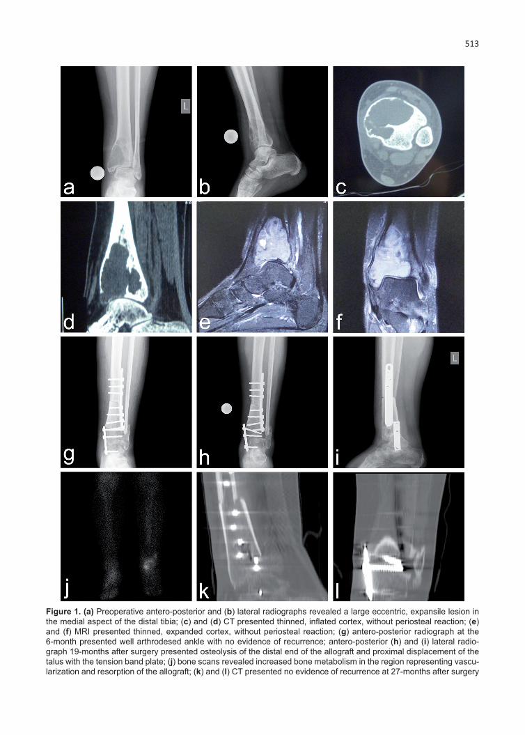
Plain X-rays (Figure 1 a, b), computed tomog-raphy (CT) (Figure 1 c, d), and magnetic resonance imaging (MRI) (Figure 1 e, f) presented a large ec-centric, expansive lesion in the medial aspect of the distal tibia suggestive of a giant cell tumor of bone. Laboratory tests were in normal ranges. An open biopsy was performed and a giant cell tumor of bone was diagnosed. Microscopically, the lesion was presented by proliferating uniform oval mono-nuclear cells scattered around the background of numerous osteoclast-type giant cells. According to the radiological classification of Campanacci et al., the tumor was classified as a grade 2 lesion [6].
513
Figure 1. (a) Preoperative antero-posterior and (b) lateral radiographs revealed a large eccentric, expansile lesion in the medial aspect of the distal tibia; (c) and (d) CT presented thinned, inflated cortex, without periosteal reaction; (e) and (f) MRI presented thinned, expanded cortex, without periosteal reaction; (g) antero-posterior radiograph at the 6-month presented well arthrodesed ankle with no evidence of recurrence; antero-posterior (h) and (i) lateral radio-graph 19-months after surgery presented osteolysis of the distal end of the allograft and proximal displacement of the talus with the tension band plate; (j) bone scans revealed increased bone metabolism in the region representing vascu-larization and resorption of the allograft; (k) and (l) CT presented no evidence of recurrence at 27-months after surgery
Georgiev and Slavchev. Giant cell tumor of the distal tibia514
J Clin Exp Invest www.jceionline.org Vol 4, No 4, December 2013
The condition, its prognosis, the possible treatment modalities and complications were discussed with the patient. Among the surgical alternatives, we considered curettage with reconstruction using an allograft, bone cement and an autograft, resection and arthrodesis, or joint replacement. Decision was made, with the patient, to perform en-block resec-tion of the distal tibia, reconstruction with a struc-tural tibial allograft, and ankle arthrodesis. Precise preoperative planning was used concerning the resection and arthrodesis. Using an anterior ap-proach the left distal tibia was exposed extraperios-teally and resected at 9.5 cm proximal to the ankle joint. The articular surface of the talar dome was removed and a distal oblique fibular osteotomy was performed. The allograft was cut to size and fixed with a 4.5 mm DCP. Thereafter, for arthrodesis of the ankle, an oblique 4.5 mm compression cancel-lous screw from the metaphysis of the allograft to the talus was inserted. During the insertion of the second cancellous screw a partial fracture of the al-lograft occurred. After that, the ankle was arthrod-esed with a tension band plate on the medial side. No adjuvant agents were used. The wound was closed over a deep suction drain. A short leg cast was applied and the leg was non-weight bearing for 6 weeks. On suture removal, which was performed through a window in the plaster cast, an area of partial-thickness skin necrosis was detected on the dorsum of the foot, measuring 3 cm by 5 cm. The lesion was managed with enzymatic debridement and healed for 3 weeks. At 6-months follow-up, the patient had a smooth healed scar with a painless and well arthrodesed ankle with no evidence of re-currence (Figure 1g). At 19 months after surgery, there was osteolysis of the distal end of the allograft and proximal displacement of the talus with the ten-sion band plate on the medial side of the ankle with the foot remaining plantigrade (Figure 1 h, i). Bone scans revealed increased bone metabolism in the region representing resorption of the allograft (Fig-ure 1j). Thereafter, the patient underwent treatment with bisphosphonates. On CT, no signs of continu-ing bone resorption and no evidence of recurrence was detected at 27-months follow-up (Figure 1 k, l). The patient had pain-free stable ankle and unlim-ited ambulation. Her subtalar and midtarsal motion measured 0° of dorsiflexion/supination and 15° of plantar flexion/pronation.
DISCUSSION
GCTB is described as a locally invasive tumor with a high rate of recurrence and a possibility of mainly pulmonary metastases or transformation into a ma-
lignancy [1-4]. The diagnosis of this tumor requires precise assessment of the clinical findings, imaging modalities, and histopathologic evaluation [2,10,11].
In the current literature, GCTB is described as a predominantly osteoclastogenic stromal cell tumor of mesenchymal origin [12]. It is composed of three cell types - the spindle-like stromal cells, mononuclear monocyte cells, and multinucleated giant cells [12-16]. The multinucleated giant cells which mimic osteoclasts are principally responsible for the extensive bone resorption that is character-istic of GCTB [16]. However, the stromal cells are the main neoplastic component of GCTB and have been shown to express and secrete a variety of chemotactic factors to enlist pathologic components [12,16]. Mononuclear monocyte cells are consid-ered to be either reactive macrophages or osteo-clast precursors [12,16].
The main clinical symptoms are non-specific and include pain of variable severity, local swelling, tenderness of the affected area, and limited range of motion of the adjacent joint [2,3,17]. The duration of symptoms usually varies from two to six months. Rarely, a pathologic fracture may be the first symp-tom [2,3,17].
Imaging studies are essential for the diagnosis of GCTB [2,11,17]. On conventional radiographs this tumor typically presents as a purely lytic eccen-tric lesion, with expansion and thinning of the cor-tex. Periosteal reaction is usually absent [1,3,7,17]. Campanacci et al. classified GCTB in three grades: grade 1 is static form with minimal involvement of the cortex; grade 2 presents with thinned and ex-panded cortex and in grade 3 the lesion penetrates the cortex and has a soft tissue component [6]. As with any suspicious bone lesion, full staging with MRI and CT should be undertaken [17,18]. CT is useful in the evaluation of the cortical bone and could clearly present the thinning of the cortex and subchondral bone, the pathologic fracture, the peri-osteal reaction, and the absence of matrix miner-alization [3,19]. In the cases of cortical destruction and soft-tissue tumor extension MRI is superior to CT in delineation of GCTB. The tumor will appear with a non-homogenous signal on magnetic reso-nance imaging: low in T1-weighted images and high in T2-weighted images. Bone scintigraphy could also be used for evaluation of giant cell tumor of bone [2,3,17].
Various limb salvage techniques for the distal tibia have been described in literature: extended curettage with a large window, high speed burring, and filling of the cavity with bone cement or bone graft; resection and ankle arthrodesis; resection

Georgiev and Slavchev. Giant cell tumor of the distal tibia 515
J Clin Exp Invest www.jceionline.org Vol 4, No 4, December 2013
and reconstruction with porous tantalum spacer; re-section of the tumor followed by placement of an ex-ternal fixator for segmental bone transport and en-doprosthetic replacement [7,20-25]. Autografts and allografts are associated with high rates of healing when used for arthrodesis. In most cases, arthrod-esis of the ankle joint provides excellent stability of the ankle and very good functional outcome [7]. However, due to the high pressure in the ankle joint after resection of the distal tibia and arthrodesis of the ankle, the allograft could collapse or fracture. Infections, non-union, osteolysis, iatrogenic frac-ture of the allografts were also reported [27]. In such cases these complications could compromise the ankle arthrodesis. In cases of bone resorption, bisphosphonates may be a reasonable option due to the reported evidence of inhibiting bone resorp-tion in human and animal trials [26]. Rarely, in cases when surgery is not feasible, irradiation for the treat-ment of GCTB could also be an alternative option [28].
GCT of bone is a locally aggressive tumor with a high tendency to recur after removal. The rates of recurrence after simple curettage ranged from 12-65% as compared with 12-27% after curettage and adjuvant treatment and 0-12% after resection [1,4,29-31]. In cases of GCTB affecting the hand and foot the recurrence rate is higher in comparison with GCTB in more conventional sites [8,10]. GCTB could metastasize in up to 10% of patients. Most commonly the metastatic spread occurs after repeti-tive local recurrences [10,11].
In conclusion, GCTB of the foot and ankle is a rare lesion. Prognosis, treatment, and results are directly dependent on early diagnosis and adequate therapy. In this report, we present a rare case of GCTB of the distal tibia treated with en-bloc resec-tion and ankle arthrodesis. This treatment modality leads to good results.
REFERENCES
1. Balke M, Schremper L, Gebert C,et al. Giant cell tumor of bone: treatment and outcome of 214 cases. J Can-cer Res Clin Oncol 2008;134:969-978.
2. Georgiev GP, Stokov L. Giant cell tumor of bone. Bulg J Orthop Trauma 2012;49:34-39.
3. Szendröi M. Giant-cell tumour of bone. J Bone Joint Surg Br 2004;86:5-12.
4. Turcotte RE, Wunder JS, Isler MH, et al. Giant cell tu-mor of long bone: a Canadian Sarcoma Group study. Clin Orthop Relat Res 2002;397:248-258.
5. Klenke FM, Wenger DE, Inwards CY, Rose PS, Sim FH. Giant cell tumor of bone: risk factors for recur-rence. Clin Orthop Relat Res 2011;469:591-599.
6. Campanacci M, Giunti A, Olmi R. Giant-cell tumours of bone: a study of 209 cases with long term follow up in 130. Ital J Orthop Traumatol 1975;1:249-277.
7. Cribb GL, Cool P, Hill SO, Mangham DC. Distal tibial gi-ant cell tumour treated with curettage and stabilisation with an Ilizarov frame. Foot Ankle Surg 2009;15:28-32.
8. Kamath S, Jane M, Reid R. Giant cell tumour around the foot and ankle. J Foot Ankle Surg 2006;12:99-102.
9. Biscaglia R, Bacchini P, Bertoni F. Giant cell tumor of the bones of the hand and foot. Cancer 2000;88:2022-2032.
10. Matev B, Georgiev H, Georgiev GP. Giant cell tumor of the fourth metacarpal: case report and literature re-view. J Radiother Med Oncol 2012;18:73-77.
11. Babazadeh S, Broadhead ML, Slavin JL, et al. Giant cell tumour of metacarpal diaphysis. Eur J Radiol Ex-tra 2010;75:31-36.
12. Kim Y, Nizami S, Goto H, Lee FY. Modern interpre-tation of giant cell tumor of bone: predominantly osteoclastogenic stromal tumor. Clin Orthop Surg 2012;4:107-116.
13. Georgiev GP, Landzhov B, Slavchev S, et al. Localiza-tion of matrix metalloproteinase-2 in giant cell tumor of bone. Compt Rend Acad Bulg Sci 2012;65:1285-1288.
14. Georgiev GP, Landzhov B, Slavchev S, et al. Com-parative electron microscopic and immunohistochem-ical study of stromal cells in giant cell tumor of bone. Scripta Scient Med 2013,45:19-22.
15. Georgiev GP, Georgiev H, Landzhov B. Ultrastructural study of giant cell tumor of bone. Orthop Rheumatol 2012;2:13-15.
16. Cowan RW, Singh G. Giant cell tumor of bone: a basic science perspective. Bone 2013;52:238-246.
17. Murphey MD, Nomikos GC, Flemming DJ, et al. Imaging of giant cell tumor and giant cell reparative granuloma of bone: radiologic-pathologic correlation. Radiographics 2001;21:1283-1309.
18. Lee MJ, Sallomi DF, Munk PL, et al. Pictorial review: giant cell tumours of bone. Clin Radiol 1998;53:481-489.
19. Levine E, De Smet AA, Neff JR. Role of radiologic imaging in management planning of giant cell tumor of bone. Skeletal Radiol 1984;12:79-89.
20. Ajit Singh V, Nasirudin N, Bernatt M. Endoprosthet-ic reconstruction for giant cell tumors of the distal tibia: A short term review. Asia-Pacific J Clin Oncol 2013;9:182-189.
21. Alsulaimani SA, Turcotte RE; Canadian Orthopaedic Oncology Society (CANOOS) collaborators. Iterative curettage is sssociated with local control in giant cell tumors involving the distal tibia. Clin Orthop Relat Res. 2013;471:2668-2674.
22. Bami M, Nayak AR, Kulkarni S, et al. Giant cell tu-mor of lower end of tibia. Case Rep Orthop. 2013; doi: 10.1155/2013/429615.
Georgiev and Slavchev. Giant cell tumor of the distal tibia516
J Clin Exp Invest www.jceionline.org Vol 4, No 4, December 2013
23. Economopoulos K, Barker L, Beauchamp C, Claridge R. Case report: reconstruction of the distal tibia with porous tantalum spacer after resection for giant cell tumor. Clin Orthop Relat Res 2010;468:1697-1701.
24. Lampert C. Ankle joint prosthesis for bone defects. Orthopade. 2011;40:978-983.
25. Pаn KL, Chan WH. Curettage and cementation in gi-ant cell tumour of the distal tibia using polypropylene mesh for containment: a case report. Malaysian Or-thop J 2010;4:51-53.
26. Seo SW, Cho SK, Storer SK, Lee FY. Zoledronate reduces unwanted bone resorption in intercalary bone allografts. Int Orthop. 2010;34:599-603.
27. Rödl RW, Ozaki T, Hoffmann C, et al. Osteoarticular allograft in surgery for high-grade malignant tumours of bone. J Bone Joint Surg Br 2000;82:1006-1010.
28. Roeder F, Timke C, Zwicker F, et al. Intensity modu-lated radiotherapy (IMRT) in benign giant cell tumors-a single institution case series and a short review of the literature. Radiat Oncol 2010;5:18.
29. Prosser GH, Baloch KG, Tillman RM, Carter SR, Grimer RJ. Does curettage without adjuvant therapy provide low recurrence rates in giant-cell tumors of bone? Clin Orthop Relat Res 2005;435:211-218.
30. Errani C, Ruggieri P, Asenzio MA, et al. Giant cell tu-mor of the extremity: a review of 349 cases from a single institution. Cancer Treat Rev 2010;36:1-7.
31. Kivioja AH, Blomqvist C, Hietaniemi K, et al. Cement is recommended in intralesional surgery of giant cell tumors: a Scandinavian Sarcoma Group study of 294 patients followed for a median time of 5 years. Acta Orthop 2008;79:86-93.


![Recurrent primary mediastinal giant cell tumor of soft tissue ......Discussion Giant cell tumor of soft tissue (GCT-ST) is a rare tumor. GCT-ST, which resembles osseous GCT [2], broadly](https://img.dokumen.tips/doc/110x75/608dfb4a7de20e33185e8616/recurrent-primary-mediastinal-giant-cell-tumor-of-soft-tissue-discussion.jpg)