Embed Size (px)
Citation preview

Supplementary materials An asymmetric interface between the regulatory particle and core particle of the proteasome Geng Tian, Soyeon Park, Min Jae Lee, Bettina Huck, Fiona McAllister, Christopher P. Hill, Steven P. Gygi, and Daniel Finley *corresponding author: [email protected]
Nature Structural & Molecular Biology doi:10.1038/nsmb.2147

Supplementary �igure 1. Sequence alignment of the C-terminal tails of Rpt1–6 (a) and the N-terminal regions of α1–α7 (b) from various species. Black, grey and light-grey shading indicate 100%, 80% and 60% conservation respectively, over the entire α or Rpt family. The conserved residue corres–ponding to K66 in the T. acidophilum proteasome CP is highlighted by a blue arrow and the non-conserved residue mutated to cysteine within each α pocketis highlighted by a yellow arrow. Abbreviations are: ag: Ashbya gossypii; at: Arabidopsis thaliana; ce: Caenorhabditis elegans; dm: Drosophila melanogaster; dr: Danio rerio; hs: Homo sapiens; kl: Kluyveromyces lactis; mg: Magnaporthe grisea; pf: Plasmodium falciparum; sc: Saccharomyces cerevisiae.
ba
Nature Structural & Molecular Biology doi:10.1038/nsmb.2147

c
d e f
ba
RP2-CP
RP-CP
CP
RP2-CP
RP-CP
CP
RP2-CP
RP-CP
CP
Supplementary �igure 2. Evaluation of the effects of introduced cysteines on growth rate and on structural stability of the proteasome. Growth assays of wild type and mutant strains bearing introduced cysteines in α subunits (a), Rpt proteins (b), and six identi�ied Rpt–α pairs (c). Spot assays of cell growth were carried out with 0.05 OD₆₀₀ of cells grown in YPD, resuspended in 250 μl of H₂O, and serially diluted in �ive-foldincrements. The cells were then spotted on YPD plates followed by incubation at 30°C for 48 hr. Note that the RPN11–TevProA allele was not present in these strains. The α4N79C mutant used in all these studies is produced from a strain expressing α4C32A C46A (TG644), which shows the same growth phenotype as wild type under various conditions (Fig. S2 and data not shown). (d–f) Total cell lysates of the same set of strains were analyzed by native PAGE followed by overlay assay with LLVY–AMC. Early stationary phase cells were lysed by grinding under liquid N₂. 50 μg of protein were loaded for each strain. Several of the strains are mildly hypomorphic, which is most readily indicated by the enhanced levels of CP in extracts of Rpt4-Rpt6 (ref. 1).
Nature Structural & Molecular Biology doi:10.1038/nsmb.2147
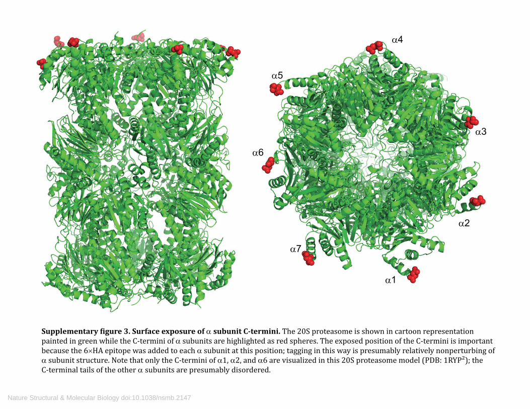
α1
α2
α3
α4
α5
α6
α7
Supplementary �igure 3. Surface exposure of α subunit C-termini. The 20S proteasome is shown in cartoon representation painted in green while the C-termini of α subunits are highlighted as red spheres. The exposed position of the C-termini is important because the 6×HA epitope was added to each α subunit at this position; tagging in this way is presumably relatively nonperturbing of α subunit structure. Note that only the C-termini of α1, α2, and α6 are visualized in this 20S proteasome model (PDB: 1RYP²); the C-terminal tails of the other α subunits are presumably disordered.
Nature Structural & Molecular Biology doi:10.1038/nsmb.2147

P91P91
trans- cis-
R134
D99
a b c
d e
Supplementary �igure 4. Pairing between t- and c-type Rpts. In each PAN dimer, one subunit exhibits a cis con�iguration of the main chain at proline 91, while its partner contains a trans-proline residue. The former is known as a c-type subunit and the latter t-type. The intra-dimer interface of PAN is also characterized by two residues within neighboring OB domains (Asp99 from the c-type subunit and Arg134 from the t-type) that form a salt-bridge³,⁴. The eukaryotic Rpt ring assembles via dimeric precursors, each composed of one c-type and one t-type sub-unit⁵-⁷. This �igure shows that the Rpt proteins of yeast proteasomes are speci�ically paired as Rpt1–Rpt2, Rpt4–Rpt5, and (by default) Rpt3–Rpt6. This conclusion is in agreement with Tomko et al³. Residues for crosslinking are those implicated in formation of the intradimer salt bridge⁴. (a) Cartoon representation of a dimeric pair of t-type PAN subunit and c- type subunit. t-type PAN is represented in magenta and c-type PAN in cyan. Insets show the con�iguration of P91 in each type of subunit. The intradimer salt bridge is highlighted in sticks mode. (b–c) Identi�ication of the Rpt1-Rpt2 pair. Crosslinking experiments were performed on base subcomplexes from wild type yeast or mutant yeasts, and analyzed by SDS-PAGE and immunoblotting. Mutant proteins were Rpt1-R173C, Rpt2-E111C, and the double, Rpt1-R173C Rpt2-E111C. The two blots are loaded with the same samples but probed with antibodies to either Rpt1 or Rpt2, as indicated. Crosslinked products are marked by arrows. (d–e) Identi�ication of the Rpt4-Rpt5 pair. Mutant proteins were Rpt4-R145C, Rpt5-E84C, and Rpt4-R145C Rpt5-E84C. Methods as in panel b–c except that whole proteasomes were used for crosslinking.
Nature Structural & Molecular Biology doi:10.1038/nsmb.2147

Supplementary �igure 5. Whole cell lysate screening for crosslinking partners of α2, α6, and α7 subunits. With whole cell lysates, no crosslinked product was detected for α2 (panel a) paired with any of the Rpts. Results for α6 (panel b) and α7 (panel c) were inconclusive due to a nonspeci�ic crosslinked product with a size similar to that of the speci�ic Rpt-α pair observable using puri�ied proteasomes (Fig. 5). Several lanes are underloaded (lanes 1 and 2 of Fig. S5a, lanes 3–6 of Fig. S5b). As shown in Fig. 5a and 5g of the main text, these nonspeci�ic crosslinked products are formed with cysteines from neighboring α subunits, i.e., C117 of α5 and C113 of α6, respectively. Mutating these cysteines to alanines gives rise to little or no growth phenotype, as shown in panels e and f.
a b c
d e
Nature Structural & Molecular Biology doi:10.1038/nsmb.2147

Supplementary �igure 6. RP-CP association is not signi�icantly affected by the presence of different nucleotides. (a) α5-T82C Rpt1-N467C proteasomes were subjected to crosslinking after 30 min incubation with 1 mM ATP, 1 mM ADP, or 1mM ATP following a 30 min pre-incubation with 1 mM ADP. Crosslinking was assayed by SDS-PAGE followed by immunoblotting. (b) Incubation with either ATP or ADP does not affect the distribution of proteasome species (RP₂–CP, RP–CP and CP) for either wild type or mutant (α5-T82C Rpt1-N467C) proteasomes.
a b
Nature Structural & Molecular Biology doi:10.1038/nsmb.2147
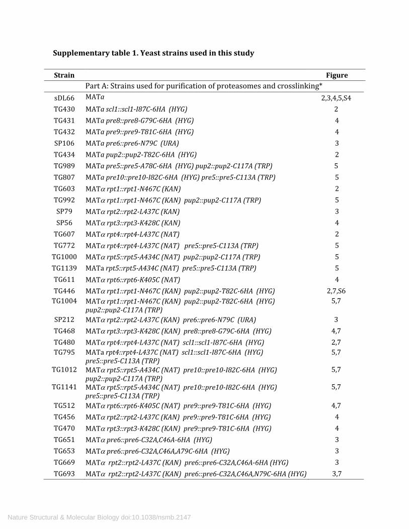
Supplementary table 1. Yeast strains used in this study
Strain Figure Part A: Strains used for purification of proteasomes and crosslinking*
sDL66 MATa
2,3,4,5,S4 TG430 MATa scl1::scl1-I87C-6HA (HYG) 2 TG431 MATa pre8::pre8-G79C-6HA (HYG) 4 TG432 MATa pre9::pre9-T81C-6HA (HYG) 4 SP106 MATa pre6::pre6-N79C (URA) 3 TG434 MATa pup2::pup2-T82C-6HA (HYG) 2 TG989 MATa pre5::pre5-A78C-6HA (HYG) pup2::pup2-C117A (TRP) 5 TG807 MATa pre10::pre10-I82C-6HA (HYG) pre5::pre5-C113A (TRP) 5 TG603 MATα rpt1::rpt1-N467C (KAN) 2 TG992 MATα rpt1::rpt1-N467C (KAN) pup2::pup2-C117A (TRP) 5 SP79 MATα rpt2::rpt2-L437C (KAN) 3 SP56 MATα rpt3::rpt3-K428C (KAN) 4
TG607 MATα rpt4::rpt4-L437C (NAT) 2 TG772 MATα rpt4::rpt4-L437C (NAT) pre5::pre5-C113A (TRP) 5
TG1000 MATα rpt5::rpt5-A434C (NAT) pup2::pup2-C117A (TRP) 5 TG1139 MATa rpt5::rpt5-A434C (NAT) pre5::pre5-C113A (TRP) 5 TG611 MATα rpt6::rpt6-K405C (NAT) 4 TG446 MATα rpt1::rpt1-N467C (KAN) pup2::pup2-T82C-6HA (HYG) 2,7,S6
TG1004 MATα rpt1::rpt1-N467C (KAN) pup2::pup2-T82C-6HA (HYG) pup2::pup2-C117A (TRP)
5,7
SP212 MATα rpt2::rpt2-L437C (KAN) pre6::pre6-N79C (URA) 3 TG468 MATα rpt3::rpt3-K428C (KAN) pre8::pre8-G79C-6HA (HYG) 4,7 TG480 MATα rpt4::rpt4-L437C (NAT) scl1::scl1-I87C-6HA (HYG) 2,7 TG795 MATa rpt4::rpt4-L437C (NAT) scl1::scl1-I87C-6HA (HYG)
pre5::pre5-C113A (TRP) 5,7
TG1012 MATα rpt5::rpt5-A434C (NAT) pre10::pre10-I82C-6HA (HYG) pup2::pup2-C117A (TRP)
5,7
TG1141 MATα rpt5::rpt5-A434C (NAT) pre10::pre10-I82C-6HA (HYG) pre5::pre5-C113A (TRP)
5,7
TG512 MATα rpt6::rpt6-K405C (NAT) pre9::pre9-T81C-6HA (HYG) 4,7 TG456 MATα rpt2::rpt2-L437C (KAN) pre9::pre9-T81C-6HA (HYG) 4 TG470 MATα rpt3::rpt3-K428C (KAN) pre9::pre9-T81C-6HA (HYG) 4 TG651 MATα pre6::pre6-C32A,C46A-6HA (HYG) 3 TG653 MATα pre6::pre6-C32A,C46A,A79C-6HA (HYG) 3 TG669 MATα rpt2::rpt2-L437C (KAN) pre6::pre6-C32A,C46A-6HA (HYG) 3 TG693 MATα rpt2::rpt2-L437C (KAN) pre6::pre6-C32A,C46A,N79C-6HA (HYG) 3,7
Nature Structural & Molecular Biology doi:10.1038/nsmb.2147

*All strains listed in part A have a background genotype of sDL66 (ref. 8): lys2-801 leu2-3, 2-112 ura3-52 his3-Δ200 trp1-1 rpn11::RPN11-TEVProA(HIS3). All Rpt salt-bridge and α subunit mutants were initially constructed in strain sDL66. Rpt C-terminal Cys mutants were made by transformation of strain DF5 (ref. 9) and subsequent dissection of the diploids. Double mutants were constructed by mating and tetrad analysis. Note α1 is encoded by SCL1, α2 by PRE8, α3 by PRE9, α4 by PRE6, α5 by PUP2, α6 by PRE5, and α7 by PRE10. **All strains listed in part B have a background genotype of SUB61 (ref. 9):lys2-801 leu2-3, 2-112 ura3-52 his3-Δ200 trp1-1).
TG199 MATα rpt1::rpt1-R173C(TRP1) S4 TG203 MATα rpt2::rpt2-E111C(HYG) S4 TG211 MATα rpt4::rpt4-R145C(NAT) S4 TG215 MATα rpt5::rpt5-E84C(KAN) S4 TG223 MATα rpt1::rpt1-R173C(TRP1) rpt2::rpt2-E111C(HYG) S4 TG247 MATα rpt4::rpt4-R145C(NAT) rpt5::rpt5-E84C(KAN) S4
Part B: Strains used for spotting assay and native PAGE of total cell lysate**
Sub61 MATα
S2,S4 TG577 MATa scl1::scl1-I87C-6HA (HYG) S2 TG579 MATa pre8::pre8-G79C-6HA (HYG) S2 TG581 MATa pre9::pre9-T81C-6HA (HYG) S2 TG646 MATa pre6::pre6-C32A C46A N79C-6HA (HYG) S2 TG585 MATa pup2::pup2-T82C-6HA (HYG) S2 TG587 MATa pre5::pre5-A78C-6HA (HYG) S2 TG589 MATa pre10::pre10-I82C-6HA (HYG) S2 SP47 MATα rpt1::rpt1-N467C (KAN) S2
TG641 MATα rpt2::rpt2-L437C (KAN) S2 TG643 MATα rpt3::rpt3-K428C (KAN) S2 SP304 MATα rpt4::rpt4-L437C (NAT) S2 SP309 MATα rpt5::rpt5-A434C (NAT) S2 SP311 MATα rpt6::rpt6-K405C (NAT) S2 TG530 MATα rpt1::rpt1-N467C (KAN) pup2::pup2-T82C-6HA (HYG) S2 TG681 MATα rpt2::rpt2-L437C (KAN) pre6::pre6- C32A C46A N79C-6HA (HYG) S2 TG629 MATα rpt3::rpt3-K428C (KAN) pre8::pre8-G79C-6HA (HYG) S2 TG536 MATα rpt4::rpt4-L437C (NAT) scl1::scl1-I87C-6HA (HYG) S2 TG562 MATα rpt5::rpt5-A434C (NAT) pre10::pre10-I82C-6HA (HYG) S2 TG568 MATα rpt6::rpt6-K405C (NAT) pre9::pre9-T81C-6HA (HYG) S2
Nature Structural & Molecular Biology doi:10.1038/nsmb.2147

Supplementary table 2. Yeast strains used to screen for Rpt-α pair crosslinking
Rpt1-N467C
Rpt2-L437C
Rpt3-K428C
Rpt4-L437C
Rpt5-A434C
Rpt6-K405C Figure
α1-I87C TG522 TG612 TG627 TG536 TG550 TG564 2
α2-G79C TG524 TG615 TG629 TG538 TG552 TG566 S5
α3-T81C TG526 TG617 TG630 TG540 TG554 TG568 4
α4-N79C TG528 TG619 TG633 TG542 TG556 TG570 3
α4-W.T. TG1021 TG1023 TG1025 TG1027 TG1029 TG1031 3
α5-T82C TG530 TG621 TG635 TG544 TG558 TG572 2
α6-A78C TG532 TG623 TG637 TG546 TG560 TG574 S5
α6-A78C a5-C117A
TG973 TG975 TG977 TG979 TG981 TG983 5
α7-I82C TG534 TG625 TG639 TG548 TG562 TG576 S5 α7-I82C
a6-C113A TG777 TG779 TG781 TG783 TG1137 TG787 5
All strains in this table are α mating type and have the background genotype of SUB61 (ref. 9) : lys2-801 leu2-3, 2-112 ura3-52 his3-Δ200 trp1-1. The marker for each mutated gene is as listed in table S1 part A.
Nature Structural & Molecular Biology doi:10.1038/nsmb.2147
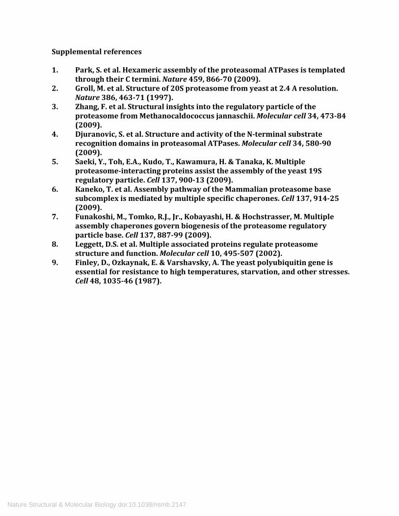
Supplemental references 1. Park, S. et al. Hexameric assembly of the proteasomal ATPases is templated
through their C termini. Nature 459, 866-70 (2009). 2. Groll, M. et al. Structure of 20S proteasome from yeast at 2.4 A resolution.
Nature 386, 463-71 (1997). 3. Zhang, F. et al. Structural insights into the regulatory particle of the
proteasome from Methanocaldococcus jannaschii. Molecular cell 34, 473-84 (2009).
4. Djuranovic, S. et al. Structure and activity of the N-terminal substrate recognition domains in proteasomal ATPases. Molecular cell 34, 580-90 (2009).
5. Saeki, Y., Toh, E.A., Kudo, T., Kawamura, H. & Tanaka, K. Multiple proteasome-interacting proteins assist the assembly of the yeast 19S regulatory particle. Cell 137, 900-13 (2009).
6. Kaneko, T. et al. Assembly pathway of the Mammalian proteasome base subcomplex is mediated by multiple specific chaperones. Cell 137, 914-25 (2009).
7. Funakoshi, M., Tomko, R.J., Jr., Kobayashi, H. & Hochstrasser, M. Multiple assembly chaperones govern biogenesis of the proteasome regulatory particle base. Cell 137, 887-99 (2009).
8. Leggett, D.S. et al. Multiple associated proteins regulate proteasome structure and function. Molecular cell 10, 495-507 (2002).
9. Finley, D., Ozkaynak, E. & Varshavsky, A. The yeast polyubiquitin gene is essential for resistance to high temperatures, starvation, and other stresses. Cell 48, 1035-46 (1987).
Nature Structural & Molecular Biology doi:10.1038/nsmb.2147


![Clinical Neurophysiology Volume Issue 2013 [Doi 10.1016_j.clinph.2013.12.094] Kim, Soyeon; Liu, Zhongxu; Glizer, Daniel; Tannock, Rosemary; Wo -- Adult ADHD and Working Memory- Neural](https://img.dokumen.tips/doc/110x75/577cd43d1a28ab9e789800b9/clinical-neurophysiology-volume-issue-2013-doi-101016jclinph201312094.jpg)