Embed Size (px)
Citation preview

Genetics Material
Fatchiyah, Ph.D.
http://fatchiyah.lecture.ub.ac.id

Chemical Composition of the
Body
“Because living things, including humans, are composed
only of chemicals, it is absolutely essential for a
(physiology) student to have a basic understanding of
chemistry.”
Sylvia Mader..

What is the genetic material?
George Mendel:
Thomas Morgan, in his experiments with fruit flies,
described genetic recombination, and demonstrated
that traits were to inherited together to varying
degrees.
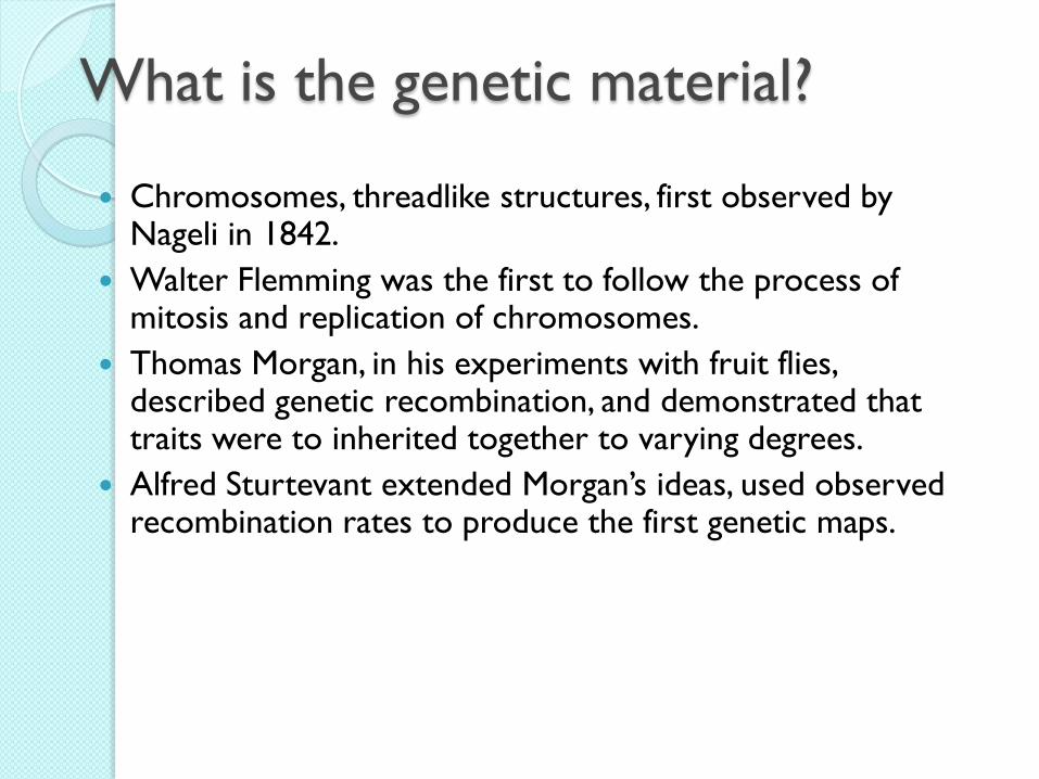
Chromosomes, threadlike structures, first observed by Nageli in 1842.
Walter Flemming was the first to follow the process of mitosis and replication of chromosomes.
Thomas Morgan, in his experiments with fruit flies, described genetic recombination, and demonstrated that traits were to inherited together to varying degrees.
Alfred Sturtevant extended Morgan’s ideas, used observed recombination rates to produce the first genetic maps.
What is the genetic material?
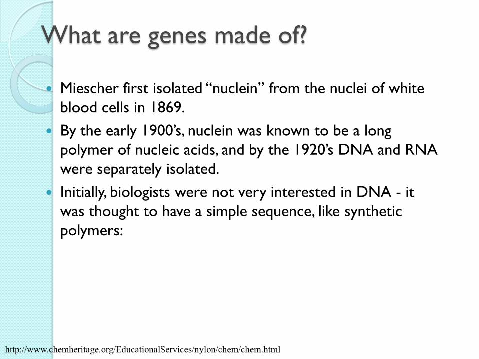
Miescher first isolated “nuclein” from the nuclei of white
blood cells in 1869.
By the early 1900’s, nuclein was known to be a long
polymer of nucleic acids, and by the 1920’s DNA and RNA
were separately isolated.
Initially, biologists were not very interested in DNA - it
was thought to have a simple sequence, like synthetic
polymers:
What are genes made of?
http://www.chemheritage.org/EducationalServices/nylon/chem/chem.html

The function of genes
Beadle and Tatum produced strong evidence via
mutation experiments with the mold Neurospora that
genes direct the production of proteins (1941)
◦ Produced mutant strain using irradiation
◦ Some mutant strains would not grow on conventional media,
but would grow on media with supplements (e.g. vitamin B6)
◦ The role of proteins as enzymes, and the part they play in
metabolism, was already understood at this time; the evidence
suggested that some inherited mutations knocked out specific
elements of metabolic machinery (i.e. proteins).

The genetics material: early studies
<1940s protein chemistry
1868 F Miescher, nuclei cell have nuclein
1910 Levine tetranucleotide hypothesis as DNA structure
1927 Grifftith, transformation studies Diplococcus
pneumoniae, virulent and avirulent strains
1944 O Avery, C McLeod, M McCarty: transforming
principle in Bacteria, the event led to acceptance of DNA
as the genetics material

Genes are made of DNA
Griffith showed that bacteria could be “transformed”
◦ pneumococcus colonies come in two varieties, “rough” (R) and “smooth” (S). S colonies are infectious, R are not.
◦ Kill S colony with heat, mix dead bacteria with R cells, inject into mouse. Mouse gets sick and dies; can isolate S bacteria from carcass.
Avery isolated the chemical components of S bacteria, demonstrated that the transforming factor was DNA.

Frederick Griffith 1928
Transformation - process in which one
strain of bacteria is changed by a gene or
genes from another strain of bacteria
Does material genetics Protein or DNA?

Avery, C McLeod, M McCarty’s ExperimentDNA is transforming factor
Heat-killed
IIIS cells cultured
Extract carbohydrates, lipids and protein
homogenize
Treat by protease
Treat by ribonuclease
Treat by deoxy-ribonuclease
Transformation occursNo Transformation
occurs
IIR+IIIS filtrate
IIR+RNase-treated IIIS filtrate
IIR+DNase-treated IIIS filtrate
Recovery IIIS filtrate
IIR+Protease-treatedIIIS filtrate
Assay for transformation
Active factor is not RNA
Active factor is not Protein
Active factor is DNA
Control: IIIS contains active
factor

Hershey-Chase Experiment 1952
Good scientists are naturally skeptical.
Hershey-Chase are testing to see if DNA
is the molecule that carries genetic
information.
Bacteriophage - virus that infects
bacteria
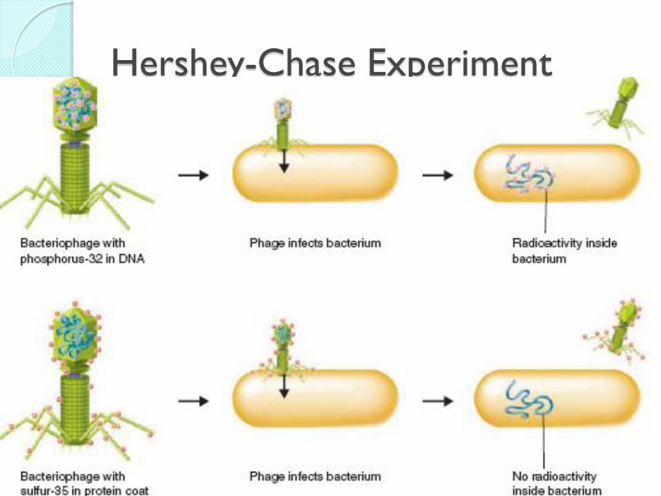
Hershey-Chase Experiment

DNA, what is it?
RNA, what is it?
DNA Replication, how?
Differences and Similarities

DNA: The Facts
DNA has a Double Helix shape. This shape is due to hydrogen bonds.

D.N.A. STRUCTURE DNA is also known as
deoxyribonucleic acid. It is a polymer, which is made up of smaller, similar molecules, which coil together to form chains. DNA is described as a (double helix). This is because it forms a 3D Structure. A DNA molecule can be copied perfectly over and over again.

Nucleotides

“backbone” of nucleic acid
The “backbone” of the nucleic
acid is formed by the sugar and
phosphate pairs.
Nitrogen containing base.
A Pentose sugar.
A phosphate group.
The “rungs” are formed by paired nitrogenous bases.
◦ Nitrogenous bases
complementary pair
A + T (U)
C + G..

Hydrogen bonds
Hydrogen bonds are special (polar) covalent bonds that are very important to physiology
Bonds formed between the hydrogen end (+ charged) of a polar molecule and the – end of any other polar molecule or highly electronegative atom (e.g. P, N, O) are called hydrogen bonds.
These hydrogen bonds are very important because they alter the physical and chemical properties of many molecules (especially water)..

The Essential Structure of DNA

Why DNA structure is ds?
Pauling & Carey structure of nucleat acid
Chargaff demonstrated that the ratio of A/T
in genomic DNA was a constant, and
likewise G/C
Wilkins and Franklin collected x-ray
diffraction data for fibers of DNA, and
determined that it had a helical structure.
Watson & Crick

Chargaff : the ratio of A/T in genomic
DNA
4/10/2011 Fatchiyah, Ph.D. JBUB 21
They also concluded that this percentage of bases in a DNA molecule
is independent of age, nutritional state, environment of the organism
studied.
It appears that human and e-coli bacteria obey a Chargaff’s rule which states that In
every species, the percent of Adenine almost exactly equals that of Thymine, and
the percent of Guanine is essentially identical to that of Cytosine.
Species Adenine Thymine Guanine Cytosine
Human 31.0 31.5 19.1 18.4
Fruit fly 27.3 27.6 22.5 22.5
Corn 25.6 25.3 24.5 24.6
Mold 23.0 23.3 27.1 26.6
Escherichia 24.6 24.3 25.5 25.6
Bacillius Subtillis 28.4 29.0 21.0 21.6

Rosalind Franklin 1950
X-Ray Diffraction of DNA Clues from the X-
Ray
◦ Coiled (forming
Helix)
◦ Double-stranded
◦ Nitrogeneous
bases are in the
center

Watson & Crick
Francis Crick – British physicist
James Watson – American Biologist
◦ Building a 3D model of DNA
◦ Franklin’s X-Ray opened their eyes to the
Double Helix
Watson and Crick’s model of DNA
was a double helix, in which two
strands were wound around each
other.

Structure of DNA
Watson & Crick put these clues together
with simple MOLECULAR MODELING studies to deduce THE STRUCTURE OF DOUBLE-STRANDED DNA, and also to suggest the mechanism for copying DNA
Here’s the original paper:http://www.nature.com/genomics/human/watson-crick/index.html
A- and B-DNA – right-handed helix,
Z-DNA – left-handed helix
B-DNA – fully hydrated DNA in vivo,
10 base pairs per turn of helix

RNA Structure
RNA is generally single stranded◦ Can fold and create complicated structure◦ Multiple types of RNA, each with a different function
Sugar-phosphate groups form the backbone of the molecule◦ Nucleotides are organized 5’ to 3’
Bases form the center of the molecule

Material Genetik pada virus
Terdapat di nukleus, sitoplasma
Bentuk Linier, single strand
Struktur kimiawi:
1. Gula penthose, disebut ribonucleosa
2. Asam phosphat
3. Basa Nitrogen:
Purin: Adenin, Guanin
Pyrimidin: Sitosin, Urasil
Type RNA:
mRNA, messenger RNA
rRNA, ribosomal RNA
tRNA, transfer RNA
RNA: Ribonucleic Acid
Backbone
RNA
mRNA mempunyai half life yang pendek mempertahankan homogenitas
Ju
mla
h m
RN
A p
ers
el
waktu
Laju transkripsi

Types of RNA
mRNA: messenger
RNA. It is the copy of
RNA that is made in
the nucleus and travels
outside the cell
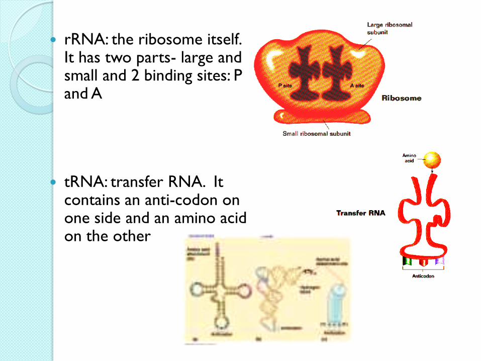
rRNA: the ribosome itself. It has two parts- large and small and 2 binding sites: P and A
tRNA: transfer RNA. It contains an anti-codon on one side and an amino acid on the other

Genes code for proteins using
symbolic information
Gene sequences code for protein sequences via a
symbolic code, the genetic code. This code is used nearly
universally by living organisms; it is one of the most
ancient shared characteristics of living things.
The “words” of the genetic code are nucleotide triplets
called codons. Each codon codes for at most one amino
acid.
Codons that do not code for any amino acids, called
nonsense or stop codons, terminate a coding region of
the gene. They serve as “punctuation marks”

•Codon tersusun atas 3 nukleotida (triplet) yg mengkode
informasi untuk satu asam amino, terbentuk 64 macam
• dari 64 mengkode 20 asam amino, beberapa asam amino
dikode lebih dari 1 codon
• bersifat UNIVERSAL untuk semua organisme
• Start codon, initiation codon, kodon awal/pembuka adalah
AUG (RNA) atau ATG (DNA)
• Stop codon or termination codon adalah UAA, UAG dan
UGA. Karena ketiga kodon ini tidak mengkode asam amino
apapun disebut juga nonsense-codon
Kode Genetik

mRNA-amino acid chart


4/10/2011 fatchiyah Dept Biology UB 33
Structure of Amino Acid subclass

Chromosome, DNA, & gene

Genes
Genes are short
sections of
chromosomes
http://www.accessexcellence.org

Chromosomal Structure of the Genetic
Material

Structure of a Typical Eukaryotic
Gene – the b-Globin Gene

Prokaryotic gene structure
P O A B C UTR
Intronless,
polysistronic
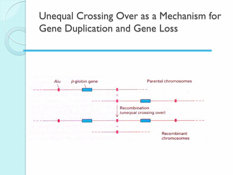
Unequal Crossing Over as a Mechanism for
Gene Duplication and Gene Loss

The Impact of the Complexity of Gene
Structure on Gene Expression

mRNA
Transcription
The Central Dogma of SYNTHESIS PROTEIN
Cell
Polypeptide
(protein)
TranslationRibosome
Reversetanscription
DNA

42
Eukaryotic Gene Structure
5’ - Promoter Exon1 Intron1 Exon2 Terminator – 3’
UTR splice splice UTR
transcription
translation
Poly A
protein

43
Prokaryotic Gene Structure
Promoter CDS
Terminator
transcription
Genomic DNA
mRNA
protein
UTR UTR
translation

44
From Gene to Protein

DNA
Cytoplasm
Nucleus
G AAAAAA
Export
Degradation etc.G AAAAAA
Control of Gene Expression
G AAAAAA
RNAProcessing
mRNA
RNA
Transcription
Translation
Packaging
Modification
Transportation
Degradation

How do DNA Replicate?
Replication is the process by which copies
of DNA
Cells of living organisms and made on
daily basis and most of the older cells die
as well.
So there are many generation and dying
of cell.
are made
The Replication Challenge
Size of an average human chromosome
130 million bp
Rate of replication
~ 50 bp per sec
Fidelity of replication

1. Enzymes unwind DNA
2. Enzymes split “unzip” double helix
3. The enzyme, DNA polymerase, finds and attaches the corresponding N-base
4. Each “old” stand serves as a template and is matched up with a new stand of DNA
5. New helixes wind back up.

DNA Replication
A – C – T – T – G – G – A – C
T – G – A – A – C – C – T - G

1) Semiconservative model:
Daughter DNA molecules contain one parental
strand and one newly-replicated strand
2) Conservative model:
Parent strands transfer information to an
intermediate (?), then the intermediate gets copied.
The parent helix is conserved, the daughter
helix is completely new
3) Dispersive model:
Parent helix is broken into fragments, dispersed,
copied then assembled into two new helices.
New and old DNA are completely dispersed
Models for DNA replication

(a) Hypothesis 1:
Semi-conservative
replication
(b) Hypothesis 2:
Conservative replication
Intermediate molecule
(c) Hypothesis 3:
Dispersive replication
MODELS OF DNA REPLICATION

Meselson and Stahl
Semi-conservative replication of DNA
Isotopes of nitrogen (non-radioactive) were used in this experiment

Generations
0
0.3
0.7
1.0
1.1
1.5
1.9
2.5
3.0
4.1
0 and 1.0
mixed
0 and 4.1
mixed
HH
HL
LL + HL
HH
HL
HL LL LL LH
Equilibrium Density
Gradient
Centrifugation
Detection of
semiconservative
replication in E. coli
by density-gradient
centrifugation. The
position of a band of
DNA depends on its
content of 14N amd 15N. After 1.0
generation, all the
DNA molecules are
hybrids containing
equal amounts of 14N
and 15N

Origin
5’
3’
3’
5’
UNIDIRECTIONAL REPLICATION
Origin
5’
3’
3’
5’
BIDIRECTIONAL REPLICATION
Replication can be Uni- or Bidirectional

Replication of the Genetic Material
Small chromosomes use a single origin
Replication of large chromosomes requires multiple origins
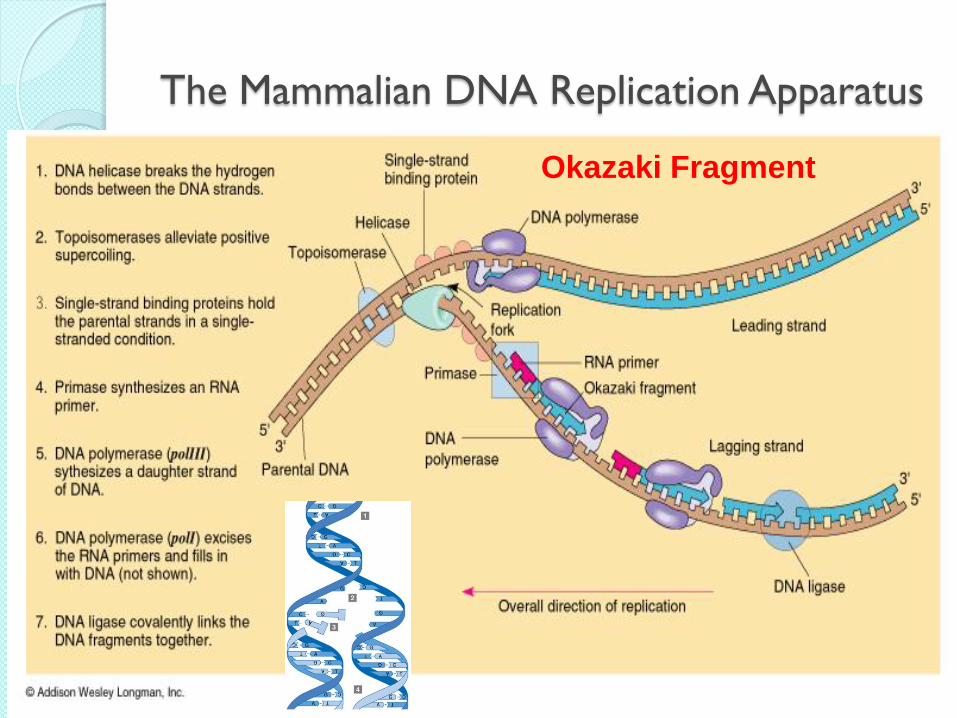
The Mammalian DNA Replication Apparatus
Okazaki Fragment

Subsequent
hydrolysis of
PPi drives the
reaction forward
Nucleotides are added at the 3'-end of the strand
The 5’ to 3’ DNA polymerizing activity

Why the exonuclease activities?
The 3'-5' exonuclease activity serves a proofreading function
It removes incorrectly matched bases, so that the polymerase can try again.

The DNA Polymerase Family
A total of 5 different DNAPs have been reported in E. coli
DNAP I: functions in repair and replication
DNAP II: functions in DNA repair (proven in
1999)
DNAP III: principal DNA replication enzyme
DNAP IV: functions in DNA repair (discovered in
1999)
DNAP V: functions in DNA repair (discovered in
1999)

DNA Polymerase III
The "real" replicative polymerase in E. coli
It’s fast: up to 1,000 dNTPs added/sec/enzyme
It’s highly processive: >500,000 dNTPs added
before dissociating
It’s accurate: makes 1 error in 107 dNTPs added,
with proofreading, this gives a final error rate of 1
in 1010 overall.

Proof reading activity
of the 3’ to 5’ exonuclease.
DNAPI stalls if the incorrect
ntd is added - it can’t add the
next ntd in the chain
Proof reading activity is slow
compared to polymerizing
activity, but the stalling of
DNAP I after insertion of an
incorrect base allows the
proofreading activity to
catch up with the polymerizing
activity and remove the
incorrect base.