Embed Size (px)
Citation preview

FINAL DRAFT FOR BIOMED 101
GENETICALLY MAGNETIZED PLGA
NANOPARTICLES FOR INTRAVENOUS DRUG
AND GENE TARGETING
by
Jeong-Yeol Yoon
Date: Dec 10, 1999
Ph.D. Student
Biomedical Engineering IDP
University of California, Los Angeles
Email: [email protected]

4
GENETICALLY MAGNETIZED PLGA NANOPARTICLES FOR INTRAVENOUS
DRUG AND GENE TARGETING
Jeong-Yeol Yoon
New drug targeting system is proposed by incorporating PLGA – poly(D,L-lactide-co-glycolide) –
nanoparticles and genetically synthesized magnetic nanoparticles. By taking the advantages of PLGA (good
biocompatibility, biodegradability, and small size for intravenous delivery) and magnetite (rapid and easy
treatments), a powerful drug/gene carrier is made. Recently, many people worked about the synthesis of
PLGA nanoparticles for drug delivery (not targeting), and the synthesis of magnetic nanoparticles as a
contrast agent for MRI (magnetic resonance imaging). However, there are no published reports for
magnetic targeting with PLGA nanoparticles. The main problem is ‘how we can incorporate magnetite and
biocompatible carrier (PLGA) together.’ We will use the lipid-layered magnetic nanoparticles made by
microorganisms, with recombinant DNA technology. This lipid layer gives biocompatibility to the
magnetic particles and this compatibility enables to be easily incorporated with PLGA. The main objectives
are (1) synthesis of genetically magnetized PLGA nanoparticles, and (2) their application to the drug and
gene (DNA) targeting.
Keywords: drug targeting, gene targeting, gene therapy, PLGA, magnetite, nanoparticles, recombinant
DNA technology.

5
I. IDENTIFICATION AND SIGNIFICANCE OF THE PROBLEM
Nowadays, drug delivery (including both controlled release and drug targeting) becomes more popular
both in academia and industry than ever. The main foci are now shifting to gene delivery for therapeutic
purposes and the guided targeting to specific organs. An extremely small-sized carrier is also required for
the intravenous injection to prevent the carrier’s clogging up in vessels – the size below 70 nm is generally
demanded, primarily due to the size constraints within the capillary vessels. The carrier should also be
biodegradable and biocompatible. PLGA – poly(D,L-lactide-co-glycolide) – nanoparticles, with PVAL –
poly(vinyl alcohol) – as a stabilizer, are generally considered as an acceptable carrier to meet the above all
requirements. Many problems still remain such as instability, long-term incompatibility with blood cells
and tissues, and non-zero-order release behaviors.
Targeting methodology is another problem. One of the most favored methods for guided targeting is a
magnetic one, especially with magnetite (Fe3O4). However, conventional magnetite particles are so big that
they cannot be incorporated within the nanoparticles. A recently introduced alternative is ferrofluid, but no
work has been published that incorporates PLGA nanoparticles and the ferrofluid. The main problem is the
incompatibility between PLGA and the ferrofluid. The advantages of magnetic targeting – rapidity and easy
treatment – are too beneficial to be abandoned.
Therefore, for the magnetic targeting of drugs (or genes), we must choose one method from the
followings: (Note: particle size cannot be sacrificed – it must be smaller than 70 nm.)
(1) To use alternative materials (for example, polystyrene-based latex particles) rather than PLGA, that
can be easily incorporated with ferrofluid or magnetite. This alternative generally has lower
biocompatibility than PLGA.
(2) To adopt alternative targeting method, such as using site-specific enzymes or using targeting by
antibodies, rather than magnetic targeting. Targeting efficiency are generally lowered comparing with
the magnetic one.
Table 1 summarizes several reviews on the intravenous drug targeting (Guiot and Couvreur, 1986,
Rembaum and Tokes, 1988, Tsuruta et al., 1993). PS (polystyrene) and its derivatives were widely used as
carriers at first, but people gradually moved to PEU (polyether urethane) and PLGA to increase the
biocompatibility. Conversely, these new carriers’ incorporation with magnetite is more difficult than PS,
hence these carriers are currently used only for controlled release, not targeting.
Targeting methodolgy is also shown in Table 1 (Guiot and Couvreur, 1986, Rembaum and Tokes, 1988,
Kompala and Todd, 1991). Magnetic targeting is superior to other methods in all aspects (except in site-
specific efficiency), but making biocompatible nano-sized one remains as a big problem. Therefore, the use
of magnetic particles is confined to ex vivo, except the use as an MRI (magnetic resonance imaging)
contrast agent.

6
Table 1. Current status in intravenous drug targeting.
---------------------------------------------------------------------------------------------------------------------------------
Carrier selection Biocompatibility Incorporation with magnetite
---------------------------------------------------------------------------------------------------------------------------------
PS and its derivatives Poor Good
PEU and its derivatives Not bad Not bad
PLGA and its derivatives Good Poor
Proteins (albumin, dextran) Good Not tested yet
---------------------------------------------------------------------------------------------------------------------------------
Targeting methodology Speed Treatment Site-specific
---------------------------------------------------------------------------------------------------------------------------------
Magnetic Fastest Easy Good
Enzymatic Rather slow Complex Arguable
Immunological Fast Complex Excellent
---------------------------------------------------------------------------------------------------------------------------------
Note. ‘Carrier selection’ information is based on the results of controlled release system, not targeting or
site-specific system.
7
II. BACKGROUND, TECHNICAL APPROACH, AND ANTICIPATED BENEFITS
II.1. Background
Previous Applications Using Magnetic Beads
Magnetite (Fe3O4) has been widely used in many organic and inorganic chemical applications due to its
ease of treatment, but it is recently highlighted in the biomedical area. Magnetite particles can be easily and
rapidly separated by an externally applied magnetic field. Due to the ease and rapidity of response, these
particles are alternatives for biomedical applications. Cell separation and MRI (magnetic resonance
imaging) were the first areas that magnetite was used in biomedical applications.
(1) Cell Separation – Many different kinds of cells are tested in clinical labs, and these cells must be
separated for specific purpose. Two methods are commonly used (Kompala and Todd, 1999) – flow
cytometry and immunomagnetic cell separation (IMCS). Although there exists a state-of-the-art flow
cytometry, IMCS is commonly used in most labs, because IMCS is faster and easier to handle than
flow cytometry. In IMCS, polymer beads containing magnetite are attached with monoclonal
antibodies, which can be specifically bound to target cells. An external magnetic field keeps these
beads stationary (Schwarz and Contescu, 1999); so the target cells are bound to the flow system and
other cells flows through. For polymer beads, magnetized polystyrene (PS) is the most common one
used (these beads are commercially available). Magnetite does not appear to sustain permanent
magnetization; hence magnetic interaction between particles is negligible in aqueous media (Sohn et
al., 1996). However, in our opinion, there probably exist a stability problem, because these magnetized
beads have relatively high PDI (polydispersity index) values, which means the size distribution is
broad (Ding et al., 1998). This mainly results from the polymerization methods – dispersion
polymerization with solvent evaporation technique – which necessarily leads to broad size distribution.
In addition, for the same reason, these particles are so big that they cannot be used for intravenous
injection. A recent study by Partington et al. (1999) concerns the synthesis of magnetic nanoparticles
Magnetic force
Target cell
Magnetic beads magnetite core
with monoclonal
antibodies
Flow Antibody Normal cell
Figure 1. Immunomagnetic Cell Separation

8
for use with cell separation. However, there still exists a problem that they cannot be incorporated with
polymers, which can be used to carry drugs or genes (Schwarz and Contescu, 1999).
(2) MRI (magnetic resonance imaging) – magnetite particles have been used as a magnetic resonance
contrast agents. These particles are small enough to prevent the clogging problems, but this application
is still plagued by problems in incorporating them with polymers. Recently, Zaitsev et al. (1999)
published a paper about the optical properties of polymer-coated magnetic nanoparticles, but made no
attempts to use it as a drug/gene carrier. Biocompatibility with human tissues/organs and drugs/genes
continues to be a persistent problem for these particles.
(3) Other applications: magnetite particles have also been used as an immobilization matrix (Bahar and
Celebi, 1998), and also as a solid-phase matrix of ELISA (enzyme linked immunosorbent assay) (Kala
et al., 1997).
PLGA Nanoparticles as a Drug Carrier
Nanoparticles are one of the most promising dosage forms of potential formulations for site-specific
drug delivery systems including drug targeting. Special interest has been focused on the particles prepared
from polyesters, such as poly(D,L-lactide-co-glycolide) (PLGA), poly(D,L-lactide) (PLA), polyglycolide
(PGA). This is largely due to their biocompatibility and resorbability via natural pathways. Moreover, the
Food and Drug Administration (FDA) has approved the use of polymers prepared from glycolic acid and
lactic acid for in vivo use. These polymers do not need surgical removal after the completion of drug
dosage.
These nanoparticles were made by the following methods:
(1) Emulsion-evaporation method (in 1980’s): This method utilizes the o/w (oil-in-water) emulsion. Oil-
phase solvent is removed by evaporation, thus forming particles. From this reason, this method is also
called as ‘Solvent Evaporation Method.’ The size of resulting particles are generally over 1 μm, thus
nanoparticle formation is difficult with this method (Guiot and Couvreur, 1986, Ibrahim et al., 1991).
(2) Emulsion-diffusion method (in 1990’s): This method was evolved from the ‘salting-out’ procedure
(Allemann et al., 1992, 1993). The difference between emulsion-evaporation and emulsion-diffusion
methods is that the particle formation is induced by diffusion, not evaporation. Nanoparticle formation
is possible in this method, with the help of stabilizers. At first, benzyl alcohol was used as an oil-phase
solvent, and poly(vinyl alcohol) (PVAL) was used as stabilizers (Leroux et al., 1995). Quintanar-
Guerrero et al. (1998) used propylene carbonate (PC) as a solvent instead of benzyl alcohol, to reduce
the toxicity of oil-phase solvent. Labhasetwar et al. (1998) also reported that using
didodecyldimethylammonioum bromide (DMAB) as a co-stabilizer could enhance the loading level of
drugs.
9
Gene Delivery and Targeting for Gene Therapy
Many diseases are related to defects in genes. Therefore, site-specific gene delivery may be of great
promise to those who suffer from diseases with genetic origin. Nowadays, the genes can be synthesized for
therapeutic purpose very easily. The mass of genes (DNA’s) can be increased through PCR (polymerase
chain reaction). (Glick and Pasternak, 1998).
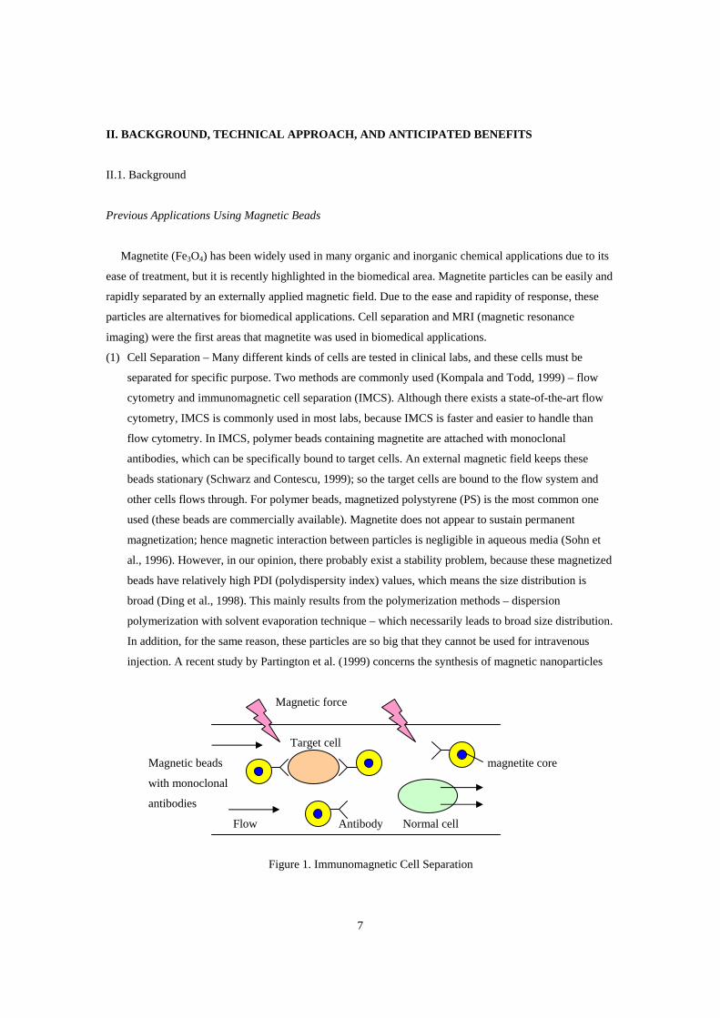
Genes, in cDNA form that has no introns (cDNA is made by reverse transcription from mRNA), are
carried by a biocompatible matrix. For site-specific delivery, both enzymatic and magnetic methods can be
used. Genes can be attached to the surface of carrier beads, or they can be encapsulated into porous
nanocapsules that mimic the eukaryotic nuclei (Figure 2).
ss mRNA
reverse
transcription ds cDNA
beads with attached genes porous microcapsules
containing genes
Figure 2. Two methods for site-specific gene targeting
Biocompatible Magnetic Particles by Microorganisms
Matsunaga et al. (1998) reported that certain types of microorganisms are capable of producing
biocompatible magnetic nanoparticles. The microorganism is Magnetospirillum sp. AMB-1, and it
synthesizes intracellular particles of magnetite (Fe3O4). With the help of recombinant DNA technology, the
gene for this magnetite production (macA gene) was cut by restriction enzymes (BamHI, SacI), and ligated
to plasmid vector pRK415.
They only suggested its use with immunoassays, but it has the potential for magnetic drug/gene
targeting as well. This magnetite has high biocompatiblity, due to the biologic manufacture, especially the
lipid layer on its surface. This biocompatibility is important for PLGA incorporation.

10
II.2. Technical Approach and Anticipated Benefits
Our approach for new method of magnetic drug/gene targeting is as follows:
Step 1. Production of magnetic particles by recombinant DNA technique
With this technique, we can avoid the incompatibility problem between conventional magnetite or
ferrofluid and PLGA. This procedure is schematically illustrated in Figure 3.
Magnetospirillum sp. AMB-1
BamHI SacI
KpnI SpnI
Plasmid vector
pRK415 (10.5 kbp) mRNA with magA gene
Recombinant
vector magA protein
with magA
magnetic nanoparticles
with lipid layer
Figure 3. Biocompatible magnetic nanoparticle formation
(1) Preparation of plasmid vector template: a plasmid vector, pRK415 will be used. Its size is 10.5 kbp.
Two restriction enzymes, BamHI and SacI will cut this vector to make a template for magA gene
expression.

11
(2) Preparation of magA gene: mRNA (which has no introns) containing magA gene will be separated
from Magnetospirillum sp. AMB-1. Two restriction enzymes, KpnI and SpnI will digest this mRNA to
obtain magA gene.
(3) Preparation of recombinant vector: magA gene will be ligated to the plasmid vector template. This
recombinant vector will be inserted in E. coli to express magA protein.
(4) Production of lipid-layered nanoparticles: magA protein will produce lipid-layerd, biocompatible
magnetic nanoparticles (BMNP).
Step 2. Incorporation the biocompatible magentic nanoparticles (BMNP) with PLGA
The resulting magnetic particles have lipid layer, which provides intrinsic biocompatibility. Adding
PLGA will yield the core-shell structure. The following is a summary for making the structured
biocompatible magnetized PLGA nanoparticles. Figure 4 also illustrates this procedure.
PLGA
Drug/Gene External
PC Water magnetic field
(solvent)
Stabilizers
BMNP
Water Homogenizing Stirring Removing non-magnetic
with adding water particles
Figure 4. Procedure for biocompatible magnetized PLGA nanoparticle production
(1) Preparation of organic (oil) phase: PLGA (and drugs/genes) are dissolved in organic solvent, PC (after
Quintanar-Guerrero et al., 1998). PLGA can be fluorescent-labeled with rhodamine B, for tissue
localization studies.
(2) Preparation of aqueous phase: Stabilizers, PVAL and DMAB (after Labhasetwar et al., 1998) are
dissolved in water. We will also dissolve BMNP in aqueous phase; thus micelles with BMNP can be
generated at this stage. BMNP disperse very well in aqueous phase because they are covered with a
stable lipid layer (Matsunaga and Takeyama, 1998).
(3) Emulsification: Mixing and homogenization of the above two phases yield an o/w emulsion. High-
speed homogenizer required in this step.

12
(4) Nanoparticle formation: Water is subsequently added to the o/w emulsion, leading to the diffusion of
PLGA (and drugs/genes) to the micelles in aqueous phase. This yields the core-shell structured
nanoparticle formation. Figure 5 schematically illustrates the nanoparticle formation.
(5) Removal of non-magnetic nanoparticles: There probably exist non-magnetized nanoparticles, that is,
PLGA nanoparticles without BMNP. These non-specific nanoparticles can be easily removed by an
external permanent magnet (Schwarz and Contescu, 1999).
(6) Purification: Core-shell structured nanoparticles are purified by dialysis. Freeze-drying produces a
homogeneous free-flowing powder.
lipid layer
stabilizer
diffusion
PLGA (+ drug/gene) droplet BMNP micelle
PLGA
(+ drug/gene) Core-shell structured
magnetic PLGA nanoparticle
BMNP
Figure 5. Nanoparticle formation
Step 3. In vitro study (for phase II)
Some feasibility tests should be performed in vitro prior to in vivo experiments. Following is a list of
factors that could determine the PLGA-BMNP feasibility for biomedical use.
(1) Stability: The particles should have enough stability to overcome non-Newtonian behavior in vivo
conditions. This can be measured through HLB (hydrophile-lipophile balance), CMC (critical micelle
concentration), and hydrophobicity values (Shaw 1992). Surface tension meter can calculate HLB and
CMC values. Hydrophobicity can be measured by labeling pyrene (strongly hydrophobic material) to
the stabilizers.
(2) Particle size distribution: Nanoparticles should also be uniform in size to prevent an aggregation.
Dynamic light scattering (DLS) should be used for size determination because the PLGA-BMNP’s are
smaller than 0.1 μm.

13
(3) Drug/gene content: The particles should have sufficient loading amount of drugs/genes. Centrifuging
and spectroscopy techniques will be adopted.
(4) Movement of particles in a magnetic field: The particles should behave properly in blood vessels
where magnetic field is applied. Clogging with the vessels, self-aggregation, and decomposition are
not allowed for nanoparticles. Mathematical simulation and experimental certification will be
simultaneously performed. The magnetic force will be applied as a function of flat, sine-wave, or
impulse (Figure 6).
Magnetic force
Various forms of blood flow
Figure 6. Motion of nanoparticles in blood vessels
Step 4. In vivo study (for phase III)
In vivo experiments for animal will be performed. Localization, retention, and bioavailability of the
nanoparticles will be evaluated. Fluorescent-labeled (rhodamine B) nanoparticles will be used to determine
the tissue localization and persistence of them of them in the tissue.
Injury will be induced in rats, then PLGA-BMNP will be infused to them. External permanent magnets
will be attached to the skin (just above the specific tissue or organ) of rats. The rats will be killed with
varying the time (such as 1 day, 7 days, and 1 month), then the above parameters – localization, retention,
and bioavailability will be measured.

14
III. PHASE I TECHNICAL OBJECTIVES
The long-term goal of this program is to demonstrate the targeted drug delivery using magnetized
particles in vivo. Good biocompatibility of carriers, good stability of magnetized-carriers, as well as
rapidity and ease of magnetic targeting, is required.
Detail objectives of phase I are listed in the following:
1. To produce magnetized nanoparticles with recombinant DNA technology
The optimal conditions for the production of biocompatible magnetic nanoparticles (BMNP) should be
established; the concentrations of restriction enzymes, the separation of non-specific recombinant
vectors, and the culture conditions for magA gene expression in E. coli. Size distribution of BMNP
should also be determined at this stage.
2. To encapsulate these particles in PLGA
The above BMNP will be encapsulated in PLGA (plus drugs/genes) by emulsion-diffusion method.
The optimal experimental conditions should be established; homogenizer speed, stirring rpm, stabilizer
concentration, and the rate of water addition.
The followings are the technical objectives of phases II and III.
3. To demonstrate the stability and sufficient drug loading (phase II)
Various parameters (HLB, CMC, hydrophobicity, and drug/gene content) will be obtained to secure
the particle stability and the sufficient drug loading.
4. To demonstrate ability to control the particles in a magnetic field (phase II)
Mathematical modeling and experimental certification will be carried out to prevent the clogging, self-
aggregation, and decomposition of nanoparticles.
5. In vivo study (phase III)
In vivo experiments for animal will be performed to evaluate localization, retention, and bioavailability
of the nanoparticles.

15
IV. PHASE I RESEARCH PLAN
Task 1. Gene Expression with Recombinant DNA Techniques
The objective of this task is to make biocompatible magnetic nanoparticles (BMNP) that can be
controlled by magnetic fields. The overall procedure will follow after Matsunaga et al. (1998). Details are
listed in the following:
1. Preparation of plasmid vector template
BamHI and SacI (restriction enzymes) will digest plasmid vector pRK415 (10.5 kbp) to make a
template for magA insertion. A gel electrophoresis will determine the efficiency of restriction enzymes.
Digestive conditions will be changed after this analysis.
2. Preparation of magA gene
The mRNA containing magA gene will be separated from Magnetospirillum sp. AMB-1 by a gel
electrophoresis. Two restriction enzymes, KpnI and SpnI, will digest this mRNA to obtain macA gene.
After sequencing of this gene, PCR technique will be used to amplify the macA gene. (Two primers are
required for PCR.) Gene sequencing and PCR apparatuses are required for this step. For PCR, Primers
and dNTP’s are required in excess, and Taq DNA polymerase is required for the stability in high
temperature.
3. Preparation of recombinant vector
With the help of blunt-end ligase (the above restriction enzymes makes a blunt-end), magA gene will
be ligated to plasmid vector template. This recombinant vector will be inserted in E. coli to obtain
magA protein. Batch fermentation is required for the gene expression in E. coli.
4. Production of BMNP
magA protein will make lipid-layerd BMNP. BMNP will be separated from cell cultures by
centrifugation. Optimal rpm should be determined to prevent the breaking-up of BMNP. Further
purification will be made by an external magnetic field, shown in the following figure.
Stationary BMNP External magnetic field
Flow Other materials
(moving)
Figure 7. Purification of BMNP from cell culture

16
5. Characterization of BMNP
The size distribution will be determined by DLS (dynamic light scattering). Raman spectroscopy,
conductometric titration, and zeta potential analysis will give additional information of the surface
properties. A recent study by Vo-Dinh et al. (1999) reports that the surface-enhanced Raman scattering
(SERS) can be used for analysis of biological compounds in multicomponent environment. Therefore,
we will use SERS technique to determine the lipid content in BMNP. Conductometric titration and zeta
potential analysis gives direct information of electrostatic stability (Yoon et al., 1996, 1998b, 1999).
The stability will be measured by HLB and CMC values, through surface tension meter.
The overall scheme was already illustrated in Figure 3. The following table summarizes the materials
and apparatus required for task 1.
Table 2. Materials and Apparatus Required for Task 1
---------------------------------------------------------------------------------------------------------------------------------
Step Materials Apparatus required
---------------------------------------------------------------------------------------------------------------------------------
Step 1 – plasmid vector template pRK415 Gel electrophoresis
BamHI, SacI
Step 2 – magA gene Magnetospirillum sp. AMB-1 Gel electrophoresis
KpnI, SpnI Gene sequencing apparatus
Primers PCR apparatus
dNTP
Taq DNA polymerase
Step 3 – recombinant vector Step 1 and 2 products Fermenter
Blunt-end ligase
E. coli, cell culture
Step 4 – BMNP Step 3 product Centrifuge
Column with magnetic field
Step 5 – characterization Step 4 product DLS
SERS
Conductometric titrator
Zeta potential analyzer
Surface tension meter
---------------------------------------------------------------------------------------------------------------------------------

17
Task 2. Magnetized PLGA nanoparticle formation
In this task, PLGA and BMNP will be incorporated into one, resulting core-shell structure (core:
magnetic beads, shell: PLGA). The overall scheme was already illustrated in Figure 4, and details are listed
in the following:
1. Preparation of organic (oil) phase
PLGA (plus drugs or genes) will be dissolved in organic solvent, PC, after Quintanar-Guerrero et al.
(1998).
2. Preparation of aqueous phase
Stabilizers, PVAL and DMAB will be dissolved in water, after Labhasetwar et al. (1998). Then BMNP
will be added to this aqueous phase, thus forming micelle with BMNP core. (Figure 5). The amount of
PVAL will determine the final size and stability of PLGA-BMNP, and that of DMAB will determine
the drug/gene content.
3. Emulsification
The above two phases will be mixed together and homogenized at high speed. Mixing ratio and
homogenizer speed will be varied. The o/w emulsion will be made at this step. PLGA (plus
drugs/genes) exist in organic (oil) phase, and BMNP in aqueous (water) phase.
4. Nanoparticle formation
Water will be subsequently added to the o/w emulsion, leading to the diffusion of PLGA (plus
drugs/genes) to the micelles in aqueous phase. This yields the core-shell structured nanoparticle
formation, and it was illustrated in Figure 5. The only variable in this step is the rate of water addition.
5. Removal of non-magnetic nanoparticles
The non-specific nanoparticles (which has no BMNP in core) will be removed by the same method
illustrated in Figure 7.
6. Purification
Resulting magnetized PLGA nanoparticles will be purified by dialysis and freeze-drying.
7. Characterization
The characterization of magnetized PLGA nanoparticles will be performed by a similar method of task
1. Size distribution will be measured by DLS, surface acidity by conductometric titration, and
electrostatic stability by zeta potential analyzer (Yoon et al., 1996, 1998b, 1999). Further
characterization studies – HLB, CMC values, hydrophobicity, and drug/gene content – will be
performed in phase II.
Table 3 summarizes the materials and apparatus required for task 2.

18
Table 3. Materials and Apparatus Required for Task 2
---------------------------------------------------------------------------------------------------------------------------------
Step Materials Apparatus required
---------------------------------------------------------------------------------------------------------------------------------
Step 1 – organic phase PLGA
Drug/Gene
PC
Step 2 – aqueous phase PVAL, DMAB
BMNP
Water
Step 3 – emulsification Step 1 and 2 products Homogenizer
Step 4 – nanoparticle Step 3 product (o/w emulsion) Stirrer
formation Water Micro tubing pump
Step 5 – removal of non- Step 4 product Column with magnetic field
magnetic particles
Step 6 – purification Step 5 product Dialysis
Freeze-dryer
Step 7 – characterization Step 6 product DLS
Conductometric titrator
Zeta potential analyzer
---------------------------------------------------------------------------------------------------------------------------------
19
V. RELATED RESEARCH AND DEVELOPMENT
Major recent researches (biomedical application of magnetic particles or non-magnetic nanoparticles)
are already mentioned in previous sections. These works can be classified into several categories:
1. Microparticles, non-magnetized
Yoon et al., 1996, 1998b, 1999, for protein adsorption study
Yoon et al., 1998a, Lee et al., 1998, for protein separation
- Microparticles generally have the size between 0.1~10 μm. J.-Y. Yoon (who will work as a
graduate student researcher in this project) have published several works with the microparticles
ranging 0.2~0.4 μm to reveal the interactions between proteins and polymer surfaces. This a priori
knowledge will be useful for studying drug-PLGA interaction.
2. Microparticles, magnetized
Bahar and Celebi, 1998, for immoblization of enzymes
Liberti and Pino, US Patent, for cell separation
Ding et al., 1998, for cell separation
- Magnetized microparticles for cell separation generally have large size, ranging 1~100 μm.
Especially, cell separation requires large particles due to the size of living cells. These works are
intended for ex vivo use, and not applicable for in vivo use.
Kala et al., 1997, for immunoassay
Sohn et al., 1996, for optical study
- Magnetic microparticles were tested for bio-separation and immunoassay in these works. The size
of particles are relatively small (below 1 μm), because they employed a light scattering technique.
3. Nanoparticles, non-magnetized
Allemann et al., 1993, for controlled release of drugs
Leroux et al., 1995, establishment of emulsion-diffusion method
Quintanar-Guerrero et al., 1998, change of solvent to PC to increase biocompatibility
Labhatsetwar et al., 1998, introduction of DMAB for the increase in drug content
- All of the above works were related with the synthesis of nano-sized PLA drug carrier. Allemann
et al. established a salting-out procedure, and Leroux et al. developed an emulsion-diffusion
method to make nanoparticles. Quintanar-Guerrero et al. and Labhatsetwar et al. improved
biocompatibility and drug content, respectively.
Maruyama et al., 1999, for gene delivery
- A recent study by Maruyama et al. dealt with gene (DNA) delivery, instead of drug. Their primary
objective was gene therapy (to treat the disease caused by a gene defect). Non-magnetized
nanoparticles were used.

20
4. Nanoparticles, magnetized
Matsunaga et al., 1998, for MRI
- Biocompatible, magnetized nanoparticles were prepared by the recombinant DNA technology.
Their primary concern is to use them as an MRI contrast agent, hence they did not incorporated
with any polymer carriers.
Partington et al., 1999, for cell separation
- Large-sized magnetized PS (polystyrene) particles are generally used for cell separation, but these
people used nano-sized particles to improve the selectivity. However, they eventually made
microparticles, because the nanoparticles were assembled together while passing several steps.
Sonti et al., 1997, for DNA separation
- The authors made a nanocluster from magnetic nanoparticles to separate DNA. These particles
lack biocompatibility and stability, hence they are not suitable for in vivo use.
Our only concern is group 4, because we need magnetized drug/gene targeting and nano-size for in vivo
use. No attempts have been made to use magnetized nanoparticles as a drug carrier. While it is possible to
make magnetic nanoparticles, there are no published reports dealing with the incorporation of these
magnetite nanoparticles and carriers. Partington et al. eventually made microparticles for cell separation
(nanoparticles are not suitable for cell separation media, they are so small). The particles that Sonti et al.
made are actually not particles; it is nanoclusters. Therefore, only the work by Matsunaga et al. is valuable,
although they made without any carriers or drugs and just applied to MRI.
The previous works performed by J.-Y. Yoon (which dealt with the interfacial phenomena between
biomolecules and polymeric particle surfaces) will make a basis for Task 2 in Phase I, especially for drug
content and releasing behavior.

21
VI. KEY PERSONNEL AND BIBLIOGRAPHY
Professor Robin L. Garrell (Department of Chemistry and Biochemistry & Biomedical Engineering
Program, University of California, Los Angeles) will serve as principal investigator. She will supervise the
whole program, and also take charge of the polymer synthesis section. Professor Garrell has over 15 years
of experience in organic/inorganic synthesis for biological applications, especially in surface chemistry.
Graduate student researcher Jeong-Yeol Yoon (Biomedical Engineering Program, University of California,
Los Angeles) will take charge of the characterization section, as well as actual experimental section.
22
VII. FACILITIES AND EQUIPMENT
Biomedical Engineering Program in University of California, Los Angeles has wide resources ranging
from School of Engineering and Applied Sciences, School of Medicine, School of Dentistry, as well as
some departments from College of Letters and Science. Followings are the required facilities for this
program, and all of these are available upon request.
a) Spectrophotometer: UV/Vis, FT-IR, AA, Raman
b) Chromatography: HPLC, TLC, GPC, GC
c) Electron Microscope: SEM, TEM, AFM
d) Surface characterization: Zeta Potential Analyzer, SERS, Surface Tension Meter, Auto-titrator
e) Size analysis: DLS
f) Gel Electrophoresis Equipment, Gene Sequencing Apparatus, PCR Apparatus
g) Fermenter
h) Dialyzer, Freeze-dryer
i) Homogenizer, Centrifuge, Magnetic Stirrer, Micro Tubing Pump
j) Organic Synthetic Laboratory

23
VIII. CONSULTANTS
Following people are anticipated to work as consultants for DNA manipulation, sequencing, and
recombinant technologies, for magA gene expression in task 1.
Professor James C. Liao, Department of Chemical Engineering & Biomedical Engineering Program,
University of California, Los Angeles
Professor Imke Schroeder, Department of Microbiology and Molecular Genetics & Biomedical
Engineering Program, University of California, Los Angeles

24
IX. POTENTIAL COMMERCIAL APPLICATIONS
Although the method of site-specific magnetic targeting is primitive comparing with the enzymatic and
immunological targeting, the ease of treatment and the rapidity are still attractive. Numerous studies were
published for the biocompatible magnetic nanoparticles, and for the biocompatible drug carriers. However,
incorporating these two materials into one still remains a great problem. If this problem is solved (with the
method we suggested in this proposal), it will be applied to the following commercial applications.
(1) Site-specific drug targeting
Drugs are encapsulated into PLGA (shell side of our proposed new carrier), and they are injected into
the blood vessels. Drugs are localized on the specific tissue/organ with the help of an external
paramagnet (attached to the skin of patient) or an internal paramagnet (inserted into the specific
tissue/organ by a surgeon). Internal paramagnet should be treated with biocompatible materials to
prevent graft-vs-host disease.
(2) Site-specific gene targeting for gene therapy
Almost all diseases are originated from the defects of genetic codon. Therefore, inserting a ‘right’ gene
into the patients’ body can treat the disease. However, this external gene occasionally causes problems
due to the excess amount of proteins produced. Site-specific targeting technique can fix this problem,
and our new carrier is anticipated to improve the efficiency of localization as well as the cost. The
technical aspects are the same as drug targeting.

25
X. CURRENT AND PENDING SUPPORT
No work substantially similar to that proposed here is being conducted at this time, nor is any pending.
XI. EQUIVALENT PROPOSALS
No proposal substantially similar to this has been submitted to any other agency.

26
REFERENCES
1. Allemann, E., R. Gurny, and E. Doelker, “Preparation of Aqueous Polymeric Nanodispersions by a
Reversible Salting-Out Process – Influence of Process Parameters on Particle Size,” Int. J. Pharm. 87
(1992) 247-253.
2. Allemann, E., J.C. Leroux, R. Gurny, E. Doelker, “In Vitro Extended-Release Properties of Drug-
Loaded Poly(DL-lactic acid) Nanoparticles Produced by a Salting-Out Procedure,” Pharm. Res. 10
(1993) 1732-1737.
3. Bahar, T., and S.S. Celebi, “Characterization of Glucoamylase Immobilized on Magnetic Poly(styrene)
Particles,” Enzyme Microbial Technol., 23 (1998) 301-304.
4. Ding, X.B., Z.H. Sun, G.X. Wan, and Y.Y. Jiang, “Preparation of Thermosensitive Magnetic Particles
by Dispersion Polymerization,” Reactive Functional Polymers 38 (1998) 11-15.
5. Glick, B.R., and J.J. Pasternak, “Molecular Biotechnolohgy: Principles and Applications of
Recombinant DNA,” 2nd Edn., ASM Press, Washington, DC (1998).
6. Guiot, P., and P. Couvreur, Eds., “Polymeric Nanoparticles and Microspheres,” CRC Press, Boca
Raton, FL (1986).
7. Ibrahim, H., C. Bindschaedler, E. Doelker, P. Burgi, and R. Gurny, “Concept and Development of
Ophtalmic Pseudo-Latexes Triggered by pH,” Int. J. Pharm. 77 (1991) 211-219.
8. Kala, M., K. Bajaj, and S. Sinha, “Magnetic Bead Enzyme-Linked Immunosorbent Assay (ELISA)
Detects Antigen-Specific Binding by Phage-Displayed scFv Antibodies That Are Not Detected with
Conventional ELISA,” Anal. Biochem. 254 (1997) 263-266.
9. Kompala, D.S., and P. Todd, Eds., “Cell Separation Science and Technology,” American Chemical
Society, Washington, DC (1991).
10. Labhasetwar, V., C.X. Song, W. Humphrey, R. Shebuski, and R.J. Levy, “Arterial Uptake of
Biodegradable Nanoparticles: Effect of Surface Modifications,” J. Pharm. Res. 87 (1998) 1229-1234.
11. Lee, J.H., J.-Y. Yoon, and W.-S. Kim, “Continuous Separation of Serum Proteins Using a Stirred Cell
Charged with Carboxylated and Sulfonated Microspheres,” Biomed. Chromatogr. 12 (1998) 330-334.
12. Leroux, J.C., E. Allemann, E. Doelker, and R. Gurny, “New Approach for the Preparation of
Nanoparticles by an Emulsification-Diffusion Method,” Eur. J. Pharm. Biopharm. 41 (1995) 14-18.
13. Liberti, P., M.A. Pino, “Resuspendable Coated Magnetic Particles and Stable Magnetic Particle
Suspensions,” US Patent 5,597,531.
14. Maruyama, A., T. Ishihara, J.-S. Kim, S.W. Kim, and T. Akaike, “Design of Multi-Layered
Nanoparticles as a DNA Carrier,” Colloids Surfaces A 153 (1999) 439-443.
15. Matsunaga, T., and H. Takeyama, “Biocompatible magnetized nanoparticle Formation and
Application,” Supramolecular Sci. 5 (1998) 391-394.
16. Partington, K.M., E.J. Jenkinson, and G. Anderson, “A Novel Method of Cell Separation Based on
Dual Parameter Immunomagnetic Cell Selection,” J. Immunol. Methods 223 (1999) 195-205.

27
17. Quintanar-Guerrero, D., A. Ganem-Quintanar, E. Allemann, H. Fessi, E. Doelker, “Influence of the
Stabilizer Coating Layer on the Purification and Freeze-Drying of Poly(D,L-lactic acid) Nanoparticles
Prepared by an Emulsion-Diffusion Technique,” J. Microencapsulation 15 (1998a) 107-119.
18. Rembaum, A., and Z.A. Tokes, Eds., “Microspheres: Medical and Biological Applications,” CRC
Press, Boca Raton, FL (1988).
19. Schwarz, J.A., and C.I. Contescu, Eds., “Surfaces of Nanoparticles and Porous Materials,” Marcel
Dekker, New York, NY (1999).
20. Shaw, D.J., “Introduction to Colloid and Surface Chemistry,” 4th edn., Butterworth-Heinemann,
Oxford (1992).
21. Sohn, D., P.S. Russo, A. Davila, D.S. Poche, and M.L. McLaughlin, “Light Scattering Study of
Magnetic Latex Particles and Their Interaction with Polyelectrolytes,” J. Colloid Interface Sci. 177
(1996) 31-44.
22. Sonti, S.V., and A. Bose, “DNA Isolation Using Avidin-Coated Magnetic Nanoclusters,” Colloids
Surfaces B 8 (1997) 199-204.
23. Tsuruta, T., T. Hayashi, K. Kataoka, K. Ishihara, and Y. Kimura, “Biomedical Applications of
Polymeric Materials,” CRC Press, Boca Raton, FL (1993).
24. Vo-Dinh, T., D.L. Stokes, G.D. Griffin, M. Volkan, U.J. Kim, and M.I. Simon, “Surface-Enhanced
Raman Scattering (SERS) Method and Instrumentation for Genomics and Biomedical Analysis,” J.
Raman Spectroscopy 30 (1999) 785-793.
25. Yoon, J.-Y., J.-H. Kim, and W.-S. Kim, “Interpretation of Protein Adsorption Phenomena onto
Functional Microspheres,” Colloids Surfaces B 12 (1998a) 15-22.
26. Yoon, J.-Y., J.-H. Kim, and W.-S. Kim, “The Relationship of Interaction Forces in the Protein
Adsorption onto Polymeric Microspheres,” Colloids Surfaces A 153 (1999) 413-419.
27. Yoon, J.-Y. J.H. Lee, J.-H. Kim, and W.-S. Kim, “Separation of Serum Proteins with Uncoupled
Microsphere Particles in a Stirred Cell,” Colloids Surfaces B 10 (1998b) 365-377.
28. Yoon, J.-Y., H.-Y. Park, J.-H. Kim, and W.-S. Kim, “Adsorption of BSA on Highly Carboxylated
Microspheres – Quantitative Effects of Surface Functional Groups and Interaction Forces,” J. Colloid
Interface Sci. 177 (1996) 613-620.
29. Zaitsev, V.S., D.S. Filimonov, I.A. Presnyakov, R.J. Gambino, and B. Chu, “Physical and Chemical
Properties of Magnetite and Magnetite-Polymer Nanoparticles and Their Colloidal Dispersion,” J.
Colloid Interface Sci. 212 (1999) 49-57.
![Supplementary Information: Spontaneous in- ight assembly of … · 2019. 5. 20. · magnetized cube, (c) uniformly [111] magnetized magnetic cube, (d) nine dipole model for [111]](https://img.dokumen.tips/doc/110x75/6103491b59433746e0180327/supplementary-information-spontaneous-in-ight-assembly-of-2019-5-20-magnetized.jpg)