Embed Size (px)
Citation preview
Article
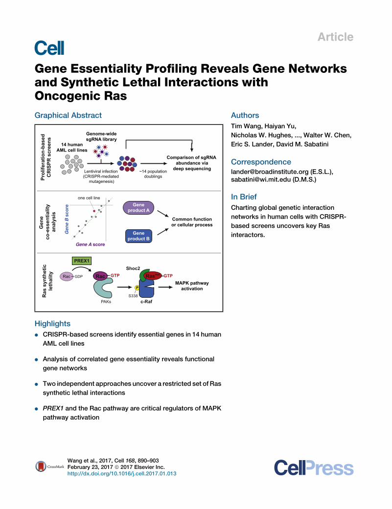
Gene Essentiality Profiling Reveals Gene Networks
and Synthetic Lethal Interactions withOncogenic RasGraphical Abstract
Rac GTP
PAKs
Rac GDP
PREX1
c-Raf
MAPK pathwayactivationP
S338
Shoc2
~14 populationdoublings
Genome-widesgRNA library
Geneproduct A
Geneproduct B
Gene A score
Gen
e B
sco
re
one cell line
Common functionor cellular process
Lentiviral infection(CRISPR-mediated
mutagenesis)
14 humanAML cell lines
Comparison of sgRNA abundance via
deep sequencing
Ras
syn
thet
icle
thal
ityPr
olife
ratio
n-ba
sed
CR
ISPR
scr
eens
Gen
e co
-ess
entia
lity
anal
ysis
Rasmut GTP
Highlights
d CRISPR-based screens identify essential genes in 14 human
AML cell lines
d Analysis of correlated gene essentiality reveals functional
gene networks
d Two independent approaches uncover a restricted set of Ras
synthetic lethal interactions
d PREX1 and the Rac pathway are critical regulators of MAPK
pathway activation
Wang et al., 2017, Cell 168, 890–903February 23, 2017 ª 2017 Elsevier Inc.http://dx.doi.org/10.1016/j.cell.2017.01.013
Authors
Tim Wang, Haiyan Yu,
Nicholas W. Hughes, ..., Walter W. Chen,
Eric S. Lander, David M. Sabatini
[email protected] (E.S.L.),[email protected] (D.M.S.)
In Brief
Charting global genetic interaction
networks in human cells with CRISPR-
based screens uncovers key Ras
interactors.
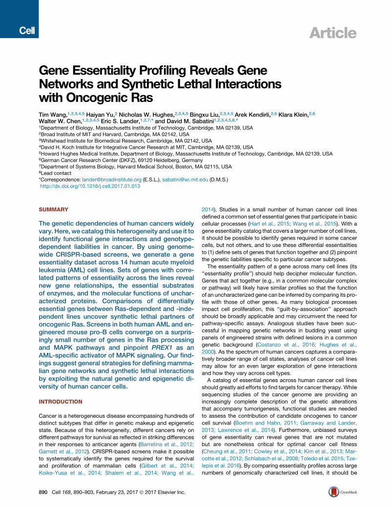
Article
Gene Essentiality Profiling Reveals GeneNetworks and Synthetic Lethal Interactionswith Oncogenic RasTim Wang,1,2,3,4,5 Haiyan Yu,2 Nicholas W. Hughes,2,3,4,5 Bingxu Liu,2,3,4,5 Arek Kendirli,2,6 Klara Klein,2,6
Walter W. Chen,1,2,3,4,5 Eric S. Lander,1,2,7,* and David M. Sabatini1,2,3,4,5,8,*1Department of Biology, Massachusetts Institute of Technology, Cambridge, MA 02139, USA2Broad Institute of MIT and Harvard, Cambridge, MA 02142, USA3Whitehead Institute for Biomedical Research, Cambridge, MA 02142, USA4David H. Koch Institute for Integrative Cancer Research at MIT, Cambridge, MA 02139, USA5Howard Hughes Medical Institute, Department of Biology, Massachusetts Institute of Technology, Cambridge, MA 02139, USA6German Cancer Research Center (DKFZ), 69120 Heidelberg, Germany7Department of Systems Biology, Harvard Medical School, Boston, MA 02115, USA8Lead contact
*Correspondence: [email protected] (E.S.L.), [email protected] (D.M.S.)
http://dx.doi.org/10.1016/j.cell.2017.01.013
SUMMARY
The genetic dependencies of human cancers widelyvary. Here, we catalog this heterogeneity and use it toidentify functional gene interactions and genotype-dependent liabilities in cancer. By using genome-wide CRISPR-based screens, we generate a geneessentiality dataset across 14 human acute myeloidleukemia (AML) cell lines. Sets of genes with corre-lated patterns of essentiality across the lines revealnew gene relationships, the essential substratesof enzymes, and the molecular functions of unchar-acterized proteins. Comparisons of differentiallyessential genes between Ras-dependent and -inde-pendent lines uncover synthetic lethal partners ofoncogenic Ras. Screens in both human AML and en-gineered mouse pro-B cells converge on a surpris-ingly small number of genes in the Ras processingand MAPK pathways and pinpoint PREX1 as anAML-specific activator of MAPK signaling. Our find-ings suggest general strategies for defining mamma-lian gene networks and synthetic lethal interactionsby exploiting the natural genetic and epigenetic di-versity of human cancer cells.
INTRODUCTION
Cancer is a heterogeneous disease encompassing hundreds of
distinct subtypes that differ in genetic makeup and epigenetic
state. Because of this heterogeneity, different cancers rely on
different pathways for survival as reflected in striking differences
in their responses to anticancer agents (Barretina et al., 2012;
Garnett et al., 2012). CRISPR-based screens make it possible
to systematically identify the genes required for the survival
and proliferation of mammalian cells (Gilbert et al., 2014;
Koike-Yusa et al., 2014; Shalem et al., 2014; Wang et al.,
890 Cell 168, 890–903, February 23, 2017 ª 2017 Elsevier Inc.
2014). Studies in a small number of human cancer cell lines
defined a common set of essential genes that participate in basic
cellular processes (Hart et al., 2015; Wang et al., 2015). With a
gene essentiality catalog that covers a larger number of cell lines,
it should be possible to identify genes required in some cancer
cells, but not others, and to use these differential essentialities
to (1) define sets of genes that function together and (2) pinpoint
the genetic liabilities specific to particular cancer subtypes.
The essentiality pattern of a gene across many cell lines (its
‘‘essentiality profile’’) should help decipher molecular function.
Genes that act together (e.g., in a common molecular complex
or pathway) will likely have similar profiles so that the function
of an uncharacterized gene can be inferred by comparing its pro-
file with those of other genes. As many biological processes
impact cell proliferation, this ‘‘guilt-by-association’’ approach
should be broadly applicable and may circumvent the need for
pathway-specific assays. Analogous studies have been suc-
cessful in mapping genetic networks in budding yeast using
panels of engineered strains with defined lesions in a common
genetic background (Costanzo et al., 2016; Hughes et al.,
2000). As the spectrum of human cancers captures a compara-
tively broader range of cell states, analyses of cancer cell lines
may allow for an even larger exploration of gene interactions
and how they vary across cell types.
A catalog of essential genes across human cancer cell lines
should greatly aid efforts to find targets for cancer therapy. While
sequencing studies of the cancer genome are providing an
increasingly complete description of the genetic alterations
that accompany tumorigenesis, functional studies are needed
to assess the contribution of candidate oncogenes to cancer
cell survival (Boehm and Hahn, 2011; Garraway and Lander,
2013; Lawrence et al., 2014). Furthermore, unbiased surveys
of gene essentiality can reveal genes that are not mutated
but are nonetheless critical for optimal cancer cell fitness
(Cheung et al., 2011; Cowley et al., 2014; Kim et al., 2013; Mar-
cotte et al., 2012; Schlabach et al., 2008; Toledo et al. 2015; Tze-
lepis et al. 2016). By comparing essentiality profiles across large
numbers of genomically characterized cell lines, it should be
NB
4 re
p. 2
OC
I-A
ML2
OC
I-A
ML5
HE
L
TH
P-1
P31
/FU
J
Mol
m-1
3
SK
M-1
MV
4;11
EO
L -1
Mon
oMac
1
TF
-1
OC
I-A
ML3
PL-
21
0.7
0.8
0.9
Cor
rela
tion
with
NB
4 re
plic
ate
1 (r
)
Celllines:
A B
C
D14 human
AML cell lines
Lentiviralinfection
~14 populationdoublings
Genome-widesgRNA library
Comparison of sgRNA barcode abundance via
deep sequencing
DNA replicationProteasome
RibosomeRNA polymerase
Spliceosome
Genes rankedby average CS
Ave
rage
CS 0
-4
-2[ ( )]CRISPR genescore (CS)
final sgRNA abundance
initial sgRNA abundance= average log2
0
40
0
2
Slid
ing
win
dow
scor
e (S
WS
)lo
g 2(C
N/2
)
Genomic position
SWS:
Hig
h
Low
HEL
9p24.1
High (<12)Low
E F
-4
0
02
8
CS
DN
A c
opy
num
ber
(CN
)
Genes ranked by position
JAK2
9p24.1
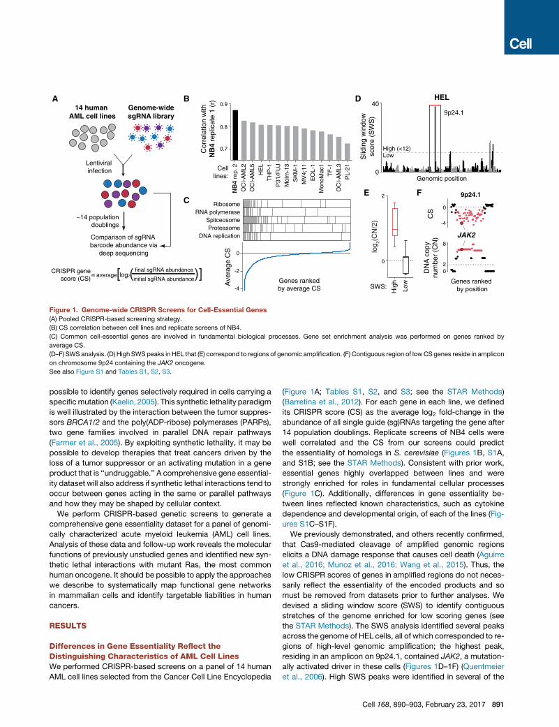
Figure 1. Genome-wide CRISPR Screens for Cell-Essential Genes
(A) Pooled CRISPR-based screening strategy.
(B) CS correlation between cell lines and replicate screens of NB4.
(C) Common cell-essential genes are involved in fundamental biological processes. Gene set enrichment analysis was performed on genes ranked by
average CS.
(D–F) SWS analysis. (D) High SWS peaks in HEL that (E) correspond to regions of genomic amplification. (F) Contiguous region of lowCS genes reside in amplicon
on chromosome 9p24 containing the JAK2 oncogene.
See also Figure S1 and Tables S1, S2, S3.
possible to identify genes selectively required in cells carrying a
specificmutation (Kaelin, 2005). This synthetic lethality paradigm
is well illustrated by the interaction between the tumor suppres-
sors BRCA1/2 and the poly(ADP-ribose) polymerases (PARPs),
two gene families involved in parallel DNA repair pathways
(Farmer et al., 2005). By exploiting synthetic lethality, it may be
possible to develop therapies that treat cancers driven by the
loss of a tumor suppressor or an activating mutation in a gene
product that is ‘‘undruggable.’’ A comprehensive gene essential-
ity dataset will also address if synthetic lethal interactions tend to
occur between genes acting in the same or parallel pathways
and how they may be shaped by cellular context.
We perform CRISPR-based genetic screens to generate a
comprehensive gene essentiality dataset for a panel of genomi-
cally characterized acute myeloid leukemia (AML) cell lines.
Analysis of these data and follow-up work reveals the molecular
functions of previously unstudied genes and identified new syn-
thetic lethal interactions with mutant Ras, the most common
human oncogene. It should be possible to apply the approaches
we describe to systematically map functional gene networks
in mammalian cells and identify targetable liabilities in human
cancers.
RESULTS
Differences in Gene Essentiality Reflect theDistinguishing Characteristics of AML Cell LinesWe performed CRISPR-based screens on a panel of 14 human
AML cell lines selected from the Cancer Cell Line Encyclopedia
(Figure 1A; Tables S1, S2, and S3; see the STAR Methods)
(Barretina et al., 2012). For each gene in each line, we defined
its CRISPR score (CS) as the average log2 fold-change in the
abundance of all single guide (sg)RNAs targeting the gene after
14 population doublings. Replicate screens of NB4 cells were
well correlated and the CS from our screens could predict
the essentiality of homologs in S. cerevisiae (Figures 1B, S1A,
and S1B; see the STAR Methods). Consistent with prior work,
essential genes highly overlapped between lines and were
strongly enriched for roles in fundamental cellular processes
(Figure 1C). Additionally, differences in gene essentiality be-
tween lines reflected known characteristics, such as cytokine
dependence and developmental origin, of each of the lines (Fig-
ures S1C–S1F).
We previously demonstrated, and others recently confirmed,
that Cas9-mediated cleavage of amplified genomic regions
elicits a DNA damage response that causes cell death (Aguirre
et al., 2016; Munoz et al., 2016; Wang et al., 2015). Thus, the
low CRISPR scores of genes in amplified regions do not neces-
sarily reflect the essentiality of the encoded products and so
must be removed from datasets prior to further analyses. We
devised a sliding window score (SWS) to identify contiguous
stretches of the genome enriched for low scoring genes (see
the STAR Methods). The SWS analysis identified several peaks
across the genome of HEL cells, all of which corresponded to re-
gions of high-level genomic amplification; the highest peak,
residing in an amplicon on 9p24.1, contained JAK2, a mutation-
ally activated driver in these cells (Figures 1D–1F) (Quentmeier
et al., 2006). High SWS peaks were identified in several of the
Cell 168, 890–903, February 23, 2017 891
other cell lines; the genes within these peaks were also present
at high copy number (Figures S1G–S1H). Thus, this simple
filtering procedure, which does not rely on DNA copy number in-
formation, can be used to identify genes whose low CS are likely
artifactual and thus potentially confounding to downstream
analyses.
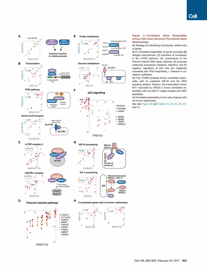
Correlated Gene Essentiality across Cell Lines RevealsFunctional Gene RelationshipsGenes acting in the same cellular pathway should show similar
patterns of essentiality across cell lines, raising the possibility
that functional gene networks can be mapped through correla-
tion-based analysis of gene essentiality profiles (Figure 2A). To
obtain biologically meaningful gene associations, comparisons
must be made between genes showing significant differences
in essentiality between lines. Therefore, we chose the most var-
iably essential genes as a query set and searched for co-essen-
tial partners for each of these genes. These associations reveal
known and novel gene relationships that encompass several
types of functional interactions.
Many sets of highly correlated genes encoded physically
interacting proteins, including heterodimers involved in tran-
scription (LDB1 and LMO2), PI3-Kg signaling (PIK3CG
and PIK3R5), amino acid transport (SLC7A5 and SLC3A2),
and components of two complexes in the mTOR pathway,
mTORC2 (MAPKAP1, MLST8, and RICTOR) and GATOR1
(NPRL2, NPRL3, and DEPDC5) (Figures 2B, 2C, and S2A).
This analysis also identified larger protein complexes; nearly
all non-redundant components of the Fanconi anemia DNA
repair machinery and the GM-CSF receptor pathway clustered
tightly together (Figures 2D and S2B). Other sets of genes en-
coded enzymes catalyzing successive reactions in metabolic
pathways (Figure 2E).
Interestingly, we identified a single case of anti-correlation be-
tween the p53 tumor suppressor gene (TP53) and its negative
regulators (Figure 2F). The sgRNAs targeting TP53 provided a
selective advantage (indicated by positive CS) to cells with
wild-type, but not mutant, p53. In these same lines, four negative
regulators of p53, TERF1, a telomere-binding factor; PPM1D, a
p53-induced phosphatase; MDM2, an E3 ubiquitin ligase for
p53; and MDM4, an inhibitor of p53 transactivation, were selec-
tively required, as their loss presumably induced p53-mediated
cell-cycle arrest or apoptosis (Figure S2C).
The correlation analysis also revealed several unexpected
gene relationships. For example, the Furin protease cleaves
and activates a diverse array of cytokines and growth factor re-
ceptors, but in our dataset the essentiality of FURIN correlated
very highly with that of only one its substrates, the insulin-like
growth factor receptor (IGF1R), and its adaptor IRS2 (Bassi
et al., 2005) (Figure 2G). This suggests that IGF1R processing
may be the only essential function of Furin in cells grown in
culture.
We could also examine the opposite problem: identifying en-
zymes responsible for the maturation of a precursor protein.
Activation of the transcription factor Nrf-1 (NFE2L1) involves
retrotranslocation of Nrf-1 into the cytosol via the ER-associ-
ated degradation (ERAD) pathway, deglycosylation by PNGase
(NGLY1) in the ER, and partial proteolytic digestion by an uniden-
892 Cell 168, 890–903, February 23, 2017
tified protease (Radhakrishnan et al., 2014). Our dataset showed
correlated essentiality between NFE2L1, NGLY1, and the endo-
peptidase DDI2 (Figure 2G). These patterns of gene essentiality
suggest that DDI2 may be the unknown protease that cleaves
Nrf-1. Indeed, very recent work in C. elegans indicates that the
homolog of DDI2 (C01G5.6) does act on the worm version
Nrf-1 (Lehrbach and Ruvkun, 2016).
Our analysis also predicted associations between genes for
which no functional relationship has been previously established
(Figure 2H). Lastly, several genes of unknown function, such as
C1orf27 and C17orf89, had correlated essentialities with genes
encoding components of well-characterized pathways, suggest-
ing that they may represent new pathway members. We per-
formed extensive follow-up experiments to determine whether
this was indeed the case
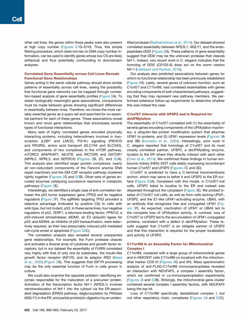
C1orf27 Interacts with UFSP2 and Is Required fordeUFMylationThe essentiality of C1orf27 correlated with (1) the essentiality of
several genes encoding components of the UFMylation machin-
ery, a ubiquitin-like protein modification system that attaches
UFM1 to proteins, and (2) UFM1 expression levels (Figures 3A
and 3B) (Komatsu et al., 2004). Interestingly, prior work in
C. elegans reported that homologs of C1orf27 and its most
closely correlated partner, UFSP2, a deUFMylating enzyme,
localize to the ER where they directly interact with each other
(Chen et al., 2014). We confirmed these findings in human em-
bryonic kidney (HEK)-293T cells stably expressing recombinant
human C1orf27 and UFSP2 (Figures 3C and 3D).
C1orf27 is predicted to have a C-terminal transmembrane
anchor, which may serve to tether it and UFSP2 to the ER sur-
face (Figure S3A). Consistent with this model, in C1orf27-null
cells, UFSP2 failed to localize to the ER and instead was
dispersed throughout the cytoplasm (Figure 3E). We probed ly-
sates of C1orf27 null cells, as well as from cells lacking UFM1,
UFSP2, and the E1-like UFM1-activating enzyme, UBA5, with
an antibody that recognizes free and conjugated UFM1 (Fig-
ure 3F). As expected, inactivation of UFM1 or UBA5 led to
the complete loss of UFMylation activity. In contrast, loss of
C1orf27 or UFSP2 led to the accumulation of UFM1-conjugated
proteins, consistent with a defect in deUFMylation. These re-
sults suggest that C1orf27 is an obligate partner of UFSP2
and that this interaction is required for the proper localization
and activity of UFSP2.
C17orf89 Is an Assembly Factor for MitochondrialComplex IC17orf89, clustered with a large group of mitochondrial genes
and in HEK293T cells C17orf89 co-localized with the mitochon-
drial marker COX IV (Figures 3G and 3H). Mass spectrometric
analysis of anti-FLAG-C17orf89 immunoprecipitates revealed
an interaction with NDUFAF5, a complex I assembly factor,
which we confirmed in co-immunoprecipitation experiments
(Figures 3I and S3B). Strikingly, the mitochondrial gene cluster
contained several complex I assembly factors, with NDUFAF5
being the top hit.
Loss of C17orf89 specifically destabilized complex I, but
not other respiratory chain, complexes (Figures 3J and S3C).
A
B
H
-2 -1 1
-2
-1
1
PS
IP1
CS
PHF15 CS
r = 0.95
ULK
3 C
S
SPAST CS
-4 -2
-3
-2
-1r = 0.97
Target genetranscription
Lineage-specificTFs
LDB1LMO2
-4 -2
-4
-2
LMO
2 C
S
LDB1 CS
r = 0.92
Transcription
tetrahyrdofolate (THF)
SHMT2
MTHFD2
5,10-methylene-THF
10-formyl-THFNADPH
NADP+
Glycine
Serine-2 -1
-3
-2
-1
SH
MT
2 C
S
MTHFD2 CS
r = 0.98
Folate metabolism
Large, neutralamino acids
SLC7A5
SLC3A2
-2 -1
-2
-1
SLC
7A5
CS
SLC3A2 CS
r = 0.91
Amino acid transport
PIK3R5PIK3CG
PIP3PIP2
Regulatorysubunit
Catalyticsubunit
-2 -1
-3
-2
-1
PIK
3R5
CS
PIK3CG CS
r = 0.92
PI3K pathway
C
Geneproduct A
Geneproduct B
G
E
Gene A CS
Gen
e B
CS
one cell line
Common functionor cellular process
SLC2A1
HK2
ATP ADP
Glucose
G6P F6PGPI
Glu
Glucose metabolism
-2 -1 1 2
-2
2
HK2 CS
GPISLC2A1
DDI2
ER
RetrotranslocatedNFE2L1 (Nrf-1)
NGLY1
ActiveNFE2L1
Nrf-1 processing
-4 -2
-2
-1
DDI2 CS
NGLY1NFE2L1
IRS2
MatureIGF1R
FURIN
IGF1Rprecusor
IGF1R processing
IRS2IGF1R
-1
-4
-2
FURIN CS
Co-essential genes with no known relationship
F
D
-3 -2 -1
-4
-2
FANCF CS
FANCCC17orf70FANCABRIP1FANCGFANCIFANCLUBE2TFANCE
Fanconi anemia pathway
RICTOR
MAPKAP1MLST8
mTOR complex 21
-2 -1 1
-2
-1
MAPKAP1 CS
MLST8RICTOR
MTOR
GAP activitytowards RagA/B
DEPDC5
NPRL2 NPRL3
RagC/D
RagA/B
-1 1
-3
-2
-1
1
GATOR1 complex
NPRL2 CS
NPRL3DEPDC5
mutantwild-type
-1 1 2 3
-4
-2
TP53 CS
MDM4MDM2TERF1PPM1D
p53 signaling
p53 status:
Figure 2. Correlated Gene Essentiality
across Cell Lines Uncovers Functional Gene
Relationships
(A) Strategy for identifying functionally related sets
of genes.
(B–F) Correlated essentiality of genes encoding (B)
obligate heterodimers, (C) members of complexes
in the mTOR pathway, (D) components of the
Fanconi anemia DNA repair pathway, (E) enzymes
catalyzing successive metabolic reactions, and (F)
negative regulators of p53 that are negatively
correlated with TP53 essentiality. r, Pearson’s cor-
relation coefficient.
(G) Top: FURIN protease shows correlated essen-
tiality with its substrate IGF1R and the IRS2
signaling adaptor. Bottom: the transcription factor
Nrf-1 (encoded by NFE2L1) shows correlated es-
sentiality with the NGLY1 deglycosylase and DDI2
peptidase.
(H) Correlated essentiality of two sets of genes with
no known relationship.
See also Figure S2 and Tables S1, S2, S3, S6, S7,
and S8.
Cell 168, 890–903, February 23, 2017 893
A
UFSP2 GRP94 merge
sgAAVS1
sgC1orf27-1
sgC1orf27-2
UFM1
Raptor
P31/F
UJ
KMS-28B
M
L-363SKM-1
MOLT-1
6
NB4KM-H
2
SUP-T1
KE-97
C1o
rf27
CS
B
D F
G
Celllines:
IP:FLAG
celllysate
HEK-293T cells stably expressing:
transfectedcDNAs:
C
E
H
J
Atp5a(Complex V)
Uqcrc2(Complex III)
Sdhb(Complex II)
MT-CO2(Complex IV)
Ndufb8(Complex I)
Raptor
sgC17orf89-1
FLAG-C17orf89sgC17orf89-2
sgAAVS1
Nomo-1 cells stably expressing:
+
--
+-
-+
--
-+
+-
+-
-+
--
-
0
1.5
Oxy
gen
cons
umpt
ion
rate
(nm
ol/m
in)
Nomo-1 cells stably expressing:
sgAAVS1sgC17orf89-1FLAG-C17orf89
-+-
-++
+--
K
FLAG-
C17orf89 COX IV merge
FLAG-
UFSP2HA-
C1orf27 GRP94 merge
HEK-293T cellsexpressing:
UFM1
GAPDH
sgAAVS1
sgUBA5
sgUFM1
sgUFSP2
sgC1o
rf27-
1
sgC1o
rf27-
2
sgAAVS1
Free UFM1
UFMylated UBA5
UFMylated UFC1
HA-C1orf27
FLAG-Rap2A-GFPFLAG-UFSP2
FLAG-UFSP2FLAG-Rap2A-GFP
HA-metap2HA-C1orf27
FLAG-Rap2A-GFPFLAG-UFSP2
HA-metap2(s.e.)
HA-C1orf27 (l.e.)
+--+
++ +
+ ---
-53
51
53
51
kD
C17orf89 CS
-2
-2
PDE12NDUFAF5NDUFS5COQ7NDUFAF7NDUFC2TIMMDC1NDUFA6ACAD9NDUFA2
NUBPLNDUFS2CPOXNDUFA9MTO1NDUFAF4YBEYNDUFAF1NDUFV1NDUFAF3
IP:FLAG
celllysate
FLAG-C17orf89
FLAG-Rap2A-GFP
HA-NDUFAF5
HA-metap2
HA-NDUFAF5
FLAG-C17orf89
FLAG-Rap2A-GFP
*
53
39
53
39
kD
IHEK-293T cells
stably expressing: FLAG-C17orf89FLAG-Rap2A-GFP
HA-metap2HA-NDUFAF5
+--+
++ +
+ ---
-transfected
cDNAs:
C1orf27 CS
-2
-2
UFSP2UFC1UBA5UFL1UFM1
Genes rankedby correlation
Genes ranked by correlation
Figure 3. Correlated Essentiality Analysis Re-
veals Function of Two Uncharacterized Genes
(A) Correlated essentiality of C1orf27 with members
of the UFMylation pathway.
(B) UFM1 levels correlate with C1orf27 essentiality.
(C) Recombinant C1orf27 andUFSP2 interact. Rap2A
and metap2 served as control bait and prey proteins.
s.e., short exposure. l.e., long exposure.
(D) Micrograph of a HEK293T cell stably expressing
FLAG-UFSP2 and HA-C1orf27. GRP94 is an ER
marker.
(E) C1orf27 is required for the proper localization of
UFSP2 in HEK293T cells.
(F) C1orf27 loss results in accumulation of UFMylated
proteins. GAPDH served as a loading control.
(G) Correlated essentiality of C17orf89 with members
of the OXPHOS pathway.
(H) Micrograph of a HEK293T cell stably expressing
FLAG-C17orf89. COX IV is a mitochondrial marker.
(I) Recombinant C17orf89 and NDUFAF5 interact. *,
non-specific band.
(J and K) C17orf89 loss (J) destabilizes mitochondrial
complex I and (K) reduces oxygen consumption.
Raptor served as a loading control. Error bars
represent SD from four replicate wells.
Scale bar, 5 mm.
See also Figure S3 and Tables S1, S2, S3.
Consistent with a defect in OXPHOS, C17orf89-null cells con-
sumed oxygen at a profoundly reduced rate and required the
addition of pyruvate to the media to maintain optimal prolifera-
tion (Figures 3K and S3D). Importantly, expression of a sgRNA-
resistant cDNA rescued these phenotypes. These findings
indicate that C17orf89 encodes a component of the complex I
assembly machinery and are in agreement with very recent
work which characterized C17orf89 via a proteomics-based
approach (Floyd et al., 2016).
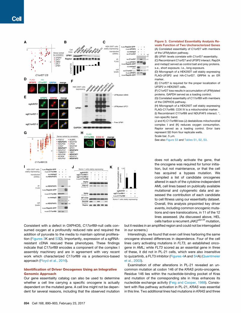
Identification of Driver Oncogenes Using an IntegrativeGenomic ApproachOur gene essentiality catalog can also be used to determine
whether a cell line carrying a specific oncogene is actually
dependent on the mutated gene. A cell line might not be depen-
dent for several reasons, including that the observed mutation
894 Cell 168, 890–903, February 23, 2017
does not actually activate the gene, that
the oncogene was required for tumor initia-
tion, but not maintenance, or that the cell
has acquired a bypass mutation. We
compiled a list of candidate oncogenes
altered in each of the cytokine-independent
AML cell lines based on publically available
mutational and cytogenetic data and as-
sessed the contribution of each candidate
to cell fitness using our essentiality dataset.
Overall, this analysis pinpointed key driver
events, including common oncogenic muta-
tions and rare translocations, in 11 of the 12
lines assessed. (As discussed above, HEL
cells harbor a recurrent JAK2V617F mutation,
but it resides in an amplified region and could not be interrogated
in our screens.)
Interestingly, we found that even cell lines harboring the same
oncogene showed differences in dependence. Four of the cell
lines carry activating mutations in FLT3, an established onco-
gene in AML; while FLT3 scored as an essential gene in three
of these, it did not in PL-21 cells, which were also insensitive
to quizartinib, a FLT3 inhibitor (Figures 4A and S4A) (Quentmeier
et al., 2003).
Examination of other alterations in PL-21 revealed an un-
common mutation at codon 146 of the KRAS proto-oncogene.
Residue 146 lies within the nucleotide-binding pocket of Kras
and mutation of the corresponding site in Hras enhances its
nucleotide exchange activity (Feig and Cooper, 1988). Consis-
tent with Ras pathway activation in PL-21, KRAS was essential
in this line. Two additional lines hadmutations in KRAS and three
A B
C D
Figure 4. Identification of Driver Oncogenes
via an Integrative Genomic Approach
(A) Genomic information and gene essentiality data
identify driver oncogenes. JAK2 is a known driver
in HEL cells, but resides in an amplicon and cannot
be assessed in our screen.
(B) PDGFRA and RAF1 participate in oncogenic
gene fusions. Only gene-fusion-targeting sgRNAs
are depleted.
(C) RNA sequencing of OCI-AML2 pinpoints a
discontinuity in coverage between exons 4 and 5 of
RAF1. PL-21 served as a control.
(D) Immunoblotting using an antibody against the
C terminus of c-Raf identifies a 90-kDa protein in
OCI-AML2. Raptor served as a loading control.
See also Figure S4 and Tables S1, S2, S3.
others in NRAS. In all cases, the mutant Ras isoform was selec-
tively essential, whereas wild-type Ras lines did not require any
of the individual Ras isoforms.
Our library includes on average ten sgRNAs tiled across the
body of each gene allowing for fine-scale analysis of gene fu-
sions. EOL-1 cells harbor a recurrent FIP1L1-PDGFRA fusion
gene; in these cells, only sgRNAs targeting the fused portion of
PDGFRA scored, resulting in an atypical, position-dependent
pattern of sgRNA depletion (Figure 4B). Translocation partners
of the KMT2A (MLL) oncogene showed similar patterns in
MV4;11 and THP-1 cells (Figure S4B).
We applied this ‘‘partial gene essentiality’’ signature to search
for translocated genes in OCI-AML2, which harbors no recurrent
oncogenic drivers. Remarkably, our analysis uncovered RAF1,
which encodes c-Raf, a major Ras effector that regulates the
MAPK signaling cascade. Consistent with a previous report,
RNA sequencing revealed a chimeric transcript spanning exon 4
of MBNL1 and exon 5 of RAF1 that results in the production of a
90-kDa gene product (Figures 4C and 4D) (Klijn et al., 2015). This
unique rearrangement removes the N-terminal autoinhibitory
domain of c-Raf and likely leads to MAPK pathway activation.
Together, these results illustrate how functional data derived
from loss-of-function screens can be integratedwith genomic in-
formation to identify and validate driver oncogenes.
Two Independent Screening Approaches RevealCommon Synthetic Lethal Interactions withOncogenic RasWealsousedour data to identify genes that are selectively essen-
tial in cell lines carrying particular drivermutations—that is, which
have synthetic lethal interactions with the mutated gene. Such
genes will typically not be mutated and thus cannot be reliably
detected through genome sequencing. They are of significant
interest because they may provide drug targets in tumors where
the cancer-causing genes cannot readily
be targeted, for example, in those driven
by the loss of tumor suppressor genes or
by oncogenes that have proven difficult
to inhibit directly.
Mutations in the Ras family of
GTPases (KRAS, NRAS, and, less
frequently, HRAS) are commonly found in many human
cancers, including AML, and are associated with poor clinical
prognoses (Cox et al., 2014). Ras controls a diverse array of
cellular processes through many downstream effectors. As
each of these effector pathways is implicated in various as-
pects of Ras-driven tumorigenesis across different cellular
contexts, it has been difficult to dissect the contribution of
each pathway to the overall survival and proliferation of cancer
cells. Furthermore, it is even less clear if Ras hyper-activation
may somehow confer dependence on other, unrelated cellular
pathways. Systematic screening approaches have greatly
accelerated efforts to find liabilities in Ras-driven cancers
(Barbie et al., 2009; Luo et al., 2009a). Here, we employed
two independent screening strategies to search for synthetic
lethal partners of oncogenic Ras (Figure 5A).
In our initial approach, we looked for genes that showed differ-
ential essentiality across the 12 cytokine-independent AML cell
lines in our panel. Comparisons between the six Ras-dependent
and six Ras-independent revealed five genes that were required
only in the context of oncogenic Ras. Two genes (RCE1 and
ICMT) are involved in the maturation of Ras. Two additional
genes (RAF1 and SHOC2) are involved in MAPK pathway
signaling. The final gene, PREX1, did not immediately fit in either
category and is discussed later in its own section.
Ras is synthesized as an inactive precursor in the cytosol and
converted into its mature membrane-associated form through
three enzymatic steps: (1) prenylation of the CAAX box by farne-
syltransferase (FTase) or geranylgeranyltransferase I (GGTase I),
(2) cleavage of the terminal AAX residues by Ras converting
enzyme (Rce1), and (3) methylation of the terminal cysteine res-
idue by isoprenylcysteine carboxyl methyltransferase (Icmt).
FNTB, which encodes a subunit of the FTase, was essential in
all cell lines screened, suggesting that FTase acts on a univer-
sally essential protein (Figure S5A). RCE1 and ICMT, however,
Cell 168, 890–903, February 23, 2017 895
Exp
erim
enta
l ap
pro
ach
Mouse isogenic cellsHuman cancer cell line panel
Wild-typeBa/F3 cells
Transduce Cas9-GFPPick clonePi
Transduce NRASG13D-RFPFACS and withdraw IL-3
TTrans
pells
CGBa/F3 cells
CGNBa/F3 cells
+IL-3–IL-3
NRASdependent state
NRASindependent state
14 AMLcell lines
6 RASmutant lines
6 RASwild-type lines
2 GM-CSF-dependent lines
(removed)
THP-1
P31/FUJ
SKM-1
OCI-AML2
HEL
Molm-13
MV4;11
EOL-1
MonoMac1OCI-AML3
PL-21
NB4
NR
AS
KR
AS
Genome-wide screen fordifferentially essential genes
Genome-wide screens fordifferentially essential genes
Dep
end
ency
val
idat
ion
KR
AS
CS
-6
-4
-2
0
2
NR
AS
CS
-6
-4
-2
0
2
KR
AS
mu
t
NR
AS
mu
t
RA
SW
T
KR
AS
mu
t
NR
AS
mu
t
RA
SW
T
Ruxolitinib (μM)0.0 0.6 1.2
0.0
0.5
1.0
Selumetinib (nM)
0 150 3000.0
0.5
1.0
CG Ba/F3 (+IL-3)CGN Ba/F3 (+IL-3)CGN Ba/F3 (–IL-3)
Rel
ativ
e vi
abili
ty (d
ay 3
)
Rel
ativ
e vi
abili
ty (d
ay 3
)
-6 -3 3 6
-6
-3
3
6
Shoc2
Rps6ka1 Raf1
Mapk1
Rce1
Icmt
Differential CS, replicate 1(–IL-3 CS - +IL-3 CS)
Diff
eren
tial C
S, r
eplic
ate
2(–
IL-3
CS
- +
IL-3
CS
)RC
E1
CS
-4
-2
0
ICM
T C
S
-2
0
RA
F1
CS
-4
-2
0
SH
OC
2 C
S
-4
-2
0
Top
gen
e h
its
Ras GTP
c-Raf(Raf1)
Shoc2
Mek
Erk(Mapk1)
Rsk(Rps6ka1)
A
CRas CAAX
Ras CF
OMe
Ras CF
Ras CAAXF
FTase
Rce1
Icmt
Ras maturationpathway
MAPK pathway
B SKM-1(KRAS mutant)
SHOC2
Gapdh
sgAAVS1
sgSHOC2-
1
sgSHOC2-
2
-Erk1/2(T202/Y204)
Erk1/2
-Mek1(S217/221)
Mek1
P
P
sgAAVS1
sgSHOC2-
1
sgSHOC2-
2
OCI-AML2(RAF1 translocated)
c-Raf*
Cells stablyexpressing:
RA
Sm
ut
RA
SW
T
RA
Sm
ut
RA
SW
T
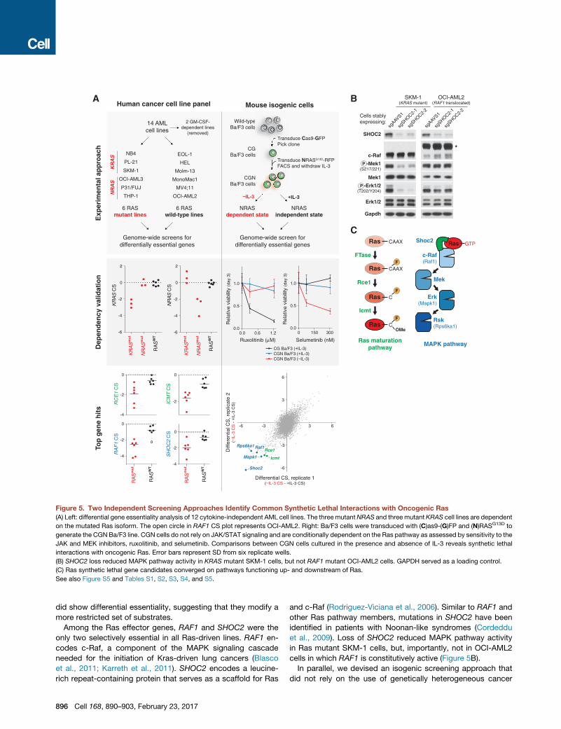
Figure 5. Two Independent Screening Approaches Identify Common Synthetic Lethal Interactions with Oncogenic Ras
(A) Left: differential gene essentiality analysis of 12 cytokine-independent AML cell lines. The three mutantNRAS and three mutant KRAS cell lines are dependent
on the mutated Ras isoform. The open circle in RAF1 CS plot represents OCI-AML2. Right: Ba/F3 cells were transduced with (C)as9-(G)FP and (N)RASG13D to
generate the CGN Ba/F3 line. CGN cells do not rely on JAK/STAT signaling and are conditionally dependent on the Ras pathway as assessed by sensitivity to the
JAK and MEK inhibitors, ruxolitinib, and selumetinib. Comparisons between CGN cells cultured in the presence and absence of IL-3 reveals synthetic lethal
interactions with oncogenic Ras. Error bars represent SD from six replicate wells.
(B) SHOC2 loss reduced MAPK pathway activity in KRAS mutant SKM-1 cells, but not RAF1 mutant OCI-AML2 cells. GAPDH served as a loading control.
(C) Ras synthetic lethal gene candidates converged on pathways functioning up- and downstream of Ras.
See also Figure S5 and Tables S1, S2, S3, S4, and S5.
did show differential essentiality, suggesting that they modify a
more restricted set of substrates.
Among the Ras effector genes, RAF1 and SHOC2 were the
only two selectively essential in all Ras-driven lines. RAF1 en-
codes c-Raf, a component of the MAPK signaling cascade
needed for the initiation of Kras-driven lung cancers (Blasco
et al., 2011; Karreth et al., 2011). SHOC2 encodes a leucine-
rich repeat-containing protein that serves as a scaffold for Ras
896 Cell 168, 890–903, February 23, 2017
and c-Raf (Rodriguez-Viciana et al., 2006). Similar to RAF1 and
other Ras pathway members, mutations in SHOC2 have been
identified in patients with Noonan-like syndromes (Cordeddu
et al., 2009). Loss of SHOC2 reduced MAPK pathway activity
in Ras mutant SKM-1 cells, but, importantly, not in OCI-AML2
cells in which RAF1 is constitutively active (Figure 5B).
In parallel, we devised an isogenic screening approach that
did not rely on the use of genetically heterogeneous cancer
cell lines. For this purpose, we screened Ba/F3 cells, a murine
pro-B cell line, which we engineered to express oncogenic
NRAS (CGN Ba/F3) (Figure 5A; Tables S4 and S5; see the
STAR Methods). CGN Ba/F3 cells cultured in the absence of
IL-3 were dependent on Ras/MAPK signaling, but, critically,
this dependence was relieved by the addition of IL-3. Therefore,
we could identify Ras-associated vulnerabilities by comparing
gene essentiality between these two conditions. Notably,
because the genetic background of the cells remains fixed in
this experiment, differences in essentiality can be directly attrib-
uted to Ras dependency (Figure S5C).
Replicate screens revealed a common set of genes selectively
required in the absence of IL-3. Remarkably, Shoc2, Raf1, Rce1,
and Icmt all scored in the top 0.1% of all genes indicating a very
high degree of overlap between the two screening approaches.
Additional MAPK pathwaymembers (Braf,Rps6ka1, andMapk1)
scored strongly aswell.BRAF andMAPK1 did show a differential
essentiality in the human AML lines but were dispensable
in some of the mutant Ras lines presumably because they ex-
pressed redundant members of these kinase families (Fig-
ure S5A). We also identified an Nras-specific dependency. After
methylation by Icmt, Nras, but not the major Kras isoform (Kras-
4B), is palmitoylated.Golga7/GOLGA7, which encodes a subunit
of the palmitoyltransferase, scored in CGN Ba/F3 and two of the
threemutantNRAS AML lines, but not in any of themutant KRAS
or wild-type Ras lines (Swarthout et al., 2005). Other genes, such
as the ubiquitin-specific peptidase Usp32/USP32, scored
strongly in CGN Ba/F3 cells and a subset of the mutant Ras
AML lines; the biological basis for its selective essentiality re-
mains to be defined.
These two independent screening approaches converged
on a restricted set of common dependencies required for
the survival and proliferation of Ras-driven cancers. Intrigu-
ingly, the majority of these genes are involved in the matura-
tion of Ras itself and the downstream MAPK signaling
pathway (Figure 5C).
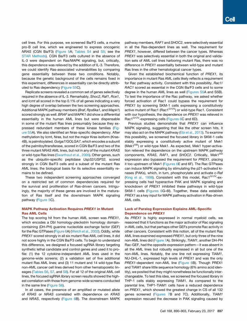
MAPK Pathway Activation Requires PREX1 in MutantRas AML CellsThe top scoring hit from the human AML screen was PREX1,
which encodes a Dbl homology-pleckstrin homology domain-
containing (DH-PH) guanine nucleotide exchange factor (GEF)
for the RacGTPases (Figure 6A) (Welch et al., 2002). Oddly, while
PREX1 scored strongly in all six mutant Ras AML cell lines, it did
not score highly in the CGN Ba/F3 cells. To begin to understand
this difference, we designed a focused sgRNA library targeting
synthetic lethal candidate and control genes and used it to pro-
file: (1) the 12 cytokine-independent AML lines used in the
genome-wide screens; (2) a validation set of five additional
mutant Ras AML lines; and (3) 11 mutant and 14 wild-type Ras
non-AML cancer cell lines derived from other hematopoietic lin-
eages (Tables S6, S7, and S8). For all 12 of the original AML cell
lines, the focused sgRNA library screen results showed the high-
est correlation with those from genome-wide screens conducted
in the same line (Figure S6).
In all cases, the presence of an amplified or mutated allele
of KRAS or NRAS correlated with dependence on KRAS
and NRAS, respectively (Figure 6B). The downstream MAPK
pathwaymembers,RAF1 and SHOC2, were selectively essential
in all the Ras-dependent lines as well. The requirement for
PREX1, however, differed between the cancer types. Whereas
PREX1 was selectively essential in both the original and valida-
tion sets of AML cell lines harboring mutant Ras, there was no
difference in PREX1 essentiality between wild-type and mutant
Ras lines in the other hematological cancer types.
Given the established biochemical function of PREX1, its
importance in mutant Ras AML cells likely reflects a requirement
for Rac pathway activity. Consistent with this possibility, Rac1/
RAC1 scored as essential in the CGN Ba/F3 cells and to some
degree in the human AML lines as well (Figures S5A and S5B).
To test the importance of the Rac pathway, we asked whether
forced activation of Rac1 could bypass the requirement for
PREX1 by screening SKM-1 cells expressing a constitutively
active mutant of Rac1 (Rac1G12V) or wild-type Rac1. Consistent
with our hypothesis, the dependence on PREX1 was relieved in
Rac1G12V-expressing cells (Figures 6C and 6D).
Previous studies demonstrate that PREX1 can influence
MAPK signaling, suggesting that like the other screen hits, it
may also act on theMAPK pathway (Ebi et al., 2013). To examine
this possibility, we screened the focused library in SKM-1 cells
stably expressing a constitutively active mutant of Mek1
(Mek1DD) or wild-type Mek1. As expected, Mek1 hyper-activa-
tion relieved the dependence on the upstream MAPK pathway
components, KRAS, RAF1, and SHOC2. Critically, Mek1DD
expression also bypassed the requirement for PREX1, placing
it too upstream of Mek1 (Figures 6E and 6F). The Rac GTPases
can induce MAPK signaling by stimulating the p21-activated ki-
nases (PAKs), which, in turn, phosphorylate and activate c-Raf
(King et al., 1998). Consistent with this model, Rac1G12V-ex-
pressing cells had hyperactive PAK and MAPK signaling and
knockdown of PREX1 inhibited these pathways in wild-type
SKM-1 cells (Figures 6G–6I). Together, these data establish
PREX1 as a key input for MAPK pathway activation in Ras-driven
AML cells.
Lack of Paralog Expression Explains AML-SpecificDependence on PREX1
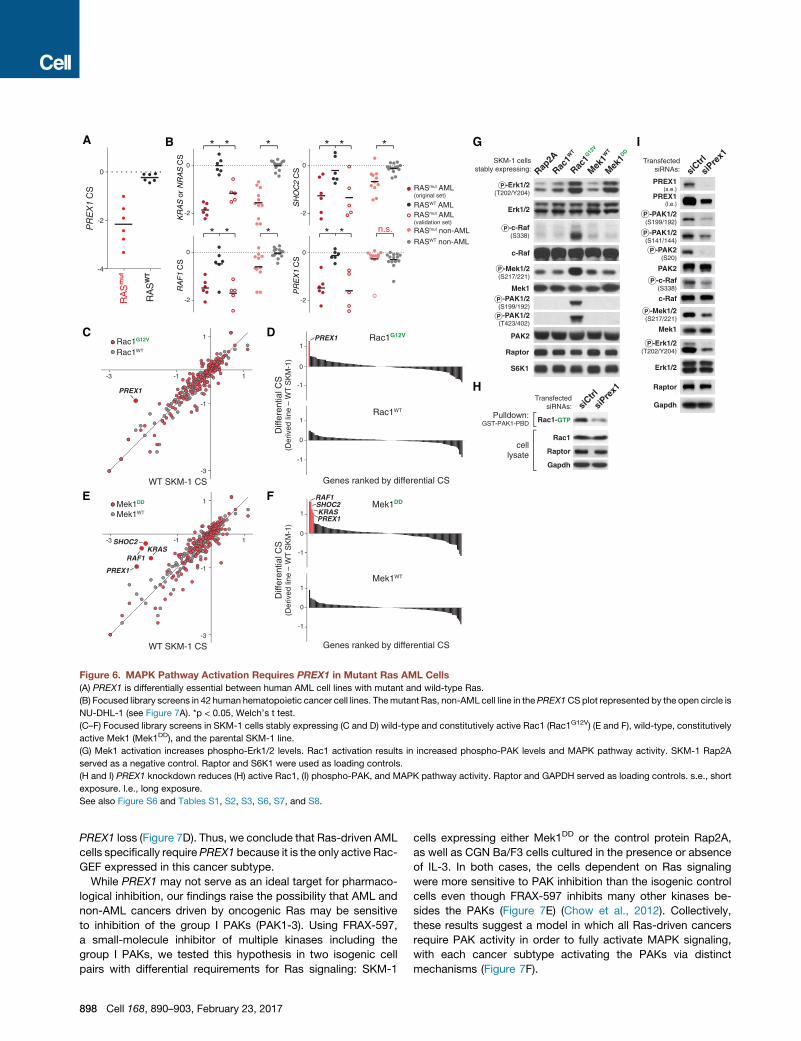
As PREX1 is highly expressed in normal myeloid cells, we
reasoned that it functions as the major activator of Rac signaling
in AML cells, but that perhaps other GEFs promote Rac activity in
other cancers. Consistent with this notion, all of the mutant Ras
AML lines examined expressed PREX1, but only three of the nine
non-AML lines did (Figure 7A). Strikingly, TIAM1, another DH-PH
Rac-GEF, had the opposite expression pattern—it was absent in
all the AML lines but robustly expressed in all but one of the
non-AML lines. Notably, the one line not expressing TIAM1,
NU-DHL-1, expressed high levels of PREX1 and was the only
PREX1-dependent non-AML line (Figure 6B). Though PREX1
and TIAM1 share little sequence homology (6% amino acid iden-
tity), we posited that theymight nonetheless be functionally inter-
changeable. To test this idea, we screened the focused library in
THP-1 cells stably expressing TIAM1. As compared to the
parental line, THP1-TIAM1 cells have a reduced dependence
on PREX1, which showed the greatest change in CS of all 132
genes screened (Figures 7B and 7C). Additionally, TIAM1
expression rescued the decrease in PAK signaling caused by
Cell 168, 890–903, February 23, 2017 897
-4
-2
0
PR
EX
1 C
S
A
D
F
C
H
G
Rap2A
Rac1W
T
Rac1G12
V
Mek1W
T
Mek1DD
SKM-1 cellsstably expressing:
Raptor
S6K1
-Erk1/2(T202/Y204)
Erk1/2
-c-Raf(S338)
c-Raf
-Mek1/2(S217/221)
Mek1
PAK2
-PAK1/2(S199/192)
-PAK1/2(T423/402)
P
P
P
P
P
-3 -1 1
-3
-1
1
-3 -1 1
-3
-1
1
Rac1WT
Rac1G12V
Mek1WT
Mek1DD
PREX1
RAF1KRAS
SHOC2
PREX1
siCtrl
siPre
x1
Raptor
Gapdh
-Erk1/2(T202/Y204)
Erk1/2
c-Raf
Mek1
PAK2
P
P
P
P
PREX1(s.e.)
-PAK1/2(S199/192)
PREX1(l.e.)
-c-Raf(S338)
P
-PAK1/2(S141/144)
-PAK2(S20)
siCtrl
siPre
x1
Pulldown:GST-PAK1-PBD
celllysate
-1
0
1
-1
0
1
-1
0
1
-1
0
1Rac1WT
Rac1G12V
Mek1WT
Mek1DDRAF1SHOC2KRASPREX1
PREX1
E
Diff
eren
tial C
S(D
eriv
ed li
ne –
WT
SK
M-1
)
Genes ranked by differential CS
Genes ranked by differential CSWT SKM-1 CS
WT SKM-1 CS
ITransfected
siRNAs:
TransfectedsiRNAs:
Raptor
Gapdh
Rac1
Rac1-GTP
RASmut AML(original set)
RASWT AMLRASmut AML(validation set)
RASWT non-AML
RASmut non-AML
B
-2
0
-2
0
-2
0
-2
0
n.s.* ** * *
* * ** * *
KR
AS
or
NR
AS
CS
RA
F1
CS
SH
OC
2 C
SP
RE
X1
CS
RA
Sm
ut
RA
SW
T
-Mek1/2(S217/221)P
Diff
eren
tial C
S(D
eriv
ed li
ne –
WT
SK
M-1
)
Figure 6. MAPK Pathway Activation Requires PREX1 in Mutant Ras AML Cells
(A) PREX1 is differentially essential between human AML cell lines with mutant and wild-type Ras.
(B) Focused library screens in 42 human hematopoietic cancer cell lines. Themutant Ras, non-AML cell line in thePREX1CSplot represented by the open circle is
NU-DHL-1 (see Figure 7A). *p < 0.05, Welch’s t test.
(C–F) Focused library screens in SKM-1 cells stably expressing (C and D) wild-type and constitutively active Rac1 (Rac1G12V) (E and F), wild-type, constitutively
active Mek1 (Mek1DD), and the parental SKM-1 line.
(G) Mek1 activation increases phospho-Erk1/2 levels. Rac1 activation results in increased phospho-PAK levels and MAPK pathway activity. SKM-1 Rap2A
served as a negative control. Raptor and S6K1 were used as loading controls.
(H and I) PREX1 knockdown reduces (H) active Rac1, (I) phospho-PAK, and MAPK pathway activity. Raptor and GAPDH served as loading controls. s.e., short
exposure. l.e., long exposure.
See also Figure S6 and Tables S1, S2, S3, S6, S7, and S8.
PREX1 loss (Figure 7D). Thus, we conclude that Ras-driven AML
cells specifically require PREX1 because it is the only active Rac-
GEF expressed in this cancer subtype.
While PREX1 may not serve as an ideal target for pharmaco-
logical inhibition, our findings raise the possibility that AML and
non-AML cancers driven by oncogenic Ras may be sensitive
to inhibition of the group I PAKs (PAK1-3). Using FRAX-597,
a small-molecule inhibitor of multiple kinases including the
group I PAKs, we tested this hypothesis in two isogenic cell
pairs with differential requirements for Ras signaling: SKM-1
898 Cell 168, 890–903, February 23, 2017
cells expressing either Mek1DD or the control protein Rap2A,
as well as CGN Ba/F3 cells cultured in the presence or absence
of IL-3. In both cases, the cells dependent on Ras signaling
were more sensitive to PAK inhibition than the isogenic control
cells even though FRAX-597 inhibits many other kinases be-
sides the PAKs (Figure 7E) (Chow et al., 2012). Collectively,
these results suggest a model in which all Ras-driven cancers
require PAK activity in order to fully activate MAPK signaling,
with each cancer subtype activating the PAKs via distinct
mechanisms (Figure 7F).
F
Rac GTP
PAK1/2/3
Rac GDP
TIAM1
PREX1
OR
Rac GEF
c-Raf
Rasmut GTP
MAPK pathwayactivation
P
S338
AML-specific:
Other cancers:
A
B
-3 -1 1
-3
-1
1
WT THP-1 CS
TH
P-1
TIA
M1
CS
0.0
0.2
0.4
0.6
PREX1
PREX1
Top 20 genes
D
PREX1
TIAM1
GAPDH
(s.e.)
(l.e.)
THP-1 cells stably expressing:
sgAAVS1 -+ -+
- -TIAM1 + +sgPREX1 -+- +
PAK2
P
C
-PAK1/2(S141/144)
Diff
eren
tial C
S(T
HP
-1 T
IAM
1 –
TH
P-1
)
AML non-AML
TIAM1
RagC
PREX1
THP-1
T-ALL1
KE-37
KMS-2
8BM
JJN-3
697
NU-DHL-1
*
RCH-ACV
L-363
NALM-6
THP-1
P31/F
UJ
KY-821
SKM-1
SHI-1
NB4Nom
o-1
RCH-ACV
OCI-AM
L3
PL-21
(s.e.)
(l.e.)
NRASmut KRASmut NRASmut KRASmut
E
Rap2aMek1DD
–IL-3+IL-3
SKM-1 cellsstably expressing:
CGN Ba/F3cells cultured in:
0.0
0.5
1.0
0.0
0.5
1.0
Rel
ativ
e ce
ll vi
abili
ty (
day
3)R
elat
ive
cell
viab
ility
(da
y 3)
0 2 4
FRAX 597 (µM)
0 1 2 3
FRAX 597 (µM)
**
*
*
Figure 7. Lack of Paralog Expression Ex-
plains PREX1-Dependence in AML
(A) Analysis of PREX1 and TIAM1 expression. RagC
was used as a loading control.
(B) Focused library screens in wild-type and TIAM1-
overexpressing THP-1 cells.
(C) CRISPR scores from THP-1 TIAM1 cells are
compared with those of the parental THP-1 cells to
calculate the differential CS.
(D) TIAM1 rescues sgPREX1-mediated inhibition of
PAK signaling in THP-1 cells. GAPDH served as a
loading control.
(E) Treatment of isogenic SKM-1 and Ba/F3 cell line
pairs with a group I PAK inhibitor FRAX-597. Error
bars represent SD from ten replicate wells. *p <
0.05, Welch’s t test.
(F) Proposed model of cell-type-specific PREX1
dependence.
SE, short exposure; LE, long exposure.
See also Tables S6, S7, and S8.
DISCUSSION
An Integrative Genomic Approach Reveals OncogeneDependencyCancer genome sequencing efforts have provided an increas-
ingly complete catalog of the genes altered during tumor devel-
opment (Lawrence et al., 2014). Functional studies enable a
direct assessment of the contribution of each of these genes
to cancer cell fitness (Boehm and Hahn, 2011; Garraway and
Lander, 2013). Together, these complementary approaches
should accelerate the identification of novel oncogenes and po-
tential therapeutic targets. Some cancers are driven by rare
events that are difficult to distinguish from random mutations
and thus require functional analysis to assess the significance
of an alteration (Berger et al., 2016; Starita et al., 2015; Tsang
et al., 2016). For instance, the tiled design of our libraries
enabled us to identify the essentiality of translocation events
including a rare inversion involving the RAF1 kinase in OCI-
AML2 cells.
However, mutational information alone cannot discriminate
between oncogenes required for the continued growth of cancer
cells from those solely involved in tumor
initiation. Even for cells harboring acti-
vating mutations in the same oncogene,
we found differences in essentiality (only
three of four FLT3 mutant lines required
FLT3). Thus, tomore accurately guide can-
cer treatment, functional testing of patient
tumor cells, should be considered in com-
bination with sequence analysis.
Functional Gene Network MappingUsing Correlated Gene EssentialityAnalysisThe natural variability in the genetic and
epigenetic makeup across human cancer
cell lines leads to differences in gene es-
sentiality and so provides a convenient means for defining func-
tional gene networks. Even between lines of a single subtype, we
found many genes with variable essentiality. Reasoning that
genes in the same biological pathway should show similar pat-
terns of essentiality, we used the CRISPR scores to cluster
genes into groups with correlated essentiality. Interestingly, the
scores of many gene pairs correlated linearly, with the different
cell lines showing graded, rather than binary levels of require-
ments for the genes. Our analysis uncovered several classes of
functional relationships including gene sets encoding protein
complexes, metabolic pathways, and enzyme-substrate pairs
and enabled us to determine the molecular functions of unchar-
acterized genes.
Analysis of other cancer types or across cancer types may
reveal additional interactions and surveying across media condi-
tions or in the presence of chemical compounds may also yield
valuable insights. Moreover, we anticipate that more sophisti-
cated analysis of our dataset using approaches that can detect
multi-way interactions will allow for continued discovery.
With the exception of the genes involved in p53 signaling, the
basis of the variable essentiality of all other gene clusters
Cell 168, 890–903, February 23, 2017 899
remains unclear. Such an understanding will be required in order
to exploit these pathways for cancer therapy. Similar to efforts to
predict cancer drug response, integrative approaches may help
uncover biomarkers for gene essentiality.
Screens in Established Human AML and EngineeredMouse Cell Lines Uncover a Common Set of RasSynthetic Lethal InteractionsWe focused on a special case of co-essentiality: synthetic
lethality with oncogenic Ras. In large part, our study suggests
that the development of therapies that selectively impact Ras-
dependent cancer cells will require re-focusing efforts on target-
ing select components of the Ras pathway itself.
Ras, like many small GTPases, undergoes a series of post-
translational modifications to facilitate interaction with the inner
leaflet of the plasma membrane. Efforts to block this process
have been primarily directed toward inhibition of the initial step
of the pathway catalyzed by FTase (Cox et al., 2014). However,
FTase inhibitors have been ineffective in the clinic as Kras and
Nras can be geranylgeranylated, an alternative prenylation
pathway (Whyte et al., 1997). Additionally, our results here and
from prior screens conducted in other cancer subtypes indicate
that FTase is required in all cells. In contrast to FTase, the en-
zymes catalyzing the latter two steps of the Ras processing
pathway, Rce1 and Icmt, do display synthetic lethality with onco-
genic Ras and may thus serve as therapeutic targets.
Our results provide further support for the central role of MAPK
signaling in Ras-driven cancers and suggest c-Raf as a thera-
peutic target. The unique requirement for c-Raf, but not other
Raf kinases, is consistent with only c-Raf being required in
lung cancer models driven by oncogenic Ras (Blasco et al.,
2011; Karreth et al., 2011).
A mechanistic insight from our study is the critical role of
the Rac/PAK signaling axis in promoting MAPK activity in mutant
Ras cancers. Even though the Rac GTPases activate many
downstream pathways, we found that forced expression of
constitutively active Mek1 can bypass the requirement for
PREX1. The selective essentiality of PREX1 in Ras-driven AML,
but not in the other cancer types tested, likely reflects the critical
role of PREX1 in normal myeloid cells. In neutrophils, where
PREX1 is highly expressed, host- and pathogen-derived chemo-
tactic factors trigger activation of the PI3-Kg and GPCR path-
ways (Welch et al., 2002). This results in the generation of PIP3
and free Gbg subunits which recruit PREX1 and stimulate
Rac-GEF activity. In AML cells, Gbg and PIP3 may be similarly
required to activate PREX1. We note that genes encoding two
Gb subunits (GNB1/2), a Gg subunit (GNG5), a Gbg-modulator
(PDCL), and the catalytic and regulatory subunits of PI3-Kg
(PIK3CG/PIK3R5) all showed partial Ras co-dependency (Fig-
ure S5A). We hypothesize that Ras-driven cancers originating
from other cell types rely on other Rac-GEFs, such as TIAM1
and VAV1, to activate PAK signaling.
Design of Synthetic Lethal Screens and sgRNA LibrariesThe combination of screening approaches employed here pro-
vides a guide for the design of robust screens for synthetic lethal
interactions. As illustrated by the case PREX1 in Ras-driven
AML, genetic interactions with oncogenes may occur in a cell
900 Cell 168, 890–903, February 23, 2017
context-dependent manner. Thus, it may be sensible to screen
lines of a particular cell type or to include enough cell lines
representing each cancer type. Additionally, screens across
isogenic cell lines should be employed to eliminate factors
that may confound analyses across genetically heterogeneous
cancer cell lines. Here, we screened Ba/F3 cells expressing
oncogenic NRAS in the presence and absence of IL-3. This
perturbation altered oncogene dependence, but not proliferation
rate (Figure S5C).
Microarray-based oligonucleotide synthesis enables the rapid
generation of focused sgRNA libraries for follow-up studies. As
such experiments require vastly fewer numbers of cells, many
additional cell lines can be tested. By using expanded cell line
panels representing more cancer types, the generality of the in-
teractions can be assessed and with engineered panels of lines,
epistatic relationships between hit genes defined. Moreover, it
may be possible to conduct screens using murine cancer
models and identify genes that play critical roles in vivo.
General Comments on Synthetic Lethality in CancerSynthetic lethal interactions in cancer cells can, in principle,
occur between several classes of genes. The prototypical
example is the inactivation of a so-called ‘caretaker’ gene
involved in the maintenance of genomic stability that leads
to dependence on a parallel maintenance pathway (Ashworth
et al., 2011; Kaelin, 2005). Such interactions may arise be-
tween genes involved in distinct but functionally overlapping
processes, as seen with the BRCA and PARP DNA repair
pathways, or between highly related and perhaps even inter-
changeable paralogs, such as ARID1A and ARID1B (Farmer
et al., 2005). However, this paradigm may not apply to Ras
and other genes involved in signal transduction. In contrast
to loss-of-function mutations in caretaker genes, oncogenic
mutations in growth factor signaling pathways result in hyper-
active signaling and, in most cases, render cells dependent
on the altered pathway (Luo et al., 2009b). Furthermore, as
these mutations act in a dominant fashion, they are typically
found in the heterozygous state, leaving the wild-type allele
intact.
Genes and pathways that protect cancer cells from the
diverse stresses associated with the malignant state represent
a second class of potential vulnerabilities. In comparison to
their normal counterparts, cancer cells rely to a much greater
extent on such cytoprotective pathways as they experience
elevated levels of mitotic, oxidative, proteotoxic, metabolic,
and DNA damage-related stress (Luo et al., 2009b). While
many of these stresses can be experimentally induced by the
expression of specific oncogenes, they are almost universally
found in established tumors regardless of genotype (Courtois-
Cox et al., 2008). Thus, it is unclear whether these liabilities
can be linked to any particular oncogene per se or if they arise
as a secondary consequence of the increased genomic insta-
bility and mitotic index characteristic of all cancer cells. Indeed,
chaperones, such as Hsp90, act as ‘‘genetic hubs’’ and show
epistasis with hundreds of client proteins, including several
oncogenic kinases (Whitesell and Lindquist, 2005). More
comprehensive studies that compare various genetically
defined malignant and pre-malignant cells are needed to

pinpoint the specific features of the oncogenic state that sensi-
tize cells to inhibition of individual stress response pathways.
Importantly, as full inhibition of many of these pathways is likely
to be lethal, gene knockdown approaches, such as CRISPRi,
may be better suited to interrogate them (Gilbert et al., 2014;
Horlbeck et al., 2016).
The only consistent differences in gene essentiality between
the mutant and wild-type Ras cells in our study were in genes
closely connected to Ras itself (Ras post-translational process-
ing and MAPK signaling). Extensive experimental evidence in
Ras-driven cell lines and in murine cancer models supports the
importance of these pathways. Our data are in general agree-
ment with findings from our correlated essentiality analysis—as
with other pathways and complexes, cells that require Ras
also require other genes that act in concert with Ras to promote
survival and proliferation. We anticipate that screens for syn-
thetic lethal partners of other driver oncogenes will uncover
similar networks of ancillary genes that may serve as attractive
targets for therapy. More broadly, through the systematic appli-
cation of CRISPR-based screens, it should be possible to
comprehensively identify the acquired vulnerabilities of human
cancers.
STAR+METHODS
Detailed methods are provided in the online version of this paper
and include the following:
d KEY RESOURCES TABLE
d CONTACT FOR REAGENT AND RESOURCE SHARING
d EXPERIMENTAL MODEL AND SUBJECT DETAILS
B Cell Lines and Genomic Annotations
B Cell Culture Conditions
d METHOD DETAILS
B Virus Production and Transduction
B Vector Construction
B Generation of Isogenic Cell Lines for CRISPR
Screening
B Genome-wide CRISPR Screening
B Genome-wide sgRNA Library Construction
B Secondary CRISPR Screening
B Antibodies
B Cell Lysis and Immunoblotting
B Immunoprecipitation Studies
B Immunofluorescence
B Seahorse Analysis
B siRNA Experiments
B RNA Sequencing
B Short-Term Proliferation Assays
B Sanger Sequencing
d QUANTIFICATION AND STATISTICAL ANALYSIS
B Genome-wide CRISPR Screening
B Secondary CRISPR Screening
B Comparative Essentiality Testing
B Copy Number Peak Analysis
B Correlated Gene Essentiality Analysis
d DATA AND SOFTWARE AVAILABILITY
B Data Resources
SUPPLEMENTAL INFORMATION
Supplemental Information includes six figures and eight tables and can be
found with this article online at http://dx.doi.org/10.1016/j.cell.2017.01.013.
AUTHOR CONTRIBUTIONS
T.W., E.S.L., and D.M.S. designed the research; T.W., H.Y., and N.W.H.
conducted the screens; T.W., H.Y., N.W.H., B.L., A.K., K.K., and W.W.C. con-
ducted other experiments; T.W. analyzed the data; and T.W., E.S.L., and
D.M.S. wrote the paper.
ACKNOWLEDGMENTS
The authors would like to thank W.C. Comb, M.L. Valenstein, and K.M. Krupc-
zak for assistance and L. Chantranupong, R.A. Saxton, and C.H. Adelmann for
manuscript review. This work was supported by the NIH (CA103866 to D.M.S.;
F31 CA189437 to T.W), the National Human Genome Research Institute
(2U54HG003067-10) (to E.S.L.), and the MIT Whitaker Health Sciences Fund
(to T.W.). D.M.S. is an investigator of the Howard Hughes Medical Institute.
E.S.L. directs The Broad Institute, which holds patents and has filed patent ap-
plications on technologies related to CRISPR-Cas 9. E.S.L. has no personal
financial interest in the work in the paper. D.M.S. and T.W. are co-founders
of and D.M.S is a consultant to KSQ Therapeutics, Inc., which is using
CRISPR-based genetic screens to identify to drug targets. T.W., D.M.S., and
E.S.L. are inventors on a patent for functional genomics using CRISPR-Cas
(US 15/141,348).
Received: October 19, 2016
Revised: December 30, 2016
Accepted: January 11, 2017
Published: February 2, 2017
REFERENCES
Aguirre, A.J., Meyers, R.M., Weir, B.A., Vazquez, F., Zhang, C.-Z., Ben-David,
U., Cook, A., Ha, G., Harrington, W.F., Doshi, M.B., et al. (2016). Genomic copy
number dictates a gene-independent cell response to CRISPR-Cas9 targeting
(Cancer Disc). http://dx.doi.org/10.1158/2159-8290.CD-16-0154.
Ashworth, A., Lord, C.J., and Reis-Filho, J.S. (2011). Genetic interactions in
cancer progression and treatment. Cell 145, 30–38.
Barbie, D.A., Tamayo, P., Boehm, J.S., Kim, S.Y., Moody, S.E., Dunn, I.F.,
Schinzel, A.C., Sandy, P., Meylan, E., Scholl, C., et al. (2009). Systematic
RNA interference reveals that oncogenic KRAS-driven cancers require
TBK1. Nature 462, 108–112.
Barretina, J., Caponigro, G., Stransky, N., Venkatesan, K., Margolin, A.A., Kim,
S., Wilson, C.J., Lehar, J., Kryukov, G.V., Sonkin, D., et al. (2012). The Cancer
Cell Line Encyclopedia enables predictive modelling of anticancer drug sensi-
tivity. Nature 483, 603–607.
Bassi, D.E., Fu, J., Lopez de Cicco, R., and Klein-Szanto, A.J.P. (2005). Pro-
protein convertases: ‘‘master switches’’ in the regulation of tumor growth
and progression. Mol. Carcinog. 44, 151–161.
Berger, A.H., Brooks, A.N., Wu, X., Shrestha, Y., Chouinard, C., Piccioni, F.,
Bagul, M., Kamburov, A., Imielinski, M., Hogstrom, L., et al. (2016). High-
throughput Phenotyping of Lung Cancer Somatic Mutations. Cancer Cell 30,
214–228.
Blasco, Rafael B., Francoz, S., Santamarıa, D., Canamero, M., Dubus, P.,
Charron, J., Baccarini, M., and Barbacid, M. (2011). c-Raf, but not B-Raf, is
essential for development of K-Ras oncogene-driven non-small cell lung car-
cinoma. Cancer Cell 19, 652–663.
Boehm, J.S., and Hahn, W.C. (2011). Towards systematic functional charac-
terization of cancer genomes. Nat. Rev. Genet. 12, 487–498.
Chantranupong, L., Scaria, S.M., Saxton, R.A., Gygi, M.P., Shen, K., Wyant,
G.A., Wang, T., Harper, J.W., Gygi, S.P., and Sabatini, D.M. (2016). The
Cell 168, 890–903, February 23, 2017 901
CASTOR proteins are arginine sensors for the mTORC1 pathway. Cell 165,
153–164.
Chen, C., Itakura, E., Weber, K.P., Hegde, R.S., and de Bono, M. (2014). An ER
complex of ODR-4 and ODR-8/Ufm1 specific protease 2 promotes GPCR
maturation by a Ufm1-independent mechanism. PLoS Genet. 10, e1004082.
Cheung, H.W., Cowley, G.S., Weir, B.A., Boehm, J.S., Rusin, S., Scott, J.A.,
East, A., Ali, L.D., Lizotte, P.H., Wong, T.C., et al. (2011). Systematic inves-
tigation of genetic vulnerabilities across cancer cell lines reveals lineage-spe-
cific dependencies in ovarian cancer. Proc. Natl. Acad. Sci. USA 108,
12372–12377.
Chow, H.Y., Jubb, A.M., Koch, J.N., Jaffer, Z.M., Stepanova, D., Campbell,
D.A., Duron, S.G., O’Farrell, M., Cai, K.Q., Klein-Szanto, A.J.P., et al. (2012).
p21-Activated kinase 1 is required for efficient tumor formation and progres-
sion in a Ras-mediated skin cancer model. Cancer Res. 72, 5966–5975.
Cong, L., Ran, F.A., Cox, D., Lin, S., Barretto, R., Habib, N., Hsu, P.D., Wu, X.,
Jiang,W., Marraffini, L.A., and Zhang, F. (2013). Multiplex genome engineering
using CRISPR/Cas systems. Science 339, 819–823.
Cordeddu, V., Di Schiavi, E., Pennacchio, L.A., Ma’ayan, A., Sarkozy, A., Fo-
dale, V., Cecchetti, S., Cardinale, A., Martin, J., Schackwitz, W., et al. (2009).
Mutation of SHOC2 promotes aberrant protein N-myristoylation and causes
Noonan-like syndrome with loose anagen hair. Nat. Genet. 41, 1022–1026.
Costanzo,M., VanderSluis, B., Koch, E.N., Baryshnikova, A., Pons, C., Tan, G.,
Wang, W., Usaj, M., Hanchard, J., Lee, S.D., et al. (2016). A global genetic
interaction network maps a wiring diagram of cellular function. Science 353,
pii: aaf1420.
Courtois-Cox, S., Jones, S.L., and Cichowski, K. (2008). Many roads lead to
oncogene-induced senescence. Oncogene 27, 2801–2809.
Cowley, G.S., Weir, B.A., Vazquez, F., Tamayo, P., Scott, J.A., Rusin, S., East-
Seletsky, A., Ali, L.D., Gerath, W.F.J., Pantel, S.E., et al. (2014). Parallel
genome-scale loss of function screens in 216 cancer cell lines for the identifi-
cation of context-specific genetic dependencies. Sci. Data 1, 140035.
Cox, A.D., Fesik, S.W., Kimmelman, A.C., Luo, J., and Der, C.J. (2014). Drug-
ging the undruggable RAS: Mission possible? Nat. Rev. Drug Discov. 13,
828–851.
Ebi, H., Costa, C., Faber, A.C., Nishtala, M., Kotani, H., Juric, D., Della Pelle, P.,
Song, Y., Yano, S., Mino-Kenudson, M., et al. (2013). PI3K regulates MEK/ERK
signaling in breast cancer via the Rac-GEF, P-Rex1. Proc. Natl. Acad. Sci. USA
110, 21124–21129.
Farmer, H., McCabe, N., Lord, C.J., Tutt, A.N.J., Johnson, D.A., Richardson,
T.B., Santarosa,M., Dillon, K.J., Hickson, I., Knights, C., et al. (2005). Targeting
the DNA repair defect in BRCA mutant cells as a therapeutic strategy. Nature
434, 917–921.
Feig, L.A., and Cooper, G.M. (1988). Relationship among guanine nucleotide
exchange, GTP hydrolysis, and transforming potential of mutated ras proteins.
Mol. Cell. Biol. 8, 2472–2478.
Floyd, B.J., Wilkerson, E.M., Veling, M.T., Minogue, C.E., Xia, C., Beebe, E.T.,
Wrobel, R.L., Cho, H., Kremer, L.S., Alston, C.L., et al. (2016). Mitochondrial
protein interaction mapping identifies regulators of respiratory chain function.
Mol. Cell 63, 621–632.
Forbes, S.A., Beare, D., Gunasekaran, P., Leung, K., Bindal, N., Boutselakis,
H., Ding, M., Bamford, S., Cole, C., Ward, S., et al. (2015). COSMIC: exploring
the world’s knowledge of somatic mutations in human cancer. Nucleic Acids
Res. 43, D805–D811.
Garnett, M.J., Edelman, E.J., Heidorn, S.J., Greenman, C.D., Dastur, A., Lau,
K.W., Greninger, P., Thompson, I.R., Luo, X., Soares, J., et al. (2012). System-
atic identification of genomicmarkers of drug sensitivity in cancer cells. Nature
483, 570–575.
Garraway, L.A., and Lander, E.S. (2013). Lessons from the cancer genome.
Cell 153, 17–37.
Giaever, G., Chu, A.M., Ni, L., Connelly, C., Riles, L., Veronneau, S., Dow, S.,
Lucau-Danila, A., Anderson, K., Andre, B., et al. (2002). Functional profiling of
the Saccharomyces cerevisiae genome. Nature 418, 387–391.
902 Cell 168, 890–903, February 23, 2017
Gibson, D.G., Young, L., Chuang, R.Y., Venter, J.C., Hutchison, C.A., 3rd, and
Smith, H.O. (2009). Enzymatic assembly of DNA molecules up to several
hundred kilobases. Nat. Methods 6, 343–345.
Gilbert, L.A., Horlbeck, M.A., Adamson, B., Villalta, J.E., Chen, Y., Whitehead,
E.H., Guimaraes, C., Panning, B., Ploegh, H.L., Bassik, M.C., et al. (2014).
Genome-scale CRISPR-mediated control of gene repression and activation.
Cell 159, 647–661.
Hart, T., Chandrashekhar, M., Aregger, M., Steinhart, Z., Brown, K.R., Ma-
cLeod, G., Mis, M., Zimmermann, M., Fradet-Turcotte, A., Sun, S., et al.
(2015). High-resolution CRISPR screens reveal fitness genes and genotype-
specific cancer liabilities. Cell 163, 1515–1526.
Horlbeck, M.A., Gilbert, L.A., Villalta, J.E., Adamson, B., Pak, R.A., Chen, Y.,
Fields, A.P., Park, C.Y., Corn, J.E., Kampmann, M., and Weissman, J.S.
(2016). Compact and highly active next-generation libraries for CRISPR-medi-
ated gene repression and activation. eLife 5, e19760.
Hughes, T.R., Marton, M.J., Jones, A.R., Roberts, C.J., Stoughton, R., Armour,
C.D., Bennett, H.A., Coffey, E., Dai, H., He, Y.D., et al. (2000). Functional dis-
covery via a compendium of expression profiles. Cell 102, 109–126.
Kaelin, W.G., Jr. (2005). The concept of synthetic lethality in the context of anti-
cancer therapy. Nat. Rev. Cancer 5, 689–698.
Karreth, F.A., Frese, K.K., DeNicola, G.M., Baccarini, M., and Tuveson, D.A.
(2011). C-Raf is required for the initiation of lung cancer by K-Ras(G12D). Cancer
Discov. 1, 128–136.
Kim, H.S., Mendiratta, S., Kim, J., Pecot, C.V., Larsen, J.E., Zubovych, I., Seo,
B.Y., Kim, J., Eskiocak, B., Chung, H., et al. (2013). Systematic identification of
molecular subtype-selective vulnerabilities in non-small-cell lung cancer. Cell
155, 552–566.
King, A.J., Sun, H., Diaz, B., Barnard, D., Miao, W., Bagrodia, S., and Marshall,
M.S. (1998). The protein kinase Pak3 positively regulates Raf-1 activity through
phosphorylation of serine 338. Nature 396, 180–183.
Klijn, C., Durinck, S., Stawiski, E.W., Haverty, P.M., Jiang, Z., Liu, H., Degen-
hardt, J., Mayba, O., Gnad, F., Liu, J., et al. (2015). A comprehensive transcrip-
tional portrait of human cancer cell lines. Nat. Biotechnol. 33, 306–312.
Koike-Yusa, H., Li, Y., Tan, E.P., Velasco-Herrera, Mdel.C., and Yusa, K.
(2014). Genome-wide recessive genetic screening in mammalian cells with a
lentiviral CRISPR-guide RNA library. Nat. Biotechnol. 32, 267–273.
Komatsu, M., Chiba, T., Tatsumi, K., Iemura, S., Tanida, I., Okazaki, N., Ueno,
T., Kominami, E., Natsume, T., and Tanaka, K. (2004). A novel protein-conju-
gating system for Ufm1, a ubiquitin-fold modifier. EMBO J. 23, 1977–1986.
Lawrence, M.S., Stojanov, P., Mermel, C.H., Robinson, J.T., Garraway, L.A.,
Golub, T.R., Meyerson, M., Gabriel, S.B., Lander, E.S., and Getz, G. (2014).
Discovery and saturation analysis of cancer genes across 21 tumour types.
Nature 505, 495–501.
Lehrbach, N.J., and Ruvkun, G. (2016). Proteasome dysfunction triggers acti-
vation of SKN-1A/Nrf1 by the aspartic protease DDI-1. eLife 5, e17721.
Luo, J., Emanuele, M.J., Li, D., Creighton, C.J., Schlabach, M.R., Westbrook,
T.F., Wong, K.-K., and Elledge, S.J. (2009a). A genome-wide RNAi screen
identifies multiple synthetic lethal interactions with the Ras oncogene. Cell
137, 835–848.
Luo, J., Solimini, N.L., and Elledge, S.J. (2009b). Principles of cancer therapy:
oncogene and non-oncogene addiction. Cell 136, 823–837.
Marcotte, R., Brown, K.R., Suarez, F., Sayad, A., Karamboulas, K., Krzyza-
nowski, P.M., Sircoulomb, F., Medrano, M., Fedyshyn, Y., Koh, J.L.Y., et al.
(2012). Essential gene profiles in breast, pancreatic, and ovarian cancer cells.
Cancer Discov. 2, 172–189.
Munoz, D.M., Cassiani, P.J., Li, L., Billy, E., Korn, J.M., Jones, M.D., Golji, J.,
Ruddy, D.A., Yu, K., McAllister, G., et al. (2016). CRISPR screens provide a
comprehensive assessment of cancer vulnerabilities but generate false-posi-
tive hits for highly amplified genomic regions. Cancer Discov. 6, 900–913.
Quentmeier, H., Reinhardt, J., Zaborski, M., and Drexler, H.G. (2003). FLT3
mutations in acute myeloid leukemia cell lines. Leukemia 17, 120–124.
Quentmeier, H., MacLeod, R.A.F., Zaborski, M., and Drexler, H.G. (2006).
JAK2 V617F tyrosine kinase mutation in cell lines derived from myeloprolifer-
ative disorders. Leukemia 20, 471–476.
Radhakrishnan, S.K., den Besten, W., and Deshaies, R.J. (2014). p97-depen-
dent retrotranslocation and proteolytic processing govern formation of active
Nrf1 upon proteasome inhibition. eLife 3, e01856.
Rodriguez-Viciana, P., Oses-Prieto, J., Burlingame, A., Fried, M., and
McCormick, F. (2006). A phosphatase holoenzyme comprised of
Shoc2/Sur8 and the catalytic subunit of PP1 functions as an M-Ras
effector to modulate Raf activity. Mol. Cell 22, 217–230.
Schlabach, M.R., Luo, J., Solimini, N.L., Hu, G., Xu, Q., Li, M.Z., Zhao, Z., Smo-
gorzewska, A., Sowa, M.E., Ang, X.L., et al. (2008). Cancer proliferation gene
discovery through functional genomics. Science 319, 620–624.
Shalem, O., Sanjana, N.E., Hartenian, E., Shi, X., Scott, D.A., Mikkelsen, T.S.,
Heckl, D., Ebert, B.L., Root, D.E., Doench, J.G., and Zhang, F. (2014).
Genome-scale CRISPR-Cas9 knockout screening in human cells. Science
343, 84–87.
Starita, L.M., Young, D.L., Islam, M., Kitzman, J.O., Gullingsrud, J., Hause,
R.J., Fowler, D.M., Parvin, J.D., Shendure, J., and Fields, S. (2015). Massively
parallel functional analysis of BRCA1 RING domain variants. Genetics 200,
413–422.
Subramanian, A., Tamayo, P., Mootha, V.K., Mukherjee, S., Ebert, B.L., Gil-
lette, M.A., Paulovich, A., Pomeroy, S.L., Golub, T.R., Lander, E.S., and Me-
sirov, J.P. (2005). Gene set enrichment analysis: a knowledge-based approach
for interpreting genome-wide expression profiles. Proc. Natl. Acad. Sci. USA
102, 15545–15550.
Swarthout, J.T., Lobo, S., Farh, L., Croke, M.R., Greentree, W.K., Deschenes,
R.J., and Linder, M.E. (2005). DHHC9 and GCP16 constitute a human protein
fatty acyltransferase with specificity for H- and N-Ras. J. Biol. Chem. 280,
31141–31148.
Toledo, C.M., Ding, Y., Hoellerbauer, P., Davis, R.J., Basom, R., Girard, E.J.,
Lee, E., Corrin, P., Hart, T., Bolouri, H., et al. (2015). Genome-wide CRISPR-
Cas9 screens reveal loss of redundancy between PKMYT1 and WEE1 in glio-
blastoma stem-like cells. Cell Rep. 13, 2425–2439.
Trapnell, C., Roberts, A., Goff, L., Pertea, G., Kim, D., Kelley, D.R., Pimentel,
H., Salzberg, S.L., Rinn, J.L., and Pachter, L. (2012). Differential gene and tran-
script expression analysis of RNA-seq experiments with TopHat and Cufflinks.
Nat Protoc. 7, 562–578.
Tsang, Y.H., Dogruluk, T., Tedeschi, P.M., Wardwell-Ozgo, J., Lu, H., Espitia,
M., Nair, N., Minelli, R., Chong, Z., Chen, F., et al. (2016). Functional annotation
of rare gene aberration drivers of pancreatic cancer. Nat. Commun. 7, 10500.
Tzelepis, K., Koike-Yusa, H., De Braekeleer, E., Li, Y., Metzakopian, E., Dovey,
O.M., Mupo, A., Grinkevich, V., Li, M., Mazan, M., et al. (2016). A CRISPR
dropout screen identifies genetic vulnerabilities and therapeutic targets in
acute myeloid leukemia. Cell Rep. 17, 1193–1205.
Wang, T., Wei, J.J., Sabatini, D.M., and Lander, E.S. (2014). Genetic screens in
human cells using the CRISPR-Cas9 system. Science 343, 80–84.
Wang, T., Birsoy, K., Hughes, N.W., Krupczak, K.M., Post, Y., Wei, J.J.,
Lander, E.S., and Sabatini, D.M. (2015). Identification and characterization of
essential genes in the human genome. Science 350, 1096–1101.
Welch, H.C.E., Coadwell, W.J., Ellson, C.D., Ferguson, G.J., Andrews, S.R.,
Erdjument-Bromage, H., Tempst, P., Hawkins, P.T., and Stephens, L.R.
(2002). P-Rex1, a PtdIns(3,4,5)P3- and Gbetagamma-regulated guanine-
nucleotide exchange factor for Rac. Cell 108, 809–821.
Whitesell, L., and Lindquist, S.L. (2005). HSP90 and the chaperoning of cancer.
Nat. Rev. Cancer 5, 761–772.
Whyte, D.B., Kirschmeier, P., Hockenberry, T.N., Nunez-Oliva, I., James, L.,
Catino, J.J., Bishop, W.R., and Pai, J.K. (1997). K- and N-Ras are geranylger-
anylated in cells treated with farnesyl protein transferase inhibitors. J. Biol.
Chem. 272, 14459–14464.
Cell 168, 890–903, February 23, 2017 903
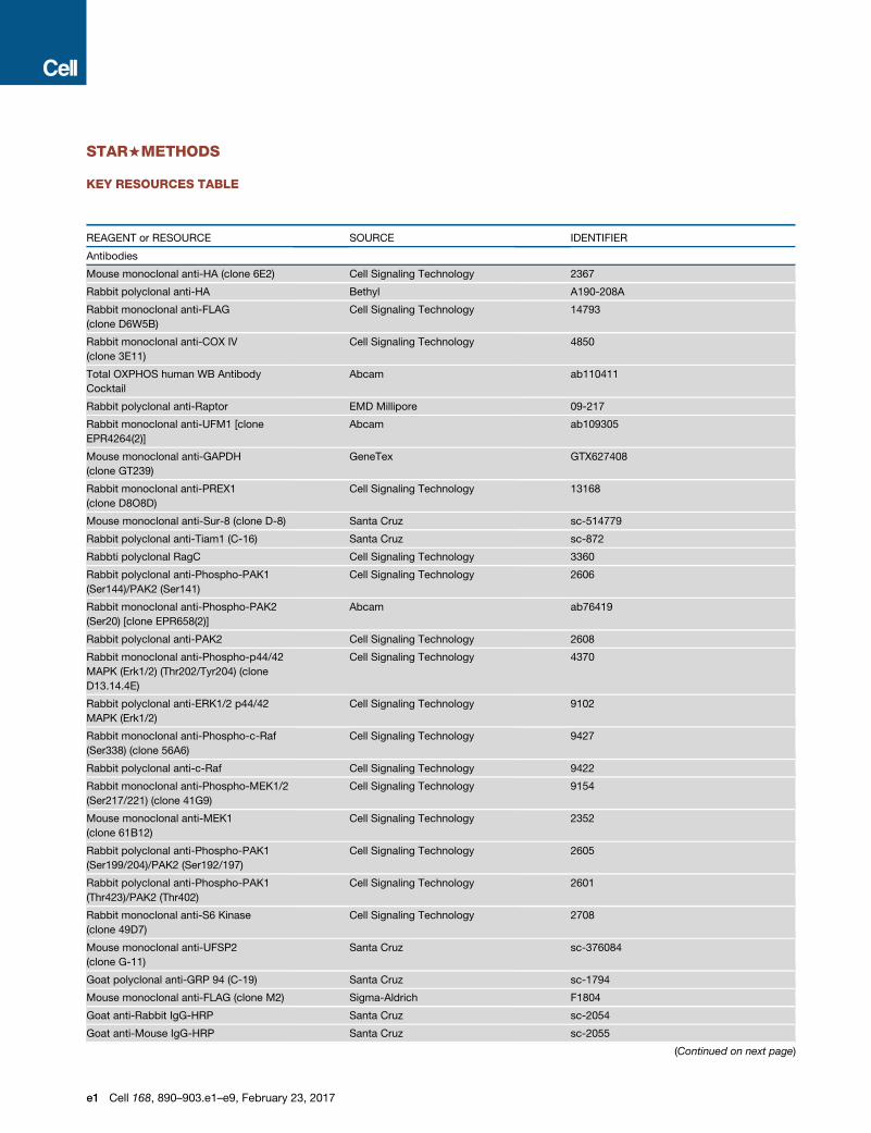
STAR+METHODS
KEY RESOURCES TABLE
REAGENT or RESOURCE SOURCE IDENTIFIER
Antibodies
Mouse monoclonal anti-HA (clone 6E2) Cell Signaling Technology 2367
Rabbit polyclonal anti-HA Bethyl A190-208A
Rabbit monoclonal anti-FLAG
(clone D6W5B)
Cell Signaling Technology 14793
Rabbit monoclonal anti-COX IV
(clone 3E11)
Cell Signaling Technology 4850
Total OXPHOS human WB Antibody
Cocktail
Abcam ab110411
Rabbit polyclonal anti-Raptor EMD Millipore 09-217
Rabbit monoclonal anti-UFM1 [clone
EPR4264(2)]
Abcam ab109305
Mouse monoclonal anti-GAPDH
(clone GT239)
GeneTex GTX627408
Rabbit monoclonal anti-PREX1
(clone D8O8D)
Cell Signaling Technology 13168
Mouse monoclonal anti-Sur-8 (clone D-8) Santa Cruz sc-514779
Rabbit polyclonal anti-Tiam1 (C-16) Santa Cruz sc-872
Rabbti polyclonal RagC Cell Signaling Technology 3360
Rabbit polyclonal anti-Phospho-PAK1
(Ser144)/PAK2 (Ser141)
Cell Signaling Technology 2606
Rabbit monoclonal anti-Phospho-PAK2
(Ser20) [clone EPR658(2)]
Abcam ab76419
Rabbit polyclonal anti-PAK2 Cell Signaling Technology 2608
Rabbit monoclonal anti-Phospho-p44/42
MAPK (Erk1/2) (Thr202/Tyr204) (clone
D13.14.4E)
Cell Signaling Technology 4370
Rabbit polyclonal anti-ERK1/2 p44/42
MAPK (Erk1/2)
Cell Signaling Technology 9102
Rabbit monoclonal anti-Phospho-c-Raf
(Ser338) (clone 56A6)
Cell Signaling Technology 9427
Rabbit polyclonal anti-c-Raf Cell Signaling Technology 9422
Rabbit monoclonal anti-Phospho-MEK1/2
(Ser217/221) (clone 41G9)
Cell Signaling Technology 9154
Mouse monoclonal anti-MEK1
(clone 61B12)
Cell Signaling Technology 2352
Rabbit polyclonal anti-Phospho-PAK1
(Ser199/204)/PAK2 (Ser192/197)
Cell Signaling Technology 2605
Rabbit polyclonal anti-Phospho-PAK1
(Thr423)/PAK2 (Thr402)
Cell Signaling Technology 2601
Rabbit monoclonal anti-S6 Kinase
(clone 49D7)
Cell Signaling Technology 2708
Mouse monoclonal anti-UFSP2
(clone G-11)
Santa Cruz sc-376084
Goat polyclonal anti-GRP 94 (C-19) Santa Cruz sc-1794
Mouse monoclonal anti-FLAG (clone M2) Sigma-Aldrich F1804
Goat anti-Rabbit IgG-HRP Santa Cruz sc-2054
Goat anti-Mouse IgG-HRP Santa Cruz sc-2055
(Continued on next page)
e1 Cell 168, 890–903.e1–e9, February 23, 2017
Continued
REAGENT or RESOURCE SOURCE IDENTIFIER
Mouse monoclonal anti-rabbit IgG
(Conformation Specific) (clone L27A9)
(HRP Conjugate)
Cell Signaling Technology 5127
Donkey anti-Goat IgG (H+L) Secondary
Antibody, Alexa Fluor 488 conjugate
Thermo Fisher Scientific A-11055
Donkey anti-Mouse IgG (H+L) Secondary
Antibody, Alexa Fluor 488 conjugate
Thermo Fisher Scientific A-21202
Donkey anti-Mouse IgG Secondary
Antibody, Alexa Fluor 568 conjugate
Thermo Fisher Scientific A-10037
Donkey anti-Rabbit IgG (H+L) Secondary
Antibody, Alexa Fluor 568 conjugate
Thermo Fisher Scientific A-10042
Donkey anti-Mouse IgG (H+L) Secondary
Antibody, Alexa Fluor 647 conjugate
Thermo Fisher Scientific A-31571
Chemicals, Peptides, and Recombinant Proteins
Anti-FLAG M2 Affinity Gel Sigma-Aldrich A2220
X-tremeGENE 9 DNA Transfection Reagent Roche 06365787001
TaKaRa Ex Taq DNA Polymerase TaKaRa RR001A
Cell-Tak Cell and Tissue Adhesive CORNING 354240
FLAG peptide (sequence DYKDDDDK) Biopolymers Core, Koch Institute N/A
Sodium pyruvate Sigma-Aldrich 113-24-6
Human GM-CSF Miltenyi Biotec 130-093-862
Recombinant murine IL-3 PeproTech 213-13
FRAX 597 Selleckchem S7271
Ruxolitinib Selleckchem S1378
Selumetinib Selleckchem S1008
Quizartinib LC Laboratories Q-4747
Critical Commercial Assays
XF24 Extracellular Flux Analyzer Seahorse Bioscience N/A
CellTiter-Glo Luminescent Cell
Viability Assay
Promega G7570
Active Rac1 Detection Kit Cell Signaling Technology 8815
Nucleofector Device Lonza N/A
Cell Line Nucleofector Kit V Lonza 1003
QIAamp DNA Blood Maxi Kit QIAGEN 51192
QIAamp DNA Blood Midi Kit QIAGEN 51183
Deposited Data
Cell line mutational data (Barretina et al., 2012) https://www.broadinstitute.org/ccle
Cell line mutational data (Forbes et al., 2015) http://cancer.sanger.ac.uk/cell_lines
DNA copy number data (Barretina et al., 2012) https://www.broadinstitute.org/ccle
Cell line microarray expression data (Barretina et al., 2012) https://www.broadinstitute.org/ccle
Experimental Models: Cell Lines
OCI-AML5 CCLE ACC-247
TF-1 CCLE ACC-334
697 CCLE ACC-42
HPB-ALL CCLE ACC-483
Jurkat CCLE ACC-282
KE-37 CCLE ACC-46
MOLT-16 CCLE ACC-29
RCH-ACV CCLE ACC-548
(Continued on next page)
Cell 168, 890–903.e1–e9, February 23, 2017 e2
Continued
REAGENT or RESOURCE SOURCE IDENTIFIER
SEM CCLE ACC-546
SUP-T1 CCLE ACC-140
Reh CCLE ACC-22
TALL-1 CCLE ACC-521
NALM-6 CCLE ACC-128
NB-4 CCLE ACC-207
MOLM-13 CCLE ACC-554
HEL CCLE ACC-11
PL-21 CCLE ACC-536
MV4;11 CCLE ACC-102
EOL-1 CCLE ACC-386
OCI-AML2 CCLE ACC-99
OCI-AML3 CCLE ACC-582
P31/FUJ CCLE JCRB0091
MonoMac1 CCLE ACC-252
SKM-1 CCLE ACC-547
THP-1 CCLE ACC-16
SHI-1 DSMZ ACC-645
NOMO-1 J. D. Griffin ACC-542
Ba/F3 J. D. Griffin ACC-300
KY821 JCRB JCRB0105
ML-2 DSMZ ACC-15
ML-1 R. Polakiewicz ECACC 88113007
HT CCLE ACC-567
KM-H2 CCLE ACC-8
L-428 CCLE ACC-197
MC116 CCLE ACC-82
Mino CCLE ACC-687
NU-DHL-1 CCLE ACC-583
RL CCLE ACC-613
SU-DHL-4 CCLE ACC-495
U-937 CCLE ACC-5
JJN-3 CCLE ACC-541
KMS-26 CCLE JCRB1187
KE-97 CCLE RCB1435
KMS-28BM CCLE JCRB1192
L-363 CCLE ACC-49
Recombinant DNA
pLenti-Cas9-GFP This paper Addgene 86145
LentiCRISPR-sgC17orf89-1 This paper Addgene 86137
LentiCRISPR-sgC17orf89-3 This paper Addgene 86136
LentiCRISPR-sgC17orf89-2 This paper Addgene 86135
LentiCRISPR-sgUFSP2 This paper Addgene 86134
LentiCRISPR-sgUFM1 This paper Addgene 86133
LentiCRISPR-sgUBA5 This paper Addgene 86132
LentiCRISPR-sgC1orf27-2 This paper Addgene 86131
LentiCRISPR-sgC1orf27-1 This paper Addgene 86130
LentiCRISPR-sgSHOC2-2 This paper Addgene 86129
(Continued on next page)
e3 Cell 168, 890–903.e1–e9, February 23, 2017
Continued
REAGENT or RESOURCE SOURCE IDENTIFIER
LentiCRISPR-sgSHOC2-1 This paper Addgene 86128
LentiCRISPR-sgPREX1 This paper Addgene 86127
lentiCRISPR-AAVS1 sgRNA (Wang et al., 2015) Addgene 70661
pMXs3-NRAS G13D This paper Addgene 86144
pMXs3-TIAM1 This paper Addgene 86143
pMXs2-MEK1 DD This paper Addgene 86142
pMXs2-MEK1 This paper Addgene 86141
pMXs2-RAP2A-GFP This paper Addgene 86140
pMXs2-RAC1 G12V This paper Addgene 86139
pMXs2-RAC1 This paper Addgene 86138
pMXs-C17orf89-FLAG This paper Addgene 86126
pMXs-NDUFAF5-HA This paper Addgene 86125
pMXs-RAP2A-GFP This paper Addgene 86124
pMXs-UFSP2-FLAG This paper Addgene 86123
pMXs-HA-C1orf27 This paper Addgene 86122
pRK5-HA-metap2 (Chantranupong et al., 2016) N/A
Genome-wide human sgRNA library This paper and (Wang et al., 2015) N/A
Genome-wide murine sgRNA library This paper N/A
Focused human sgRNA library This paper N/A
Sequence-Based Reagents
Primers for Illumina sequencing This paper See the STAR Methods Genome-wide
CRISPR screening
Primers for sgRNA quantification This paper See the STAR Methods Genome-wide
CRISPR screening
Primers for genotyping KRAS This paper See the STAR Methods
Sanger sequencing
Primers for genotyping NRAS This paper See the STAR Methods
Sanger sequencing
Individual sgRNA target sequences This paper See the STARMethods Vector construction
Genome-wide human sgRNA library This paper and Wang et al., 2015 See Table S2
Genome-wide murine sgRNA library This paper See Table S4
Focused human sgRNA library This paper See Table S7
Software and Algorithms
TopHat version 2.0.13 (Trapnell et al., 2012) http://cole-trapnell-lab.github.io/cufflinks
R version 2.15.1 The R Project https://www.r-project.org/
GSEA (Subramanian et al., 2005) http://software.broadinstitute.org/gsea/
Prism version 6.0.1 GraphPad https://www.graphpad.com
python version 2.6.8 Python software foundation https://www.python.org/
CONTACT FOR REAGENT AND RESOURCE SHARING
Requests for further information and resources may be directed to Lead Contact David M. Sabatini ([email protected]).
EXPERIMENTAL MODEL AND SUBJECT DETAILS
Cell Lines and Genomic AnnotationsML-1 cells were a gift from R. Polakiewicz of Cell Signaling Technology. Ba/F3 and Nomo-1 cells were a gift from J. D. Griffin of the
Dana Farber Cancer Institute. ML-2 and SHI-1 cells were obtained from the DSMZ cell bank; KY-821 cells from the JCRB cell bank;
and all other lines from the Cancer Cell Line Encyclopedia (CCLE). Genomic information was obtained from the CCLE and from the
canSAR database. All cell lines obtained from the CCLE, DSMZ and JCRB were subjected to STR profiling and mycoplasma testing.
Cell 168, 890–903.e1–e9, February 23, 2017 e4
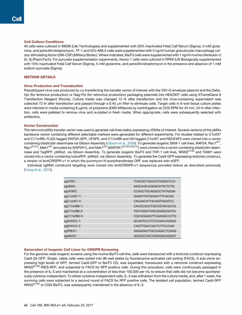
Cell Culture ConditionsAll cells were cultured in IMDM (Life Technologies) and supplemented with 20% Inactivated Fetal Calf Serum (Sigma), 5 mM gluta-
mine, and penicillin/streptomycin. TF-1 and OCI-AML5 cells were supplemented with 5 ng/ml human granulocyte-macrophage col-
ony-stimulating factor (GM-CSF) (Miltenyi Biotec). Where indicated, Ba/F3 cells were supplementedwith 1 ng/mlmurine interleukin-3
(IL-3) (PeproTech). For pyruvate supplementation experiments, Nomo-1 cells were cultured in RPMI (US Biologicals) supplemented
with 10% Inactivated Fetal Calf Serum (Sigma), 5 mM glutamine, and penicillin/streptomycin in the presence and absence of 1 mM
sodium pyruvate (Sigma).
METHOD DETAILS
Virus Production and TransductionPseudotyped virus was produced by co-transfecting the transfer vector of interest with the VSV-G envelope plasmid and the Delta-
Vpr (for lentivirus production) or Gag-Pol (for retrovirus production) packaging plasmids into HEK293T cells using XTremeGene 9
Transfection Reagent (Roche). Culture media was changed 12 hr after transfection and the virus-containing supernatant was
collected 72 hr after transfection and passed through a 0.45 mm filter to eliminate cells. Target cells in 6-well tissue culture plates
were infected in media containing 8 mg/mL of polybrene (EMD Millipore) by centrifugation at 2220 RPM for 45 min. 24 hr after infec-
tion, cells were pelleted to remove virus and re-plated in fresh media. When appropriate, cells were subsequently selected with
antibiotics.
Vector ConstructionThe retroviral pMXs transfer vector was used to generate cell lines stably expressing cDNAs of interest. Several versions of the pMXs
backbone vector containing different selectable markers were generated for different experiments. For studies related to C1orf27
and C17orf89, FLAG-tagged RAP2A-GFP, UFSP2, and C17orf89 and HA-tagged C1orf27 and NDUFAF5 were cloned into a vector
containing blasticidin deaminase via Gibson Assembly (Gibson et al., 2009). To generate isogenic SKM-1 cell lines, RAP2A, Rac1WT,
Rac1G12V, Mek1WT (encoded byMAP2K1), and Mek1DD (MAP2K1S218D;S221D) were cloned into a vector containing blasticidin deam-
inase and TagRFP, pMXs2, via Gibson Assembly. To generate isogenic Ba/F3 and THP-1 cell lines, NRASG13D and TIAM1 were
cloned into a vector containing turboRFP, pMXs3, via Gibson Assembly. To generate the Cas9-GFP expressing lentiviral construct,
a version of lentiCRISPR-v1 in which the puromycin N-acetyltransferase ORF was replaced with eGFP.
Individual sgRNA constructs targeting were cloned into lentiCRISPR-v1 (sequences provided below) as described previously
(Cong et al., 2013).
sgUFM1: TCACGCTGACGTCGGACCCA
sgUBA5: AAGCAGCAGAACATACTCTG
sgUFSP2: CCAGCTGCAGGCCTATAGGA
sgC1orf27-1: GAGATTGTGGAATTTCACAG
sgC1orf27-2: CACAACATTACAGTGGATCC
sgC17orf89-1: CACGCACCTGCCGTACGCCG
sgC17orf89-2: TGTCGGCTAACGGAGCGGTG
sgC17orf89-3: CGCGGGAGTTCGAGGCCCTG
sgSHOC2-1: GCAGTCCCTCCCAGCAGAGG
sgSHOC2-2: CAGTTGACCACTCTTCCCAG
sgPREX1: GGGAGACTGCCAGACTCGGG
sgAAVS1: GGGGCCACTAGGGACAGGAT
Generation of Isogenic Cell Lines for CRISPR ScreeningFor the genome-wide isogenic screens using the murine Ba/F3 cell line, cells were transduced with a lentiviral construct expressing
Cas9-2A-GFP. Single, viable cells were sorted into 96-well plates by fluorescence-activated cell sorting (FACS). A sub-clone ex-
pressing high levels of GFP, termed Cas9-GFP or Ba/F3 CG, was expanded, transduced with a retroviral construct expressing
NRASG13D-IRES-RFP, and subjected to FACS for RFP positive cells. During this procedure, cells were continuously passaged in
the presence of IL-3 and maintained at a concentration of less than 100,000 per mL to ensure that cells did not become spontane-
ously cytokine-independent. To obtain cytokine-independent cells, IL-3 waswithdrawn from the culturemedia, and, after 1 week, the
surviving cells were subjected to a second round of FACS for RFP positive cells. The isolated cell population, termed Cas9-GFP
NRASG13D or CGN Ba/F3, was subsequently maintained in the absence of IL-3.
e5 Cell 168, 890–903.e1–e9, February 23, 2017
For screens using the validation library, SKM-1 cells were transduced with retroviral constructs expressing Rap2A, Rac1,
Rac1G12V, Mek1 (also known as MAP2K1), and Mek1DD; selected with and continuously cultured in blasticidin; and subjected to
two rounds of FACS for RFP positive cells. THP-1 cells were transduced with a retroviral construct expressing TIAM1 and subjected
to two rounds of FACS for RFP positive cells.
Genome-wide CRISPR ScreeningGenome-wide screens for all of the human and mouse cell lines was performed as described in (Wang et al., 2015) with minor mod-
ifications and the entire screening procedure was performed twice in the human NB4 and mouse Ba/F3 cells to assess reproduc-
ibility. Briefly, for each line, 240 million target cells were transduced with the viral pool to achieve an average 1000-fold coverage
of the library after selection. After 72 hr, 200million cells were selected with puromycin. An initial pool of 80million cells was harvested
for genomic DNA extraction from all of the cell lines except for THP-1 and TF-1. The remaining cells were passaged every 3 days, and
after 14 doublings, a final pool of 100 million cells was harvested for genomic DNA extraction using the QIAamp DNA Blood Maxi Kit
(QIAGEN)
sgRNA inserts were PCR amplified using Ex Taq DNA Polymerase (Takara) from 50-75 million genome equivalents of DNA from
each initial and final sample, achieving an average coverage of �275-400x of the sgRNA library. The resultant PCR products
were purified and sequenced on a HiSeq 2500 (Illumina) (primer sequences provided below) to monitor the change in the abundance
of each sgRNA between the initial and final cell populations.
Primer sequences for sgRNA quantification
Forward:
AATGATACGGCGACCACCGAGATCTACACGAATACTGCCATTTGTCTCAAGATCTA
Reverse:
CAAGCAGAAGACGGCATACGAGATCnnnnnnTTTCTTGGGTAGTTTGCAGTTTT
(nnnnnn denotes the sample barcode)
Illumina sequencing primer
CGGTGCCACTTTTTCAAGTTGATAACGGACTAGCCTTATTTTAACTTGCTATTTCTAGCTCTAAAAC
Illumina indexing primer
TTTCAAGTTACGGTAAGCATATGATAGTCCATTTTAAAACATAATTTTAAAACTGCAAACTACCCAAGAAA
Genome-wide sgRNA Library ConstructionFor genome-wide screens in the human AML cell lines, the human sgRNA library generated in (Wang et al., 2015) was used. Notably,
the gene-targeting sgRNA sequences in our library were optimized for high cleavage activity to enable more sensitive and specific
detection of cell-essential genes (Wang et al., 2015). For more complete coverage of protein-coding genes, a sub-library containing
5,401 additional sgRNAs (comprising 499 intergenic control sgRNAs and 4,902 sgRNAs targeting 497 additional protein-coding
genes) were designed, synthesized, and cloned into lentiCRISPRv1. In total, the human sgRNA library contained 187,536 constructs
targeting 18,543 protein-coding genes and 1,504 intergenic and non-targeting control sgRNAs.
Using similar guidelines for the design of highly specific and active sgRNAs, a genome-wide murine library containing 188,509
sgRNAs (comprising 199 intergenic control sgRNAs and 188,310 sgRNAs targeting 18,986 protein-coding genes) was designed,
synthesized, and cloned into pLenti-sgRNA, a lentiviral sgRNA expression vector that does not contain Cas9.
Secondary CRISPR ScreeningA pooled library containing 6,661 sgRNAs (comprising 499 intergenic control sgRNAs and 6,162 sgRNAs targeting 132 control and
candidate Ras synthetic lethal genes) was designed and cloned into lentiCRISPR-v1. When possible, up to 50 sgRNAs were de-
signed for each gene. The validation screening procedure was similar to genome-wide screens with minor modifications. 10 million
cells were used for screening and harvested during the initial and final collections. Genomic DNA was extracted using QIAamp DNA
Blood Midi Kit (QIAGEN) and 6 million genomic equivalents were processed for PCR.
AntibodiesThe following antibodies were used for immunoblotting: HA-Tag (6E2) Mouse (Cat#2367), DYKDDDDK Tag (D6W5B) (Cat#14793),
PREX1 (D8O8D) (Cat#13168), RagC (Cat#3360), p-PAK1 (S144)/PAK2 (S141) (Cat#2606), PAK2 (Cat#2608), p-MAPK (Erk1/2)
(T202/Y204) (Cat#4370), MAPK (Erk1/2) (Cat#9102), p-c-Raf (S338) (56A6) (Cat#9427), c-Raf (Cat#9422), p-MEK1/2 (S217/221)
(41G9) (Cat#9154), MEK1 (61B12) (Cat#2352), p-PAK1 (S199/204)/PAK2 (S192/197) (Cat#2605), p-PAK1 (T423)/PAK2 (T402)
(Cat#2601), p70 S6 Kinase (49D7) (Cat#2708), Mouse Anti-rabbit IgG (Conformation Specific) (L27A9) mAb (HRP Conjugate)
Cell 168, 890–903.e1–e9, February 23, 2017 e6
(Cat#5127) from Cell Signaling Technology. Goat anti-rabbit IgG-HRP (sc-2054), Goat anti-mouse IgG-HRP (sc-2055), Sur-8 (D-8)
(sc-514779), Tiam1 (C-16) (sc-872) from Santa Cruz; HA Tag (A190-208A) from Bethyl, FLAG M2 antibody (F1804) from Sigma-
Aldrich; Total OXPHOS human WB Antibody Cocktail (ab110411), UFM1 [EPR4264(2)] (ab109305), p-PAK2 (S20) [EPR658(2)]
(ab76419) from Abcam; Raptor (09-217) from EMD Millipore,; and GAPDH (GT239) from GeneTex.
The following antibodies were used for immunofluorescence: UFSP2 (G-11) and GRP94 (C-19) from Santa Cruz; COX IV (3E11),
HA-Tag (6E2), FLAG-Tag (D6W5B) from CST; FLAG-Tag (M2) from Sigma; and Donkey anti-Goat IgG (H+L) Secondary Antibody
Alexa Fluor 488 conjugate, Donkey anti-Mouse IgG (H+L) Secondary Antibody Alexa Fluor 488 conjugate, Donkey anti-Mouse
IgG Secondary Antibody Alexa Fluor 568 conjugate, Donkey anti-Rabbit IgG (H+L) Secondary Antibody Alexa Fluor 568 conjugate,
Donkey anti-Mouse IgG (H+L) Secondary Antibody Alexa Fluor 647 conjugate from Thermo Fisher.
Cell Lysis and ImmunoblottingCells were rinsed twice with ice-cold PBS and lysed with Triton lysis buffer (1% Triton X-100, 20 mM Tris-HCl [pH 7.4], 150 mMNaCl,
1 mM EDTA, 1 PhosSTOP Phosphatase Inhibitor Cocktail tablet per 25 mL buffer [Roche], 1 cOmplete, EDTA-free Protease Inhibitor
Cocktail Tablet per 25mL buffer [Roche]). The cell lysates were cleared by centrifugation at 13,000 rpm at 4�C in amicrocentrifuge for
10 min and quantified for protein amount using BCA reagent (Thermo Scientific). Protein samples were normalized for protein con-
tent, denatured by the addition of Laemmli buffer and boiling for 5 min, resolved by SDS-PAGE, and transferred to a polyvinylidene
difluoride membrane (Millipore). Immunoblots were processed and analyzed according to standard procedures and analyzed using
chemiluminescence.
Immunoprecipitation Studies5 million HEK293T cells stably expressing FLAG-tagged cDNAs were plated in 10 cm culture dishes. For co-immunoprecipitation
(co-IP) studies, cells were transfected with 3 mg of the indicated plasmids using XTremeGene 9 Transfection Reagent (Roche)
24 hr after seeding and the cell culture media was changed the following day. 72 hr after seeding, cell lysates were prepared as
described above. The FLAG-M2 affinity gel (Sigma) was washed three times with lysis buffer. 40 ml of a 50/50 slurry of the FLAG-
M2 affinity gel was then added to clarified cell lysates and incubated with rotation for 90 min at 4�C. Following IP, the beads were
washed three times with lysis buffer. For co-IP experiments, immunoprecipitated proteins were denatured by the addition of 40 ml
of Laemmli buffer and boiling for 5 min and resolved by SDS-PAGE. For FLAG-C17orf89 mass spectrometry experiments, immuno-
precipitated proteins were eluted using the FLAG peptide, resolved rom the FLAG-M2 affinity gel, resolved on 4%–12%NuPage gels
(Invitrogen), and stained with simply blue stain (Invitrogen). Each gel lane was sliced into 10-12 pieces and the proteins in each gel
slice were digested overnight with trypsin. The resulting digests were analyzed by mass spectrometry.
Immunofluorescence100 thousand HEK293T cells were seeded on 35 mm fibronectin-coated glass-bottom dishes (MatTek). 24 hr later, cells were rinsed
with PBS, fixedwith 4%paraformaldehyde in PBS for 15min, rinsedwith PBS again, permeabilized with 0.4%Triton X-100 in PBS for
12min, rinsed with PBS again, and blocked with 10% horse serum (HS) for 20min. Dishes were then incubated with primary antibody
in 10%HS for 1 hr at RT, rinsed three times with PBS, and incubated with a fluorescent secondary antibody diluted 1:400 in 10%HS
for 2 hr at RT in the dark. Finally, cells were rinsed three times with PBS and on the secondwash were incubated with DAPI for 20min.
Dishes were imaged on an Axio Observer.Z1 inverted epifluorescence microscope (Zeiss).
Seahorse AnalysisOxygen consumption of intact cells was measured using an XF24 Extracellular Flux Analyzer (Seahorse Bioscience). XF24 Cell
Culture Microplates (Seahorse Bioscience) were coated with Cell-Tak Cell and Tissue Adhesive (Corning), and seeded with 180
thousand Nomo-1 cells (100 ml) per well. The plates were centrifuged to let cells adhere to the bottom, placed in an incubator not
supplemented with CO2 for 30 min, and subsequently analyzed on the XF24 Analyzer.
siRNA ExperimentsNucleofection of siRNAs was performed using the Cell Line Nucleofector Kit V on a Nucleofector Device (Lonza) according to the
manufacturer’s instructions. Briefly, 5 million SKM-1 cells were pelleted and resuspended in 100 mL Nucleofector solution and
2 mL of either the ON-TARGETplus PREX1 siRNA SMARTpool or the siGENOME Non-Targeting siRNA Pool #1 (100 mM) and trans-
fected using the V-001 program. Cells were then resuspended in pre-warmed IMDM supplemented with 10% IFS to allow for recov-
ery and the same transfection procedure was repeated 24 hr later. 96 hr after the initial transfection, cells were lysed and processed
for either immunoblotting as described above or with the Active Rac1 Detection Kit (Cell Signaling Technology) according to the
manufacturer’s instructions to determine the cellular levels of active, GTP-bound Rac1.
RNA SequencingTranscriptomic analysis of PL-21 and OCI-AML2 cells was performed using a strand-specific RNA sequencing protocol described
previously. Briefly, total RNA was extracted using the RNeasy Mini kit (QIAGEN). 5 mg of polyA-selected RNA was fragmented
and dephosphorylated after which an ssRNA adaptor was then ligated. Reverse transcription (RT) was performed using a primer
e7 Cell 168, 890–903.e1–e9, February 23, 2017
complementary to the RNA adaptor after which a DNA adaptor was ligated onto the 30 end of the resulting cDNA product. The library
was then PCR amplified, cleaned, quantified using a TapeStation (Agilent) and sequenced on a HiSeq 2500 (Illumina). Result reads
were then mapped to the reference human genome (hg19) using TopHat.
Short-Term Proliferation AssaysATP-basedmeasurements of cellular viability were performed by plating cells in 200 mL ofmedia in 96-well plates. The number of cells
and biological replicates seeded varied depending on the cell line and the duration of the experiment. At the indicated times, 40 mL of
CellTiter-Glo reagent (Promega) was added to each well, mixed for 5 min, after which the luminescence was measured on the Spec-
traMaxM5 Luminometer (Molecular Devices). For the drug treatment experiments, FRAX-597, Ruxolitinib, and Selumetinib were ob-
tained from Selleckchem and Quizartinib from LC Laboratories.
Sanger SequencingFor a subset of the mutant Ras cell lines used in our study, KRAS andNRASwere subjected to sequencing analysis. Briefly, genomic
DNA was extracted and amplified via PCR (primer sequences listed below) to interrogate hotspots in both genes. The PCR products
were then purified and sequenced using the Sanger method.
For residues G12, G13, and A18:
KRAS1 forward: AGGCCTGCTGAAAATGACTGAA
KRAS1 reverse: AAAGAATGGTCCTGCACCAG
For residue Q61:
KRAS2 forward: CTCAGGATTCCTACAGGAAGCA
KRAS2 reverse: CACCTATAATGGTGAATATCTTCAAAT
For residues K117 and A146:
KRAS3 forward: GGACTCTGAAGATGTACCTATGG
KRAS3 reverse: TCAGTGTTACTTACCTGTCTTGT
For residues G12 and G13:
NRAS1 forward: ACAGGTTCTTGCTGGTGTGA
NRAS1 reverse: CACTGGGCCTCACCTCTATG
For residue Q61:
NRAS2 forward: GTGGTTATAGATGGTGAAACCTGT
NRAS2 reverse: TGGCAAATACACAGAGGAAGC
QUANTIFICATION AND STATISTICAL ANALYSIS
Genome-wide CRISPR ScreeningSequencing readswere aligned to the sgRNA library and the abundance of each sgRNAwas calculated. A small number of sgRNAs in
both the human andmouse libraries have identical target sequences because they targetmultiplemembers of the same highly redun-
dant gene family. Reads mapping to these sequences are assigned to all matching sgRNAs. As the human sgRNA library is
comprised of three separate DNA plasmid sub-pools (due to limitations of microarray-based sgRNA synthesis), the counts of the
sgRNA within each sub-pool are quantile normalized against each other for each of the initial and final AML samples. The sgRNA
counts from all of the initial cell populations of the AML lines and of the two replicate initial Ba/F3 cell populations were combined
to generate the human and mouse initial reference datasets, respectively. For each initial reference dataset, sgRNAs with less
than 50 counts were removed from downstream analyses. The log2 fold-change in abundance of each sgRNAwas calculated for final
population samples for each of the cell lines after adding a count of one as a pseudocount. Gene-based CRISPR scores (CS) were
defined as the average log2 fold-change in the abundance of all sgRNAs targeting a given gene between the initial and final cell pop-
ulations and calculated for all screens. The CS reported for the NB4 cell line and the isogenic Ba/F3 experiments was the average of
two independent replicate experiments.
Secondary CRISPR ScreeningCRISPR gene scores were calculated as with the genome-wide screens with slight modifications. sgRNAs with less than 10,00
counts in the initial dataset were removed from the downstream analysis and a pseudocount of 10 was added prior to the log2fold-change calculation. Lastly, CRISPR scores were quantile normalized across of all the cell lines screened.
Cell 168, 890–903.e1–e9, February 23, 2017 e8
Comparative Essentiality TestingTo compare human gene essentiality with yeast gene essentiality 1-to-1 human-yeast homologs mappings were obtained from the
Ensembl Gene release 79 database. Human genes common to the selected genome-wide CRISPR screen datasets were used for
comparison. Each dataset was ranked by their respective scores and used to predict the essentiality of yeast homologs (Giaever
et al., 2002). The sensitivity and specificity of these predictions were analyzed using receiver operator characteristic (ROC) curves
and the area under the ROC curve was used as the performance metric.
Copy Number Peak AnalysisThe sliding window score (SWS) for a given gene in a given cell line was defined as the number of nearby geneswith a CS in the lowest
3% of all genes in that cell line. For each gene, a window of the 20 nearest ‘upstream’ and 20 nearest ‘downstream’ flanking genes
was chosen for analysis. As some genomic regions contain many bona fide essential genes (e.g., histone gene clusters), genes
essential in all lines were removed prior to the SWS calculation. For this purpose, the average CS of each gene across all cell lines
was calculated and genes in the lowest 15% were removed. For each of the remaining genes, the SWS was calculated in each cell
line. Genes with SWS > 12 were designated as high SWS genes and removed from the correlated gene essentiality analysis.
Correlated Gene Essentiality AnalysisTo maximize the likelihood of identifying biologically meaningful relationship between genes, (1) genes essential in most of the cell
lines, (2) genes only essential in a single line or which display erythroid-specific essentiality and (3) genes with low variability in CS
across the 14 cell lines were removed from the analysis. For (1), genes for which the second lowest CS was less than �1 were
removed. For (2), to assess if a gene was selectively essential in any single cell line, pairwise Pearson’s correlation coefficients
were calculated between the CS profile of each gene across the 14 cell lines and a 14x14 identity matrix. To assess if a gene was
selectively essential in the two erythroid lines, a Pearson’s correlation coefficient was calculated between the CS profile of each
gene and a vector containing 14 binary variables in which the two variables corresponding to the erythroid lines are set to ‘1’ with
the remaining set to ‘0’. If the maximum absolute value of any of these coefficients was greater than 0.8, the gene was removed.
For (3), the variance of the CS profile each gene across the 14 cell lines was obtained. The top 2,000 genes showing the highest vari-
ance were included in the correlated essentiality analysis. Select sets of genes with high correlation were highlighted and/or chosen
for follow-up validation.
DATA AND SOFTWARE AVAILABILITY
Data ResourcesData resources can be found in Tables S3, S5, and S8. Additional sgRNA-level data and custom scripts for analysis of genome-wide
screens are available at: http://sabatinilab.wi.mit.edu/wang/2017/.
e9 Cell 168, 890–903.e1–e9, February 23, 2017
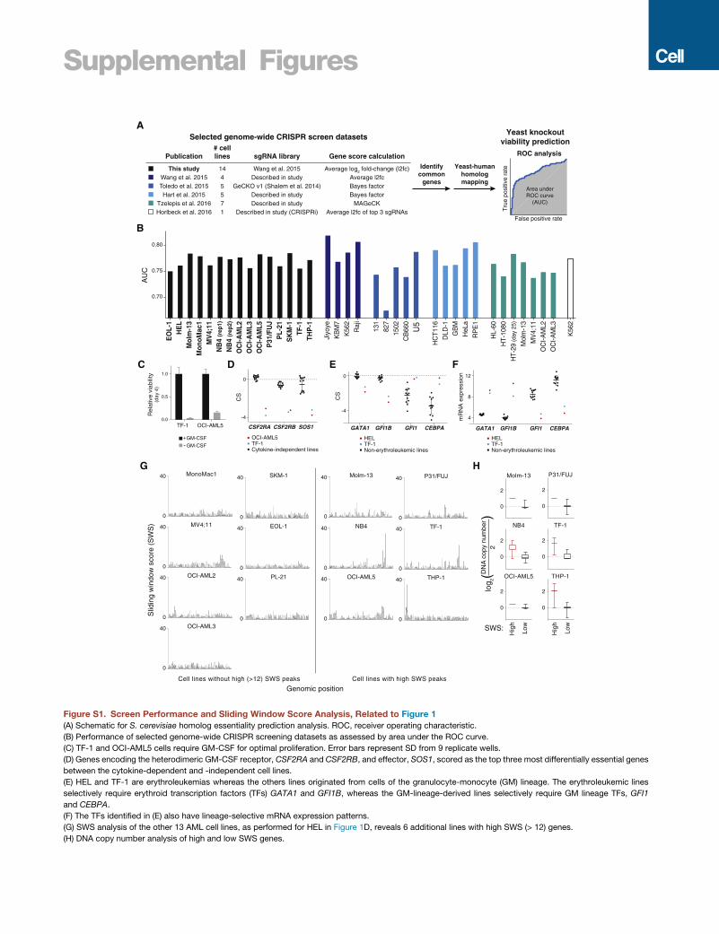
Supplemental Figures
EO
L-1
HE
LM
olm
-13
Mon
oMac
1M
V4;
11N
B4
(rep
1)
NB
4 (r
ep2)
OC
I-AM
L2O
CI-A
ML3
OC
I-AM
L5P
31/F
UJ
PL-
21S
KM
-1TF
-1TH
P-1
Jiyo
yeK
BM
7K
562
Raj
i
131
827
1502
CB
660
U5
HC
T116
DLD
-1
GB
M
HeL
a
RP
E1
HL-
60H
T-10
80H
T-29
(day
25)
Mol
m-1
3M
V4;
11O
CI-A
ML2
OC
I-AM
L3
K56
2
0.70
0.75
0.80
AU
CSelected genome-wide CRISPR screen datasets
Yeast-humanhomologmapping
This studyWang et al. 2015Toledo et al. 2015Hart et al. 2015
Tzelepis et al. 2016Horlbeck et al. 2016
1445571
Wang et al. 2015Described in study
GeCKO v1 (Shalem et al. 2014)Described in studyDescribed in study
Described in study (CRISPRi)
Average log2 fold-change (l2fc)Average l2fcBayes factorBayes factor
MAGeCKAverage l2fc of top 3 sgRNAs
Publication# celllines sgRNA library Gene score calculation
Identifycommon
genes
Yeast knockoutviability prediction
A
B
Molm-13
0
2
NB4
0
2
OCI-AML5
0
2
P31/FUJ
0
2
TF-1
0
2
THP-1
0
2
MonoMac1
0
40 Molm-13
0
40
MV4;11
0
40 NB4
0
40
OCI-AML2
0
40 OCI-AML5
0
40
OCI-AML3
0
40
P31/FUJ
0
40SKM-1
0
40
TF-1
0
40EOL-1
0
40
THP-1
0
40PL-21
0
40
Slid
ing
win
dow
sco
re (
SW
S)
Genomic position
G
log 2(
)D
NA
cop
y nu
mbe
r2
SWS:
Hig
h
Low
H
Cell lines without high (>12) SWS peaks Cell lines with high SWS peaks
Hig
h
Low
Tru
e po
sitiv
e r a
te
False positive rate
ROC analysis
Area underROC curve
(AUC)
CS
mR
NA
exp
ress
ion
-4
0
GATA1 GFI1B GFI1 CEBPA
4
8
12
Non-erythroleukemic lines
HELTF-1
Non-erythroleukemic lines
HELTF-1
DC
TF-1 OCI-AML50.0
0.5
1.0
GM-CSF
GM-CSF
Rel
ativ
e vi
abili
ty(d
ay 4
)
+
-Cytokine-independent lines
OCI-AML5TF-1
-4
0
CSF2RA CSF2RB SOS1
CS
E F
GATA1 GFI1B GFI1 CEBPA
Figure S1. Screen Performance and Sliding Window Score Analysis, Related to Figure 1(A) Schematic for S. cerevisiae homolog essentiality prediction analysis. ROC, receiver operating characteristic.
(B) Performance of selected genome-wide CRISPR screening datasets as assessed by area under the ROC curve.
(C) TF-1 and OCI-AML5 cells require GM-CSF for optimal proliferation. Error bars represent SD from 9 replicate wells.
(D) Genes encoding the heterodimeric GM-CSF receptor, CSF2RA and CSF2RB, and effector, SOS1, scored as the top three most differentially essential genes
between the cytokine-dependent and -independent cell lines.
(E) HEL and TF-1 are erythroleukemias whereas the others lines originated from cells of the granulocyte-monocyte (GM) lineage. The erythroleukemic lines
selectively require erythroid transcription factors (TFs) GATA1 and GFI1B, whereas the GM-lineage-derived lines selectively require GM lineage TFs, GFI1
and CEBPA.
(F) The TFs identified in (E) also have lineage-selective mRNA expression patterns.
(G) SWS analysis of the other 13 AML cell lines, as performed for HEL in Figure 1D, reveals 6 additional lines with high SWS (> 12) genes.
(H) DNA copy number analysis of high and low SWS genes.
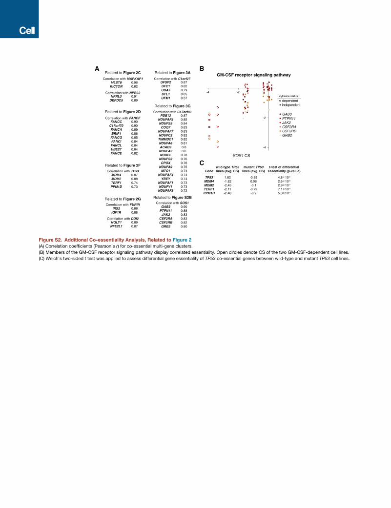
-4 -2
-4
-2GAB3PTPN11JAK2CSF2RACSF2RBGRB2
SOS1 CS
GM-CSF receptor signaling pathway
independentdependent
cytokine status:
MLST8RICTOR
NPRL3DEPDC5
FANCCC17orf70FANCABRIP1
FANCGFANCIFANCLUBE2TFANCE
MDM4MDM2TERF1PPM1D
IRS2IGF1R
NGLY1NFE2L1
UFSP2UFC1UBA5UFL1UFM1
PDE12NDUFAF5NDUFS5
COQ7NDUFAF7NDUFC2TIMMDC1NDUFA6ACAD9
NDUFA2NUBPL
NDUFS2CPOX
NDUFA9MTO1
NDUFAF4YBEY
NDUFAF1NDUFV1
NDUFAF3
GAB3PTPN11
JAK2CSF2RACSF2RB
GRB2
0.960.82
0.910.89
0.900.900.890.860.850.840.840.840.82
0.870.880.740.73
0.880.88
0.890.87
0.870.820.790.650.57
0.870.850.840.830.830.820.820.810.80.8
0.780.760.760.750.740.740.740.730.730.72
0.900.880.830.830.820.80
Correlation with MAPKAP1
Correlation with NPRL2
Correlation with FANCF
Correlation with TP53
Correlation with FURIN
Correlation with DDI2
Correlation with C1orf27
Correlation with C17orf89
Correlation with SOS1Related to Figure 2G
Related to Figure 2C
Related to Figure S2B
Related to Figure 3G
Related to Figure 3A
Related to Figure 2F
Related to Figure 2D
A B
C
TP53MDM4MDM2TERF1PPM1D
1.62-1.82-2.45-2.11-2.48
-0.390.08-0.1-0.79-0.9
4.8 X 10⁻⁵
2.6 X 10⁻⁵
2.9 X 10⁻⁷
7.1 X 10⁻⁴
5.3 X 10⁻⁴
Genewild-type TP53lines (avg. CS)
mutant TP53lines (avg. CS)
t-test of differentialessentiality (p-value)
Figure S2. Additional Co-essentiality Analysis, Related to Figure 2
(A) Correlation coefficients (Pearson’s r) for co-essential multi-gene clusters.
(B) Members of the GM-CSF receptor signaling pathway display correlated essentiality. Open circles denote CS of the two GM-CSF-dependent cell lines.
(C) Welch’s two-sided t test was applied to assess differential gene essentiality of TP53 co-essential genes between wild-type and mutant TP53 cell lines.
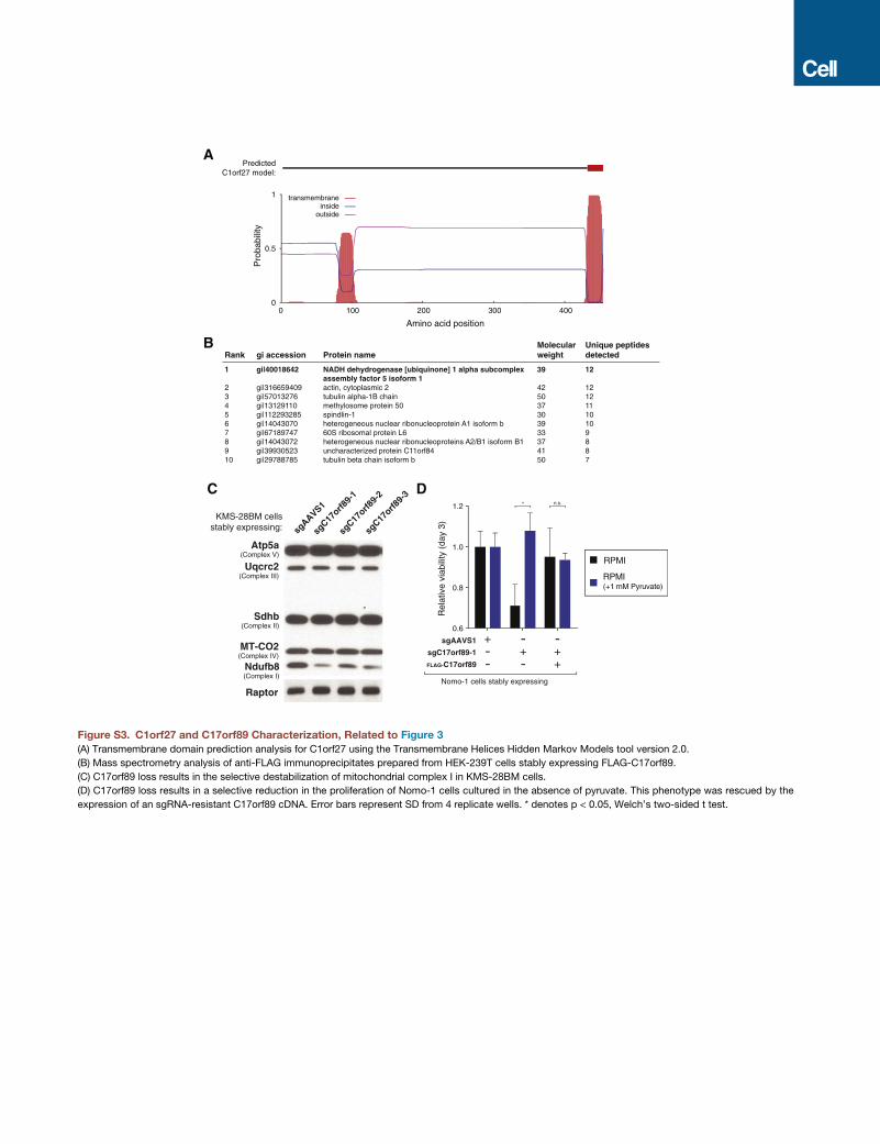
Atp5a(Complex V)
Uqcrc2(Complex III)
Sdhb(Complex II)
MT-CO2(Complex IV)
Ndufb8(Complex I)
Raptor
KMS-28BM cellsstably expressing:
0.6
0.8
1.0
1.2
RPMI
RPMI(+1 mM Pyruvate)
Rel
ativ
e vi
abili
ty (
day
3)
-+-
-++
+--
DC
Nomo-1 cells stably expressing
sgAAVS1
sgC17orf89-1
FLAG-C17orf89
sgC17
orf89-
1
sgC17
orf89-
3
sgC17
orf89-
2
sgAAVS1
1
2345678910
NADH dehydrogenase [ubiquinone] 1 alpha subcomplex assembly factor 5 isoform 1 actin, cytoplasmic 2tubulin alpha-1B chainmethylosome protein 50 spindlin-1heterogeneous nuclear ribonucleoprotein A1 isoform b 60S ribosomal protein L6 heterogeneous nuclear ribonucleoproteins A2/B1 isoform B1 uncharacterized protein C11orf84tubulin beta chain isoform b
gi|40018642
gi|316659409gi|57013276gi|13129110gi|112293285gi|14043070gi|67189747gi|14043072gi|39930523gi|29788785
39
425037303933374150
12
12121110109887
Rank gi accession Protein nameMolecularweight
Unique peptidesdetected
B
0
0.5
1
100 200 300 400
Pro
babi
lity
transmembraneinside
outside
0
PredictedC1orf27 model:
Amino acid position
A
* n.s.
Figure S3. C1orf27 and C17orf89 Characterization, Related to Figure 3
(A) Transmembrane domain prediction analysis for C1orf27 using the Transmembrane Helices Hidden Markov Models tool version 2.0.
(B) Mass spectrometry analysis of anti-FLAG immunoprecipitates prepared from HEK-239T cells stably expressing FLAG-C17orf89.
(C) C17orf89 loss results in the selective destabilization of mitochondrial complex I in KMS-28BM cells.
(D) C17orf89 loss results in a selective reduction in the proliferation of Nomo-1 cells cultured in the absence of pyruvate. This phenotype was rescued by the
expression of an sgRNA-resistant C17orf89 cDNA. Error bars represent SD from 4 replicate wells. * denotes p < 0.05, Welch’s two-sided t test.
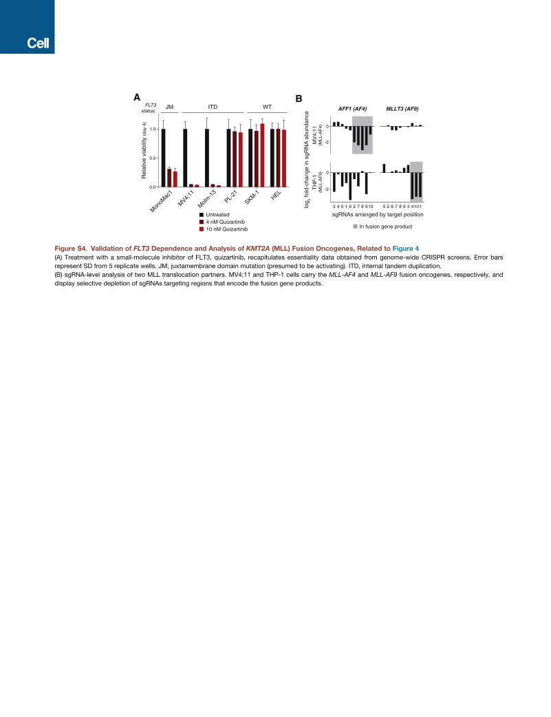
-2
0
-2
3 4 5 1 6 2 7 8 910 5 2 6 7 8 9 3 4101
0
AFF1 (AF4) MLLT3 (AF9)
TH
P-1
(MLL
-AF
9)M
V4;
11(M
LL-A
F4)
log 2
fold
-cha
nge
in s
gRN
A a
bund
ance
sgRNAs arranged by target position
In fusion gene product
A B
0.0
0.5
1.0
Untreated4 nM Quizartinib10 nM Quizartinib
HEL
Molm
-13
SKM-1
MV4;
11
Mon
oMac
1
PL-21
Rel
ativ
e vi
abili
ty (d
ay 4
)
JM ITD WTFLT3status:
Figure S4. Validation of FLT3 Dependence and Analysis of KMT2A (MLL) Fusion Oncogenes, Related to Figure 4
(A) Treatment with a small-molecule inhibitor of FLT3, quizartinib, recapitulates essentiality data obtained from genome-wide CRISPR screens. Error bars
represent SD from 5 replicate wells. JM, juxtamembrane domain mutation (presumed to be activating). ITD, internal tandem duplication.
(B) sgRNA-level analysis of two MLL translocation partners. MV4;11 and THP-1 cells carry the MLL-AF4 and MLL-AF9 fusion oncogenes, respectively, and
display selective depletion of sgRNAs targeting regions that encode the fusion gene products.
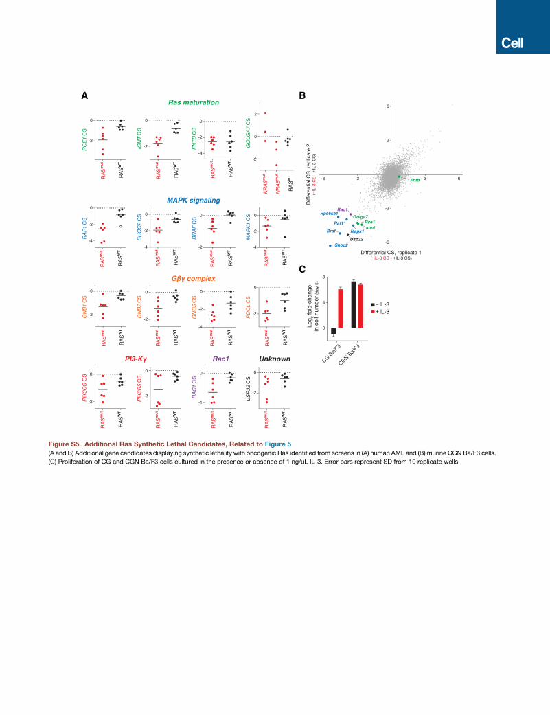
Figure S5. Additional Ras Synthetic Lethal Candidates, Related to Figure 5
(A and B) Additional gene candidates displaying synthetic lethality with oncogenic Ras identified from screens in (A) human AML and (B) murine CGN Ba/F3 cells.
(C) Proliferation of CG and CGN Ba/F3 cells cultured in the presence or absence of 1 ng/uL IL-3. Error bars represent SD from 10 replicate wells.
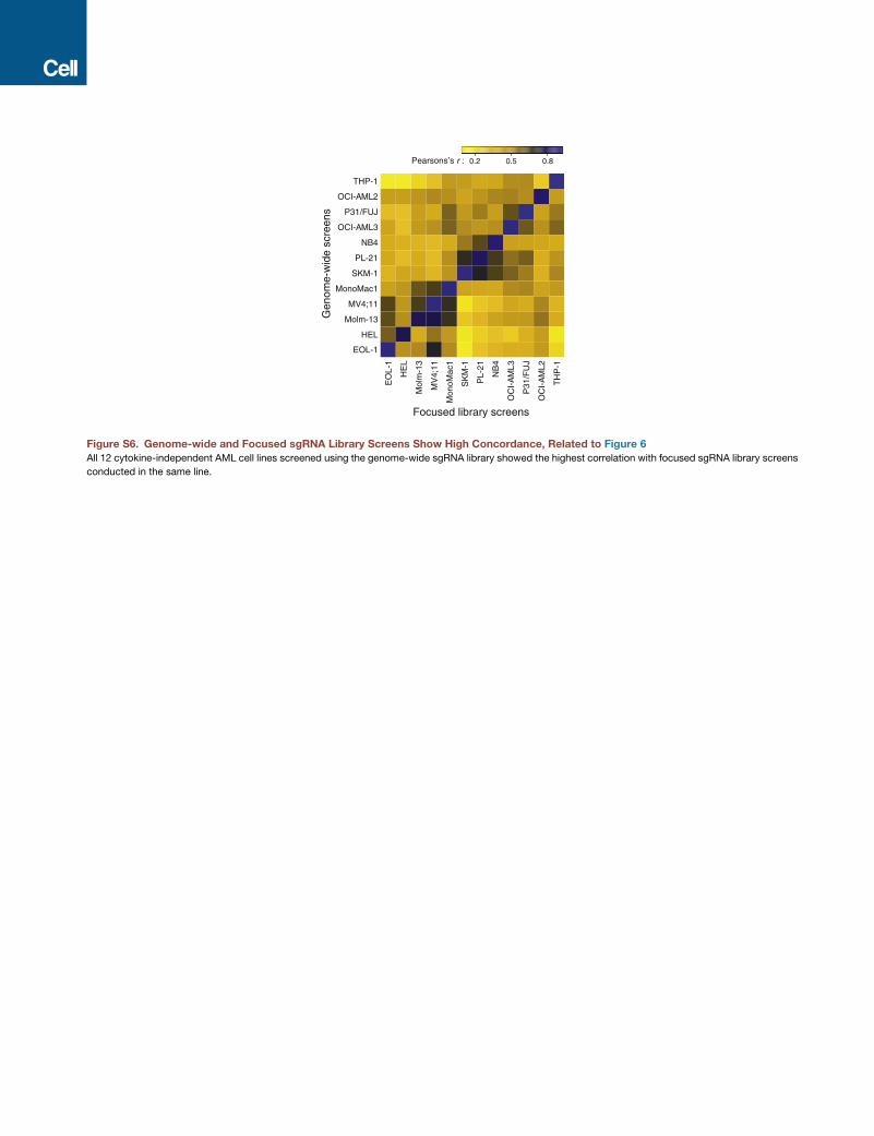
NB4
OCI-AML2
HEL
THP-1
P31/FUJ
Molm-13
SKM-1
MV4;11
EOL-1
MonoMac1
OCI-AML3
PL-21
Gen
ome-
wid
e sc
reen
s
NB
4
OC
I-A
ML2
HE
L
TH
P-1
P31
/FU
J
Mol
m-1
3
SK
M-1
MV
4;11
EO
L-1
Mon
oMac
1
OC
I-A
ML3
PL-
21
Focused library screens
0.2Pearsons’s r : 0.80.5
Figure S6. Genome-wide and Focused sgRNA Library Screens Show High Concordance, Related to Figure 6
All 12 cytokine-independent AML cell lines screened using the genome-wide sgRNA library showed the highest correlation with focused sgRNA library screens
conducted in the same line.