Embed Size (px)
Citation preview

RESEARCH ARTICLE Open Access
Functional variants of humanpapillomavirus type 16 demonstrate hostgenome integration and transcriptionalalterations corresponding to their uniquecancer epidemiologyRobert Jackson1,2, Bruce A. Rosa3, Sonia Lameiras4, Sean Cuninghame1,5, Josee Bernard1,6, Wely B. Floriano7,Paul F. Lambert8, Alain Nicolas9 and Ingeborg Zehbe1,5,6*
Abstract
Background: Human papillomaviruses (HPVs) are a worldwide burden as they are a widespread group of tumourviruses in humans. Having a tropism for mucosal tissues, high-risk HPVs are detected in nearly all cervical cancers.HPV16 is the most common high-risk type but not all women infected with high-risk HPV develop a malignanttumour. Likely relevant, HPV genomes are polymorphic and some HPV16 single nucleotide polymorphisms (SNPs)are under evolutionary constraint instigating variable oncogenicity and immunogenicity in the infected host.
Results: To investigate the tumourigenicity of two common HPV16 variants, we used our recently developed,three-dimensional organotypic model reminiscent of the natural HPV infectious cycle and conducted various“omics” and bioinformatics approaches. Based on epidemiological studies we chose to examine the HPV16Asian-American (AA) and HPV16 European Prototype (EP) variants. They differ by three non-synonymous SNPs inthe transforming and virus-encoded E6 oncogene where AAE6 is classified as a high- and EPE6 as a low-risk variant.Remarkably, the high-risk AAE6 variant genome integrated into the host DNA, while the low-risk EPE6 variantgenome remained episomal as evidenced by highly sensitive Capt-HPV sequencing. RNA-seq experiments showedthat the truncated form of AAE6, integrated in chromosome 5q32, produced a local gene over-expression and alarge variety of viral-human fusion transcripts, including long distance spliced transcripts. In addition, differentialenrichment of host cell pathways was observed between both HPV16 E6 variant-containing epithelia. Finally, in thehigh-risk variant, we detected a molecular signature of host chromosomal instability, a common property of cancercells.
Conclusions: We show how naturally occurring SNPs in the HPV16 E6 oncogene cause significant changes in theoutcome of HPV infections and subsequent viral and host transcriptome alterations prone to drive carcinogenesis.Host genome instability is closely linked to viral integration into the host genome of HPV-infected cells, which is akey phenomenon for malignant cellular transformation and the reason for uncontrolled E6 oncogene expression. Inparticular, the finding of variant-specific integration potential represents a new paradigm in HPV variant biology.
Keywords: Human papillomavirus, HPV16, E6 oncogene variants, Organotypic rafts, Viral integration,Transcriptomics, Pathogen-host relationship
* Correspondence: [email protected] Development and Biomarker Exploration, Thunder Bay RegionalResearch Institute, Thunder Bay, Ontario, Canada5Northern Ontario School of Medicine, Lakehead University, Thunder Bay,Ontario, CanadaFull list of author information is available at the end of the article
© The Author(s). 2016 Open Access This article is distributed under the terms of the Creative Commons Attribution 4.0International License (http://creativecommons.org/licenses/by/4.0/), which permits unrestricted use, distribution, andreproduction in any medium, provided you give appropriate credit to the original author(s) and the source, provide a link tothe Creative Commons license, and indicate if changes were made. The Creative Commons Public Domain Dedication waiver(http://creativecommons.org/publicdomain/zero/1.0/) applies to the data made available in this article, unless otherwise stated.
Jackson et al. BMC Genomics (2016) 17:851 DOI 10.1186/s12864-016-3203-3

BackgroundApproximately 20 % of human cancers are caused byinfectious agents [1], including >500,000 patients di-agnosed annually with human papillomavirus (HPV)associated cancers. Oncogenic HPV, denoted as “high-risk”, is the primary risk factor for cervical cancerdue to its exclusive tropism for mucosal tissues [2, 3].Upon persistent infections of the cervical mucosa,oncogenic HPVs can cause progression from low- tohigh-grade cervical intraepithelial neoplasias that,without ablative treatment, may develop into invasivecarcinomas. At the molecular level HPV is a double-stranded DNA virus and, to date, the sequences ofover 200 types have been described [4]. The ~8 kbpgenome of HPV contains 8 functional open readingframes (ORFs) that encode 5 early gene products (E1,E2, E5, E6 and E7) and 3 late gene products (E4, L1and L2). While E1 and E2 are involved in DNA repli-cation and transcriptional regulation of the viral gen-ome [5], HPV’s potent tumourigenicity is primarilydue to E6 [6], E7 [7], and E5 [8]. L1 and L2 arestructural proteins that self-assemble to form icosahe-dral capsids [9], while the fused product of ORFs E1and E4 (E1^E4) is most abundant in the productiveviral life cycle, coinciding with the onset of viralDNA amplification [10].Among the HPV types, HPV16 (a member of species
Alphapapillomavirus 9) is the most prevalent in cervicalcancers. Intriguingly, and perhaps related to its preva-lence, the HPV16 genome is polymorphic. Evolutionaryanalyses have revealed that the worldwide diversity ofHPV16 genomes evolved for over 200,000 years [11],leading to five phylogenetic branches representing iso-lates from Africa, Europe, Asia and the Americas [12].Furthermore, each branch can be further dissected intointratypic single nucleotide polymorphisms (SNPs) orvariants differing in their host persistence and frequencyof detection in human pre-cancers and cancers(reviewed in [13]). The tumourigenic differences of theseSNPs have been ascribed largely to those within the E6oncogene [14–17]. The Asian-American (AAE6) andEuropean Prototype (EPE6) are common HPV16genome variants which differ by six SNPs in their E6genes, three of which are non-synonymous, leading tothe 151-residue AAE6 protein differing by three amino-acids: Q14H, H78Y, and L83V [18] (with residue 14 and83 being under Darwinian constraint [19]).Epidemiological studies showed that the AAE6
genome variant is a higher risk factor for dysplasia aswell as an earlier onset of invasive tumours thanEPE6 [20–26]. As well, AAE6 has a greater trans-forming, migratory, and invasive potential than EPE6when retrovirally transduced into primary humankeratinocytes during recent long-term in vitro
immortalization studies [27–30]. These resultssuggested that coding changes in E6 have strongmechanistic and functional consequences for infectionand thus contribute to marked differences in cancerrisk of HPV16 variants.To decipher the fundamental biology of HPVs and
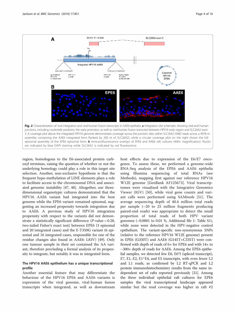
their tumourigenic features in a model system, theorganotypic 3D infection model (raft culture) has theadvantage of allowing reproducible and simultaneousepithelial differentiation and hence the occurrence of anactive viral life cycle ([31]; Fig. 1). Thus, using engi-neered human epithelium resembling in vivo conditionsbased on near-diploid immortalized keratinocytes (NIKS[32]) we recently elucidated the phenotypic characteris-tics of both E6 gene variants in the context of the fullHPV16 genome [31], building upon previous work onthe effects of transduction with the E6 or E6/E7 genesonly [27, 28, 33]. Using the organotypic model weobserved that the AAE6 genome drives tumourigenesisby increasing epithelial proliferation, disrupting routinedifferentiation and apoptosis, evading the innateimmune system and promoting immortalization [31].Interestingly, we also observed that the differences inhost epithelia histologically classified as mild keratinizing(EPE6) or moderate (AAE6) dysplasia were reflective ofincreased oncogene (E6 and E7) expression in AAE6 cul-tures and loss of productive life cycle (decreased E2,E1^E4, and L2). Together these observations lead us tosuspect integration of the AAE6 viral DNA into the hostgenome [31], a common phenomenon during HPV-induced tumourigenesis (reviewed in [34]).Here, to further advance our mechanistic understand-
ing of the impact of these common but epidemiologi-cally and clinically important E6 SNPs, we conducted an“-omics” analysis on the NIKS-based organotypic epithe-lia containing the HPV16 variants AAE6 and EPE6(Fig. 1). Modern deep sequencing techniques have beenused to study HPV [35–39], but only recently in thecontext of intratypic variants [40], and not using anorganotypic epithelial model with full viral variantgenomes. Instead, our complete approach allowed acomparison of these variants with regards to theirintegration capacity and subsequent transcriptional con-sequences in close to in vivo conditions, resulting inviral integration and a molecular signature of hostchromosomal instability for AAE6 only.
Results and discussionViral integration in the HPV16 AAE6 but not EPE6epitheliumTo permit the viral life cycle in a raft culture system, wetransfected the keratinocytes, prior to rafting, withcomplete viral genomes containing either the HPV16EPE6 or AAE6 variant. A similar technique was used in
Jackson et al. BMC Genomics (2016) 17:851 Page 2 of 16

a recent study to successfully study varicella zoster virus[41], providing a keratinocyte model and a “global” per-spective of all changes in host transcription in responseto a pathogen. As illustrated in Fig. 1, over a 14 day dif-ferentiation process, we observed that the NIKS werenormal epithelia whereas NIKS with HPV16 EPE6 ex-hibited a mild dysplasia and NIKS with HPV16 AAE6exhibited a moderate dysplasia.To examine the HPV status of these cells we used the
highly sensitive and high-throughput DNA capture andsequencing technique named Capt-HPV [42]. We pre-pared genomic DNA from epithelia of both EPE6 andAAE6. Then, after double capture on the HPV probes,we performed 2 × 151 nt paired-end sequencing (seeMethods). As expected, we readily identified numerousHPV reads in both epithelial cultures. The sequencingreads of the E6 coding region confirmed the positive in-fection of the epithelia by the AAE6 and EPE6 variants.However, as we hypothesized [31], the physical genomicstatus of HPV was clearly different. In the EPE6 epithe-lia, the reads covered the entire HPV genome indicativeof its episomal state (Fig. 2a) whereas only a fraction ofthe virus genome was detectable in the AAE6 epithelia,indicative of its integration into the host genome. Fur-thermore, in the case of EPE6, no human-viral junctionreads were detected while the integrated AAE6 viral gen-ome was truncated and several human-viral junctionreads were identified in AAE6 epithelia. The integratedviral sequence was from nt 2453 (within HPV16 E1gene) and nt 5780 (within HPV16 L1 gene) and thus
includes the E6 and E7 oncogenes. Precisely, the inser-tion of the HPV16 AAE6 variant occurred between thent position 149,347,294 and 149,347,305 of chromosome5. Mechanistically, this is a simple “end-out” integrationevent with a typical two junction, co-linear (2J-COL) sig-nature [42], associated with a very short 11 bp deletionof the host genome, and two overlapping nucleotides be-tween viral and human sequence at each junction(Fig. 2a). Functionally, the insertion occurred within the5q32 sub-band region, and more precisely, within thefirst intron of the SLC26A2 gene, approximately 13 kbupstream of its third exon.Based on the Dr.VIS (Viral Integration Site) v2.0 data-
base of HPV16 integration sites [43], this exact region(5q32) of integration is not frequent, but potentially re-current as it was found in 2 out of 878 previously docu-mented sites. The nearest fragile site was 13 Mbupstream of this integration site: FRA5C, 5q31.1. Sincerepeated regions might be prone to genome rearrange-ments and therefore prone to HPV integration, wescanned the adjacent regions using the UCSC hg19 gen-ome browser RepeatMasker track for human repeat ele-ments and found a nearby 158 bp long interspersednuclear element (LINE): L1MB5 located from Chr5 ntposition 149,347,143 to 149,347,300. Indeed, L1MB5-derived sequences have been documented as break-points, such as in the human genes HPRT [44], CYP2C[45], and in proximity of genes containing the ubiquitinligase Mib-herc2 domain, which mediates Notch signal-ling [46]. Strikingly, this domain contains the Hect
Fig. 1 The HPV16 genome and our experimental epithelial model. Contained within the viral protein capsid (top left, not to scale relative to skin) is the7.9 kb HPV16 genome, comprised of eight viral genes. Over a 14 day differentiation process we grew three-dimensional organotypic epithelia, or raftcultures, using near-diploid immortalized keratinocytes (NIKS) and primary human fibroblasts embedded in collagen-based dermal matrix. To permitthe viral life cycle in this culture system we transfected the keratinocytes, prior to rafting, with complete viral genomes containing either the EuropeanPrototype or Asian-American variant of HPV16 E6 (EPE6 or AAE6, respectively). NIKS represented normal epithelia, NIKS with HPV16 EPE6 was a milddysplasia (indicated by thickening and some suprabasal proliferation), whereas NIKS with HPV16 AAE6 was a moderate dysplasia (indicated by a greaternumber of suprabasal proliferating cells and abnormal cellular phenotypes, including micronuclei). Additionally, HPV16 viral integration was detectedin AAE6 epithelia
Jackson et al. BMC Genomics (2016) 17:851 Page 3 of 16
region, homologous to the E6-associated protein carb-oxyl terminus, raising the question of whether or not theunderlying homology could play a role in this target siteselection. Another, non-exclusive hypothesis is that thefrequent hypo-methylation of LINE elements plays a roleto facilitate access to the chromosomal DNA and associ-ated genomic instability [47, 48]. Altogether, our three-dimensional organotypic cultures demonstrated that theHPV16 AAE6 variant had integrated into the hostgenome while the EPE6 variant remained episomal, sug-gesting an increased propensity towards integration dueto AAE6. A previous study of HPV16 integrationpropensity with respect to the variants did not demon-strate a statistically significant difference (P-value = 0.28,two-tailed Fisher’s exact test) between EPE6 (3 episomaland 20 integrated cases) and the E-T350G variant (6 epi-somal and 16 integrated cases, responsible for one of theresidue changes also found in AAE6: L83V) [49]. Onlyone tumour sample in their set contained the AA vari-ant, therefore precluding a formal analysis of its propen-sity to integrate, but notably it was in integrated form.
The HPV16 AAE6 epithelium has a unique transcriptionalprofileAnother essential feature that may differentiate thebehaviour of the HPV16 EPE6 and AAE6 variants isexpression of the viral genome, viral-human fusiontranscripts when integrated, as well as downstream
host effects due to expression of the E6/E7 onco-genes. To assess these, we performed a genome-wideRNA-Seq analysis of the EPE6 and AAE6 epitheliausing Illumina sequencing of total RNAs (seeMethods), mapping first against our reference HPV16W12E genome [GenBank AF125673]. Viral transcrip-tomes were visualized with the Integrative GenomicsViewer (IGV) [50], while viral gene counts and vari-ant calls were performed using SAMtools [51]. Theaverage sequencing depth of 40.4 million total readsper sample (~20 to 25 million fragments producingpaired-end reads) was appropriate to detect the smallproportion of total reads of both HPV variantgenomes (~0.0001 to 0.01 %, Additional file 1: Table S1),while none were detected in the HPV-negative controlepithelium. The variant-specific non-synonymous SNPs(relative to the reference HPV16 W12E genome) presentin EPE6 (G350T) and AAE6 (G145T+C335T) were con-firmed with depth of reads of 6× for EPE6 and with 14× to~300× depth of reads for AAE6. Among the EPE6 epithe-lial samples, we detected few E6, E6*I (spliced transcript),E7, E1, E2, E1^E4, and E5 transcripts, with even fewer L2and L1 reads, as confirmed by L2 RT-qPCR and L2protein immunohistochemistry results from the same in-dependent set of rafts reported previously [31]. Amongthe three individual epithelial raft cultures for EPE6samples the viral transcriptional landscape appearedsimilar but the read coverage was higher in raft #2
Fig. 2 Characterization of viral integration and viral-human fusion transcripts in AAE6 epithelia. a Integration site schematic showing viral and humanjunctions, including nucleotide positions, the early promotor, as well as viral-human fusion transcript between HPV16 early region and SLC26A2 exon3. A coverage plot above the integrated HPV16 genome demonstrates coverage across the junction sites within SLC26A2 (5482 reads across a 4978 ntassembly containing the AAE6 integrated form flanked by 200 nt of SLC26A2), while a circular coverage plot on the right shows the fullepisomal assembly of the EPE6 episomal form. b Immunofluorescence overlays of EPE6 and AAE6 raft cultures (400× magnification). Nucleiare indicated by blue DAPI staining while SLC26A2 is indicated by red fluorescence
Jackson et al. BMC Genomics (2016) 17:851 Page 4 of 16

due to an overall higher abundance of viral tran-scripts in this sample (Fig. 3a). In contrast, thetranscriptional landscape for the three AAE6 sampleswas more homogenous (Fig. 3a), further emphasizedin a clustered heatmap (Fig. 3b). Abundant full-lengthE6, E6*I, E7, and only truncated E1 and L1 tran-scripts were detected. Full-length E1, E2, E1^E4, andL2 reads were absent in AAE6 epithelia, consistentwith the Capt-HPV data reported above and ourprevious RT-qPCR results and DNA copy numberanalyses on these molecules [31].To quantitatively account for sample variance, we also
performed differential expression analysis of the viralgene counts using DESeq [52]. DESeq software tests fordifferential expression in library size-corrected countdata using a negative binomial distribution model. In
agreement with our previous RT-qPCR results [31], wefound significantly more E6 (24.05 fold higher, P < 10−10)and E7 (17.30 fold higher, P < 10−10) counts in triplicateAAE6 rafts in comparison to triplicate EPE6 rafts (Fig. 3c).Taken together, analyses of viral transcriptome datarevealed that the AAE6 viral transcriptome significantlydiffers from that of EPE6 in a manner that is indicative ofintegration, with increased E6 and E7 levels [53–55]. Evi-dently, AAE6 transcriptome profiles are lacking E2 andhave increased E6/E7 oncogene expression, perhaps dueto loss of transcriptional repression by E2. We thereforereasoned that the increased levels of E6/E7 expressionbetween the variants were ultimately due to their viralintegration status, as we hypothesized in our phenotypicstudy, and confirmed by Capt-HPV, leading to a signifi-cant effect on the host transcriptome [31].
Fig. 3 The HPV16 transcriptome in EPE6 and AAE6 organotypic rafts. a Linear viral gene map. Viral RefSeq ([GenBank: AF125673], HPV16 W12Egenome) alignment from each individual raft culture was visualized using IGV [50]. The y-axis (coverage) is log2 scaled. Total number of viral reads aregiven on the right-hand side of each track. b Heatmap & clustering analysis of viral transcriptome on DESeq normalized counts: viral genes vs samplereplicates. Two distinct sample clusters matched EPE6 and AAE6 replicates respectively, clustering independently of each other. Within the high-variabilityEPE6 cluster, replicate 1 and 3 were clustered together. Within the low-variability AAE6 cluster, replicate 1 and 2 were clustered together. As well, AAE6epithelia converged on consistently high viral transcription (specifically E6/E7). From the viral gene perspective, two distinct clusters were identified: E6, E7,E1, and L1 in one, and E2, E4, E5, and L2 in another. Within the first primary cluster, E6 and E7 cluster close together, as expected given they are expressedtogether as a multi-cistronic transcript. E1 and L1 also cluster together, constituting the truncated transcripts on the periphery of the non-transcribedregion within AAE6 samples. In the second primary cluster, E5 and L2 cluster together, independent of E2 and E4 which is transcribedonly in EPE6 samples. E2 and E4 expression unsurprisingly clusters together given that E4 is contained within the E2 ORF. c Scatterplot ofaverage viral gene expression for EPE6 samples (x-axis) and AAE6 samples (y-axis). The axes (DESeq normalized gene counts) are log10scaled. Significant differential gene expression is denoted by marker colour. Dashed line represents equal expression
Jackson et al. BMC Genomics (2016) 17:851 Page 5 of 16

Nature of viral-human fusion transcripts detected inHPV16 AAE6 epitheliumThe integration of HPV16 genomes into host chromo-somes is a frequent phenomenon associated withcarcinogenesis, and not only modifies the expression ofHPV-encoded E6 and E7 oncogenes (Fig. 3a), but canalso trigger the expression of fusion viral-humanmRNAs [34, 56]. Since the virus can integrate into avariety of positions in the human genome, these fusiontranscripts are specific to each integration site. In recentyears, following the introduction of high-throughputsequencing techniques, multiple softwares for detectingpathogen sequences in host sequence data have becomeavailable [38, 57–63]. Here, to identify the viral-humanfusion transcripts expressed in our epithelia, we used theViralFusionSeq (VFS) software [61, 64]. VFS was chosenover alternatives due to its optimization for RNA-Seqdata from the Illumina platform, the ability to define ourown reference virus genome, as well as the full suite offusion transcript discovery techniques it uses. Using thistechnique, only the AAE6 rafts yielded viral-humanfusion transcripts (Table 1), providing further evidenceof viral integration as well as its transcriptional impact.In accordance with the structure of the HPV integra-
tion, the transcript breakpoints mapped to either the E1or L1 HPV16 ORF. Alternative splicing was detectedwith the viral nucleotide position at the fusion site ofone class of the viral-human fusion transcripts (Fig. 3a):nt 880 (splice donor, SD) in the E1 gene [65]. This is thesame SD site for the E1^E4 splice transcript typicallyexpressed in the late stage of the viral life cycle [66], andpreviously shown to be expressed in our EPE6 epithelia[31]. HPV16 viral-human fusion transcripts are oftendetected with a breakpoint at this natural splice donorsite [56, 67, 68], and the coverage plot for AAE6 showsdecreased coverage for transcripts downstream of thisE1 SD site, supporting the hypothesis of alternative spli-cing. With respect to the L1 breakpoints, the typical L1splice acceptor (SA) site is at nt 5639 [65], but notablyin our study, the viral-human fusion transcripts here had
a putative downstream SA site at nt 5778. Interestingly,the coverage plot of the viral transcriptome shows nt5778 as the site where L1 coverage begins to be detectedin AAE6 rafts (Fig. 3a), so we reasoned that this discrep-ancy in SA site could be due to either a cryptic SA sitein the HPV16 W12E genome (although not found previ-ously in the literature) or simply due to integrationtruncating the upstream region of L1.Next, we mapped the human portion of the fusion
transcripts using VFS’s clipped-seq (CS) and read-pair(RP) methods. Confirmed by both these methods, twofusions mapped to the human chromosome location5q32, occurring within the solute carrier family 26(anion exchanger), member 2 (SLC26A2) and phospho-diesterase 6A, cGMP-specific, rod, alpha (PDE6A)human ORFs (Table 1). Strikingly, along with detectionof fusion transcripts with these genes, we detected a sig-nificant increase in the expression of human genes fromthis region in AAE6 epithelia compared to normal epi-thelia, namely SLC26A2 (114.19 fold increase, P = 2.14 ×10−173) and colony-stimulating factor 1 receptor (CSF1R,407.82 fold increase, P = 4.70 × 10−112, which was onlydetected as RP fusion reads by VFS, and not confirmedby CS). This observation is in agreement with otherswho have found that, in numerous cervical carcinomasacross multiple high-risk HPV types, HPV integrationleads to an increase in the expression of genes adjacentto integration loci [69]. To explain the molecular basisof this cis-effect, it has been proposed to be theresult of viral promotor-driven expression or somaticgenome amplification at the integration site [70, 71].In the present case, this last hypothesis is unlikelybecause the AAE6 integration produced a clean 11 bpdeletion of the target region that led to two co-linearviral-human junctions (2J-COL), which is not associ-ated with gene amplification [42].Functional human fusion proteins can be formed due
to chromosomal translocations in cancer cells [72]. Theelucidation of novel protein-coding viral-human fusiontranscripts is particularly intriguing due to their
Table 1 Integration loci detected by ViralFusionSeq
Sample Mapped human transcript† Gene description Chromosome location HPV transcript breakpoint(s)‡
AAE6 SLC26A2 Solute carrier family 26, member 2 5q32 E1, L1
PDE6A Cyclic GMP- Phosphodiesterase 6A alpha subunit 5q32 E1, L1
EPE6 None – – –
NIKS None – – –
Viral-human fusion transcripts were discovered using ViralFusionSeq’s [61]: clipped-sequence (CS) and read-pair (RP) modules. Detected by at least 1 RP and CSevent (†). As detected by CS method (‡). VFS uses two methods to detect viral-human fusion transcripts. The Clipped-Seq (CS) method detects viral fusion transcriptbreakpoints with a read that maps to both viral and human sequences, while the Read-Pair (RP) analysis detects transcripts with read ends mapped separately to theviral and human genome [61]. We required candidate viral fusion transcripts to be supported by at least 1 CS and 1 RP event in order to improve its stringency [64].Although RP events were more abundant in our samples, CS analysis provided single-base resolution of viral-human fusion transcript breakpoints. In particular, weidentified an average of 1.33 +/− 1.53 CS transcripts in EPE6 and 7.66 +/− 6.66 in AAE6. We detected no RP transcripts in EPE6, while 118.66 +/− 7.23 were found inAAE6 rafts. While one RP transcript was detected in a NIKS control culture, this read was not confirmed by the CS method of VFS and therefore not considered as avalid event
Jackson et al. BMC Genomics (2016) 17:851 Page 6 of 16

potentially functional roles within host cells. Usingimmunofluorescence for the expressed portion of theSLC26A2 protein in formalin-fixed and paraffin embed-ded (FFPE) rafts, we determined that SLC26A2 proteinexpression was aberrantly high in AAE6 compared toEPE6, supposedly as a result of its viral-human fusionand increased transcription (Fig. 2b). This translatedfusion protein contains exon 3 of the transmembraneprotein SLC26A2, previously known as diastrophic dys-plasia sulfate transporter (DTDST) [73], which encodesthe carboxy-terminal cytoplasmic sulfate transporter andanti-sigma factor (STAS) domain [74]. We cannot findany evidence in the literature of this unique viral-humanfusion protein in other HPV-integrated samples. Overall,these chimeric molecules are unique for each sampleand to the specific integration site, with presentlyunknown effect on host cell functions, an aspect to befurther researched due to its importance for understand-ing mechanisms of tumourigenesis as well as in theemerging field of personalized medicine.
The HPV16 AAE6 epithelium reveals a signature ofchromosomal instability conducive to host genomeintegrationIntegration of HPV DNA into the host genome is con-sidered to be a key factor for cervical cancer develop-ment [67, 75, 76], but the cellular events that initiate theintegration process (and selection of insertion sites)remain to be better understood. A reasonable hypothesisis that the integration is triggered by a rare and stochas-tic target site event, such as a replicative fork stalling oran accidental chromosome double-strand break, leadingto an ultimate use of the viral DNA for repair via recom-bination, template switching (FoSTeS) and/ormicrohomology-mediated break-induced replication(MMBIR) ([42, 71, 77], and references within each). In-deed, infections with pathogens can cause chromosomalinstability by inactivating the host DNA damage re-sponse [78]. For HPV, this has been linked to the expres-sion of both HPV16 E6 and E7 oncoproteins, affectingthe infected cell’s genome integrity [79–82]. A model ofearly carcinogenesis due to HPV16 E6 and E7 suggeststhat this chromosomal instability is caused by uncon-trolled proliferation, leading to an insufficient nucleotidepool that cannot support normal replication [83]. Alter-natively, E6 alone, through the inactivation of p53, canpromote chromosomal instability, at least during earlyonset of carcinogenesis [84]. Presently, HPV16 AAE6demonstrated enhanced integration propensity overEPE6 and exhibited increased E6 and E7 oncogeneexpression, which is in accordance with elevated E6 andE7 levels reported in other studies [53–55]. Thisenhanced integration ability is based on AAE6’s greaterproliferation ability, leading to chromosomal instability.
The underlying mechanism of its increased cellgrowth is the result of a deregulated sugar metabol-ism (Warburg effect), as we reported previously [28]and currently under study (Cuninghame et al., inpreparation: unpublished observations).To assess the host chromosomal instability in our
HPV16 variant epithelia, we examined our RNA-Seqdata to detect the CIN70 gene expression signature [85],which has been applied as a prognostic marker incervical cancer [86] and more generally as a significantindicator to predict clinical outcome across multiplecancer types [85]. This signature is derived from 18 geneexpression datasets (with genes ranked based on theircorrelation to functional aneuploidy). The CIN70 scorerelative to HPV-negative NIKS was significantly higherin AAE6 compared to EPE6 epithelia (2.32 fold higher,P = 0.02 by Welch’s T-test), indicating a signature of hostchromosomal instability in AAE6 epithelia (Fig. 4a). Fur-thermore, as a morphological sign of chromosomalinstability, we detected micronuclei (MN) in AAE6 butnot EPE6 or NIKS FFPE H&E-stained epithelia (Fig. 4b).MN were reported to be present in higher grade cervicalintraepithelial neoplastic lesions and invasive cervicalcancer [87] and mechanistically have been associatedwith hallmarks of genomic instability [88].
HPV16 AAE6 epithelium exhibits a proliferatingphenotype as a consequence of viral integration into thehost genomeMore broadly, our RNA-Seq data led us to examine glo-bal changes in host gene expression. Our previous studydemonstrated enhanced tumourigenesis by the fullHPV16 genome with AAE6 [31], while another studypresented altered gene expression by the AA variant[89]. Work by other groups have studied the down-stream pathways in the AA variant [90, 91], and haveutilized high-throughput techniques to investigategenetic variation within HPV16 [39, 40, 92], but this isthe first study investigating the downstream pathwaysaffected by the HPV16 variants in an organotypic epithe-lial model using next-generation sequencing. Wehypothesized two scenarios that can be associated withthese findings and analyzed in our present study: i) theglobal gene expression profile within AAE6-infected epi-thelium would differ significantly from that of EPE6 andii) significant gene expression differences in the host duenot only to the actions of the viral oncogenes E6 and E7,but also as a result of integration [56]. A global “-omics”technique, RNA-Seq, was required to sufficiently addressour hypotheses around the functional relevance of theAA variant in epithelia. We assessed host differentialgene expression using DESeq [52] to determine how itreflected the unique viral gene expression profiles in-duced in human epithelium undergoing differentiation.
Jackson et al. BMC Genomics (2016) 17:851 Page 7 of 16

Strikingly, NIKS, which contain no virus genome, hadzero significant differentially expressed genes comparedto EPE6, at a false-discovery rate (FDR) of 10 %(Additional file 2: Figure S1). NIKS to AAE6 had 3006significant differentially expressed genes (Additional file 2:Figure S2, Additional file 3 for list of differentiallyexpressed genes between NIKS and AAE6). Of thesegenes, 1312 were down-regulated while 1694 were up-regulated in AAE6 compared to NIKS. The lack of any dif-ferentially expressed genes between NIKS and EPE6organotypic epithelial cultures was surprising, but consist-ent with the similarity between the NIKS and EPE6cultures monitored with respect to basal and suprabasalkeratinocyte proliferation assessed by BrdU-incorporation,p53 and p16INK4A by immunohistochemistry and IFN-κby RT-qPCR [31]. Phenotypically, these results suggestthat the episomal expression of the EPE6 variant in ourmodel does not have a significant tumourigenic effect.Since our 3D culture model specifically captures earlytumourigenesis, with only a 2-week growth period andlow initial viral copy number, very small gene expressiondifferences in a homogenized epidermal sample are notexpected to be easily detected with global transcriptomictechniques. On the other hand, AAE6 significantlyperturbed a high number of human genes, demonstratingits ability to cause a wide-range of host molecular changesconsistent with tumourigenesis. Compared to EPE6,AAE6 had 1666 significant differentially expressed genes(Additional file 2: Figure S3, Additional file 3 for list ofdifferentially expressed genes between EPE6 and AAE6).Of these genes, 666 were down-regulated while 1000 wereup-regulated in AAE6 compared to EPE6. Additionaldiscussion of the top-ten most significant down- and up-regulated genes for each pair-wise comparison is providedin Additional file 4. To further investigate the differential
gene expression data we applied two additional bioinfor-matics analyses: gene ontology (GO) biological processterm enrichment (Additional file 5 for GO output, Figs. 5and 6), as well as co-expression analysis and visualizationusing networks (Fig. 7). Finally, we also compared the pair-wise lists of differentially expressed genes to determine thenumber of common and unique genes among each set(Fig. 8): 1541 genes unique to the NIKS comparison, 201unique to the EPE6 comparison, and 1465 common be-tween them. Overall, these bioinformatics analyses highlightthe global effects of AAE6 on host epithelia due to its inte-gration event, increased E6/E7 expression, and perhaps inpart functional differences due to the AAE6 oncoprotein it-self: increased proliferation and decreased differentiation.
ConclusionsWe have systematically characterized the viral integra-tion process of a common high-risk HPV16 variant andits consequences for the affected host cell. This and earl-ier work lend themselves to propose a model of in-creased tumourigenicity in human keratinocyte epitheliawhere AAE6’s enhanced ability to proliferate leads tochromosomal instability. In such an environment, thehost genome may be susceptible to viral integration sub-sequently increasing E6/E7 oncogene expression and ul-timately driving additional tumourigenic changes.Previously, we performed phenotypic studies of theEPE6 and AAE6 variants in a 3D raft model of early car-cinogenesis [31] and determined the functional differ-ences of these variants in longitudinal monolayer cellcultures [27–30]. While necessary for studying the virallife cycle, limitations of the current organotypic modelare the lack of immune components, vasculature, andthe complexity of tissue heterogeneity that arises. Ourcurrent study builds on the foundation of these
Fig. 4 Chromosomal instability signature and micronuclei in AAE6 epithelia. a The CIN70 score relative to HPV-negative NIKS was significantlyhigher in AAE6 compared to EPE6 epithelia (2.32 fold higher, P = 0.02 by Welch’s T-test). Mean values are shown with error bars representingstandard deviation (n = 3). Statistical significance (P < 0.05) denoted by “*”. b Haematoxylin and eosin micrographs of FFPE AAE6 epithelia, 400×cropped, micronuclei indicated by arrow. Close-up shows micronucleus and normal-sized nucleus within same cell
Jackson et al. BMC Genomics (2016) 17:851 Page 8 of 16

Fig. 5 Gene Ontology (GO) terms enriched in highly significant differentially expressed genes in AAE6 vs. NIKS. The Term Enrichment Serviceavailable on the AmiGO 2 website [104] was used to determine enriched GO (biological process) terms among (a) down-regulated and (b)up-regulated genes. Only the top ten GO terms are shown for each. See Additional file 4 for discussion
Fig. 6 Gene Ontology (GO) terms enriched in highly significant differentially expressed genes in AAE6 vs. EPE6. The Term Enrichment Serviceavailable on the AmiGO 2 website [104] was used to determine enriched GO (biological process) terms among (a) down-regulated and (b)up-regulated genes. Only the top ten GO terms are shown for each. See Additional file 4 for discussion
Jackson et al. BMC Genomics (2016) 17:851 Page 9 of 16

investigations. We have applied a wide range of molecu-lar analyses, creating a framework which can benefitfuture virus-host interaction studies with various orga-notypic cell culture models. A variant-specific integra-tion is worth reporting and should be furtherinvestigated, with additional samples from independentdonors, as it represents a new paradigm in HPV variantbiology. Here we report a viable integration mechanismin a robust viral life cycle model for AAE6. The findingsof the current and other studies reported by us [27–31],
and others [89–91], are consistent with cancer epidemi-ology studies demonstrating that the HPV16 AA variantis a higher risk factor for high-grade intraepithelialneoplasia and progression to invasive cervical cancer[22–24, 89]. In the future, HPV variant genotyping couldbe used as a clinical prognostic factor for patient-centered health services, while the role of individual hostgenomics on integration, including characterization ofintegration sites, will be important to consider forpersonalized medicine approaches.
Fig. 7 Co-expression networks of highly significant (a) down-regulated and (b) up-regulated genes in AAE6 vs. EPE6. a Four discrete clusters ofdown-regulated and co-expressed genes were observed. Only co-expressed genes with a Pearson correlation coefficient greater than 0.95 are shown.Clusters are labelled by number and functionally annotated with their significantly enriched biological process. Nodes = gene, denoted by genesymbol; node colour = white to red with down-regulation (fold change) in AAE6 from EPE6; edge thickness = increases with Pearson correlationcoefficient. b Five discrete clusters of up-regulated and co-expressed genes were observed. Only clusters co-expressed genes with a Pearsoncorrelation coefficient greater than 0.996 and are shown, to narrow down the number of genes displayed. Clusters are labelled by number andfunctionally annotated with their significantly enriched biological process. Nodes = gene, denoted by gene symbol; node colour =white to green withup-regulation (fold change) in AAE6 over EPE6; edge thickness = increases with Pearson correlation coefficient. See Additional file 4 for discussion
Jackson et al. BMC Genomics (2016) 17:851 Page 10 of 16

MethodsCell linesAs described by us previously [31], we used the Normal/Near-Diploid Immortalized Keratinocytes (NIKS) cellline [32] to establish 3D organotypic epithelia cultures.These spontaneously immortalized cells were originallyderived from neonatal human foreskin and are non-tumourigenic, though contain an additional long armpiece of chromosome 8 (8q). In monolayer they aregrown on mitomycin-C-treated Swiss mouse J2/3T3fibroblast feeder layers [32], while primary human fore-skin fibroblasts (ATCC CRL-2097) are incorporated intothe dermal equivalent of organotypic NIKS cultures [31].
Detection of integrated papillomavirus sequences bynext-generation DNA-Seq: Capt-HPVDNA-Seq was used to confirm the presence and locationof the viral integration sites in the human genome usingDNA extracted from formalin-fixed paraffin embedded(FFPE) samples which had been prepared previously [31].DNA was extracted using the DNeasy Blood and TissueKit (QIAGEN, Cat# 69504) with the recommended pre-treatment for FFPE samples and the optional RNase treat-ment. To overcome the limitations of traditionaltechniques, such as DIPS-PCR (Detection of IntegratedPapillomavirus Sequences by ligation mediated PCR), weused an unbiased and state-of-the-art next-generationDNA sequencing technique for detecting HPV viral inte-gration sequences in our samples [42]. Library prepar-ation, sequence capture, and high-throughput sequencing
was carried out at the Institut Curie on an Illumina MiSeqplatform with a V2 Nano chip (~1 × 10⁶ total reads) with2 × 151 base pair read length. Analysis of sequencing datawas performed using the Galaxy platform [93–95], withthe primary goal of detecting the viral-human junction sitelocations. Packages used were FASTQ Groomer [96],Bowtie2 [97], Picard MarkDuplicates [98], SAMtoolsBAM-to-SAM and Filter SAM [51].
RNA-Seq library preparation and sequencingIsolation of high-quality total RNA from the epitheliumof organotypic keratinocyte cultures containing full-length HPV16 E6 variant genomes, European Prototype(EPE6) and Asian-American (AAE6), was describedpreviously [31]. Our keratinocyte model was grown for14 days to allow simultaneous epithelial differentiationand occurrence of an active viral life cycle. Total RNAfor EPE6, AAE6, and HPV16 negative cultures (NIKS),three organotypic raft cultures (n = 3) each, were sentfor library preparation and sequencing at The Centre forApplied Genomics, Hospital for Sick Children, Toronto,Canada. RNA-Seq libraries were prepared by IlluminaTruSeq® RNA Sample Preparation kit followed bysequencing using an Illumina HiSeq® 2500 platform withIllumina v3 chemistry. One lane of multiplexed, paired-end, 2 × 101 base pair sequencing was performed withnine samples: yielding an average of 40.4 million totalreads (~20 to 25 million fragments) per sample(Additional file 1: Table S2).
Viral variant read alignment, mapping, and coverageplottingThe human papillomavirus type 16 W12E isolate genome[GenBank: AF125673] [54, 99] was used as a viral refer-ence sequence since it was the parental sequence modifiedby site-directed mutagenesis to generate the EPE6 andAAE6 viral genomes used in this study [31]. Only thethree non-synonymous nucleotide changes differentiatedEPE6 and AAE6 genomes: EPE6 was made by mutatingthe parental W12E genome at G350T while AAE6 wasmutated at G145T and C335T. Prior to alignment andmapping, Bowtie2 [97] was used to build a reference indexfor HPV16 using the AF125673 W12E isolate RefSeq.TopHat2 [100] was used for alignment to our viral RefSeq.Variant-specific non-synonymous SNPs were confirmedby variant calling with SAMtools [51]. The Broad Insti-tute’s Integrative Genomics Viewer (IGV) [50] was used tovisualize alignment coverage for each sample. Gene-levelcounts of the HPV16 W12E ORF’s were generated usingSAMtools [51], and normalized with library-size correc-tion factors using the Bioconductor project DESeq [52] inthe statistical environment R [101]. DESeq was also usedfor differential viral gene expression analysis. DESequses a default false discovery rate (FDR) of 10 % for
Fig. 8 Venn diagram of differentially expressed genes common andunique to each pairwise comparison. Of the 3006 differentiallyexpressed (DE) genes in NIKS vs AAE6 and the 1666 differentiallyexpressed (DE) genes in EPE6 vs AAE6 there were 1541 genesunique to the NIKS comparison, 1465 common between them, and201 unique to the EPE6 comparison. No genes were up-regulated inone set of a pair-wise comparison (either NIKS vs EPE6 or EPE6 vsAAE6) while down-regulated in the other
Jackson et al. BMC Genomics (2016) 17:851 Page 11 of 16

its binomial statistical inference tests to determinedifferentially expressed genes. Clustered heatmaps ofnormalized viral gene counts were generated usingthe gplots package [102].
Identification of viral-human fusion transcriptsViralFusionSeq (VFS) [61] was used, with default param-eters, to identify any viral-human fusion transcripts ineach of our sample RNA-Seq datasets. As with viralalignment by TopHat2 (described above), the W12Egenome was used as a reference sequence for VFS.Briefly, VFS is a Perl script that searches in high-throughput sequencing data (RNA or DNA-Seq) forviral-human fusion transcripts, which are present as aresult of viral integration events into host DNA. Thissoftware uses read pair (RP) and clipped sequences (CS)to accurately discover and identify viral-fusion sequences[61]. Additionally, VFS is able to reconstruct fusion tran-scripts by a targeted de novo assembly process. Thesemethods allow us to identify, with single-base resolution,viral-human fusion transcripts present within our epi-thelial cultures. Viral-human fusion transcripts werecompared to known HPV16 integration sites and fusiontranscripts with assistance from the database of diseaserelated viral integration sites (Dr. VIS v2.0, [43]).We sought to perform protein-level confirmation of
highly expressed viral-human fusion transcripts contain-ing exons from human targets SLC26A2 and CSF1R.SLC26A2 protein expression was detected in raft cul-tures by immunofluorescence, as described previously[31]. Based on the viral-human fusion RNA-Seq data,the primary antibody (rabbit polyclonal, 1:500 dilution,Bethyl Laboratories Inc., Cat. No. A304-467A) waschosen to have specificity for translated exon 3 (epitopebetween amino acid residue 689 and 739). Although alsohighly up-regulated, no suitable commercial antibodywas found for CSF1R exons 20 to 22.
Human read alignment, mapping, and count generationRead alignment, mapping, and count generation for thehuman reference genome (hg19, UCSC nomenclaturefor GRCh37) was performed by The Centre for AppliedGenomics, Hospital for Sick Children, Toronto, Canada.TopHat2 [100] was used for RefSeq while gene- andexon-level counts were generated using HTSeq [103].Number of reads and percentage of human RefSeq readsdefined as aligned, exon, and exon-exon are reported inAdditional file 1: Table S2 for each sample analyzed.
Differential expression analysis of human transcriptomeDifferential analysis of pair-wise human gene-levelcounts between NIKS and EPE6, NIKS and AAE6, andEPE6 and AAE6 were performed using the Bioconductorproject DESeq [52] package implemented in the
statistical environment R [101]. Raw gene counts fromHTSeq were first normalized by estimating the samplelibrary sizes (Additional file 1: Table S3) and applyingthe size-factor correction to all counts within a givensample. A dispersion plot was made to visualize the vari-ance estimation step prior to differential expressioninference (Additional file 2: Figure S4). A clustered heat-map with hierarchical dendrograms was used to showoverall sample and biological replicate clustering: thegene expression profile of AAE6 samples was distinctfrom EPE6 and NIKS (control) samples (Additional file 2:Figure S5). Although EPE6 replicate 3 and NIKS repli-cate 1 cluster outside of their specific sample group,viral RNA-Seq analysis has confirmed these sampleID’s are correct, and that their grouping is likely aresult of the minor host transcriptomic difference be-tween NIKS and EPE6 cultures. DESeq uses a defaultfalse discovery rate (FDR) of 10 % for its binomialstatistical inference tests to determine differentiallyexpressed genes. However, for downstream analyses ofdown- and up-regulated genes we used a more strin-gent adjusted P-value cut-off of 10−5.
CIN70 scoring and micronuclei detectionHost chromosomal instability was assessed, using nor-malized human gene count data from our RNA-Seq ex-periments, by calculating a CIN70 gene expressionsignature score [85] for EPE6 and AAE6 relative to NIKSepithelia. For each of the 70 genes, a normalized humangene count ratio was calculated for all EPE6 and AAE6samples relative to the average of the NIKS samples.Relative ratio values were then averaged for all 70 genesin each sample and a Welch’s T-test, for unequal vari-ance, was used to determine whether there was a statisti-cally significant difference in host chromosomalinstability signature between EPE6 and AAE6 epithelia.We used a significance level of P < 0.05. As a morpho-logical assessment of chromosomal instability wescreened haematoxylin and eosin-stained sections fromformalin-fixed and paraffin-embedded NIKS, EPE6, andAAE6 epithelia for micronuclei (MN). These aberrantnuclei structures [88] were detected using light micros-copy with high-magnification (at least 400×).
Gene set enrichment analysis and networksEnrichment of host biological processes of differentiallyexpressed human genes was determined using the GeneOntology (GO) Term Enrichment Service hosted on theAmiGO 2 website [104]. Only biological processes wereincluded. Terms were considered significantly enrichedif the Bonferroni-corrected P-value was less than 0.05.To aid in the visual interpretation of down- and up-regulated gene sets, co-expression networks wereconstructed with Cytoscape software [105]. Pearson
Jackson et al. BMC Genomics (2016) 17:851 Page 12 of 16

correlation coefficients were calculated for each gene-gene pairwise comparison in highly significant down-and up-regulated genes between AAE6 and EPE6(Additional file 6 for down- and up-regulated gene-gene pairwise comparisons, respectively). Pearson cor-relation coefficient cut-offs used for networking wereselected strategically to produce small distinct clustersof genes, since setting the threshold too low resultsin all nodes connected, and setting the threshold toohigh results in a lack of clusters.
Additional files
Additional file 1: Viral and human read tables. Table S1. Viral readssummary. Overall, viral reads make up ~0.0001 to 0.01 % of the totalreads, while human reads make up 80 to 85 % of the total reads (theremaining reads are unmapped, to either viral or human sequences).Table S2. Human RefSeq alignment statistics for all samples. NIKS wereHPV16 negative organotypic keratinocyte cultures while EPE6 and AAE6were cultures containing the full genome of HPV16 with either EuropeanPrototype E6 or Asian-American E6 variants, respectively. “Aligned” refersto reads overlapping exons, “Exon” refers to reads completely within anexon, and “Exon-Exon” refers to reads overlapping exon junctions. Table S3.Human library size factor for all samples. Library size factors derived fromDESeq [52]. (DOCX 15 kb)
Additional file 2: DESeq plots. Figure S1. Plot of normalized meancounts versus log2 fold change for the contrast NIKS versus EPE6. Redpoints represent genes that have significant differential expressionbetween the two conditions (false-discovery rate of 10 %, adjusted P <0.1). No genes were significantly differentially expression between NIKSand EPE6. Figure S2. Plot of normalized mean counts versus log2 foldchange for the contrast NIKS versus AAE6. Red points represent genesthat have significant differential expression between the two conditions(false-discovery rate of 10 %, adjusted P < 0.1). In total, 3006 genes weresignificantly differentially expression between NIKS and EPE6. Figure S3.Plot of normalized mean counts versus log2 fold change for the contrastEPE6 versus AAE6. Red points represent genes that have significantdifferential expression between the two conditions (false-discovery rateof 10 %, adjusted P < 0.1). In total, 1666 genes were significantlydifferentially expressed between NIKS and EPE6. Figure S4. Empirical andfitted dispersion values plotted against the mean of the normalizedhuman gene-level counts. Red line represents fitted dispersion over theempirical values (black dots). Figure S5. Heatmap of Euclidean distancesbetween human gene-level counts of samples. Heatmap and clusteringwas performed after DESeq variance-stabilizing transformation of humangene-level count data. (DOCX 204 kb)
Additional file 3: DESeq output. Significant differential expression outputfor NIKS and AAE6 contrast as well as EPE6 and AAE6 contrast. (XLSX 430 kb)
Additional file 4: Follow-up discussion of host expression analysis.Additional discussion of differential gene expression analysis, pathway-level enrichment, and co-expression networks. Tables S4-S7. top-tenmost significant down- or up-regulated genes in AAE6 compared to NIKSor EPE6. (DOCX 30 kb)
Additional file 5: GO output. Significantly enriched GO terms (biologicalprocesses) for NIKS and AAE6 contrast as well as EPE6 and AAE6 contrast.(XLSX 28 kb)
Additional file 6: Pearson correlations Pearson correlation coefficientsfor gene-gene pairwise comparisons of down- and up-regulated genesfor EPE6 and AAE6 contrast. (XLSX 298 kb)
AcknowledgementsThank you to Dr. Allyson Holmes at the Institut Curie for her valuablefeedback and collaboration on the DNA-Seq experiments. Special thanks goto Melissa Togtema for her insightful comments while preparing the
manuscript as well as Darryl Willick for his help in setting up andmaintaining the Galaxy platform hosted at the Lakehead University HighPerformance Computing Centre (LUHPCC).
FundingThis work was supported by Natural Sciences and Engineering ResearchCouncil of Canada (NSERC) grants to IZ (#355858-2008, #435891-2013,#RGPIN-2015-03855), NSERC Alexander Graham Bell Canada GraduateScholarship-Doctoral (CGS-D) to RJ (#454402-2014), NSERC Alexander GrahamBell Canada Graduate Scholarship-Masters (CGS-M) to SC (#442618-2013), andan NSERC Undergraduate Student Research Award (USRA) to JB (#483630-2015).The funding bodies had no role in study design, data collection, data analysisand interpretation, or preparation of the manuscript.
Availability of data and materialsRaw sequence data used in this article can be accessed via the SequenceRead Archive (SRA), study accession number SRP055094 (http://www.ncbi.nlm.nih.gov/sra/SRP055094) and National Center for BiotechnologyInformation (NCBI) BioProject, accession number PRJNA275642 (http://www.ncbi.nlm.nih.gov/bioproject/PRJNA275642). Remaining supporting datacan be accessed as Additional files, while software and tools used have beencited throughout the Methods section.
Authors’ contributionsThis interdisciplinary study was initially conceived by IZ and RJ refined thebioinformatics portion in collaboration with BR, WF, SL, and AN. RJ, PL, andIZ designed and carried out the 3D organotypic skin culturing experiments.IZ and PL contributed reagents, materials, and methods for culturingexperiments. RJ, BR, SC and JB performed RNA-Seq and follow-up data analyses.AN contributed reagents, materials, and methods for DNA sequencing. SL andRJ performed DNA-Seq and follow-up analyses. RJ, BR, SL, SC, JB, WF, PL, AN,and IZ contributed to data interpretation. All authors contributed to writing thepaper with RJ being the lead author and IZ having considerable input into thewriting. All authors have read and approved the final manuscript.
Authors’ informationNot applicable.
Competing interestsThe authors declare that they have no competing interests.
Consent for publicationNot applicable.
Ethics approval and consent to participateNot applicable.
Author details1Probe Development and Biomarker Exploration, Thunder Bay RegionalResearch Institute, Thunder Bay, Ontario, Canada. 2Biotechnology Program,Lakehead University, Thunder Bay, Ontario, Canada. 3McDonnell GenomeInstitute, Washington University School of Medicine, St. Louis, MO, USA. 4NGSplatform, Institut Curie, PSL Research University, 26 rue d’Ulm, 75248 Paris,Cedex, France. 5Northern Ontario School of Medicine, Lakehead University,Thunder Bay, Ontario, Canada. 6Department of Biology, Lakehead University,Thunder Bay, Ontario, Canada. 7Department of Chemistry, LakeheadUniversity, Thunder Bay, Ontario, Canada. 8McArdle Laboratory for CancerResearch, University of Wisconsin School of Medicine and Public Health,Madison, WI, USA. 9Institut Curie, PSL Research University, Centre National dela Recherche Scientifique UMR3244, Sorbonne Universités, Paris, France.
Received: 19 May 2016 Accepted: 25 October 2016
References1. Bouvard V, Baan R, Straif K, Grosse Y, Secretan B, Ghissassi FE, et al. A review of
human carcinogens—Part B: biological agents. Lancet Oncol. 2009;10:321–2.2. zur Hausen H. Papillomavirus infections—a major cause of human cancers.
BBA-Rev Cancer. 1996;1288:F55–78.3. zur Hausen H. Papillomaviruses and cancer: from basic studies to clinical
investigations. Nat Rev Cancer. 2002;2:342–50.
Jackson et al. BMC Genomics (2016) 17:851 Page 13 of 16

4. Kocjan BJ, Bzhalava D, Forslund O, Dillner J, Poljak M. Molecular methods foridentification and characterization of novel papillomaviruses. Clin MicrobiolInfect. 2015;21:808–16.
5. Doorbar J, Quint W, Banks L, Bravo IG, Stoler M, Broker TR, et al. The biologyand life-cycle of human papillomaviruses. Vaccine. 2012;30:F55–70.
6. Vande Pol SB, Klingelhutz AJ. Papillomavirus E6 oncoproteins. Virology.2013;445:115–37.
7. Roman A, Münger K. The papillomavirus E7 proteins. Virology.2013;445:138–68.
8. Maufort JP, Williams SM, Pitot HC, Lambert PF. Human papillomavirus 16 E5oncogene contributes to two stages of skin carcinogenesis. Cancer Res.2007;67:6106–12.
9. Conway MJ, Meyers C. Replication and assembly of human papillomaviruses. JDent Res. 2009;88:307–17.
10. Middleton K, Peh W, Southern S, Griffin H, Sotlar K, Nakahara T, et al.Organization of human papillomavirus productive cycle during neoplasticprogression provides a basis for selection of diagnostic markers. J Virol.2003;77:10186–201.
11. Bernard HU. The clinical importance of the nomenclature, evolution andtaxonomy of human papillomaviruses. J Clin Virol. 2005;32:1–6.
12. Yamada T, Manos MM, Peto J, Greer CE, Munoz N, Bosch FX, et al. Humanpapillomavirus type 16 sequence variation in cervical cancers: a worldwideperspective. J Virol. 1997;71:2463–72.
13. Burk RD, Harari A, Chen Z. Human papillomavirus genome variants. Virology.2013;445:232–43.
14. Grodzki M, Besson G, Clavel C, Arslan A, Franceschi S, Birembaut P, et al.Increased risk for cervical disease progression of French women infectedwith the human papillomavirus type 16 E6-350G variant. Cancer EpidemiolBiomarkers Prev. 2006;15:820–2.
15. Zehbe I, Wilander E, Delius H, Tommasino M. Human papillomavirus 16 E6variants are more prevalent in invasive cervical carcinoma than theprototype. Cancer Res. 1998;58:829–33.
16. Zehbe I, Voglino G, Delius H, Wilander E, Tommasino M. Risk of cervical cancerand geographical variations of human papillomavirus 16 E6 polymorphisms.Lancet. 1998;352:1441–2.
17. Zehbe I, Voglino G, Wilander E, Delius H, Marongiu A, Edler L, et al. p53 codon72 polymorphism and various human papillomavirus 16 E6 genotypes are riskfactors for cervical cancer development. Cancer Res. 2001;61:608–11.
18. Cornet I, Gheit T, Franceschi S, Vignat J, Burk RD, Sylla BS, et al. Humanpapillomavirus type 16 genetic variants: phylogeny and classification basedon E6 and LCR. J Virol. 2012;86:6855–61.
19. Chen Z, Terai M, Fu L, Herrero R, DeSalle R, Burk RD. Diversifying selection inhuman papillomavirus type 16 lineages based on complete genomeanalyses. J Virol. 2005;79:7014–23.
20. Xi LF, Koutsky LA, Galloway DA, Kiviat NB, Kuypers J, Hughes JP, et al.Genomic variation of human papillomavirus type 16 and risk for high gradecervical intraepithelial neoplasia. J Natl Cancer Inst. 1997;89:796–802.
21. Villa LL, Sichero L, Rahal P, Caballero O, Ferenczy A, Rohan T, et al. Molecularvariants of human papillomavirus types 16 and 18 preferentially associatedwith cervical neoplasia. J Gen Virol. 2000;81:2959–68.
22. Berumen J, Ordonez RM, Lazcano E, Salmeron J, Galvan SC, Estrada RA, et al.Asian American variant of human papillomavirus 16 and risk for cervicalcancer: a case–control study. J Natl Cancer Inst. 2001;93:1325–30.
23. Xi LF, Koutsky LA, Hildesheim A, Galloway DA, Wheeler CM, Winer RL, et al.Risk for high-grade cervical intraepithelial neoplasia associated with variantsof human papillomavirus types 16 and 18. Cancer Epidemiol BiomarkersPrev. 2007;16:4–10.
24. Zuna RE, Moore WE, Shanesmith RP, Dunn ST, Wang SS, Schiffman M, et al.Association of HPV16 E6 variants with diagnostic severity in cervicalcytology samples of 354 women in a US population. Int J Cancer.2009;125:2609–13.
25. Schiffman M, Rodriguez AC, Chen Z, Wacholder S, Herrero R, Hildesheim A,et al. A population-based prospective study of carcinogenic humanpapillomavirus variant lineages, viral persistence, and cervical neoplasia.Cancer Res. 2010;70:3159–69.
26. Freitas LB, Chen Z, Muqui EF, Boldrini NAT, Miranda AE, Spano LC, et al.Human Papillomavirus 16 Non-European Variants Are PreferentiallyAssociated with High-Grade Cervical Lesions. PLoS One. 2014;9:e100746.
27. Zehbe I, Richard C, DeCarlo CA, Shai A, Lambert PF, Lichtig H, et al. Humanpapillomavirus 16 E6 variants differ in their dysregulation of humankeratinocyte differentiation and apoptosis. Virology. 2009;383:69–77.
28. Richard C, Lanner C, Naryzhny S, Sherman L, Lee H, Lambert PF, et al. Theimmortalizing and transforming ability of two common humanpapillomavirus 16 E6 variants with different prevalences in cervical cancer.Oncogene. 2010;29:3435–45.
29. Niccoli S, Abraham S, Richard C, Zehbe I. The Asian-American E6 variantprotein of human papillomavirus 16 alone is sufficient to promoteimmortalization, transformation, and migration of primary human foreskinkeratinocytes. J Virol. 2012;86:12384–96.
30. Togtema M, Jackson R, Richard C, Niccoli S, Zehbe I. The humanpapillomavirus 16 European-T350G E6 variant can immortalize but nottransform keratinocytes in the absence of E7. Virology. 2015;485:274–82.
31. Jackson R, Togtema M, Lambert PF, Zehbe I. Tumourigenesis Driven by theHuman Papillomavirus Type 16 Asian-American E6 Variant in a Three-Dimensional Keratinocyte Model. PLoS One. 2014;9:e101540.
32. Allen-Hoffmann BL, Schlosser SJ, Ivarie CA, Sattler CA, Meisner LF, O’ConnorSL. Normal growth and differentiation in a spontaneously immortalizednear-diploid human keratinocyte cell line. NIKS J Invest Dermatol.2000;114:444–55.
33. Schütze DM, Snijders PJ, Bosch L, Kramer D, Meijer CJ, Steenbergen RD.Differential In Vitro Immortalization Capacity of Eleven, Probable High-RiskHuman Papillomavirus Types. J Virol. 2014;88:1714–24.
34. Poreba E, Broniarczyk JK, Gozdzicka-Jozefiak A. Epigenetic mechanisms invirus-induced tumorigenesis. Clin Epigenetics. 2011;2:233–47.
35. Mine KL, Shulzhenko N, Yambartsev A, Rochman M, Sanson GF, LandoM, et al. Gene network reconstruction reveals cell cycle and antiviralgenes as major drivers of cervical cancer. Nat Commun. 2013;4:1806.
36. Khoury JD, Tannir NM, Williams MD, Chen Y, Yao H, Zhang J, et al. TheLandscape of DNA Virus Associations Across Human Malignant CancersUsing RNA-Seq: An Analysis of 3775 Cases. J Virol. 2013;87:8916–26.
37. Bryant D, Onions T, Raybould R, Flynn Á, Tristram A, Meyrick S, et al. mRNAsequencing of novel cell lines from human papillomavirus type‐16 relatedvulval intraepithelial neoplasia: Consequences of expression of HPV16 E4and E5. J Med Virol. 2014;86:1534–41.
38. Chandrani P, Kulkarni V, Iyer P, Upadhyay P, Chaubal R, Das P, et al. NGS-based approach to determine the presence of HPV and their sites ofintegration in human cancer genome. Br J Cancer. 2015;112:1958–65.
39. Cullen M, Boland J, Schiffman M, Zhang X, Wentzensen N, Yang Q, et al.Deep sequencing of HPV16 genomes: A new high-throughput tool forexploring the carcinogenicity and natural history of HPV16 infection.Papillomavirus Research. 2015;1:3–11.
40. Lavezzo E, Masi G, Toppo S, Franchin E, Gazzola V, Sinigaglia A, et al.Characterization of Intra-Type Variants of Oncogenic Human Papillomavirusesby Next-Generation Deep Sequencing of the E6/E7 Region. Viruses. 2016;8:79.
41. Jones M, Dry IR, Frampton D, Singh M, Kanda RK, Yee MB, et al. RNA-seqanalysis of host and viral gene expression highlights interaction betweenvaricella zoster virus and keratinocyte differentiation. PLoS Pathog.2014;10:e1003896.
42. Holmes A, Lameiras S, Jeannot E, Marie Y, Castera L, Sastre-Garau X, et al.Mechanistic signatures of HPV insertions in cervical carcinomas. GenomeMed. 2016;1:16004.
43. Yang X, Li M, Liu Q, Zhang Y, Qian J, Wan X, et al. Dr.VIS v2.0: an updateddatabase of human disease-related viral integration sites in the era ofhigh-throughput deep sequencing. Nucl Acids Res. 2015;43:D887–92.
44. Williams M, Rainville IR, Nicklas JA. Use of inverse PCR to amplify andsequence breakpoints of HPRT deletion and translocation mutations.Environ Mol Mutagen. 2002;39:22–32.
45. Zhou S. Cytochrome P450 2D6: structure, function, regulation andpolymorphism. CRC Press; 2016 Feb 24
46. del Rosario RC, Rayan NA, Prabhakar S. Noncoding origins of anthropoidtraits and a new null model of transposon functionalization. Genome Res.2014;24:1469–84.
47. Richards KL, Zhang B, Baggerly KA, Colella S, Lang JC, Schuller DE, et al.Genome-wide hypomethylation in head and neck cancer is morepronounced in HPV-negative tumors and is associated with genomicinstability. PLoS One. 2009;4:e4941.
48. Baba Y, Watanabe M, Murata A, Shigaki H, Miyake K, Ishimoto T, et al. LINE-1hypomethylation, DNA copy number alterations, and CDK6 amplification inesophageal squamous cell carcinoma. Clin Cancer Res. 2014;20:1114–24.
49. Xu B, Chotewutmontri S, Wolf S, Klos U, Schmitz M, Dürst M, et al. Multiplexidentification of human papillomavirus 16 DNA integration sites in cervicalcarcinomas. PLoS One. 2013;8:e66693.
Jackson et al. BMC Genomics (2016) 17:851 Page 14 of 16

50. Robinson JT, Thorvaldsdóttir H, Winckler W, Guttman M, Lander ES, Getz G,et al. Integrative genomics viewer. Nat Biotechnol. 2011;29:24–6.
51. Li H, Handsaker B, Wysoker A, Fennell T, Ruan J, Homer N, et al. The sequencealignment/map format and SAMtools. Bioinformatics. 2009;25:2078–9.
52. Anders S, Huber W. Differential expression analysis for sequence count data.Genome Biol. 2010;11:R106.
53. Durst M, Bosch FX, Glitz D, Schneider A, zur Hausen H. Inverse relationshipbetween human papillomavirus (HPV) type 16 early gene expression andcell differentiation in nude mouse epithelial cysts and tumors induced byHPV-positive human cell lines. J Virol. 1991;65:796–804.
54. Jeon S, Allen-Hoffmann BL, Lambert PF. Integration of humanpapillomavirus type 16 into the human genome correlates with a selectivegrowth advantage of cells. J Virol. 1995;69:2989–97.
55. Daniel B, Rangarajan A, Geetasree M, Elizabeth V, Krishna S. The linkbetween integration and expression of human papillomavirus type 16genomes and cellular changes in the evolution of cervical intraepithelialneoplastic lesions. J Gen Virol. 1997;78:1095–101.
56. Lace MJ, Anson JR, Klussmann JP, Wang DH, Smith EM, Haugen TH, et al.Human papillomavirus type 16 (HPV-16) genomes integrated in head andneck cancers and in HPV-16-immortalized human keratinocyte clonesexpress chimeric virus-cell mRNAs similar to those found in cervical cancers.J Virol. 2011;85:1645–54.
57. Hawkins TB, Dantzer J, Peters B, Dinauer M, Mockaitis K, Mooney S, etal. Identifying viral integration sites using SeqMap 2.0. Bioinformatics.2011;27:720–2.
58. Westermann AJ, Gorski SA, Vogel J. Dual RNA-seq of pathogen and host.Nat Rev Microbiol. 2012;10:618–30.
59. Bonfert T, Csaba G, Zimmer R, Friedel CC. Mining RNA–Seq Data forInfections and Contaminations. PLoS One. 2013;8:e73071.
60. Chen Y, Yao H, Thompson EJ, Tannir NM, Weinstein JN, Su X. VirusSeq:software to identify viruses and their integration sites using next-generationsequencing of human cancer tissue. Bioinformatics. 2013;29:266–7.
61. Li JW, Wan R, Yu CS, Wong N, Chan TF. ViralFusionSeq: accurately discoverviral integration events and reconstruct fusion transcripts at single-baseresolution. Bioinformatics. 2013;29:649–51.
62. Wang Q, Jia P, Zhao Z. VirusFinder: Software for Efficient and AccurateDetection of Viruses and Their Integration Sites in Host Genomes throughNext Generation Sequencing Data. PLoS One. 2013;8:e64465.
63. Katz JP, Pipas JM. SummonChimera infers integrated viral genomes withnucleotide precision from NGS data. BMC Bioinformatics. 2014;15:348.
64. Lau CC, Sun T, Ching AKK, He M, Li JW, Wong AM, et al. Viral-HumanChimeric Transcript Predisposes Risk to Liver Cancer Development andProgression. Cancer Cell. 2014;25:1–15.
65. Johansson C, Schwartz S. Regulation of human papillomavirus gene expressionby splicing and polyadenylation. Nat Rev Microbiol. 2013;11:239–51.
66. Doorbar J. The papillomavirus life cycle. J Clin Virol. 2005;32:7–15.67. Wentzensen N, Ridder R, Klaes R, Vinokurova S, Schaefer U, Doeberitz M.
Characterization of viral-cellular fusion transcripts in a large series of HPV16and 18 positive anogenital lesions. Oncogene. 2002;21:419–26.
68. Kraus I, Driesch C, Vinokurova S, Hovig E, Schneider A, von Knebel DoeberitzM, et al. The majority of viral-cellular fusion transcripts in cervicalcarcinomas cotranscribe cellular sequences of known or predicted genes.Cancer Res. 2008;68:2514–22.
69. Ojesina AI, Lichtenstein L, Freeman SS, Pedamallu CS, Imaz-Rosshandler I,Pugh TJ, et al. Landscape of genomic alterations in cervical carcinomas.Nature. 2014;506:371–5.
70. Peter M, Stransky N, Couturier J, Hupé P, Barillot E, de Cremoux P, et al.Frequent genomic structural alterations at HPV insertion sites in cervicalcarcinoma. J Pathol. 2010;221:320–30.
71. Akagi K, Li J, Broutian TR, Padilla-Nash H, Xiao W, Jiang B, et al. Genome-wide analysis of HPV integration in human cancers reveals recurrent, focalgenomic instability. Genome Res. 2014;24:185–99.
72. Rabbitts TH. Chromosomal translocations in human cancer. Nature.1994;372:143–9.
73. Hästbacka J, Kerrebrock A, Mokkala K, Clines G, Lovett M, Kaitila I, et al.Identification of the Finnish founder mutation for diastrophic dysplasia(DTD). Eur J Human Genet. 1999;7:664–7.
74. Sharma AK, Rigby AC, Alper SL. STAS domain structure and function. CellPhysiol Biochem. 2011;28:407–22.
75. Pett M, Coleman N. Integration of high‐risk human papillomavirus: a keyevent in cervical carcinogenesis? J Pathol. 2007;212:356–67.
76. Bodelon C, Vinokurova S, Sampson JN, den Boon JA, Walker JL, Horswill MA,et al. Chromosomal copy number alterations and HPV integration in cervicalprecancer and invasive cancer. Carcinogenesis. 2016;37:188–96.
77. Hu Z, Zhu D, Wang W, Li W, Jia W, Zeng X, et al. Genome-wide profiling ofHPV integration in cervical cancer identifies clustered genomic hot spotsand a potential microhomology-mediated integration mechanism. NatGenet. 2015;47:158–63.
78. Weitzman MD, Weitzman JB. What’s the damage? The impact of pathogenson pathways that maintain host genome integrity. Cell Host Microbe.2014;15:283–94.
79. White AE, Livanos EM, Tlsty TD. Differential disruption of genomic integrityand cell cycle regulation in normal human fibroblasts by the HPVoncoproteins. Genes & Dev. 1994;8:666–77.
80. Kessis TD, Connolly DC, Hedrick L, Cho KR. Expression of HPV16 E6 or E7increases integration of foreign DNA. Oncogene. 1996;13:427–31.
81. Duensing S, Lee LY, Duensing A, Basile J, Piboonniyom SO, Gonzalez S, et al. Thehuman papillomavirus type 16 E6 and E7 oncoproteins cooperate to inducemitotic defects and genomic instability by uncoupling centrosome duplicationfrom the cell division cycle. Proc Natl Acad Sci U S A. 2000;97:10002–7.
82. Duensing S, Münger K. The human papillomavirus type 16 E6 and E7oncoproteins independently induce numerical and structural chromosomeinstability. Cancer Res. 2002;62:7075–82.
83. Bester AC, Roniger M, Oren YS, Im MM, Sarni D, Chaoat M, et al. Nucleotidedeficiency promotes genomic instability in early stages of cancerdevelopment. Cell. 2011;145:435–46.
84. Havre PA, Yuan J, Hedrick L, Cho KR, Glazer PM. p53 inactivation by HPV16 E6results in increased mutagenesis in human cells. Cancer Res. 1995;55:4420–4.
85. Carter SL, Eklund AC, Kohane IS, Harris LN, Szallasi Z. A signature ofchromosomal instability inferred from gene expression profiles predictsclinical outcome in multiple human cancers. Nat Genet. 2006;38:1043–8.
86. How C, Bruce J, So J, Pintilie M, Haibe-Kains B, Hui A, et al. Chromosomalinstability as a prognostic marker in cervical cancer. BMC Cancer. 2015;15:1.
87. Samanta S, Dey P, Nijhawan R. Micronucleus in Cervical IntraepithelialLesions and Carcinoma. Acta Cytol. 2011;55:42–7.
88. Zhang CZ, Spektor A, Cornils H, Francis JM, Jackson EK, Liu S, et al.Chromothripsis from DNA damage in micronuclei. Nature. 2015;522:179–84.
89. Sichero L, Sobrinho JS, Villa LL. Oncogenic potential diverge among humanpapillomavirus type 16 natural variants. Virology. 2012;432:127–32.
90. Hochmann J, Sobrinho JS, Villa LL, Sichero L. The Asian-American variant ofhuman papillomavirus type 16 exhibits higher activation of MAPK and PI3K/AKT signaling pathways, transformation, migration and invasion of primaryhuman keratinocytes. Virology. 2016;492:145.
91. Zacapala-Gómez AE, Del Moral-Hernández O, Villegas-Sepúlveda N, Hidalgo-Miranda A, Romero-Córdoba SL, Beltrán-Anaya FO, et al. Changes in globalgene expression profiles induced by HPV 16 E6 oncoprotein variants incervical carcinoma C33-A cells. Virology. 2016;488:187–95.
92. Muller E, Brault B, Holmes A, Legros A, Jeannot E, Campitelli M, et al.Genetic profiles of cervical tumors by high-throughput sequencing forpersonalized medical care. Cancer Med. 2015;4:1484–93.
93. Giardine B, Riemer C, Hardison RC, Burhans R, Elnitski L, Shah P, et al.Galaxy: a platform for interactive large-scale genome analysis. GenomeRes. 2005;15:1451–5.
94. Goecks J, Nekrutenko A, Taylor J. Galaxy: a comprehensive approach forsupporting accessible, reproducible, and transparent computationalresearch in the life sciences. Genome Biol. 2010;11:R86.
95. Blankenberg D, Kuster GV, Coraor N, Ananda G, Lazarus R, Mangan M, et al.Galaxy: a web‐based genome analysis tool for experimentalists. Curr ProtocMol Biol. 2010;Chapter 19:Unit 19.10.1-21
96. Blankenberg D, Gordon A, Von Kuster G, Coraor N, Taylor J, Nekrutenko A.Manipulation of FASTQ data with Galaxy. Bioinformatics. 2010;26:1783–5.
97. Langmead B, Salzberg SL. Fast gapped-read alignment with Bowtie 2. NatMethods. 2012;9:357–9.
98. Picard Tools. http://broadinstitute.github.io/picard/. Accessed 13 May 201699. Flores ER, Allen-Hoffmann BL, Lee D, Sattler CA, Lambert PF. Establishment
of the human papillomavirus type 16 (HPV-16) life cycle in an immortalizedhuman foreskin keratinocyte cell line. Virology. 1999;262:344–54.
100. Trapnell C, Roberts A, Goff L, Pertea G, Kim D, Kelley DR, et al. Differentialgene and transcript expression analysis of RNA-seq experiments withTopHat and Cufflinks. Nat Protoc. 2012;7:562–78.
101. R Core Team. R: a language and environment for statistical computing. RFoundation for Statistical Computing. 2013. http://www.R-project.org
Jackson et al. BMC Genomics (2016) 17:851 Page 15 of 16

102. gplots Package for R. http://cran.r-project.org/web/packages/gplots/gplots.pdf103. Anders S, Pyl PT, Huber W. HTSeq — A Python framework to work with
high-throughput sequencing data. bioRxiv. 2014. doi:10.1101/002824.104. Carbon S, Ireland A, Mungall CJ, Shu S, Marshall B, Lewis S. AmiGO: online
access to ontology and annotation data. Bioinformatics. 2009;25:288–9.105. Smoot ME, Ono K, Ruscheinski J, Wang PL, Ideker T. Cytoscape 2.8: new
features for data integration and network visualization. Bioinformatics.2011;27:431–2.
• We accept pre-submission inquiries
• Our selector tool helps you to find the most relevant journal
• We provide round the clock customer support
• Convenient online submission
• Thorough peer review
• Inclusion in PubMed and all major indexing services
• Maximum visibility for your research
Submit your manuscript atwww.biomedcentral.com/submit
Submit your next manuscript to BioMed Central and we will help you at every step:
Jackson et al. BMC Genomics (2016) 17:851 Page 16 of 16