Embed Size (px)
Citation preview

Instructions for use
Title Functional study on a common hydrolase enzyme in plants:Chlorophyllase contributes to a defense mechanism of plantsagainst insect herbivores
Author(s) 胡, 学運
Issue Date 2013-09-25
DOI 10.14943/doctoral.k11116
Doc URL http://hdl.handle.net/2115/57897
Type theses (doctoral)
File Information Xueyun_Hu.pdf
Hokkaido University Collection of Scholarly and Academic Papers : HUSCAP
Functional study on a common hydrolase enzyme in
plants: Chlorophyllase contributes to a defense
mechanism of plants against insect herbivores
by
Xueyun Hu
A Dissertation
Submitted to the Graduate School of Life Science, Hokkaido University
in partial fulfillment of the Requirements for the Degree of Doctor of Philosophy in Life
Science
Graduate Course of Biosystem Science
Graduate School of Life Science
Hokkaido University, Sapporo, Japan
August 2013

CONTENTS
List of Abbreviations .................................................................................................................................. 1
General abstract .......................................................................................................................................... 2
Chapter 1: General Introduction ............................................................................................................... 7
1.1 Chlorophyll degradation and chlorophyllase.................................................................................... 8
1.2 Plant defense against insect herbivores ........................................................................................... 9
1.3 The aims and main finding of the investigation ............................................................................. 11
Chapter 2: Simple extraction methods that prevent the artifactual conversion of chlorophyll to
chlorophyllide during pigment isolation from leaf samples .................................................................. 13
Abstract .................................................................................................................................................. 14
2.1 Introduction .................................................................................................................................... 16
2.2 Methods ........................................................................................................................................... 20
2.2.1 Chemicals ................................................................................................................................. 20
2.2.2 Plant materials......................................................................................................................... 20
2.2.3 Chlorophyll extraction and analysis ....................................................................................... 21
2.2.4 HPLC separation of photosynthetic pigments ...................................................................... 23
2.2.5 Time course experiments ........................................................................................................ 23
2.3 Results ............................................................................................................................................. 24
2.3.1 Use of 80% and pure acetone as extraction solvents results in the formation of Chlide ... 24
2.3.2 CLH is responsible for the formation of Chlide during Chl extraction ............................. 25
2.3.3 Stability of Chl and Chlide in pure acetone .......................................................................... 26
2.3.4 Chlide formation is suppressed with rapid boiling of leaves ............................................... 27
2.3.5 Pure acetone extraction at sub-zero temperatures............................................................... 28
2.3.6 Extraction of Chl by DMF or ethanol ................................................................................... 29
2.3.7 Comparison of the Chl extraction methods in different plant species ............................... 30
2.4 Discussion........................................................................................................................................ 31
2.5 Figures, Table and Legends ........................................................................................................... 37
Chapter 3: Plants use chlorophyll-derivatives for defense against insect herbivores through CLH
activity ........................................................................................................................................................ 56
Abstract .................................................................................................................................................. 57
3.1 Introduction .................................................................................................................................... 59
3.2 Materials and methods .................................................................................................................. 67

3.2.1 Plant materials......................................................................................................................... 67
3.2.2 Pigment analysis ...................................................................................................................... 68
3.2.3 Arabidopsis transformation and confocal microscopic analysis of the localization of eYFP
............................................................................................................................................................ 68
3.2.4 Preparation of membrane and soluble fractions .................................................................. 69
3.2.5 Isolation intact chloroplasts and vacuole .............................................................................. 70
3.2.6 Membrane fractionation ......................................................................................................... 70
3.2.7 Immunoblot analysis ................................................................................................................ 71
3.2.8 CLH Assay ............................................................................................................................... 72
3.2.9 Pigments preparation ............................................................................................................. 73
3.2.10 Pigments feeding experiments .............................................................................................. 74
3.2.11 Leaf homogenization experiment ........................................................................................ 75
3.2.12 Plant feeding experiment ...................................................................................................... 76
3.3 Results .............................................................................................................................................. 76
3.3.1 Both the expression levels of CLH1 and total CLH activity were induced by MeJA
treatment. ........................................................................................................................................... 76
3.3.2 The clh1-1 mutant showed a similar Chl breakdown rate with WT even during MeJA-
induced senescence ............................................................................................................................ 78
3.3.3 CLH1 is located to both tonoplast and ER membrane rather than chloroplast ................ 80
3.3.4 Chlide a and Pheide a delayed the development of Spodoptera litura larvae, and also
reduced the survival rates. ............................................................................................................... 84
3.3.5 Chlide a was formed after leaf cells were disrupted by homogenization .......................... 85
3.3.6 CLH is involved in plant defense against insect herbivores ................................................ 86
3.4 Discussion........................................................................................................................................ 87
3.5 Figures, Table and Legends ........................................................................................................... 99
Chapter 4: Conclusion ............................................................................................................................ 121
Referances ................................................................................................................................................ 125
List of Publications ................................................................................................................................. 139
Acknowledgments ................................................................................................................................... 140
1
LIST OF ABBREVIATIONS
Chl, Chlorophyll
Chlide, Chlorophyllide
CLH, Chlorophyllase
HPLC, High performance liquid chromatography
MeJA, Methyl-jasmonate
DMF, N, N’-dimethylformamide
YFP, Yellow fluorescent protein
Pheide, Pheophorbide
WT, Wild type
ER, Endoplasmic reticulum
PM, Plasma membrane
Suc, Sucrose

2
GENERAL ABSTRACT

3
Insect herbivores and plants have been living together for a long history. During
co-evolution, plants have developed many different defense systems to protect
themselves from insect herbivores. Nearly all classes of secondary metabolites
were used as chemical defense compounds. However, whether chlorophyll (Chl)-
derivatives can be used for defense against insect herbivores are unknown.
Chlorophyllase (CLH), which catalyzes the release of the phytol chain from
Chlorophyll (Chl) to produce chlorophyllide (Chlide), is long considered to be
involved in the first step of Chl breakdown. However, Schenk et al presented
evidence showing that CLH was not essential for Chl breakdown during dark-
induced senescence (Schenk et al. 2007). Arabidopsis contains two isoforms of
CLH (CLH1 and CLH2), and it was hypothesized that under some stressful
conditions or in the presence of methyl-jasmonate (MeJA) CLH1 is involved in
Chl breakdown (Azoulay Shemer et al. 2008).
To examine the possible involvement of CLH1 in MeJA-induced Chl breakdown,
we carefully analyzed that Chlide content in Arabidopsis leaves. We evaluated
Chlide produced during Chl extraction by comparing different extraction methods
with wild-type (WT) and mutant Arabidopsis leaves that lack the major isoform of
CLH. The results suggested that almost no Chlide existed in leaves neither
presence nor absence of MeJA treatment. Therefore, CLH1 might not be involved
in Chl degradation under MeJA treatment conditions.

4
It was suggested that during pigment extraction procedures with acetone, the
phytol side chain of Chl is sometimes removed, forming Chlide, which affects Chl
measurement when high performance liquid chromatography (HPLC) is employed
for Chl analysis. Here, several extraction methods were compared to provide
alternatives to researchers who utilize HPLC for the analysis of Chl levels. As a
result, the following three methods are recommended. In the first method, leaves
are briefly boiled prior to extraction. In the second method, grinding or
homogenization of leaves is performed at sub-zero temperatures. In the third
method, N, N'-dimethylformamide (DMF) is used for the extraction of pigments.
When compared, the first two methods eliminated almost all Chlide-forming
activity in Arabidopsis thaliana and other three tested species. However, DMF
effectively suppressed the activity of CLH only in the leaves of Arabidopsis. All
three methods evaluated in this study reduce the artifactual production of Chlide
and are thus suitable for pigment extraction for HPLC analysis. The advantage and
disadvantage of each method are discussed.
About the function of CLH, direct evidence was also collected. We analyzed the
clh mutants which lack single or both CLH isoforms after MeJA-treatment. The
results showed that the clh knockout lines were still able to degrade Chl at the
same rate as wild types. Subsequently, by membrane fractionation and an analysis
of the localization of the fusion of CLH1 and yellow fluorescent protein, we found

5
that CLH1 was located outside the chloroplasts, and it was located to the tonoplast
and endoplasmic reticulum (ER).
Finally, we demonstrate here that Chlide and pheophorbide (Pheide) are poisonous
to the larvae of a common generalist herbivore Spodoptera litura. In addition,
significantly more larvae were dead and the development of the larvae was delayed
by feeding Chlide and Pheide. While by feeding the same concentration of Chl,
survival ratio and development rate of the larvae could not show significant
difference comparing with the control. To mimic the leaf cells after they were
eaten by larvae, the leaves were homogenized under either pH8 or pH10 conditions.
The Chlide amount was subsequently analyzed by HPLC after the leaf homogenate
was incubated at room temperature. The result showed that much Chlide was
produced in the leaves mixture of CLH1 overexpression lines and WT, while only
little Chlide in the clh1-1 mutant. Finally we fed Spodoptera litura with WT, clh1-
1 and three CLH1-overexpression lines for 11 days. Although almost all of the
larvae who ate WT and the clh1-1mutant were survived, the average survival rates
of larvae were significantly decreased after they ate the leaves of three CLH1-
overexpression lines. Taken together, we conclude that CLH1 is not involved in
Chl breakdown even during MeJA-induced senescence in Arabidopsis. However,
we demonstrate that plants use Chl-derivatives for defense against insect

6
herbivores through CLH activity. This system is quite convenient for defense
against insect herbivores and widely exists in the plant kingdom.
Keywords: Chlorophyllase; Methyl jasmonate; HPLC; Chlorophyll-derivatives;
Insect herbivore; Defense

7
CHAPTER 1: GENERAL INTRODUCTION

8
1.1 Chlorophyll degradation and chlorophyllase
In our common home, the earth, the color change is one of the most significant
events. The appearance of chlorophyll (Chl) in terrestrial plants during springtime
and its disappearance in autumn that can even be seen from outer space (Eckhardt
et al. 2004). Therefore, Chl biosynthesis and degradation would be very important
biochemical reactions for land plants and algaes. Although the research of Chl
degradation started much later than Chl biosynthesis research, almost all enzymes
which were proposed to involve in Chl breakdown have been identified (Tsuchiya
et al. 1999) (Pružinská et al. 2003) (Pružinská et al. 2007) (Schelbert et al. 2009),
except for Mg-dechelatase which catalyze the conversion of Chl a to pheophytin a
by removing Mg2+
. Among them the function of chlorophyllase (CLH) in Chl
degradation is controversial. CLH is the enzyme which catalyze the conversion of
Chl to chlorophyllide (Chlide) by removing the phytol side chain. Since CLH was
discovered in 1912 (Stoll 1912), for a long time, CLH was believed to be the first
step of Chl breakdown (Hendry et al. 1987) (Drazkiewicz and Krupa 1991) (A
Tanaka and R Tanaka, 2006) (H rtensteiner, 2006), because the activity is very
high and is conserved in almost all of the higher plants and green algae (Barrett
and Jeffrey 1964) (Drazkiewicz and Krupa 1991) (MCM Chen et al. 2012).
However, when the Arabidopsis CLH knockout mutants are available, Schenk and
coworkers found CLH was not essential for dark induced-senescence-related Chl

9
breakdown (Schenk et al. 2007). However, another research group presented
evidence to indicate that citrus CLH was responsible for ethylene-induced fruit
color break (Azoulay Shemer et al., 2008). In Arabidopsis, there are two isoforms
of CLH which are named as chlorophyllase1 (CLH1) and chlorophyllase2 (CHL2)
(Tsuchiya et al. 1999). Although the expression of CLH2 is constitutive, The
CLH1 expression level is sharply induced by MeJA, wounding and coronatine (a
toxin produced by the bacterium Pseudomonas syringae) (Benedetti et al. 1998)
(Tsuchiya et al. 1999). Therefore, it was suggested that CLH1 involved in Chl
degradation under stress or phytohormone-induced leaf senescence (Azoulay
Shemer et al. 2008).
On the other hand, the reported levels of Chlide was considerably varied even in
the same Arabidopsis wild-type (WT) line (columbia) among literature (Benedetti
and Arruda 2002) (Kariola et al. 2005) (Yang et al. 2012) (Schenk et al. 2007). If
the levels of Chlide fluctuates in vivo under different conditions, it would be
reasonable to assume that CLH is involved in chlorophyll degradation under some
conditions, under which CLH expression level is enhanced.
1.2 Plant defense against insect herbivores
Plants and insect herbivores have been living together for more than 350 million
years (War et al. 2012). Although plants cannot move freely like animals, they are

10
never just passively eaten by insect herbivores. In fact, plants are using many
strategies to defense against insect herbivores. Some plants have developed
structural defense systems, such as trichomes, thick leaves with wax, spinescent
leaves or stems. These structures can discourage consumption (Hanley et al. 2007).
On the other hand, almost all of the plants have evolved a plethora of different
chemical defenses. The compounds of chemical defenses with chemical properties
that directly deter herbivores from feeding on a plant. Although some heavy metal
can be concentrated from the environment by some plant species for defense, such
as lead, zinc and nickel (R Boyd 2012). In most cases, chemical defenses are
known as organic chemical defenses. Organic chemical compounds which are
produced by plants as secondary metabolites are often used for defense against
insect herbivores. They cover nearly all classes of (secondary) metabolites: some
are constitutive; others are inducible by insect attack (Wittstock and Gershenzon
2002) (Mithöfer and Boland 2012). Some compounds act indirectly via the
attraction of the predators of insect herbivores, also known as volatile organic
chemicals (Dicke et al. 1990) (A Kessler and IT Baldwin 2001) (Degenhardt 2009).
While many others act directly on the insect herbivores (Mithöfer and Boland
2012). In this group, many secondary metabolites which are well known as
defense compounds including Alkaloids, Cyanogenic glycosides, terpenoids,
phenolics and so on. In addition, amino acids and even peptides are also used as a

11
defense (T Huang et al. 2011) (Habib and Fazili 2007). As Chl degradation
intermediate, Chl-derivatives’ other physiological function was largely unknown.
It was reported that Pheide a possess cytotoxic activity to cancer cell (Sowemimo
et al. 2012). In Arabidopsis, light-independent cell death can be induced by
accumulation of Pheide a (Hirashima et al. 2009). However, whether plants use
Chl-derivatives for defense against insect herbivores have not reported yet.
1.3 The aims and main finding of the investigation
Accurate analysis the content of Chl and its derivatives by high performance liquid
chromatography (HPLC) is one of the basic technology in the Chl metabolite
research field nowadays. However, during Chl extraction and HPLC analysis,
artifactual productions are easily formed by enzymatic or non-enzymatic reaction
other than the true content in the leaves. In chapter 2, I noticed that artifactual
Chlide production is easily formed during Chl extraction by normal extraction
methods. I evaluated the artifactual Chlide production during Chl extraction by
comparing different extraction methods with wild-type and mutant Arabidopsis
leaves that lack the major isoform of CLH. Finally, several methods were
compared to provide alternatives to researchers who utilize HPLC for the analysis
of Chl levels without artifactual Chlide production. Three other species Glebionis

12
coronaria, Pisum sativum L. and Prunus sargentii Rehd. were also used to test
these methods.
In chapter 3, I clarified the function of CLH. Firstly, I demonstrated that CLH1
was not involved in Chl breakdown with methyl-jasmonate (MeJA) treatment,
even when the expression level of CLH1 was much induced. On the other hand,
both CLH1-YFP (C-terminal fusion) and the native CLH1 was located outside of
the chloroplast. I subsequantly found plants utilize Chl-derivatives (Chlide and
Pheide) for defense against insect herbivores through CLH activity, only after plant
leaves were eaten by insect herbivores. This is the first report demonstrated that
plants use Chl-derivatives for defense against insect herbivores.

13
CHAPTER 2: SIMPLE EXTRACTION
METHODS THAT PREVENT THE
ARTIFACTUAL CONVERSION OF
CHLOROPHYLL TO CHLOROPHYLLIDE
DURING PIGMENT ISOLATION FROM LEAF
SAMPLES

14
ABSTRACT
High performance liquid chromatography (HPLC) is becoming widely used for the
measurement of photosynthetic pigments because it provides an accurate
determination of a variety of photosynthetic pigments simultaneously. This
technique has a drawback compared with conventional spectroscopic techniques,
because it is more prone to structural modification of pigments during extraction,
thus potentially generating erroneous results. During pigment extraction
procedures with acetone, the phytol side chain of chlorophyll (Chl) is sometimes
removed, forming chlorophyllide (Chlide), which affects chlorophyll measurement
using HPLC. We evaluated the artifactual Chlide production during Chl extraction
by comparing different extraction methods with wild-type (WT) and mutant
Arabidopsis leaves that lack the major isoform of CLH. Several extraction methods
were compared to provide alternatives to researchers who utilize HPLC for the
analysis of Chl levels. As a result, the following three methods are recommended.
In the first method, leaves are briefly boiled prior to extraction. In the second
method, grinding and homogenization of leaves are performed at sub-zero
temperatures. In the third method, N, N'-dimethylformamide (DMF) is used for
the extraction of pigments. When compared, the first two methods eliminated
almost all Chlide-forming activity in Arabidopsis thaliana, Glebionis coronaria,
Pisum sativum L. and Prunus sargentii Rehd. However, DMF effectively

15
suppressed the activity of CLH only in the leaves of Arabidopsis. Chlide
production in leaf extracts is predominantly an artifact. All three methods
evaluated in this study reduce the artifactual production of Chlide and are thus
suitable for pigment extraction for HPLC analysis. The boiling method would be a
practical choice when leaves are not too thick. However, it may convert a small
fraction of Chl a into pheophytin a. Extraction at sub-zero temperatures is suitable
for all plant species examined in this study but may be tedious for a large number
of samples and it needs liquid nitrogen and equipment for grinding leaves. Using
DMF as an extractant is simple and suitable with Arabidopsis samples. However,
this solvent cannot completely block the formation of Chlide in thicker leaves.
Key words: chlorophyll extraction, chlorophyllide, chlorophyllase

16
2.1 Introduction
Chlorophyll (Chl) analysis has been conducted in numerous studies due to the
importance of this pigment in the physiology of plants. Chl is involved in the
absorption and transfer of light energy, and electron transfer, all of which are vital
processes in photosynthesis. Chl content can change in response to biotic and
abiotic stresses such as pathogen infection (Mur et al. 2010), and light stress
(Brouwer et al. 2012), (Kitajima and Hogan 2003). Thus, quantification of Chl
provides important information about the effects of environments on plant growth
(Bianchi and Findlay 1990) (Rosevear et al. 2001) (Oserkowsky 1933) (Cartelat et
al. 2005) (Schlemmer et al. 2005).
Historically, spectroscopic methods have been most frequently used for Chl
measurement because they provide a quick, accurate and inexpensive estimation of
Chl concentration (Arnon 1949) (Lichtenthaler 1987) (Porra et al. 1989). However,
conventional spectroscopic methods, where bulk photosynthetic pigments are
measured in the same cuvette, have limitations in their ability to simultaneously
measure multiple photosynthetic pigments due to the overlapping absorption
spectra of these pigments. For this reason, it has become more common to
separate photosynthetic pigments by high-pressure liquid chromatography (HPLC)
prior to spectrophotometric analysis (Z Li et al. 2009) (EH Kim et al. 2009). When
separating pigments by HPLC, extra care must be taken since HPLC analysis is

17
prone to the artifactual modification of pigments. In particular, cleavage of the
phytol chain of Chl molecules readily occurs with the use of common extraction
solvents such as 80% acetone (Venketeswaran 1965). The products of Chl
hydrolysis are chlorophyllide (Chlide) and free phytol. Since Chlide has the same
absorption spectra as Chl in the visible light spectrum and phytol does not,
cleavage of the phytol chain does not affect the values obtained using conventional
spectroscopic methods of Chl determination when samples are extracted with
organic solvents. However, due to the polar nature of Chlide, it is readily
separated from Chl with HPLC, and thus the artifactual formation of Chlide can
result in erroneous data using HPLC-based determination of Chl concentration.
Conversion of Chl to Chlide induced by the extraction agent reduces the apparent
concentration of Chl in samples. It is usually difficult to distinguish whether or not
the Chlide detected during HPLC analysis is an artifact or a natural product. In
fact, Chlide has been considered a natural product in leaves without examining the
basis of its formation (Benedetti and Arruda 2002), (Kariola et al. 2005). In order
to avoid possible misinterpretation of Chl levels, it is essential to employ extraction
methods that result in a minimal amount of conversion of Chl to Chlide.
It has been reported that the hydrolase enzyme, chlorophyllase (CLH) catalyzes the
formation of Chlide during pigment extraction (Bacon and Holden 1967) (Fig. 2-

18
1A). This enzyme is unusually stable in high concentrations of organic solvents
such as 50-70% aqueous acetone (Holden 1961) (Mcfeeters et al. 1971). Higher
plants contain one or two isoforms of this enzyme (Hörtensteiner, 2006) and
Arabidopsis has two CLH isoforms encoded by CLH1 and CLH2 genes,
respectively (Tsuchiya et al. 1999). CLH1 encodes the isoform of CLH that
accounts for the majority of CLH in Arabidopsis leaves. CLH1 gene expression is
significantly upregulated by methyl-jasmonate (MeJA), a phytohormone mediating
various biotic and abiotic signaling pathways (Cheong and Choi 2003). In contrast,
CLH2 is constitutively expressed and only represents a minor fraction of CLH
activity (Schenk et al. 2007). In the present study, we assessed how much Chlide is
formed during pigment extraction compared to the amount that naturally occurs in
leaves. In a subsequent analysis, we then examined whether or not CLH is
involved in Chlide formation during extraction by comparing its formation in
leaves of wild-type and an Arabidopsis mutant which is deficient in CLH activity.
Collectively, these experiments indicated that the majority of Chlide detected in
extracts obtained using 80% acetone or pure acetone is produced during pigment
extraction through the reaction catalyzed by CLH. We also compared three
different methods of pigment extraction that were previously reported in literature.
Bacon and Holden (Bacon and Holden 1967) reported that CLH activity could be
suppressed by boiling leaves for a period of 5 min. They also indicated, however,

19
that the boiling treatment also removes Mg2+
from Chl (Bacon and Holden 1967).
We found that, in the case of Arabidopsis leaves, CLH can be inactivated and Mg2+
removal from Chl can be reduced when samples were only boiled for 5 sec. In the
method of Schenk et al (Schenk et al. 2007), leaves were first ground into powder
in liquid nitrogen and pigments were subsequently extracted in buffered acetone
cooled to -20ºC. We found this method is very efficient when processing a
relatively small number of samples. Finally, we tested the use of N, N'-
dimethylformamide (DMF) as an extraction agent to eliminate the formation of
Chlide during sample preparation. Although Moran and Porath (Moran and Porath
1980) reported that Chl is stable in this solvent, they did not characterize the effect
of DMF on Chlide formation. In our study, DMF was capable of extracting
pigments without enabling the conversion of Chl to Chlide in Arabidopsis,
however, for the other species which we have tested in this study, DMF cannot
completely suppress the activity of CLH. Collectively, all three methods (boiling
leaf sample, freezing leaf samples in liquid nitrogen with the use of pre-
cooled acetone, and the use of DMF as an extraction agent) were superior to the
methods only using 80% or pure acetone for the extraction of photosynthetic
pigments. It is important to understand pros and cons of each method and choose
an appropriate one for each plant species and for the purpose of pigment analysis.

20
2.2 Methods
2.2.1 Chemicals
Acetone (HPLC grade, 99.7% purity) was purchased from Wako Pure Chemical
Industries, Ltd, Osaka, Japan. DMF (Guaranteed Reagent Grade), ethanol (HPLC
grade) and other solvents (Guaranteed Reagent Grade) were purchased from
Nacalai Tesque, Inc., Kyoto, Japan.
2.2.2 Plant materials
WT Arabidopsis (Columbina-0 ecotype) and a T-DNA insertion line
(SALK_124978; designated as clh1-1, (Schenk et al. 2007)) was primarily used in
this study. Plants were grown in soil under long-day conditions (16 h light/8 h
dark) in growth chambers under fluorescent light (70-90 mol photons m-2
s-1
) at
23ºC. For pigment extraction, leaves (7th to 9th leaves counting from the bottom
of the plant) were harvested either after 4 weeks or after a period of 8 weeks to
allow natural senescence. For the MeJA treatment, 4-week-old plants were sprayed
with 100 μM MeJA in 0.1% ethanol, 0.01% Tween 20, or solvent control (0.1%
ethanol, 0.01% Tween 20), and kept for 3 days at the same growth conditions. For
dark-induced senescence, the 7th-9th leaves of 4-week-old plants were detached
and placed on wet filter paper (3 mM MES buffer, pH 5.8, with or without 50 μM
MeJA) and incubated in complete darkness at 23ºC for up to 3 days. In addition to
Arabidopsis, three other plant species were tested in this study. Glebionis

21
coronaria (garland chrysanthemum) adult plants were purchased from a supermarket,
and their mature leaves were used for the experiments. Pisum sativum L. (pea)
was grown in soil under long-day conditions (16 h light/8 h dark) for seven days in
growth chambers under fluorescent light (70-90 mol photons m-2
s-1
) at 23ºC.
Then, young leaves were harvested for pigment extraction. Young leaves of
Prunus sargentii Rehd. (North Japanese hill cherry) were collected from the
campus of Hokkaido University, Japan in mid-May.
2.2.3 Chlorophyll extraction and analysis
Leaves were harvested and the fresh weight (18-30 mg) of each sample was
recorded. Leaves were then frozen in liquid nitrogen and stored at -80ºC in a deep
freezer. In most experiments described in this study, pigments were extracted by
immersing leaves in organic solvents for 10 to 48 hours. Incubation time, and the
organic solvent were varied experiment to experiment, which is described in the
result section. The procedure described below is common to all extraction
methods used in this study unless otherwise noted. Firstly, tubes with leaves were
removed from the liquid nitrogen and 1 ml of organic solvents cooled to 4ºC or -
30ºC was immediately added to each tube and incubated at 4ºC in the dark for 12 h
for Arabidopsis, 20 h for G. coronaria, 48 h for pea and 10 h for cherry leaves.
The length of incubation was determined for each plant species by preliminary
experiments. For Arabidopsis leaves, the results of longer incubation in acetone at

22
-30ºC in the dark for 4 days were described in the results section as well. The 80%
acetone employed in this study contained 20% (v/v) 0.2 M Tris-HCl pH 8.
In the boiling method, the leaves were dipped into boiling water for 5 or 10 sec.
The leaves were then placed on filter paper to absorb excess water and then
homogenized in pure acetone at room temperature. Alternatively, four-degree
acetone was added to the boiled samples in a 2-ml microtube. Tubes were then
kept at 4ºC for 12 h for Arabidopsis, 20 h for G. coronaria , 48 h for pea and 10 h
for cherry leaves. After incubation, extracts were transferred to a glass vial and
analyzed using HPLC as described below.
In the second extraction method, acetone was cooled to -30ºC prior to its use. An
aluminum metal box (BioMedical Science Co. Ltd., Tokyo, Japan) for holding
sample tubes during shaking was cooled in liquid nitrogen for 30 min prior to its
use. Two-ml microcentrifuge tubes containing leaf samples and homogenization
beads were frozen in liquid nitrogen and then stored at -80ºC Acetone was added to
the tubes containing the frozen samples while they were in the nitrogen-cooled
metal box. Homogenization of the tissue was performed immediately by shaking
the sample tubes containing the homogenization beads and leaf samples in an
automatic bead shaker (Shake Master, BioMedical Science Co. Ltd, Tokyo, Japan).

23
In the third extraction method, 1 ml of DMF cooled to 4ºC was immediately added
to frozen leaves. The samples were then incubated at 4ºC in the dark for 12 h for
Arabidopsis, 20 h for G. coronaria, 48 h for pea and 10 h for cherry leaves.
Subsequently, the organic solvent was recovered by centrifugation and its pigment
composition was determined by HPLC as described below.
2.2.4 HPLC separation of photosynthetic pigments
The microtubes containing the homogenized samples were subjected to
centrifugation at 15,000 rpm for 5 min at 4ºC and the resulting supernatant was
analyzed by HPLC with a Symmetry C8 column (150 mm in length, 4.6 mm in i.d.;
Waters, Milford, MA, USA) according to the method of Zapata et al. (Zapata et al.
2000) . Elution profiles were monitored by measuring absorbance at 410 nm.
Pigments used as standards were purchased from Juntec Co. Ltd. (Odawara, Japan).
2.2.5 Time course experiments
Leaf samples from 4-week-old Arabidopsis plants were immersed in pure acetone
in 2.0 mL-microtubes that were held in a metal box that had been pre-cooled with
liquid nitrogen. The sample tubes were then transferred to a tube rack and
incubated at ambient temperature for the times indicated in Figure 4. After a
prescribed time, the tubes were returned to the nitrogen-cooled metal box to
terminate the incubation. Pigments were extracted from samples at a sub-zero

24
temperature while tubes were in the cooled metal box by adding stainless beads
and shaking the box in a bead shaker (Shake Master).
2.3 Results
2.3.1 Use of 80% and pure acetone as extraction solvents results in the
formation of Chlide
It was our objective to provide a simple and reliable method to extract chlorophyll
for HPLC analysis that would be free from artifacts. In order to achieve this goal,
we started with one of the simplest methods to extract pigments and attempted to
improve it. We first compared the two conventional methods in which pigments
are extracted from Arabidopsis leaf samples by soaking them in 80% or pure
acetone for 12 hours at 4ºC. Since CLH has been reported to be active in aqueous
acetone but precipitated in pure acetone (Holden 1961) (Mcfeeters et al. 1971), it
was expected that Chlide would only be produced in the 80%-acetone extracts. In
line with this expectation, we determined that nearly 70% of the combined Chl and
Chlide a content was composed of Chlide a in the 80%-acetone extracts (Fig. 2-2).
In contrast, only a small amount of Chlide a was produced in pure acetone (Fig. 2-
2). Therefore, it is likely that most of the Chlide a detected in 80% acetone was
formed during extraction or after extraction. Since Chl a is much more abundant
than Chl b and the trend of Chlide b formation was similar to that of Chlide a, we
only describe the results on Chl a and Chlide a in the present study.

25
CLH activity increases in leaves that are either senescent (MT Rodríguez et al.
1987) or wounded (Kariola et al. 2005), and in response to MeJA treatment
(Tsuchiya et al. 1999). Thus, we examined Chlide formation using naturally-
senescent leaves from 8-week-old Arabidopsis plants, those in which senescence
was induced by a 4-day dark treatment, and those in which Chl breakdown was
induced by MeJA. Eight to ten percent of the Chl content in naturally-senescent,
dark-treated, and MeJA-treated leaves was composed of Chlide (Figure 2-3) even
when pure acetone was used. These results indicate that pure acetone does not
sufficiently suppress the formation of Chlide during Chl extraction.
2.3.2 CLH is responsible for the formation of Chlide during Chl extraction
The experiments described above indicated that the majority of Chlide is formed
during extraction. In the next series of experiments, we examined the time-course
of Chlide formation during acetone extraction. We also tested whether or not CLH
was involved in Chlide formation in pure acetone by comparing Chlide formation
during extraction using Arabidopsis leaves from WT and a clh1-1 mutant that lacks
the major isoform of CLH. After immersing leaves in pure acetone, the leaves were
incubated in pure acetone for up to 6 min (Fig. 2-4). In this series of experiments,
Chlide formation was also compared in leaves from WT plants that had been either
treated or not with 50 μM MeJA because the CLH activity is more evident after
MeJA treatment (Fig. 2-4).

26
Production of Chlide was negligible unless incubation was at an ambient
temperature (Fig. 2-4). Increasing incubation time at an ambient temperature
resulted in elevated amounts of Chlide (Fig. 2-4). MeJA-treated WT leaves
yielded a higher amount of Chlide (up to 12% of the total Chl plus Chlide after 6
min of incubation) compared to non-treated WT leaves. In contrast, extracts from
leaves from clh1-1 plants only contained a trace amount of Chlide with or without
MeJA treatment (Fig. 2-4). These results are consistent with the report of Schenk
et al. (Schenk et al. 2007), in which they detected a much lower level of Chlide in
acetone extracts of dark-incubated clh1-1 leaves compared to extracts from dark-
incubated WT leaves. These results indicate that the major isoform of CLH is
responsible for Chlide formation during extraction.
2.3.3 Stability of Chl and Chlide in pure acetone
In our next line of investigation, we tested the stability of Chl and Chlide in pure
acetone. Chlide production was evident from an extraction of WT leaves in pure
acetone at ambient temperatures for 6 min (Fig. 2-4 and Table 2-1). Specifically,
the pigments were extracted by grinding the leaves in pure acetone with stainless
steel balls and the extracts were separated from cell debris by centrifugation. This
procedure yielded approximately 200 nmol/gFW (fresh weight) Chlide in the
extract. In contrast, Chlide levels were less than 10 nmol/gFW in the absence of
the 2 – 6 min incubation. Extracts were also maintained for one day in darkness at

27
room temperature and their Chl and Chlide levels were measured using HPLC. No
significant changes in Chl or Chlide levels were observed in the one-day-old
extracts (Table 2-1). Therefore, it is likely that the majority of Chlide detected in
the extracts were formed during the extraction or homogenization procedures.
2.3.4 Chlide formation is suppressed with rapid boiling of leaves
In order to provide a simple Chl extraction method that is less affected by CLH
activity, we assessed if CLH activity could be inactivated by boiling leaves. Bacon
and Holden (Bacon and Holden 1967) have already reported that CLH activity can
be suppressed by boiling leaves for 5 min. However, they also found that this
treatment destroys some pigments. Therefore, we examined whether shorter
(approximately 5 or 10 sec) periods of boiling can adequately suppress CLH
activity while avoiding pigment decomposition. In this experiment, mature
Arabidopsis leaves were sprayed with 50 μM MeJA and subsequently harvested.
After collection they were dipped in boiling water for 5 or 10 sec, and then soaked
in pure acetone. For pigment extraction, leaves were homogenized in acetone
using stainless beads or kept in pure acetone overnight at 4ºC.
Leaves from WT plants that were homogenized without boiling yielded 94
nmol/gFW and 46 nmol/gFW Chlide a when they were treated or not with MeJA,
respectively, prior to extraction (Fig. 2-5A). In contrast, Chlide a was below a

28
detectable level in leaves that were not treated with MeJA when the leaves were
boiled for 5 or 10 sec before Chl extraction. When MeJA-treated WT leaves were
boiled for 5 or 10 sec prior to extraction, only 3.6 and 1.4 nmol/gFW Chlide a were
detected, respectively (Fig. 2-5A). The combined sum of Chl and Chlide were not
affected by boiling (Fig. 2-5A and 2-5B), indicating that the significant pigment
losses, observed when leaves were subjected to 5 min boiling (Bacon and Holden
1967) , did not occur when the brief boiling procedure was used. The overall
profiles of detectable photosynthetic pigments obtained by HPLC were not altered
by boiling except for pheophytin a. This pigment increased slightly from 30
nmol/gFW to 50 nmol/gFW in boiled samples. These levels represented
approximately 0.2% to 0.3% of total Chl a levels, and are almost negligible in the
HPLC profiles (Fig. 2-6B).
To further simplify the extraction method, boiled leaves were kept overnight in
pure acetone at 4ºC. Chlide formation in both MeJA-treated and non-treated
boiled WT leaves were negligible after overnight incubation (Fig. 2-5C and 2-5D),
indicating that rapid boiling almost completely inactivated CLH activity.
2.3.5 Pure acetone extraction at sub-zero temperatures
Schenk et al. (Schenk et al. 2007) have already demonstrated that Chlide formation
could be minimized if leaf samples were ground in liquid nitrogen and extracted

29
with acetone cooled to -20ºC. In this study, we attempted to simplify their method.
We evaluated whether or not Chl could be extracted just by immersing frozen
leaves in pure acetone cooled to -30ºC Our data indicated that a substantial
amount of Chl remained in leaf tissue after an overnight incubation in -30ºC
acetone (data not shown). After 4 days of incubation, the majority of Chl had been
extracted as evidenced by the white appearance of extracted leaves (data not
shown). HPLC analysis showed that only trace amounts of Chlide were detected
in samples obtained from WT leaves that had been treated or not with MeJA (Fig.
2-6 and 2-7), indicating that CLH activity was negligible at -30ºC.
2.3.6 Extraction of Chl by DMF or ethanol
DMF is reported to efficiently extract Chl without the need for homogenization
(Inskeep and Bloom 1985). In the present study, we tested whether or not Chlide
formation occurs during extraction with DMF. WT leaves, treated or not with
MeJA, were incubated for 12 h in DMF at 4ºC. Only a trace amount of Chlide was
detected (Fig. 2-7). Modification of pigment structure that impacts the profiles
obtained by HPLC separation of major photosynthetic pigments, including Chl a,
did not occur with the use of DMF (Fig. 2-6).
We also examined the ability of ethanol to extract chlorophyll in this study. WT
leaves, those were either treated or not with MeJA, were incubated for 12 h in
ethanol at 4ºC. This solvent did not extract all of the chlorophyll from leaves after

30
a 12-h incubation, as evidence by the leaves retaining some greenish color.
Additionally, this solvent induced significant modifications of the pigments (Fig.
2-6).
2.3.7 Comparison of the Chl extraction methods in different plant species
For testing the general utility of the three methods described as above, Chl was
extracted from the leaves of three other plant species, namely, Glebionis coronaria
(garland chrysanthemum), Pisum sativum L. (pea) and Prunus sargentii Rehd.
(North Japanese hill cherry) (Fig. 2-8A-F). Chlide a was detected in all three
species when pigments were extracted by homogenizing leaves in pure acetone at
room temperature or by immersing leaves in 4ºC pure acetone. These results
indicate that all three species possess CLH activity. Among these species, the
largest accumulation of Chlide was observed with pea leaves when the pigments
were extracted by immersing leaves in pure acetone at 4ºC, which converted 20%
of Chl a to Chlide a (Fig. 2-8D). In contrast, the G. coronaria leaves did not show
high CLH activity, which yielded only 2% of Chlide a compared to total Chl a
levels by the acetone immersing method (Fig. 2-8B). The sub-zero temperature
extraction yielded negligible amounts of Chlide from all three samples (Fig. 2-8A-
F), demonstrating that the Chlide formed during the other extraction methods was
predominantly an artifact. The boiling method worked well with the leaves of G.
coronaria, which formed negligible amounts of Chlide a (Fig. 2-8A and 2-8B).

31
This method resulted in the formation of small amounts Chlide a with pea and
cherry leaves (Fig. 2-8C-F). The Chlide a levels in this method were in a similar
range with Arabidopsis, which was about 1% or less of total Chl a (Fig. 2-8D and
2-8 F). The DMF extraction method did not work as well for G. coronaria, pea
and cherry leaves as it did for Arabidopsis. This method allowed formation of
Chlide a up to 10% of total Chl a in pea leaves (Fig. 2-8D). Moreover, immersing
leaves in DMF (48h) and acetone extracted only half of pigments compared to
other methods (Fig. 2-8C). Interestingly, short boiling before immersing leaves in
pure acetone drastically improved the extraction efficiency of pigments (Fig. 2-8C).
2.4 Discussion
It has been reported that the commonly used method of extraction of
photosynthetic pigments with aqueous acetone sometimes results in artifactual
Chlide formation (Holden 1961) (Schenk et al. 2007). In the present study, we
determined the quantity of Chlide formation before, during or after extraction of
these pigments using several different methods of extraction. By suppressing CLH
activity during extraction, we demonstrated that only trace amounts of Chlide, if
any, are present in the cells prior to extraction (see Fig. 2-4, 2-5, 2-7 and 2-8). We
also showed that both Chl and Chlide are stable in acetone after extraction (Table

32
2-1). Therefore, it is unlikely that Chlide is formed in the solvents after the
extraction procedure is completed. Based on our collective results, we concluded
that Chlide is formed during the extraction process. We speculate that Chlide is
formed when acetone infiltrates the tissue, or when the tissue is homogenized in
acetone. During these processes, the actual concentration of acetone to which cells
are exposed may increase gradually rather than immediately, thus allowing an
opportunity for aberrant enzymatic reactions to occur. Although CLH is known to
precipitate in pure acetone, it is capable of remaining highly active in lower
concentrations of aqueous acetone (Tsuchiya et al. 1997) (Holden 1961).
Therefore, it is likely that CLH catalyzes the formation of Chlide during extraction
until the actual acetone concentration reaches nearly 100%. This hypothesis
explains the differential effects of DMF on Chlide formation during Chl extraction
from different plant species (Fig. 2-7 and 2-8). DMF suppressed Chlide formation
in Arabidopsis leaves almost perfectly, while it allowed Chlide formation in other
plant species whose leaves are thicker than Arabidopsis (Fig. 2-8). These
observations can be explained by assuming that DMF infiltrate slower in thicker
leaves than in thinner leaves.
The hypothesis mentioned as above raises the question why CLH is active only
after the tissue is homogenized with organic solvents or soaked in organic solvents.
A possible answer to this question may be that CLH is active in cells but separated

33
from Chl in cells. Schenk et al. (Schenk et al. 2007) used CLH-GFP targeting
experiments and confirmed that CLH is localized outside of chloroplasts. If CLH
is indeed separated from Chl in intact cells, the homogenization of cells or
immersion of tissue in acetone may disrupt cell structures and enable CLH to act
on Chl.
Chlide has been long considered to be an intermediate of both Chl biosynthesis and
breakdown (KI Takamiya et al. 2000) (Hörtensteiner, 2006). Hörtensteiner and co-
workers (Schenk et al. 2007) (Schelbert et al. 2009), however, suggested that
Chlide is not a true intermediate of Chl breakdown, at least during leaf senescence
in Arabidopsis. Instead, they indicated that Chl is degraded via pheophytin (Fig. 2-
1B). Our results are consistent with the Chl breakdown model of Hörtensteiner
and coworkers (Schenk et al. 2007) (Schelbert et al. 2009). The majority of Chlide
detected in acetone extracts of leaf pigments in our experiments were formed by
the action of CLH during extraction (see Fig. 2-3 and 2-4). These results suggest
that plants only accumulate a small amount (if any) of Chlide in cells under either
normal growth conditions ((Schenk et al. 2007) and this study) or when exposed to
MeJA.
We compared three methods that suppress CLH activity with a conventional
acetone-extraction method. In the first method, Arabidopsis leaves were boiled for

34
a short time (5 or 10 sec). This procedure almost completely suppressed Chlide
formation with Arabidopsis and G. coronaria leaves. Bacon and Holden (Bacon
and Holden 1967) already reported that a 5 minute period of boiling eliminates
Chlide formation. Their boiling time, however, appears to have been too long
since they observed extensive decomposition of the pigments (Bacon and Holden
1967). In principle, the boiling time used in this procedure should be optimized for
each plant species but we do not suggest boiling leaves for more than 10 sec for
most plant species (see Fig. 2-8). Thicker leaves may necessitate a longer boiling
time. For example, we found that a 30 sec boiling time worked well to eliminate
CLH activity in mulberry leaves in our laboratory (data not shown). This method
appears to have another advantage in increasing the extraction efficiency of
pigments from thicker leaves such as pea leaves when pigments are extracted by
immersing leaves in organic solvents (see Fig. 2-8C). Thus, the boiling method
combined with the use of DMF as an extractant would be worth testing when
pigments are extracted from thicker leaves. A possible drawback of the boiling
method is the potential for additional types of modification to Chl molecules. For
instance, we observed a slight increase in pheophytin a concentration in our
extracts (Fig. 2-6) indicating that 0.1 to 0.2% Chl a might be converted to
pheophytin a by boiling. Thus, the boiling method is recommended in studies
where the quantitation of pheophytin a is not being considered.

35
In the second method, frozen leaves were ground at sub-zero temperatures in a
metal box that was cooled with liquid nitrogen. The leaves were then
homogenized in pure acetone cooled to -30ºC using an automatic bead shaker,
Shake Master. The use of this shaker facilitates the processing of a relatively large
number of samples. It is also possible to use cooled mortar and pestles for
grinding leaves at sub-zero temperatures. However, this approach may be
laborious and time-consuming when the analysis of a large number of samples is
required. In addition, the recovery of a sufficient amount of solvent from a mortar
can be problematic when only a small amount of sample tissue is used or available
(Moran and Porath 1980). Therefore, the usage of a mortar and pestle with this
method is recommended only when a relatively small number of samples need to
be analyzed and when a sufficient amount of tissue is available for each sample.
Another limitation of this method will be a requirement of liquid nitrogen, which
might not be readily available in field research. Regardless of these limitations,
this method is superior to other methods in completely suppressing CLH activity in
all plant species tested in this study. This method would be suitable for
determining the minimum levels of Chlide formation.
In the third method, pigments were extracted with DMF. This solvent has been
previously used for pigment extraction (Moran and Porath 1980), (Inskeep and
Bloom 1985) but, to the best of our knowledge, was not tested for Chlide

36
formation. This solvent prevents CLH activity even during an overnight
incubation of Arabidopsis leaves at 4ºC (Fig. 2-7). Therefore, the use of DMF
appears to be the best option for extracting photosynthetic pigments from this
model organism for downstream analysis using HPLC without introducing artifacts.
However, this solvent is not as effective for G. coronaria, pea and cherry leaves as
it is for Arabidopsis leaves (Fig. 2-8). Moreover, this solvent is a possible liver
toxin (Wrbitzky 1999) and all appropriate safety guidelines should be adhered to
in its use. Although the volatility of DMF is low, it should be carefully handled in
an exterior venting fume hood. In conclusion, the use of DMF might be restricted
to Arabidopsis or similar plant species in well-ventilated laboratory conditions.

37
2.5 Figures, Table and Legends

38
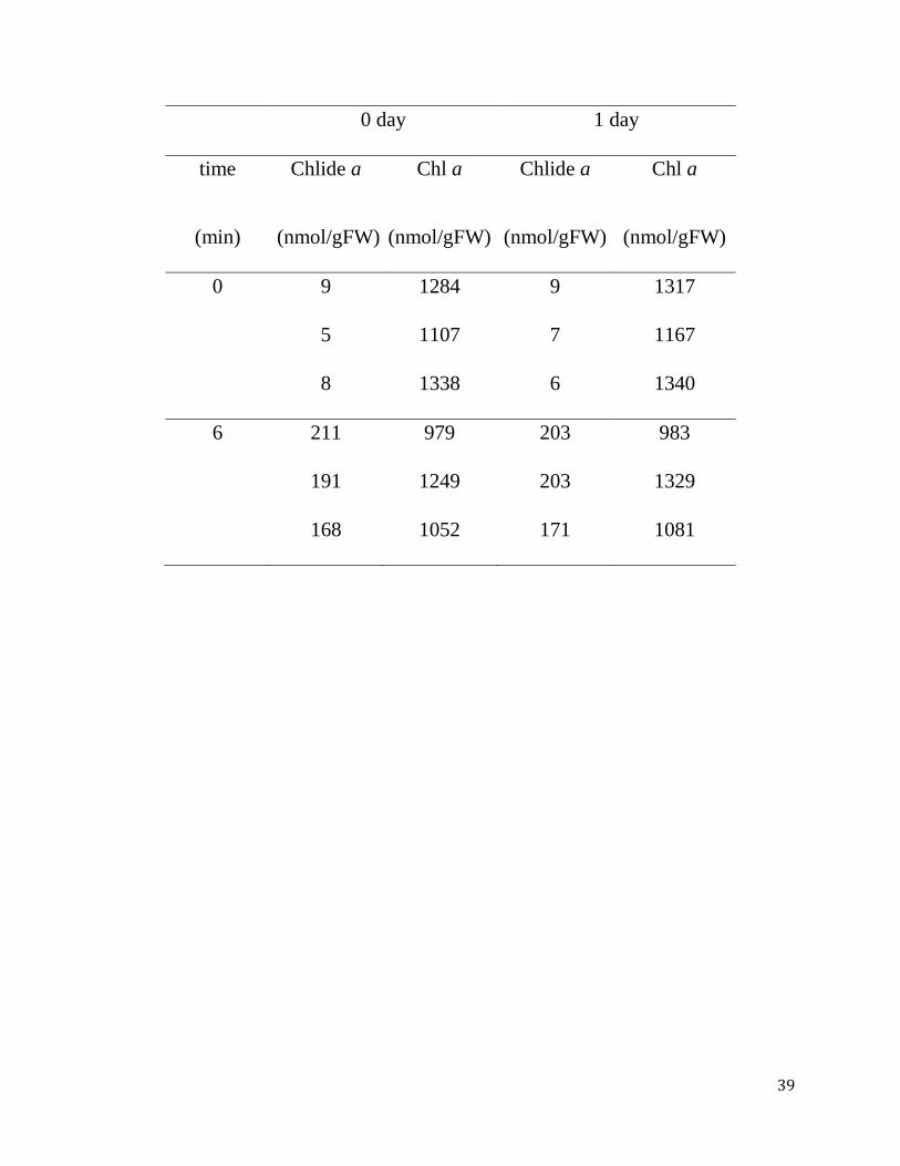
Table 2-1. Stability of chlorophyll a and chlorophyllide a after extraction with
pure acetone. Mature Arabidopsis leaves (7th - 9th leaves counting from the
bottom of the 4-week-old plants) were treated with 50 M MeJA and were kept in
complete darkness for 3 days. Chlorophyll was extracted by immersing leaves in
pure acetone as it is described in the "Time-course experiments" subsection of the
Methods section. "Time" indicates the incubation time at room temperature. After
removing the residual leaf tissue by centrifugation, the extracts were incubated for
24 hours at room temperature. Each line represents a single experiment with an
extract from a single leaf, and experiments were repeated three times in the same
conditions. Values represent chlorophyllide a or chlorophyll a concentrations per
gram fresh weight of leaves. Chl a, chlorophyll a. Chlide a, chlorophyllide a.
39
0 day 1 day
time
(min)
Chlide a
(nmol/gFW)
Chl a
(nmol/gFW)
Chlide a
(nmol/gFW)
Chl a
(nmol/gFW)
0 9 1284 9 1317
5 1107 7 1167
8 1338 6 1340
6 211 979 203 983
191 1249 203 1329
168 1052 171 1081

40
Figure 2-1

41
Figure 2-1. Comparison of the CLH reaction and a proposed in vivo
degradation pathway of chlorophyll a. A. CLH catalyzes the hydrolysis of the
ester bond of chlorophyll to form chlorophyllide and phytol. B. An in vivo
degradation pathway of chlorophyll a proposed by Hörtensteiner and coworkers
(Schelbert et al. 2009). MCS denotes magnesium dechelating substances. PPH
denotes pheophytinase.

42
Figure 2-2

43
Figure 2-2. Formation of chlorophyllide a by the extraction of chlorophyll
with pure or 80% buffered acetone. Frozen leaves were immersed in pure
acetone or 80% acetone containing 20% (v/v) Tris-HCl, pH 8 and then incubated
in these solvents for 12 h at 4ºC in the dark. After that the pigments were extracted
by grinding with stainless steel beads as described in the Methods section. A.
Levels of chlorophyll a and chlorophyllide a per gram fresh weight of leaves. B.
Chlorophyllide a levels in sample extracts expressed as the ratio of cholorophyllide
a to the sum of chlorophyll a and chlorophyllide a. Chl a, chlorophyll a. Chlide a,
chlorophyllide a. Error bars indicate standard deviations. Sample size, n = 3.

44
Figure 2-3
45
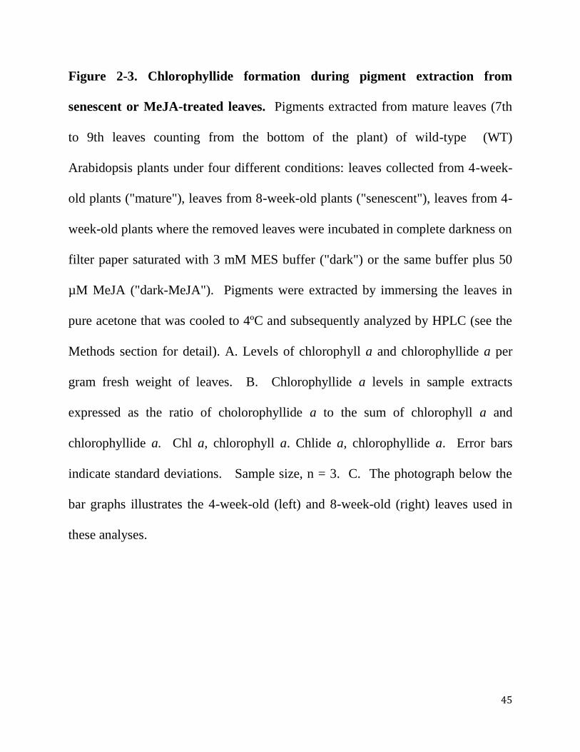
Figure 2-3. Chlorophyllide formation during pigment extraction from
senescent or MeJA-treated leaves. Pigments extracted from mature leaves (7th
to 9th leaves counting from the bottom of the plant) of wild-type (WT)
Arabidopsis plants under four different conditions: leaves collected from 4-week-
old plants ("mature"), leaves from 8-week-old plants ("senescent"), leaves from 4-
week-old plants where the removed leaves were incubated in complete darkness on
filter paper saturated with 3 mM MES buffer ("dark") or the same buffer plus 50
µM MeJA ("dark-MeJA"). Pigments were extracted by immersing the leaves in
pure acetone that was cooled to 4ºC and subsequently analyzed by HPLC (see the
Methods section for detail). A. Levels of chlorophyll a and chlorophyllide a per
gram fresh weight of leaves. B. Chlorophyllide a levels in sample extracts
expressed as the ratio of cholorophyllide a to the sum of chlorophyll a and
chlorophyllide a. Chl a, chlorophyll a. Chlide a, chlorophyllide a. Error bars
indicate standard deviations. Sample size, n = 3. C. The photograph below the
bar graphs illustrates the 4-week-old (left) and 8-week-old (right) leaves used in
these analyses.

46
Figure 2-4

47
Figure 2-4. Time course of chlorophyllide a formation during pigment
extraction. Chlorophyll was extracted by immersing leaves in pure acetone as it is
described in the "Time-course experiments" subsection of the Methods section. A.
Levels of chlorophyll a and chlorophyllide a per gram fresh weight of leaves. B.
Chlorophyllide a levels in sample extracts expressed as the ratio of chlorophyllide
a to the sum of chlorophyll a and chlorophyllide a. Chl a, chlorophyll a. Chlide a,
chlorophyllide a. Error bars indicate standard deviations. Sample size, n = 3. n.d.
= not detected.

48
Figure 2-5

49
Figure 2-5. Effect of quick boiling on the formation of chlorophyllide a
during extraction. Mature leaves (7th to 9th leaves counting from the bottom of
the plant) were collected from 4-week-old wild-type plants that had been sprayed
with 100 μM MeJA and kept for 3 days at the same growth conditions. The
collected leaves were immersed in pure acetone at room temperature directly or
were boiled for 5 and 10 sec respectively before they were immersed in pure
acetone, then they were ground with stainless steel beads by vigorous shaking (A
and B). Alternatively, pigments were extracted by immersing leaves in pure
acetone at 4ºC for 12 hours (C and D). A and C, Levels of chlorophyll a and
chlorophyllide a per gram fresh weight of leaves. B and D, Chlorophyllide a
levels in sample extracts expressed as the ratio of chlorophyllide a to the sum of
chlorophyll a and chlorophyllide a. Chl a, chlorophyll a. Chlide a, chlorophyllide
a. Error bars indicate standard deviations. Sample size, n = 3. n.d. = not detected.

50
Figure 2-6

51
Figure 2-6. The elution profiles of pigments from extracts separated by HPLC.
Representative HPLC profiles from each experiment are shown. A. HPLC profile
of pigments extracted by immersing wild-type (WT) leaves in pure acetone for 12
hours at 4ºC. B. An HPLC profile of WT pigments extracted by the quick boiling
method. C. HPLC profile of pigments extracted from leaves of WT plants by
immersing leaves in pure acetone for 96 hours at -30 ºC. D. HPLC profile of
pigments extracted from leaves of WT plants by immersing leaves in ethanol at
4ºC for 12 hours. E. HPLC profile of pigments extracted from leaves of WT
plants by immersing leaves in DMF at 4ºC for 12 hours. Peak 1, chlorophyllide b.
Peak 2, chlorophyllide a, Peak 3, pheophorbide a. Peak 4, neoxanthin. Peak 5,
violaxanthin. Peak 6, lutein. Peak 7, chlorophyll b. Peak 8, chlorophyll a. Peak 9,
pheophytin a. Peak 10, β-carotene.

52
Figure 2-7

53
Figure 2-7. Pigment extraction at -30ºC in pure acetone and at 4ºC in DMF.
Mature Arabidopsis leaves (7th to the 9th leaves counting from the bottom of the
plant) that were treated with or without MeJA (see the Method section for the
detail) were harvested and their pigments were extracted by three different
methods. In the first two methods, leaves were immersed in pure acetone at 4ºC
for 12 hours ("acetone 4ºC") and at -30ºC for 96 hours ("acetone -30ºC")
respectively. In the third method, leaves were immersed in DMF at 4ºC for 12
hours ("DMF 4ºC"). A. Levels of chlorophyll a and chlorophyllide a per gram
fresh weight of leaves. B. Chlorophyllide a levels in sample extracts expressed as
the ratio of chlorophyllide a to the sum of chlorophyll a and chlorophyllide a. Chl
a, chlorophyll a. Chlide a, chlorophyllide a. Error bars indicate standard
deviations. Sample size, n = 3. n.d. = not detected.

54
Figure 2-8

55
Figure 2-8. Comparison of extraction methods for chlorophyllide formation
with three different species (Glebionis coronaria, Pisum sativum L. and
Prunus sargentii Rehd.). Leaves were homogenated in acetone at room
temperatures ("room temperatures") or sub-zero temperatures ("sub-zero
temperatures"), respectively, by grinding leaves with steel stainless beads.
Alternatively, leaves were immersed in pure acetone at 4ºC ("acetone 4ºC") or in
pure DMF at 4ºC ("DMF 4ºC"), respectively, without the grinding procedure. In
the last pair of experiments, leaves were boiled for 5 ("boiling 5 sec") or 10 sec
("boiling 10 sec"), and then they were immersed in acetone at 4ºC for chlorophyll
extraction. The detail of the methods is described in the Methods section. A, C and
E. Levels of chlorophyll a and chlorophyllide a per gram FW of leaves. B, D and
F. Chlorophyllide a levels in sample extracts expressed as the ratio of
chlorophyllide a to the sum of chlorophyll a and chlorophyllide a. Chl a,
chlorophyll a. Chlide a, chlorophyllide a. Error bars indicate standard deviations.
Sample size, n = 3. n.d. = not detected.

56
CHAPTER 3: PLANTS USE CHLOROPHYLL-
DERIVATIVES FOR DEFENSE AGAINST
INSECT HERBIVORES THROUGH CLH
ACTIVITY

57
ABSTRACT
Insect herbivores and plants have been living together for a long history. During
co-evolution, plants have developed many different defense systems to protect
themselves from insect herbivores. Nearly all classes of secondary metabolites
were used as chemical defense compounds. However, whether chlorophyll (Chl)-
derivatives also can be used for defense against insect herbivores are unknown.
Chlorophyllase (CLH), which catalyzes the release of the phytol chain from Chl to
produce chlorophyllide (Chlide), is long considered to be involved in the first step
of Chl breakdown. However, Schenk et al presented evidence showing that CLH
was not essential for Chl breakdown during dark-induced senescence (Schenk et al.,
2007). Arabidopsis contains two isoforms of CLH (CLH1 and CLH2), and it was
hypothesized that under some stressful conditions or in the presence of methyl-
jasmonate (MeJA) CLH1 is involved in Chl breakdown. To examine the possible
involvement of CLH1 in MeJA-induced Chl breakdown, we analyzed the clh
mutants which lack single or both CLH isoforms after MeJA-treatment. The results
showed that the clh knockout lines were still able to degrade Chl at the same rate
as wild types. Subsequently, by membrane fractionation and an analysis of the
localization of the fusion of CLH1 and yellow fluorescent protein, we found that
CLH1 was located outside the chloroplasts, and it was located to the tonoplast and
endoplasmic reticulum (ER). We subsequently demonstrated that Chlide and

58
pheophorbide (Pheide) were poisonous to the larvae of a common generalist
Spodoptera litura. In addition more larvae were killed by feeding the two Chl-
derivatives, and the development of the larvae was significant delayed by feeding
Childe. In contrast, by feeding the same concentration of Chl, survival ratio and
development rate of the larvae could not show significant difference comparing
with control. To mimic the leaf cells after they were eaten by larvae, the leaves
were homogenized under either pH8 or pH10 conditions. The Chlide amount was
subsequently analyzed by HPLC after the leaf homogenate was incubated at room
temperature. The result showed that much Chlide were produced in the leaves
mixture of CLH1 overexpression lines and WT and only little in clh1-1 mutant.
Finally we fed Spodoptera litura with WT, clh1-1 and three CLH1 overexpression
lines for 11 days. Although almost all of the larvae who ate WT and clh1-1 were
survived, the average survival rates were significantly decreased when they ate the
leaves of three CLH1 overexpression lines. Taken together, we conclude that
CLH1 is not involved in Chl breakdown even during MeJA-induced senescence in
Arabidopsis. We suggest that plants utilize Chl-derivatives for defense against
insect herbivores through CLH activity. This system is quite convenient for
defense against insect herbivores and widely exists in the plant kingdom.
Keywords: Chlorophyllase; Methyl jasmonate; Chlorophyll-derivatives; Insect
herbivore; Defense

59
3.1 Introduction
Plants suffer from abiotic and biotic stress throughout their life in their natural
environment because they are sessile organisms. Except the competititon among
plants, herbivores and pathogens are the main biotic stresses. As one of the most
important group, insect herbivores have been living together with plants for more
than 350 million years (War et al. 2012) (Fürstenberg-Hägg et al. 2013). Although
the size of each insect herbivore is small, the populations and types are amazing.
Losses due to insect herbivores were estimated at 10-20% of major crops (Ferry et
al. 2004). Especially in some seasons or years when the natural conditions are quite
suitable for the growth of insect herbivores, they will become to a disaster to plants.
For example, plague development have occurred with the locust in many countries
and regions in the world (Showler 1995) (Z Liu et al. 2008). On the other hand,
plants also developed defense systems to protect themselves from insect herbivores.
Fully utilizing the defense system which produced by plants themselves was one of
the effective and environmentally-friendly strategy to protect our target plants,
especially crops. For example, maize was protected from lepidopteran larvae by
the co-cultivation with an Africa grass (melinis minutiflora) that release abundant
volatile compounds (Z.R. Khan et al. 1997) (Z.R. Khan et al. 2000). Therefore,
studying on the mechanism of plant defense against insect herbivores not only
increase our base knowledge of science, but also help us to control the pest

60
environmentally sustainable and cost-effective by improving plant resistant other
than by spraying pesticide.
The types of plant defense against insect herbivores can be divided into two large
groups: direct and indirect defense reactions. Direct defense also contains two
groups: structural and chemical defense. Structural defenses are usually barriers
trait for insect herbivores such as sclerophylly and pubescence (Agren and
Schemske 1993) (Hanley et al. 2007). Chemical defenses, except some heavy
metals such as zinc, lead and nickel can be concentrated from the environment by
some plant species for defense (Jhee et al. 2005) (R Boyd 2012), organic
compounds for defense are well reported (Mithöfer and Boland 2012). They almost
cover all classes of (second) metabolites which are produced by plants in order to
retard the normal growth of insect herbivores (I Baldwin and Preston 1999) (I
Baldwin 2001). These compounds belong to various chemical classes such as
isoprene-derived terpenoids, N-containing alkaloids, phenolic compounds and so
on (Mithöfer and Boland 2012). In addition, even amino acids, peptides and fatty
acid derivatives are used by plants for defense as well (Hylin 1969) (Ryan and
Byrne 1988) (T Huang et al. 2011) (Habib and Fazili 2007). Some of the defense
compounds specially exist in some plant species, while others ubiquitously exist in
almost all of the plants as common defense systems.

61
Instead of direct defenses, indirect defenses are that plants reduce insect herbivores
by increase predation of herbivores by luring and keeping predators on a plant with
food rewards, shelters from harsh conditions, or chemicals signaling prey
availability. The only indirect defenses that actively attract predators are volatile
organic chemicals (VOCs) (Mortensen 2013).
Some of the defenses are constitutive (pre-formed), while others are induced after
attack by insect herbivores (Wittstock and Gershenzon 2002) (M Chen 2008).
Inducible defenses incur less metabolic costs to host plants because they only
induced when insect attack happen. Inducible defenses were divided into three
steps: surveillance, signal transduction, and the production of defensive chemicals
(A Kessler and IT Baldwin 2002) (Ferry et al. 2004) (Smith and Boyko 2007).
Although there are many signal transduction pathways for defense against insect
herbivores in plants, in general, a phytohormone–jasmonates (JA) are regarded as
important signalling substances upon wounding or biotic attack (Creelman and
Mullet 1997). The JA signalling pathway is important for defense against
necrotrophic pathogens and also against insect herbivores, although the crosstalk of
JA with other two phytohormones: salicylates and ethylene also exist in plants
(KL-C Wang et al. 2002). Many plant defense genes are inducible though JA
pathway such as VSP2 and PDF1.2 (Pieterse et al. 2009).

62
Chlorophyll (Chl), the most abundant pigment on earth, is an essential component
in photosynthesis. It is responsible for harvesting solar energy in photosynthetic
antenna systems, and for charge separation and electron transport within reaction
centers. However, Chl is also a dangerous molecule and a potential cellular
phototoxin. For example, Chl and its derivatives are strong photosensitizers; that
is, when the present is excess, they will generate reactive oxygen species (ROS)
(Camp et al., 2003). As it allows for recycling of nitrogen and other nutrients for
protection from buildup of phototoxic Chl intermediates (reviewed in
Hörtensteiner, 2006), Chl degradation is vital during leaf senescence or fruit
ripening (Kräutler 2008), but also as a response to many biotic and abiotic stresses
(Benedetti and Arruda 2002) (Kariola et al. 2005) (Hörtensteiner and Kräutler
2011). Chl metabolism is highly regulated during plant development. However,
Chl degradation mechanism is less understood than that of Chl biosynthesis. A
Chl breakdown pathway had been suggested based on the identification of key
enzymes and catabolites along the pathway. In this pathway, removal of the phytol
chain by chlorophyllaase (CLH) is the first step, which converts Chl to phytol and
chlorophyllide (Chlide). Then after Mg-dechelation from Chlide, the porphyrin
ring of pheophorbide (Pheide) is oxygenolytically opened by Pheide a oxygenase
(PAO) inside of chloroplast, resulting in loss of the green color of the molecule.
After that, the intermediate product, Chl catabolite (RCC) is degraded by reductase

63
(RCCR). The product, a primary fluorescent catabolite (pFCC), is transported to
the vacuole and converted to the final non-fluorescent Chl catabolites (NCCs) by
non-enzymatic tautomerization (Hörtensteiner, 2006) (Hörtensteiner and Kräutler
2011).
CLH was discovered about 100 years ago (Stoll, 1912). In vitro, this enzyme
displays high activity to catalyze the hydrolysis of Chl to phytol and Chlide (Stoll,
1912). Afterwards, many researchers detected this enzyme’s activity and analyzed
the catalytic characterization of it in algae and green tissue of many plants (Ardao
and Vennesland, 1960) (Mcfeeters et al., 1971) (Klein and Vishniac, 1961)
(Terpstra and Lambers, 1983) (Benedetti and Arruda, 2002) (Arkus et al., 2005)
(Shioi et al. 1991). Therefore, for a long time, CLH has been considered the first
enzyme involving in the progress of Chl degradation (Hörtensteiner, 2006).
However, whether this enzyme is involved in Chl breakdown in vivo was
questioned recently, based on the research using Arabidopsis mutants in which
CLHs was absent. In Arabidopsis, there are two isoforms of CLH (CLH1 and
CLH2). Interestingly, both clh1 and clh2 single and double knockout lines were
still able to degrade Chl during dark-induced leaf senescence (Schenk et al., 2007),
which suggested that both CLH1 and CLH2 are not essential for dark-induced leaf
senescence-related Chl breakdown in Arabidopsis. Instead, another enzyme, which
was named as pheophytinase (PPH) was localized to the chloroplast and the PPH

64
knock-out lines showed a strong stay-green phenotype during dark-induced leaf
senescence (Schelbert et al., 2009). They found PPH possess the activity to
catalyze the conversion of pheophytin to Pheide, while it cannot catalyze the
conversion of Chl to Chlide although both reactions regard to removing the phytol
chain. Therefore, another Chl degradation pathway, PPH pathway should exists in
plants. PPH pathway starts with removing Mg2+
from Chl a by metal-chelating
substance, then the phytol side chain is removed from the last step product
pheophytin a to produce Pheide a by the catalyzation of PPH (Schelbert et al.,
2009). Schelbert and coworkers suggested that in dark-induced senescence, PPH
pathway takes charge of Chl degradation rather than the CLH pathway. As well as
known, under normal growth condition, Chl degradation in plants happened only in
the old leaves because of the development stage, while Chl degradation will be
started earlier and faster in leaves when plants are under stress or phytohormones
treated conditions. The age dependent and independent senescence mechanisms are
largely different with each other (Weaver et al. 1998) (BF Quirino et al. 2000).
Therefore, it is possible that the Chl degradation pathway also different between
the two types of Chl degradation. It was reported that CLH1 mRNA was induced
rapidly by a treatment of Methyl-jasmonate (MeJA), wounding, pathogen infection
and other stress conditions (Benedetti et al. 1998)(Kariola et al. 2005) while CLH2
did not respond to MeJA (Tsuchiya et al., 1999). Because MeJA is a well-known

65
phytohormone promoting senescence and Chl degradation in plants (He et al.
2002), it could be hypothesized that CLH1 was important for Chl breakdown
specially during MeJA (or other stress)-induced senescence and Chl catabolism in
Arabidopsis like citrus CLH was involved in ethylene-induced fruit color–break
(Azoulay Shemer et al. 2008).
Intracellular localization is often a key strategy to understand protein functions and
regulation in vivo and has been a controversial issue with regard to the enzyme
CLH (Azoulay Shemer et al., 2008). A lot of research reported that CLHs from
different kinds of plant were located in chloroplasts (Krossing, 1940) (Garcia and
Galindo, 1991) (Matile et al., 1997) (Trebitsh et al., 1993) (Okazawa et al., 2006).
Even until recently, it was reported that both the precursor and mature forms of
citrus CLH was located in chloroplasts during ethylene-induced Chl breakdown
(Harpaz-Saad et al., 2007) (Azoulay-Shemer et al., 2011) (Azoulay Shemer et al.,
2008). However, after cloning of the two isoforms of CLH, no typical feature of
signal sequence for chloroplast targeting was identified in CLH1, whereas CLH2
had only a weakly predicted signal sequence for chloroplasts (Tsuchiya et al.,
1999). In Arabidopsis, CLH1 is the major isoform and it contributes to the most
activity of CLHs (Schenk et al., 2007). It was hypothesized that CLH1 was located
outside of chloroplast, and there was an additional Chl degradation pathway
located outside of chloroplast (Takamiya et al. 2000). In additionally, transient

66
expression of CLH1-YFP (C-terminal fused) in Arabidopsis protoplast showed that
CLH1 was located in cytosol rather than chloroplast (Schenk et al., 2007). One
reasonable explanation was that the transient expression system was not stable for
the existence of the fusion protein, the GFP signal could not response the true
localization of CLH. The other explanation was suggested that GFP fusions may
interfere with the natural localization of CLHs due to masking of amino- and
carboxy-terminal targeting and modification signals (Tian et al., 2004) (Azoulay
Shemer et al., 2008). Therefore, to identify the localization of CLH1, it was
necessary to check the localization of CLH1-YFP which was stably overexpressed
in plants. Furthermore, the localization of native CLH1 is also required.
In this study, we aimed at elucidating what is the function of CLH. By observation
the phenotype of single and double knockout mutants of CLHs and wild type (WT)
during MeJA-induced senescence, and also the localization of CLH1, our data
showed that Arabidopsis CLH1 is not involved in Chl breakdown during MeJA-
induced senescence in Arabidopsis. However, we demonstrate that Chlide and
Pheide are poisonous to the larvae of a common generalist Spodoptera litura. After
the leaves were eaten by insect herbivores, Chlide can be produced during chewing
and digestion. The amount of Chlide formation heavily dependent on the activity
of CLH. Finally we employed WT, clh1-1 and three CLH1 overexpression lines to
test the same kind of insect larvae. Although almost all of the larvae who ate WT

67
and clh1-1 were survived, the average survival rate of larvae was significantly
decreased when they ate the three CLH1 overexpression lines. Taken together, we
conclude that CLH1 is not involved in Chl breakdown even during MeJA-induced
senescence in Arabidopsis. Here, we would like to suggest that plants use Chl-
derivatives for defense against insect herbivores through CLH activity. This system
is quite convenient for defense against insect herbivores and widely exists in the
plant kingdom.
3.2 Materials and methods
3.2.1 Plant materials
Arabidopsis T-DNA insertion lines SALK_124978 (designated clh1-1; ecotype
Columbia (col)), SAIL_646_E09 (designated clh2-2; ecotype col), clh1-1/clh2-2
were described by Schenk et al. (Schenk et al., 2007). Another T-DNA insertion
line GK-453A08 (designated pph3; ecotype col) was obtained from the European
Arabidopsis Stock Center, Nottingham, UK (Schelbert et al. 2009). GFP::cDNA
transgenic lines (Q4 (GFP-ER protein) and Q5 (GFP-δ-TIP)) were obtained from
The Arabidopsis Information Resource (TAIR, (Cutler et al. 2000)). Homozygous
plants were identified by PCR using T-DNA and gene-specific primers as listed by
Schenk et al. and Schelbert et al. (Schenk et al., 2007) (Schelbert et al., 2009).

68
Plants were grown on soil in long-day (16 h light/8 h dark) or short-day (10 h
light/14 h dark) growth conditions under fluorescent light of 70–
90 μmol photons m−2
s−1
at 23 °C for 4 weeks. For senescence, all the leaves from
each plant which was grown in long day condition were detached and placed on
wet filter paper (3 mM MES buffer, pH 5.8, with or without 50 μM MeJA) and
incubated in permanent darkness for up to 4 days.
3.2.2 Pigment analysis
The leaves were imaged by digital camera, and the sizes were calculated by ImageJ
software (http://rsbweb.nih.gov/ij/). Pigments were extracted from leaf tissue by
homogenization at -20°C with acetone (Hu et al. 2013). Extracts were centrifuged
for 5 min at 15,000 rpm at 4°C, and the supernatant was analyzed by HPLC with
Symmetry C8 column (150 mm in length, 4.6 mm in i.d.; Waters, Milford, MA,
USA) according to the method of Zapata et al. (Zapata et al., 2000). Elution
profiles were monitored by measuring absorbance at 410 nm. Standard pigments
(Chl a and Chl b) were purchased from Juntec Co. Ltd. (Odawara, Japan).
3.2.3 Arabidopsis transformation and confocal microscopic analysis of the
localization of eYFP
Complementary DNA of CLH1 was cloned from the wild-type plants. eYFP was
cloned from pEarleyGate101 plasmid (Earley et al., 2006). For C-terminal fusion

69
(CLH1-eYFP), the construct was generated by fusing the eYFP sequence to the C
terminus of CLH1, and then 5’UTR and 3’UTR of CLH1 were fused to the N and
C-terminal of the fusion sequence respectively. By performing the LR clonase
reaction, the construct cloned in pENTR was transferred into the expression vector
pEarleyGate100 (Earley et al., 2006) containing a gene conferring glufosinate
resistance (Nakagawa et al., 2007) under the control of the 35S promoter of
pBI221 (Clontech). The recombinant binary vector was introduced into the
Agrobacterium (Rhizobium radiobacter) strain GV2260. Agrobacterium was
infected into Arabidopsis using the floral dip method (Clough and Bent, 1998).
Two-week-old seedlings of transformants were selected by spray basta.
Protoplasts were isolated from mature leaves of 4-old-week plants by following the
method described by Robert and coworkers (Robert et al., 2007). Expression and
localization of the CLH1-eYFP protein in transformed transgenic plants were
examined under the confocal fluorescence microscopy (describe instrument, Nikon
Co. Ltd., Japan).
3.2.4 Preparation of membrane and soluble fractions
Leaves from 4-week-old plants grown under the short-day conditions (short day)
were harvested and lysed using a mortar and pestle under liquid nitrogen. The
fractions were prepared as described by Oka et al. (Oka et al., 2010).

70
3.2.5 Isolation intact chloroplasts and vacuole
Arabidopsis rosette leaves are harvested from 4-week-old Arabidopsis which were
grown in the short-day conditions. Intact chloroplasts were prepared essentially
according to the method that was described by Salvi et al. (Salvi et al., 2008).
Intact vacuoles were prepared according to the protocol that was described by
Robert et al. (Robert et al., 2007). Protein concentrations were determined by
Lowry analysis ((Lowry et al., 1951), with bovine serum albumin (BSA) (Sigma
Chemical) as standard protein.
3.2.6 Membrane fractionation
Arabidopsis leaves used for membrane fractionation were grown on soil for 28
days in the short-day conditions. The fractionation method was modified from
which was described by Oka et al. (Oka et al., 2010) and Wulfetange et al.
(Wulfetange et al., 2011). Leaf tissues was homogenized in buffer containing 50
mM Tris (pH 7.6 at 22°C), 20% glycerol, and 150 mM NaCl with either 5 mM
MgCl2 and 2 mM EGTA (for subsequent sucrose (Suc) gradient separation in the
presence of Mg2+
) or with only 2 mM EDTA (for subsequent Suc gradient
separation in the absence of Mg2+
). Leaves were homogenized three times at a low
speed in a blender with a 10-fold amount of buffer. The homogenates were filtered
through two layers of Miracloth (Merck) and then centrifuged for 10 min at 10,000
g at 4 °C. The supernatants were ultra-centrifuged for 30 min at 100,000 g at 4°C.

71
The resulting pellets were resuspended in 0.5 mL/g in buffer containing 50 mM
MOPS-NaOH, PH 7.0, 10% (w/v) Suc, EDTA-free complete protease inhibitor
cocktail (Sigma), and either 5 mM or 5 mM MgCl2. Then the suspension was
centrifuged at 6,000 g for 3 min to remove un-suspended small pellets. Six
hundred ml of the supernatant was loaded onto the top of the following
discontinuous Suc gradient: from bottom to top, 200 µl of 55% Suc, 450 µl of 50%,
45%, 40%, 35%, 30%, 25% and 20% Suc solution containing the same
components with the re-suspension buffer except Suc. Then the gradient was
centrifuged in a bucket rotor for 20 h at 100,000 g at 4°C. After centrifugation,
the gradient was collected in 200 µl increment fractions from top to bottom, and
the fractions were stored at -30°C before using.
3.2.7 Immunoblot analysis
Total protein was extracted from leaves using extraction buffer containing 50 mM
Tris-HCl (pH=8), 12% Sucrose, 2% LDS, and 1.5% DTT. Before SDS-PAGE
separation, all the samples were mixed with equal volume of 2×Ling's urea buffer
containing 8 M urea, 30 mM Suc, 10 mM Tris-HCl, pH 8.0, 40 mM dithiothreitol,
2% SDS, 1 mM EDTA and 10 g/mL Pyronin Y, and then they were separated on a
14% polyacrylamide gel and electroblotted to PVDF membrane. The CLH1 protein
was detected with an anti-CLH1 antiserum (rabbit) using the ECL Plus
immunodetection kit (GE Healthcare). The membrane type was identified by the

72
use of a set of polyclonal rabbit antibodies against specific markers: ER luminal
BiP2 (Agrisera), Plasma membrane (PM) marker H+-ATPase (Agrisera), tonoplast
marker V-PPase (CosmoBio, Inc.). Thylakoid membranes were identified by
HPLC analysis of the Chl content (Zapata et al., 2000).
3.2.8 CLH Assay
The method for CLH assay was modified from which was described by Tsuchiya
and coworkers (Tsuchiya et al., 1997). Leaves were homogenized with 10 times
weight of the assay buffer containing 50 mM Tris-HCl, 38 mM octylglucoside
(OG), 1% (v/v) protease inhibiter cocktail (PH=7.5, on ice). The homogenate was
centrifuged for 10 min at 15,000 rpm at 4 °C. The supernatant was transferred into
a 1.5 mL microcentrifuge tube. The pellet was re-extracted with 10 times weight of
the assay buffer, and the extract was pooled in the same 1.5 mL microcentrifuge
tubes. To 375 µl of an extract (or buffer only as the control), 125 µl of acetone
containing Chl a (500 µg/ml) was added and incubated for 10 min at 37ºC. The
reaction was stopped by adding 1 ml of hexane and 0.5 ml of acetone to the
reaction mixture. Then the total mixture was vigorously shaken and centrifuged for
5 min at 10,000 g at 4°C, so that the Chlide a by the enzyme reaction was
partitioned into the lower aqueous layer. After centrifugation at 10,000 g for 5
min, Chlide was partitioned into the lower aqueous acetone layer, so that the lower
layer was recovered to a new tube. The concentrations of Chlide a was determined

73
by measuring an absorbance at 667 nm using the absorption coefficients of 76.79
mM-1
cm-1
for Chlide a (Porra et al., 1989). Chlide a amount which was produced
during a unit time was defined as the activity of CLH. The relative activity of CLH
in 50 µM MeJA treated leaves for 2 days was considered as 100%.
3.2.9 Pigments preparation
Pheide a was purchased from Wako Pure Chemical Industries, Ltd. Japan.
For preparing Chlide, the method was modified from the Müller and coworkers’
method (AH Müller et al. 2011). Glebionis coronaria (garland chrysanthemum)
leaves was purchased from the supermarket in Sapporo, Japan. Leaves of G.
coronaria were ground in liquid nitrogen using mortar and pestle. The fine powder
(5–10 g) was suspended in 20 ml of acetone, and then 20 ml of Tricine–NaOH
(0.2 M, pH 8.0) was added. After shaking at 220 min-1
for 2 h at room temperature
in the dark, the leaf debris was removed by centrifugation and the green
supernatant was repeatedly extracted with 40 ml of hexane until the epiphase was
colorless. The leaf debris can be used for extracting Chlide additional twice
following the same procedure. Diethyl ether was added twice to the pigment
solutions, and the green epiphases were combined and dried in N2.
For preparing Chl, the fine leaf powder was suspended in the same volume of
acetone, first shaking at 220 min-1
for 2 h at room temperature in the dark, then 20

74
ml of Tricine–NaOH (0.2 M, pH 8.0) was added. The leaf debris was removed by
centrifugation and the green supernatant was extracted with 40 ml of hexane, then
pigments appeared in the hexane phase were collected and dried in N2.
Before HPLC purification, small amount of pure acetone was added to the dried
pigments in order to dissolve all of the pigments. For HPLC purification, C8
column and solvent A and B of mobile phase which was reported by Zapata and
coworkers were used (Zapata et al. 2000). Different gradient profiles were adjusted
for separating Chlide a and Chl a (Table 3-1). For purification Chlide, The flow
rate was fixed at 9 m1 min-l.
After getting the target pigments in the HPLC solvent, Diethyl ether and water
were added immediately to concentrate target pigments into the Diethyl ether
phase. After the green epiphases were combined, small volume of green epiphases
was taken out and dried in N2. Then 80% acetone (acetone:water (v:v) =8:2 ) was
added to dissolve the pigments, and then the concentration was analyzed by
spectrophotometer (Porra et al. 1989). Later the total amount of pigments was
calculated. Finally, all of the green epiphases were dried in N2 and stored in deep
freezer.
3.2.10 Pigments feeding experiments

75
Pigments were first dissolved into the same amount of DMF, then mixed with
artificial food. The final concentration of DMF was 0.1% in food with 400 nmol/g
Chlide a, 200 nmol/g Chlide a, 400 nmol/g Chlide a, 400 nmol/g Chl a
respectively, and control without any pigment but same concentration of DMF.
Each group including 40 Spodoptera litura larvae which were just hatched. The
same amount of food mixed with different kinds or concentrations of pigments
were fed to each larva which was separated from each other by plastic tubes. The
growth status of each larva was recorded at 11th day.
3.2.11 Leaf homogenization experiment
CLH1-YFP were overexpressed in the background of clh1-1. Three stable
overexpreesion lines, WT and clh1-1 were used for this experiment. Plants were
grown in soil under short-day conditions (10 h light/14 h dark) in growth chambers
under fluorescent light (70-90 mol photons m-2
s-1
) at 23ºC. Each sample including
0.3 g mature fresh leaves from 7-week-old plants. They were subsequently
homogenized by glass homogenizer with 1ml pH8 (Tris-HCl) or pH10 (0.1 M
Na2CO3-NaHCO3) buffer respectively. Then the leaf mixture was kept at room
temperature (25 ) for 0.5 h or 2 h. After incubation, pigments were extracted by
pre-cooled pure acetone (-30 ) (Hu et al. 2013) and analyzed by HPLC using
Zapata’s method (Zapata et al. 2000).

76
3.2.12 Plant feeding experiment
The same three stable CLH1 overexpression lines, WT and clh1-1 mutant were
grown in soil under short-day conditions (10 h light/14 h dark) in growth chambers
under fluorescent light (70-90 mol photons m-2
s-1
) at 23ºC. Twelve uniform plants
from each plant line were fed by 60 Spodoptera litura larvae which were just
hatched (each plant was eaten by 5 larvae). Then larvae growth and survival status
were observed at 11th day.
3.3 Results
3.3.1 Both the expression levels of CLH1 and total CLH activity were induced
by MeJA treatment.
It was suggested that Arabidopsis CLH1 expression levels were induced by MeJA
treatment. However, these results were just showed at the transcription levels
(Benedetti et al. 1998) (Tsuchiya et al. 1999). To demonstrate that CLH1 is really
induced by MeJA at translation levels and also all the induced CLH1 is active, the
following experiments were performed.
Firstly, the specificity of the antibody for CLH1 was checked by comparing the
western-blotting signals between WT and clh1-1 mutant. The 7-8th leaves
(counting from the bottom) of 4-week-old plants were treated under the condition

77
which was described in Fig. 3-1. Total protein was extracted and analyzed by
immunoblotting using an anti-CLH1 antiserum. WT showed a signal at about 35
KD, which is consistent with the size of CLH1 protein estimated from the CLH1
coding sequence. In contrast, no signal was obtained from the protein extracts of
the clh1-1 mutant (Fig. 3-1), indicating that the antibody is specific to CLH1.
Although we attempted to detect CLH2 protein with an anti-CLH2 antiserum, we
could not detect the specific signal of CLH2 in WT.
Then we analyzed the content of CLH1 during dark senescence with or without 50
µM MeJA treatment. Consistent with the reported patterns of mRNA expression
(Tsuchiya et al. 1999) (Kariola et al. 2005), the expression level of CLH1 was
slightly induced during dark-induced senescence in the absence of MeJA and it
was much enhanced (more than two times ) by treating with 50 µM MeJA (Fig. 3-
1) .
In vitro measured CLH activity was correlated to the CLH1 protein level (Fig. 3-2).
Compared to the leaves before senescence, CLH activity was slightly increased
after 2 days dark treatment without MeJA treatment. However, the WT leaves in
the same condition treating with 50 μM MeJA, CLH activity was induced more
than two times (Fig. 3-2). Hence, the CLH protein level was increased by darkness
and it was enhanced by MeJA treatment. Since the CLH activity and protein levels

78
are correlated during MeJA treatment, it is concluded that the activation of CLH is
not necessary. On the other hand, in the clh1-1 mutant which lack of CLH1, only
about 10% of CLH activity were retained. In addition, MeJA treatment could not
enhance the activity of CLH in this mutant. It is possible that CLH2 contributes to
the activity observed in the clh1-1 mutant. These results implied that CLH1
contributes most of the activity of CLH in Arabidopsis.
3.3.2 The clh1-1 mutant showed a similar Chl breakdown rate with WT even
during MeJA-induced senescence
Although CLH1 was absent in the clh1-1 mutant, we could not observe any
difference in the phenotype of green plants between the clh1-1 mutant and its
ecotype (Fig. 3-3). That is reasonable, because the knockout plants which absence
enzymes which involved in Chl catabolism, usually no any visible difference
phenotype before senescence, compared with WT (Pružinská et al. 2003) (Kusaba
et al., 2007) (Schelbert et al., 2009) (Meguro et al., 2011).
It was suggested that CLH1 might be involved in Chl degradation only under some
stress conditions (Azoulay Shemer et al. 2008). Because under stress conditions,
Chl degradation happened much faster and CLH1 expression levels were also
much enhanced. For example, MeJA promotes leaf senescence and Chl
degradation (He et al. 2002), at the same time, it also enhances the expression
levels of CLH1 ((Tsuchiya et al. 1999) (Kariola et al. 2005) and this study, see Fig.

79
3-1 and 3-2). To check the possibility of this hypothesis, we performed dark-
induced leaf senescence with or without 50 µM MeJA treatment. All leaves from
4-week-old plants of the different lines were detached and kept in dark with buffer
in the absence or presence 50 µM MeJA for 4 days. The results were shown in
Figure 3-3. Without MeJA treatment, the leaves of both single and double mutants
of CLHs turned yellow as fast as WT during dark incubation, whereas in the pph
knockout mutant (It was demonstrated that PPH is involved in Chl degradation in
Arabidopsis), all leaves remained green after 4 days of dark incubation. The
average Chl content per unit area of leaves was also measured (Fig. 3-3). Decrease
in Chl content was not suppressed in both of the clh1-1 and clh1-1/2-2 double
mutants. In other words, the decrease of Chl content in the clh1-1 and clh1-1/2-2
mutants were similar with WT. If CLHs were involved in Chl degradation like
PPH, the Chl degradation rate should be slower in clh mutants.
The rate of senescence and Chl degradation was strongly accelerated by MeJA
treatment in WT (Fig. 3-3). However, the Chl degradation rate was also enhanced
in clh mutants by treating with 50 µM MeJA. Additionally, the Chl degradation
rate of leaf in CLHs mutants was similar with WT. On the contrary, the leaves of
PPH mutant showed stay-green phenotype and retained much more Chl than the
other lines after MeJA treatment (Fig. 3-3). At the same time, we noticed that the
content of Chl was retained in-dependent MeJA treatment in PPH mutant. These

80
results implied that the PPH pathway for Chl degradation can be promoted by
MeJA treatment. In other words, MeJA promotes Chl degradation through the PPH
pathway rather than CLH.
Mutants of the enzymes for Chl metabolism are known to accumulate intermediate
molecules of Chl synthesis or degradation. Altering the expression of CLH1 in
transgenic Arabidopsis caused changes in the Chl-to-Chlide ratio was reported
(Benedetti and Arruda 2002). To investigate the effect of absence of CLHs on Chl
metabolism, Chl and its derivatives were extracted from leaves of the WT and clh
mutants and analyzed by HPLC. We found that the content of Chl a and Chl b
were similar between the WT and CLH mutants before and after dark-induced
senescence with and without 50 µM MeJA treatment (data are not shown). And we
also noticed that there was almost no Chlide detected by HPLC if we always
extract pigments at sub-zero temperatures (Fig. 2-4).
3.3.3 CLH1 is located to both tonoplast and ER membrane rather than
chloroplast
CLH1-eYFP (C-terminal fusion) was over-expressed in Arabidopsis to determine
the subcellular localization of CLH1. After getting the stable CH1-eYFP over-
expression lines, protoplasts were isolated from the leaves and they were observed
by using a confocal microscope. From the fluorescent image, it was clear that the
fluorescence of eYFP (yellow) was located outside of the chloroplast (red), and it

81
was most likely located outside of the chloroplast, and on some membrane
structure (Fig. 3-4). This result was according to the transient expression
experiment which was reported in 2007 (Schenk et al., 2007). Comparing the
localization of GFP marker of ER (GFP-ER protein) and tonoplast (GFP-δ-TIP)
(Fig. 3-4) (Cutler et al., 2000), it was difficult to demonstrate CLH1-eYFP was
located to the ER or tonoplast, or both of them.
It was reported that when target protein was fused with GFP, the localization of
this target protein would be interfered due to masking of amino- and carboxy-
terminal targeting and modification signals (Tian et al., 2004). Therefore, the
localization of native CLH1 would be required.
To examine the localization of native CLH1 in Arabidopsis leaves, we first
prepared membrane and soluble fractions from WT leaves and checked the CLH1
by immunoblotting. The result showed that CLH1 was detected in the membrane
fraction rather than soluble fraction (Fig. 3-5). The marker proteins, BiP2 for the
ER and V-PPase for the tonoplast were mainly detected in the membrane fraction,
suggesting the majority CLH1 is associated with the membrane. However, a little
amount of CLH1 was detected in the soluble fraction. At the same time, we
detected a little amount of ER marker protein BiP2 in the soluble fraction. This
signal should be the contamination of membrane proteins into the soluble fraction.

82
It was reported that CLH1 was detected in vegetative vacuole by proteome analysis
besides the reports of the chloroplast localization (Carter et al. 2004). In this study,
both intact chloroplast and vacuole were isolated from WT and the clh1-1 mutant,
and then the samples were used to determine CLH1 by immunoblotting. In WT,
CLH1 was concentrated in the vacuole sample, while we could not detect any
CLH1 in the sample of chloroplasts even 3 times of the total protein amount were
loaded (Fig. 3-5). This result showed that native CLH1 is located to the tonoplast
rather than to the chloroplast (Fig. 3-5).
In order to confirm the tonoplast localization of CLH1 and to test whether CLH1 is
also located to other kinds of membrane, we try to separate different types of
membrane. Then the sucrose (Suc) density gradient centrifugation experiments
were performed. Total subcellular fractions were extracted from leaves by
membrane extraction buffer containing either Mg2+
or EDTA. The association of
ribosomes with the ER is Mg2+
-dependent, therefore depletion of Mg2+
from the
extraction buffer results in dissociation of ribosomes from the ER, then the ER will
be redistributed to lower Suc density position, comparing with the ER which
associated by ribosome after fractionation (Lord et al., 1973) (Lord, 1987). After
Suc density gradient centrifugation of the WT membrane sample, and sample
collection (see Materials and Methods section), the distribution of CLH1 and
marker proteins was analyzed by immune-blotting. The position of the membrane-

83
bound fraction of the ER marker BiP2 was clearly shifted upon depletion of Mg2+
ions from the extraction buffer, whereas the pattern obtained for the markers of the
plasma membrane (PM) and the tonoplast were almost independent of the
presence or absence of Mg2+
ions (Fig. 3-6). Relative Chl content, which was
measured as a marker for the thylakoid membrane, also did not show a Mg2+
dependent shift like BiP2. However, the position of CLH1 in the gradient shared
the similar characteristic Mg2+
shift with BiP2, and it was much different from the
position of plasma membrane (PM) (from 15 to 17) and thylakoid membrane (from
13 to 17) in the gradient under both absence and presence of Mg2+
. These results
implied that CLH1 located to the ER rather than the PM and thylakoid membrane.
However, we also noticed that the positions of CLH1 and BiP2 showed differences
under both absence and presence of Mg2+
condition. On the other hand, the
overlapping patterns between CLH1 and tonoplast were also observed under both
conditions. In the absence of Mg2+
, CLH1 could be detected in fractions from 5 to
13. While BiP2 was concentrated in fraction 5 and 9 to 15, and V-PPase was
concentrated in fractions from 9 to 15. In the presence of Mg2+
, CLH1-containing
fractions were detected mainly in fractions 11 to 17. Under the same condition,
BiP2 was concentrated in fractions from 11-19 and V-PPase was concentrated in
the fractions of 9-17 (Fig. 3-6). Taken together, the results indicated that both the
tonoplast and ER associated with CLH1.

84
It was reported that citrus CLH was imported into the chloroplast during ethylene-
induced Chl breakdown (Azoulay Shemer et al., 2008). So we also check whether
CLH1 could import into chloroplasts after phytohormone (MeJA) treatment. We
performed membrane fractionation on Suc density gradient centrifugation without
Mg2+
after the leaves were incubated with 50 µM MeJA added buffer for 3 days.
The immuno-blotting results showed that the positions of CLH1 and markers in the
gradient were very similar with the results using leaf sample which before
senescence (Fig. 3-6, 3-7). Most of the CLH1 was not detected at the similar
patterns of relative high Chl content (thylakoid marker). However, the position of
CLH1 in the gradient was also similar with BiP2 and V-PPase. Taken together, we
concluded CLH1 is located to both the ER and tonoplast with or without MeJA
treatment in Arabidopsis leaves.
3.3.4 Chlide a and Pheide a delayed the development of Spodoptera litura
larvae, and also reduced the survival rates.
To demonstrate whether Chlide and Pheide are potential poison to insect
herbivores, We check the poisonousness of Chlide a and Pheide a by comparing
with Chl a. After the larvae of Spodoptera litura were hatched, the larvae of
similar sizes were selected and were fed with artificial food that was mixed with
the same concentration of N, N’-dimethylformamide (DMF) but different kinds
and concentrations of pigments (see Materials and methods section). After feeding

85
for 11 days, the growth and development status of each larva was surveyed (Fig.
3-8). The result showed that in the control and Chl a 400 nmol/g groups, the
development of larvae were similar with each other. More than 80% of the larvae
which were fed without pigments or 400 nmol/g Chl a added food developed to the
4th instar. However, only 56% and 63% of larvae developed to 4th instar which
were fed with Chlide a 400 nmol/g and Pheide a 400 nmol/g added food
respectively. About 61% larvae developed to 4th instar after they were fed with
200 nmol/g Chlide a. The statistic analysis showed that the development rate was
significantly delayed by feeding with 400 nmol/g Chlide a, however, it was not
delayed by feeding with the same concentration of Chl a. We also noticed that the
larvae who were fed with Chlide a or Pheide a also showed higher death rates than
those fed with Chl a. The results showed Chlide a and Pheide a delay larvae
development and even kill a population of larvae. These results indicate that
possibility of plants use the two Chl-derivatives to defense against insect
herbivores.
3.3.5 Chlide a was formed after leaf cells were disrupted by homogenization
We have demonstrated that CLH1 was located outside of chloroplasts, while Chl
was inside of chloroplasts. Moreover, there was almost no Chlide existed in leaves
(see Chapter 2). We expected that only after leaf cells were disrupted by insect
herbivores in larvae’s mouth and gut, Chlide and Pheide would be produced. In the

86
midgut of insect larvae, the variation of pH values exists among different species.
The midgut of some insect larvae including lepidopteran species is alkaline (Dow
1992). Therefore, to mimic the leaves after they have been eaten and disrupted in
the mouth and midgut of different insect species, Arabidopsis leaves were
homogenized at pH8 or pH10 (Fig. 3-9). After homogenization, Chlide was
detected in all of the plant lines which we have tested. Under the same condition,
Chlide a/Chl a ratio in WT was much higher than in the clh1-1 mutant, but lower
than in the CLH1-YFP overexpression lines. Comparing with pH8 conditions,
about 1 times more Chlide was formed in pH10 conditions. After leaves were
homogenized and incubated at room temperature for 0.5 h, the Chlide a/Chl a ratio
in WT was 0.08 and 0.2 under pH8 and pH10 conditions respectively, While in
C1Y-15 lines, the Chlide a/Chl a ratio was as high as 0.2 and 0.5 respectively (Fig.
3-9 ). Comparing with 0.5 h, we found the Chlide a/Chl a ratio was just slightly
increased when incubation was performed at the same condition for 2 h. The
results suggested that CLH activity was high and the reaction takes place in the
early stage after leaf cells were just disrupted.
3.3.6 CLH is involved in plant defense against insect herbivores
In order to demonstrate whether CLH was really involved in plant defense against
insect herbivores, plants feeding experiments were subsequently performed. After
feeding the young larvae (just after hatching) of Spodoptera litura for 11 days,

87
almost all of the larvae who fed with the WT and clh1-1 mutant were survived.
Statistic analysis showed that there was no significant difference between the two
larvae groups. Comparing with the larvae fed with the WT and clh1-1 mutant, the
average survival rates of larvae who ate the three CLH1-YFP overexpression lines
were significantly decreased under 0.05 significant levels (Fig. 3-10). These results
indicated that CLH1 was involved in plant defense against insect herbivores.
3.4 Discussion
In green leaves, an enzyme that catalyzes the hydrolysis of Chl to phytol and
Chlide was discovered about 100 years ago (Stoll, 1912) (Willstätter and Stoll
1913). It was named as CLH. After that, many studies have been done on CLH,
and it has been widely considered to exist in higher plants and algal (Jeffrey and
Hallegraeff, 1987) (Shioi et al. 1991). For a long time, scientists proposed and
believed that CLH was the first enzyme that involved in Chl degradation pathway
(Hörtensteiner, 2006). Along with the development of modern molecular genetics,
it was possible to analyze the function of CLH in vivo, by using the information
obtained from the CLH gene sequence and the clh mutants which lack CLH
proteins. One of the Arabidopsis CLH isoforms, CLH1 was first named as
ATHCOR1, because it was cloned as a gene in response to coronatine, MeJA and
wounding in Arabidopsis (Benedetti et al., 1998). It was found that it is expressed

88
in seedlings, young and mature leaves, flowers and siliques, but its expression is
not detected in roots. In the next year, Tsuchiya and coworkers cloned two
homologous genes from Arabidopsis those encode two enzymes (CLH1 and CLH2)
which possessed CLH activity in vitro (Tsuchiya et al., 1999). CLH1 mRNA was
induced rapidly by a treatment of MeJA, and a high mRNA level was maintained
up to 9 h. CLH2 was constitutively expressed at a low level in the rosette leaves
and flowers, and did not respond to MeJA. Interestingly, no typical feature of a
signal sequence for chloroplast was identified in CLH1, and also CLH2 has only a
weakly predicted signal sequence for chloroplasts (Tsuchiya et al., 1999). In
addition, both CLH1 and CLH2 fused with GFP was located in cytosol when they
were transiently expressed in Arabidopsis protoplast (Schenk et al., 2007). Based
on these findings, two hypothesizes were proposed. Firstly, another Chl
degradation pathway in which CLH1 is involved exists outside of chloroplasts in
plants (Takamiya et al., 2000). Another hypothesis was that CLH1 can be imported
into chloroplasts and degrade Chl after some stress treatment. The second was
constructed based on the research on citrus CLH (Azoulay-Shemer et al., 2008).
However, through studying the CLH1 and CLH2 knockout single and double
mutants, no obvious difference was observed between these mutants and WT
during natural and dark-induced senescence and Chl breakdown in Arabidopsis

89
(Schenk et al., 2007). Our results were in accordance with this report when leaf
senescence and Chl degradation was induced in darkness.
However, it was reported that citrus CLH functions as a rate-limiting enzyme in
Chl catabolism during ethylene-induced fruit color-break (Harpaz-Saad et al., 2007)
(Azoulay Shemer et al. 2008). For CLH1 in Arabidopsis, the expression of the clh1
gene was daftly induced by MeJA treatment (Tsuchiya et al., 1999). Because
MeJA is also a well-known phytohormone that promotes leaf senescence (He et al.,
2002), it is suggested that Chl was degraded by CLH1 during MeJA-induced
senescence (Azoulay Shemer et al., 2008). Our results showed that after treating
leaves with 50 µM MeJA for 4 days in darkness, Chl degradation rate was strongly
accelerated by treating with MeJA in both WT and the CLH1 knockout mutant.
Although both CLH protein and activity were enhanced by MeJA treatment (Fig.
3-1 and 3-2), the Chl degradation rate was also similar between the same age of
leaves of clh1-1(CLH1 deficient mutant) and WT. In addition, the CLH1 and
CLH2 double mutant also degrade Chl at the similar rate with WT after MeJA
treatment (Fig. 3-3). These results suggested that the expression level of CLH1 was
not related to the Chl degradation rate. Absence of either or both of CLH1 and
CLH2 had no obvious effect on Chl metabolism in Arabidopsis. During the
experiment, we noticed that similar content of Chl was retained in the pph mutant
under the absence and presence of 50 µM MeJA. The results implied that MeJA

90
treatment promoted Chl degradation though the PPH degradation pathway rather
than CLH pathway. Taken together, we would like to suggest that CLH1 is not
involved in Chl degradation even in the presence of MeJA during leaf senescence.
The intracellular localization of CLH has also been studied for a long time
(Krossing, 1940) (Ardao and Vennesland, 1960) (Terpstra, 1974) (Tarasenko et al.,
1986) (Matile et al., 1997). There were certain discrepancies concerning the
localization of CLH among these reports, a consensus seemed to be its localization
in the chloroplasts. It is reasonable to assume that CLH is present inside of the
chloroplast if it is involved in the first step of Chl degradation, because its substrate,
Chl is degraded inside of the chloroplast till it becomes colorless products
(Hörtensteiner, 2006). The intermediate pigments during degradation, such as
Chlide, Pheide and the enzyme PAO are all located in the chloroplast
(Hörtensteiner, 2006). In addition, CLHs which were fused with GFP both at the
C-terminal and N-terminal were located in the cytosol when they were transient
expression in the protoplasts of Arabidopsis (Schenk et al., 2007). It was suggested
that GFP fusions may interfere with the natural localization of CLHs due to
masking of amino- and carboxy-terminal targeting and modification signals (Tian
et al., 2004) (Azoulay Shemer et al., 2008). Therefore, to demonstrate the
localization of CLH1, it was necessary to check the localization of natural CLH1
without any modification.

91
CLH has been considered as a membrane protein ( Lambers et al., 1984) (Matile et
al., 1997), but no potential membrane spanning region was found in any of the
CLHs cloned. It was unclear whether natural CLHs are a soluble protein or
membrane associated protein or whether both forms exist in plants (Ogura, 1972)
(Kuroki et al., 1981). Firstly, we detected CLH1 in the membrane fraction (Fig. 3-
5). So we would like to conclude that CLH1 is a membrane associated protein.
Furthermore, there was another publication showing that CLH1 was detected in
vacuole samples by proteomic analysis (Carter et al., 2004). To confirm that CLH1
was located to the tonoplast, intact vacuole and chloroplast were both isolated from
WT and clh1-1 respectively. The results showed that CLH1 was concentrated in
the vacuole sample and no CLH1 signal was observed in the chloroplast sample.
The result clearly implied that CLH1 was located to the tonoplast rather than
chloroplasts. In this work, the results of membrane fractionation with and without
Mg2+
told us that CLH1 also located to ER besides the tonoplast. In diatom P.
Tricornutum, CLH was found as a glycoprotein (Terpstra, 1981) (Terpstra et al.,
1986), it was also assumed that native CaCLH was modified by glycosylation at
the Asn residue, suggesting a modification (glycosylation) in ER, because ER is a
major site for the glycosylation of proteins. For CaCLH, Tsuchiya and coworker
tried many times to isolate the gene encoding chloroplast-type CLH by the
heterologous screening of a C. album cDNA library, but they failed (Tsuchiya et

92
al., 1999). In addition, CaCLH possesses a signal sequence at the N terminus for
ER (Tsuchiya et al., 1999), however, there was no signal sequence for ER retention
and 11 aa residues existed between the putative cleavage site in ER and the N
terminus of the putative mature CaCLH, so it was assumed that CaCLH was
transported to other organelles, for example vacuole. Taking into account this
observation and the results of our study (Fig. 3-5, 3-6), we conclude that CLH1 in
Arabidopsis cell was located to both the tonoplast and ER. We also performed
membrane fractionation using leaves which were kept in darkness and treated with
MeJA for three days. The results showed that the intracellular localization of
CLH1 was not changed after MeJA treatment. So we conclude that CLH1 is not
transported into chloroplast after MeJA treatment. That was much different from
the citrus CLH, which is located in the chloroplast in ethylene-induced fruit color-
break (Azoulay Shemer et al., 2008).
On the other hand, It was also suggested that Chl is transported outside of
chloroplast and it is subsequently degraded by CLH1 in Arabidopsis (Takamiya et
al., 2000). One kind of protein, water-soluble Chl protein (WSCP) are assumed to
act as Chl carrier proteins transporting Chls from thylakoid membranes to the
active site of CLH to initiate the process of Chl degradation (Matile et al., 1997).
However, WSCP is only exists in several plant groups, and the Chl-binding
function of WSCP was just demonstrated in vitro (Kamimura et al. 1997) (Satoh et

93
al. 1998) (Takahashi et al. 2012). However, CLH exists in plant and algal
kingdom universally (Stoll, 1912) (Jeffrey and Hallegraeff, 1987) (Shioi et al.
1991). On the other hand, Chl was degraded inside of chloroplast before it was
degraded to a colorless intermediate (Hörtensteiner, 2006), except in a few cases
that some Chl or its green derivatives was detected in vacuoles of tobacco and
Arabidopsis (Martíne et al., 2008) (Wada et al., 2008). In tobacco, Chl a was
detected in senescence-associated vacuoles (SAVs) at a relatively slow rate but not
in leaves treated with ethephon. In Arabidopsis, small chloroplast without stroma
was detected inside of vacuole when autophagy was happening (Wada et al., 2008).
It was reported that autophagy happens in dark-induced senescence leaves (Wada
et al., 2008), but we could not detect any obvious difference about the Chl
degradation rate between CLH mutants and WT during dark-induced senescence
(Fig. 3-3). Taken together, we suggested that even some of Chl was degraded in
the vacuole when autophagy happening, CLH1 also not involved in Chl
degradation. Because small chloroplasts were located inside of vacuole when
autophagy happened (Wada et al., 2008), while CLH1 was associated to tonoplast
and ER membrane. Chl SAVs may be degraded by other enzymes or non-
enzymatic tautomerization in acidic vacuole.
It was reported that citrus CLH is a rate-limiting enzyme in Chl catabolism and it
was located to the chloroplast in ethylene-induced fruit (Azoulay Shemer et al.
94
2008), the function of CLH may be different among different plant systems (Amir-
Shapira et al., 1987) (Azoulay-Shemer et al., 2011). Combining the intracellular
localization of CLH1 with the phenotype of clh1 and CLHs double knockout lines
during dark-induced senescence with or without MeJA, we conclude that CLH1 is
not involved in Chl breakdown during MeJA-induced senescence in Arabidopsis.
What is the true function of CLH1 in Arabidopsis? It was reported that CLH1 may
play a role in the response to pathogens and wounding in Arabidopsis (Kariola et
al., 2005). We suggest one CLH work model: when the leaves are infected by
pathogens or bitten by herbivores, plant cell structure will be disrupted. CLH
subsequently may have a chance to react with Chl to produce Chlide and its
catabolites. In fact, when leaf cells were disrupted, much Childe was formed easily
both under pH8 and pH10 conditions.
In higher plants, Chl degradation is an important catabolic process of leaf
senescence and fruit ripening. In addition, it also occurs as a response to many
biotic and abiotic stresses (Hörtensteiner and Kräutler 2011). Therefore, Chl
degradation has been studied for many years. With the key enzymes identified, the
degradation pathway was almost clarified (Tsuchiya et al. 1999) (Pružinská et al.
2003) (Pružinská et al. 2007) (Schelbert et al. 2009) ( Hörtensteiner and Kräutler
2011). On the other hand, some researchers found some of the Chl degradation
intermediates have some special physiological functions for plants and other

95
organisms. For example, in Arabidopsis, light-independent cell death can be
induced by accumulation of Pheide a (Hirashima et al. 2009). In addition, Pheide a
possess cytotoxic activity to cancer cells (Sowemimo et al. 2012). Then we
compared the hydrophilicity of Pheide a with Chl a. By removing the phytol side
chain and Mg2+
from Chl a, Pheide a becomes to a hydrophilic compound, while
Chl a is a hydrophobic compound. Because Mg2+
cannot affect the hydrophilic
ability of the pigments, so Chlide is also a hydrophilic compound. The results of
pigments feeding experiments implied Chlide a and Pheide a were poisonous,
while Chl a was not. We expected that plants use Chl for photosynthesis, on the
other hand, the Chl-derivatives can be used to defense against insect herbivores.
To minimize the risk of self-intoxication, defense compounds are often stored in
the compartments of limited metabolic activity (Mithöfer and Boland 2012). One
of the famous examples is hydrogen cyanide (HCN), which is released from
cyanogenic glycosides. Cyanogenic glycosides are not toxic and are stored in the
vacuole, whereas the enzyme - glycosidase is present in the cytoplasm. Only after
the cells were disrupted by the consumption of insect herbivore, the enzyme and
the substrate get chance to react with each other. Subsequently, acetone
cyanohydrin is released, which can be converted into HCN and acetone either
spontaneously or by a hydroxynitrilelyase (Vetter 2000). We expected that the
plants may use Chlide and Pheide to defense against insect herbivores through

96
CLH activity after leaves were consumed by insects. After homogenization
experiments were performed to mimic the leaf cells were disrupted by insect
eating and digestion. Chlide was formed and the amount was positively correlated
with the CLH1 expression level. In addition, the CLH1 overexpression lines were
more poisonous to the larvae of Spodoptera litura (Fig. 3-10). While almost all the
larvae were survived after feeding WT and the clh1-1 mutant. Statistic analysis
showed that there was no significant difference between the two larvae groups.
However, plants show variation in the amount of CLH (Bacon and Holden 1967)
(Shioi et al. 1991). CLH involved defense system maybe not important for
Arabidopsis, however, it should be one of the important defense system for the
plants which have the capacity to induce more CLH after insect attack happen
such as Glebionis coronaria (garland chrysanthemum) (Shioi et al. 1991). On the
other hand, different insects and different stages of insects will show different
tolerance level to the poisonousness of Chlide and Pheide. It is possible some
insects more sensitive to this plant defense system than Spodoptera litura which
was employed in this study. Lastly, plants utilize Chl-derivatives as chemical
defense compounds through CLH activity possibly more effectively to chewing
insects because large amount or leaf cells were broken during chewing and
digestion. While piercing-sucking insects, such as aphid, they usually take the
tissue juice by piercing and sucking without many leaf cells disruption, therefore,

97
only little amount of Chlide and Pheide will produced in the body of such kind of
insect.
Comparing the results of pigment feeding and plant feeding experiments, we
noticed that although more larvae were killed by feeding Chlide or Pheide, and
also by feeding CLH1-overexpression leaves, the significant delaying of
development only observed in the pigments feeding experiment (Fig. 3-8 and Fig.
3-10). In his study, the growth conditions of the pigments feeding experiment
were controlled very similar for each larva. While in the leaf feeding experiment,
because plants contain many defense systems, other defense systems will interfere
the function of Chl-derivatives system. Except some larvae were killed directly by
all the defense systems, the development of each larva varied obviously dependent
on their micro-environment. Therefore, no obvious difference on development
among the larvae in different groups which were fed with different plant lines.
Using Chl-derivatives as chemical defense compounds are quite convenient and
low cost. Firstly, the defense mechanism is safe for the plants themselves (some
defense compounds are also toxic to the plants themselves), before consumption by
the insects, no Chlide and Pheide are formed. Secondly, the substrate Chl is
universally and abundantly exists in the plant kingdom. Without insect attack, Chl
is the major pigments for photosynthesis. On the other hand, CLH also exists in

98
almost all of the plant species (Drazkiewicz and Krupa 1991) (MCM Chen et al.
2012). Therefore, this defense system should widely exists in the plant kingdom.
Thirdly, unlikely nicotin and some other defense compounds, long pathway
biosynthesis and long distance transportation is not necessary for defense against
insect herbivores by using Chl-derivatives as defense compounds. This is the first
reports about plants employ Chl-derivatives as defense compounds against
chewing insect herbivores. In future, the mechanism of action of these Chl-
derivatives to insect larvae, the importance of this defense system for different
plants and the sensitivity of different insects to this system should be clarified.

99
3.5 FIGURES, TABLE AND LEGENDS

100
Table 3-1 Gradient profiles for separating Chlide a and Chl a

101
Figure 3-1

102
Figure 3-1. The change of CLH content during senescence with or without 50
µM MeJA. Lane 1, wild type (col) before senescence; lane 2, clh1-1 before
senescence; lane 3 and 4, clh1-1; lane 5 and 6, col dark induced senescence
without and with 50 µM respectively for 2 days; lane 7 and 8, col dark induced
senescence without and with 50 µM respectively for 4 days; lane 9 and 10, col dark
induced senescence without and with 50 µM respectively for 6 days.

103
Figure 3-2
0
20
40
60
80
100
120
before senescence dark dark +50 µM MeJA
Rel
ativ
e ch
loro
phyll
ase
acti
vit
y
clh1-1
col
104
Figure 3-2. Determination of relative activity of CLH in vitro before and after
senescence with or without 50 µM MeJA. All the leaves were 7 8th leaves
(counted from the bottom of the plant), and before or after 2 days senescence. CLH
activity in 50 µM MeJA treated leaves was employed as the 100% standard. Error
bars indicate standard deviations. Sample size, n = 3.

105
Figure 3-3

106
Figure 3-3. The effect of MeJA on the senescence of Arabidopsis leaves.
Leaves from each plant were detached and kept in darkness for 4 d in the absence
or presence of 50 µM MeJA. A. Images of the leaves during senescence. B. Total
chlorophyll content in cm2 of leaf. The leaf numbers were counted from the bottom
of the 4-week-old plant. Error bars indicate standard deviations. Sample size, n = 3.

107
Figure 3-4

108
Figure 3-4. The CLH1-YFP(yellow fluorescent protein) image of isolated
protoplast of 4-week-old transgenic plants. GFP-ER protein is a green
fluorescent protein (GFP)-tagged endoplasmic reticulum (ER) marker line; And
GFP-δ-TIP is a GFP-tagged vacuole membrane (tonoplast) marker line. Red color
is the fluorescent of chlorophyll.

109
Figure 3-5

110
Figure 3-5. Subcellular localization of CLH1 analyzed by separation of
membrane and soluble fraction and isolation of organelles. Membrane and
soluble fraction were loaded with 2 µg total protein; leaf and chloroplast were
loaded with 12 µg total protein; while vacuole samples were loaded with 4 µg total
protein. membrane: membrane fraction; soluble: soluble protein enrich fraction.

111
Figure 3-6

112
Figure 3-6. Membrane fractionation showing co-fractionation of CLH1 with
both the tonoplast and ER. Sucrose density gradient centrifugation of microsome
fraction with or without Mg2+
. After centrifugation, resulting gradient was
separated into 20 fractions and then the fractions with odd number were analyzed
by immunoblotting for CLH1, H+
-ATPase (plasma membrane (PM) marker), BIP2
(ER marker), V-PPase (tonoplast marker). Thylakoid membranes were identified
by HPLC based on relative chlorophyll content(%).

113
Figure 3-7

114
Figure 3-7. Localization of CLH1 in fractionated membranes from leaves in
which senescence was induced under dark plus 50 µM MeJA treatment for 3
days. After sucrose density gradient centrifugation of microsome fraction without
Mg2+
, resulting gradient was separated into 20 fractions and then the odd number
fractions were analyzed by immunoblotting for CLH1, H+-ATPase (PM marker),
BIP2 (ER marker), V-PPase (tonoplast marker). Thylakoid membranes were
identified by HPLC based on relative chlorophyll content(%).

115
Figure 3-8
0
10
20
30
40
50
60
70
80
90
100
control chl a 400 nmol/g chlide a 200nmol/g
chlide a 400nmol/g
pheide 400nmol/g
Pe
rce
nta
ge o
f e
ach
de
velo
pm
en
t st
age
dead
1st instar
2nd instar
3rd instar
4th instar
control Chl a Chlide a Chlide a Pheide a 400 nmol/g 200 nmol/g 400 nmol/g 400 nmol/g
a a ab ab b

116
Figure 3-8. Chlide a and Pheide a delayed the development of the larvae of
Spodoptera litura. More than 35 larvae were feeding in each group, each larva was
separated and fed by the same amount of artificial food which was mixed with
different pigments for 11 days. DMF was employed to dissolve pigments and the
final concentration in the food was 0.1%. Chl a. chlorophyll a, Chlide a.
chlorophyllide a, pheide a. pheophorbide a. The Kruskal.test and R version 3.0.1
software were employed for data statistic analysis, a and b renote p<0.05.

117
Figure 3-9

118
Figure 3-9 Chlide formation after leaves were homogenized in pH8 and pH10
buffer respectively. The leaves were homogenized by following the procedure
which was described in the Materials and Methods section. 0.5 h and 2 h represent
the period during which samples were incubated at 25 after they were
homogenized. C1Y: CLH1-YFP (clh1-1 genetic background). In these lines, CLH1
was overexpressed by the driving of 35S promoter. Error bars indicate standard
deviations. Sample size, n = 3.

119
Figure 3-10
Three CLH1 overexpression lines CLH1 mutant
a a b b b
0
20
40
60
80
100
120
WT clh1-1 C1Y-5 C1Y-13 C1Y-15
Ave
rage
pe
rce
nta
ge o
f su
rviv
ed
la
rvae
dead
survival
a a b b b

120
Figure 3-10 Average percentage of survived larvae by feeding Arabidopsis
lines. Each plant line including 12 plants, and each plant was eaten by 5 insects.
Insects were fed for 11 days. The student t-test was employed for data statistic
analysis. A and B renote p<0.01, a and b renote p<0.05.

121
CHAPTER 4: CONCLUSION

122
In this dissertation, in order to demonstrate whether plants use Chl-derivatives to
defense against insect herbivores, two Chl-derivatives which were named as
Chlide and Pheide was selected as the candidates because of their hydrophilic
properity that is different from Chl, which shows hydrophobic characteristic.
However, both in normal and stressed leaves, almost no Chlide was detected.
By the way, we noticed that Chlide is easily formed during Chl extraction at
normal temperatures, even at 4 condition. Then we demonstrated the formation
of artifactual Chlide was because of the activity of the enzyme which was named
as CLH. To suppress the activity of CLH during Chl extraction, and also analysis
Chl more accurate by HPLC, we developed three simple methods to extract Chl.
The boiling method would be a practical choice when leaves are not too thick.
However, it may convert a small fraction of Chl a into pheophytin a. Extraction at
sub-zero temperatures is suitable for all plant species examined in this study but
may be tedious for a large number of samples and it needs liquid nitrogen and
equipment for grinding leaves. Using DMF as an extractant is simple and suitable
with Arabidopsis samples. However, this solvent cannot completely block the
formation of Chlide in thicker leaves such as pea leaves.
Chlide and phytol side chains are the products of the CLH reaction on Chl.
However, there was no Chlide exists in leaves. We started to doubt the function of

123
CLH which was considered to be involved in the first step of chlorophyll
degradation. Besides CLHs were not essential for the dark induced Chl breakdown
(Schenk et al. 2007). We demonstrated that the major isoform of CLH in
Arabidopsis (CLH1) was not involved in Chl breakdown with MeJA treatment,
even the expression level of it was much induced. On the other hand, both CLH1-
YFP (C-terminal fusion) and the native CLH1 was located outside of the
chloroplast. Then we hypothesized that Chlide and Pheide were used for defense
against insect herbivores only after the leaves were consumed by the herbivores.
We performed leaf homogenization to mimic the conditions where the leaf cells
were disrupted by insect bites. When leaf cells were disrupted, much Childe was
formed easily both under pH8 and pH10 conditions. The results of pigments
feeding experiments implied Chlide a and Pheide a were poisonous to insect larvae,
while Chl a was not. In addition, the Arabidopsis in which CLH1 was
overexpressed showed more poisonous to the larvae. Finally, we would like to
make the conclusion that plants use Chlide and Pheide for defense against insect
herbivores through CLH activity, only after plant leaves were eaten by insect
herbivores. The CLH involved defense system maybe not important for
Arabidopsis, however, it should be one of the important defense system for the
plants which possess high activity of CLH.

124
Using Chl-derivatives as chemical defense compounds are quite convenient and
low cost. Firstly, the defense mechanism is safe to the plants themselves. Secondly,
the substrate Chl is universally and abundantly exists in the plant kingdom. The
enzyme CLH also exists in almost all of the plant species (Drazkiewicz and Krupa
1991) (MCM Chen et al. 2012). Without insect attack, Chl is the major pigments
for photosynthesis. Thirdly, unlikely nicotin and some other defense compounds,
long pathway biosynthesis and long distance transportation is not necessary for
defense against insect herbivores by using Chl-derivatives as defense compounds.
In this study, we identified a new class of defense compounds. The affection of
them for different class or different stage of chewing insects, and the mechanism of
toxicity of them to insects are open for future research.

125
REFERANCES

126
Agren J, Schemske DW. 1993. The cost of defense against herbivores: an
experimental study of trichome production in Brassica rapa. The American naturalist 141: 338-350.
Amir-Shapira D, Goldschmidt E E, Altman A. 1987. Chlorophyll catabolism in
senescing plant tissues: In vivo breakdown intermediates suggest different
degradative pathways for Citrus fruit and parsley leaves. Proceedings of the
National Academy of Sciences of the United States of America 84: 1901-1905.
Ardao C, Vennesland B. 1960. Chlorophyllase activity of Spinach chloroplastin.
Plant physiology 35: 368-371.
Arkus KAJ, Cahoon EB, Jez JM. 2005. Mechanistic analysis of wheat
chlorophyllase. Archives of biochemistry and biophysics 438: 146-155.
Arnon DI. 1949. Copper Enzymes in Isolated Chloroplasts. Polyphenoloxidase in
Beta Vulgaris. Plant physiology 24: 1-15.
Azoulay Shemer T, Harpaz-Saad S, Belausov E, Lovat N, Krokhin O, Spicer
V, Standing KG, Goldschmidt Eliezer E, Eyal Y. 2008. Citrus chlorophyllase
dynamics at ethylene-induced fruit color-break: a study of chlorophyllase
expression, posttranslational processing kinetics, and in situ intracellular
localization. Plant physiology 148: 108-118.
Azoulay-Shemer T, Harpaz-Saad S, Cohen-Peer R, Mett A, Spicer V, Lovat N,
Krokhin O, Brand A, Gidoni D, Standing KG, Goldschmidt E E, Eyal Y. 2011.
Dual N- and C-terminal processing of Citrus chlorophyllase precursor within the
plastid membranes leads to the mature enzyme. Plant & cell physiology 52: 70-83.
Azoulay-Shemer T, Harpaz-Saad Y, Eyal Y, Goldschmidt E. 2009. Pathway of
Chlorophyll Breakdown in Citrus Fruit Peel, as Compared with Arabidopsis
Leaves and Other Plant Systems. II International Symposium on Citrus
Biotechnology: ISHS Acta Horticulturae 892.
Bacon MF, Holden M. 1967. Changes in chlorophylls resulting from various
chemical and physical treatments of leaves and leaf extracts. Phytochemistry 6:
193-210.
Baldwin I. 2001. An ecologically motivated analysis of plant-herbivore
interactions in native tobacco. Plant Physiology 127: 1449-1458.

127
Baldwin I, Preston C. 1999. The eco-physiological complexity of plant responses
to insect herbivores. Planta 208: 137-145.
Barrett J, Jeffrey SW. 1964. Chlorophyllase and formation of an atypical
chlorophyllide in marine algae. Plant physiology 39: 44-47.
Benedetti CE, Arruda P. 2002. Altering the expression of the chlorophyllase
gene ATHCOR1 in transgenic Arabidopsis caused changes in the chlorophyll-to-
chlorophyllide ratio. Plant physiology 128: 1255-1263.
Benedetti CE, Costa CL, Turcinelli SR, Arruda P. 1998. Differential expression
of a novel gene in response to coronatine, methyl jasmonate, and wounding in the
Coi1 mutant of Arabidopsis. Plant physiology 116: 1037-1042.
Bianchi TS, Findlay S. 1990. Plant pigments as tracers of emergent and
submergent macrophytes from the Hudson River. Can J Fish Aquat Sci 47: 492-
494.
Boyd R. 2012. Elemental defenses of plants by metals. Nature Education
Knowledge 3: 57.
Brouwer B, Ziolkowska A, Bagard M, Keech O, Gardeström P. 2012. The
impact of light intensity on shade-induced leaf senescence. Plant, cell &
environment 35: 1084-1098.
Camp RGL, Przybyla D, Ochsenbein C, Laloi C, Kim C, Danon A, Wagner D,
Hideg É, Göbel C, Feussner I, Nater M, Apel K. 2003. Rapid induction of
distinct stress responses after the release of singlet oxygen in Arabidopsis. The Plant cell 15: 2320-2332.
Cartelat A, Cerovic ZG, Goulas Y, Meyer S, Lelarge C, Prioul JL, Barbottin
A, Jeuffroy MH, Gate P, Agati G, Moya I. 2005. Optically assessed contents of
leaf polyphenolics and chlorophyll as indicators of nitrogen deficiency in wheat
(Triticum aestivum L.). Field crops research 91: 35-49.
Carter C, Pan S, Zouhar J, Avila EL, Girke T, Raikhel NV. 2004. The
vegetative vacuole proteome of Arabidopsis thaliana reveals predicted and
unexpected proteins. The Plant cell 16: 3285-3303.

128
Chen MCM, Chao PY, Huang MY, Yang JH, Yang ZW, Lin KH, Yang CM.
2012. Chlorophyllase activity in green and non-green tissues of variegated plants.
South African Journal of Botany 81: 44-49.
Chen M. 2008. Inducible direct plant defense against insect herbivores: a review.
Insect science 15: 101-114.
Cheong JJ, Choi YD. 2003. Methyl jasmonate as a vital substance in plants.
Trends in genetics : TIG 19: 409-413.
Clough SJ, Bent AF. 1998. Floral dip: a simplified method for Agrobacterium-
mediated transformation of Arabidopsis thaliana. : Plant J 16: 735-743.
Creelman R, Mullet J. 1997. Biosynthesis and action of jasmonates in plants.
Annual review of plant biology 48: 355-381.
Cutler SR, Ehrhardt DW, Griffitts JS, Somerville CR. 2000. Random
GFP::cDNA fusions enable visualization of subcellular structures in cells of
Arabidopsis at a high frequency. Proceedings of the National Academy of Sciences
of the United States of America 97: 3718-3723.
Degenhardt J. 2009. Indirect defense responses to herbivory in grasses. Plant physiology 149: 96-102.
Dicke M, Sabelis M, Takabayashi J, Bruin J, Posthumus M. 1990. Plant
strategies of manipulating predatorprey interactions through allelochemicals:
prospects for application in pest control. Journal of Chemical … 16: 3091-3118.
Dow JAT. 1992. pH gradients in lepidopteran midgut. Journal of Experimental
Biology 172: 355-375.
Drazkiewicz M, Krupa Z. 1991. The participation of chlorophyllase in
chlorophyll metabolism. Acta Societatis Botanicorum Poloniae 60: 139-154.
Earley KW, Haag JR, Pontes O, Opper K, Juehne T, Song K, Pikaard CS.
2006. Gateway-compatible vectors for plant functional genomics and proteomics.
Plant J 45: 616-629.
Eckhardt U, Grimm B, Hörtensteiner Stefan. 2004. Recent advances in
chlorophyll biosynthesis and breakdown in higher plants. Plant molecular biology
56: 1-14.

129
Ferry N, Edwards MG, Gatehouse J a, Gatehouse AMR. 2004. Plant-insect
interactions: molecular approaches to insect resistance. Current opinion in biotechnology 15: 155-161.
Fürstenberg-Hägg J, Zagrobelny M, Bak S. 2013. Plant Defense against Insect
Herbivores. International journal of molecular sciences 14: 10242-10297.
Garcia A, Galindo L. 1991. Chlorophyllase in citrus leaves: localization and
partial purification of the enzyme. : Photosynthetica 25: 105-111.
Habib H, Fazili khalid M. 2007. Plant protease inhibitors, a defense strategy in
plants.pdf. Biotechnology and Molecular Biology Review 2: 68-85.
Hanley mick E, Lamont BB, Fairbanks MM, Rafferty CM. 2007. Plant
structural traits and their role in anti-herbivore defence. Perspectives in Plant Ecology, Evolution and Systematics 8: 157-178.
Harpaz-Saad S, Azoulay T, Arazi T, Ben-Yaakov E, Mett A, Shiboleth YM,
Hörtensteiner S, Gidoni D, Gal-On A, Goldschmidt E, Eyal Y. 2007.
Chlorophyllase is a rate-limiting enzyme in chlorophyll catabolism and is
posttranslationally regulated. The Plant cell 19: 1007-1022.
He Y, Fukushige H, Hildebrand DF, Gan Susheng. 2002. Evidence supporting a
role of jasmonic acid in Arabidopsis leaf senescence. Plant physiology 128: 876-
884.
Hendry GAF, Houghton JD, Brown SB. 1987. The degradation of chlorophyll -
a biological enigma. New Phytologist 107: 255-302.
Hirashima M, Tanaka Ryouichi, Tanaka Ayumi. 2009. Light-independent cell
death induced by accumulation of pheophorbide a in Arabidopsis thaliana. Plant & cell physiology 50: 719-29.
Holden M. 1961. The breakdown of chlorophyll by chlorophyllase. Biochem J 78:
359-364.
Hu X, Tanaka Ayumi, Tanaka Ryouichi. 2013. Simple extraction methods that
prevent the artifactual conversion of chlorophyll to chlorophyllide during pigment
isolation from leaf samples. Plant methods 9: 19.

130
Huang T, Jander G, de Vos M. 2011. Non-protein amino acids in plant defense
against insect herbivores: representative cases and opportunities for further
functional analysis. Phytochemistry 72: 1531-1537.
Hylin J. 1969. Toxic peptides and amino acids in foods and feeds. Journal of Agricultural and Food Chemistry 17: 492-496.
Hörtensteiner S. 2006. Chlorophyll degradation during senescence. Annual review of plant biology 57: 55-77.
Hörtensteiner Stefan. 2006. Chlorophyll degradation during senescence. Annual review of plant biology 57: 55-77.
Hörtensteiner Stefan, Kräutler B. 2011. Chlorophyll breakdown in higher plants.
Biochimica et biophysica acta 1807: 977-988.
Inskeep WP, Bloom PR. 1985. Extinction coefficients of chlorophyll a and B in
n,n-dimethylformamide and 80% acetone. Plant physiology 77: 483-485.
Jeffrey SW, Hallegraeff GM. 1987. Chlorophyllase distribution in ten classes of
phytoplankton, a problem for chlorophyll analysis. Marine Ecology Progress Series 35: 293-304.
Jhee EM, Boyd RS, Eubanks MD. 2005. Nickel hyperaccumulation as an
elemental defense of Streptanthus polygaloides (Brassicaceae): influence of
herbivore feeding mode. The New phytologist 168: 331-344.
Kamimura Y, Mori T, Yamasaki T, Katoh S. 1997. Isolation, properties and a
possible function of a water-soluble chlorophyll a/b-protein from brussels sprouts.
Plant & cell physiology 38: 133-138.
Kariola T, Brader G, Li Jing, Palva ET. 2005. Chlorophyllase 1, a damage
control enzyme, affects the balance between defense pathways in plants. The Plant cell 17: 282-294.
Kessler A, Baldwin IT. 2001. Defense function of herbivore-induced plant
volatile emissions in nature. Science 291: 2141-2144.
Kessler A, Baldwin IT. 2002. Plant responses to insect herbivory: the emerging
molecular analysis. Annual review of plant biology 53: 299-328.

131
Khan Z.R., Ampong-Nyarko K, Chiliswa P, Hassanali A, Kimani S, Lwande
W, Overholt WA, Pickett J. A., Smart LE, Wadhams L. J., Woodcock C. M. 1997. Intercropping increases parasitism of pests. Nature 388: 631-632.
Khan Zeyaur R., Pickett John A., Berg J van den, Wadhams Lester J.,
Woodcock Christine M. 2000. Exploiting chemical ecology and species diversity:
stem borer and striga control for maize and sorghum in Africa. Pest management
science 56: 957-962.
Kim EH, Li XP, Razeghifard R, Anderson JM, Niyogi KK, Pogson BJ, Chow
WS. 2009. The multiple roles of light-harvesting chlorophyll a/b-protein
complexes define structure and optimize function of Arabidopsis chloroplasts: a
study using two chlorophyll b-less mutants. Biochimica et biophysica acta 1787:
973-984.
Kitajima K., Hogan KP. 2003. Increases of chlorophyll a/b ratios during
acclimation of tropical woody seedlings to nitrogen limitation and high light. Plant, cell & environment 26: 857-865.
Klein AO, Vishniac W. 1961. Activity and partial purification of chlorophyllase
in aqueous systems. The Journal of biological chemistry 236: 2544-2547.
Krossing G. 1940. Versuche zur lokalisation einiger fermente in den
verschiedenen zellbestandteilen der spinatblätter. : Biochem Z 305: 359-373.
Kräutler B. 2008. Chlorophyll breakdown and chlorophyll catabolites in leaves
and fruit. Photochemical & photobiological sciences : Official journal of the
European Photochemistry Association and the European Society for Photobiology
7: 1114-20.
Kuroki M, Shioi Y, Sasa T. 1981. Purification and properties of soluble
chlorophyllase from tea leaf sprouts. : Plant Cell Physiol 22: 717-725.
Kusaba M, Ito Hisashi, Morita R, Iida S, Sato Y, Fujimoto M, Kawasaki S,
Tanaka Ryouichi, Hirochika H, Nishimura M, Tanaka Ayumi. 2007. Rice
NON-YELLOW COLORING1 is involved in light-harvesting complex II and
grana degradation during leaf senescence. The Plant cell 19: 1362-1375.
Lambers JJW, Terpstra W, Levine Y. 1984. Studies on the action mechanism of
the membrane enzyme chlorophyllase, I. Binding of the fluorescence probe 4,4-
132
bis(1-anilino-8-naphthalene sulfonate) [bis(ANS)], influence of Mg2+ and intrinsic
lipids. : Bioehimica et Biophysica Acta 789: 188-196.
Li Z, Ahn TK, Avenson TJ, Ballottari M, Cruz J a, Kramer DM, Bassi R,
Fleming GR, Keasling JD, Niyogi KK. 2009. Lutein accumulation in the absence
of zeaxanthin restores nonphotochemical quenching in the Arabidopsis thaliana
npq1 mutant. The Plant cell 21: 1798-1812.
Lichtenthaler HK. 1987. Chlorophylls and carotenoids: Pigments of
photosynthetic biomembranes. Methods in enzymology 148: 350-382.
Liu Z, Shi X, Warner E, Ge Y, Yu D, Ni S, Wang H. 2008. Relationship
between oriental migratory locust plague and soil moisture extracted from MODIS
data. International Journal of Applied Earth Observation and Geoinformation 10:
84-91.
Lord J, Kagawa T, Moore T, Beevers H. 1973. Endoplasmic reticulum as the
site of lecithin formation in castor bean endosperm. The Journal of Cell Biology 57:
659-667.
Lord JM. 1987. Isolation of endoplasmic reticulum: General principles, enzymatic
markers, and endoplasmic reticulum-bound polysomes. Methods in Enzymology
148: 576-584.
Lowry OH, Rosebrough NJ, Farr AL, Randall RJ. 1951. Protein measurement
with the Folin phenol reagent. Journal of Bology Chemistry 193: 265.
Martínez DE, Costa ML, Gomez FM, Otegui MS, Guiamet JJ. 2008.
“Senescence-associated vacuoles” are involved in the degradation of chloroplast
proteins in tobacco leaves. The Plant journal 56: 196-206.
Matile P, Schellenberg M, Vicentini F. 1997. Localization of chlorophyllase in
the chloroplast envelope. Planta 201: 96-99.
Mcfeeters RF, Chichester CO, Whitaker JR. 1971. Purification and properties
of chlorophyllase from Ailanthus altissima (tree-of-heaven). Plant physiology 47:
609-618.
Meguro M, Ito H, Takabayashi A, Tanaka R, Tanaka A. 2011. Identification of
the 7-hydroxymethyl chlorophyll a reductase of the chlorophyll cycle in
Arabidopsis. The Plant Cell 23: 3442-3453.

133
Mithöfer A, Boland W. 2012. Plant defense against herbivores: chemical aspects.
Annual review of plant biology 63: 431-450.
Moran R, Porath D. 1980. Chlorophyll determination in intact tissues using n,n-
dimethylformamide. Plant physiology 65: 478-479.
Mortensen B. 2013. Plant Resistance against Herbivory. Nature Education Knowledge 4: 5.
Mur LAJ, Aubry S, Mondhe M, Kingston-Smith A, Gallagher J, Timms-
Taravella E, James C, Papp I, Hörtensteiner Stefan, Thomas H, Ougham H.
2010. Accumulation of chlorophyll catabolites photosensitizes the hypersensitive
response elicited by Pseudomonas syringae in Arabidopsis. The New phytologist
188: 161-174.
Müller AH, Gough SP, Bollivar DW, Meldal M, Willows RD, Hansson M.
2011. Methods for the preparation of chlorophyllide a: an intermediate of the
chlorophyll biosynthetic pathway. Analytical biochemistry 419: 271-276.
Nakagawa T, Kurose T, Hino T, Tanaka K, Kawamukai M, Niwa Y, Toyooka
K, Matsuoka K, Jinbo T, Kimura T. 2007. Development of series of
gatewaybinaryvectors, pGWBs, for realizingefficient construction of fusion genes
for plant transformation. : J of Bioscience and Bioengineering 104: 34-41.
Ogura N. 1972. Studies on chlorophyllase in tea leaves III. Properties of soluble
and insoluble chlorophyllases. Plant & cell physiology 13: 971-979.
Oka T, Saito F, Shimma Y, Yoko-o T, Nomura Y, Matsuoka K, Jigami Y.
2010. Characterization of endoplasmic reticulum-localized UDP-D-galactose:
hydroxyproline O-galactosyltransferase using synthetic peptide substrates in
Arabidopsis. Plant physiology 152: 332-340.
Okazawa A, Tango L, Itoh Y, Fukusaki E, Kobayashi A. 2006. Characterization
and subcellular localization of chlorophyllase from Ginkgo biloba. Zeitschrift für Naturforschung. C, Journal of biosciences 61: 111-117.
Oserkowsky J. 1933. Quantitative relation between chlorophyll and iron in green
and chlorotic pear leaves. Plant physiology 8: 449-468.

134
Pieterse CMJ, Leon-Reyes A, Van der Ent S, Van Wees SCM. 2009.
Networking by small-molecule hormones in plant immunity. Nature chemical biology 5: 308-316.
Porra RJ, Thompson WA, Kriedemann PE. 1989. Determination of accurate
extinction coefficients and simultaneous equations for assaying chlorophylls a and
b extracted with four different solvents: verification of the concentration of
chlorophyll standards by atomic absorption spectroscopy. Biochimica et biophysica acta 975: 384-394.
Pružinská A, Anders I, Aubry S, Schenk N, Tapernoux-Lüthi E, Müller T,
Kräutler B, Hörtensteiner S. 2007. In vivo participation of red chlorophyll
catabolite reductase in chlorophyll breakdown. The Plant cell 19: 369-387.
Pružinská A, Tanner G, Anders I, Roca M, Hörtensteiner S. 2003. Chlorophyll
breakdown: pheophorbide a oxygenase is a Rieske-type iron-sulfur protein,
encoded by the accelerated cell death 1 gene. Proceedings of the National Academy of Sciences of the United States of America 100: 15259-15264.
Quirino BF, Noh YS, Himelblau E, Amasino RM. 2000. Molecular aspects of
leaf senescence. Trends in plant science 5: 278-282.
Robert S, Zouhar J, Carter C, Raikhel N. 2007. Isolation of intact vacuoles
from Arabidopsis rosette leaf-derived protoplasts. Nature protocols 2: 259-262.
Rodríguez MT, Gonzélez MP, Linares JM. 1987. Degradation of chlorophyll
and chlorophyllase activity in senescing barley leaves. Journal of plant physiology
129: 369-374.
Rosevear MJ, Young AJ, Johnson GN. 2001. Growth conditions are more
important than species origin in determining leaf pigment content of British plant
species. Functional ecology 15: 474-480.
Ryan M, Byrne O. 1988. Plant-insect coevolution and inhibition of
acetylcholinesterase. Journal of chemical ecology 14: 1965-1975.
Salvi D, Rolland N, Joyard J, Ferro M. 2008. Purification and proteomic
analysis of chloroplasts and their sub-organellar compartments. Organelle
Proteomics: Meth Mol Biol 432: 19-36.

135
Satoh H, Nakayama K, Okada M. 1998. Molecular cloning and functional
expression of a water-soluble chlorophyll protein, a putative carrier of chlorophyll
molecules in cauliflower. The Journal of biological chemistry 273: 30568-30575.
Schelbert S, Aubry S, Burla B, Agne B, Kessler F, Krupinska K,
Hörtensteiner S. 2009. Pheophytin pheophorbide hydrolase (pheophytinase) is
involved in chlorophyll breakdown during leaf senescence in Arabidopsis. The
Plant cell 21: 767-785.
Schenk N, Schelbert S, Kanwischer M, Goldschmidt E E, Dörmann P,
Hörtensteiner S. 2007. The chlorophyllases AtCLH1 and AtCLH2 are not
essential for senescence-related chlorophyll breakdown in Arabidopsis thaliana.
FEBS letters 581: 5517-5525.
Schenk N, Schelbert S, Kanwischer M, Goldschmidt Eliezer E, Dörmann P,
Hörtensteiner Stefan. 2007. The chlorophyllases AtCLH1 and AtCLH2 are not
essential for senescence-related chlorophyll breakdown in Arabidopsis thaliana.
FEBS letters 581: 5517-5525.
Schlemmer MR, Francis DD, Shanahan JF, Schepers JS. 2005. Remotely
measuring chlorophyll content in corn leaves with differing nitrogen levels and
relative water content. Agronomy journal 97: 106-112.
Shioi Y, Tatsumi Y, Shimokawa K. 1991. Enzymatic degradation of chlorophyll
in Chenopodium album. Plant and cell physiology 32: 87-93.
Showler AT. 1995. Locust (Orthoptera: Acrididae) Outbreak in Africa and Asia,
1992-1994: An Overview. American Entomologist 41: 179-185.
Smith C, Boyko E. 2007. The molecular bases of plant resistance and defense
responses to aphid feeding: current status. Entomologia Experimentalis et Applicata 122: 1-16.
Sowemimo A, Venter M van de, Baatjies L, Koekemoer T, Adesanya S, Lin W.
2012. Cytotoxic compounds from the leaves of Combretum paniculatum vent.
African Journal of Biotechnology 11: 4631-4635.
Stoll A. 1912. Über Chlorophyllase und die Chlorophyllide, Ph.D. thesis, Nr. 49,
ETH Zürich.

136
Takahashi S, Yanai H, Nakamaru Y, Uchida A, Nakayama K, Satoh H. 2012.
Molecular Cloning, Characterization and Analysis of the Intracellular Localization
of a Water-Soluble Chlorophyll-Binding Protein from Brussels Sprouts (Brassica
oleracea var. gemmifera).pdf. Plant and Cell Physiology.
Takamiya KI, Tsuchiya T, Ohta H. 2000. Degradation pathway(s) of chlorophyll:
what has gene cloning revealed? Trends in plant science 5: 426-431.
Tanaka A, Tanaka R. 2006. Chlorophyll metabolism. Current Opinion in Plant Biology 9: 248-255.
Tarasenko LG, Khodasevich EV, Orlovskaya KI. 1986. Location of
chlorophyllase in chloroplast membranes. Photobiochem Photobiophys 12: 119-
121.
Terpstra W. 1974. Properties of chloroplasts and chloroplastfragments, as
deduced from internalchlorophyll → chlorophyllideconversion. Zeitschrift für Pflanzenphysiologie 71: 129-143.
Terpstra W. 1981. Identification of chlorophyllase as a glycoprotein. FEBS letters
126: 231-235.
Terpstra W, Lambers JWJ. 1983. Interactions between chlorophyllase,
chlorophyll a, plant lipids and Mg 2+. Biochimica et Biophysica Acta 746: 23-31.
Terpstra W, Lambers JWJ, Levine Y. 1986. Studies on chlorophyllase: I.
Release of the enzyme from photosynthetic membranes of Phaeodacylum
tricornutum. Photobiochem Photobiophys 11: 249-255.
Tian GW, Mohanty A, Chary S, Li S, Paap B, Drakakaki G, Kopec C, Li J,
Ehrhardt D, Jackson D, Rhee S, Raikhel NV, Citovsky V. 2004. High-
throughput fluorescent tagging of full-length Arabidopsis gene products in planta.
Plant physiology 135: 25-38.
Trebitsh T, Goldschmidt E E, Riov J. 1993. Ethylene induces de novo synthesis
of chlorophyllase, a chlorophyll degrading enzyme, in Citrus fruit peel.
Proceedings of the National Academy of Sciences of the United States of America
90: 9441-9445.

137
Tsuchiya T, Ohta H, Masuda T, Mikami B, Kita N, Shioi Y, Takamiya K.
1997. Purification and characterization of two isozymes of chlorophyllase from
mature leaves of Chenopodium album. Plant & cell physiology 38: 1026-1031.
Tsuchiya T, Ohta H, Okawa K, Iwamatsu A, Shimada H, Masuda T,
Takamiya Kenichiro. 1999. Cloning of chlorophyllase, the key enzyme in
chlorophyll degradation: Finding of a lipase motif and the induction by methyl
jasmonate. Proc Natl Acad Sci USA 96: 15362-15367.
Venketeswaran S. 1965. Studies on the isolation of green pigmented callus tissue
of tobacco and its continued maintenance in suspension cultures. Physiologia plantarum 18: 776-789.
Vetter J. 2000. Plant cyanogenic glycosides. Toxicon 38: 11-36.
Wada S, Ishida H, Izumi M, Yoshimoto K, Ohsumi Y, Mae T, Makino A. 2008.
Autophagy plays a role in chloroplast degradation during senescence in
individually darkened leaves. Plant physiology 149: 885-893.
Wang KL-C, Li H, Ecker JR. 2002. Ethylene biosynthesis and signaling
networks. The Plant cell 14 Suppl: S131-S151.
War AR, Paulraj MG, Ahmad T, Buhroo AA, Hussain B, Ignacimuthu S,
Sharma HC. 2012. Mechanisms of plant defense against insect herbivores. Plant signaling & behavior 7: 1306-1320.
Weaver LM, Gan S, Quirino B, Amasino RM. 1998. A comparison of the
expression patterns of several senescence-associated genes in response to stress
and hormone treatment. Plant molecular biology 37: 455-469.
Willstätter R, Stoll A. 1913. Die Wirkungen der Chlorophyllase. In
Untersuchungen über Chlorophyll, R. Willstätter and A. Stoll, eds (Berlin: Verlag Julius Springer).172-187.
Wittstock U, Gershenzon J. 2002. Constitutive plant toxins and their role in
defense against herbivores and pathogens. Current opinion in plant biology 5: 300-
307.
Wrbitzky R. 1999. Liver function in workers exposed to N,N-dimethylformamide
during the production of synthetic textiles. International archives of occupational and environmental health 72: 19-25.

138
Wulfetange K, Lomin SN, Romanov GA, Stolz A, Heyl A, Schmülling T. 2011.
The cytokinin receptors of Arabidopsis are located mainly to the endoplasmic
reticulum. Plant physiology 156: 1808-1818.
Yang S, Zeng X, Li T, Liu M, Zhang S, Gao S, Wang Y, Peng C, Li L, Yang C.
2012. AtACDO1, an ABC1-like kinase gene, is involved in chlorophyll
degradation and the response to photooxidative stress in Arabidopsis. Journal of
experimental botany 63: 3959-3973.
Zapata M, Rodríguez F, Garrido JL. 2000. Separation of chlorophylls and
carotenoids from marine phytoplankton, a new HPLC method using a reversed
phase C8 column and phridine-containing mobile phases. Marine ecology progress
series 195: 29-45.

139
LIST OF PUBLICATIONS
Xueyun Hu, Ayumi Tanaka, Ryouichi Tanaka. Simple extraction methods that
prevent the artifactual conversion of chlorophyll to chlorophyllide during pigment
isolation from leaf samples. Plant Methods, 2013, 9:19

140
ACKNOWLEDGMENTS
I wish to express my sincerely gratitude to my supervisors Prof. Ayumi Tanaka and Dr.
Ryouichi Tanaka, for devoted guidance, pertinent advice, significant discussion, kindly
suggestion and critical review throughout my PhD study and the writing of this thesis. And also
thanks Prof. Kotaro Yamamoto for support and encouragement during my research.
I am also thankful to Satoru Makita for performing the difficult insect herbivore experiments.
Together with Dr. Kazuyuki Tomida and Dr. Shinsuke Sano for their supporting the insect
experiments. All of them are working in Nippon Soda Co. Ltd.
I am very grateful to Dr. Atsushi Takabayashi and Dr. Hisashi Ito for their technical advises,
significant discussion and also help in my daily life. In a short, I am greatly thankful to all
members in our laboratory and Hokkaido University for their assistance, help and friendliness.
I am also very grateful to Mrs. Junko Kishimoto and Mrs. Sachiko Tanaka for their technical
assistance and help in my daily life in Japan.
I sincerely thank Dr. Masanori Ochiai for silkworms providing and isolation the midgut of
silkworm, as well as significant discussion, Dr. Tohru Tsuchiya for CLH antibody providing, Dr.
Stefan Hörtensteiner for providing mutants of CLH, and Dr. Hideaki Hasegawa for helping the
statistic analysis.
This research was supported by the Ministry of Education, Culture, Sports, Science and
Technology, Japan, a Grant-in-Aid for Scientific Research, no. 23570042. The financial support
by China Scholarship Council and Hokkaido University is gratefully acknowledged.
At the last but not the least I would like to express my heartfelt thanks to my parents and
family for their encouragement, supporting and kindness all through my university life.
Xueyun Hu
Sapporo, August 2013