Embed Size (px)
Citation preview

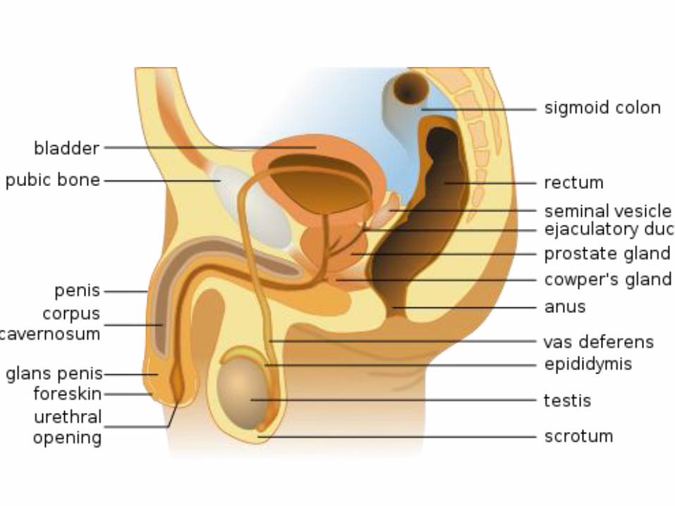
Functional Reproductive Anatomy
of the Male
• Many Individual Organs
– Acting in concert
• Produce
• Deliver
– Sperm to female tract
• Basic Components
– Spermatic cords
– Scrotum
– Testes
– Excurrent duct system
– Accessory glands
– Penis
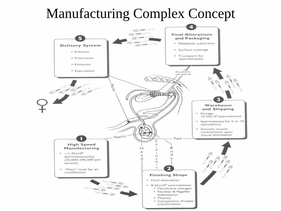
Manufacturing Complex Concept

Testicular Descent

Testicular Descent
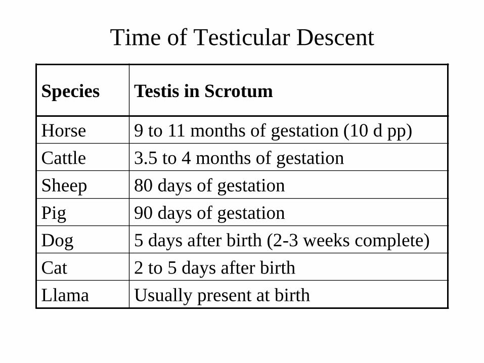
Time of Testicular Descent
Species Testis in Scrotum
Horse 9 to 11 months of gestation (10 d pp)
Cattle 3.5 to 4 months of gestation
Sheep 80 days of gestation
Pig 90 days of gestation
Dog 5 days after birth (2-3 weeks complete)
Cat 2 to 5 days after birth
Llama Usually present at birth

Cryptorchidism
• Failure of the testis to
fully descend into the
scrotum
– Unilateral
– Bilateral
• Sterile
– Abdominal
– Inguinal
• Most Common
– Boars
– Dogs
– Stallions
• Breed effects
• Least common
– Bulls
– Rams
– Bucks
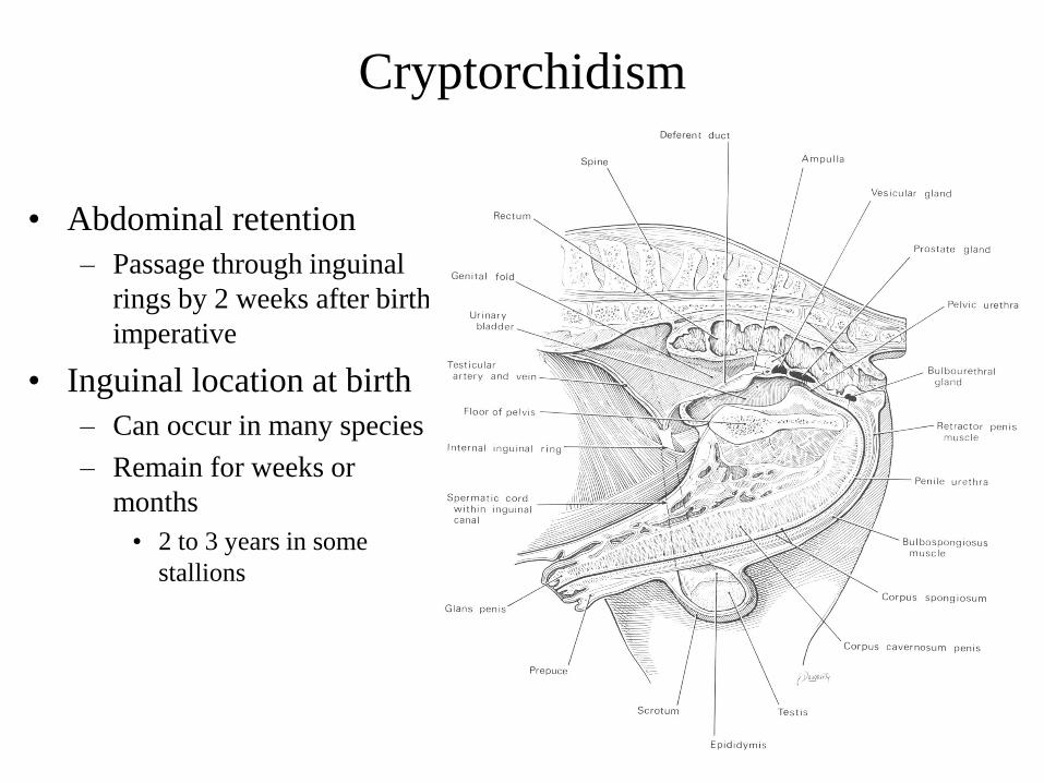
Cryptorchidism
• Abdominal retention
– Passage through inguinal
rings by 2 weeks after birth
imperative
• Inguinal location at birth
– Can occur in many species
– Remain for weeks or
months
• 2 to 3 years in some
stallions

Cryptorchidism
• Causes for concern
– Reduced fertility
– Genetic component
• Mode of inheritance unclear
– Autosomal recessive in sheep & swine?
– Neoplasia
– Spermatic cord torsion
– Androgen production

Spermatic Cord
• Extends from inguinal ring to suspend testis in scrotum
• Contains
– Testicular artery
– Testicular veins
• Pampiniform plexus
– Lymphatics
– Nerves
– Ductus deferens
– Cremaster muscle*

• Testicular arteries
– R: off aorta
– L: off left renal artery
• Testicular veins
– R: off vena cava
– L: off left renal vein
Vascular Supply to the Testes

Scrotum
• Present in most male mammals
• Diverticulum of the abdomen
– Two scrotal sacs
• Four major layers
– Parietal tunica vaginalis
– Scrotal fascia
– Tunica dartos
– Skin
• Sweat glands
CC Urethra
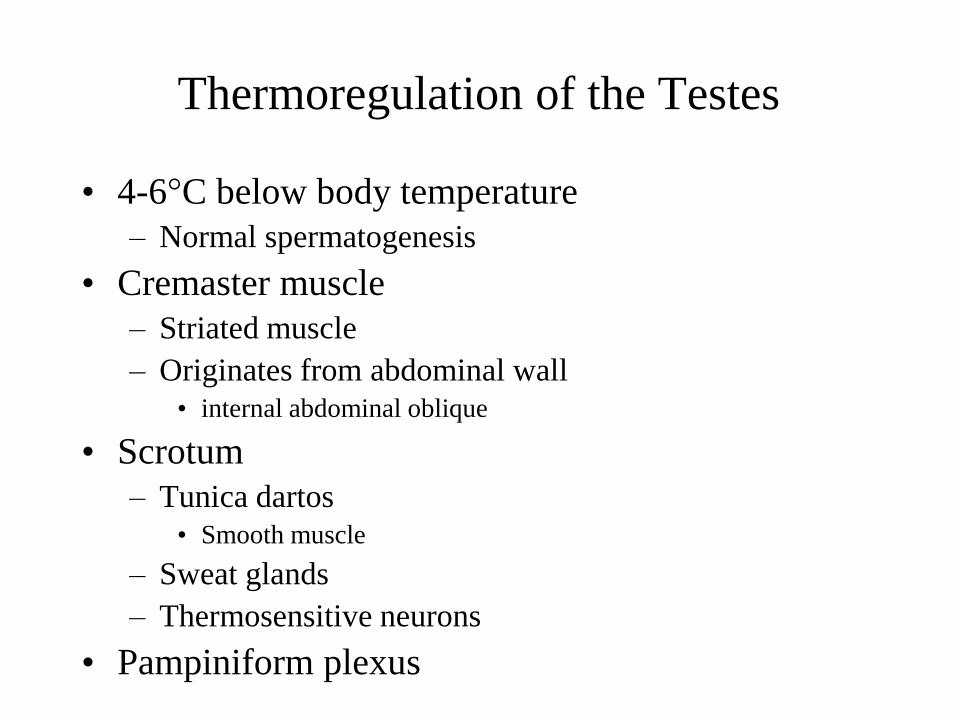
Thermoregulation of the Testes
• 4-6°C below body temperature
– Normal spermatogenesis
• Cremaster muscle
– Striated muscle
– Originates from abdominal wall
• internal abdominal oblique
• Scrotum
– Tunica dartos
• Smooth muscle
– Sweat glands
– Thermosensitive neurons
• Pampiniform plexus

Pampiniform Plexus
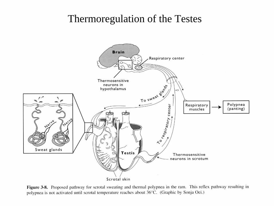
Thermoregulation of the Testes

Testis
• Primary Functions
– Exocrine: Spermatozoa
– Endocrine: Hormones
• Androgens & Estrogens
• Facilitate
– Spermatogenesis
– Sexual differentiation
– Development of 2°
sexual characteristics
– Libido
– Proteins
• Scrotal Orientation
– Varies with species

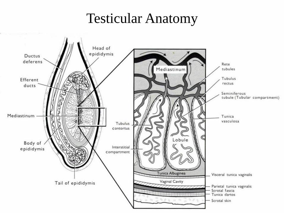
Testicular Anatomy
• Capsule
– Visceral vaginal tunic
– Tunica albuginea
• Penetrates parenchyma
• Divides testis into lobules
• Joins mediastinum
• Supporting layer
• Rhythmic contractions
• Parenchyma
• Mediastinum
– Rete Tubules
• Efferent ducts
Testicular Anatomy
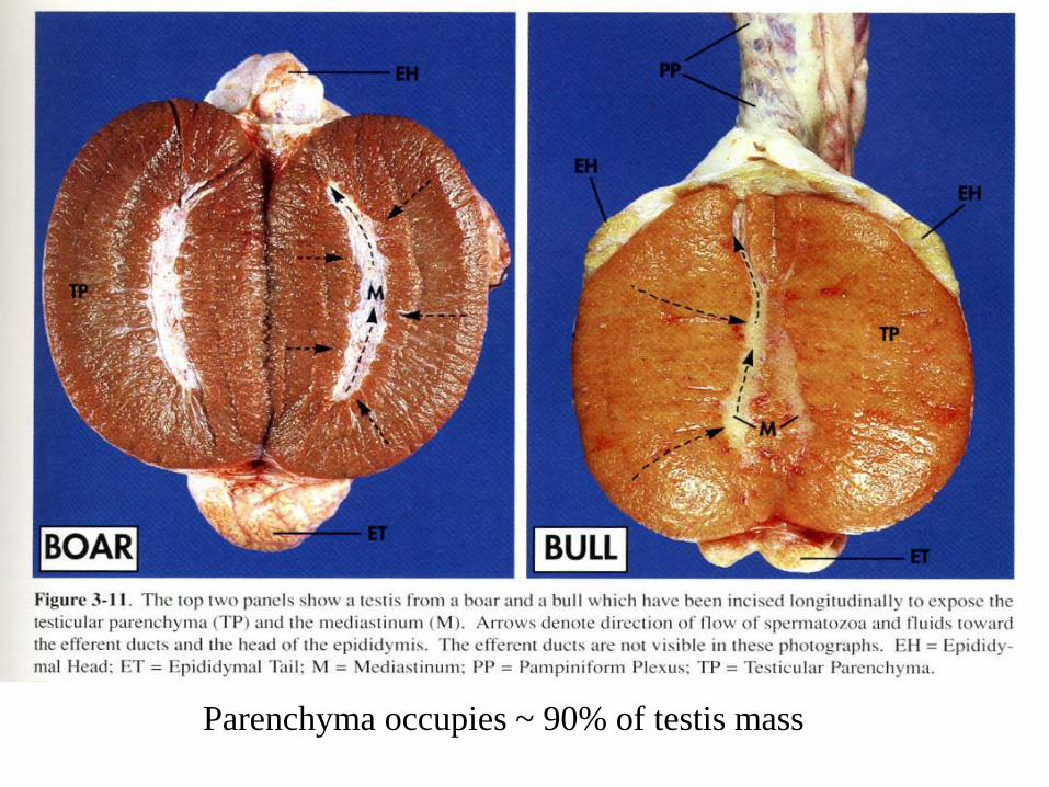
Parenchyma occupies ~ 90% of testis mass
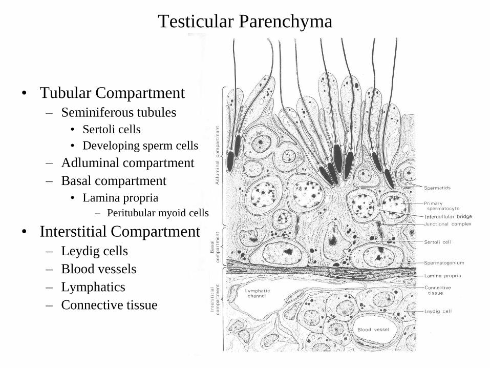
Testicular Parenchyma
• Tubular Compartment
– Seminiferous tubules
• Sertoli cells
• Developing sperm cells
– Adluminal compartment
– Basal compartment
• Lamina propria
– Peritubular myoid cells
• Interstitial Compartment
– Leydig cells
– Blood vessels
– Lymphatics
– Connective tissue

Testicular Parenchyma
• Seminiferous tubules 70%
• Interstitial Component 30%
– Leydig cells 12-18%
– Other interstitial cells 1%
• Fibroblasts
• Lymphocytes
• Mast cells
– Non-cellular components 5-10%
– Blood vessels 3-4%
– Lymphatics 1%

Sertoli Cells (nurse cells; sustentacular cells)
• Somatic cells
– FSH receptors
– Testosterone receptors
– Support & nutrition to developing germ cells
– Phagocytic
• Degenerating germ cells
• Residual bodies
• Number related to sperm production
– Fixed after puberty?
• Cell to cell communication
• Rich in SER
• Equivalent of granulosa

Sertoli Cells
Secretory activity
– Estrogens
– Proteins
• Inhibin
– gonadal glycoprotein
» preferentially inhibits secretion of FSH
– 2 active forms: Inhibin A and inhibin B (α & β subunits)
– FSH stimulates secretion by increasing α subunit
• Sulfated glycoproteins
– SGP-1
» Fertility acquisition
– SGP-2
» Facilitates movement thru testis
• Transferrin
– Iron transport for spermatogenesis
• Androgen Binding Protein
• Mitogenic peptide

Sertoli Cells
• Tight Junctions
– Blood Testis Barrier
– Separate compartments
• Basal
• Adluminal
– Isolates spermatocytes
& spermatids
• General circulation
• Serum enzymes
• High molecular weight
components
• Immune system

Leydig Cells (intersitial cells)
• 1° site of steroidogenesis
– Testosterone
• Spermatogenesis
• 2° sex characteristics
• Sex drive
– Estrogen in stallions
• Aromatase
• Stimulated by LH
– cAMP & protein kinase
– phosphorylation of ribosomal protein, protein turnover
– CSCC enzyme
– Response potentiated by Sertoli cell products
CSCC = Cholesterol Side Chain Cleavage Enzyme

Leydig Cells
• Equivalent of theca interna
• Abundance of
– Smooth ER (80% v/v)
• Cholesterol synthesis
– Mitochondria (10% v/v)
• Rate limiting SCC step
• Number & volume
– Age
– Season

Hypothalamic Pituitary Testicular Axis
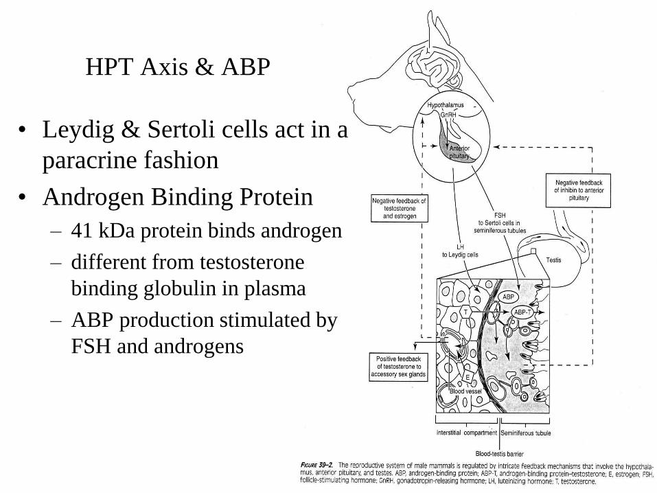
HPT Axis & ABP
• Leydig & Sertoli cells act in a
paracrine fashion
• Androgen Binding Protein
– 41 kDa protein binds androgen
– different from testosterone
binding globulin in plasma
– ABP production stimulated by
FSH and androgens

HPT Axis & ABP
• ABP Functions
– Local [T] & [DHT]
• Interstitium & seminiferous
tubules
– 70 mg/g parenchyma vs.
– 400 pg/ml serum in circulation
• Essential for normal
spermatogenesis
– Androgens to epididymis
T = Testosterone
DHT - Dihydrotestosterone

Rete Testis
• Network of
intercommunicating channels
• Lined with cells ranging from
squamous to columnar
• Fluid phase and adsorptive
endocytosis
• Carry sperm to epididymis
• Centrally located
– Ungulates
– Carnivores
– Rabbits
• Located along the epididymal
edge in primates

Efferent Ducts
• Link rete testis to epididymis
• Fluid resorption
• High enzymatic activity
– Acid phosphatase,
– Esterase
– Beta-glucuronidase
– Carbonic anhydrase activity
• Two main cell types
– Principle: columnar with
prominent intercellular junctions
• Fluid resorption
– Ciliated
• Fluid movement
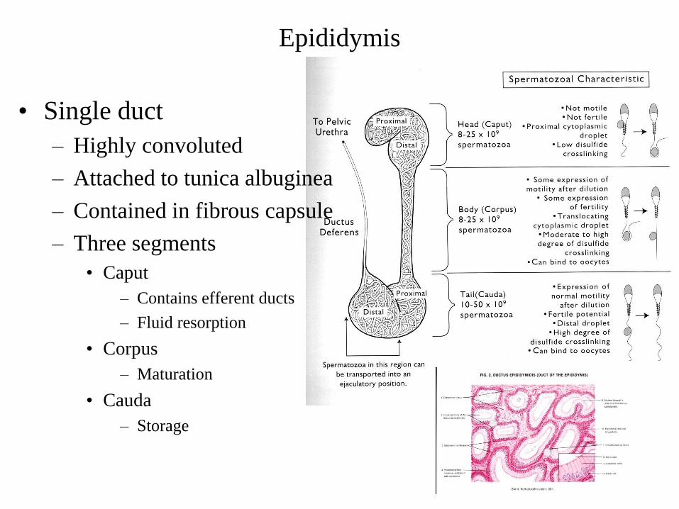
Epididymis
• Single duct
– Highly convoluted
– Attached to tunica albuginea
– Contained in fibrous capsule
– Three segments
• Caput
– Contains efferent ducts
– Fluid resorption
• Corpus
– Maturation
• Cauda
– Storage
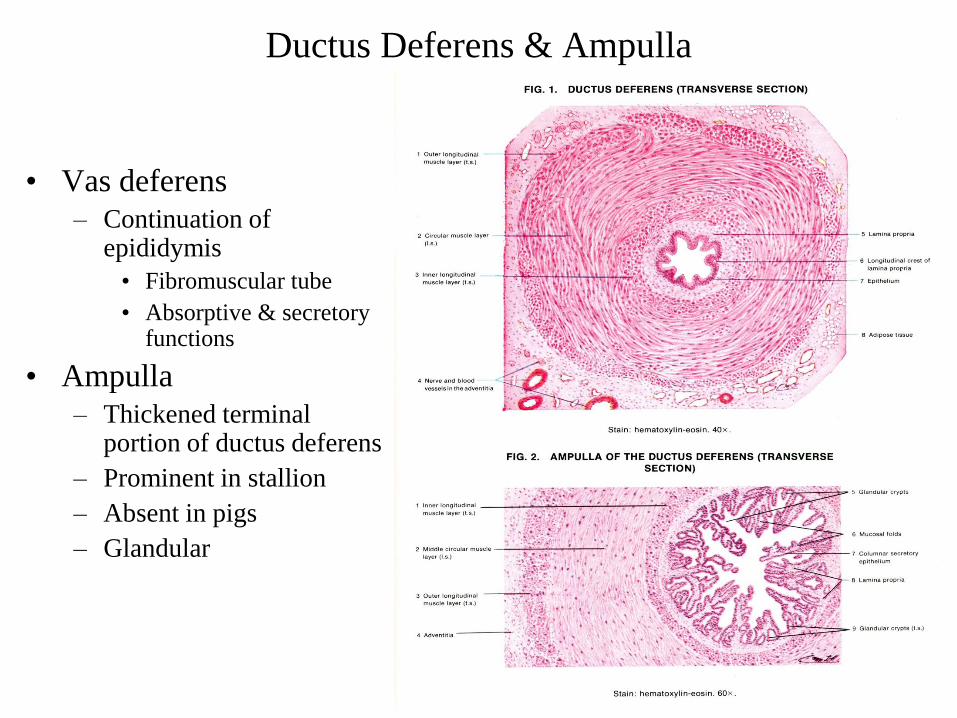
Ductus Deferens & Ampulla
• Vas deferens
– Continuation of epididymis
• Fibromuscular tube
• Absorptive & secretory functions
• Ampulla
– Thickened terminal portion of ductus deferens
– Prominent in stallion
– Absent in pigs
– Glandular

Accessory Sex Glands
• Ampullae
• Vesicular Glands
– Seminal vesicles
• Prostate
• Bulbourethral glands

Vesicular Glands Seminal Vesicles
• Lobular
– Bull
– Ram
– Boar
• Sac-like
– Human
– Stallion
– Rat
– Guinea pig
• Provides
– Volume
– Protein
– Sugars
– Salts

Vesicular Glands Seminal Vesicles
• Absent in
– Carnivores
– Lagomorphs
– Marsupials
– Cetaceans
– Some primates
– Others
• Do not store sperm

Prostate
• Present in all mammals
• Compound tubuloalveolar
gland
– Boars & dogs
• Large component of ejaculate
• Discrete and disseminate
portions
• Varying morphology
– Rats
• Ventral, lateral and dorsal
portions
– Each with multiple ducts
• Anterior prostate
– Coagulating gland
– Empties through a single duct

Bulbourethral Gands Cowpers glands
• Present in most mammals
– Pre-ejaculate fluid
• Cleanses urethra & reduces
acidity
– Bull – Ram
– Stallion – Man
– Very large in boars
• Gel fraction
– Sialic acid
• Absent in
– Dogs – Mustelids
– Bears – Aquatic mammals
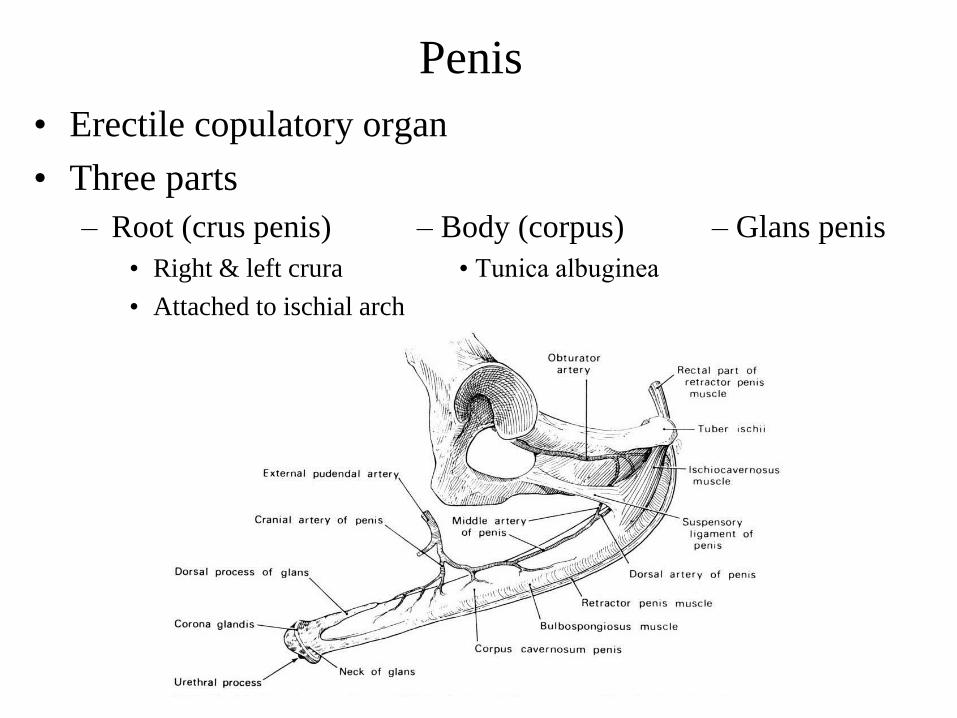
Penis
• Erectile copulatory organ
• Three parts
– Root (crus penis) – Body (corpus) – Glans penis
• Right & left crura • Tunica albuginea
• Attached to ischial arch

• Corpus cavernosum
– Majority of interior
penile shaft
– Spongy erectile tissue
• Smooth muscle
• Corpus spongiosum
– Surrounds urethra
– Extends to glans
• Very prominent in
stallion - belling

Penile Musculature
• Retractor penis
– Paired
– Attachments
• Caudal vertebrae
• Anal sphincter
• Tunica albugiea
• Ischiocavernosus
– Paired
– Inserts on crus penis
– Important in erection
• Compresses crus penis
• Bulbospongiosus
– Overlaps root of penis
– Covers bulbourethral glands
• Urethralis
– Encloses pelvic urethra
– Covers bulbourethral glands

Species Differences
• Bull, Ram, Buck
– Fibroelastic
– Sigmoid flexure
– Long urethral process
• Ram and buck
• Boar
– Fibroelastic
– Sigmoid flexure
– Corkscrew glans
– Preputial diverticulum
• Stallion
– Vascular
– Belling of glans
– Fossa glandis &
urethral sinus
• Dog
– Vascular
– Os penis (baculum)
– Bulbus glandis


Erection • Relaxation of retractor penis
• Penile rigidity
– Increase arterial inflow + decreased venous outflow
• Ischiocavernosus muscle contraction
– Relaxation and engorgement of corpus cavernosum
• Tremendous increase in intracavernous pressure
• Stallion and man increase diameter dramatically
• Little change in diameter with fibroelastic penis


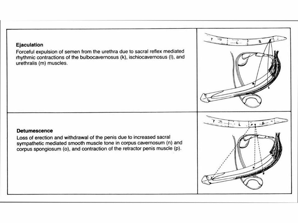

Ejaculatory Process