Embed Size (px)
Citation preview
Tang et al. BMC Complementary and Alternative Medicine (2018) 18:118 https://doi.org/10.1186/s12906-018-2171-3
RESEARCH ARTICLE Open Access
Fructus Ligustri Lucidi modulates estrogenreceptor expression with no uterotrophiceffect in ovariectomized rats
Yu-qing Tang1†, Cheng Li1†, Xue-jiao Sun1, Yi Liu1, Xi-ting Wang1, Yu-bo Guo1, Li-li Wang2, Ru-feng Ma1,Jian-zhao Niu1, Min Fu3, Dong-wei Zhang4* and Yu Li1*Abstract
Background: Accumulating evidence suggests that Fructus Ligustri Lucidi (FLL) plays a beneficial role in preventingthe development of osteoporosis. However, the effects of FLL on estrogen receptor (ER) α and ERβ expressionsremain unknown. Therefore, in the current study we attempted to probe into the effects of FLL on ERα and ERβexpressions in femurs, tibias and uteri of ovariectomized (OVX) rats.
Methods: The OVX rats were orally administrated with FLL water extract (3.5 g/kg/day) for 12 weeks. The uteri,femurs, tibias and serum were harvested from rats. The serum levels of estrogen (E2), luteinizing hormone (LH) andfollicle-stimulating hormone (FSH) were determined by ELISA. The expressions of ERα and ERβ in the femurs andtibias as well as uteri were analysed by western blot and immunohistochemical staining.
Results: FLL treatment did not increase uterus relative weight in OVX rats. Further, FLL treatment increased ERαexpression in the femurs and tibias, and enhanced ERβ expression in the uteri of OVX rats. However, the resultedexpression of ERα was stronger than that of ERβ in OVX rats in response to FLL treatment. Meanwhile,administration with FLL to OVX rats increased FSH and LH but did not increase E2 level in the serum.
Conclusion: FLL treatment shows tissue selection on ERα and ERβ expressions in the femurs and tibias as well asuteri of OVX rats without uterotrophic effect, which may offer the scientific evidence of the efficiency and safety ofits clinical application.
Keywords: Fructus Ligustri Lucidi (FLL), Osteoporosis, Estrogen receptor, Femurs, Tibias, Uteri
BackgroundAs life expectancy around world shows dramatic rise inthe last several decades, osteoporosis, one of commonchronic metabolic diseases among the elders, has becomea prevalent public health problem owing to its high mor-bidity and mortality [1]. According to the report fromInternational Osteoporosis Foundation, women are at ahigher risk of developing osteoporosis than men becauseof the estrogen deficiency after their menopause [2]. Assuch, classical hormone replacement therapy (HRT) is
* Correspondence: [email protected]; [email protected]†Equal contributors4Diabetes Research Center, Beijing University of Chinese Medicine, Beijing100029, People’s Republic of China1Traditional Chinese Medicine School, Beijing University of Chinese Medicine,Beijing 100029, People’s Republic of ChinaFull list of author information is available at the end of the article
© The Author(s). 2018 Open Access This articInternational License (http://creativecommonsreproduction in any medium, provided you gthe Creative Commons license, and indicate if(http://creativecommons.org/publicdomain/ze
widely used to prevent both menopausal symptoms andosteoporotic fractures [3]. Estrogen promotes bone ac-crual through estrogen receptor (ER) α and ERβ [4]. Inaddition, deletion of ERα in female mice exhibits reduc-tion of bone mass and strength [5, 6], whereas increasedexpression of ERα in endothelium is associated with riskof developing breast and uterine cancer, which are alsomain side effects induced by HRT treatment [7, 8]. More-over, selective activation of ERβ contributes to inhibitionof breast cell proliferation and is also one of optimal tar-gets to elicit beneficial estrogen-like activities [4, 9, 10].Therefore, discovery and development of selective ERagonist remains a need for osteoporosis treatment.Fructus Ligustri Lucidi (FLL) is the ripe fruit derived
from the evergreen tree Ligustrum lucidum Ait. It is acommon herbal medicine widely used in traditional
le is distributed under the terms of the Creative Commons Attribution 4.0.org/licenses/by/4.0/), which permits unrestricted use, distribution, andive appropriate credit to the original author(s) and the source, provide a link tochanges were made. The Creative Commons Public Domain Dedication waiverro/1.0/) applies to the data made available in this article, unless otherwise stated.
Tang et al. BMC Complementary and Alternative Medicine (2018) 18:118 Page 2 of 8
Chinese medicine (TCM) formula for the management ofosteoporosis [11]. FLL extracts have been demonstrated toimprove bone quality in diabetic mice [12] and growingfemale rats [13] through regulation of calcium metabolismvia stimulating parathyroid production. We [14] andothers [15] also demonstrated that aqueous extracts ofFLL improved bone mineral density (BMD) and bonemicrostructure in ovariectomized (OVX) rats viaregulation of collagen metabolism. In addition, FLL etha-nol extracts also promote mesenchymal stem cells differ-entiation [16]. However, little is known about the effect ofFLL on ER expression in OVX rats. Therefore, the presentstudy is aimed to explore the effects of FLL on ERα andERβ expressions in the femurs, tibias and uteri as well asits effects on uterus weight in OVX rats.
MethodsReagents and chemicalsPentobarbital sodium was purchased from Sigma-Aldrich (St. Louis, USA), Estradiol valerate (EV) tab-lets were bought from Bayer (Monheim, German). Ratmonoclonal anti-ERα antibody (ab3575) and mousemonoclonal anti-ERβ (ab288) were purchased fromAbcam (Cambridge, UK). All the other chemicals, ex-cept specially identified, were obtained from BeijingSinopharm Chemical (Beijing, China).
Preparation of FLL water extractsFLL was bought from Beijing TongRenTang (Beijing,China) and authenticated by Professor Zexin Ma (TCMmuseum at Beijing University of Traditional ChineseMedicine (BUCM)). For preparation of FLL water ex-tracts, 100 g of raw FLL was grinded into powder anddissolved in 1000 ml of distilled water by continuousstirring for 48 h under low temperature. Then the aque-ous extracts were collected by centrifugation (4000 rpmat 4 °C for 10 min). And the supernatants were har-vested and lyophilized to obtain a powder (20 g).
AnimalsFemale 12-week-old Sprague Dawley rats (200 ± 20 g)were purchased from Beijing SiBeiFu Animal Technol-ogy company (license number: SCXK (Beijing) 2014-0037, Beijing, China). The animals were housed in theclean level conditions (certification number: SCXK(Beijing) 2011-0024) at BUCM with the temperature of22 ± 1 °C, humidity of 55 ± 5%, and a 12 h-light/darkcycle. All rats had free access to tap water and standardchow. All procedures in this study were approved by theAnimal Care Committee of BUCM, Beijing, China.
OVX rat model establishmentAfter 1 week of acclimation, the OVX rats were estab-lished by removing the bilateral ovaries from the
corresponding anesthetized rats. The sham controlgroups were performed by removing the equal volumeof fat surrounding the bilateral ovaries. One week aftersurgery, the OVX animals were randomly divided intothree groups of 9 rats in each, named OVX control,OVX + EV and OVX+ FLL, respectively. For the treat-ment, the rats in the OVX + FLL group were orally admin-istrated with the water extracts of FLL (3.5 g/kg/day). Therats in the OVX + EV group were orally administratedwith EV tablets (0.1 mg/kg/day). The rats in OVX controlgroup and Sham control group were orally administratedwith the same volume of distilled water.After 12 weeks of administration, rats were anesthetized
by intraperitoneal injection with 1% pentobarbital sodium(0.4 ml/100 g, i.p.). Subsequently, blood was collectedfrom the heart by puncture. Then the rats were sacrificedby cervical dislocation. After that, the uteri, femurs andtibias were harvested for the following experiments.
Uterus coefficientAfter trimming off the fat and absorbing the excess sur-rounding fluid, the wet weight of the uterus was re-corded with analytical balance. Then, the uteri were cutinto pieces just above the junction with the cervix. Halfof the uteri were stored in liquid nitrogen until use.Another half of it was fixed in 10% neutral buffered for-malin for histological analysis. Uterus coefficient wasdetermined by uterus wet weight divided by the corre-sponding body weight of the rat (g/100 g).
Estrogen (E2), luteinizing hormone (LH) and follicle-stimulating hormone (FSH)E2, LH and FSH were determined by ELISA (CUSABIO,China) according to the manufacturer’s instructions. Allthe samples were evaluated in duplicates.
Immunohistochemical stainingThe femurs and uteri were fixed with 10% neutral buff-ered formalin. Furthermore, the tibias were decalcifiedin 15% ethylenediaminetetraacetic acid (EDTA) buffer(pH 7.4) for 90 days. After that, the femurs and uteriwere dehydrated in graded ethanol, defatted in xylene,and embedded in paraffin. Then, 5 μm sections weredeparaffinized in xylene and rehydrated with gradedethanol. Subsequently, the sections were incubated with3% H2O2 and antigen retrieval solution (0.1 M sodiumcitrate buffer, pH 6.0) followed by incubation with 10%goat serum in phosphate-buffered saline (PBS) for30 min to block nonspecific binding sites. The sectionswere then incubated with the primary antibodies (anti-ERα antibody (1:500) or anti-ERβ antibody (1:500)) over-night at 4 °C. The next day, after washing in PBS, thesections were incubated with biotinylated anti-rat sec-ondary antibody for 30 min and with peroxidase for

Table 1 The uterus coefficient in the different groups of rats
Groups Number Uterus weight/Body weight (g/100 g)
Sham 9 0.2132 ± 0.0312*
OVX 9 0.0357 ± 0.0143
OVX + EV 9 0.1493 ± 0.0240*
OVX + FLL 9 0.0322 ± 0.0137
*Compared with OVX group rats, *P < 0.05
Table 2 Serum levels of E2, LH and FSH in the different groupsof rats
Groups Number E2 (pg/ml) LH (mUI/ml) FSH (mUI/ml)
Sham 9 15.5975 ± 5.0579* 2.1711 ± 0.8571* 0.8411 ± 0.3071*
OVX 9 6.65 ± 2.3853 3.8744 ± 0.8100 1.8837 ± 0.3301
OVX + EV 9 11.9755 ± 1.0535* 2.5850 ± 0.4829* 1.2087 ± 0.1795*
OVX + FLL 9 6.8842 ± 1.9301 3.0455 ± 0.5554* 1.4512 ± 0.2801*
*Compared with OVX group rats, *P < 0.05
Tang et al. BMC Complementary and Alternative Medicine (2018) 18:118 Page 3 of 8
10 min according to the SP staining system (BeijingZhongShan JinQiao, Beijing, China). Diaminobenzidine(DAB) was used as the substrate for color developmentand visualization under the microscope. For controls,the primary antibodies were replaced by non-immunizedgoat serum. The slides were then taken for histopatho-logical evaluations. The results of immunohistochemicalstaining were quantified by Image Pro-Plus software(version 6, SPSS Inc., Chicago, IL, USA) and the integraloptical density (IOD) values were recorded. The mea-surements were performed by two investigators whowere blinded regarding the animals’ treatment groups.
Western blot analysisThe uteri were placed in a 1.5 ml Eppendorf tube andwashed with PBS twice, and then were cut with scissorsand grounded. The tibias were prepared by lyophilizingand grinding. After that, the samples were lysed in a buf-fer containing 20 mM Tris–HCl, pH 7.5, 0.1% (v/v)Igepal, 6 mM sodium deoxycholate, 150 mM NaCl,2 mM ethyleneglycoltetraacetic acid (EGTA), 2 mMEDTA, 0.1 mM Na2SO4, 20 mM NaF, and a protease in-hibitor cocktail tablet (Roche, German). The lysates werecentrifuged at 10,000 g for 15 min at 4 °C, and pro-tein concentrations in the supernatants were deter-mined by BCA protein assay kit (Applygene, China).Then 50 μg/lane of proteins were loaded into 10%polyacrylamide gel, and transferred onto nitrocellulosemembrane, and then incubated with the primary anti-body (anti-ERα or anti-ERβ) and the correspondingHRP labeled secondary antibody. The membraneswere developed using enhanced chemiluminescencesolution. The images were captured with Bio-Radbioimaging system. The gray values of the blots werequantified using the Image J software (NIH, Bethesda,MD), and normalized with the corresponding β-actin(1:2000) as the internal control.
Statistical analysisData were expressed as the mean ± standard deviation(SD). One-way analysis of variance (ANOVA) was per-formed between multiple groups using SPSS software(Version 20.0) when homogeneity of variance and nor-mality were met. Otherwise, Dunnett’s T3 and Nonpara-metric tests were conducted between multiple groups,respectively. P values less than 0.05 were considered tobe statistically significant.
ResultsEffects of FLL on the alterations of uterus coefficient inOVX ratsThe uterus coefficient of the rats were shown in Table 1.As expected, ovariectomy resulted in a significant reduc-tion in the relative uterus weight of the rats. The uterus
coefficient in the OVX control group was only around16% of that in the Sham control group. EV treatment for12 weeks significantly increased the uterus coefficient inOVX rats (P < 0.05). By contrast, FLL treatment did notincrease the uterus coefficient in OVX rats.
Effects of FLL on E2, LH and FSH levels in serumAs shown in Table 2, serum E2 level was decreased, andserum LH and FSH levels were increased in the OVXcontrol group rats as compared to those of rats in theSham control group. The administration of EV and FLLto OVX rats for 12 weeks significantly decreased serumLH and FSH levels (P < 0.05). However, FLL treatmentdid not increase serum E2 in OVX rats (P > 0.05).
Effects of FLL on the expressions of ERα and ERβ in thefemurs and tibias of OVX ratsThe effects of FLL on ERα and ERβ expressions in thefemurs were assessed by immunohistochemical staining.As shown in Figs. 1 and 2, ERα and ERβ expressions inthe femurs of the OVX control group were significantlydecreased (P < 0.01), when compared with those of ratsin the Sham control group. Both FLL and EV treatmentsignificantly increased ERα and ERβ expressions in thefemurs of the OVX rats (P < 0.05 or 0.01) when com-pared to those in the OVX control group.Furthermore, the effects of FLL on ERα and ERβ ex-
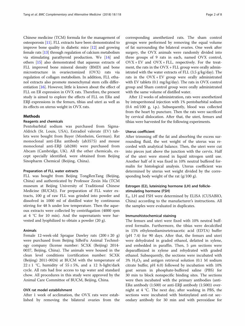
pressions in the tibias of the rats in different groupswere also evaluated by western blot. As shown in Fig. 3,FLL treatment did markedly increase ERα expression inresponse to ovariectomy (P < 0.01). By contrast, FLLtreatment showed a trend toward increasing ERβ expres-sion in OVX rats, but the differences did not reach thestatistically significant level when compared to those ofrats in the OVX control group.

**
ER
exp
ress
ion
in f
emur
sIO
D v
alue
**
**
0
600
400
1000
800
200
ShamOVX
a
b
Group
OVX+EV OVX+FLL
Sham OVX
×400
×400
OVX+EV
OVX+FLL
Fig. 1 The representative images (a) and images analysis results ofimmunohistochemical staining (b) showing the effect of FLL on ERαexpression in the femurs of the rats. The arrows illustrated theexpression and distribution of ERα. Data are presented as mean ± SD.**P < 0.01 compared with the OVX control group
Sham OVXa
b
ER
exp
ress
ion
in f
emur
sIO
D v
alue
**
**
*
ShamOVX
Group0
600
400
1000
800
200
×400
×400
OVX+EV OVX+FLL
OVX+EV
OVX+FLL
Fig. 2 The representative images (a) and images analysis results ofimmunohistochemical staining (b) showing the effect of FLL on ERβexpression in the femurs of the rats. The arrows illustrated theexpression and distribution of ERβ. Data are presented as mean ± SD.*P < 0.05 or **P < 0.01 compared with the OVX control group
Tang et al. BMC Complementary and Alternative Medicine (2018) 18:118 Page 4 of 8
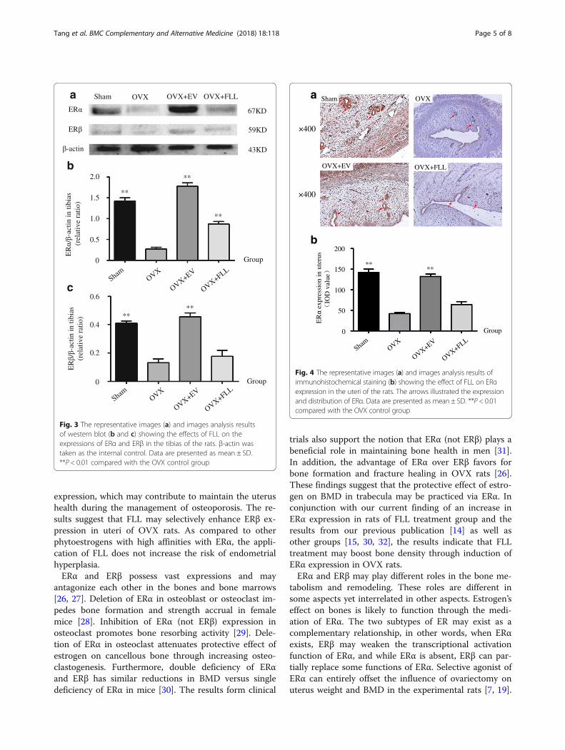
Effects of FLL on the expressions of ERα and ERβ in theuteri of OVX ratsAs shown in Figs. 4 and 5, ERα and ERβ were mainlylocated in the endometrium and glandular epithelia cells,and were highly expressed in the uteri of rats in shamcontrol group and EV treatment group compared tothose of rats in the OVX control group as evaluated byimmunohistochemical staining (P < 0.01). Further, FLLtreatment did not increase ERα expression but did obvi-ously increase ERβ expression in the uteri of the OVXrats (P < 0.05). These results were also further confirmedby western blot (Fig. 6).
DiscussionIn the current study, we demonstrated that FLL treat-ment decreased serum LH and FSH levels but notincreased serum E2 level in OVX rats. FLL did not in-crease uterus relative weight in response to ovarirect-omy. In addition, FLL treatment significantly enhancedERβ expression, but left no evident influence on ERα inuteri. Moreover, FLL treatment markedly enhanced ERαexpression, but had no obvious effect on ERβ expressionin the femurs and tibias. These results suggest that FLL
may show different effects on ER expression in bonesand uteri, which may contribute to uterus health duringthe treatment of osteoporosis.Both ERα and ERβ are the main targets of estrogen
and can be either coexistent or expressed alone in vari-ous tissues [17]. ERα is inclined to be found in the ovary,breast, hypophysis, paranephros, kidney and bone, whileERβ is highly concentrated in the granulosa cells ofprostate and ovary, commonly expressed in ovary, lung,brain and testis, and less expressed in hypophysis andspinal cord [18, 19]. In the reproductive system, ERα ex-pression decreases gradually from the epithelial cells ofvagina to those of oviduct [20, 21]. In the uteri of OVXrats, the expression of ERβ are tremendously reducedand almost only ERα can be detected [22, 23]. Over-expression of ERα may lead to the hyperplasia of mam-mary glands and endometrial cells, and result in anincreased risk of breast cancer and endometrial cancer[24]. In contrast, activation of ERβ never causes the rele-vant cell proliferation, instead, it has certain effectsagainst cell proliferation [25]. In the current study, wefound that FLL treatment did increase ERβ expression inthe uteri though with no significant effect on ERα
ER
ER
67KD
43KD
**
**
**
****
ER
/-a
ctin
in ti
bias
(r
elat
ive
ratio
)
Sham OVX OVX+EV OVX+FLL
-actin
0
1.5
1.0
2.0
0.5
0
0.6
0.4
0.2
ShamOVX
Group
ShamOVX
Group
c
a
b
59KD
ER
/-a
ctin
in ti
bias
(re
lati
ve r
atio
)
OVX+EV
OVX+FLL
OVX+EV
OVX+FLL
Fig. 3 The representative images (a) and images analysis resultsof western blot (b and c) showing the effects of FLL on theexpressions of ERα and ERβ in the tibias of the rats. β-actin wastaken as the internal control. Data are presented as mean ± SD.**P < 0.01 compared with the OVX control group
****
ER
expr
essi
on in
ute
rus
IOD
val
ue0
150
100
200
50
ShamOVX
Group
Sham OVXa
b
×400
×400
OVX+EV OVX+FLL
OVX+EV
OVX+FLL
Fig. 4 The representative images (a) and images analysis results ofimmunohistochemical staining (b) showing the effect of FLL on ERαexpression in the uteri of the rats. The arrows illustrated the expressionand distribution of ERα. Data are presented as mean ± SD. **P < 0.01compared with the OVX control group
Tang et al. BMC Complementary and Alternative Medicine (2018) 18:118 Page 5 of 8
expression, which may contribute to maintain the uterushealth during the management of osteoporosis. The re-sults suggest that FLL may selectively enhance ERβ ex-pression in uteri of OVX rats. As compared to otherphytoestrogens with high affinities with ERα, the appli-cation of FLL does not increase the risk of endometrialhyperplasia.ERα and ERβ possess vast expressions and may
antagonize each other in the bones and bone marrows[26, 27]. Deletion of ERα in osteoblast or osteoclast im-pedes bone formation and strength accrual in femalemice [28]. Inhibition of ERα (not ERβ) expression inosteoclast promotes bone resorbing activity [29]. Dele-tion of ERα in osteoclast attenuates protective effect ofestrogen on cancellous bone through increasing osteo-clastogenesis. Furthermore, double deficiency of ERαand ERβ has similar reductions in BMD versus singledeficiency of ERα in mice [30]. The results form clinical
trials also support the notion that ERα (not ERβ) plays abeneficial role in maintaining bone health in men [31].In addition, the advantage of ERα over ERβ favors forbone formation and fracture healing in OVX rats [26].These findings suggest that the protective effect of estro-gen on BMD in trabecula may be practiced via ERα. Inconjunction with our current finding of an increase inERα expression in rats of FLL treatment group and theresults from our previous publication [14] as well asother groups [15, 30, 32], the results indicate that FLLtreatment may boost bone density through induction ofERα expression in OVX rats.ERα and ERβ may play different roles in the bone me-
tabolism and remodeling. These roles are different insome aspects yet interrelated in other aspects. Estrogen’seffect on bones is likely to function through the medi-ation of ERα. The two subtypes of ER may exist as acomplementary relationship, in other words, when ERαexists, ERβ may weaken the transcriptional activationfunction of ERα, and while ERα is absent, ERβ can par-tially replace some functions of ERα. Selective agonist ofERα can entirely offset the influence of ovariectomy onuterus weight and BMD in the experimental rats [7, 19].
ERβ
expr
essi
on in
ute
rus
IOD
val
ue
0
**
**
*
150
100
50
ShamOVX
Group
Sham OVXa
b
×400
×400
OVX+EV OVX+FLL
OVX+EV
OVX+FLL
Fig. 5 The representative images (a) and images analysis results ofimmunohistochemical staining (b) showing the effect of FLL on ERβexpression in the uteri of the rats. The arrows illustrated the expressionand distribution of ERβ. Data are presented as mean ± SD. *P < 0.05 or**P < 0.01 compared with the OVX control group
**
**
*
**
**
ER
ER
67KD
59KD
43KD
Sham
-actin
0
1.2
0.8
1.6
0.4
ER
/-a
ctin
in u
teru
s
(rel
ativ
e ra
tio)
ER
/-a
cti n
in u
teru
s
(rel
ativ
e ra
tio)
Group
ShamOVX
ShamOVX
Group
c
a
b
0
1.2
0.8
0.4
OVX+EV
OVX+FLL
OVX+EV
OVX+FLL
OVX OVX+EV OVX+FLL
Fig. 6 The representative images (a) and images analysis results ofwestern blot (b and c) showing the effects of FLL on theexpressions of ERα and ERβ in the uteri of the rats. β-actin was takenas the internal control. Data are presented as mean ± SD. *P < 0.05or **P < 0.01 compared with the OVX control group
Tang et al. BMC Complementary and Alternative Medicine (2018) 18:118 Page 6 of 8
The findings of our current study here also show thataqueous extract of FLL could enhance ERα expression,and have no apparent influence on ERβ expression inthe femurs and tibias of OVX rats.In the current study, we found that FLL treatment sig-
nificantly decreased the levels of LH and FSH but didnot increase E2 levels in OVX rats. Ovariectomy in ratsresults in a decrease in E2 and an increase in LH andFSH [33, 34]. Deficiency of E2 significantly promotesbone loss and aggravates uterus atrophy [34, 35]. Inhib-ition of FSH also impairs bone loss and further preventsLH release while the alteration does not play a dominantrole in the development of osteoporosis [35]. In addition,high circulating FSH contributes to endometrial atrophyin mice [36]. Increased circulating LH may be associatedwith postmenopausal “hot flushes” [37]. The increase ofFSH may contribute to uterus atrophy. The results sug-gest that FLL could alleviate postmenopausal vasomotorsymptoms, which required further investigation.In addition, we have demonstrated that FLL water
extract mainly includes salidroside, ligustroflavon, acteo-side, specnuezhenide, and oleuropein acid [38]. Cur-rently, salidroside was demonstrated to bind to ERα in
docking simulation assay [39]. So it is reasonable to de-duce that salidroside may account for the estrogen-likeeffect of FLL in OVX rats. However, further studies arestill needed to identify the contributions of each compo-nent in the FLL aqueous extract.
ConclusionIn conclusion, FLL treatment increases ERβ expressionin uteri and strengthens ERα expression in the femursand tibias as well as poses no risk of the increasing ofuterus relative weight in OVX rats. In addition, our find-ings also demonstrate that FLL has the ability of coord-inating LH and FSH levels in circulation, which maycontribute to alleviate postmenopausal vasomotor symp-toms. However, how FLL regulates ER expression inOVX rats still needs further investigation.
AbbreviationsBMD: Bone mineral density; BUCM: Beijing University of Traditional ChineseMedicine; DAB: Diaminobenzidine; E2: Estrogen;EDTA: Ethylenediaminetetraacetic acid; EGTA: Ethyleneglycoltetraacetic acid;

Tang et al. BMC Complementary and Alternative Medicine (2018) 18:118 Page 7 of 8
ER: Estrogen receptor; EV: Estradiol valerate; FLL: Fructus Ligustri Lucidi;FSH: Follicle-stimulating hormone; HRT: Hormone replacement therapy;IOD: Integral optical density; LH: Luteinizing hormone; OVX: Ovariectomized;PBS: Phosphate-buffered saline; TCM: Traditional Chinese medicine
AcknowledgementsNot applicable.
FundingThis study was supported by the Grants from the National Natural ScienceFoundation of China (No. 81573716, 81273995) and the 111 project of MOE(B07007) as well as Beijing Municipal Natural Science Foundation (7172126).
Availability of data and materialsThe datasets used and/or analysed during the current study available fromthe corresponding author on reasonable request.
Authors’ contributionsYQT and CL designed and conducted most of animal experiments anddrafted the manuscript. XJS, YiL and XTW conducted immunohistochemicalexperiments. YBG, LLW and RFM conducted western blot and analysedexperimental results. JZN and MF interpreted the results. DWZ and YuLconceived the experiments and revised the manuscript. All authors haveread and approved the final manuscript.
Ethics approvalAll procedures in this study were performed in accordance with the“Guidelines for Experimental Animal Care and Use” from the Animal CareCommittee of BUCM, Beijing, China. The protocol was approved by theAnimal Care Committee of BUCM, Beijing, China.
Consent for publicationNot applicable.
Competing interestsThe authors declare that they have no competing interests.
Publisher’s NoteSpringer Nature remains neutral with regard to jurisdictional claims inpublished maps and institutional affiliations.
Author details1Traditional Chinese Medicine School, Beijing University of Chinese Medicine,Beijing 100029, People’s Republic of China. 2Chinese Material Medica School,Beijing University of Chinese Medicine, Beijing 100029, People’s Republic ofChina. 3The Research Institute of McGill University Health Center, Montreal,Quebec H4A 3J1, People’s Republic of China. 4Diabetes Research Center,Beijing University of Chinese Medicine, Beijing 100029, People’s Republic ofChina.
Received: 18 November 2016 Accepted: 15 March 2018
References1. Kruger MC, Wolber FM: Osteoporosis: modern paradigms for last Century's
bones. Nutrients 2016;8(6):376.2. Lin X, Xiong D, Peng YQ, Sheng ZF, Wu XY, Wu XP, Wu F, Yuan LQ, Liao EY.
Epidemiology and management of osteoporosis in the People's Republic ofChina: current perspectives. Clin Interv Aging. 2015;10:1017–33.
3. Cauley JA. Estrogen and bone health in men and women. Steroids. 2015;99(Pt A):11–5.
4. Niu AQ, Xie LJ, Wang H, Zhu B, Wang SQ. Prediction of selective estrogenreceptor beta agonist using open data and machine learning approach.Drug Des Devel Ther. 2016;10:2323–31.
5. Melville KM, Kelly NH, Surita G, Buchalter DB, Schimenti JC, Main RP, Ross FP,van der Meulen MC. Effects of deletion of ERalpha in osteoblast-lineagecells on bone mass and adaptation to mechanical loading differ in femaleand male mice. J Bone Miner Res. 2015;30(8):1468–80.
6. Macari S, Ajay SL, Wyatt A, Knowles P, Szawka RE, Garlet GP, Grattan DR,Dias GJ, Silva TA. Osteoprotective effects of estrogen in the maxillary bonedepend on ERalpha. J Dent Res. 2016;95(6):689–96.
7. Matsushima H, Mori T, Ito F, Yamamoto T, Akiyama M, Kokabu T, Yoriki K,Umemura S, Akashi K, Kitawaki J. Anti-tumor effect of estrogen-relatedreceptor alpha knockdown on uterine endometrial cancer. Oncotarget.2016;7(23):34131–48.
8. Lin Z, Yin P, Reierstad S, O'Halloran M, Coon VJ, Pearson EK, Mutlu GM,Bulun SE. Adenosine A1 receptor, a target and regulator of estrogenreceptoralpha action, mediates the proliferative effects of estradiol in breastcancer. Oncogene. 2010;29(8):1114–22.
9. Cvoro A, Paruthiyil S, Jones JO, Tzagarakis-Foster C, Clegg NJ, Tatomer D,Medina RT, Tagliaferri M, Schaufele F, Scanlan TS, et al. Selective activationof estrogen receptor-beta transcriptional pathways by an herbal extract.Endocrinology. 2007;148(2):538–47.
10. de Villiers TJ, Gass ML, Haines CJ, Hall JE, Lobo RA, Pierroz DD, Rees M.Global consensus statement on menopausal hormone therapy. Climacteric.2013;16(2):203–4.
11. Cheng M, Wang Q, Fan Y, Liu X, Wang L, Xie R, Ho CC, Sun W. A traditionalChinese herbal preparation, Er-Zhi-wan, prevent ovariectomy-inducedosteoporosis in rats. J Ethnopharmacol. 2011;138(2):279–85.
12. Zhang Y, Diao TY, Wang L, Che CT, Wong MS. Protective effects of waterfraction of Fructus Ligustri Lucidi extract against hypercalciuria andtrabecular bone deterioration in experimentally type 1 diabetic mice. JEthnopharmacol. 2014;158 Pt A:239–45.
13. Lyu Y, Feng X, Zhao P, Wu Z, Xu H, Fang Y, Hou Y, Denney L, Xu Y, Feng H.Fructus Ligustri Lucidi (FLL) ethanol extract increases bone mineral densityand improves bone properties in growing female rats. J Bone Miner Metab.2014;32(6):616–26.
14. Guo Y, Wang L, Ma R, Wang L, Yang M, Tang Y, Liu C, Zhu R, Liu H, Zhao D,et al. Effects of water extract from Fructus Ligustri Lucidi on bone structure andmetabolism in ovariectomized rats. Chinese Tradit Herb Drugs. 2016;47:1155–62.
15. Guo Y, Ma R, Wang L, Liu H, Zhu R, Shi R, Zhao D, Mo F, Gao S, Zhang D.Research progress on effects and their mechanism of Ligustri Lucidi Fructusin treatment of osteoporosis. Chinese Trad Herb Drugs. 2016;47:851–6.
16. Li G, Zhang XA, Zhang JF, Chan CY, Yew DT, He ML, Lin MC, Leung PC,Kung HF. Ethanol extract of Fructus Ligustri Lucidi promotes osteogenesisof mesenchymal stem cells. Phytother Res. 2010;24(4):571–6.
17. Wong KC, Lee KS, Luk HK, Wan HY, Ho CK, Zhang Y, Wong MS. Er-xiandecoction exerts estrogen-like osteoprotective effects in vivo and in vitro.Am J Chin Med. 2014;42(2):409–26.
18. Paterni I, Granchi C, Katzenellenbogen JA, Minutolo F. Estrogen receptorsalpha (ERalpha) and beta (ERbeta): subtype-selective ligands and clinicalpotential. Steroids. 2014;90:13–29.
19. Harris HA, Katzenellenbogen JA, Katzenellenbogen BS. Characterization ofthe biological roles of the estrogen receptors, ERalpha and ERbeta, inestrogen target tissues in vivo through the use of an ERalpha-selectiveligand. Endocrinology. 2002;143(11):4172–7.
20. Couse JF, Lindzey J, Grandien K, Gustafsson JA, Korach KS. Tissue distributionand quantitative analysis of estrogen receptor-alpha (ERalpha) and estrogenreceptor-beta (ERbeta) messenger ribonucleic acid in the wild-type andERalpha-knockout mouse. Endocrinology. 1997;138(11):4613–21.
21. Baranda-Avila N, Cardoso-Rangel ME, Cerbon M, Camacho-Arroyo I,Mendoza-Rodriguez CA, Villasenor-Gaona H, Anzaldua-Arce SR. Differentialexpression of estrogen receptor alpha gene in the ampullae and isthmusregions of the rabbit oviduct during early pregnancy. Anim Reprod Sci.2010;121(3-4):286–93.
22. Cevik O, Akpinar H, Oba R, Cilingir OT, Ozdemir ZN, Cetinel S, Yoldemir T.The effect of Momordica charantia intake on the estrogen receptorsESRalpha/ESRbeta gene levels and apoptosis on uterine tissue inovariectomy rats. Mol Biol Rep. 2015;42(1):167–77.
23. Sims NA, Dupont S, Krust A, Clement-Lacroix P, Minet D, Resche-Rigon M,Gaillard-Kelly M, Baron R. Deletion of estrogen receptors reveals a regulatoryrole for estrogen receptors-beta in bone remodeling in females but not inmales. Bone. 2002;30(1):18–25.
24. Christenson ES, Jiang X, Kagan R, Schnatz P. Osteoporosis management inpost-menopausal women. Minerva Ginecol. 2012;64(3):181–94.
25. Nilsson S, Koehler KF, Gustafsson JA. Development of subtype-selectiveoestrogen receptor-based therapeutics. Nat Rev Drug Discov. 2011;10(10):778–92.
26. Chow SK, Leung KS, Qin L, Wei F, Cheung WH. Callus formation is related tothe expression ratios of estrogen receptors-alpha and -beta in ovariectomy-induced osteoporotic fracture healing. Arch Orthop Trauma Surg. 2014;134(10):1405–16.
Tang et al. BMC Complementary and Alternative Medicine (2018) 18:118 Page 8 of 8
27. Khalid AB, Krum SA. Estrogen receptors alpha and beta in bone. Bone. 2016;87:130–5.
28. Melville KM, Kelly NH, Khan SA, Schimenti JC, Ross FP, Main RP, van der MeulenMC. Female mice lacking estrogen receptor-alpha in osteoblasts havecompromised bone mass and strength. J Bone Miner Res. 2014;29(2):370–9.
29. Mano H, Hakeda Y, Kumegawa M. Estrogen directly down-regulates thebone-resorbing activity of mature osteoclasts through nuclear estrogenreceptor alpha. Cytotechnology. 2001;35(1):17–23.
30. McCauley LK, Tozum TF, Kozloff KM, Koh-Paige AJ, Chen C, Demashkieh M,Cronovich H, Richard V, Keller ET, Rosol TJ, et al. Transgenic models ofmetabolic bone disease: impact of estrogen receptor deficiency on skeletalmetabolism. Connect Tissue Res. 2003;44(Suppl 1):250–63.
31. Khosla S, Riggs BL, Atkinson EJ, Oberg AL, Mavilia C, Del MF, Melton LR,Brandi ML. Relationship of estrogen receptor genotypes to bone mineraldensity and to rates of bone loss in men. J Clin Endocrinol Metab. 2004;89(4):1808–16.
32. Li X, Song QS, Wang JY, Leng HJ, Chen ZQ, Liu ZJ, Dang GT, Song CL.Simvastatin induces estrogen receptor-alpha expression in bone, restoresbone loss, and decreases ERalpha expression and uterine wet weight inovariectomized rats. J Bone Miner Metab. 2011;29(4):396–403.
33. Cen J, Zhang H, Liu Y, Deng M, Tang S, Liu W, Zhang Z. Anti-aging effect ofestrogen on telomerase activity in ovariectomised rats–animal model formenopause. Gynecol Endocrinol. 2015;31(7):582–5.
34. Xu Y, Ding J, Ma XP, Ma YH, Liu ZQ, Lin N. Treatment with Panax ginsengantagonizes the estrogen decline in ovariectomized mice. Int J Mol Sci.2014;15(5):7827–40.
35. Rouach V, Katzburg S, Koch Y, Stern N, Somjen D. Bone loss inovariectomized rats: dominant role for estrogen but apparently not for FSH.J Cell Biochem. 2011;112(1):128–37.
36. Zhang D, Li J, Xu G, Zhang R, Zhou C, Qian Y, Liu Y, Chen L, Zhu B, Ye X,et al. Follicle-stimulating hormone promotes age-related endometrialatrophy through cross-talk with transforming growth factor beta signaltransduction pathway. Aging Cell. 2015;14(2):284–7.
37. Seidlova-Wuttke D, Hesse O, Jarry H, Christoffel V, Spengler B, Becker T,Wuttke W. Evidence for selective estrogen receptor modulator activity in ablack cohosh (Cimicifuga racemosa) extract: comparison with estradiol-17beta. Eur J Endocrinol. 2003;149(4):351–62.
38. Wang L, Ma R, Guo Y, Sun J, Liu H, Zhu R, Liu C, Li J, Li L, Chen B, et al.Antioxidant effect of Fructus Ligustri Lucidi aqueous extract inOvariectomized rats is mediated through Nox4-ROS-NF-kappaB pathway.Front Pharmacol. 2017;8:266.
39. Zhang J, Kasim V, Xie YD, Huang C, Sisjayawan J, Dwi AA, Yan XS, Wu XY,Liu CP, Yang L, et al. Inhibition of PHD3 by salidroside promotesneovascularization through cell-cell communications mediated bymuscle-secreted angiogenic factors. Sci Rep. 2017;7:43935.
• We accept pre-submission inquiries
• Our selector tool helps you to find the most relevant journal
• We provide round the clock customer support
• Convenient online submission
• Thorough peer review
• Inclusion in PubMed and all major indexing services
• Maximum visibility for your research
Submit your manuscript atwww.biomedcentral.com/submit
Submit your next manuscript to BioMed Central and we will help you at every step:


![Fructus Ligustri Lucidi preserves bone quality …...deficiency [3, 4]. As bone loss primarily occurs in populations aged 30 years and above, the burden of osteoporosis is projected](https://img.dokumen.tips/doc/110x75/5f42c7c5b48d6a680a293047/fructus-ligustri-lucidi-preserves-bone-quality-deficiency-3-4-as-bone-loss.jpg)
![Validation of the (anti-) ERα CALUX bioassay ERa Calux...validation of the ERα CALUX bioassay (agonistic and antagonistic mode) according to the OECD guidelines for validation [2]](https://img.dokumen.tips/doc/110x75/5e63fa0741abdf46ef13e550/validation-of-the-anti-er-calux-bioassay-era-calux-validation-of-the-er.jpg)


