Embed Size (px)
Citation preview

http://www.diva-portal.org
This is the published version of a paper published in Frontiers in Marine Science.
Citation for the original published paper (version of record):
Pittura, L., Avio, C G., Giuliani, M E., d’Errico, G., Keiter, S. et al. (2018)Microplastics as Vehicles of Environmental PAHs to Marine Organisms: CombinedChemical and Physical Hazards to the Mediterranean Mussels, Mytilus galloprovincialisFrontiers in Marine Science, 5(Apr): 103https://doi.org/10.3389/fmars.2018.00103
Access to the published version may require subscription.
N.B. When citing this work, cite the original published paper.
Permanent link to this version:http://urn.kb.se/resolve?urn=urn:nbn:se:oru:diva-66497
ORIGINAL RESEARCHpublished: 03 April 2018
doi: 10.3389/fmars.2018.00103
Frontiers in Marine Science | www.frontiersin.org 1 April 2018 | Volume 5 | Article 103
Edited by:
Ricardo Beiras,
University of Vigo, Spain
Reviewed by:
Laura Guimarães,
Centro Interdisciplinar de Pesquisa
Marine e Ambiental (CIIMAR), Portugal
Juan Bellas,
Instituto Español de Oceanografía
(IEO), Spain
*Correspondence:
Francesco Regoli
†These authors have contributed
equally to this work.
Specialty section:
This article was submitted to
Marine Pollution,
a section of the journal
Frontiers in Marine Science
Received: 14 January 2018
Accepted: 13 March 2018
Published: 03 April 2018
Citation:
Pittura L, Avio CG, Giuliani ME,
d’Errico G, Keiter SH, Cormier B,
Gorbi S and Regoli F (2018)
Microplastics as Vehicles of
Environmental PAHs to Marine
Organisms: Combined Chemical and
Physical Hazards to the Mediterranean
Mussels, Mytilus galloprovincialis.
Front. Mar. Sci. 5:103.
doi: 10.3389/fmars.2018.00103
Microplastics as Vehicles ofEnvironmental PAHs to MarineOrganisms: Combined Chemical andPhysical Hazards to theMediterranean Mussels, Mytilusgalloprovincialis
Lucia Pittura 1†, Carlo G. Avio 1†, Maria E. Giuliani 1, Giuseppe d’Errico 1, Steffen H. Keiter 2,
Bettie Cormier 2,3, Stefania Gorbi 1 and Francesco Regoli 1,4*
1 Laboratorio di Ecotossicologia e Chimica ambientale, Dipartimento di Scienze della Vita e dell’Ambiente, Università
Politecnica delle Marche, Ancona, Italy, 2Man-Technology-Environment Research Centre, School of Science and Technology,
Örebro University, Örebro, Sweden, 3UMR Centre National de la Recherche Scientifique 5805 EPOC, University of Bordeaux,
Talence, France, 4Consorzio Interuniversitario per le Scienze del Mare, CoNISMa, ULR Ancona, Ancona, Italy
The ubiquitous occurrence of microplastics (MPs) in the marine environment is raising
concern for interactions with marine organisms. These particles efficiently adsorb
persistent organic pollutants from surrounding environment and, due to the small
size, they are easily available for ingestion at all trophic levels. Once ingested, MPs
can induce mechanical damage, sub-lethal effects, and various cellular responses,
further modulated by possible release of adsorbed chemicals or additives. In this
study, ecotoxicological effects of MPs and their interactions with benzo(a)pyrene
(BaP), chosen as a model compound for polycyclic aromatic hydrocarbons (PAHs)
were investigated in Mediterranean mussels, Mytilus galloprovincialis. Organisms were
exposed for 4 weeks to 10 mg/L of low-density polyethylene (LDPE) microparticles
(2.34 ∗ 107 particles/L, size range 20–25µm), both virgin and pre-contaminated with
BaP (15µg/g). Organisms were also exposed for comparison to BaP dosed alone at
150 ng/L, corresponding to the amount adsorbed on microplastics. Tissue localization
of microplastics was histologically evaluated; chemical analyses and a wide battery of
biomarkers covering molecular, biochemical and cellular levels allowed to evaluate BaP
bioaccumulation, alterations of immune system, antioxidant defenses, onset of oxidative
stress, peroxisomal proliferation, genotoxicity, and neurotoxicity. Obtained data were
elaborated within a quantitative weight of evidence (WOE) model which, using weighted
criteria, provided synthetic hazard indices, for both chemical and cellular results, before
their integration in a combined index. Microplastics were localized in hemolymph, gills,
and especially digestive tissues where a potential transfer of BaP from MPs was also
observed. Significant alterations were measured on the immune system, while more
limited effects occurred on the oxidative status, neurotoxicity, and genotoxicity, with a
different susceptibility of analyzed pathways, depending on tissue, time, and typology

Pittura et al. Ecotoxicology of Microplastics in Marine Mussels
of exposure. Molecular analyses confirmed the general lack of significant transcriptional
variations of antioxidant and stress genes. The overall results suggest that microplastics
induce a slight cellular toxicity under short-term (28 days) exposure conditions. However,
modulation of immune responses, along with bioaccumulation of BaP, pose the still
unexplored risk that these particles, under conditions of more chronic exposure (months
to years) or interacting with other stressors, may provoke long-term, subtle effects on
organisms’ health status.
Keywords: microplastics, mussels, bioavailability, biomarkers, immune responses, gene transcription, weighted
criteria, hazard index
INTRODUCTION
Microplastics are particles smaller than 5mm in diameter(NOAA, 2015), now identified as the predominant componentof plastic debris in the marine environment (Goldstein et al.,2013; Eriksen et al., 2014). The huge amount of microplasticsdocumented over the past decade (Wright et al., 2013), is partlydue to the direct release of micro-debris into the ocean (Browne,2015), but in larger quantities, it depends on fragmentation ofmacro- and meso-plastic (Galgani et al., 2015; Thompson, 2015).The small dimensions of microplastics and their ubiquitouspresence in marine habitats, are key factors promoting theirinteractions with organisms (Wright et al., 2013).
Ingestion of microplastics is well-documented for severalmarine vertebrates and invertebrates, including commerciallyimportant species, which differ by trophic level, feedingstrategies, and distribution along the water column (Lusher, 2015;Phuong et al., 2016; Avio et al., 2017a; Lusher et al., 2017; Santilloet al., 2017).
Several laboratory experiments have been performed, inrecent years, to understand dynamics of particles uptake,bioaccumulation and toxicological mechanisms possibly leadingto detrimental effects in a variety of bioindicators organisms(Lusher, 2015; Phuong et al., 2016). Such studies demonstratedthat ingested microplastics can be taken up into the cells byendocytosis, retained and even traslocated to different tissues(Browne et al., 2008; Von Moos et al., 2012; Avio et al., 2015).Several effects have been described in terms of histologicalalterations, inflammatory reactions, and ecotoxicologicalresponses at cellular, biochemical, and molecular levels, butalso in terms of modulations of physiological functions suchas respiration, nutrition, reproduction and growth (Avio et al.,2015; Paul-Pont et al., 2016; Pedà et al., 2016; Détrée andGallardo-Escárate, 2017; Karami et al., 2017).
Harmful consequences of microplastics to marine organismsmay also derive from the possible transfer of hazardous chemicalsassociated to the plastic during manufacturing or adsorbed fromthe environment (Rochman et al., 2013; Wright et al., 2013).In this respect, microplastics can efficiently concentrate organicpollutants from surrounding seawater, due to the hydrophobicnature of these compounds and to the high surface/volumeratio of the small particles (Liu et al., 2016), with a sorptioncapacity that varies by plastic polymers and considered chemicals(Rochman, 2015).
Although the ingestion of microplastics does not certainlyrepresent the main route of exposure to organic xenobioticsfor aquatic animals, when compared with other environmentalsources (i.e., water, sediments, food web) (Koelmans et al., 2016;Lohmann, 2017; Wang and Wang, 2018), plastic particles havethe peculiar characteristic to combine a physical stress with achemical challenge (Rochman, 2015). In this respect, studiesaddressing the ecotoxicological risk of microplastics in themarine environment, should consider both the individual effectsof particles and chemicals, as well as their interactions, possiblycausing synergistic, additive, or antagonistic effects (Syberg et al.,2015).
While in field conditions it is virtually impossible todistinguish adverse effects caused by exposure to microplastics,chemicals, or their combined effects, controlled laboratoryexperiments remain a necessary approach to understand suchmechanisms of toxicological action.
In the present study, the contribution of microplastics tobenzo(a)pyrene (BaP) bioavailability and the onset of adverseeffects caused by pristine and contaminated particles wereevaluated at cellular, biochemical, and transcriptional levels,using mussels Mytilus galloprovincialis, as biological model.These organisms have high ecological and commercial relevancein the Mediterranean Sea, where microplastics contaminationis also of particular concern (Lusher, 2015). Organisms wereexposed for 4 weeks to 10mg/L of virgin low density polyethylene(LDPE) microparticles, one of the most common polymers infloating debris (Cózar et al., 2015; Suaria et al., 2016), BaPchosen as representative compound for polycyclic aromatichydrocarbons (PAHs), and to BaP pre-treated particles (LDPE-BaP). Selected levels of microplastics are at least two orders ofmagnitude higher than those observed in the Mediterranean(Suaria et al., 2016) and more similar to those of the CalifornianCurrent System (5.33 mg/L, Gilfillan et al., 2009) and of NorthPacific Central Gyre (3.02mg/L, Moore et al., 2001; Sussarelluet al., 2016). The high dose of microplastics was chosen in ourstudy to explore potential long-term mechanism of action ofthese particles after 28 days of exposure.
Chemical analyses of BaP and histological examinations wereperformed in digestive glands, gills, and hemolymph to confirmmicroplastics ingestion, translocation, and bioaccumulation indifferent tissues. A wide battery of biomarkers was measuredat both cellular and transcriptional levels including lysosomal,immunological, and antioxidant responses, markers of neuro and
Frontiers in Marine Science | www.frontiersin.org 2 April 2018 | Volume 5 | Article 103

Pittura et al. Ecotoxicology of Microplastics in Marine Mussels
genotoxicity, peroxisomal proliferation, lipid peroxidation, andoxidative stress. Results were further elaborated and integratedwithin a weight of evidence (WOE) model which provideda quantitative evaluation of hazard based on the extent ofBaP accumulation, as well as on the toxicological relevanceand magnitude of variations observed at cellular level. Overall,the study was expected to provide additional insights onpotential ecotoxicological risk of microplastics and their role intransferring chemical pollutants to marine biota.
MATERIALS AND METHODS
Sorption of Benzo(a)Pyrene onMicroplastic ParticlesLow density polyethylene (LDPE) particles (20–25µm) werepurchased from Micro Powders, Inc. (USA), while BaP wasobtained from Sigma-Aldrich.
The adsorption of BaP on LDPE was obtained mixing 4 g ofLDPE micropowder in 32ml of double-deionized water, spikedwith 80 µl of BaP stock solution (1 µg/µL of BaP in toluene,purity 96%, SOLVECO). After 2 days in continuous rotation atthe lowest speed (20 rpm, in 40ml amber glass vials with Teflonlids), the solution was filtered on glass microfiber filters, rinsedwith double-deionized water and dried by vacuum evaporationto obtain contaminated microplastic debris.
To confirm the adsorption of BaP on microplastics, an aliquotof 0.25 g treated-LDPE was extracted in 2.5mL of hexane, ultra-sonicated for 30min, and centrifugated for 10min. Supernatantwas reduced to a volume of 1.5ml using a nitrogen stream;500 µL toluene were added and the volume further reducedto 500 µL. GC vials were filled with 100 ng recovery standardperylene D12 (Chiron) (2 ng/µL in toluene, 50 µL added)and 500 µL of extract transferred. Concentrations of BaP werequantified using a high-resolution GC-MS system (MicromassAutopspec Ultima), separation on a 30m (0.25mm i.d., 25µmfilm thickness) DB-5MS column (J&W Scientific, Folsom,USA). Quality assurance/quality control procedures included theinternal standard method using labeled standards. Referencemicroplastic (virgin microplastic) was tested in triplicates,
TABLE 1 | Primer pair sequences, amplicon size, annealing temperatures, and
Genbank accession numbers of genes analyzed in quantitative PCR in the
digestive gland of mussels.
Gene Primer sequences Amplicon
size (bp)
Annealing
T (◦C)
Accession
number
cat Fwd: CGACCAGAGACAACCCACCa 132 55 AY743716
Rev: GCAGTAGTATGCCTGTCCATCCa
Se-gpx Fwd: AGCCTCTCTCTGAGGAACAACTG 166 55 FL499839
Rev: TGGTCGAACATGCTCAAGGGC
gstpi Fwd: TCCAGTTAGAGGCCGAGCTGAb 172 55 AF527010
Rev: CTGCACCAGTTGGAAACCGTCb
hsp70 Fwd: GGTGGTGAAGACTTTGACAACAGc 295 62 AY861684
Rev: CTAGTTTGGCATCGCGTAGAGCc
aox1 Fwd: ACAGTCGTGCAAAACAGGGAC 153 62 EF525542
Rev: CTGCTGCTTCAACCAACCTGG
aCanesi et al., 2007; bCanesi et al., 2008; cCellura et al., 2006.
spiked with internal standard solutions before extraction,and spiked with recovery standard before GC/MS-analysis.BaP was quantified by use of five points calibration curves.Relative standard deviation (RSD) of the triplicates was <15%.Quantification standards were analyzed after every 10 or 12sample. Procedure blanks were included in all batches, the limitof detection (LOD) was defined as mean concentration in blanks+3 times the standard deviations. The absorbed concentrationresulted approximately 15 µg BaP/g of LDPE.
Experimental DesignSpecimens of M. galloprovincialis (6 ± 1 cm shell length) wereobtained in March 2017 from a local farm in an unpollutedarea of Central Adriatic Sea (Ancona) and acclimated for 15days to laboratory conditions in glass aquaria with aeratedartificial seawater (ASW; Instant Ocean R© at salinity 37 p.s.u. and18± 1◦C.
A total of 720 organisms were randomly distributed intotwelve 20 L- glass-aquaria and exposed, in triplicates, to one of thefollowing conditions for 4 weeks: (1) control (CTRL); (2) virginLDPE (10 mg/L corresponding to 2.34 ∗ 107 particles/L); (3) BaPalone (150 ng/L); (4) BaP-treated polyethylene (LDPE-BaP) (15µg BaP/g LDPE). BaP was dissolved in acetone which had a finalconcentration of 0.0015%, previously shown to have no effects onexposed organisms (Giannapas et al., 2012; Grintzalis et al., 2012;Avio et al., 2015).
The microplastics concentration (10 mg/l) is much lower thanthose used in previous exposures to mussels (Von Moos et al.,2012; Wegner et al., 2012; Avio et al., 2015), but still higherthan the maximum levels detected in the Mediterranean Sea(0.026 mg/L) (Suaria et al., 2016). Although in the range of levelsmeasured in California Current System andNorth Pacific CentralGyre (Gilfillan et al., 2009; Sussarellu et al., 2016), it was chosento highlight the possible onset of long-term effects after 28 daysof exposure. The administered dose of BaP (150 ng/l) was basedon the amount of BaP adsorbed on microplastics and it alsorepresents an environmentally realistic value, lower than thosefrequently used to assess ecotoxicological effects of BaP in marineinvertebrates (Marigómez and Baybay-Villacorta, 2003; Pan et al.,2009; Ren et al., 2015; Banni et al., 2017; Rey-Salgueiro et al.,2017).
Water was daily changed in each tank and virgin, pre-treatedmicroplastics and BaP redosed. Mussels were fed 12 h priorthe water change with a commercial mixture of zooplankton(50–300µm) for filter-feeding organisms, and no mortality wasobserved during the experiment. To avoid the stratification ofparticles in the surface of the aquaria, air bubbling and motionpumps were used (Coral R©, 250lt/h).
Organisms were collected after 7, 14, and 28 days ofexposure. Hemolymph, digestive glands and gills were rapidlyremoved from 60 specimens (20 from each tank) for eachtreatment, pooled in 20 samples (each containing tissues ofthree specimens), frozen in liquid nitrogen and maintained at−80◦C for chemical, biochemical, molecular, and histochemicalanalyses. An aliquot of hemolymph was immediately processedfor lysosomal neutral red retention time assay (NRRT),phagocytosis activity, granulocytes/hyalinocytes ratio, and DNA
Frontiers in Marine Science | www.frontiersin.org 3 April 2018 | Volume 5 | Article 103

Pittura et al. Ecotoxicology of Microplastics in Marine Mussels
damage (Comet Assay), while another aliquot was fixed inCarnoy’s solution (3:1 methanol, acetic acid) for the microscopicevaluation of micronuclei frequency.
Chemical Analyses of benzo(a)pyreneBenzo(a)pyrene in mussels digestive glands and gills wasanalyzed in samples extracted in 0.5M potassium hydroxideand methanol (1:10 w:v) with microwave at 55◦C for 15min(Benedetti et al., 2014). Centrifugation was performed for5min at 1,000 × g, and resulting methanolic solutions,concentrated in speedvac, were finally purified with solid phaseextraction (Octadecyl C18, 500mg × 6mL, Bakerbond). A finalvolume of 1mL was recovered with pure, analytical HPLCgradient grade acetonitrile, before analyses were performedwith water–acetonitrile gradient and fluorimetric detection.Appropriate pure standard solutions (EPA 610 PolynuclearAromatic Hydrocarbons Mix) were used to identify BaP by theretention time. Quality assurance and quality control (QA/QC)included processing blank and reference samples (mussel tissuesSRM 2977, NIST); concentrations obtained for the SRM werealways within the 95% confidence interval of certified value. Thewater content in tissues was determined and concentrations ofBaP expressed as ng/g dry weight (d.w.).
Histological and Biochemical AnalysesPresence and histological localization of plastic particles wereevaluated in cryostatic sections (20µm thick) of gills anddigestive glands, and in hemolymph smears. After staining withHaematoxylin and Eosin, slides were observed through polarizedlight microscopy. No quantitative assessment was performed andresults on microplastics in tissues are thus of descriptive andqualitative nature.
Standardized protocols were used for measurement ofbiomarkers in tissues of control and exposed organisms (Regoliand Winston, 1998; Bocchetti et al., 2008; Baršiene et al.,2012; Gorbi et al., 2013; Benedetti et al., 2014). Detailedmethods have been given elsewhere (Avio et al., 2015) for thefollowing typologies of effects: immunological alterations of
hemocytes in terms of lysosomal membrane stability (NRRT),phagocytosis activity and granulocytes/hyalinocytes ratio (G/Hratio); neurotoxic responses in hemocytes and gills measuredas enzymatic activity of acetylcholinesterase (AChE); cellularand oxidative stress biomarkers in digestive tissues, i.e., acyl-CoA oxidase (AOX), antioxidant defenses (catalase glutathioneS-transferases, glutathione peroxidases, glutathione reductase,glutathione), total oxyradical scavenging capacity (TOSC),content of malondialdehyde (MDA), and neutral lipids (NL);genotoxic effects in hemolymph measured as DNA strand breaksand micronuclei frequency (MN).
Molecular AnalysesTranscriptional responses were measured in digestive glandsfor some antioxidant and stress genes including catalase(cat), glutathione peroxidase Se-dependent isoform (Se-gpx),glutathione S-transferase pi-isoform (gstpi), acyl CoA oxidase1 (aox1), heat shock protein 70 (hsp70). Selected genes reflectat molecular level some of the responses also measured at thefunctional, catalytic level, and they are all typical responses tocellular stress.
For mRNA isolation and cDNA synthesis, total RNA waspurified from tissues using the Hybrid-RTM purification kit(GeneAll R©), according to the manufacturer’s protocol. TotalRNA concentrations were measured by Nano-Drop ND-1000 UV-Visible Spectrophotometer (NanoDrop Technologies,Wilmington, DE, USA). RNA quality was verified on agarose-formaldehyde gel. Total cDNA was generated by RT-PCR(Reverse Transcription-Polymerase Chain Reaction) from 1 µgof total RNA for each sample using combined oligo(dT) andrandom hexamer primers (iScript cDNA Synthesis Kit, Bio-Rad).
Absolute quantitative real-time PCRs (qPCRs) wereperformed with gene-specific primer pairs (Table 1) andmRNA levels of individual target genes were quantified throughthe SYBR green method in StepOnePlus R© Real-Time PCRSystem (Applied Biosystems). Each 15 µl DNA amplificationreaction contained 7.5 µl of SYBR Select Master Mix (LifeTechnologies), 5 µl of total cDNA (synthesized as described
FIGURE 1 | Concentrations of benzo(a)pyrene in digestive glands (A) and gills (B) of mussels exposed for 7, 14, and 28 days to various treatments (CTRL, control;
LDPE, virgin low density polyethylene; BaP, benzo(a)pyrene alone; LDPE-BaP, benzo(a)pyrene-contaminated polyethylene). Data are expressed as ng/g dry weight
(mean values ± standard error, n = 4); different letters indicate significant differences between groups of means within the same time of exposure (post-hocNewman-Keuls comparison).
Frontiers in Marine Science | www.frontiersin.org 4 April 2018 | Volume 5 | Article 103

Pittura et al. Ecotoxicology of Microplastics in Marine Mussels
above and diluted 1:5), and 200 nM of each forward and reverseprimers. The real-time PCR program included an enzymeactivation step at 95◦C (2min) and 40 cycles each composed by15 s at 95◦C, 15 s at the annealing temperature (Table 1), and1min at 72 ◦C. The absence of a specific amplifications waschecked by including negative controls lacking cDNA templateand by a melting analysis (1min at 95◦C, 10 s at 65◦C, andfluorescence detection at increasing temperature between 65 and95◦C).
For each target gene, serial dilutions of known amountsof plasmid containing the amplicon of interest were usedas standards. Samples and standards were run in duplicatein the same run. A calibration curve was built by plottingcycle threshold (Ct)-values vs. log copy numbers. Ct-values ofunknown samples were converted into mRNA copy number byinterpolating the standard plot. Obtained data from the sameexperimental group (n = 4) were averaged and expressed asmRNA copy number per µg of total RNA.
Statistical Analyses and Hazard IndicesEvaluationAnalysis of variance (Two-way ANOVA) was used to evaluatethe effects of various treatments, time of exposure and their
interactions on investigated parameters. Combined effects ofmicroplastics and BaP were further assessed by post-hoccomparisons (Newman-Keuls) between LDPE, BaP, and LDPE-BaP. Level of significance was set at p < 0.05, homogeneityof variance was checked by Cochram C and mathematicaltransformation applied if necessary. Multivariate statisticalanalyses (principal component analysis, PCA) were applied tobiomarkers data in order to discriminate between differentexposure conditions; a threshold factor loading of 0.6 was usedas cut-off value.
A quantitative and software-assisted WOE model(Sediqualsoft) was applied to elaborate results of BaPbioavailability and biomarkers analyses and to summarizespecific hazard indices. Whole calculations, detailed flow-charts,rationale for weights, thresholds, and expert judgments havebeen fully given elsewhere (Piva et al., 2011; Benedetti et al.,2012) and successfully applied to several multidisciplinarystudies (Piva et al., 2011; Benedetti et al., 2012, 2014, 2016; Regoliet al., 2014; Avio et al., 2015; Bebianno et al., 2015; Mezzelaniet al., 2016; Nardi et al., 2017).
Briefly, the elaboration of Hazard Quozient for bioavailability(HQBA) was calculated by the increase of BaP tissueconcentration in exposed organisms in respect to controls,corrected for the significance of the difference and assigned
TABLE 2 | Results of two-way analysis of variance for the biological responses in mussels, M. galloprovincialis, exposed to different treatments (LDPE, BaP, and
LDPE-BaP) for different times (7, 14, and 28 days).
Treatment Time Interaction
dF F p value dF F p value dF F P value
BaP in digestive gland 3 50.72 P < 0.001 2 1.892 ns
BaP in gill 3 41.52 P < 0.001 2 3.379 P < 0.05 6 1.482 ns
Neutral Red Retention Time 3 20.55 P < 0.001 2 2.100 ns
Phagocytosis activity 3 16.02 P < 0.001 2 46.19 P < 0.001 6 11.32 P < 0.001
G/H ratio 3 19.76 P < 0.001 2 15.02 P < 0.001 6 3.176 P < 0.05
Acetylcholinesterase in hemolymph 3 1.482 ns 2 10.30 P < 0.001
Acetylcholinesterase in gills 3 1.417 ns 2 4.702 P < 0.05
Micronuclei 3 3.365 P < 0.05 2 3.267 P < 0.05 6 1.621 ns
DNA TAIL 3 0.136 ns 2 2.695 ns
Acyl CoA oxidase 3 1.311 ns 2 3.621 P < 0.05
Neutral lipis 3 3.197 P < 0.05 2 0.056 ns
Catalase 3 0.632 ns 2 15.75 P < 0.001
Glutathione S-transferases 3 0.270 ns 2 2.003 ns
Glutathione reductase 3 1.117 ns 2 16.16 P < 0.001
Glutathione peroxidases total 3 3.419 P < 0.05 2 4.722 P < 0.05 ns
Glutathione peroxidases Se-dip 3 0.628 ns 2 3.943 P < 0.05
Total glutathione 3 2.376 ns 2 0.108 ns
TOSC OH 3 1.490 ns 2 4.269 P < 0.05
TOSC ROO 3 0.165 ns 2 2.870 ns
Malondialdehyde 3 1.553 ns 2 16.51 P < 0.001
catalase 3 1.539 ns 2 32.30 P < 0.001
Se-dependent glutathione peroxidases 3 0.156 ns 2 6.975 P < 0.01
glutathione S-transferases pi class 3 0.909 ns 2 16.03 P < 0.01
acyl CoA oxidase 3 2.724 ns 2 2.505 ns
heat shock protein 70 3 4.620 P < 0.01 2 12.16 P < 0.001 ns
DNA TAIL, single DNA strand breaks; TOSC, total oxyradical scavenging capacity toward peroxyl (ROO•) and hydroxyl (•OH) radical; Df (degrees of freedom). F- and P-value are
reported.
Frontiers in Marine Science | www.frontiersin.org 5 April 2018 | Volume 5 | Article 103

Pittura et al. Ecotoxicology of Microplastics in Marine Mussels
to one of five classes of effect, Absent (no increase comparedto control concentrations), Slight (up to 2.6-folds increase),Moderate (up to 6.5-folds increase), Major (up to 13-foldsincrease), Severe (more than 13-folds increase, Piva et al., 2011).
For elaboration of biomarkers results, each response has aweight based on its toxicological relevance (from 1 to 3), anda specific threshold defining changes of biological relevancewhich consider the possibility of biphasic responses and thedifferent responsiveness among tissues (Piva et al., 2011). Eachbiomarker variation is compared to its specific threshold (effect),corrected for the weight of the response and the statisticalsignificance of the difference in comparison to control values.The Hazard Quotient for biomarkers (HQBM) is calculatedwithout considering the contribution of responses with an effect<1 (lower than threshold), the average for those with an effectup to 2-folds compared to the threshold and the summation (6)for the responses more than 22-folds greater than the respectivethreshold (Piva et al., 2011):
HQBM =
N∑
j=1EffectW(j)1<Effect(j)≤2
numbiomark1<Effect(j)≤2+
M∑
k=1
EffectW(k)Effect(j)>2
The level of cumulative HQBM is summarized in one of fiveclasses of hazard for biomarkers, from Absent to Severe (Pivaet al., 2011).
The hazard indices elaborated for bioavailability andbiomarker results are normalized to a common scale and finally
integrated within a classical WOE approach which assigns one offive classes of risk, from Absent to Severe (Piva et al., 2011).
RESULTS
Chemical analyses revealed amarked bioaccumulation inmusselsexposed to either BaP alone or LDPE-BaP, in both digestive glandand gills (Figures 1A,B, Table 2). After 7 days of exposure, levelsof BaP in the digestive glands were significantly enhanced, thenremaining almost constant until the end of exposure and withoutsignificant differences as a function of time in organisms exposedto contaminated microplastics or to BaP alone (Figure 1A,Table 2). Gills exhibited rapid accumulation of BaP in organismsexposed to the chemical alone where the elevated concentrationmeasured after 7 days did not further change (Figure 1B). Onthe other hand, in gills of mussels treated with contaminatedmicroplastics, BaP levels significantly increased until the end ofexposure at 28 days when values were similar to those of BaPtreatment (Figure 1B, Table 2).
Histological analyses revealed the presence of microparticlesin hemolymph, gills and digestive glands and no qualitativedifferences were observed between organisms treated with virginLDPE or contaminated LDPE-BaP, as well as between differenttimes of exposure (7, 14, and 28 days). Particles were observedinside hemocytic cells (Figure 2A), in the lamellae of gills(Figure 2B) and in digestive glands, where numerous aggregatescould be observed in the intestinal lumen (Figure 2C) and, to alower extent, inside the digestive tubules (Figure 2D) and in theintestinal epithelium.
Immunological responses of hemocytes exhibited statisticallysignificant variations (Figures 3–C, Table 2). A significant
FIGURE 2 | Polarized-light microscopy images showing the presence of microplastic particles in hemolymph (A), gills (B), gut lumen and epithelium (C), digestive
tubules (D).
Frontiers in Marine Science | www.frontiersin.org 6 April 2018 | Volume 5 | Article 103

Pittura et al. Ecotoxicology of Microplastics in Marine Mussels
FIGURE 3 | Immunological biomarkers in mussels exposed for 7, 14, and 28 days to various treatments (CTRL, control; LDPE, virgin low density polyethylene; BaP,
benzo(a)pyrene alone; LDPE-BaP, benzo(a)pyrene-contaminated polyethylene). NRRT: neutral red retention time (A), Phagocytosis (B), Granulocytes/Hyalinocytes
ratio (C). Data are expressed as mean values ± standard error, n = 4; different letters indicate significant differences between groups of means within the same time of
exposure (post-hoc Newman-Keuls comparison).
destabilization of lysosomal membrane stability was observedin mussels exposed to various treatments (Figure 3A, Table 2);post-hoc comparison revealed a marked effect of BaP andLDPE-BaP after 7 and 14 days of exposure, while no differenceswere obtained among different treatments after 28 days(Figure 3A). Phagocytosis exhibited significant changes as afunction of treatment and time, with a temporary increaseafter 7 days in mussels exposed to virgin polymer and to BaPalone, while a significant decrease appeared at longer times inall experimental conditions (Figure 3B, Table 2). Granulocytes-hyalinocytes ratio was significantly affected by treatment withmarked increase caused by with BaP after 7 and 14 days, whileno effects were observed in mussels exposed to both virginand contaminated LDPE (Figure 3C, Table 2): after 28 days nodifferences were observed between exposed and control groups(Figure 3C).
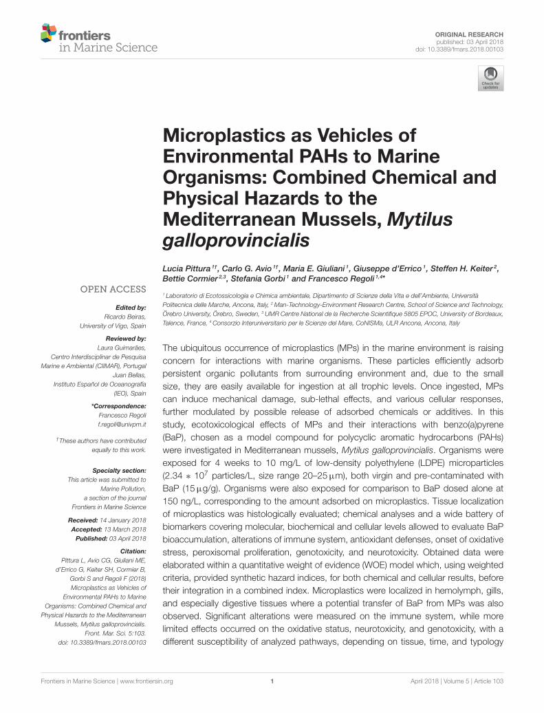
Acetylcholinesterase showed significant effects as a functionof time with a slight decrease in hemolymph and a slightincrease in gills after 7 days of exposure to all the treatments(Figures 4A,B, Table 2): no significant variations were observedbetween different treatments (Table 2).
DNA strand breaks in hemocytes were always comparable forvarious treatments and times of exposure (Figure 4C, Table 2),while micronuclei showed a significant increase in musselsexposed to BaP and BaP contaminated LDPE after 14 days ofexposure (Figure 4D, Table 2).
Peroxisomal AOX did not significantly vary in any treatments,although a clear trend of inhibition was observed over time inmussels exposed to LDPE (Figure 4E, Table 2). A slight increaseof neutral lipids was observed in mussels exposed to BaP and BaPcontaminated microplastics particularly after 7 days (Figure 4F,Table 2).
Antioxidant defenses revealed minor fluctuations caused byvarious treatments, with only a slightly higher oxidative pressureafter 28 days of exposure to BaP (Figures 5A–F, Table 2). Thelimited pro-oxidant challenge was further supported by MDA,showing a moderate increase only after 7 days in musselsexposed to LDPE and BaP (Figure 5I), and by general lack ofvariations for TOSC toward both peroxyl and hydroxyl radicals(Figures 5G,H, Table 2).
The results on molecular analyses confirmed the absenceof statistically significant differences between treatments onmRNA levels of antioxidants cat, gst-pi, Se-gpx, and of aox1(Figures 6A–D, Table 2). Generally higher transcriptional levelswere measured for cat and gst-pi in mussels after 28 daysindependently on exposure treatment, while fluctuating levels ofSe-gpx mRNA were observed in mussels treated with BaP andwith LDPE-BaP (Figures 6A-C, Table 2). Transcriptional levelsof hsp70 appeared downregulated by various treatments after7 days, while a significant increase was observed in organismsexposed to LDPE for 14 days (Figure 6E).
The PCA carried out on the whole set of biomarkers produceda two-dimensional pattern explaining 54% of total variance(Figure 7). Although a quite large percentage remained to beexplained, obtained results indicated a clear separation betweenspecimens exposed at different treatments for different times.After 7 days (Blu ellipse), LDPE and LDPE-BaP treated musselsseparated from the other groups, at 14 days (Red ellipse) musselstreated with BaP and LDPE-BaP were more differentiated,while after 28 days (Green ellipse) the effects of BaP alonebecame more evident, producing a clear separation betweensuch experimental group and other treatments (Figure 7). Theparameters determining the separation along the PC1 axis wererelated to immune system responses (G/H ratio), neurotoxiceffects (AchE), and antioxidant system (catalase, glutathione-S-transferase, glutathione reductase, glutathione peroxidase Se-dep, TOSC •OH and ROO•), and AOX. On the otherside, genotoxic effects (micronuclei), neutral lipids (NL), totalglutathione (TGSH), total glutathione peroxidases (GPX_CHP),and phagocytosis activity determined the separation along thePC2 axis.
Elaboration of data with weighted criteria summarized asSevere the hazard index for bioavailability in mussels exposedto BaP or BaP contaminated LDPE at all exposure periods(Figure 8). On the other hand, based on the magnitude ofvariations exhibited by various biomarkers, their statisticalsignificance of such differences and the toxicological relevance ofeach biological endpoint, the model summarized the hazard forcellular responses as Slight for organisms exposed to BaP, virgin,and contaminated LDPE, and Moderate only for organisms
Frontiers in Marine Science | www.frontiersin.org 7 April 2018 | Volume 5 | Article 103
Pittura et al. Ecotoxicology of Microplastics in Marine Mussels
FIGURE 4 | Biomarkers in mussels exposed for 7, 14, and 28 days to various treatments (CTRL, control; LDPE, virgin low density polyethylene; BaP, benzo(a)pyrene
alone; LDPE-BaP, benzo(a)pyrene-contaminated polyethylene). ACh-E: acetylcholinesterase in haemolymph (A) and gills (B); DNA TAIL %: fragmentation of DNA (C);
MN/1000: frequency of micronuclei (D); AOX: Acyl CoA Oxidase (E); Neutral Lipids (F). Data are expressed as mean values ± standard error, n = 4; different letters
indicate significant differences between groups of means within the same time of exposure (post-hoc Newman-Keuls comparison).
exposed to BaP after 14 days (Figure 8). The integration ofhazard indices elaborated for bioavailability and biomarker dataresulted in a combined WOE effect classified as Slight formussels exposed to virgin LDPE andMajor for those treated withboth contaminated LDPE and BaP alone, without variations atdifferent times of exposure.
DISCUSSION
The increase of plastics and microplastics in marine ecosystemshas raised concern on their impact to marine organisms, and
several species have been shown to ingest these particles underexperimental and wild conditions (Cole et al., 2011; Lusheret al., 2013; De Witte et al., 2014; Avio et al., 2015, 2017b;Devriese et al., 2015; Paul-Pont et al., 2016; Sussarellu et al., 2016;Murphy et al., 2017). The capability of microplastics to efficientlyadsorb chemical pollutants from the environment (Avio et al.,2017a) poses an additional risk although there is not yet clearevidence that microplastics ingestion has adverse consequenceson the health status of marine species, especially under long termconditions.
In this respect, the present study was aimed to provide newinsights on the capability of microplastics to transfer adsorbed
Frontiers in Marine Science | www.frontiersin.org 8 April 2018 | Volume 5 | Article 103

Pittura et al. Ecotoxicology of Microplastics in Marine Mussels
FIGURE 5 | Levels of catalase (A), glutathione reductase (B), glutathione S-transferases (C), sum of Se-dependent and Se-independent glutathione peroxidases (D),
Se-dependent glutathione peroxidases (E), total glutathione (F), total oxyradical scavenging capacity (TOSC) toward peroxyl (•OOR) radicals (G), total oxyradical
scavenging capacity (TOSC) toward hydroxyl (•OH) radicals (H), malondialdehyde (MDA) (I) in mussels exposed for 7, 14, and 28 days to various treatments (CTRL,
control; LDPE, virgin low density polyethylene; BaP, benzo(a)pyrene alone; LDPE-BaP, benzo(a)pyrene-contaminated polyethylene). Data are expressed as mean
values ± standard error, n = 4; different letters indicate significant differences between groups of means within the same time of exposure (post-hoc Newman-Keuls
comparison).
pollutant to organisms after ingestion and to evaluate potentialecotoxicological effects of virgin and contaminatedmicroplastics,using the Mediterranean mussel M. galloprovincialis as modelmarine organism. Although the selected level of microplastics(10 mg/L) appears higher than environmental data, it is worthyto note that a direct comparison between experimental andfield values is not necessarily appropriate. Reported seawaterconcentrations are typically referred to microplastics >200µm,while natural levels are still unknown for smaller particles, likethose used in the present study (20–25µm), which representthe size range preferentially ingested by filter feeding organisms.Considering the need to characterize the ecotoxicologicalpotential of such biologically relevant microplastics, at thepresent state of knowledge, concentrations of fewmg/L are still inan ecologically relevant range to evaluate in laboratory conditionsthe disturbance of cellular pathways, possibly involved in long-term responses to small microplastics.
Our results revealed that microplastics can act as efficientvehicles of chemical pollutants. Bioaccumulation analysesshowed a marked and rapid enhancement of BaP concentrationsin digestive gland of mussels exposed to LDPE-BaP, reaching
a steady state after 7 days and values comparable to thoseobserved in BaP treated mussels. This result corroborates thehypothesis of a marked release of BaP from microplastics and anelevated bioconcentration process in tissues under physiologicalgut conditions, as previously suggested by other authors (Teutenet al., 2009; Bakir et al., 2014; Avio et al., 2015). A slightly differenttrend was observed for bioaccumulation of BaP in gills: LDPE-BaP treated mussels exhibited only a moderate increase duringthe initial phases of exposure, reaching tissue concentrationssimilar to those observed in BaP exposed mussels only after28 days. While a rapid uptake in gills can be explained by thedirect contact of this tissue with the chemical dissolved in water(Banni et al., 2017), the slower accumulation from contaminatedmicroplastics may, at least partly derive from primary desorptionof BaP in digestive tissues and a secondary transfer of thischemical to gills.
The possibility that BaP measured in LDPE-BaP treatedorganisms can reflect the presence of still un-excreted particlesmore than a real tissue accumulation, can be considered asnegligible. Concentrations higher than 15 and 30 ng/g weremeasured in gills and digestive glands, respectively; assuming
Frontiers in Marine Science | www.frontiersin.org 9 April 2018 | Volume 5 | Article 103

Pittura et al. Ecotoxicology of Microplastics in Marine Mussels
FIGURE 6 | Transcriptional responses in the digestive glands of mussels exposed for 7, 14, and 28 days to various treatments (CTRL, control; LDPE, virgin low
density polyethylene; BaP, benzo(a)pyrene alone; LDPE-BaP, benzo(a)pyrene-contaminated polyethylene). cat, catalase (A); gst-pi, pi-class glutathione S-transferase
(B); Se-gpx, selenium-dependent glutathione peroxidase (C); aox1, acyl-CoA oxidase, isoform 1 (D); hsp70, heat shock protein 70 (E). Data are expressed as mean
values ± standard error, n = 4; different letters indicate significant differences between groups of means within the same time of exposure (post-hoc Newman-Keuls
comparison).
that all the measured BaP was still adsorbed on microplastics,we should expect at least 1mg of particles for each gram of gilltissue (corresponding to 2.34 ∗ 105 particles), and at least 2mg(4.68 ∗ 105 particles) for each gram of digestive gland. A similarassumption is excluded by histological analyses that confirmedthe presence of particles in those tissues, but with much morelimited numbers, particularly in gills where only a few and sparsemicroplastics were observed.
Uptake and tissue distribution of microplastics has alreadybeen investigated in marine bivalves such as the musselsMytilus edulis and M. galloprovincialis exposed to virgin andcontaminated polyethylene and polystyrene (Browne et al., 2008;Von Moos et al., 2012; Avio et al., 2015). Although these studiesused extremely high concentrations of microplastics (up to threeorder of magnitude greater than in the present work), they wereimportant in demonstrating the initial uptake of particles at thegill’s surface throughmicrovilli activity and endocytosis, while viaciliae movement in the stomach, intestine and digestive tubulesare responsible for a second pathway mediated by accumulationwithin the lysosomal compartment (Von Moos et al., 2012). Ourobservations almost reflected the above mechanisms of uptake,with aggregates of particles observed within intestinal lumen anddigestive tissues, lower occurrence in gills, and some particlesnoticed also inside hemocytes, as previously documented inother experiments (Browne et al., 2008; Von Moos et al., 2012).Histological analyses were of qualitative nature, but no markeddifferences in the amount of microparticles were visible forvarious treatments and times of exposure, thus supporting a shortretention time of such particles in mussels, as reported in fishexposed to microbeads (Grigorakis et al., 2017).
Significant immunological effects were observed onhemocytes lysosomal membrane stability, phagocytosis, andgranulocytes/hyalinocytes ratio. The impairment of immunesystem has already been measured in marine organisms exposedto microplastics by several authors (Von Moos et al., 2012;Avio et al., 2015; Paul-Pont et al., 2016). Lysosomes, besiderepresenting major sites for intracellular sequestration anddetoxification of xenobiotics, have been also demonstrated assensitive organelles toward micro- and nano-plastics (Regoli,1992; Petrovic et al., 2004; Moore et al., 2006; Canesi et al.,2012; Avio et al., 2015; Nardi et al., 2017). The destabilizationof lysosomal membrane caused by LDPE or BaP alone, wassynergistically enhanced in mussels exposed to LDPE-BaP,particularly after 7 days and, to a lower extent, 14 days ofexposure. Effects of various treatments were observed alsofor phagocytosis which initially increased in mussels exposedto LDPE and BaP, while decreasing at longer periods as aconsequence of BaP, virgin, and contaminated LDPE: similareffects might be due to an overload of sequestering capacity ofhemocytes by microplastics, and to the well-known inhibitoryaction of PAHs on this function (Wootton et al., 2003; Hannamet al., 2010). Interestingly, LDPE and LDPE-BaP did not affect thegranulocytes/hyalinocytes ratio that was statistically increasedonly by BaP until 14 days. The changes of immune parametersobserved in this study are not a surprise given the characteristicsof plastic particles, and the physical stress that potentially inducein hemocytes, further modulated with a chemical challenge inmussels exposed to LDPE-BaP.
Our results did not reveal significant effects on AChEactivity neither in hemolymph nor in gills, although both the
Frontiers in Marine Science | www.frontiersin.org 10 April 2018 | Volume 5 | Article 103

Pittura et al. Ecotoxicology of Microplastics in Marine Mussels
FIGURE 7 | Multivariate PCA analysis on biomarker data in mussels exposed to various microplastics treatments: CTRL, control; LDPE, virgin low density
polyethylene; BaP, benzo(a)pyrene alone; LDPE-BaP, benzo(a)pyrene-contaminated polyethylene.
tissues exhibited after 7 days a clear trend toward reduced orenhanced values, respectively. The only moderate and temporarymodulation of AChE may reflect the low exposure period.However, cholinesterasic effects of microplastics still deservescientific attention due to the abundance of these particles inthe marine environment and their suggested role in influencingvarious physiological and behavioral responses controlled byneurological mechanisms (Oliveira et al., 2013; Avio et al., 2015;Mattsson et al., 2017; Ribeiro et al., 2017).
No variations were measured on levels of DNA strandbreaks in organisms exposed to microplastics (both virgin andcontaminated) or to BaP. A high DNA fragmentation hadbeen previously measured in mussels exposed to polyethylenemicroplastics (Avio et al., 2015), but the more elevated amountof particles used in those treatments (1.5 vs. 0.01 g/L of thisstudy) can explain the different results. Similarly, the lack ofDNA fragmentation in BaP treated mussels might reflect the lowexperimental concentration as compared to those frequently usedfor assessing ecotoxicological effects of BaP in mussels (Pan et al.,2009; Banni et al., 2017): in this respect, no formation of DNAadducts or strand breaks was observed in mussels exposed to 300ng/L of BaP for 24 days (Ching et al., 2001).
Some authors have suggested that microplastics ingestion canpotentially cause pseudo-satiety in mussels, thus lowering fattyacids metabolization (Kühn et al., 2015). The AOX, one of theenzymes involved in fatty acid oxidation (Cajaraville et al., 1997;Bilbao et al., 2009) did not show significant effects neither atcatalytic nor at transcriptional levels. Content of neutral lipidstended to increase in mussels exposed to BaP and LDPE-BaP,confirming a typical effect of this chemical in inducing lipidosis
in digestive gland ofmussels (Livingstone and Farrar, 1984; Gorbiet al., 2008).
Treatments with virgin and contaminated microplastics didnot affect the oxidative status of mussels, and only minorfluctuations of a few enzymes (glutathione S-transferases andglutathione reductase) were observed, without clear trends asa function of treatment or time of exposure. Responses ofantioxidant systemwere investigated also atmolecular level, sincetranscriptional changes might be more sensitive than enzymaticbiomarkers, despite more useful in revealing “exposure” ratherthan functional “effects” at cellular level (Giuliani et al., 2013;Regoli and Giuliani, 2014). Also these analyses exhibited minorand not significant variations, allowing to exclude an oxidativechallenge, as further supported by the lack of effects on thetotal antioxidant capacity and peroxidation processes in musselsexposed to virgin and contaminated LDPE. The lower levels ofparticles used in this study, might explain the different resultson oxidative effects in comparison to other studies in whichmussels exposed to microplastics exhibited significant changesof antioxidant defenses (Avio et al., 2015; Paul-Pont et al., 2016;Détrée and Gallardo-Escárate, 2017; Ribeiro et al., 2017).
A transient upregulation of hsp70 was observed only after14 days in mussels exposed to virgin LDPE, suggesting aresponse toward the physical disturbance caused by the ingestionof such particles. Enhanced levels of these proteins are ageneric biomarker of stress, acting in mussels as a first line ofdefense to cope with environmental challenges (Franzellitti andFabbri, 2005; Heindler et al., 2017). The effects of contaminatedmicroplastics were more similar to those of BaP, with lack ofstatistical changes and a trend toward lower values of hsp70,
Frontiers in Marine Science | www.frontiersin.org 11 April 2018 | Volume 5 | Article 103

Pittura et al. Ecotoxicology of Microplastics in Marine Mussels
FIGURE 8 | Weighted elaboration of bioaccumulation and biomarkers data in mussels exposed for 7, 14, 28 days to LDPE, BaP, and LDPE-BaP. The assigned
classes of hazard are given. Treatments: LDPE, virgin low density polyethylene; BaP, Benzo(a)pyrene alone; LDPE-BaP, Benzo(a)pyrene-contaminated polyethylene.
supporting a limited responsiveness of these proteins to theprevalence of a chemical stress.
The overall evaluation of biomarker results by multivariatePCA provided a clear separation between times and typologiesof exposure, highlighting a shift from a physical to a chemicalstress. After 7 days, the main effects were those inducedby microplastics (possibly reflecting a physical challenge),followed at 14 days by those combined of microplasticswith BaP, while at longer exposure conditions effects ofBaP prevailed on those induced by microplastics (chemicalimpact). The multivariate analysis indicated that the majority ofobserved immunological, lysosomal, and cholinesterasic effectswere influenced by polymer (LDPE), while genotoxicity andantioxidant defenses were mostly related to BaP. The impactof LDPE-BaP appeared more biologically relevant with timeof exposure, suggesting that energy resources were initiallydirected to activate primary mechanisms of defense towardthe physical stress of particles, while later the chemical stressassumed the major role in biological disturbance. A similardelay of chemical-induced toxic effects was previously observedin fish Pomatoschistus microps exposed to microplastics andorganic compounds, where these particles acted as a transitorymechanism of protection toward chemical insult (Oliveira et al.,2013).
The overall data were elaborated according to the weightedcriteria of the Sediqualsoft model to synthesize the biologicalsignificance of bioaccumulation results and cellular responsesin mussels exposed to virgin and contaminated microplastics.
The bioavailability of BaP was classified as Severe for both thechemical dosed alone and for LDPE-BaP, since concentrationsincreased from 15- to 60-folds in tissues of exposed musselscompared to controls. On the other hand, the toxicologicalhazard calculated from the number, magnitude and biologicalimportance of biomarkers was typically Slight for all thetreatments, raising to Moderate only in BaP exposed musselsafter 14 days. The combination of chemical and cellular hazardsprovided a WOE index Slight for mussels exposed to virginLDPE, and Major for those exposed to BaP and LDPE-BaPfor all the periods. Considering the similarity of biologicaleffects observed after 28 days, it is quite obvious that the finalevaluation of the risk caused by virgin and contaminated LDPEwas greatly influenced by the marked accumulation of BaP,further corroborating the still unexplored possibility of indirect,long-term consequences of released chemicals.
In conclusion, this study confirmed that microplastics cantransfer adsorbed organic contaminants like BaP to tissuesof marine organisms, providing an additional experimentalevidence to the role of these particles as source of chemicalbioaccumulation. Both virgin and contaminated microplasticsdid not induce marked ecotoxicological effects at molecularand cellular levels after 28 days of exposure. However, theobserved susceptibility of the immune system, the accumulationof BaP and the probable shift from physical to chemicalchallenge, suggest that the toxicological risk of microplastics formarine organisms is probably low, but not negligible. Additionalstudies are needed to elucidate conditions of chronic exposure
Frontiers in Marine Science | www.frontiersin.org 12 April 2018 | Volume 5 | Article 103
Pittura et al. Ecotoxicology of Microplastics in Marine Mussels
and whether interactions of particles with other stressors mayprovoke long term, subtle effects on organisms’ health status.
ETHICS STATEMENT
The study was exempt from the above requirements because theydo not apply to invertebrates which were used in this study.
AUTHORS CONTRIBUTIONS
LP, CA, SG, and FR: Conceived the study; SK and BC: Preparedthe contaminated microplastics; LP and CA: Performed the
experiments; LP, CA, and MG: Made laboratory analyses, GdE:Statistical and weighted elaboration of data; LP, CA, SG, andFR: Wrote the manuscript, FR: Edited and reviewed the finalversion of the manuscript which all the authors approved beforesubmission.
ACKNOWLEDGMENTS
This work has been financially supported withinEphemare Project (by JPI Oceans). Centro AssistenzaEcologica (Ancona) partially funded the Ph.D. fellowshipto LP.
REFERENCES
Avio, C. G., Cardelli, L. R., Gorbi, S., Pellegrini, D., and Regoli, F. (2017b).Microplastics pollution after the removal of the costa concordia wreck: firstevidences from a biomonitoring case study. Environ. Pollut. 227, 207–214.doi: 10.1016/j.envpol.2017.04.066
Avio, C. G., Gorbi, S., and Regoli, F. (2017a). Plastics and microplastics in theoceans: from emerging pollutants to emerged threat. Mar. Environ. Res. 128,2–11. doi: 10.1016/j.marenvres.2016.05.012
Avio, C. G., Gorbi, S., Milan, M., Benedetti, M., Fattorini, D., d’Errico,G., et al. (2015). Pollutants bioavailability and toxicological riskfrom microplastics to marine mussels. Environ. Pollut. 198, 211–222.doi: 10.1016/j.envpol.2014.12.021
Bakir, A., Rowland, S. J., and Thompson, R. C. (2014). Enhanced desorption ofpersistent organic pollutants from microplastics under simulated physiologicalconditions. Environ. Pollut. 185, 16–23. doi: 10.1016/j.envpol.2013.10.007
Banni, M., Sforzini, S., Arlt, V. M., Barranger, A., Dallas, L. J., Oliveri,C., et al. (2017). Assessing the impact of Benzo [a] pyrene on marinemussels: application of a novel targeted low density microarraycomplementing classical biomarker responses.PloS ONE 12:e0178460.doi: 10.1371/journal.pone.0178460
Baršiene, J., Rybakovas, A., Garnaga, G., and Andreikenait,e, L. (2012).Environmental genotoxicity and cytotoxicity studies inmussels before and afteran oil spill at the marine oil terminal in the Baltic Sea. Environ. Monit. Assess.
184, 2067–2078. doi: 10.1007/s10661-011-2100-0Bebianno, M. J., Pereira, C. G., Rey, F., Cravo, A., Duarte, D., d’Errico, G., et al.
(2015). Integrated approach to assess ecosystem health in harbor areas. Sci.Total Environ. 514, 92–107. doi: 10.1016/j.scitotenv.2015.01.050
Benedetti, M., Ciaprini, F., Piva, F., Onorati, F., Fattorini, D., Notti,A., et al. (2012). A multidisciplinary weight of evidence approachfor classifying polluted sediments: integrating sediment chemistry,bioavailability, biomarkers responses and bioassays. Environ. Int. 38, 17–28.doi: 10.1016/j.envint.2011.08.003
Benedetti, M., Gorbi, S., Fattorini, D., d’Errico, G., Piva, F., Pacitti, D.,et al (2014). Environmental hazards from natural hydrocarbons seepage:integrated classification of risk from sediment chemistry, bioavailability andbiomarkers responses in sentinel species. Environ. Pollut. 185, 116–126.doi: 10.1016/j.envpol.2013.10.023
Benedetti, M., Lanzoni, I., Nardi, A., d’Errico, G., Di Carlo, M., Fattorini,D., et al. (2016). Oxidative responsiveness to multiple stressors in the keyAntarctic species, Adamussium colbecki: interactions between temperature,acidification and cadmium exposure. Mar. Environ. Res. 121, 20–30.doi: 10.1016/j.marenvres.2016.03.011
Bilbao, E., Cajaraville, M. P., and Cancio, I. (2009). Cloning and expressionpattern of peroxisomal β-oxidation genes palmitoyl-CoA oxidase,multifunctional protein and 3-ketoacyl-CoA thiolase in mussel Mytilus
galloprovincialis and thicklip grey mullet Chelon labrosus. Gene 443, 132–142.doi: 10.1016/j.gene.2009.05.008
Bocchetti, R., Fattorini, D., Pisanelli, B., Macchia, S., Oliviero, L., Pilato, F., et al.(2008). Contaminant accumulation and biomarker responses in caged mussels,
Mytilus galloprovincialis, to evaluate bioavailability and toxicological effects ofremobilized chemicals during dredging and disposal operations in harbourareas. Aquat. Toxicol. 89, 257–266. doi: 10.1016/j.aquatox.2008.07.011
Browne, M. A. (2015). “Sources and pathways of microplastics to habitats,” inMarine Anthropogenic Litter, eds M. Bergmann, L. Gutow, and M. Klages(London: Springer), 229–244.
Browne, M. A., Dissanayake, A., Galloway, T. S., Lowe, D. M., and Thompson,R. C. (2008). Ingested microscopic plastic translocates to the circulatorysystem of the mussel, Mytilus edulis (L.). Environ. Sci. Technol. 42, 5026–5031.doi: 10.1021/es800249a
Cajaraville, M. P., Orbea, A., Marigómez, I., and Cancio, I. (1997). Peroxisomeproliferation in the digestive epithelium of mussels exposed to the wateraccommodated fraction of three oils. Comp. Biochem. Physiol. C 117, 233–242.doi: 10.1016/S0742-8413(97)00057-1
Canesi, L., Borghi, C., Ciacci, C., Fabbri, R., Lorusso, L. C., Vergani, L., et al. (2008).Short-term effects of environmentally relevant concentrations of EDCmixtureson Mytilus galloprovincialis digestive gland. Aquat. Toxicol. 87, 272–279.doi: 10.1016/j.aquatox.2008.02.007
Canesi, L., Borghi, C., Ciacci, C., Fabbri, R., Vergani, L., and Gallo, G.(2007). Bisphenol-A alters gene expression and functional parametersin molluscan hepatopancreas. Mol. Cell. Endocrinol. 276, 36–44.doi: 10.1016/j.mce.2007.06.002
Canesi, L., Ciacci, C., Fabbri, R., Marcomini, A., Pojana, G., and Gallo, G. (2012).Bivalve molluscs as a unique target group for nanoparticle toxicity. Mar.
Environ. Res. 76, 16–21. doi: 10.1016/j.marenvres.2011.06.005Cellura, C., Toubiana, M., Parrinello, N., and Roch, P. (2006). HSP70 gene
expression in Mytilus galloprovincialis hemocytes is triggered by moderateheat shock and Vibrio anguillarum, but not by V. splendidus or Micrococcus
lysodeikticus. Dev. Comp. Immunol. 30, 984–997. doi: 10.1016/j.dci.2005.12.009Ching, E. W. K., Siu, W. H. L., Lam, P. K. S., Xu, L. H., Zhang, Y. Y.,
Richardson, B. J., et al. (2001). DNA adduct formation and DNA strandbreaks in green-lipped mussels (Perna viridis) exposed to benzo[a]pyrene:dose- and time-dependent relationships. Mar. Pollut. Bull. 42, 603–610.doi: 10.1016/S0025-326X(00)00209-5
Cole, M., Lindeque, P., Halsband, C., and Galloway, T. S. (2011). Microplasticsas contaminants in the marine environment: a review. Mar. Pollut. Bull. 62,2588–2597. doi: 10.1016/j.marpolbul.2011.09.025
Cózar, A., Sanz-Martín, M., Mart,í, E., González-Gordillo, J. I., Ubeda, B., Gálvez,J. Á., et al. (2015). Plastic accumulation in the Mediterranean Sea. PLoS ONE
10:e0121762. doi: 10.1371/journal.pone.0121762DeWitte, B., Devriese, L., Bekaert, K., Hoffman, S., Vandermeersch, G., Cooreman,
K., et al. (2014). Quality assessment of the blue mussel (Mytilus edulis):comparison between commercial and wild types.Mar. Pollut. Bull. 85, 146–155.doi: 10.1016/j.marpolbul.2014.06.006
Détrée, C., and Gallardo-Escárate, C. (2017). Polyethylene microbeads inducetranscriptional responses with tissue-dependent patterns in the musselMytilus galloprovincialis. J. Molluscan Stud. 83, 220–225. doi: 10.1093/mollus/eyx005
Devriese, L. I., van der Meulen, M. D., Maes, T., Bekaert, K., Paul-Pont, I., Frère, L.,et al. (2015). Microplastic contamination in brown shrimp (Crangon crangon,
Frontiers in Marine Science | www.frontiersin.org 13 April 2018 | Volume 5 | Article 103

Pittura et al. Ecotoxicology of Microplastics in Marine Mussels
Linnaeus 1758) from coastal waters of the Southern North Sea and Channelarea.Mar. Pollut. Bull. 98, 179–187. doi: 10.1016/j.marpolbul.2015.06.051
Eriksen, M., Lebreton, L. C. M., Carson, H. S., Thiel, M., Moore, C. J., Borerro,J. C., et al. (2014). Plastic pollution in the world’s oceans: more than 5 trillionplastic pieces weighing over 250,000 tons afloat at sea. PLoS ONE 9:e111913.doi: 10.1371/journal.pone.0111913
Franzellitti, S., and Fabbri, E. (2005). Differential HSP70 gene expression in theMediterranean mussel exposed to various stressors. Biochem. Biophys. Res.
Commun. 336, 1157–1163. doi: 10.1016/j.bbrc.2005.08.244Galgani, F., Hanke, G., and Maes, T. (2015). “Global distribution, composition and
abundance of marine litter”, inMarine Anthropogenic Litter, eds M. Bergmann,L. Gutow, and M. Klages (London: Springer), 29–56.
Giannapas, M., Loukas, K., and Stefanos, D. (2012). Generation of free radicalsin haemocytes of mussels after exposure to low molecular weight PAHcomponents: immune activation, oxidative and genotoxic effects. Comp.
Biochem. Physiol. C 155, 182–189. doi: 10.1016/j.cbpc.2011.08.001Gilfillan, L. R., Ohman, M. D., Doyle, M. J., and Watson, W. (2009). Occurrence
of plastic micro-debris in the Southern California Current system Cal. Coop.
Ocean. Fish. 50, 123–133.Giuliani, M. E., Benedetti, M., Arukwe, A., and Regoli, F. (2013). Transcriptional
and cata- lytic responses of antioxidant and biotransformation pathwaysin mussels, Mytilus galloprovincialis, exposed to chemical mixtures. Aquat.Toxicol. 134, 120–127. doi: 10.1016/j.aquatox.2013.03.012
Goldstein, M. C., Titmus, A. J., and Ford, M. (2013). Scales of spatial heterogeneityof plastic marine debris in the northeast Pacific Ocean. PLoS ONE 8:e80020.doi: 10.1371/journal.pone.0080020
Gorbi, S., Avio, G. C., Benedetti, M., Totti, C., Accoroni, S., Pichierri,S., et al. (2013). Effects of harmful dinoflagellate Ostreopsis cf. ovataexposure on immunological, histological and oxidative responses ofmussels Mytilus galloprovincialis. Fish Shellfish Immun. 35, 941–950.doi: 10.1016/j.fsi.2013.07.003
Gorbi, S., Virno Lamberti, C., Notti, A., Benedetti, M., Fattorini, D.,Moltedo, G., et al. (2008). An ecotoxicological protocol with cagedmussels Mytilus galloprovincialis, for monitoring the impact of anoffshore platform in the Adriatic sea. Mar. Environ. Res. 65, 34–49.doi: 10.1016/j.marenvres.2007.07.006
Grigorakis, S., Mason, S. A., and Drouillard, K. G. (2017). Determination of the gutretention of plastic microbeads and microfibers in goldfish (Carassius auratus).Chemosphere 169, 233–238. doi: 10.1016/j.chemosphere.2016.11.055
Grintzalis, K., Christos, D. G., and Stefanos, D. (2012). Total thiol redox status asa potent biomarker of PAH-mediated effects on mussels.Mar. Environ. Res. 81,26–34. doi: 10.1016/j.marenvres.2012.08.004
Hannam, M. L., Bamber, S. D., Galloway, T. S., Moody, A. J., and Jones, M.B. (2010). Effects of the model PAH phenanthrene on immune function andoxidative stress in the haemolymph of the temperate scallop Pecten maximus.Chemosphere 78, 779–784. doi: 10.1016/j.chemosphere.2009.12.049
Heindler, F. M., Alajmi, F., Huerlimann, R., Zeng, C., Newman, S. J.,Vamvounis, G., et al. (2017). Toxic effects of polyethylene terephthalatemicroparticles and Di (2-ethylhexyl) phthalate on the calanoid copepod,Parvocalanus crassirostris. Ecotoxicol. Environ. Saf. 141, 298–305.doi: 10.1016/j.ecoenv.2017.03.029
Karami, A., Groman, D. B., Wilson, S. P., Ismail, P., and Neela, V. K.(2017). Biomarker responses in zebrafish (Danio rerio) larvae exposed topristine low-density polyethylene fragments. Environ. Pollut. 223, 466–475.doi: 10.1016/j.envpol.2017.01.047
Koelmans, A. A., Bakir, A., Burton, G. A., and Janssen, C. R. (2016). Microplasticas a vector for chemicals in the aquatic environment: critical review andmodel-supported reinterpretation of empirical studies. Environ. Sci. Technol.50: 3315–3326. doi: 10.1021/acs.est.5b06069
Kühn, S., Bravo Rebolledo, E. L., and van Franeker, J. A. (2015). “Deleterious effectsof litter on marine life,” in Marine Anthropogenic Litter, eds M. Bergmann, L.Gutow, and M. Klages (London: Springer), 75–116.
Liu, L., Fokkink, R., and Koelmans, A. A. (2016). Sorption of polycyclicaromatic hydrocarbons to polystyrene nanoplastic. Environ. Toxicol. Chem. 35,1650–1655. doi: 10.1002/etc.3311
Livingstone, D. R., and Farrar, S. V. (1984). Tissue and subcellular distributionof enzyme activities of mixed-function oxygenase and benzo[a]pyrenemetabolism in the common musselMytilus edulis L. STOTEN 39, 209–235.
Lohmann, R. (2017). Microplastics are not important for the cycling andbioaccumulation of organic pollutants in the oceans—but should microplasticsbe considered POPs themselves? Integr. Environ. Assess. Manag. 13, 460–465.doi: 10.1002/ieam.1914
Lusher, A. (2015). “Microplastics in the marine environment: distribution,interactions and effects,” in Marine Anthropogenic Litter, eds M. Bergmann, L.Gutow, and M. Klages (London: Springer), 245–307.
Lusher, A. L., McHugh, M., and Thompson, R. C. (2013). Occurrenceof microplastics in the gastrointestinal tract of pelagic and demersalfish from the English Channel. Mar. Pollut. Bull. 67, 94–99.doi: 10.1016/j.marpolbul.2012.11.028
Lusher, A. L., Welden, N. A., Sobral, P., and Cole, M. (2017). Sampling, isolatingand identifying microplastics ingested by fish and invertebrates. Anal. Methods
9, 1346–1360. doi: 10.1039/C6AY02415GMarigómez, I., and Baybay-Villacorta, L. (2003). Pollutant-specific and general
lysosomal responses in digestive cells of mussels exposed to model organicchemicals. Aquat. Toxicol. 64, 235–257. doi: 10.1016/S0166-445X(03)00056-0
Mattsson, K., Johnson, E. V., Malmendal, A., Linse, S., Hansson, L. A., andCedervall, T. (2017). Brain damage and behavioural disorders in fish inducedby plastic nanoparticles delivered through the food chain. Sci. Rep. 7:11452.doi: 10.1038/s41598-017-10813-0
Mezzelani, M., Gorbi, S., Da Ros, Z., Fattorini, D., d’Errico, G., Milan, M.,et al. (2016). Ecotoxicological potential of non-steroidal anti-inflammatorydrugs (NSAIDs) in marine organisms: bioavailability, biomarkers and naturaloccurrence in Mytilus galloprovincialis. Mar. Environ. Res. 121, 31–39.doi: 10.1016/j.marenvres.2016.03.005
Moore, C. J., Moore, S. L., Leecaster, M. K., and Weisberg, S. B. (2001).A comparison of plastic and plankton in the north Pacific centralgyre. Mar. Pollut. Bull. 42, 1297–1300. doi: 10.1016/s0025-326x(01)00114-x
Moore, M. N., Allen, J. I., McVeigh, A., and Shaw, J. (2006). Lysosomal andautophagic reactions as predictive indicators of environmental impact inaquatic animals. Autophagy 2, 217–220. doi: 10.4161/auto.2663
Murphy, F., Russell, M., Ewins, C., and Quinn, B. (2017). The uptakeof macroplastic & microplastic by demersal & pelagic fish in theNortheast Atlantic around Scotland. Mar. Pollut. Bull 122, 353–359.doi: 10.1016/j.marpolbul.2017.06.073
Nardi, A., Mincarelli, L. F., Benedetti, M., Fattorini, D., d’Errico, G., and Regoli,F. (2017). Indirect effects of climate changes on cadmium bioavailabilityand biological effects in the Mediterranean mussel Mytilus galloprovincialis.Chemosphere 169, 493–502. doi: 10.1016/j.chemosphere.2016.11.093
NOAA (2015). A NOAA MDP Research Project Focuses on Types and Abundance
of Microplastics. Detecting Microplastics in the Marine Environment. Availableonline at: http://marinedebris.noaa.gov/research/detecting-microplastics-marine-environment
Oliveira, M., Ribeiro, A., Hylland, K., and Guilhermino, L. (2013). Single andcombined effects of microplastics and pyrene on juveniles (0+ group) of thecommon goby Pomatoschistus microps (Teleostei, Gobiidae). Ecol. Indic. 34,641–647. doi: 10.1016/j.ecolind.2013.06.019
Pan, L., Ren, J., and Zheng, D. (2009). Effects of benzo (a) pyrene exposure onthe antioxidant enzyme activity of scallop Chlamys farreri. Chinese J. Oceanol.Limnol. 27, 43–53. doi: 10.1007/s00343-009-0043-x
Paul-Pont, I., Lacroix, C., Fernández, C. G., Hégaret, H., Lambert, C., Le Goïc,N., et al. (2016). Exposure of marine mussels Mytilus spp. to polystyrenemicroplastics: toxicity and influence on fluoranthene bioaccumulation.Environ. Pollut. 216, 724–737. doi: 10.1016/j.envpol.2016.06.039
Pedà, C., Caccamo, L., Fossi, M. C., Gai, F., Andaloro, F., Genovese, L., et al. (2016).Intestinal alterations in European sea bass Dicentrarchus labrax (Linnaeus,1758) exposed to microplastics: preliminary results. Environ. Pollut. 212,251–256. doi: 10.1016/j.envpol.2016.01.083
Petrovic, S., Semenci,c, L., Ozretic, B., and Ozretic, M. (2004). Seasonal variationsof physiological and cellular biomarkers and their use in the biomonitoringof north Adriatic coastal waters (Croatia). Mar. Pollut. Bull. 49, 713–720.doi: 10.1016/j.marpolbul.2004.05.004
Phuong, N. N., Zalouk-Vergnoux, A., Poirier, L., Kamari, A., Châtel, A.,Mouneyrac, C., et al. (2016). Is there any consistency between the microplasticsfound in the field and those used in laboratory experiments? Environ. Pollut.211, 111–123. doi: 10.1016/j.envpol.2015.12.035
Frontiers in Marine Science | www.frontiersin.org 14 April 2018 | Volume 5 | Article 103
Pittura et al. Ecotoxicology of Microplastics in Marine Mussels
Piva, F., Ciaprini, F., Onorati, F., Benedetti, M., Fattorini, D., Ausili, A., et al.(2011). Assessing sediment hazard through a weight of evidence approachwith bioindicator organisms: a practical model to elaborate data fromsediment chemistry, bioavailability, biomarkers and ecotoxicological bioassays.Chemosphere 83, 475–485. doi: 10.1016/j.chemosphere.2010.12.064
Regoli, F. (1992). Lysosomal responses as a sensitive stress index in biomonitoringheavymetal pollution.Mar. Ecol. Prog. Ser. 84, 63–69. doi: 10.3354/meps084063
Regoli, F., and Giuliani, M. E. (2014). Oxidative pathways of chemical toxicityand oxidative stress biomarkers in marine organisms. Mar. Environ. Res. 93,106–117. doi: 10.1016/j.marenvres.2013.07.006
Regoli, F., andWinston, G.W. (1998). Applications of a newmethod formeasuringthe total oxyradical scavenging capacity in marine invertebrates.Mar. Environ.
Res. 46, 439–442. doi: 10.1016/S0141-1136(97)00119-0Regoli, F., Pellegrini, D., Cicero, A. M., Nigro, M., Benedetti, M., Gorbi, S., et al.
(2014). A multidisciplinary weight of evidence approach for environmentalrisk assessment at the Costa Concordia wreck: integrative indices from musselwatch.Mar. Environ. Res. 96, 92–104. doi: 10.1016/j.marenvres.2013.09.016
Ren, X., Pan, L., and Wang, L. (2015). Toxic effects upon exposure to benzo[a] pyrene in juvenile white shrimp Litopenaeus vannamei. Environ. Toxicol.Pharmacol. 39, 194–207. doi: 10.1016/j.etap.2014.08.006
Rey-Salgueiro, L., Martínez-Carballo, E., Cid, A., and Simal-Gándara, J.(2017). Determination of kinetic bioconcentration in mussels after shortterm exposure to polycyclic aromatic hydrocarbons. Heliyon 3:e00231.doi: 10.1016/j.heliyon.2017.e00231
Ribeiro, F., Garcia, A. R., Pereira, B. P., Fonseca, M., Mestre, N. C., Fonseca, T. G.,et al. (2017). Microplastics effects in Scrobicularia plana.Mar. Pollut. Bull. 122,379–391. doi: 10.1016/j.marpolbul.2017.06.078
Rochman, C. M. (2015). “The complex mixture, fate and toxicity of chemicalsassociated with plastic debris in the marine environment,” in Marine
Anthropogenic Litter, eds M. Bergmann, L. Gutow, and M. Klages (London:Springer), 117–140.
Rochman, C. M., Hoh, E., Kurobe, T., and Teh, S. J. (2013). Ingested plastictransfers hazardous chemicals to fish and induces hepatic stress. Sci. Rep.3:3263. doi: 10.1038/srep03263
Santillo, D., Miller, K., and Johnston, P. (2017). Microplastics as contaminants incommercially important seafood species. Integr. Environ. Assess. Manag. 13,516–521. doi: 10.1002/ieam.1909
Suaria, G., Avio, C. G., Mineo, A., Lattin, G. L., Magaldi, M. G., Belmonte, G., et al.(2016). The Mediterranean plastic soup: synthetic polymers in Mediterraneansurface waters. Sci. Rep. 6:37551. doi: 10.1038/srep37551
Sussarellu, R., Suquet, M., Thomas, Y., Lambert, C., Fabioux, C., Pernet,M. E. J., et al. (2016). Oyster reproduction is affected by exposure to
polystyrene microplastics. Proc. Natl. Acad. Sci. U.S.A. 113, 2430–2435.doi: 10.1073/pnas.1519019113
Syberg, K., Khan, F. R., Selck, H., Palmqvist, A., Banta, G. T., Daley, J., et al (2015).Microplastics: addressing ecological risk through lessons learned. Environ.Toxicol. Chem. 34, 945–953. doi: 10.1002/etc.2914
Teuten, E. L., Saquing, J. M., Knappe, D. R., Barlaz, M. A., Jonsson, S.,Björn, A., et al. (2009). Transport and release of chemicals from plastics tothe environment and to wildlife. Philos. Trans. R. Soc. B 364, 2027–2045.doi: 10.1098/rstb.2008.0284
Thompson, R. C. (2015). “Microplastics in the marine environment: sources,consequences and solutions,” in Marine Anthropogenic Litter, eds M.Bergmann, L. Gutow, and M. Klages (London: Springer), 185–200.
Von Moos, N., Burkhardt-Holm, P., and Köhler, A. (2012). Uptake and effectsof microplastics on cells and tissue of the blue mussel Mytilus edulis L.after an experimental exposure. Environ. Sci. Technol. 46, 11327–11335.doi: 10.1021/es302332w
Wang, W., and Wang, J. (2018). Different partition of polycyclic aromatichydrocarbon on environmental particulates in freshwater: microplasticsin comparison to natural sediment. Ecotox. Environ. Safe. 147, 648–655.doi: 10.1016/j.ecoenv.2017.09.029
Wegner, A., Besseling, E., Foekema, E. M., Kamermans, P., and Koelmans,A. A. (2012). Effects of nanopolystyrene on the feeding behavior of theblue mussel (Mytilus edulis L.). Environ.Toxicol. Chem. 31, 2490–2497.doi: 10.1002/etc.1984
Wootton, E. C., Dyrynda, E. A., Pipe, R. K., and Ratcliffe, N. A. (2003).Comparisons of PAH-induced immunomodulation in three bivalve molluscs.Aquat. Toxicol. 65, 13–25. doi: 10.1016/S0166-445X(03)00098-5
Wright, S. L., Thompson, R. C., and Galloway, T. S. (2013). The physical impactsof microplastics on marine organisms: a review. Environ. Pollut. 178, 483–492.doi: 10.1016/j.envpol.2013.02.031
Conflict of Interest Statement: The authors declare that the research wasconducted in the absence of any commercial or financial relationships that couldbe construed as a potential conflict of interest.
Copyright © 2018 Pittura, Avio, Giuliani, d’Errico, Keiter, Cormier, Gorbi and Regoli.
This is an open-access article distributed under the terms of the Creative Commons
Attribution License (CC BY). The use, distribution or reproduction in other forums
is permitted, provided the original author(s) and the copyright owner are credited
and that the original publication in this journal is cited, in accordance with accepted
academic practice. No use, distribution or reproduction is permitted which does not
comply with these terms.
Frontiers in Marine Science | www.frontiersin.org 15 April 2018 | Volume 5 | Article 103