Embed Size (px)
Citation preview

[Frontiers in Bioscience, d701-718, July 20, 1998]
701
EXCITATORY AMINO ACID NEUROTRANSMISSION. PATHWAYS FOR METABOLISM, STORAGE ANDREUPTAKE OF GLUTAMATE IN BRAIN
Monica Palmada and Josep Joan Centelles
Departament de Bioquímica i Biologia Molecular, Facultat de Química, Universitat de Barcelona, Martí i Franquès, 1,08028-Barcelona, Spain
Received 5/7/98 Accepted 5/23/98
TABLE OF CONTENTS
1. Abstract2. Introduction3. Excitatory amino acids receptors
3.1. Ionotropic glutamate receptors3.1.1. NMDA receptors3.1.2. Non-NMDA receptors
3.2. Metabotropic glutamate receptors4. Excitatory amino acids transporters
4.1. EAAT1 and GLAST14.2. EAAT2 and GLT-14.3. EAAT3 and EAAC14.4. EAAT44.5. EAAT54.6. Synaptic vesicles glutamate transporter
5. Metabolic interactions between neurons and astrocytes5.1. Glutamine synthetase (E.C. 6.3.1.2.)5.2. Glutaminase (E.C. 3.5.1.2.)5.3. Glutamate dehydrogenase (E.C. 1.4.1.2.-4.)5.4. Transaminases5.5. Glutamic acid decarboxylase (E.C. 4.1.1.15.)
6. Dysfunction of excitatory amino acid neurotransmission7. References
1. ABSTRACT
In the nervous system, glutamate is an excitatoryaminoacid which at higher concentrations has beenimplicated in a number of disorders. Glutamate is stored inpresynaptic vesicles and is released by calcium-dependentexocytosis. After its action on ionotropic receptors (iGluR,related to ionic channels) or metabotropic receptors(mGluR, related to metabolic formation of secondmessengers), glutamate can be removed from the synapticcleft through two processes: re-uptake back into pre-synaptic terminals or diffusion out of synaptic cleft foruptake by glial cells. This is achieved by glutamatetransporters. In pre-synaptic terminals, glutamate is packedinto the specialized secretory vesicles by means of aspecific vesicular transporter. The level of glutamateavailable for neurosecretion is regulated by the vesiculartransport activity. In order to achieve a proper concentrationof the neurotransmitter in synaptic vesicles, glutamate mustbe synthesized. Glutamine is obtained in astroglial cells fromthe glutamate reuptaken, and as it has no neurotransmitteractivity, it is the metabolite which regenerates glutamate inneurones (glutamate-glutamine cycle). Moreover, glutamateis also obtained from glucose by an intermediate of TCAcycle. In this paper we want to introduce some aspects ofglutamate biosynthesis and release: glutamate receptors,neurotransmitter uptake by the glutamate transporters and
neurotransmitter inactivation and new formation bymetabolism.
2. INTRODUCTION
Aspartate and glutamate are the simple andcommon excitatory amino acids, and the inhibitory ones areGABA, glycine and taurine. Of these amino acids, two areintimately associated with CO2 fixation in the brain, and maybe therefore particularly relevant to central ventilation drive,namely, glutamate and GABA. These two amino acids areintimately related to CO2 fixation in the brain and are derivedfrom the same amino acid, glutamine. Their metabolism isinterrelated, and they have opposite and profound effectscentrally on ventilatory and cardiovascular functions.Specifically, glutamate may well be the key centralneurotransmitter released with stimulation of peripheralchemoreceptors (1,2). Metabolism of glutamate and GABAin the brain are also related to ammonia metabolism.Decrease in brain ammonia is associated with a reduction inα-ketoglutarate and glutamic acid content and an increase inglutamine via the detoxification pathway for NH3.
The influx of glutamate from plasma across theblood-brain barrier is much lower than the efflux of

Excitatory Amino Acid Neurotransmission
702
glutamate from brain (3-5). Therefore, metabolism of brainglutamate must play an important role in regulating the brainglutamate level. Due to the many different roles associatedto glutamate, it is not surprising that its synthesis andmetabolism are complex. Thus, administration of the twogeneral precursors, glucose and acetate, in a labeled formindicates that glucose and acetate are synthesizing glutamatevia two different citrate cycles in brain. Glucose yields a lowspecific radioactivity of glutamine via glutamate whereas theopposite is true for acetate. Other metabolites behaving asglucose include glycerol, lactate, pyruvate, α-ketoglutarateand β-hydroxybutyrate; whereas propionate, butyrate,citrate, leucine, GABA, aspartate, proteins and ammoniabehave as acetate (6). It seems that glucose is metabolized inneurones (7,8) whereas acetate in astroglial cells (9), wherethere is a higher glutamine synthase activity (10,11).
In this paper three key aspects of the amino acidneurotransmitter glutamate are reviewed: receptors, transportand metabolism (see figure 1 for a summary).
3. EXCITATORY AMINO ACIDS RECEPTORS
Excitatory aminoacids (aspartate and glutamate)act through two broad classes of receptors: ion channel-linked ionotropic receptors (iGluR) and metabotropicreceptors (mGluR), which are coupled with G-proteinsinducing intracellular messenger cascades (12).
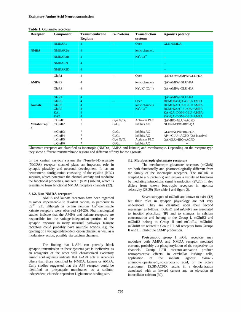
There are different types of ionotropic andmetabotropic glutamate receptors in both neurones and glialcells. Table I shows a summary of the main characteristicsof those receptors. Classification of receptors has beenpossible due to the different affinity to specific agonists andantagonists. A summary of those molecules are given infigure 2.
Glial cells, in particular astrocytes, appear torespond to a great variety of neurotransmitters, includingglutamate, hormones and growth factors with activation ofmetabotropic pathways, which may lead to intracellular pHand/or calcium changes. The distribution of different sets ofglutamate receptors in different brain regions may classifyneurones and glial cells, and appears to be functionally ofsome significance, both in normal physiological processesas well as during pathological states. The excitatoryneurotransmitter glutamate and its large receptor family isprobably the most versatile and complex signaling system inthe mammalian brain, and possibly also the most susceptiblefor pathological disturbances.
3.1. Ionotropic glutamate receptorsFour main subtypes of glutamate-gated channels
have been characterized pharmacologically and they havebeen named according to their preferred agonist, N-methyl-D-aspartate (NMDA), high affinity kainate (KA), alpha-amino-3-hydroxy-5-methyl-4-isoxazole propionate (AMPA),and 2-amino-4-phosphobutyrate (AP4). For each of those
agonists, a large diversity of receptors have been described(see table 1).
The most studied receptor is NMDA (13,14), andbecause of this, glutamate ionotropic receptors are oftennamed as NMDA and non-NMDA. Activation of theionotropic NMDA and non-NMDA receptors increasestransmembrane calcium and sodium fluxes, whereas themetabotropic glutamate receptor activation results ingeneration of inositol triphosphate and inhibition ofadenylate cyclase (15). Nevertheless, metabotropicreceptors are also related in phosphorylation of NMDA andnon-NMDA receptors.
It seems that glutamate ionotropic receptors,specially NMDA receptors, are related withneurodegenerative diseases, as act when glutamateconcentration increases. Thus, a noncompetitive NMDAreceptor antagonist (memantine) can be used for thetreatment of Alzheimer´s disease (16). It has also been seenthat injection of kainate causes selective neuronaldegeneration similar to that of Huntington´s disease (17).Evidence from animal models suggests that antagonists ofthe different glutamate receptors might be beneficial inParkinson´s disease, Huntington´s chorea and amyotrophiclateral sclerosis but the relevance of these models to thehuman disease is not clear. However, the identification ofnumerous receptor subtypes in addition to variabilities ofdistribution and multiple modulatory sites will provide newsolutions to these common neurological disorders.
3.1.1. NMDA receptorsNMDA-type ionotropic receptors have not been
demonstrated in glial cells, but only in neurons. Thosereceptors contain (a) a transmitter binding site, which bindsglutamate; (b) a regulatory or coactivator site, which bindsglycine, (c) a site within the channel that binds phencyclidineand related compounds, (d) a voltage-dependent Mg2+
binding site , and (e) an inhibitory divalent cation site thatbinds Zn2+. Glycine greatly enhances the actions of NMDAagonists but has no action by itself. Interaction ofphencyclidine and related anesthetics to NMDA receptorsreproduce most of the symptoms of schizophrenia. A majoradvance in the understanding of the NMDA receptor wasthe demonstration by McDonald and Johnston (18) andFlatman et al. (19) that NMDA-induced responses arevoltage-dependent. The agonist-induced currents aregreatest at moderately depolarized potentials (-30 to -20mV) and are reduced at both more hyperpolarized anddepolarized potentials. Consequently, NMDA receptoraction is suppressed at the normal resting potential. Thework of Nowak et al. (20) and Mayer et al. (21)demonstrated that the voltage dependency of the NMDAreceptor is attributable to extracellular Mg2+ ions that blockthe ion channel only at potentials more negative than -20 or -30 mV.

Excitatory Amino Acid Neurotransmission
703
Figure 1. Glutamate mobilization at glutamatergic synapses. Glutamate stored in synaptic vesicles is released by fusion of thosevesicles to the presynaptic membrane. Stimulation of ionotropic glutamate receptors (iGluR) by glutamate opens associated ionchannels and induces an excitatory postsynaptic potential. Metabotropic glutamate receptors (mGluR) are associated with Gproteins and, depending on the subtype they increase IP or affect cAMP concentration. Glutamate is removed from the synapticcleft through two processes: re-uptake back into pre-synaptic terminals and diffusion out of synaptic cleft to be re-uptaken by glialcells. The rapid re-uptake of glutamate is believed to be mediated by a Na+ and K+ depending high-affinity glutamate transporter(EAAC1, with Km for glutamate of 12µM). The low glutamate concentration is re-uptaked in glial cells by a higher-affinitytransporter (GLT-1, with Km of 2 µM). The low affinity glial cells transporter (GLAST, with Km of 77 µM) is able to transportaspartate and glutamate and it is a mechanism of protection in order that glutamate concentration doesn’t increase too much inbrain, because of its “excitotoxic” effect. Glutamate in glial cells is thought to be metabolized to glutamine (by glutaminesynthetase) and to α-ketoglutarate (by glutamate dehydrogenase or glutamate oxaloacetate transaminase). Those metabolites can beused as precursors of glutamate synthesis in pre-synaptic neurones. Glutamate is packed again into synaptic vesicles by a Na+
independent transport driven by the internal positive membrane potential generated by the vesicular ATP-dependent H+ transport.

Excitatory Amino Acid Neurotransmission
Figure 2. Some of the agonists and antagonists of glutamate receptors. Classification of glutamate receptors is possible due to theeffect of some agonists and antagonists. Here it is shown the chemical structures of the most common used.
Excitatory Amino Acid Neurotransmission
705
Table 1. Glutamate receptorsReceptor Component Transmembrane
RegionsG-Proteins Transduction
systemsAgonists potency
NMDAR1 4 -- Open GLU>NMDA
NMDA NMDAR2A 4 ionic channels --
NMDAR2B 4 Na+, Ca++ --
NMDAR2C 4 --
NMDAR2D 4 --
GluR1 4 -- Open QA>DOM≈AMPA>GLU>KA
AMPA GluR2 4 ionic channels QA>AMPA>GLU>KA
GluR3 4 Na+, K+ (Ca++) QA>AMPA>GLU>KA
GluR4 4 QA>AMPA>GLU>KAGluR5 4 -- Open DOM>KA>QA≈GLU>AMPA
Kainate GluR6 4 ionic channels DOM>KA>QA>GLU>AMPAGluR7 4 Na+, Ca++ DOM>KA>GLU>QA>AMPAKA1 4 KA>QA>DOM>GLU>AMPAKA2 4 KA>QA>DOM>GLU>AMPAmGluR1 7 Gq o Gi/Go Activates PLC QA>IBO≈GLU>tACPD
Metabotropic
mGluR2 7 Gi/Go Inhibits AC GLU≈tACPD>IBO>QA
mGluR3 7 Gi/Go Inhibits AC GLU≈tACPD>IBO>QAmGluR4 7 Gi/Go Inhibits AC AP4>GLU>tACPD (QA inactive)mGluR5 7 Gq o Gi/Go Activates PLC QA>GLU≈IBO>tACPDmGluR6 7 Gi/Go Inhibits AC --
Glutamate receptors are classified as ionotropic (NMDA, AMPA and kainate) and metabotropic. Depending on the receptor typethey show different transmembrane regions and different affinity for the agonists.
In the central nervous system the N-methyl-D-aspartate(NMDA) receptor channel plays an important role insynaptic plasticity and neuronal development. It has anheteromeric configuration consisting of the epsilon (NR2)subunits, which potentiate the channel activity and modulatethe functional properties, and zeta 1 (NR1) subunit, which isessential to form functional NMDA receptors channels (22).
3.1.2. Non-NMDA receptorsAMPA and kainate receptors have been regarded
as rather impermeable to divalent cations, in particular toCa2+ (23), although in certain neurons Ca2+-permeablekainate receptors were observed (24-26). Pharmacologicalstudies indicate that the AMPA and kainate receptors areresponsible for the voltage-independent portion of thesynaptic response in many neuronal pathways. Kainatereceptors could probably have multiple actions, e.g. theopening of a voltage-independent cation channel as well as amodulatory action, possibly via calcium channels.
The finding that L-AP4 can potently blocksynaptic transmission in these systems yet is ineffective asan antagonist of the other well characterized excitatoryamino acid agonists indicate that L-AP4 acts at receptorsothers than those identified by NMDA, kainate or AMPA.Early studies suggested that the AP4 receptor could beidentified in presynaptic membranes as a sodium-independent, chloride-dependent L-glutamate binding site.
3.2. Metabotropic glutamate receptorsThe metabotropic glutamate receptors (mGluR)
are both functionally and pharmacologically different fromthe family of the ionotropic receptors. The mGluR iscoupled to a G protein(s) and evokes a variety of functionsby mediating intracellular signal transduction (27,28). It alsodiffers from known ionotropic receptors in agonistsselectivity (28,29) (See table 1 and figure 2).
Seven subtypes of mGluR are known to exist (12)but their roles in synaptic physiology are not veryunderstood. They are classified upon their secondmessenger as follows: mGluR1 and mGluR5 are associatedto inositol phosphate (IP) and to changes in calciumconcentration and belong to the Group I. mGluR2 andmGluR3 belong to Group II and mGluR4, mGluR6-mGluR8 are related to Group III. All receptors from GroupsII and III inhibit the cAMP production.
Postsynaptic group I mGlu receptors maymodulate both AMPA and NMDA receptor mediatedcurrents, probably via phosphorylation of the respective ionchannels. Group II/III receptor-activation produceneuroprotective effects. In cerebellar Purkinje cells,application of the mGluR agonist trans-1-aminocyclopentane-1,3-dicarboxylic acid, or the activeenantiomer, 1S,3R-ACPD, results in a depolarizationassociated with an inward current and an elevation ofintracellular calcium (30).

Excitatory Amino Acid Neurotransmission
706
Figure 3. Structural model of EAAC1. EAAC1 transporterhas 10 transmembrane sections. It is a glycoprotein with 3external glycosilation sites (GS) and has 5 internal potentiallyphosphorylations sites (PS). Domains corresponding toregions of extended sequence identity with GLT-1 andGLAST are shown as filled circles.
4. EXCITATORY AMINO ACIDS TRANSPORTERS
Uptake of the acidic amino acids from theextracellular enviroment is mediated by sodium-dependenttransport systems of high affinity. Five subtypes of high-affinity glutamate transporters have been clonedindependently (31-35). Although they have differentnomenclature, the actual nomenclature used for humanglutamate transporters is EAAT (excitatory amino acidtransporter) followed by a number. Those transporters havebeen related to the previous known transporters from otherspecies by amino acid sequence identity. Here we describethe structure, distribution, functional expression andpharmacology of these transporter subtypes.
4.1. EAAT1 and GLAST1EAAT1 has a molecular weight of 59.5 kDa and
96% amino acid sequence identity with the rat GLAST1sequence (33). GLAST is a specific transporter for L-glutamate and L-aspartate and plays an important role in thetermination of neurotransmitter signals on excitatorysynapses. It is specifically expressed in brain beingdistributed in glial cells. In cerebellum, GLAST message isspecifically expressed in the striatum gangliosum made upprimarily by Purkinje and Bergmann glia cells (33).
Storck et al. (33) isolated and sequenced a 3 kbclone from a rat brain cDNA library. The predictedsequence of the polypeptide consists on a 543 amino acidresidues protein. A tentative model for the rat GLASTprotein comprises six α-helical membrane-spanningsegments in its N-terminal half with approximately the samespacing as the first six helices of previously reportedneurotransmitter transporters (see figure 3).
Significant sequence similarity of GLAST to theglutamate transporters of E. coli (36), Bacillusstaerothermophilus and Bacillus caldotenax and to thedicarboxylate transporters of Rhizobium meliloti andRhizobium leguminosarium (37). The overall aminoacidsequence identities range from 26 to 32%. It shares nosequence similarity with the E. coli Na+-dependent glutamatetransporter (38,39) or the cloned mammalianneurotransmitter transporters studies so far (40,41). Thetransporter function has been verified by aminoacid uptakeand electrophysiological studies in the Xenopus laevisoocyte system.
The results provide evidence for GLAST-1carrying out a high affinity, sodium-dependent L-glutamatetransport with a proposed stoichiometry of 3 Na+, 1 L-glutamate-/1K+. There is no evidence for the cotransport ofprotons (42). Aminoacid uptake shows saturation kinetics:Km,(L-glu)=77 ±27 mM (33) and other amino acids includingL-alanine, L-leucine, L-glutamine, L-arginine and L-methionine are not transported in significant amounts. DL-threo-3-hydroxy-aspartate, a strong inhibitor of the Na+-dependent glutamate uptake (43) and capable of causingneuronal degeneration (44), is a potent inhibitor of GLAST.A decrease in Na+-dependent L-glutamate transport inpatients with Alzheimer's disease has been reported (45).Whether GLAST plays a pivotal or ancillary role in thesedisorders remains to be elucidated.
4.2. EAAT2 and GLT-1EAAT2 has a molecular weight of 62.1 kDa and a
95% identity with the correct sequence (46) of rat GLT-1(32). GLT-1 is expressed in astrocytes and maintains thelow extracellular glutamate concentration of approximately1µM below neurotoxic levels. The L-glutamate transportercDNA was obtained by immunoscreening of a λZap libraryfrom rat brain (47) with an antibody raised against thepurified glial transporter. The cDNA sequence predicts aprotein of 573 amino acids with 8-9 putative transmembraneα-helices (32) (figure 3). It is a glycoprotein with 2 potentialN-linked glycosylation sites and 2 phosphorylation sites(48). GLT-1 does not share significant overall homologywith any known eukariotic protein, including the Na+/glucosetransporter and the growing superfamily of neurotransmittertransporters but with the protein-coupled L-glutamatetransporter glt-p of E.coli.
The transport system has three substrates: Na+, L-glutamate and K+ (49-51). In an electrogenic process, thetransporter takes up Na+ and L-glutamate and extrudes K+.Like the others (Na+-K+)-coupled L-glutamate transporters(52), GLT-1 is stereospecific, being strongly inhibited by L-glutamate but not by the same concentration of D-glutamate.The apparent Km in intact cells is 10 µM higher than inmembrane preparations (53) and in the purified andreconstituted transporter (54).
Glutamate analogues: L-aspartate, D-aspartate,cysteine sulphinate, threo-3-hydroxy-DL-aspartate and L-trans-pyrolidine-2,4-dicarboxylate are competitive inhibitorsof GLT-1. L-trans-pyrolidine-2,4-dicarboxylate inhibits

Excitatory Amino Acid Neurotransmission
707
transport of L-glutamate but does not prevent it frombinding to its receptors (55). The GLT-1 system is potentlyinhibited by dihydrokainate (DHK) and L-α-aminoadipate(L-AAD), while the EAAC1 system (see 2.3.) is inhibited byL-AAD and is essentially DHK insensitive.
The decrease in GLT-1 function cannot beattributed to selective astrocytic degeneration as there is noloss of staining of the astrocytic marker, GFAP. In fact,previous studies have suggested that astrogliosis and not aloss of astrocytes may be present in ALS motor cortex (56).Studies have been carried out to try to identify the reasonfor the selective loss of GLT-1 in ALS. There is noevidence for a decrease in gene expression as there was nochange in the levels of the mRNA coding for the protein inALS patients (57). Other possible explanations for a loss ofGLT-1 could involve translation mechanisms whereby thegene is transcribed into mRNA but defects in the translationcould interfere with the synthesis of the protein.Alternatively, the protein may be translated normally but maybe much more unstable than normal. Finally, the decrease inactivity may be due to posttranslational modifications,possibly induced by an increase in the levels of cellular freeradicals.
4.3. EAAT3 and EAAC1EAAT3 has a molecular weight of 57.2 kDa and
92% identity with the rabbit sequence termed EAAC1 (58).EAAC1 is of both neuronal and epithelial origin. In brain itprovides a presynaptic glutamate uptake mechanism toterminate the action of released glutamate at glutamatergicsynapses. But it is also expressed in γ-aminobutyric acid(GABA)-ergic cerebellar Purkinje cells, where it providesglutamate as a precursor for GABA synthesis.
To isolate the cDNA encoding rabbit EAAC1,Kanai and Hediger (58) screened a rabbit intestinal cDNAlibrary for their ability to induce [14C] glutamate uptake inXenopus oocytes. They isolated a cDNA which encodes a524-residue protein. This protein has approximately 10putative membrane-spanning regions but alternative modelswith a different number of membrane-spanning regions canbe constructed. EAAC1 has a significant homology to theH+-coupled gltP glutamate transporters of E. coli., B.stearothermophiles and B. caldotenax and to the dctAdicarboxylate transporter of Rhizobium meliloti. They havesequence indentities ranging between 27% and 32%. Thereis neither homology to the Na+-Cl--dependentGABA/neurotransmitter transporter family (40,41,59) nor tothe E. coli Na+/glutamate transporter gltS (38).
EAAC1-mediated transport is electrogenic anddependent on extracellular Na+ but not on Cl-. Studies insalamander retinal glial cells and in oocytes expressingEAAC1 revealed that glutamate transport coupled to thecotransport of two Na+ and the countertransport of one K+
and one OH- (60).
Due to the presence of a large hydrophobicstretch near the C terminus (residues 357 to 444, figure 3),alternative models with a different number of membrane
spanning regions are concievable. The N-terminal part ofEAAC1 is hydrophilic and lacks a signal peptide. A motif-like cluster of serine residues is present (residues 331-334).Similar clusters have been identified in the ligand-bindingsites of receptors for acetylcholine and biogenic amines.
The function and pharmacology of the expressedprotein are characteristic of the high-affinity glutamatetransporter already identified in neuronal tissues. Membranecurrents were measured in Xenopus oocytes injected withEAAC1 cRNA. L-glutamate, L-aspartate and D-aspartateevoked inward currents, almost with the same amplitude,whereas currents induced by D-glutamate and L- or D-homocysteinate were smaller. The glutamate receptorligands NMDA and kainate did not induce current whereasquisqualate evoked a significant inward current. EAAC1-mediated L-glutamate transport is saturable and the Km valueis 12,2±1,2 µM, indicative of a high-affinity transport (31).
The abnormal glutamate transport that isassociated with certain neurodegenerative diseases andwhich occurs during ischemia and anoxia could be due toabnormalities in the function of this protein.
4.4. EAAT4EAAT4 is a human L-aspartate/L-glutamate
transporter which is expressed in the cerebellum and atlower levels, in brain stem, cortex and hippocampus. cDNAencoding EAAT4 was obtained from human cerebellarmessenger RNA (34). The amino acid sequence exhibits65%, 41% and 48 % amino-acid identity to the humanglutamate transporters EAAT1, 2 and 3 respectively.
EAAT4-mediated transport is electrogenic,dependent on Na+ and K+ and it has properties of a ligand-gated chloride channel. Its transport is saturable and the Km
values derived from current measurements are Km (L-asp)=184±0.46 µM and Km(L-glu)=3.3±0.4 µM (34).Pharmacological properties of EAAT4 are similar to thosepreviously described to the other glutamate transporters.
Structural analogs of glutamate, L-trans-2,4-pyrrolidine dicarboxylic acid (PDC), L-quisqualate and L-α-aminoadipate, elicit currents in oocytes expressing EAAT4.This current is predominantly carried by chloride ions andchloride-dependent glutamate transport activities have beenreported in synaptosomes and other brain preparations butthey have not demonstrated the cotransport of chloride ionswith substrates (61,62). This chloride conductance is notblocked by endogenous oocyte chloride channel blockers.Thereafter, EAAT4 functions as a transporter, reducing theamount of neurotransmitter available for activatingpostsynaptic receptors and as a glutamate-gated chloridechannel, modifying the neuronal excitability by ist capacityfor enhancing chloride permeability.
4.5. EAAT5EAAT5 is a human glutamate transporter whose
mRNA is mostly expressed in the retina. Arriza et al. (35)isolated the transporter EAAT5 from a salamander retinaglutamate transporter cDNA to screen a human retinal λgt10

Excitatory Amino Acid Neurotransmission
708
cDNA library. The gene product is 560 amino acid residuesin length. EAAT5 has 46% identity with EAAT1, 43%identity with EAAT4, 37% with EAAT3 and 36% identitywith EAAT2.
The amino- and carboxy- terminal sequences ofthe excitatory amino acid transporters are poorly conserved.In EAAT5 C-terminus there is a sequence motif found insynaptic membrane proteins: E-S or T-X-V-COOH (63).The C-termini interact with several PDZ (a modular protein-binding motif) domains in PSD-95 (postsynaptic density-95kDa protein). The presence of this PDZ domain suggeststhat the transporter is a component of the signaltransduction pathway. Its channel-like properties mayindicate a role in retinal physiology different fromneurotransmitter clearance.
EAAT5 uptake is sodium- and voltage-dependentand chloride-independent. It has stereospecificity for L-glutamate versus D-glutamate and L-aspartate versus D-aspartate with Km values for L-glutamate and L-aspartate of64± 6µM and 13±5µM, respectively. L-trans-pyrrolidine-2,4-dicarboxylic acid (tPDC) and threo-β-hydroxyaspartate(THA) are potent blockers but neither generated a currentwith a voltage dependence similar to that of glutamate (35).However, ion substitution experiments show that thecurrents seen are carried by chloride ions. These propertiesare similar to those described in retinal neurons suggestingthat the transporter can be involved in visual processing(35).
4.6. Synaptic vesicles glutamate transporterGlutamate is preserved in secretory vesicles at
pre-synaptic terminals. A unique group of proteins found onsecretory vesicles are the transporters needed for theaccumulation of neurotransmitters from cytoplasm intothese vesicles. The vesicular glutamate transporter is onebetween at least four different vesicular transporter typeswhich have been biochemically identified (64,65). Synaptic vesicles and microvesicles, enclosed inendocrine cells like pinealocytes, possess an activeglutamate-specific transporter that is dependent on theextravesicular Cl- concentration, on an electrochemicalproton gradient across the vesicle membrane (66-69) and onthe temperature (67). The dependence of glutamate uptake on ATP-generated proton electrochemical potential was analysed in ahighly purified preparation of synaptic vesicles from ratbrain (70). The glutamate anion is transported into synapticvesicle by a Na+-independent vesicle transport, driven by theinternal positive membrane potential generated by thevesicular ATP-dependent H+ transport (66,67,70). Anyway,it seems that glutamate uptake is solely dependent on ∆ψ ,suggesting that protons are not directly involved.
The vesicle carrier has a low substrate affinity(Km= 1.6 mM) and is highly specific for L-glutamate (5 mMD-glutamate reduces uptake by 30% while 5 mM L-aspartatehas no effect) (71). Other glutamate analogues which interact
with various glutamate receptor subtypes don’t affecttransport (71). It has been observed a good correlationbetween acidification and inhibition of glutamate uptake byglutamate analogues such as 1-aminocyclohexane-trans-1,3-dicarboxylic acid (72). Glutamine, aspartate and GABA donot inhibit L-glutamate uptake (67).
Due to the fact that the transporter has not beencloned yet, there is no knowledge about its structure, sitesinvolved in vesicular transporter function nor the sites thatdetermine substrate specificity. Nevertheless, futuremolecular analysis will help us to understand some eventssuch as cell-specific expression, specific targeting,bioenergetic properties and neuropharmacology.
5. METABOLIC INTERACTIONS BETWEENNEURONS AND ASTROCYTES
If one tries to envisage the metabolic capabilitiesrequired to maintain glutamatergic, aspartatergic, andGABA-ergic transmission (Glu, Asp, and GABA) in asimple cell type residing behind a blood-brain barrierthrough which there is rapid exchange of CO2, H2O, andNH3 and a rapid net influx of glucose, but no netaccumulation of either glutamate, aspartate, GABA, orglutamine (73), one would end up with a cell like thatsketched in figure 4. Neurons are able to synthesize bothglutamate and GABA from glutamine, and astrocytes formand release glutamine (which has no transmitter activity)after accumulation of either glutamate or GABA.
Extracellular glutamate is, to a larger extent,accumulated into astrocytes both in the intact brain (74) andin cultured cells (75,76), but much of the accumulatedglutamate (how much probably depends upon theexperimental conditions) is degraded as a metabolic fuel toCO2 and H2O in astrocytes and thus not converted toglutamine (75,77). New glutamate and GABA precursormolecules will, therefore, have to be synthesized fromglucose. Since carboxylation of pyruvate to oxaloacetate, anintermediate of the TCA cycle, occurs in astrocytes, netsynthesis of α-ketoglutarate in the TCA cycle can also takeplace in these cells.
The rate of glutamate metabolism to CO2 is high inastrocytes but not in the two neuronal types, especially notin the glutamatergic cerebellar granule cells. The rapid CO2
formation in astrocytes from glutamate appears to representmainly a net utilization of glutamate (78), not just an isotopeexchange between glutamate and α-ketoglutarate. Utilizationof glutamate or glutamine as a metabolic substrate is notrestricted to cultured cells but has also been observed inbrain slices and dissociated cell preparations (79,80). Aslong as the glutamate utilized as a metabolic fuel originally isproduced from glucose behind the blood-brain barrier, thisis not a violation of the well-established fact that the entireadult brain in vivo under normal conditions almostexclusively utilizes glucose as its substrate for energymetabolism (81).

Excitatory Amino Acid Neurotransmission
709
Figure 4. Schematic diagram of glutamate/glutamine and GABA/glutamine cycles. In neurons, glutamate and γ-aminobutyric acid(GABA) are synthesized from glucose or glutamine. Neurotransmitter released in synaptic cleft can then interact with receptor sites.To terminate this effect, transport to astroglial cells is performed with higher affinity than to neuron cells. In astroglial cells there is ahigher glutamine synthase activity and glutamate and GABA are metabolized to glutamine, which has no neurotransmitter effectsand can be recycled to neurons to form glutamate or GABA.
Exposure to very high potassium concentrations(> 20 mM) causes depolarization and a depolarization-induced increase in free intracellular calcium concentration insynaptosomes (82), neurons, and astrocytes (83) andenhances metabolic interactions between these two celltypes by facilitating glutamate (and GABA) release fromneurons and CO2 fixation in astrocytes, which in turnpromotes astrocytic formation of transmitter precursors forneurons. During exposure to slightly elevated potassium. Inthe following section, we describe some properties of theenzymes involved in the glutamic acid metabolism (figure 5).
5.1. Glutamine synthetase (E.C. 6.3.1.2.)Glutamine synthetase catalyzes several reactions
(84), although the main reaction is the following one:
Glu + NH3 + ATP <-----> Gln + ADP + Pi
The enzyme has two important functions:assimilation of ammonia and biosynthesis of glutamine. Theenzyme from brain has been studied in rat, ox, sheep, pig
and human. It has been isolated from a variety of sourcesand the proteins vary greatly in their ability to catalyze thereverse reaction. With the mammalian enzyme, however, theforward rate relative to the reverse rate is about 10 to 1 (85).
The purification of the enzyme usually proceedsthrough four steps consisting on an acetone powder extract,precipitation by acid, hydroxylapatite and DEAE-cellulosecolumn chromatography (86); with a yield of 15-30% and apurification of about 200-fold.
Glutamine synthetase is composed of eightsubunits which are identical (44,000-50,000 daltons)showing rather similar amino acid composition (84). Theenzyme has a molecular weight of 400,000 daltons,distributed in two tetramers (87). Distribution of glutaminesynthetase and glutaminase are uneven in central nervoussystem. Berl (88) determined the distribution of glutaminesynthetase in 16 brain areas of the adult rat. The site ofglutamine synthesis, from glutamate amidation via glutaminesynthase (GS), is in glial cells (89); and with a cytosoliclocation (9). Additionally, glia cells contain carbonic

Excitatory Amino Acid Neurotransmission
710
Figure 5. Glutamate metabolism. Glutamate from mammalian brain can be obtained from glutamine (glutaminase) or from α-ketoglutarate (glutamate dehydrogenase and glutamate oxalacetate transaminase). Degradation of glutamate can generate α-ketoglutarate by reversible reactions, GABA (glutamate decarboxylase) or glutamine (glutamine synthetase).
anhydrase, which catalyzes the CO2 hydration reaction andHCO3
- formation. Therefore, glia cells could be important inacid-base regulation and related amino acid metabolism inCNS. Glutamine formed by GS can move into adjacentnerve endings where it is utilized either in metabolicpathways unrelated or not directly related toneurotransmitter glutamate and GABA synthesis or information of neurotransmitter glutamate and GABA viaglutaminase and glutamate decarboxylase, respectively.
The enzyme is irreversibly inhibited by methioninesulfoxamine (MSO) (90). ATP and magnesium arenecessary for the binding of glutamate to the enzyme,whereby it becomes activated. Tate et al. (84) calculated inrat liver Km for ATP = 2.3 mM; and Deul et al. (86) Km forammonia = 0.3 mM. Certain anions, particularly bicarbonateand chloride, activate the enzyme when nonsaturatingconcentrations of L-glutamate are used. Although liverglutamine synthase is activated by 2-oxoglutarate, brainenzyme is less affected by this compound (84). Themammalian enzymes are inhibited by inorganic phosphateand carbamyl phosphate (91). This effect can be due to thereaction of ATP synthesis from ADP and carbamylphosphate, catabolized also by glutamine synthetase.
5.2. Glutaminase (E.C. 3.5.1.2.)The reverse reaction of glutamine synthase is
catalyzed by the ubiquitous enzyme glutaminase, which is
present in both neurons and astrocytes (92). Nevertheless,glutaminase is predominantly a neuronal enzyme and it hasbeen localized in mitochondria (93,94). Pig brainmitochondria have been shown to contain two major formsof glutaminase, one soluble located in the matrix and onemembrane-bound enzyme located in the inner membraneand both activated by phosphate (95).
Soluble glutaminase has been purified from pigbrain (96,97) and some other sources (98-100). The solubleenzyme is present as a dimer (101) with a relatively lowspecific activity and a highly aggregated form of the enzymewith a considerably greater specific activity. By anincubation with phosphate, the soluble enzyme is reversiblyand slowly aggregated (101-103). Because of thiscomplicated regulatory behavior, some evidences have beensuggested that the membrane-bound and soluble enzymemay have different function in the brain (95).
Kinetic studies of the two enzymes have beenperformed by Nimmo and Tipton (101) and they show Km
values for glutamine of about 0.8-1.4 mM and 3.4-9.7 mM.One of the products, glutamate, inhibits the enzymestrongly, whereas the other product ammonia has only aslight inhibitory action on the enzyme. Glutamate inhibitionis mixed (Ki
slope= 1.6 mM and Kiintercept= 3.3 mM).
In astrocytes exposed to 1.2 mM valproate,glutaminase activity increased 80% in primary culture by day
Excitatory Amino Acid Neurotransmission
711
2 and remained elevated by day 4; glutamine synthetaseactivity was decreased 30% (104).
5.3. Glutamate dehydrogenase (E.C. 1.4.1.2.-4.)Glutamate dehydrogenases catalyze the following
reversible reaction:
L-glutamate + H20 + NAD(P) <-----> α-ketoglutarate + NH3
+ NAD(P)H
although it appears that the reaction velocity is higher whenthe formation of glutamate is studied.
Three different enzymes are considered dependingon the use of NAD/NADH (E.C. 1.4.1.2.), NADP/NADPH(E.C. 1.4.1.4.) or both (E.C. 1.4.1.3.) as coenzymes. Neuralglutamate dehydrogenase (E.C. 1.4.1.3.) can use bothcoenzymes, although it has been shown that NAD is usedmore effectively than NADP (105). The direction of neuralglutamic dehydrogenase (E.C. 1.4.1.3.) activity appears tobe regulated in part by the tissue NAD(P)/NAD(P)Hconcentration ratio (105, 106). In rat brain GDH activityexists in two distinct forms differing in solubility, kineticparameters, resistence to heat inactivation and allostericproperties (107). These forms have been designed solubleand particulate GDH (107,108).
When the ratio is high, e.g., in the absence ofglucose, oxidative deamination of glutamate occurs. In thepresence of glucose, when the ratio falls, and α-ketoglutarate is not rate limiting, reductive amination of α-ketoglutarate is favored. Kinetic parameters have beenstudied for both directions of the reaction (105).
GDH’s from various organs of the same speciesare similar, if not identical. Complete cross-reaction occursbetween antibodies induced by bovine liver GDH andextracts of bovine spleen, brain and heart. Most animalGDH are inhibited by GTP and activated by ADP (109).GDH from bovine brain has been partially purified byGrisolia et al. (106) and shows a specificity either for NADor NADP. This enzyme has a molecular weight of 332,000daltons, as judged from the amino acid sequence of the sixidentical subunits (110).
As compared with other enzymes, the Vmax ofGDH in synaptic mitochondria from rat brain is 20-40%lower than Vmax of aspartate aminotransferase, but 4-5foldhigher than Vmax of phosphate dependent glutaminase (111).
5.4. TransaminasesTransaminases play an important role in the
aminoacid metabolism, as they are able to catabolize areversible transference of an amino group from anaminoacid to a ketoacid acceptor. Those enzymes usepyridoxal phosphate as a coenzyme, which will perform thetransference of the amino group. Among thosetransaminases, aspartate aminotransferase or glutamateoxalacetate transaminase (E.C. 2.6.1.1.) catabolizes thereversible transference of amino group of Asp to α-
ketoglutarate and generates oxalacetate and glutamate. Therehave been found two isoenzymes of aspartateaminotransferase in animals: a citoplasmic and amitochondrial. Both are dimeric proteins of 45,000 daltonsand 2,000 daltons subunits (112).
Aspartate amino transferase (AAT) activity seemsto be higher than glutamate dehydrogenase (GDH). The ratioAAT/GDH is between 10 and 20 in rat brain (113) or insquid giant nerve (114). Transamination of glutamate byusing aspartate amino transferase generates aspartate, whichis also a neurotransmitter; whereas glutamate dehydrogenaseyields ammonia but not another neurotransmitter. The higheractivity of AAT can be therefore due to prevent the toxicaction of ammonia.
Another transaminase is alanine aminotransferase(E.C. 2.6.1.2.), an enzyme which transfers amino fromalanine to α-ketoglutarate to yield pyruvate and glutamate.This enzyme presents also a cytoplasmic and amitochondrial isoenzymes.
5.5. Glutamic acid decarboxylase (E.C. 4.1.1.15.)The enzyme removes the α-carboxyl group of
glutamate to produce a γ-carboxyl amino acid called γ-amino butyric acid (GABA). This decarboxylation ofglutamate to GABA is not very different fromdecarboxylation of L-DOPA or tryptophan to dopamine andserotonin. Like those enzymes, glutamic acid decarboxylaserequires the cofactor pyridoxal phosphate (vitamin B6).
The enzyme is highly substrate specific, althoughHomola and Dekker (115) showed that some glutamateanalogs can also be decarboxylated.
6. DYSFUNCTION OF EXCITATORY AMINOACIDNEUROTRANSMISSION
It is important to maintain low levels (1-3µM) ofextracellular glutamate as excessive receptor stimulation orexcessive ammonium generated by the glutamatedehydrogenase can lead to neural injury and/or death(“excitotoxicity”).
Glutamate appears to be remarkably potent andrapidly acting neurotoxin. Exposure to 100 µM glutamatefor 5 min is enough to destroy large numbers of culturedcortical neurons (116). By the way, glutamate neurotoxicitymay be blocked by antagonist compounds and attenuatedby antagonists added after glutamate exposure (116).
Neurodegenerative diseases, such as Alzheimer'sdisease (AD), Huntington’s disease (HD), Parkinson’sdisease (PD) and amyotrophic lateral sclerosis (ALS),particularly affect old people and result in: i) a modificationof the individual personality; ii) the need for constant helpfrom relatives; iii) a high economical costs for family andinstitutions. In Spain alone, over 500.000 people are affectedwith those diseases with this increasing as the agedpopulation increases. Due to the increased lifespan in

Excitatory Amino Acid Neurotransmission
712
developed societies, neurodegenerative diseases have beenincreased in the European Union and represent one third ofdeaths (after cancer and cardiovascular problems). It isthough that with Alzheimer's disease alone, at the beginningof next century, 8 million Europeans will be affected.
Many reports have been published on increasedlevels of glutamate in certain neurodegenerative diseasessuch as AD, HD, PD and ALS (117-120). Increasedextracellular glutamate has also been implicated in the onsetof neurodegeneration associated with hypoxic damage(stroke). The release in glutamate following hypoxia hasbeen suggested to be due, at least in part, to a calcium-independent mechanism following the reversal of theneuronal glutamate uptake carrier (121). This increase inextracellular glutamate acts afterwards postsynaptically toincrease cellular calcium levels with subsequent cell death.
The mechanisms by which glutamate is increasedin neurodegenerative diseases is unknown. The cause of thisincreased extracellular glutamate has been ascribed to adecrease in the activity of glutamate dehydrogenase (PD)(122) or a decrease in the number of Na+-glutamatetransporters (ALS, HD) (123). As it is a neurotoxic, it islikely that the high glutamate concentration observed inneurodegenerative diseases are the most likely cause of theneurodegeneration.
Marangos et al. (124) stated the glutamatehypothesis for early Alzheimer’s disease: “Cell death due toneuronal toxicity could result from excessive synthesis orrelease of glutamate or a glutamate-like substance, faultyglutamate reuptake, decreased glutamate degradation, ordecreased inhibition of excitatory neurons. Any of theseaberrant processes could, early in the disease, increase locallevels of glutamate and so initiate a slow, progressivedegeneration and eventual death of neurons”.
Several post-mortem studies have compared brainglutamate levels in Alzheimer’s disease to those in controlsubjects. Some investigators (125-127) have found lowerglutamate levels in the frontal cortex and the temporal cortexof Alzheimer’s patients than in control subjects. Cerebrospinal fluid (CSF) concentration of free glutamate wassignificantly higher in patients with Alzheimer’s disease thanin comparison subjects (117). It should be noted thatmeasurements of glutamate in CSF are likely to give betterapproximation of glutamate concentrations at synapses thanare plasma glutamate concentrations.
In Huntington’s disease, glutamate and GABAconcentrations decrease in striatum and caudate nucleusfrom brain (118,128). However, no reduction at all wasobserved in the frontal cortex of patients. A likely possibilityis that the low glutamate content of the caudate and theputamen in HD results from chronic failure of the normalreuptake mechanism for glutamate released at synapses, withor without any excessive rate of release of thisneurotransmitter. This possibility is supported by the findingof Cross et al. (129), who observed large reductions inhigh-affinity glutamate uptake sites in autopsy specimens ofcaudate and putamen from HD patients.
If either excessive release or decreased reuptakeof glutamate occurred in the striatum in HD, concentrationsof glutamate might become high at synapses, with resultingdamage to neurons. Some of the excess glutamateaccumulate in synaptic clefts in HD would be carried awayin the extracellular fluid, thus eventually causing a loweredglutamate content in striatal tissue. This fact is supported byan increased glutamate concentration in CSF of living HDpatients as it was observed for Alzheimer’s disease patients.
In Parkinson’s disease, Schapire et al. (130)demonstrated a reduced activity of complex I of themitochondrial respiratory chain in the region of substantianigra in brain. Deficiencies on complex II and IV have beenalso observed in muscle biopsia from PD patients (131).Cedarbaum et al. (122) observed a deficiency of GDH butnot on pyruvate dehydrogenase complex. Since complex Iis the point of entry for reducing equivalents (as NADH) tothe respiratory chain, a decrease in complex I activity mightresult in feedback endproduct inhibition of GDH. Decreasedlevels of GDH might exert an excitotoxic glutamate effectvia NMDA receptors on striatal dopamine nerve terminals(132) and contribute to cellular degeneration in PD.
Amyotrophic lateral sclerosis (ALS) is a diseaseresulting in degeneration of the motor cortex, the brain-stemand the spinal chord. While there are a number ofhypotheses underlying this disease, an increase inglutamatergic neurotransmission has been proposed as a keyevent in the disease onset and recently, this has beenascribed, at least in part, to a defect in the function of theGLT-1 glutamate transporter which is localized onastrocytes (123).
Recent in vitro studies have demonstrated that ablockade of the GLT-1 transporter either by transportinhibitors or by an antisense approach resulted in a slow,selective loss of motor neurons, thus strengthening the casefor a critical role for GLT-1 in the etiology of ALS(120,133). Furthermore, this decrease in uptake does notappear to be associated with a decrease in transporterexpression levels (57) and there is no evidence of specificprotein mutations associated with the disease. Because ofthe potential role of free radicals in ALS and the upset infunction of the superoxide dismutase enzyme in the familialform of ALS, previous studies have suggested that anincrease in free radicals may impair the function of GLT-1(134).
The role of free radicals in the etiology of ALShas been strengthened by the selection of point mutations inthe cytosolic Cu/Zn superoxide dismutase (SOD-1)associated with the familial form of ALS (FALS) (135).These changes were not detected in control individuals anddo not represent normal allelic variants. SOD-1 catalyses thedismutation of the superoxide radical (O2
-) to hydrogenperoxide (H2O2) and represents the first line of defenseagainst oxygen toxicity. The mechanism responsible fortissue damage associated with reduced SOD activityremains to be defined. Direct toxicity due to the superoxideradical is probably minor in comparison to the generation of

Excitatory Amino Acid Neurotransmission
713
the hydroxyl radical (OH.) which is much more reactive. Inaddition, the superoxide may interact with endogenouslyformed nitric oxide to form peroxynitrite which can oxidizemethionine residues in proteins and peptides as well as thiolsand thioethers (136). It has therefore been suggested that anincrease in free radicals in ALS may be, at least in part,responsible for the upset in functioning of the GLT-1transporter and that the disease may have both a free radicaland excitatory amino acid basis.
Glutamate, and particularly the glutamatetransporter system, have also been implicated in theischemic damage associated with anoxia/hypoxia (stroke).For the first few minutes of ischemia, there is a slow acidshift of the cellular pH with a slow rise of extracellularpotassium concentration and a subsequent decrease inextracellular sodium and calcium. The rise in potassiumdepolarizes the cells to around -20mV (anoxicdepolarization) and the release of glutamate (137). Themechanism by which glutamate is released in ischemia hasbeen controversial. Some reports suggest that the release iscalcium-dependent, suggesting conventional vesicularrelease, while others claim that the release is calcium-independent, implying a non-exocytotic mechanism such asthe reversed operation of the glutamate uptake carrier (138).
7. REFERENCES
1. Hoop B., Masjedi M.-R., Shih V.E. & Kazemi H.: Brainglutamate metabolism during hypoxia and peripheralchemodenervation. J. Appl. Physiol. 69, 147-154 (1990)
2. Housley G.D. & Sinclair J.D.: Localization by kainic acidlesions of neurones transmitting the carotid chemoreceptorstimulus for respiration in rat. J. Physiol. Lond., 406, 99-114(1988)
3. Oldendorf W.H.: Brain uptake of radiolabeled aminoacids, amines, and hexoses after arterial injection. Am JPhysiol. 221(6), 1629-1639 (1971)
4. Oldendorf W.H. & Szabo J.: Amino acid assignment toone of three blood-brain barrier amino acid carriers. Am JPhysiol. 230(1), 94-98 (1976)
5. Pratt O.E.: The transport of metabolizable substancesinto the living brain. Adv Exp Med Biol. 69, 55-75 (1976)
6. Clarke D.D., Ronan E.J., Dicker E. & Tirri L.: Ethanoland its relation to amino acid metabolism in brain. InMetabolic Compartmentation and Neurotransmission. Eds:Berl S., Clarke D.D., Schneider D. Plenum Press, NewYork. 449-460 (1975)
7. Balazs R., Machiyama Y., Hammond B.J., Julian T. &Richter D.: The operation of the γ-aminobutyrate bypath ofthe tricarboxylic acid cycle in brain tissue in vitro. Biochem.J. 116, 445-467 (1970)
8. Machiyama Y., Balazs R., Hammond B.J., Julian T. &Richer D.: The metabolism of GABA and glucose in
potassium stimulated brain tissue in vitro. Biochem. J., 116,469-482 (1970)
9. Norenberg M.D. & Martínez-Hernández A.: Finestructural localization of glutamine synthetase in astrocytesof rat brain. Brain Res., 161, 303-310 (1979)
10. Berl S., Lajtha A. & Waelsch H.: Amino acid andprotein metabolism. VI. Cerebral compartments of glutamicacid metabolism. J. Neurochem. 7, 186-197 (1961)
11. van den Berg C.J., Matheson D.F., Ronda G.,Reijnierse G.L.A., Blokhuis G.G.D., Kroon M.C., ClarkeD.D. & Garfinkel D.: A model of glutamate metabolism inbrain: a biochemical analysis of a heterogenous structure. InMetabolic Compartmentation and Neurotransmission. Eds:Berl S., Clarke D.D., Schneider D. Plenum Press, NewYork. 709-723 (1975)
12. Tanabe Y., Masu M., Ishii T., Shigemoto R. &Nakanishi S.: A family of metabotropic glutamate receptors.Neuron. 8(1), 169-179 (1992)
13. Watkins J.C. & Collingridge G.L.: In The NMDAreceptor. Oxford: Oxford Univ. Press. (1989)
14;. Daw N.W., Stein P.S.G. & Fox K.: The role of NMDAreceptors in information processing. Annu. Rev. Neurosci.16, 207-222 (1993)
15. Thomas RJ: Excitatory amino acids in health anddisease. Journal of the American Geriatrics Society 43(11),1279-1289 (1995)
16. Muller W.E., Mutschler E. & Riederer P.:Noncompetitive NMDA receptor antagonists with fast open-channel blocking kinetics and strong voltage-dependency aspotential therapeutic agents for Alzheimer's dementia.Pharmacopsychiatry 28(4), 113-124 (1995)
17. Coyle J.T: An animal model for Huntington's disease.Biological Psychiatry 14(2),251-276 (1979)
18. McDonald J.W. &Johnston M.V.: Pharmacology of N-methyl-D-aspartate-induced brain injury in an in vivoperinatal rat model. Synapse. 6(2), 179-188 (1990)
19. Flatman J.A., Schwindt P.C. & Crill W.E.: Theinduction and modification of voltage-sensitive responses incat neocortical neurons by N-methyl-D-aspartate. Brain Res.363, 62-77 (1986)
20. Nowak L., Bregetowski P., Ascher P., Herbet A. &Prochiantz A.: Magnesium gates glutamate-activatedchannels in mouse central neurones. Nature 307, 462-465(1984)
21. Mayer M.L., McDermott A.B., Westbrook G.L., SmithS.J. & Barker J.L.: Agonist- and voltage-gated calcium entryin cultured mouse spinal cord neurons under voltage clampmeasured using arsenazo III. J. Neurosci. 7, 3230-3244(1987)

Excitatory Amino Acid Neurotransmission
714
22. Watanabe M.: Dynamic regulation of the NMDAreceptor channel subunits in the central nervous system andtheir involvement in synaptic plasticity and development.Kaibogaku Zasshi - Journal of Anatomy 71(5),517-522(1996)
23. Mayer M.L. & Westbrook G.L.: The physiology ofexcitatory amino acids in the vertebrate central nervoussystem. Prog. Neurobiol. 28, 197-276 (1987)
24. Murphy S.N., Thayer S.A. & Miller R.J.: The effects ofexcitatory amino acids on intracellular calcium in singlemouse striatal neurons. J. Neurosci. 7, 4145-4158 (1987)
25. Iino M., Ozawa S. & Tsuzuki K.: Permeation ofcalcium through excitatory amino acid receptor channels incultured rat hippocampal neurones. J. Physiol. (London)424, 151-165 (1990)
26. Gilbertson T.A., Scobey R. & Wilson M.: Permeationof calcium ions through non-NMDA glutamate channels inretinal bipolar cells. Science 251, 1613-1615 (1991)
27. Sugiyama H., Ito I. & Hirono C.: A new type ofglutamate receptor linked to inositol phospholipidmetabolism. Nature 325, 531-533 (1987)
28. Schoepp D., Bockaert J. & Sladeczek F.:Pharmacological and functional characteristics ofmetabotropic excitatory amino acid receptor. TrendsPharmacol. Sci. 11, 508-515 (1990)
29. Sugiyama H., Ito I. & Watanabe S.: Glutamate receptorsubtypes may be classified into two major categories: astudy on Xenopus oocytes injected with rat brain mRNA.Neuron 3, 129-132 (1989)
30. Batchelor A.M, Madge D.J. & Garthwaite J.: Synapticactivation of metabotropic glutamate receptors in the parallelfibre-Purkinje cell pathway in rat cerebellar slices.Neuroscience 63(4), 911-915 (1994)
31. Fei Y.J, Kanai Y., Nussberger S., Ganapathy V.,Leibach F.H., Romero M.F., Singh S.K., Boron W.F. &Hediger M.A.: Expression cloning of a mammalian proton-coupled oligopeptide transporter. Nature 368, 563-566(1994)
32. Pines G, Danbolt N.C., Bjoras M., Zhang Y., BendahanA., Eide L., Koepsell H., Storm-Mathisen J., Seeberg E. &Kanner B.I.: Cloning and expression of a rat brain L-glutamate transporter. Nature 360, 464-467 (1992)
33. Storck T, Schulte S., Hofmann K. & Stoffel W.:Structure, expression, and functional analysis of a Na(+)-dependent glutamate/aspartate transporter from rat brain.Proceedings of the National Academy of Sciences of theUnited States of America 89(22),10955-10959 (1992)
34. Fairman W.A, Vandenberg R.J., Arriza J.L., KavanaughM.P. & Amara S.G.: An excitatory amino-acid transporterwith properties of a ligand-gated chloride channel. Nature375, 599-603 (1995)
35. Arriza J.L., Eliasof S., Kavanaugh M.P. & Amara S.G.:Excitatory amino acid transporter 5, a retinal glutamatetransporter coupled to a chloride conductance. Proceedingsof the National Academy of Sciences of the United States ofAmerica 94(8),4155-4160 (1997)
36. Tolner B., Poolman B., Wallace B. & Konings W.N.:Revised nucleotide sequence of the gLTP gene whichencodes the proton glutamate aspartate transport protein ofEscherichia-Coli k-12. Journal of Bacteriology 174, 2391-2393 (1992)
37. Jiang J., Gu B., Albright L.M. & Nixon B.T.:Conservation between coding and regulatory elements ofRhizobium-Meliloti and Rhizobium-leguminosarum DCTgenes. Journal of Bacteriology 171 (10), 5244-5253 (1989)
38. Deguchi Y., Yamato I. & Anraku Y.: Nucleotidesequence of gltS, the Na+/glutamate symport carrier gene ofEscherichia coli B. Journal of Biological Chemistry265(35), 21704-21708 (1990)
39. Kalman M., Gentry D.R. & Cashel M.: Characterizationof the Escherichia-Coli k-12 GLT-S glutamate permeasegene. Molecular & General Genetics 225 (3), 379-386(1991)
40. Guastella J., Nelson N., Nelson H., Czyzyk L., KeynanS., Miedel M.C., Davidson N., Lester H.A. & Kanner B.I.:Cloning and expression of a rat brain GABA transporter.Science 249, 1303-1306 (1990)
41. Shimada S., Kitayama S., Lin C.L., Patel A.,Nanthakumar E., Gregor P., Kuhar M. & Uhl G.: Cloningand expression of a cocaine-sensitive dopamine transportercomplementary DNA. Science 254, 576-578 (1991)
42. Kloeckner U., Storck T., Conradt M. & Stoffel W.:Electrogenic L glutamate uptake in Xenopus-laevis oocytesexpressing a rat brain L glutamate-L-aspartate transporterGLAST-1. Journal of Biological Chemistry 268 (20),14594-14596 (1993)
43. Balcar V.J. & Johnston G.A.R.: The structuralspecificity of the high affinity uptake of L-glutamate and L-aspartate by rat brain slices. Journal of Neurochemistry 19,2657-2666 (1972)
44. Mc Bean G.J. & Roberts P.J.: Neurotoxicity of L-glutamate and DL-threo-3-hydroxyaspartate in the ratstriatum. Journal of Neurochemistry 44, 247-254 (1985)
45. Palmer A.M., Procter A.W., Stratmann G.C. & BowenD.M.: Excitatory amino-acid-releasing and cholinergicneurons in Alzheimer´s disease. Neuroscience Letters 66(2),199-204 (1986)
46. Kanner B.I.: Glutamate transporters from brain: a novelneurotransmitter transporter family. FEBS Lett. 325, 95-99(1993)
47. Keinanen K, Wisden W., Sommer B., Werner P., HerbA., Verdoorn T.A., Sakmann B. & Seeburg P.H.: A family

Excitatory Amino Acid Neurotransmission
715
of AMPA-selective glutamate receptors. Science 249, 556-560 (1990)
48. Lehre K.P., Levy L.M., Ottersen O.P., Storm-MathisenJ. & Danbolt N.C.: Differential expression of two glutamatetransporters in rat brain: quantitative andimmunocytochemical observations. J. Neurosci. 15:1835-1853 (1995)
49. Kanner B.I & Schuldiner S.: Mechanism of transort andstorage of neurotransmitters. Critical Reviews inBiochemistry 22 (1), 1-38 (1987)
50. Nicholls D. & Attwell D.: The release and uptake ofexcitatory amino acids. Trends in PharmacologicalSciences 11 (11), 462-468 (1990)
51. Pines G. & Kanner B.I.: Counterflow of L-glutamate inplasma membrane vesicles and reconstituted preparationsfrom rat brain. Biochemistry 29 (5),11209-11214 (1990)
52. Balcar V.J. & Johnston G.A.R.: Glutamate uptake bybrain slices and its relation to the depolarization of neuronesby acidic amino acids. Journal of Neurobiology 3, 295-301(1972)
53. Kanner B.I. & Sharon I.: Active transport of L-glutamate by membrane vesicles isolated from rat brain.Biochemistry 17, 3949-3953 (1978)
54. Danbolt N.C., Pines G. & Kanner B.I.: Purification andreconstitution of the sodium and potassium-coupledglutamate transport glycoprotein from rat brain.Biochemistry 29 (28), 6734-6740 (1990)
55.Bridges R.J., Stanley M.S., Anderson M.W., CotmanC.W. & Chamberlin A.R.: Conformationally definedneurotransmitter analogues selective inhibition of glutamateuptake by one pyrrolidine-2,4-dicarboxylate diastereomer.Journal of Medicinal Chemistry 34 (2), 717-725 (1991)
56. Nagy D., Kato T. & Kushner P.D.: Reactive astrocytesare widesprad in the cortical gray matter of amyotrophiclateral sclerosis. J. Neurosci. Res. 38, 336-347 (1994)
57. Bristol, L.A. & Rothstein, J.D.: Glutamate transportergene expression in amyotrophic lateral sclerosis motorcortex. Ann. Neurol. 39:676-679 (1996)
58. Kanai Y. & Hediger M.A.: Primary structure andfunctional characterization of a high-affinity glutamatetransporter. Nature 360, 467-471 (1992)
59. Blakely R.D., Berson H.E., Fremeau R.T. Jr., CaronM.G., Peek M.M., Prince H.K. & Bradley C.C.: Cloningand expression of a functional serotonin transporter from ratbrain. Nature 354, 66-70 (1991)
60. Bouvier M., Szatkowski M., Amato A. & Attwell D.:The glial cell glutamate uptake carrier countertransports pH-changing anions. Nature 360, 471-474 (1992)
61. Zaczek R., Arlis S., Markl A., Murphy T., Drucker H.& Coyle J.T.: Characteristics of chloride-dependentincorporation of glutamate into brain membranes argueagainst a receptor binding site. Neuropharmacology 20 (4),281-288 (1987)
62. Pin J.P., Bockaert J. & Recasens M.: The calcium-chloride- dependent L-[3H]glutamate binding: a newreceptor or a particular transport process?. FEBS Letters175, 31-36 (1984)
63. Sheng M.: PDZs and receptor/channel clustering:rounding up the latest suspects. Neuron 17(4), 759-67(1996)
64. Kanner B.I. & Schuldiner S.: Mechanism of transportand storage of neurotransmitters. CRC Crit. Rev. Biochem.22, 1-38 (1987)
65. Johnson R.: Accumulation of biological amines inchromaffin granules: a model for hormone andneurotransmitter transport. Physiol. Rev. 68, 232-307 (1988)
66. Burger P.M, Mehl E., Cameron P.L., Maycox P.R.,Baumert M., Lottspeich F., DeCamilli P. & Jahn R.:Synaptic vesicles immunoisolated from rat cerebral cortexcontain high levels of glutamate. Neuron 3:715-720 (1989)
67. Naito S. & Ueda T.: Adenosine triphosphatase-dependent uptake of glutamate into protein I-associatedsynaptic vesicles. J. Biol. Chem. 258, 696-699 (1983)
68. Hartiger J. & Jahn R.: An anion binding site thatregulates the glutamate transporter of synaptic vesicles. J.Biol. Chem. 268(31), 23122-23127 (1993)
69. Moriyama Y. & Yamamoto A.: Microvesicles isolatedfrom bovine pineal gland specifically accumulate L-glutamate. FEBS Lett. 367, 233-236 (1995)
70. Maycox P.R., Deckwerth T., Hell J.W. & Jahn R.:Glutamate uptake by brain synaptic vesicles. J. Biol. Chem.263, 15423-15428 (1988)
71. Naito S. & Ueda T.: Characterization of glutamateuptake into synaptic vesicles. J. Neurochem. 44, 99-109(1985)
72. Moriyama Y. & Yamamoto A.: Vesicular L-glutamatetransporter in microvesicles from bovine pineal glands.Driving force, mechanism of chloride anion activation, andsubstrate specificity. J. Biol. Chem 270(38), 22314-22320(1995)
73. Sokoloff L., Takahashi S., Gotoh J., Driscoll B.F. &Law M.J.: Contribution of astroglia to functionally activatedenergy metabolism. Dev. Neurosci., 18(5-6), 344-52 (1996)
74. McLennan H.: The autoradiographic localization of L-[3H]glutamate in rat brain tissue. Brain Res. 115, 139-144(1976)

Excitatory Amino Acid Neurotransmission
716
75. Hertz L. & Schousboe A.: Role of astrocytes incompartmentation of amino acid and energy metabolism. InAstrocytes. Eds: Federoff S., Vernadakis A. AcademicPress, New York. Vol. 2, 179-208 (1986)
76. Hertz L. & Schousboe A.: Metabolism of glutamate andglutamine in neurons and astrocytes in primary cultures. InGlutamine and glutamate in mammals. Eds: Kvamme E.CRC Press, Boca Raton, Fla. Vol. 2, 39-55 (1988)
77. Hertz L., Yu A.C.H. & Schousboe A.: Uptake andmetabolism of malate in cultured neurons and astrocytes. J.Neurosci. Res. 33, 289-396 (1992)
78. Hertz L., Peng L., Westergaard N., Yudkoff M. &Schousboe A.: Neuronal-astrocytes interactions inmetabolism of transmitter amino acids of the glutamatefamily. In Alfred Benzon Symposium. No. 32: Drugresearch related to neuroactive amino acids. (Schousboe A.& Diemer N. eds.) Munksgaard, Copenhagen. (1992)
79. Yu A.C.H. & Hertz L.: Metabolic sources of energy inastrocytes. In Glutamine, glutamate and GABA in the centralnervous system. (Hertz L., Kvamme E., McGeer E.G. &Schousboe A. eds.) Alan R. Liss, New York. (1983)
80. Tildon J.T. & Roeder L.M.: Glutamine oxidation bydissociated cells and homogenates of rat brain: kinetics andinhibitor studies. J. Neurochem. 42, 26-35 (1984)
81. Solokoff L.: The brain as a chemical machine. InNeuronal-astrocytic interactions: their implications in normaland pathological brain function (Yu A., Hertz L., NorenbergM.D., Sykova E. & Waxman S. eds.) Prog. Brain Res. 94(1992)
82. Erecinska M., Nelson D. & Chance B.: Depolarizationinduced changes in cellular energy production. Proc. Natl.Acad. Sci. USA 88, 7600-7604 (1991)
83. Code W.E., White H.S. & Hertz L.: The effect ofmidazolam on calcium signaling in astrocytes. Ann. N.Y.Acad. Sci. 625, 430-432 (1991)
84. Tate S.S., Leu F.-Y. & Meister A.: Rat liver glutaminesynthetase. Preparation, properties, and mechanism ofinhibition by carbamyl phosphate. J. Biol. Chem., 247 (17),5312-5321 (1972)
85. Woolfolk C.A., Shapiro B. & Stadtman E.R.:Regulation of glutamine synthetase. I. Purification andproperties of glutamine synthetase from Escherichia coli.Arch. Bioch. Biophys., 116, 117-192 (1966)
86. Deuel T.F., Louie M. & Lerner A.: Glutaminesynthetase from rat liver. Purification, properties, andpreparation of specific antisera. J. Biol. Chem., 253 (17),6111-6118 (1978)
87. Wilk S., Meister A., Haschemeyer R.H.: Dissociation ofnative octameric brain glutamine synthetase to a tetramer by
treatment with N-acetylimidazole. Biochemistry 9(10), 2039-2043 (1970)
88. Berl S.: Glutamine synthetase: determination of itsdistribution in brain development. Biochemistry 5, 916-922(1966)
89. Cooper A.J.W., Veraga F. & Duffy T.E.: Cerebralglutamide synthetase. In Glutamine, glutamate and GABA inthe central nervous system. Eds: Hertz L., Kvamme E.,McGeer E.G., Schousboe A. New York: Liss. 77-93 (1983)
90. Weyne J., VanLeuven F., Kazemi H. and Leusen I.(1978) Selected brain amino acids and ammonium duringchronic hypercapnia in conscious rats. J. Appl. Physiol. 44,333-339 (1978)
91. Tatibana M. & Ito K.: Carbamyl phosphate synthetaseof the hematopoietic mouse spleen and the control ofpyrimidine biosynthesis. Biochem-Biophys-Res-Commun.26(2), 221-227 (1967)
92. Hogstad S., Svenneby G., Torgner I. Aa., Kvamme E.,Hertz L. & Schousboe A.: Glutaminase in neurons andastrocytes cultured from mouse brain: kinetic properties andeffect of phosphate, glutamate and ammonia. Neurochem.Res. 13(4), 383-388 (1988)
93. Ward H.K. & Bradford H.F.: Relative activities ofglutamine synthetase and glutaminase in mammaliansynaptosomes. J. Neurochem., 33, 339-342 (1979)
94. Kaneko T., Urade Y., Watanabe Y. & Mizuno N.:Production, characterization, and immunohistochemicalapplication of monoclonal antibodies to glutaminase purifiedfrom rat brain. J. Neurosci. 7, 302-309 (1987)
95. Nimmo G.A. & Tipton K.F.: The distribution of solubleand membrane-bound forms of glutaminase in pig brain. J.Neurochem., 33, 1089-1094 (1979)
96. Nimmo G.A. & Tipton K.F.: Purification of solubleglutaminase from pig brain. Biochem. Pharmacol. 29, 359-367 (1980)
97. Svenneby G., Torgner I.A. & Kvamme E.: Purificationof phosphate-dependent pig brain glutaminase. J.Neurochem. 20, 1217-1224 (1973)
98. Kvamme E., Svenneby G. & Tveit B.: In MolecularBasis of some aspects of mental activity. Eds: Walaas O.Academic Press, New York 211-219 (1966)
99. Curthoys N.P., Kuhlenschmidt T., Godfrey S.S. &Weiss R.F.: Phosphate-dependent glutaminase from ratkidney. Cause of increased activity in response to acidosisand identity with glutaminase from other tissues. Arch.Biochem. Biophys. 172(1), 162-167 (1976)
100. Horowitz M.L. & Knox W.E.: A phosphate activatedglutaminase in rat liver different from that in kidney andother tissues. Enzymol. Biol. Clin. Basel. 9(4), 241-255(1968)
Excitatory Amino Acid Neurotransmission
717
101. Nimmo G.A. & Tipton K.F.: Kinetic comparisonsbetween soluble and membrane-bound glutaminasepreparations from pig brain. FEBS Letters, 57-64 (1981)
102. Svenneby G.: Time and temperature dependentactivation of pig brain glutaminase. J. Neurochem. 19, 165-174 (1972)
103. Kvamme E. & Torgner I. Aa.: The effect of acetyl-coenzyme A on phosphate-activated glutaminase from pigkidney and brain. Biochem. J. 137, 525-530 (1974)
104. Collins R.M. Jr., Zielke H.R. & Woody R.C.:Valproate increases glutaminase and decreases glutaminesynthetase activities in primary cultures of rat brainastrocytes. J. Neurochem., 62, 1137-1143 (1994)
105. Palmada M. & Centelles J.J.: The effect of pH onglutamate dehydrogenase from bovine brain mitochondria.Submitted (1998)
106. Grisolia S., Quijada C.L. & Fernandez M.: Glutamatedehydrogenase from yeast and from animal tissues.Biochim. Biophys. Acta, 81, 61 (1964)
107. Colon A., Plaitakis A., Perakis A., Berl S. and ClarkeD.D.: Purification and characterization of a soluble and aparticulate glutamate dehydrogenase from rat brain. J.Neurochem. 46, 1811-1819 (1986)
108. Plaitakis A., Berl S. & Yahr M.D. Neurologicaldisorders associated with deficiency of glutamatedehydrogenase. Ann. Neurol. 15, 144-153 (1984)
109. Frieden C.: In “The role of nucleotides for thefunction and conformation of enzymes” (H.M. Kalckar etal., eds.), p. 194. Munksgaard. Copenhagen (1969)
110. Moon K. & Smith E.L.: Sequence of bovine liverglutamate dehydrogenase. 8. Peptides produced by specificchemical cleavages; the complete sequence of the protein. J.Biol. Chem. 248(9), 3082-3088 (1973)
111. Dennis S.C. & Clark J.B.: The pathway of glutamatemetabolism in rat brain mitochondria. Biochem. J. 168, 521-527 (1977)
112. Obaru K., Tsuzuki T., Shimada C. & Shimada K.(1986) Structural organization of the mouse aspartateaminotransferase isoenzyme genes. Introns antedate thedivergence of cytosolic and mitochondrial isoenzyme genes.J. Mol. Biol. 200, 13-22 (1986)
113. Erecinska M. & Silver I.A.: Metabolism and role ofglutamate in mammalian brain. Prog. Neurobiol. 35, 245-296(1990)
114. García R.A.G. & Villegas J.: Aspartateaminotransferase and glutamate dehydrogenase activities inthe squid giant nerve. J. Neurochem. 64, 437-442 (1995)
115. Homola A.D. & Dekker E.E.: Decarboxylation ofgamma-hydroxyglutamate by glutamate decarboxylase of
Escherichia coli (ATCC 11246) Biochemistry 6(8), 2626-2634 (1967)
116. Choi D.W., Maulucci-Gedde M. &Kriegstein A.R.:Glutamate neurotoxicity in cortical cell culture. J. Neurosci.7(2), 357-368 (1987)
117. Pomara N., Singh R., Deptula D., Chou J.C.-Y.,Schwartz M.B. & LeWitt P.A.: Glutamate and other CSFamino acids in Alzheimer’s disease. Am. J. Psychiatry 149,251-254 (1992)
118. Perry T.L. & Hansen S.: What excitotoxin kills striatalneurons in Huntington’s disease? Clues from neurochemicalstudies. Neurology 40, 20-24 (1990)
119. Plaitakis AP, Constantakakis E. & Smith J.: Theneuroexcitotoxic amino acids glutamate and aspartate arealtered in the spinal cord and brain in amyotrophic lateralsclerosis. Ann. Neurol. 24, 446-449 (1988)
120. Rothstein J.D., Dykes-Hoberg M., Pardo C.A., BristolL.A., Jin L., Kuncl R.W., Kanai Y., Hediger M.A., WangY., Schielke J.P. & Welty D.F.: Knockout of glutamatetransporters reveals a major role for astroglial transport inexcitotoxicity and clearance of glutamate. Neuron 16, 675-686 (1996)
121. Szatkowski M. & Attwell D.: Triggering and executionof neuronal death in brain ischaemia: two phases ofglutamate release by different mechanisms. TrendsNeurosci. 17(9), 359-365 (1994)
122. Cedarbaum J.M., Sheu K.-F.R., Harding B.J., BlassJ.P., Javoy-Agid F. & Agid Y.: Deficiency of glutamatedehydrogenase in postmortem brain samples fromparkinsonian putamen. Annals Neurol. 28(1), 111-112(1990)
123. Rothstein, J.D., Van Kammen, M., Levey, A.I.,Martin, L., Kunel, R.W. (1995) Selective loss of glialglutamate transporter GLT-1 in amyotrophic lateralsclerosis. Ann. Neurol. 38:73-84.
124. Marangos W.F., Greenamyre J.T., Penny J.B., YoungA.B.: Glutamate dysfunction in Alzheimer’s disease: anhypothesis. Trends Neurosci. 10, 65-68 (1987)
125. Arai H., Kobayashi K., Ichimiya Y., Kosaka K. &Iisuka R.: Free amino acids in post-mortem cerebral corticesfrom patients with Alzheimer-type dementia. Neurosci. Res.2, 486-490 (1985)
126. Procter A.W., Palmer A.M., Francis P.T., Lowe S.L.,Neary D., Murphy E., Doshi R. & Bowen D.M.: Evidenceof glutamatergic denervation and possible abnormalmetabolism in Alzheimer’s disease. J. Neurochem. 50, 790-802 (1988)
127. Hyman B.T., VanHoeren G.W. & Damasio A.R.:Alzheimer’s disease: glutamate depletion in the hippocampalperforant pathway zone. Ann. Neurol. 22, 37-38 (1987)
128. Reynolds G.P. & Pearson S.J.: Decreased glutamic

Excitatory Amino Acid Neurotransmission
718
acid and increased 5-hydroxytryptamine in Huntington’sdisease brain. Neurosci. Lett. 78, 233-238 (1987)
129. Cross A.J., Slater P. & Reynolds G.P.: Reduced highaffinity glutamate uptake sites in the brains of patients withHuntington’s disease. Neurosci. Lett. 67, 198-202 (1986)
130. Schapira A.H., Cooper J.M., Dexter D., Jenner P.,Clark J.B. & Marsden C.D.: Mitochondrial complex Ideficiency in Parkinson’s disease. Lancet 1, 1269 (1989)
131. Bindoff-L.A., Birch-Machin M., Cartlidge N.E.,Parker W.D. Jr. & Turnbull D.M.: Mitochondrialdysfunction in Parkinson’s disease. Lancet 2, 49 (1989)
132. Clow D.W. & Jhamandas K.J.: Characterization of L-glutamate action on the release of endogenous dopaminefrom the rat caudate-putamen. J. Pharmacol. Exp. Ther.248, 722-728 (1989)
133. Rothstein J.D., Jin L, Dykes-Hoberg M. & KunclR.W.: Chronic inhibition of glutamate uptake produces amodel of slow neurotoxicity. Proc. Natl. Acad. Sci. USA90, 6591-6595 (1993)
134. Trotti D., Rossi D., Gjesdal O., Levy L.M., RacagmiG., Danbolt N.C. & Volterra A.: Peroxynitrite inhibitsglutamate transporter subtypes. J. Biol. Chem. 271(11),5976-5979 (1996)
135. Rosen D.R., Siddique T., Patterson D., FiglewiczD.A., Sapp P., Hentati A., Donaldson D., Goto J. &O'Regan J.P.: Mutations in Cu/Zn superoxide dismutasegene are associated with familial amyotrophic lateralsclerosis. Nature 1993 Jul 22;364(6435), 362 (1993)
136. Moreno J.J. & Pryor W.A.: Inactivation of alpha 1-proteinase inhibitor by peroxynitrite. Chem. Res. Toxicol.5:425-431 (1992)
137. Nicholls D. & Attwell D.: The release and uptake ofexcitatory amino acids. Trends Pharmacol Sci. 11(11), 462-468 (1990)
138. Attwell D., Barbour B., Szatkowski M.: Nonvesicularrelease of neurotransmitter. Neuron 11:401-407 (1993)
Key words: Glutamate, Receptors, Transport, Metabolism,Neurotransmission, Neurodegeneration, Brain_____________________________________________Send correspondence to: Dr Josep J. Centelles,Departament de Bioquímica i Biologia Molecular,Facultat de Química, Universitat de Barcelona, Martí iFranquès, 1, 08028-Barcelona, Spain, Tel: +34-93.402.12.17, Fax: +34-93.402.12.19, E-mail:[email protected]


![Optical monitoring of glutamate release at multiple synapses ......of glutamate release at identified synapses in organised brain tissue [20, 21], including in vivo [22]. In the present](https://img.dokumen.tips/doc/110x75/60b6f5df1a6bec75153fd1b4/optical-monitoring-of-glutamate-release-at-multiple-synapses-of-glutamate.jpg)


![[3H]Glutamate- binding Sites in Rat Brain](https://img.dokumen.tips/doc/110x75/585ad4351a28ab6e32924c15/3hglutamate-binding-sites-in-rat-brain.jpg)


