Embed Size (px)
Citation preview

Contents lists available at ScienceDirect
NeuroImage
journal homepage: www.elsevier.com/locate/neuroimage
From connectome to cognition: The search for mechanism in humanfunctional brain networks
Ravi D. Mill⁎, Takuya Ito, Michael W. ColeCenter for Molecular and Behavioral Neuroscience, Rutgers University, 197 University Ave, Newark, NJ 07120, USA
A R T I C L E I N F O
Keywords:Functional connectivityDynamic connectivityDirected connectivityMVPAMulti-modal neuroimagingComputational modeling
A B S T R A C T
Recent developments in functional connectivity research have expanded the scope of human neuroimaging,from identifying changes in regional activation amplitudes to detailed mapping of large-scale brain networks.However, linking network processes to a clear role in cognition demands advances in the theoreticalframeworks, algorithms, and experimental approaches applied. This would help evolve the field from adescriptive to an explanatory state, by targeting network interactions that can mechanistically account forcognitive effects. In the present review, we provide an explicit framework to aid this search for “networkmechanisms”, which anchors recent methodological advances in functional connectivity estimation to a renewedemphasis on careful experimental design. We emphasize how this framework can address specific questions innetwork neuroscience. These span ambiguity over the cognitive relevance of resting-state networks, how tocharacterize task-evoked and spontaneous network dynamics, how to identify directed or “effective” connec-tions, and how to apply multivariate pattern analysis at the network level. In parallel, we apply the framework tohighlight the mechanistic interaction of network components that remain “stable” across task domains andmore “flexible” components associated with on-task reconfiguration. By emphasizing the need to structure theuse of diverse analytic approaches with sound experimentation, our framework promotes an explanatorymapping between the workings of the cognitive mind and the large-scale network mechanisms of the humanbrain.
A framework for mechanistic discovery in networkneuroscience
Since the emergence of human functional neuroimaging, research-ers have sought the optimal analytic framework to link non-invasivelyacquired brain data to cognitive function. An initial focus on changes inregional activation amplitudes (Friston et al., 1994; Kanwisher, 2010)has given way to examination of functional connectivity (FC) betweenregions and large-scale networks of regions (Biswal et al., 1995;Medaglia et al., 2015; Petersen and Sporns, 2015; Raichle, 2010;Sporns, 2014). This trend provides a macroscopic parallel to the searchfor the “neural code” in animal neurophysiology, which has alsotransitioned from analysis of spiking in individual neurons to decipher-ing patterns of spatiotemporal synchronization in neuronal populations(Fries, 2005; Goldman-Rakic, 1988; Kumar et al., 2010; Laughlin andSejnowski, 2003; Siegel et al., 2015; Vaadia et al., 1995). Both humanand animal literatures have drawn inspiration from earlier connec-tionist models linking cognition to interactions within simulated net-works (Fodor and Pylyshyn, 1988; Rumelhart et al., 1986). Rather than
being confined to abstract computational models or animal neurophy-siology, recent improvements in technology (e.g. the spatial andtemporal resolution of functional magnetic resonance imaging, fMRI;Duyn, 2012; Feinberg et al., 2010; Lewis et al., 2016), methodology(e.g. source modeling of magneto-/electro-encephalography data,MEG/EEG; Brookes et al., 2011; Hipp et al., 2012), and researchstrategy (e.g. “big data” initiatives such as the Human ConnectomeProject, HCP; Van Essen et al., 2013) have rendered questions over thenetwork architecture of the human brain more amenable to directempirical investigation than ever before.
However, whilst current methods allow for the descriptive mappingof networks in unprecedented detail and complexity, there remainsscope for a clearer explanatory understanding of what and how thesenetworks compute. In this review, we outline a framework that targetsthe discovery of “network mechanisms” to enable this deeper under-standing. We operationally define a network mechanism as a set ofinteractions amongst large-scale neural populations (e.g. corticalregions) that take part in an explanation of a cognitive phenomenon.Concretely, our framework identifies explanatory network mechanisms
http://dx.doi.org/10.1016/j.neuroimage.2017.01.060Accepted 25 January 2017
⁎ Corresponding author.E-mail address: [email protected] (R.D. Mill).
NeuroImage (xxxx) xxxx–xxxx
1053-8119/ © 2017 Elsevier Inc. All rights reserved.
Please cite this article as: Mill, R.D., NeuroImage (2017), http://dx.doi.org/10.1016/j.neuroimage.2017.01.060

via two key research practices: i) careful experimental manipulations ofcognitive and neural states, in combination with ii) recently developedFC estimation methods that characterize the operation of a networkmechanism via its spatial, temporal and directional “components” (seeFigs. 1 and 2). Such network components encompass spatial topology
(the spatial configuration of network interactions; see Fig. 1a), tem-poral properties (the onset, duration and spectral features of theseinteractions; see Fig. 1b), and directional method of communication(whether interacting regions have consistent asymmetries or lags inhow they communicate; see Fig. 1c). To clarify, “components” in our
Fig. 1. FC methods provide insight into different functional components to improve the characterization of each networkmechanism. All panels depict hypotheticalnetwork components for a visuomotor task (Task A) and an audiomotor task (Task B). A) Comparing the spatial topology of network connections across different task conditions revealsspatial components of each network mechanism. Solid FC lines here (and in subsequent panels) represent the spatial component that remains “stable” across multiple task domains,whereas dashed FC lines represent components that are more “flexible” across task domains. B) Combining well-designed tasks with dynamic FC analyses allows for the temporalcomponents of network organization to be investigated at different timescales. Example network reconfigurations operating on the “fast” scale (associated with stimulus-evokedresponding; upper panel) are shown for both visuomotor and audiomotor tasks. Network dynamics operating at the “slow” scale are associated with learning (lower panel), and arevisualized for the visuomotor task (Task A) over the course of training. C) Establishing whether communication amongst networks occurs in a directed (i.e. lagged, represented byarrows) or undirected fashion serves as another key component, enabling a fuller characterization of activity propagation in the brain.
Fig. 2. Depiction of a hypothetical network mechanism comprised of spatial, temporal and directional components. Following from the previous figure, we depict anetwork mechanism underlying the emergence of two example cognitive states: A (visuomotor task state) and B (audiomotor task state). Colored lines denote connections that areflexible across these cognitive states, whereas black lines denote stable connections. The key in the lower panel distinguishes between each functional component that is superimposed inthe full mechanism in the upper panel. This should demonstrate how different functional components combine to enrich the characterization of a given network mechanism. Althoughthe field has primarily focused on describing the spatial topology of brain networks (typically in task-free rest), we argue that combining careful task manipulations with more advancedFC estimation methods can characterize how spatial, temporal and directional components operate collectively within network mechanisms to drive cognition.
R.D. Mill et al. NeuroImage (xxxx) xxxx–xxxx
2

framework serve as critical details that collectively characterize theoperation of a network mechanism (see Fig. 2). Thus, throughout weemphasize the need to improve how hypothesized network mechanismsare experimentally tested, as well as how different componentscapturing the function of a network mechanism are characterized.
The above concrete features address specific limitations in the FCliterature focusing on the task-free “resting state”, which we argue hasprovided primarily descriptive rather than explanatory insight. Ourformalization of a clear framework for experimentally testing network-level mechanisms also distinguishes the present report from previousinformative reviews of the human FC literature (Bullmore and Sporns,2009; Medaglia et al., 2015; Sporns, 2014). Additionally, whilst thesecited reviews share an emphasis on defining graph theory andsummarizing its applications to human imaging, we opt for breadthin summarizing multiple FC analysis methods beyond graph theory.Critically, this method summary is structured by how the variousanalytic approaches might advance mechanistic inquiry in networkneuroscience.
To clarify the importance of a mechanistic approach, we concep-tualize a continuum representing the overall level of understanding of asystem of interest (e.g. the brain). This continuum ranges from merelydescribing the system at one extreme, to explaining its full set of causalrelations at the other extreme. Whilst a description of the system mightbe incredibly detailed and complex, this provides little insight into thecausal relevance of these details without demonstrating how manip-ulating or perturbing the system affects its hypothesized function.Testing a network mechanism via experimental manipulation ofneurocognitive states offers a concrete means to clarify its function.This approach helps advance neuroscience along the explanatorycontinuum towards a causal and empirically verified understandingof the brain. In this conceptualization, we acknowledge that identifyinga network mechanism does not by itself achieve a full causal under-standing of its relations. This follows from suggestion that causalinference in complex systems such as the brain is a “messy anditerative” process (Illari and Williamson, 2012), requiring convergenceacross multiple analytic approaches. This convergence is necessary toprovide the breadth to better characterize a mechanism's componentsalong with empirical verification via replication of those core compo-nents across complementary methods. We emphasize that identifyingnetwork mechanisms via experimental manipulation of neural andcognitive states, in combination with methods that characterize itsspatial, temporal and directional components, is key in advancingtowards this idealized “causal” understanding of the brain.1
Indeed, there are several ways in which the identification of anetwork mechanism can be leveraged in a broader research programthat aims for causal understanding. First, network mechanisms cangenerate predictions for how to manipulate the system of interest viabrain stimulation. Such “neural” manipulations are becoming increas-ingly viable given recent developments in transcranial magneticstimulation (TMS) and transcranial direct/alternating current stimula-tion (TDCS/TACS). These methods offer the potential to non-invasivelydisrupt or enhance the connectivity between regions that are proposedto interact in a network mechanism. Linking neural manipulation of anetwork mechanism to accompanying cognitive and behavioralchanges in experimental tasks will likely strengthen causal statementsmade about that mechanism.
A second way in which network mechanisms can advance causalunderstanding is by informing computational models. Empirical work
that provides insight into network mechanisms is necessary to con-strain the set of causal factors contributing to a given cognitivephenomenon. This facilitates instantiation of that phenomenon withina simulation. In contrast, purely descriptive accounts provide insuffi-cient insight into these causal factors and therefore fail to adequatelyinform simulations. Accurate neural simulations of cognitive phenom-ena are useful for producing artificial intelligence systems (with variouspractical applications; Kumaran et al., 2016; Piekniewski et al., 2016),as well as developing interventions in clinical samples. Importantly, aresearch program can utilize the same approaches that achieve thesemore practical outcomes to test mechanistic hypotheses that extendbeyond particular applications; namely, experimental manipulations ofthe system (via neural and cognitive manipulations) and testing theplausibility of mechanisms in a causal sense via computational model-ing.
To illustrate the “causal continuum” we advocate, consider aPearson's correlation between fMRI BOLD time series of two regions(A-B) changing in a statistically reliable fashion across two taskconditions. This would serve as a spatial component in a networkmechanism, in that it suggests an explanatory link to a manipulation ofcognitive state, and therefore provides greater functional insight thanobserving that these same regions tend to correlate during task-freerest. Nevertheless, this mechanistic component would lie at the low endof the continuum ranging from “descriptive” to “causal” insight intobrain function. An advance along this continuum would result fromusing additional FC estimation methods to characterize more of thismechanism's temporal and directional components. For instance, theFC between the two regions might begin to reliably differ across taskconditions around 300ms after stimulus onset. Furthermore, thispattern of FC change might be captured by a stable directionality fromA→B that extends from 300-500ms (as revealed by a directedconnectivity method applied over sliding time windows with MEG/EEG, see section 5). Further advances along the causal continuummight be achieved by replicating the recovery of this mechanism acrossvariations in FC estimation methods and experimental paradigms,which would demonstrate robustness in explaining the cognitive stateof interest. Brain stimulation methods could then strengthen the casethat this network mechanism exerts a causal influence via perturbingthe connectivity between these two regions, and observing concomitantdisruptions in the cognitive state to which that mechanism is linked.Thus, pursuing a mechanistic program of research can readily produceprogress in advancing basic cognitive neuroscience theory, althoughachieving a satisfactory neural explanation for a given cognitivephenomenon undoubtedly requires a long-term process.
To provide some historical context, neuroscientific mechanismshave been most commonly investigated at the cellular or microscopiclevel, leading to the identification of molecular mechanisms explainingthe function of individual neurons (Hodgkin and Huxley, 1952) andlocal circuits involving small numbers of neurons (Catterall et al., 2012;Douglas and Martin, 2004). This empirical focus builds on thephilosophical concept of “supervenience” (Kim, 1993), which statesthat the properties at macroscopic levels of a complex system areeffectively “fixed” by (or are “supervenient” on) mechanisms operatingat lower microscopic levels. By extension, it is often assumed thatprocesses operating at the micro level are most mechanisticallyrelevant (Kim, 1993; Klee, 1984). However, mounting theoretical andempirical evidence suggests that applying this view to complexdeterministic systems such as the human brain is at best overlyreductive, and at worst false. For example, recent animal neurophy-siology findings highlight the adoption of neural codes by coordinatedpopulations rather than single cells as a crucial mechanism of cognitivetransfer (Fries, 2005; Gjorgjieva et al., 2014; Kumar et al., 2010; Lopesda Silva, 1991; Montijn et al., 2016). Brain simulations have gonefurther in linking mechanisms operating at the macro rather thanmicro level preferentially to the “emergence” of cognitive states (Hoelet al., 2013; Tononi et al., 1999). These findings question the utility of
1 Note that we limit use of the term “causal” to prevent associations with the causalconnectivity literature, which we consider a class of methods clarifying activationasymmetries or lags between regional time series (termed “directed functional con-nectivity”, see section 5). Rather than highlighting these methods as preferentiallyimportant to the discovery of network mechanisms, we treat them in our framework asone of three functional components comprising a given mechanism (i.e. directionalcomponents, see Figs. 1c and 2).
R.D. Mill et al. NeuroImage (xxxx) xxxx–xxxx
3
focusing only on the activation of isolated neurons and local circuits.Rather than dismissing the importance of micro level mechanisms,
we endorse a complementary form of “weak” emergence in substantiat-ing network-level mechanistic inquiry in the human brain (consistentwith Bedau (1997) and Seth (2008)). This states that whilst macro-levelproperties are underpinned by micro-level interactions (consistent withsupervenience), cognition emerges holistically from the collectiveoperation of mechanisms across all spatial scales (see also Bassettand Gazzaniga (2011)), such that reducing mechanisms from themacro to the micro scale is a non-trivial operation. In other words,reducing macro-level mechanisms to their micro-level interactionsmight be ontologically possible, but epistemologically complex(Bedau, 1997). Hence, our network mechanisms framework refutesboth reductive supervenience (prioritizing the micro scale) and a strongirreducible form of emergence (prioritizing the macro scale), in favor ofa weakly emergent holistic hierarchy. This position states that macrolevels emerge from micro interactions, with both micro and macrobeing ontologically important, but their mapping epistemologicallycomplex. This view acknowledges the complexity of the human brain,whilst validating the need to empirically study mechanisms at all levelsor organization – not just at the micro cellular scale historicallyprioritized in invasive animal research, but also the macro networkscale now available to human neuroimaging.
We hence formalize a definition of “network mechanisms” ashigher-level network interactions that build on lower-level processesconducted within discrete neural populations (themselves comprisingperhaps hundreds of thousands of neurons), together producingcognition. This definition combines our above position on the brainas operating on weak emergence within a holistic hierarchy, withCraver's more abstract “compositional” framework (Craver, 2007; seealso its extension by Illari and Williamson (2012)). It also accommo-dates the spatial resolution of current fMRI and MEG/EEG imagingmodalities, which is limited to the large-scale population level.Following from our acknowledgement of the complexity in mappingmicro to macro level mechanisms, we assume that micro levels can bereasonably summarized at higher spatial scales without majorlydetracting from the explanatory potential of network mechanisms(e.g. coordinated local neuronal populations are summarized byregional fMRI BOLD amplitudes and evoked MEG/EEG fields, Hoelet al., 2013; Lopes da Silva, 1991). Hence, a mechanism serves as anexplanation of a phenomenon (cognition) based on a characterizationof its interacting elements (networks of brain regions; Craver, 2007).Consistent with Craver and Illari & Williamson, and many of themechanistic frameworks proposed in the philosophy of science (e.g.Bechtel and Abrahamsen, 2005; Imai et al., 2013; Pearl, 2001), weemphasize the role of careful task manipulations in linking networkinteractions to statistically reliable explanations of cognitive states. Wealso advocate combining task manipulations with FC estimationmethods that characterize different functional “components” operatingwithin each mechanism (see Figs. 1 and 2). This feature of theframework is consistent with prior emphasis on elucidating “compo-nent parts and operations” of mechanisms (Bechtel and Abrahamsen,2005; Illari and Williamson, 2012).
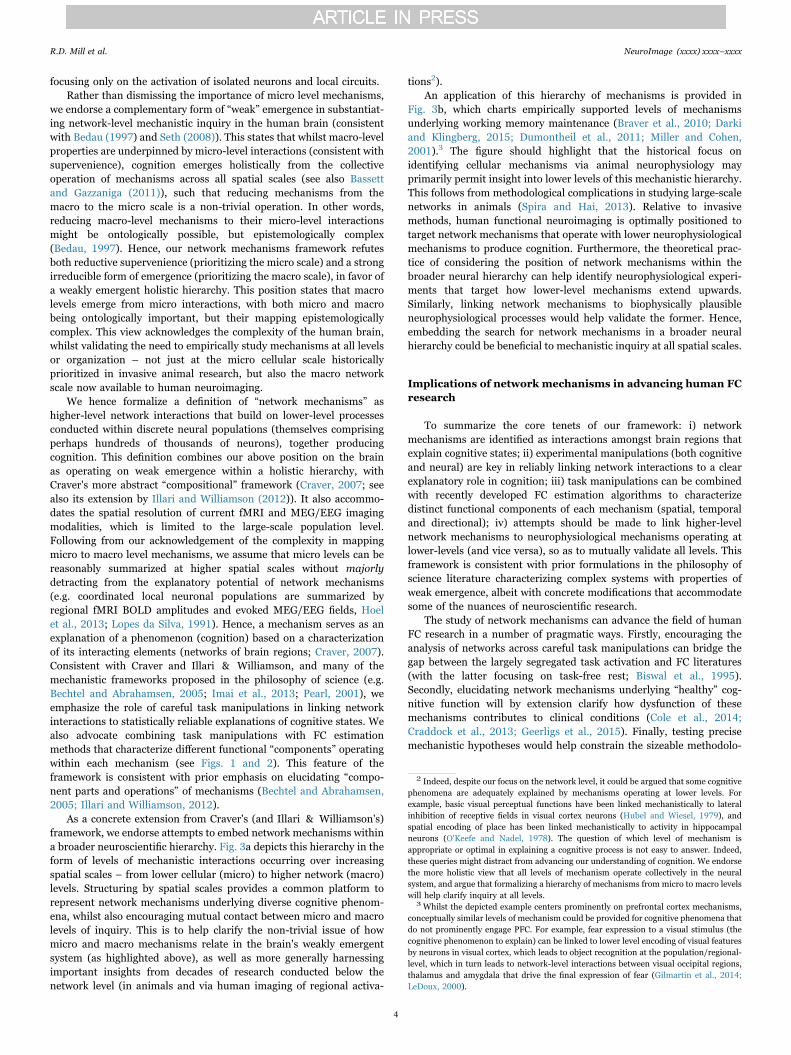
As a concrete extension from Craver's (and Illari & Williamson's)framework, we endorse attempts to embed network mechanisms withina broader neuroscientific hierarchy. Fig. 3a depicts this hierarchy in theform of levels of mechanistic interactions occurring over increasingspatial scales – from lower cellular (micro) to higher network (macro)levels. Structuring by spatial scales provides a common platform torepresent network mechanisms underlying diverse cognitive phenom-ena, whilst also encouraging mutual contact between micro and macrolevels of inquiry. This is to help clarify the non-trivial issue of howmicro and macro mechanisms relate in the brain's weakly emergentsystem (as highlighted above), as well as more generally harnessingimportant insights from decades of research conducted below thenetwork level (in animals and via human imaging of regional activa-
tions2).An application of this hierarchy of mechanisms is provided in
Fig. 3b, which charts empirically supported levels of mechanismsunderlying working memory maintenance (Braver et al., 2010; Darkiand Klingberg, 2015; Dumontheil et al., 2011; Miller and Cohen,2001).3 The figure should highlight that the historical focus onidentifying cellular mechanisms via animal neurophysiology mayprimarily permit insight into lower levels of this mechanistic hierarchy.This follows from methodological complications in studying large-scalenetworks in animals (Spira and Hai, 2013). Relative to invasivemethods, human functional neuroimaging is optimally positioned totarget network mechanisms that operate with lower neurophysiologicalmechanisms to produce cognition. Furthermore, the theoretical prac-tice of considering the position of network mechanisms within thebroader neural hierarchy can help identify neurophysiological experi-ments that target how lower-level mechanisms extend upwards.Similarly, linking network mechanisms to biophysically plausibleneurophysiological processes would help validate the former. Hence,embedding the search for network mechanisms in a broader neuralhierarchy could be beneficial to mechanistic inquiry at all spatial scales.
Implications of network mechanisms in advancing human FCresearch
To summarize the core tenets of our framework: i) networkmechanisms are identified as interactions amongst brain regions thatexplain cognitive states; ii) experimental manipulations (both cognitiveand neural) are key in reliably linking network interactions to a clearexplanatory role in cognition; iii) task manipulations can be combinedwith recently developed FC estimation algorithms to characterizedistinct functional components of each mechanism (spatial, temporaland directional); iv) attempts should be made to link higher-levelnetwork mechanisms to neurophysiological mechanisms operating atlower-levels (and vice versa), so as to mutually validate all levels. Thisframework is consistent with prior formulations in the philosophy ofscience literature characterizing complex systems with properties ofweak emergence, albeit with concrete modifications that accommodatesome of the nuances of neuroscientific research.
The study of network mechanisms can advance the field of humanFC research in a number of pragmatic ways. Firstly, encouraging theanalysis of networks across careful task manipulations can bridge thegap between the largely segregated task activation and FC literatures(with the latter focusing on task-free rest; Biswal et al., 1995).Secondly, elucidating network mechanisms underlying “healthy” cog-nitive function will by extension clarify how dysfunction of thesemechanisms contributes to clinical conditions (Cole et al., 2014;Craddock et al., 2013; Geerligs et al., 2015). Finally, testing precisemechanistic hypotheses would help constrain the sizeable methodolo-
2 Indeed, despite our focus on the network level, it could be argued that some cognitivephenomena are adequately explained by mechanisms operating at lower levels. Forexample, basic visual perceptual functions have been linked mechanistically to lateralinhibition of receptive fields in visual cortex neurons (Hubel and Wiesel, 1979), andspatial encoding of place has been linked mechanistically to activity in hippocampalneurons (O'Keefe and Nadel, 1978). The question of which level of mechanism isappropriate or optimal in explaining a cognitive process is not easy to answer. Indeed,these queries might distract from advancing our understanding of cognition. We endorsethe more holistic view that all levels of mechanism operate collectively in the neuralsystem, and argue that formalizing a hierarchy of mechanisms from micro to macro levelswill help clarify inquiry at all levels.
3 Whilst the depicted example centers prominently on prefrontal cortex mechanisms,conceptually similar levels of mechanism could be provided for cognitive phenomena thatdo not prominently engage PFC. For example, fear expression to a visual stimulus (thecognitive phenomenon to explain) can be linked to lower level encoding of visual featuresby neurons in visual cortex, which leads to object recognition at the population/regional-level, which in turn leads to network-level interactions between visual occipital regions,thalamus and amygdala that drive the final expression of fear (Gilmartin et al., 2014;LeDoux, 2000).
R.D. Mill et al. NeuroImage (xxxx) xxxx–xxxx
4

gical “model space” in brain network analysis, spanning choicesbetween multiple network node definitions, clustering algorithms andFC estimation methods. Large model spaces are increasingly the normgiven the (undoubtedly fruitful) integration of methods and algorithmsfrom fields such as computer science and engineering. Nevertheless,the increasing array of methods raises the need for principled ways toadjudicate between them. As a basis for this adjudication, we encou-rage consideration of the capabilities of a given network analysismethod to advance an explanatory understanding of cognition aboveand beyond that which is provided by previous methods.
Critically, this latter point argues against using an algorithm purelyon the basis that it has not been previously applied to human imagingdata. Rather, a given method is advocated if it has clear provisions tomake it more suitable to discovering network mechanisms overprevious approaches. For example, a novel FC estimation methodmight be endorsed based on its better treatment of non-Gaussianinformation that is known to be functionally relevant in humanimaging data (compared to methods that make Gaussian assumptions;Mumford and Ramsey, 2014). Relatedly, this mechanistic approachwould ensure that the use of advanced FC methods is structured bytheory-driven experimentation, rather than the inverse case of cogni-tive theory being tailored to the output of different methods. Thisadvocates the selection of a method a priori based on its capacity forexplanatory insight, rather than purely its novelty. The latter scenarioincreases the likelihood of a posteriori rationalization of an algorithm'soutput, which can slow the advancement of explanatory insight.
The remainder of this review outlines key methodological guide-lines to constrain the search for network mechanisms. We begin byproviding an overview of attempts to clarify the cognitive relevance offunctional networks mapped in the resting state (termed “resting-statenetworks”). Resting-state networks have become the primary researchfocus in the FC field, and hence for consistency we begin by consideringthem as potential “interacting elements” (Craver, 2007) of networkmechanisms. Critically, however, we also highlight that the resting-state literature has largely provided descriptive rather than mechanisticinsight into functional networks, given its primary emphasis onmapping networks during task-free rest. This arises from the lack ofexperimental manipulation of cognitive states, leading to poor char-
acterization of the phenomena to be explained and preventing insightinto explanatory mechanisms. Nevertheless, the fruitful method devel-opment in this field can be harnessed to reveal network mechanisms.Hence, in subsequent sections, we detail how task manipulations canbe combined with recently developed FC estimation methods tocharacterize a network mechanism's functional components – itsspatial topology, temporal properties and directional asymmetries(see Figs. 1 and 2).4 In particular, we highlight methods that cancorrect the present overreliance on outlining spatial components, at theexpense of temporal and directional ones. In the final section, we detailrecent developments in the field of multivariate pattern analysis(MVPA), which represents a broad class of algorithms that holdpotential in clarifying many network components over the “univariate”methods currently adopted.
To provide an empirical application of our framework, we concur-rently describe an example cognitive mechanism centering on interac-tions between a majority of network connections that remain spatially“stable” across task domains, and a minority of “flexible” ones (seeFig. 6 for a hierarchical depiction of this mechanism). We also suggesttemporal and directional components operating within this general“stable/flexible” mechanism. For example, we propose a temporalcomponent that splits the spatially “flexible” connections into func-tionally distinct “slow” timescale connections linked with learning, and“fast” timescale connections linked with stimulus-evoked responding(see section 4 for further details). In keeping with Fig. 3 and theaccompanying text in section 1, we also make concerted effortsthroughout to link this “stable/flexible” network mechanism to bio-physically plausible mechanisms operating at lower spatial scales.Whilst we admit that this example network mechanism is basic, itnevertheless can serve as a useful template that can be refined torepresent more functionally specific mechanisms. This furthers ourprimary aim of emphasizing the utility of “network mechanisms” as aconceptual framework to guide future FC research.
Fig. 3. Neuroscientific levels of mechanism are compositional, and organized in a weakly emergent holistic hierarchy. A) We conceptualize neuroscientific levels ofmechanism as particular neural elements interacting at successively higher spatial scales. Each mechanistic level is comprised of interactions occurring at the lower levels, such as brainnetworks being composed of a particular set of interacting brain regions. Rather than formalize a separate hierarchy for each cognitive state being explained (as in Craver (2007)), weformalize a common neural hierarchy to provide a platform to represent diverse cognitive states. This common hierarchy is structured so that each level marks an increase in spatial scale(i.e. from micro cellular scales accessible to animal neurophysiology, to higher network scales accessible to human imaging). Different cognitive states are theorized to emerge from thiscommon hierarchy via different sets of mechanistic interactions operating at each scale (as characterized by functional “components”, see Figs. 1 and 2). B) We provide an empiricallysupported example of a hierarchy of neural mechanisms underpinning working memory maintenance (the cognitive state under explanation). The schematic illustrates that cognitivestates emerge from mechanisms operating at multiple spatial scales, thereby correcting the historical over-emphasis on micro cellular mechanisms and instead validating the need formechanistic inquiry at the network level also. Note that network interactions underlying working memory maintenance are characterized using network definitions taken from theresting-state literature (e.g. “frontoparietal control network”). Note that the effects in intermediate levels may be less straightforward in reality than this simple illustration (Braver et al.,2010).
4 Whilst components other than spatial, temporal and directional are also likelyinvolved in characterizing network mechanisms (e.g. neuromodulatory components),these are the three that can be most reliably measured via current non-invasive imagingmodalities.
R.D. Mill et al. NeuroImage (xxxx) xxxx–xxxx
5
Clarifying the cognitive relevance of resting-state networks
Thus far, the dominant focus in human FC research has been onmapping spatial patterns of fMRI BOLD synchronization in the restingstate. Computing the Pearson's correlation coefficient between pairwiseregional (or voxel-wise) BOLD time series, in the absence of acontrolled task, has yielded a highly reproducible set of large-scalenetworks spanning many domains of cognitive function. These cano-nical resting-state networks include low-level sensory and motor net-works (Biswal et al., 1995; Cordes et al., 2000), as well as higher-orderdefault mode (Greicius et al., 2003), dorsal attention (Corbetta andShulman, 2002) and frontoparietal control networks (Cole andSchneider, 2007; Fox et al., 2005). Subsequent refinements to thisseed-based correlation approach have enabled more principled identi-fication of functional brain regions from which to extract BOLD timeseries (Glasser et al., 2016; Power et al., 2011), and use of data-drivennetwork definition approaches such as community detection (Poweret al., 2011; Yeo et al., 2011). Graph theoretical methods haveimproved the quantification of large-scale properties of these resting-state networks, leading to identification of highly inter-connectednetwork “hubs” (e.g. in the frontoparietal control network; Coleet al., 2010) and functionally segregated “modules” (e.g. sensory andmotor networks; Bullmore and Sporns, 2009). However, despiteevident refinement in how resting-state networks are described, thereremains much ambiguity as to precisely how cognition emerges fromthem. This ambiguity bears critically on the search for networkmechanism, and calls for more targeted attempts at studying thefunction and reconfiguration patterns of resting-state networks duringexperimentally controlled tasks.
Initial attempts to clarify the function of resting-state networksfocused on their visible spatial overlap with task-evoked activationpatterns (e.g. as identified by general linear model contrasts, Biswalet al., 1995). Studies that have used meta-analytic methods to identify“canonical” task activations (via cross-experiment correlations) acrossa number of cognitive domains have yielded high spatial similarity withtheir functionally linked resting-state networks (Laird et al., 2013;Smith et al., 2009). Such findings have informed a view that the spatialtopology of resting-state networks reflects a “history” of commonactivation during cognitive processing, likely arising from long-termsynaptic potentiation (Wig et al., 2011). The implied longitudinalrelationship between the two measures is supported by the increasein resting-state correlation observed amongst regions commonlyactivated during a task after it had been repeatedly performed (Lewiset al., 2009). However, other studies have highlighted divergences, inthat regions commonly activated in a task activation contrast oftencorrelate with separate resting-state networks (Barredo et al., 2015;Mill et al., 2015), with this lack of correspondence enhanced by morefunctionally-specific contrasts (Mennes et al., 2013). These latterfindings suggest that the relationship between resting-state networksand task activations is not one-to-one. Furthermore, the cited meta-analytic approach is poorly disposed to provide insight into networkmechanisms with clear links to cognitive function. Such comparison ofthe spatial overlap between resting-state networks and task activationpatterns between separate subject groups provides evidence only of acoarse associative (rather than explanatory) relationship betweenresting-state networks and on-task cognition.
With a view to more rigorous methods, recent work has comparedthe similarity of networks estimated across rest and task states in thesame sample of subjects. A pair of independent studies converged indemonstrating a high degree of spatial overlap between functionalnetworks estimated at rest and across a number of distinct tasks (Coleet al., 2014; Krienen et al., 2014). The shared variance in FC across restand multiple task states reached as high as 80%, suggesting theexistence of a “stable” (or “intrinsic”) resting-state network architecture(see Fig. 4a). This stable FC architecture might be the componentdriving the noted high correspondence between resting-state networks
and task activations (i.e. those identified by “general” rather thanfunctionally-specific GLM contrasts, Mennes et al., 2013). However,the residual 20% of task FC variance that is unexplained by rest FCmight still be highly relevant to more domain-specific processing.Indeed, both studies observed reliable differences in pairwise FC acrossdifferent tasks, suggesting more “flexible” network components thatunderpin network reconfiguration via variation of a small proportion ofconnections. With reference to our earlier definition of networkmechanisms (see Section 1), we theorize that cognition emerges fromthis general interaction between “stable” and “flexible” spatial networkcomponents.
Whilst this stable/flexible distinction is likely of broad importanceto diverse forms of cognition, the challenge now is to better understandthe function of each of these spatial components and how they interact.To this end, evidence from more targeted task manipulations suggeststhat “flexible” components might be preferentially localized to thefrontoparietal control resting-state network. Prior research has shownthat this network has the highest variability in its global connectivitypatterns across multiple tasks in comparison to other resting-statenetworks (Cole et al., 2013). This insight emerged from combining ageneralized psychophysiological interaction (gPPI) approach to task FCestimation (McLaren et al., 2012) with a graph theoretical measure of“global variable connectivity” (Cole et al., 2013). These methods wereapplied in a paradigm that instantiated multiple task states (thepermuted rule operations paradigm, PRO; Cole et al., 2010). Hence,as per our mechanistic framework, combining advanced FC methodswith careful task manipulation served to extend prior graph theoreticaldescription of “hubs” in the resting state (e.g. Cole et al., 2010) toreveal their explanatory cognitive role. Cognitive states might emergein a general sense from interactions between domain-general “flexiblehub” regions in the frontoparietal control network and domain-specificcontent regions in sensory-motor networks.
The extent to which the “stable” component of resting-state net-works is driven by underlying structural connectivity has also beenquestioned. Comparisons of structural networks identified via diffusionMRI (dMRI) demonstrate overlap with the spatial topology of resting-state networks, both in terms of their pairwise connections (Hagmannet al., 2008) and their graph theoretical features (e.g. overlap betweenstructural and functional hubs, van den Heuvel and Sporns, 2013).More formalized computational models have also explained a reason-able proportion of the variance in resting-state network topology fromstructural connectivity (Goni et al., 2014; Mišić et al., 2015). Thesefindings suggest that structure might indeed contribute to resting-statenetwork architecture, possibly providing the “skeleton” that constrainspaths of communication within them (Petersen and Sporns, 2015).However, computational modeling has also revealed the relationshipbetween structural and functional connectivity to be only partiallyexplanatory, and often mediated by other neurophysiological factors(Deco et al., 2009; Honey et al., 2007). In support of the outlinedstable/flexible mechanism, it might be that structural connectivityalone fails to predict more flexible functional network components thatvary across different task domains.5
To more clearly characterize task-evoked network flexibility, recentcomputational models have directly predicted task activation patterns(identified via general linear model contrasts across a number ofdistinct tasks) on the basis of resting-state FC. A set of regressionmodels trained on a large number of FC predictors and a small numberof structural predictors was able to reliably capture individual differ-ences in task activations (Tavor et al., 2016; see Fig. 4b). Notably,
5 Note that the failure to predict structural from functional connectivity could alsoarise from methodological issues, such as the use of network estimation methods(applied to both functional and structural connectivity) that do not clearly establish“direct” versus “indirect”/mediated connections. Problems with conclusively identifyingstructural connections in humans using dMRI have also been highlighted (Jones et al.,2013).
R.D. Mill et al. NeuroImage (xxxx) xxxx–xxxx
6
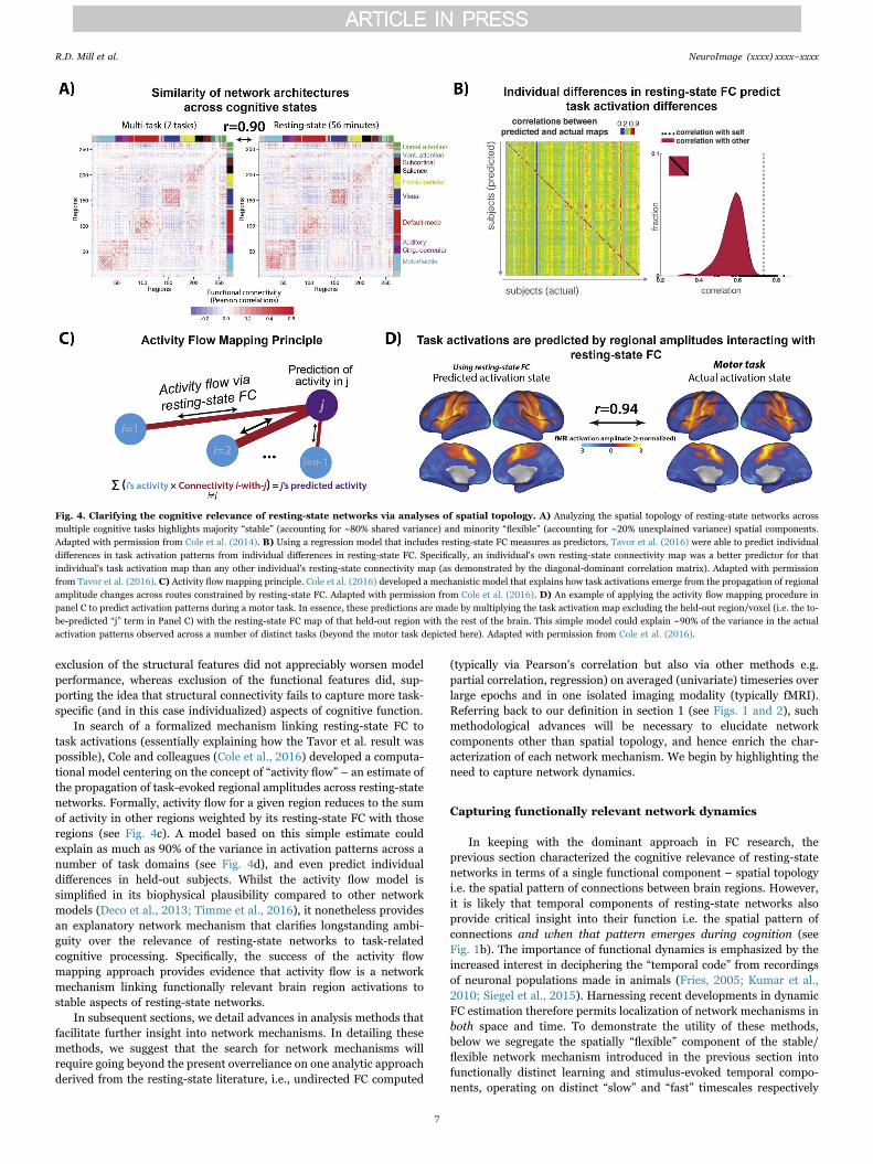
exclusion of the structural features did not appreciably worsen modelperformance, whereas exclusion of the functional features did, sup-porting the idea that structural connectivity fails to capture more task-specific (and in this case individualized) aspects of cognitive function.
In search of a formalized mechanism linking resting-state FC totask activations (essentially explaining how the Tavor et al. result waspossible), Cole and colleagues (Cole et al., 2016) developed a computa-tional model centering on the concept of “activity flow” – an estimate ofthe propagation of task-evoked regional amplitudes across resting-statenetworks. Formally, activity flow for a given region reduces to the sumof activity in other regions weighted by its resting-state FC with thoseregions (see Fig. 4c). A model based on this simple estimate couldexplain as much as 90% of the variance in activation patterns across anumber of task domains (see Fig. 4d), and even predict individualdifferences in held-out subjects. Whilst the activity flow model issimplified in its biophysical plausibility compared to other networkmodels (Deco et al., 2013; Timme et al., 2016), it nonetheless providesan explanatory network mechanism that clarifies longstanding ambi-guity over the relevance of resting-state networks to task-relatedcognitive processing. Specifically, the success of the activity flowmapping approach provides evidence that activity flow is a networkmechanism linking functionally relevant brain region activations tostable aspects of resting-state networks.
In subsequent sections, we detail advances in analysis methods thatfacilitate further insight into network mechanisms. In detailing thesemethods, we suggest that the search for network mechanisms willrequire going beyond the present overreliance on one analytic approachderived from the resting-state literature, i.e., undirected FC computed
(typically via Pearson's correlation but also via other methods e.g.partial correlation, regression) on averaged (univariate) timeseries overlarge epochs and in one isolated imaging modality (typically fMRI).Referring back to our definition in section 1 (see Figs. 1 and 2), suchmethodological advances will be necessary to elucidate networkcomponents other than spatial topology, and hence enrich the char-acterization of each network mechanism. We begin by highlighting theneed to capture network dynamics.
Capturing functionally relevant network dynamics
In keeping with the dominant approach in FC research, theprevious section characterized the cognitive relevance of resting-statenetworks in terms of a single functional component – spatial topologyi.e. the spatial pattern of connections between brain regions. However,it is likely that temporal components of resting-state networks alsoprovide critical insight into their function i.e. the spatial pattern ofconnections and when that pattern emerges during cognition (seeFig. 1b). The importance of functional dynamics is emphasized by theincreased interest in deciphering the “temporal code” from recordingsof neuronal populations made in animals (Fries, 2005; Kumar et al.,2010; Siegel et al., 2015). Harnessing recent developments in dynamicFC estimation therefore permits localization of network mechanisms inboth space and time. To demonstrate the utility of these methods,below we segregate the spatially “flexible” component of the stable/flexible network mechanism introduced in the previous section intofunctionally distinct learning and stimulus-evoked temporal compo-nents, operating on distinct “slow” and “fast” timescales respectively
Fig. 4. Clarifying the cognitive relevance of resting-state networks via analyses of spatial topology. A) Analyzing the spatial topology of resting-state networks acrossmultiple cognitive tasks highlights majority “stable” (accounting for ~80% shared variance) and minority “flexible” (accounting for ~20% unexplained variance) spatial components.Adapted with permission from Cole et al. (2014). B) Using a regression model that includes resting-state FC measures as predictors, Tavor et al. (2016) were able to predict individualdifferences in task activation patterns from individual differences in resting-state FC. Specifically, an individual's own resting-state connectivity map was a better predictor for thatindividual's task activation map than any other individual's resting-state connectivity map (as demonstrated by the diagonal-dominant correlation matrix). Adapted with permissionfrom Tavor et al. (2016). C) Activity flow mapping principle. Cole et al. (2016) developed a mechanistic model that explains how task activations emerge from the propagation of regionalamplitude changes across routes constrained by resting-state FC. Adapted with permission from Cole et al. (2016). D) An example of applying the activity flow mapping procedure inpanel C to predict activation patterns during a motor task. In essence, these predictions are made by multiplying the task activation map excluding the held-out region/voxel (i.e. the to-be-predicted “j” term in Panel C) with the resting-state FC map of that held-out region with the rest of the brain. This simple model could explain ~90% of the variance in the actualactivation patterns observed across a number of distinct tasks (beyond the motor task depicted here). Adapted with permission from Cole et al. (2016).
R.D. Mill et al. NeuroImage (xxxx) xxxx–xxxx
7

(see Fig. 1b).Mathematically diverse FC estimation methods applied in task-free
rest have provided basic evidence that resting-state networks undergodynamic fluctuations. Such methods include the use of sliding windows(e.g. 30–60 seconds) to conduct time-varying correlation (Hutchisonet al., 2013) and spatial independent components analyses (spatialICA; Allen et al., 2014), as well as more data-driven methods that donot require selection of a sliding window (e.g. temporal ICA; Smithet al., 2012). All methods converge in demonstrating that resting-statenetworks typically estimated over long durations (~5–10 minutes)undergo dynamic fluctuations over shorter periods. These “temporalnetwork states” tend to recur over the entire rest scan (Allen et al.,2014; Karahanoğlu and Van De Ville, 2015; Liu and Duyn, 2013) andacross subjects (Zalesky et al., 2014). Hence, standard static ap-proaches to FC estimation run the risk of obscuring dynamic transi-tions between temporal network states, which might be mechanisticallyrelevant to cognition.
However, whilst dynamic transitions described amongst resting-state networks are assumed to reflect transitions in cognitive states, thetask-free nature of rest FC leaves scope for alternative interpretations.This is because mere description of temporal network states during restfails to provide a clear link to emergent cognitive states. The failure toelucidate this explanatory relation opens the possibility that networkdynamics might be driven by artifactual rather than functionallyrelevant sources. For example, a recent study highlighted the contam-ination of resting-state network dynamics by head motion. This wasquantified by the reduction in a multivariate kurtosis measure ofdynamic non-stationarity after removal of high-motion time points(Laumann et al., 2016; see Fig. 5a). Reassuringly, multivariate kurtosisalso increased in task relative to rest states (after removal of high-motion time points), suggesting that functionally relevant networkdynamics are obtainable (see Fig. 5b). These findings make clear howmerely describing dynamic network states at rest provides imperfectinsight into their cognitive relevance. Consistent with the previoussection detailing spatial components of network mechanisms, we arguethat the identification of mechanistically relevant temporal componentscalls for combining dynamic FC estimation methods with careful taskmanipulations. This would link prior description of temporal statetransitions in rest FC more clearly to accompanying transitions incognitive states, as instantiated in controlled task settings.
Studies combining dynamic FC estimation with task manipulationsremain nascent, but preliminary findings suggest refinement of thepreviously introduced stable/flexible mechanism. Specifically, whereasthe spatially “stable” network component might reflect structuralconnectivity without overlaid dynamics, the “flexible” componentmight arise from structural connectivity in addition to distinct dynamicreconfiguration processes occurring over distinct timescales (seeFigs. 1b and 6). To clarify, “slow” dynamic fluctuations amongstresting-state networks have been shown to track the trajectory oflearning in sensorimotor tasks (Bassett et al., 2015; see Fig. 5c).Dynamic estimates of network modularity derived from a temporalclustering algorithm revealed a longitudinal increase (over days andweeks) in the autonomy of sensory and motor networks with learning,coupled with reduced recruitment of cognitive control networks(Bassett et al., 2015; see also Chein and Schneider (2005) and Lewiset al. (2009)). Beyond adding a temporal component to the stable/flexible mechanism, this finding supports the specialized role of higher-order cognitive control networks in actively driving learning-inducedflexibility. This extends its role as a spatial “flexible hub” introduced inthe previous section (Cole et al., 2013).
In contrast, evidence of functionally relevant network dynamicsemerging over relatively “fast” timescales was provided by Sadaghianiet al. (2015). This was enabled by trial-wise FC estimation over pre-stimulus windows in an auditory detection task. Reduced modularity indefault mode and visual networks in this baseline period (spanning~20–40 seconds) was associated with impaired performance, thereby
suggesting a mechanistic link between fast trial-evoked FC reconfigura-tion and overt behavior (see also Thompson et al. (2013)). It istempting to link these outlined “slow” learning and “fast” stimulus-evoked dynamic FC components to respective long-term synapticpotentiation and spike propagation neurophysiological substrates,given that these also differ in their slow versus fast timescales(Dobrunz and Stevens, 1997; Sejnowski, 1999; see Fig. 6). Indeed,further scrutiny of this network mechanism might reveal a cyclicrelationship linking these two temporal components, such that repeti-tions of “fast” stimulus-evoked network reconfigurations lead tolearning-induced reconfigurations emerging over a “slower” timescale,with these “slow” reconfigurations then exerting reciprocal constraintson how “fast” reconfigurations emerge across resting-state networks.Whilst such links remain speculative at this stage, it is evident thatcapturing dynamic FC processes over multiple timescales will be key incharacterizing network mechanisms that clarify multiple levels of theneural hierarchy (see Fig. 3).
However, it is also worth highlighting that the present primaryreliance on fMRI complicates the study of dynamic FC. This is due towell-known limitations in fMRI temporal resolution, arising from boththe relatively low sampling rate of the BOLD signal (that only permits amaximum frequency resolution in the ~0.75 Hz slow delta range; Lewiset al., 2016), as well as its vascular underpinnings rendering it anindirect measure of neural activity. Despite advances in sub-second TRacquisition protocols (Lewis et al., 2016; Smith et al., 2013) and blinddeconvolution methods that aim to separate the underlying neuralactivation from the HRF (detailed in the next section; Havlicek et al.,2011), analysis of dynamic FC would undoubtedly benefit from greaterinvolvement of MEG and EEG. These modalities provide millisecondresolution estimates of neural activity that are not confounded byhemodynamics.
However, this improved temporal resolution comes at the expenseof worse spatial resolution and the related issue of “field spread” – thespreading of activity from a single neural source across proximal MEG/EEG sensors. This has been shown to yield artifactual, locally dominantFC patterns (Schoffelen and Gross, 2009). However, simulations haveshown that spatial filtering approaches to MEG/EEG source modeling(e.g. linear beamforming; Van Veen et al., 1997) can attenuate fieldspread (Schoffelen and Gross, 2009). To further counteract theproblem, beamformer source modeling can be combined with FCestimation methods that exclude instantaneous or “0-lag” phaserelationships between input time series. This follows from the fact thatfield spread is entirely carried at 0-lag, albeit at the potential cost ofremoving 0-lag signal of neural origin (Nolte et al., 2004). Indeed,pairwise correlations between beamformer-modeled MEG source timeseries have recovered reasonable analogues of fMRI resting-statenetworks, both with exclusion of 0-lag phase relationships (Hippet al., 2012) as well as without (Brookes et al., 2011). Note that similarresults might be expected with EEG, given recent use of anatomicalMRI images and sophisticated models of skull tissue conductivity tocreate “individualized” head models. Such head models have beenobserved to yield equivalent EEG source modeling accuracy as withMEG (Klamer et al., 2015).
Beyond demonstrating the overlap in spatial network topologyacross fMRI and MEG, studies have begun to capitalize on advancesin MEG source modeling to target dynamics. For example, FC estima-tion over sliding windows yielded a decomposition of MEG resting-state networks into temporal network states, albeit over much shortertimescales than anticipated by fMRI studies (i.e. at millisecond-to-second resolution; Baker et al., 2014; de Pasquale et al., 2010). Anotherintriguing application of MEG/EEG source connectivity is in clarifyingwhether different network mechanisms communicate via differentfrequency bands. Thus far, MEG homologues of fMRI resting-statenetworks have emerged primarily in a moderately low frequency range(theta to beta; Brookes et al., 2011; de Pasquale et al., 2010; Hipp et al.,2012). However, a recent study that corrected for the lower signal-to-
R.D. Mill et al. NeuroImage (xxxx) xxxx–xxxx
8
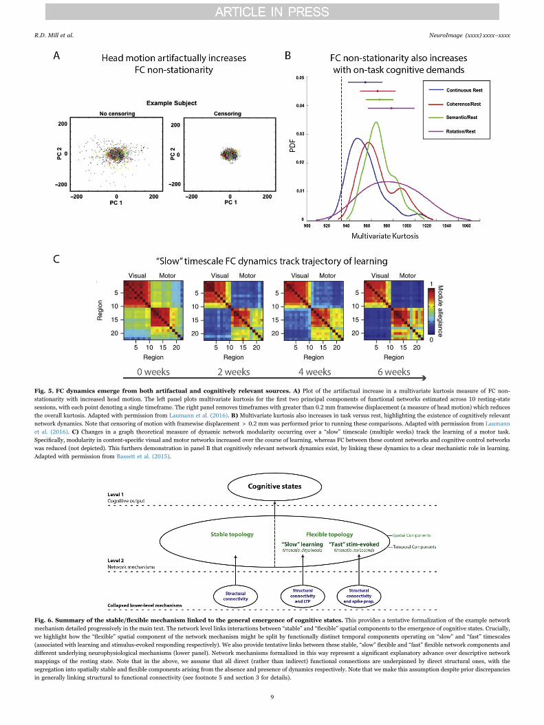
Fig. 5. FC dynamics emerge from both artifactual and cognitively relevant sources. A) Plot of the artifactual increase in a multivariate kurtosis measure of FC non-stationarity with increased head motion. The left panel plots multivariate kurtosis for the first two principal components of functional networks estimated across 10 resting-statesessions, with each point denoting a single timeframe. The right panel removes timeframes with greater than 0.2 mm framewise displacement (a measure of head motion) which reducesthe overall kurtosis. Adapted with permission from Laumann et al. (2016). B) Multivariate kurtosis also increases in task versus rest, highlighting the existence of cognitively relevantnetwork dynamics. Note that censoring of motion with framewise displacement > 0.2 mm was performed prior to running these comparisons. Adapted with permission from Laumannet al. (2016). C) Changes in a graph theoretical measure of dynamic network modularity occurring over a “slow” timescale (multiple weeks) track the learning of a motor task.Specifically, modularity in content-specific visual and motor networks increased over the course of learning, whereas FC between these content networks and cognitive control networkswas reduced (not depicted). This furthers demonstration in panel B that cognitively relevant network dynamics exist, by linking these dynamics to a clear mechanistic role in learning.Adapted with permission from Bassett et al. (2015).
Fig. 6. Summary of the stable/flexible mechanism linked to the general emergence of cognitive states. This provides a tentative formalization of the example networkmechanism detailed progressively in the main text. The network level links interactions between “stable” and “flexible” spatial components to the emergence of cognitive states. Crucially,we highlight how the “flexible” spatial component of the network mechanism might be split by functionally distinct temporal components operating on “slow” and “fast” timescales(associated with learning and stimulus-evoked responding respectively). We also provide tentative links between these stable, “slow” flexible and “fast” flexible network components anddifferent underlying neurophysiological mechanisms (lower panel). Network mechanisms formalized in this way represent a significant explanatory advance over descriptive networkmappings of the resting state. Note that in the above, we assume that all direct (rather than indirect) functional connections are underpinned by direct structural ones, with thesegregation into spatially stable and flexible components arising from the absence and presence of dynamics respectively. Note that we make this assumption despite prior discrepanciesin generally linking structural to functional connectivity (see footnote 5 and section 3 for details).
R.D. Mill et al. NeuroImage (xxxx) xxxx–xxxx
9

noise ratio at higher frequencies also recovered resting-state analoguesin the gamma band (Hipp and Siegel, 2015). Probing the functionalrelevance of such band-limited or “multiplexed” (Siegel et al., 2012)temporal components holds much promise in formalizing the pre-viously raised distinction between “slow flexible” and “fast flexible”components of stable/flexible mechanism. The former might be linkedto FC changes in lower frequencies, whereas as the latter might emergeat higher frequencies.
Overall, dynamic FC analyses have revealed that the topology ofresting-state networks masks a diversity of temporal processing overmultiple timescales. Whilst this sub-field remains at an early stage,emerging findings support a distinction between “stable” components,“slow flexible” components (linked with learning and underlyingsynaptic potentiation) and “fast flexible” components (linked withstimulus-evoked reconfiguration and underlying spike propagation;see Fig. 6). Although this distinction serves to integrate temporal andspatial components as part of a unified network mechanism (consistentwith Fig. 2), further research in this vein is undoubtedly required. Forexample, future work might probe whether dynamic FC fluctuations aredependent on or arise as a natural consequence of the propagation ofregional activations across resting-state networks, as suggested by theactivity flow mapping approach (Cole et al., 2016).
Revealing asymmetries in activity propagation via directedfunctional connectivity
The majority of research into human brain networks has focused onone FC estimation algorithm – pairwise Pearson's correlation com-puted between regional time series – which conveys whether tworegions A and B communicate in a general “undirected” sense (con-nectivity A-B). This is especially true for fMRI connectivity studies,whereas MEG/EEG connectivity has been commonly computed viaboth correlation and undirected coherence approaches. In contrast, aclass of “directed” or “effective” FC algorithms provides additionalinsight into directions of activity propagation – whether region Acommunicates downstream to region B (connectivity A→B) or viceversa (connectivity B→A). Adding directional information entails aconceptual advance over networks described via undirected FC meth-ods only, as reflected by the emphasis on parameters capturingdirectional communication in a number of computational networkmodels (for review see Woolrich and Stephan (2013)). However,directed FC methods have not been widely adopted in empiricalresearch, which is in part due to the majority of anatomical cortico-cortical connections being bidirectional (Felleman and Van Essen,1991; Markov et al., 2011). However, we have already highlighted theimperfect correspondence between functional and structural connec-tivity, and there is neurophysiological evidence of asymmetries indirected activity propagation arising from experience-dependent sy-naptic plasticity processes independent of anatomy (Zheng et al.,2011). Even if directed connections are fewer than undirected ones,they might still have particular influences on network mechanismsunderlying cognitive function. Hence, clarifying whether networkscommunicate via directed or undirected forms of activity propagationadds a distinct functional component to each network mechanism (seeFigs. 1c and 2).
The primary impediment to more widespread use of directedconnectivity has been the numerous methodological uncertaintiessurrounding these algorithms in comparison to the mathematicallysimpler undirected methods. This includes uncertainty over the choiceof directed FC algorithm from multiple mathematically diverse options,such as Granger causality (Roebroeck et al., 2005), directed coherence(Nolte et al., 2008), structural equation modeling (SEM; Gates andMolenaar, 2012), dynamic causal modeling (DCM; Chen et al., 2008;Friston et al., 2003) and Bayesian approaches (Mumford and Ramsey,2014; Patel et al., 2006). Uncertainty also persists over appropriatepre-processing steps (i.e. how to suppress noise sources whilst preser-
ving relevant fine-grained temporal information), algorithmic para-meters (e.g. the number of lagged observations included in Grangercausality models) and how to maintain computational tractability withincreasing number of input variables (e.g. the search space in model-based methods such as DCM becomes prohibitively large for > 10input variables). This uncertainty extends to the choice of imagingmodality, especially as concerns fMRI given temporal limitationsarising from its low sampling rate and hemodynamic-induced con-founds. The latter feature is particularly problematic given establishedregional and inter-subject variability in the hemodynamic responsefunction (Handwerker et al., 2004), which can give rise to “baseline”differences in regional BOLD morphology that complicate directionalinference (especially in between-subject designs; Schippers et al.,2011). Whilst MEG/EEG modalities provide a more “direct" and highertemporal resolution signal, the higher sampling rate permits contam-ination by a wider range of artifacts that can reduce overall signal-to-noise.
These methodological concerns call for concerted attempts tovalidate directed FC across algorithms and modalities, as a necessaryprecursor to applying them in the search for network mechanisms.However, validations in simulated data have yielded equivocal results.One fMRI simulation study (Smith et al., 2011) that compared theefficacy of multiple directed FC algorithms across variation in inputparameters and pre-processing steps found only modest recovery of theembedded directional ground truth (~65% maximum detection accu-racy). In contrast, a later study employing a similar multi-algorithmicvalidation, as well as explicitly modeled lags in communication at theneural level (a key difference from the generative model used in theSmith et al., 2011 study) found much higher detection accuraciesacross a number of algorithms, in both simulated fMRI and MEG/EEGdata (Wang et al., 2014).
Recently, we devised a principled approach to validating directedFC in real fMRI and source-modeled MEG data acquired in the samesubjects (Mill et al., 2016). This empirical validation sidesteps debateover the validity of various simplifying assumptions made in simula-tions (Deshpande et al., 2010; Ramsey et al., 2011). Our approachcentered on a clearly explicated “empirical ground truth” directedconnectivity pattern. This took the form of an experimentally-inducedreversal in activity propagation between auditory and visual regionsthat was predicated on the widely replicated “sensory reactivation”effect in episodic memory (e.g. Wheeler et al., 2006). This ground truthwas successfully recovered by a number of directed FC methods, suchas Granger causality and IMAGES Bayes network methods (Ramseyet al., 2011), and across fMRI and source-modeled MEG. These broadlypositive results confirm the base validity of directional algorithmsapplied to human imaging data. The findings also provide morepractical insight into the broad virtue of analyzing the experimentalmodulation of directionality estimates across task conditions (as high-lighted previously; Roebroeck et al., 2005) and the improvement infMRI results after applying “blind deconvolution”methods that removethe influence of HRF variability (Havlicek et al., 2011). Futureempirical validations might test the recovery of directed connectivityground truths spanning multiple regions, thereby testing for theremoval of “indirect” connections that can arise via pairwise methods.Nevertheless, results from the Mill et al. study support the use ofavailable algorithms in combination with task manipulations to recoverdirectional components of network mechanisms.
Methodological uncertainties have also meant that estimations ofdirectionality at the large-scale network level (beyond a subset ofregions) remain nascent, yet preliminary results have been promising.For example, analysis of lagged temporal dependencies betweencortical regions yielded a decomposition of the established fMRIresting-state networks into 8 recurring large-scale directed processingsequences (termed “lag motifs”; Mitra et al., 2015). This result suggestsa critical role for directed activity propagation in mediating theemergence of these networks. Analyses of multivariate Granger caus-
R.D. Mill et al. NeuroImage (xxxx) xxxx–xxxx
10
ality during experimentally controlled task settings have identified twolarge-scale directional influences amongst resting-state networks (in-creased dorsal attention→ventral attention connectivity, and increasedfrontoparietal control→default mode connectivity), which were linkedwith “top-down” cognitive control processes that improved behavioralperformance (Wen et al., 2013; Wen et al., 2012). These studieshighlight the potential for directed FC analyses to both refine descrip-tions of network properties in the resting state, as well as clarify the on-task mechanisms linking them to cognition.
Contingent on further validation work, future research mightexamine the role of directed activity propagation in our examplestable/flexible mechanism. For example, it might be that spatiallyflexible components linked preferentially to the frontoparietal controlnetwork enforce downstream (i.e. “top-down”) directional relation-ships with content-specific networks to coordinate processing acrossmultiple domains (i.e. frontoparietal→content networks; see Fig. 1c).More generally, future research might strive to identify whetherexplanatory network interactions are better characterized as eitherundirected/bidirectional or directional. This argues against the ideathat more widespread application of directed FC methods will revealpreviously undirected relationships to be exclusively directional.Rather, evidence supports the presence of real FC relationships lackingin any stable directionality, in the form of synchronized oscillationsobserved at multiple spatial scales (Rajagovindan and Ding, 2008;Siegel et al., 2012; Varela et al., 2001). Approaches to analyzingdirected connectivity therefore need to better provide for instances ofreal undirected connectivity. Such approaches might also accommodatea role for dynamics, given observation of transitions between directedand undirected FC relationships in animals (Gregoriou et al., 2009) andhumans (Womelsdorf et al., 2007). Indeed, linking dynamic direction-ality fluctuations to a clearer explanatory role in cognition would serveas an idealized recovery of a network mechanism's full complement ofspatial, temporal and directional components (as exemplified in Fig. 2).
Increasing the sensitivity of network components viamultivariate pattern analysis
Another feature of the standard FC estimation pipeline is theextraction of averaged time series from isolated brain voxels or asthe average across neighboring voxels within putative brain regions.Extracting such “univariate” estimates of brain activation mightocclude FC mechanisms encoded by “multivariate” representationalpatterns amongst multiple voxels or areas of cortex. Indeed, theapplication of multivariate pattern analysis (MVPA) methods tomulti-unit recordings in animals has refined relationships betweenspatiotemporal patterns in neuronal populations and cognitive func-tion (Montijn et al., 2016; Siegel et al., 2015; Stokes et al., 2013).Application of MVPA to human imaging data has provided furtherinsight into the functional relevance of regional activation amplitudesby characterizing multivariate patterns amongst voxels. In comparisonto univariate methods, MVPA algorithms can increase the sensitivity ofdetecting neural representations underlying a wide array of cognitivestates. Whilst the application of MVPA at the network level remains ina fledgling state, integrating this powerful class of algorithms holdsmuch promise in the search for network mechanisms. These tools canbe leveraged to better characterize a network mechanism, both via itsspatial components (by decoding cognitive representations in topolo-gical network patterns) and temporal components (by dynamic decod-ing of temporal network states; see Fig. 7).
To provide a brief overview (for further details see Haxby et al.(2014) and Haynes (2015)), a fundamental concept linking differentMVPA algorithms is that of a high-dimensional space spanning multi-ple brain regions/voxels (“features”) that are classified over a set ofinput samples (“observations”) as representing a particular experi-mental condition. In essence, MVPA identifies patterns of brainregions/voxels that contain “information” relevant to decoding a given
cognitive state (Kriegeskorte et al., 2006). MVPA methods includelinear and non-linear classifiers that estimate the accuracy in decodinga cognitive state from a multivariate pattern (Haxby, 2001).Representational similarity analysis (RSA; Kriegeskorte et al., 2008)can provide more detailed information as to the similarity betweenmultivariate patterns for two or more stimulus classes. This helpsdistinguish between multivariate representations that have equallyhigh classification accuracies but might be classifying via very differentfunctions. These MVPA algorithms have been observed to be moresensitive to fine-grained cognitive information than alternative uni-variate approaches (Haxby, 2001; Kriegeskorte et al., 2006). Ratherthan being confined to lower-level sensory classifications, MVPA hasalso been successful in decoding higher-order functions (Cole et al.,2011; Soon et al., 2008). Application of MVPA in a sliding windowapproach has also highlighted its utility in decoding dynamics, withabove-chance classifications observed earlier than equivalent univari-ate approaches in trial-locked analyses applied to both fMRI (Kohleret al., 2013) and MEG (Borst et al., 2016; Sudre et al., 2012). Overall,findings from the application of MVPA algorithms to task activationsemphasize their considerable utility in capturing richer and morespecific cognitive representations in space and time.
These regional activation-based findings can be extended to thesearch for mechanisms at the network level in three broad ways.Firstly, MVPA can be applied to identify more sensitive (i.e. highersignal-to-noise) spatial components via extraction of multivariatetimecourses from the pattern amongst multiple regions or voxels priorto FC estimation (see Fig. 7a). For example, Coutanche and colleagues(Coutanche and Thompson-Schill, 2013) devised a method of “infor-mational connectivity”, wherein a searchlight MVPA analysis decodesthe timepoint-by-timepoint classification accuracy across a set of voxelswith reference to a multivariate “prototype” (the mean response withinthat multivariate searchlight to a given task condition). The resultinginformational timecourses were submitted to standard pairwise corre-lations to provide a connectivity map of multivariate representations. Arelated multivariate connectivity method was found to yield morereliable rest FC maps (across sessions and subjects) than thoseobtained from univariate approaches (Geerligs et al., 2016). With aview to linking these findings to explanatory network mechanisms,future studies might seek to apply multivariate connectivity methodsduring experimentally controlled tasks, so as to capitalize on the likelyhigher signal-to-noise ratio of the input time series.
The second approach to MVPA network analysis also targets morerefined spatial components, by decoding cognitive states directly fromtopological FC patterns (see Fig. 7b). Rather than basing an MVPAclassification on activation patterns amongst input voxels, this ap-proach performs a classification based on connectivity estimatesbetween multiple regions or large-scale networks. Cole et al. (2013)demonstrated the utility of this approach by identifying more specia-lized spatial components linked to flexible network reconfiguration.Specifically, a network representational similarity analysis linked theincreasing similarity of FC patterns involving the frontoparietal controlnetwork with increasing similarity in cognitive processing across 64unique task states. Each of these task states comprised a permutationof shared low-level rules (4 motor×4 logic×4 sensory rules). Thisapproach therefore related “flexible” engagement of the frontoparietalnetwork to the “flexible” integration of these shared low-level rules innovel task contexts, consistent with the involvement of cognitivecontrol in rapid instructed task learning (Cole et al., 2013). A follow-up analysis trained a linear classifier to successfully decode the currenttask state on the basis of frontoparietal FC patterns with domain-specific networks (e.g. frontoparietal to motor network FC successfullydecoded the motor task rules). These findings highlight the use ofnetwork MVPA methods both to identify more specialized “flexible”components of the stable/flexible network mechanism, as well as indirectly decoding cognitive states that emerge from this networkmechanism. Whilst these results support a specific role for the
R.D. Mill et al. NeuroImage (xxxx) xxxx–xxxx
11

frontoparietal network in mediating “flexible” cognitive control, itwould be useful to extend this classification approach to other networkslinked with control functions (e.g. the dorsal attention network and thecingulo-opercular network, Corbetta and Shulman, 2002; Dosenbachet al., 2007).
The third application of network MVPA methods is in identifyingtemporal components of network mechanisms via dynamic decoding oftemporal states (King and Dehaene, 2014; Stokes et al., 2013). Byevaluating moment-to-moment variability of multivariate representa-tions, insight into the timescale of task-related information in specificnetworks can be gained. An extension of this dynamic MVPA approachwas recently employed in the classification of temporal network statesduring task-free rest (Chen et al., 2016; see Fig. 7c and previousdiscussion of temporal network states in section 4). Specifically, thesimilarity in multi-region spatial patterns across individual time pointswas used to identify periods during resting-state scans in which theglobal activation state was similar. A clustering algorithm applied tothe resulting “temporal similarity matrix” (left panel, Fig. 7c) identifiedtemporal network states and sub-states (middle panel, Fig. 7c), whichcritically were associated with distinct functional roles via a meta-analysis database of task activation patterns (right panel, Fig. 7c;Yarkoni et al., 2011). This study provides one example of the profitableuse of dynamic MVPA methods in elucidating clearer explanatorylinkages between spontaneous temporal network states and cognitivestates. Following from similar suggestion in section 4, future exten-sions of this dynamic network MVPA approach might instantiatecareful task manipulations to link each temporal network state directlyto a cognitive state. Future studies might also probe the correspon-dence of these fMRI states with those classified in higher temporal
resolution MEG/EEG modalities (King and Dehaene, 2014).To summarize, MVPA represents a novel class of methods that
opens up a host of possibilities for research into network mechanisms.Whilst the full scope of combining MVPA with other advanced FCmethods has yet to be realized, preliminary findings highlight theparticular utility of MVPA in elucidating more refined spatial andtemporal components within network mechanisms, and linking thesecomponents more directly to cognitive function. In this context, wehave alluded to the potential for MVPA methods to refine our stable/flexible network mechanism – from comprising interactions betweenfairly general “stable” and “flexible” spatial components, to a morespecific role for the frontoparietal control network in underpinning thelatter component.
Summary of key challenges and future directions
Whilst the preceding sections have detailed numerous advances inthe study of human brain networks, a number of key challenges areposed to the search for network mechanisms. Firstly, there remains abroad need for more principled validations of FC estimation strategies.Confidence in available methodologies is a necessary precursor togenerating meaningful mechanistic insight from them, and thereremains much ambiguity over optimal preprocessing steps (e.g. mini-mization of artifacts), choice of FC algorithm, and approaches tosignificance testing. It will be important for such validations to beapplied in empirical as well as synthetic datasets, so as to avoid debateover the assumptions made by different simulation models. As we haverepeatedly emphasized, validation of FC methods should also beaccompanied by a renewed focus on sound experimental design. This
Fig. 7. MVPA methods can identify spatial and temporal components of network mechanisms. A) Extraction of informational time series from multivariate voxel patternswithin brain regions prior to FC estimation. This approach yields network FC maps comprised of multivariate representational patterns (treating voxel activations as features) linked toparticular cognitive states, which can yield more sensitive spatial components of network mechanisms. B)MVPA can directly decode cognitive states from already estimated multivariateFC patterns (treating network connections as features). This approach also recovers spatial network components more directly linked to cognitive states. C) Empirical example of anapplication of dynamic MVPA to elucidate temporal network states. The left panel is a “temporal similarity matrix” illustrating the timepoint-by-timepoint similarity of multivariateactivation patterns. This temporal similarity matrix is clustered to identify two temporal network states that dynamically fluctuate over the course of a rest scan (State A and State B,rendered at the voxel scale in the middle panel). These temporal states are then linked to respective “task positive” and “task negative” cognitive functions via a meta-analysis of taskactivations (right panel; Yarkoni et al., 2011). Whilst direct manipulation of these states in task contexts is required to substantiate their functional roles, this dynamic MVPA approachrepresents an improvement in linking temporal states decomposed in the resting-state to clearer cognitive functions. Adapted with permission from Chen et al. (2016).
R.D. Mill et al. NeuroImage (xxxx) xxxx–xxxx
12
would increase the likelihood of providing explanatory rather thanmerely descriptive insight into functional networks. Combining ad-vanced FC estimation methods with targeted task manipulations willalso prevent over-emphasizing data-driven methods to the detriment ofcognitive theory.
We have also highlighted the need to address the present over-reliance on fMRI, given the relatively low temporal resolution of thismodality and parallel improvements in the spatial resolution of source-modeled MEG/EEG. Whilst the utility of MEG/EEG source connectiv-ity in clarifying network dynamics is fairly obvious, the use of thesemodalities might also be beneficial for MVPA, given that fMRI's lowsampling rate often means that the number of observations is not muchgreater than the number of voxel features (which increases the risk of“overfitting” noise in the data; Haxby et al., 2014). Network analysesapplied to multi-modal imaging datasets, such as simultaneouslyacquired EEG-fMRI, represents a largely unexplored but potentiallyremunerative avenue, given that both imaging modalities enablecomplementary improvements in spatial and temporal resolution.Multi-modal network analyses might also involve physiological mea-surements (e.g. pupillometry), which could clarify interactions betweencortical networks and autonomic neuromodulatory systems that areincreasingly viewed as integral to cognitive function (Aston-Jones andCohen, 2005; Mill et al., 2016; Shine et al., 2016). Furthermore,recently released large-sample electrocorticography datasets (ECoG;e.g. the Restoring Active Memory project, RAM) could be invaluable inclarifying the micro scale underpinnings of network mechanismsstudied in human imaging.
As alluded to in section 1, recent advances in combining brainstimulation technology, such as TMS and TDCS/TACS, with fMRI andEEG raise the potential to combine “neural” manipulations of brainnetworks with experimental manipulations of cognitive states. Thisapproach can strengthen causal links between a network mechanismand its cognitive function. Combining stimulation of network nodesand connections with simultaneous fMRI or EEG can clarify theircausal influence both over other network nodes and connections(Eldaief et al., 2011; O'Shea et al., 2007), as well as cognitive outcomesthrough links with on-task behavior (Polanía et al., 2012; Zanto et al.,2011). An important extension of this general approach would be todevise network stimulation protocols to treat mental disease (Fox et al.,2012). Although technological development is ongoing to enablestimulation of multiple cortical areas (greater than the 1–3 corticalsites currently feasible, see Dayan et al., 2013 for review), this class ofmethods offer the potential to directly assess the causal influence ofproposed network mechanisms.
We have also highlighted the potential to combine advanced FCmethods in novel and innovative ways. An associated goal is to proberelationships between FC and behavioral measures to capture indivi-dual differences in network processing and how these relate to taskperformance (Schultz and Cole, 2016). Such individual differenceapproaches might capitalize on recently developed MVPA algorithmsthat enable “hyperalignment” of representational spaces across sub-jects (Guntupalli et al., 2016). Relationships between FC and behaviorcan help verify that network changes across task conditions arise fromfunctionally relevant rather than artifactual sources. However, thisapproach is not infallible when applied in isolation, as supported byrecent evidence that head motion during scanning negatively correlateswith a number of individual difference measures (Siegel et al., 2016).Rather, truly explanatory network mechanisms will likely be revealedthrough a convergence across multiple methods, combining FC-beha-vioral relationships with careful task manipulations, as well as multipleFC estimation algorithms and imaging modalities. Critically, despiteour suggestion to use more advanced FC methods, it should again benoted that we encourage use of only those methods that provideadditional mechanistic insight into cognitive function, rather thanapplying a given method simply because it has never been done before.
Conclusion
The field of human functional connectivity research is tantalizinglypoised. Recent technical and methodological advances have openednew avenues of inquiry, and the challenge now is to develop optimalstrategies to navigate these avenues without getting lost in thelabyrinth of “big data”. Our goal in this review was to demonstratethat the ongoing feedback loop between FC method development andinsight into cognitive function would be fine-tuned by a specific focuson identifying network mechanisms. To exemplify this, our initialsummary of attempts to clarify the functional relevance of resting-statenetworks highlighted the interaction between spatially “stable” and“flexible” network components as a mechanism linked to the generalemergence of cognitive states. Findings from the application ofdynamic FC methods revealed fluctuations in network processingoccurring over multiple timescales, supporting further segregation ofthe flexible spatial component into “slow flexible” and “fast flexible”temporal components, each associated with memory encoding andstimulus-evoked functions respectively (see Fig. 6). Studies employingdirected FC methods and/or capitalizing on burgeoning advances inMVPA would likely further refine this general mechanism. The searchfor network mechanisms is embedded in the broader goal of neu-roscience to add functionally relevant components to each level of theneural hierarchy (depicted in Fig. 3). Hence, it remains to be seenexactly how “stable”, “slow flexible” learning and “fast flexible”stimulus-evoked network components emerge from lower-level neuro-physiological processes related to structural connectivity, synapticpotentiation and spike propagation respectively. Insights into thisgeneral cognitive mechanism will likely clarify more “specialized”mechanisms involving specific functional networks. To this end,mounting evidence suggests that the frontoparietal control networkserves as a “flexible hub”, interacting with content-specific networks todrive processing across multiple cognitive domains. Overall, theconceptual framework and advanced tools outlined above have thepotential to further our understanding of network mechanisms in bothhealth and disease.
Conflict of interest
None.
Acknowledgements
We acknowledge support by the US National Institutes of Healthunder awards K99-R00 MH096801 and R01 MH109520. The contentis solely the responsibility of the authors and does not necessarilyrepresent the official views of any of the funding agencies.
References
Allen, E.A., Damaraju, E., Plis, S.M., Erhardt, E.B., Eichele, T., Calhoun, V.D., 2014.Tracking whole-brain connectivity dynamics in the resting state. Cereb. Cortex 24(3), 663–676. http://dx.doi.org/10.1093/cercor/bhs352.
Aston-Jones, G., Cohen, J.D., 2005. An integrative theory of locus coeruleus-norepinephrine function: adaptive gain and optimal performance. Annu. Rev.Neurosci. 28 (1), 403–450. http://dx.doi.org/10.1146/annurev.neuro.28.061604.135709.
Baker, A.P., Brookes, M.J., Rezek, I.A., Smith, S.M., Behrens, T., Probert Smith, P.J.,Woolrich, M., 2014. Fast transient networks in spontaneous human brain activity.eLife 3. http://dx.doi.org/10.7554/eLife.01867.
Barredo, J., Oztekin, I., Badre, D., 2015. Ventral fronto-temporal pathway supportingcognitive control of episodic memory retrieval. Cereb. Cortex 25 (4), 1004–1019.http://dx.doi.org/10.1093/cercor/bht291.
Bassett, D.S., Gazzaniga, M.S., 2011. Understanding complexity in the human brain.Trends Cogn. Sci. 15 (5), 200–209. http://dx.doi.org/10.1016/j.tics.2011.03.006.
Bassett, D.S., Yang, M., Wymbs, N.F., Grafton, S.T., 2015. Learning-induced autonomy ofsensorimotor systems. Nat. Neurosci. 18 (5), 744–751. http://dx.doi.org/10.1038/nn.3993.
Bechtel, W., Abrahamsen, A., 2005. Explanation: a mechanist alternative. Stud. Hist.Philos. Sci. Part C: Stud. Hist. Philos. Biol. Biomed. Sci. 36 (2), 421–441. http://
R.D. Mill et al. NeuroImage (xxxx) xxxx–xxxx
13

dx.doi.org/10.1016/j.shpsc.2005.03.010.Bedau, M., 1997. Weak emergence. In: Tomberlin, J. (Ed.), Philosophical Perspectives:
Mind, Causation, and World 11. Blackwell, Malden, MA, 375–399.Biswal, B., Yetkin, F.Z., Haughton, V.M., Hyde, J.S., 1995. Functional connectivity in the
motor cortex of resting human brain using echo-planar MRI. Magn. Reson. Med. 34(4), 537–541.
Borst, J.P., Ghuman, A.S., Anderson, J.R., 2016. Tracking cognitive processing stageswith MEG: a spatio-temporal model of associative recognition in the brain.NeuroImage 141, 416–430. http://dx.doi.org/10.1016/j.neuroimage.2016.08.002.
Braver, T.S., Cole, M.W., Yarkoni, T., 2010. Vive les differences! Individual variation inneural mechanisms of executive control. Curr. Opin. Neurobiol. 20 (2), 242–250.http://dx.doi.org/10.1016/j.conb.2010.03.002.
Brookes, M.J., Woolrich, M., Luckhoo, H., Price, D., Hale, J.R., Stephenson, M.C.,Morris, P.G., 2011. Investigating the electrophysiological basis of resting statenetworks using magnetoencephalography. Proc. Natl. Acad. Sci. USA 108 (40),16783–16788. http://dx.doi.org/10.1073/pnas.1112685108.
Bullmore, E., Sporns, O., 2009. Complex brain networks: graph theoretical analysis ofstructural and functional systems. Nat. Rev. Neurosci. 10 (3), 186–198. http://dx.doi.org/10.1038/nrn2575.
Catterall, W.A., Raman, I.M., Robinson, H.P.C., Sejnowski, T.J., Paulsen, O., 2012. TheHodgkin-Huxley Heritage: from Channels to Circuits. J. Neurosci. 32 (41),14064–14073. http://dx.doi.org/10.1523/JNEUROSCI.3403-12.2012.
Chein, J.M., Schneider, W., 2005. Neuroimaging studies of practice-related change: fmriand meta-analytic evidence of a domain-general control network for learning. Cogn.Brain Res. 25 (3), 607–623. http://dx.doi.org/10.1016/j.cogbrainres.2005.08.013.
Chen, C.C., Kiebel, S.J., Friston, K.J., 2008. Dynamic causal modelling of inducedresponses. NeuroImage 41 (4), 1293–1312. http://dx.doi.org/10.1016/j.neuroimage.2008.03.026.
Chen, R.H., Ito, T., Kulkarni, K.R., Cole, M.W., 2016. Large-scale multivariate activationstates of the human brain (No. biorxiv;068221v1). Retrieved from ⟨http://biorxiv.org/lookup/doi/10.1101/068221⟩
Cole, M.W., Bagic, A., Kass, R., Schneider, W., 2010. Prefrontal dynamics underlyingrapid instructed task learning reverse with practice. J. Neurosci. 30 (42),14245–14254. http://dx.doi.org/10.1523/JNEUROSCI.1662-10.2010.
Cole, M.W., Bassett, D.S., Power, J.D., Braver, T.S., Petersen, S.E., 2014. Intrinsic andtask-evoked network architectures of the human brain. Neuron 83 (1), 238–251.http://dx.doi.org/10.1016/j.neuron.2014.05.014.
Cole, M.W., Etzel, J.A., Zacks, J.M., Schneider, W., Braver, T.S., 2011. Rapid transfer ofabstract rules to novel contexts in human lateral prefrontal cortex. Front. Hum.Neurosci. 5, 142. http://dx.doi.org/10.3389/fnhum.2011.00142.
Cole, M.W., Ito, T., Bassett, D.S., Schultz, D.H., 2016. Activity flow over resting-statenetworks shapes cognitive task activations (No. biorxiv;055194v1). Retrieved from⟨http://biorxiv.org/lookup/doi/10.1101/055194⟩
Cole, M.W., Laurent, P., Stocco, A., 2013. Rapid instructed task learning: a new windowinto the human brain's unique capacity for flexible cognitive control. Cogn. Affect.Behav. Neurosci. 13 (1), 1–22. http://dx.doi.org/10.3758/s13415-012-0125-7.
Cole, M.W., Pathak, S., Schneider, W., 2010. Identifying the brain's most globallyconnected regions. NeuroImage 49 (4), 3132–3148. http://dx.doi.org/10.1016/j.neuroimage.2009.11.001.
Cole, M.W., Repov, G., Anticevic, A., 2014. The frontoparietal control system: a centralrole in mental health. Neuroscientist 20 (6), 652–664. http://dx.doi.org/10.1177/1073858414525995.
Cole, M.W., Reynolds, J.R., Power, J.D., Repovs, G., Anticevic, A., Braver, T.S., 2013.Multi-task connectivity reveals flexible hubs for adaptive task control. Nat. Neurosci.16 (9), 1348–1355. http://dx.doi.org/10.1038/nn.3470.
Cole, M.W., Schneider, W., 2007. The cognitive control network: integrated corticalregions with dissociable functions. NeuroImage 37 (1), 343–360. http://dx.doi.org/10.1016/j.neuroimage.2007.03.071.
Corbetta, M., Shulman, G.L., 2002. Control of goal-directed and stimulus-drivenattention in the brain. Nat. Rev. Neurosci. 3 (3), 201–215. http://dx.doi.org/10.1038/nrn755.
Cordes, D., Haughton, V.M., Arfanakis, K., Wendt, G.J., Turski, P.A., Moritz, C.H.,Meyerand, M.E., 2000. Mapping functionally related regions of brain with functionalconnectivity MR imaging. AJNR. Am. J. Neuroradiol. 21 (9), 1636–1644.
Coutanche, M.N., Thompson-Schill, S.L., 2013. Informational connectivity: identifyingsynchronized discriminability of multi-voxel patterns across the brain. Front. Hum.Neurosci. 7. http://dx.doi.org/10.3389/fnhum.2013.00015.
Craddock, R.C., Jbabdi, S., Yan, C.-G., Vogelstein, J.T., Castellanos, F.X., Di Martino, A.,Milham, M.P., 2013. Imaging human connectomes at the macroscale. Nat. Methods10 (6), 524–539. http://dx.doi.org/10.1038/nmeth.2482.
Craver, C.F., 2007. Explaining the Brain: Mechanisms and the Mosaic Unity ofNeuroscience. Oxford University Press: Clarendon Press, Oxford: New York.
Darki, F., Klingberg, T., 2015. The Role of Fronto-Parietal and Fronto-Striatal Networksin the Development of Working Memory: a Longitudinal Study. Cereb. Cortex 25 (6),1587–1595. http://dx.doi.org/10.1093/cercor/bht352.
Dayan, E., Censor, N., Buch, E.R., Sandrini, M., Cohen, L.G., 2013. Noninvasive brainstimulation: from physiology to network dynamics and back. Nat. Neurosci. 16 (7),838–844. http://dx.doi.org/10.1038/nn.3422.
Deco, G., Jirsa, V.K., McIntosh, A.R., 2013. Resting brains never rest: computationalinsights into potential cognitive architectures. Trends Neurosci. 36 (5), 268–274.http://dx.doi.org/10.1016/j.tins.2013.03.001.
Deco, G., Jirsa, V., McIntosh, A.R., Sporns, O., Kotter, R., 2009. Key role of coupling,delay, and noise in resting brain fluctuations. Proc. Natl. Acad. Sci. USA 106 (25),10302–10307. http://dx.doi.org/10.1073/pnas.0901831106.
de Pasquale, F., Della Penna, S., Snyder, A.Z., Lewis, C., Mantini, D., Marzetti, L.,Corbetta, M., 2010. Temporal dynamics of spontaneous MEG activity in brain
networks. Proc. Natl. Acad. Sci. USA 107 (13), 6040–6045. http://dx.doi.org/10.1073/pnas.0913863107.
Deshpande, G., Sathian, K., Hu, X., 2010. Effect of hemodynamic variability on Grangercausality analysis of fMRI. NeuroImage 52 (3), 884–896. http://dx.doi.org/10.1016/j.neuroimage.2009.11.060.
Dobrunz, L.E., Stevens, C.F., 1997. Heterogeneity of release probability, facilitation, anddepletion at central synapses. Neuron 18 (6), 995–1008.
Dosenbach, N.U.F., Fair, D.A., Miezin, F.M., Cohen, A.L., Wenger, K.K., Dosenbach,R.A.T., Petersen, S.E., 2007. Distinct brain networks for adaptive and stable taskcontrol in humans. Proc. Natl. Acad. Sci. USA 104 (26), 11073–11078. http://dx.doi.org/10.1073/pnas.0704320104.
Douglas, R.J., Martin, K.A.C., 2004. Neuronal circuits of the neocortex. Annu. Rev.Neurosci. 27 (1), 419–451. http://dx.doi.org/10.1146/annurev.neuro.27.070203.144152.
Dumontheil, I., Roggeman, C., Ziermans, T., Peyrard-Janvid, M., Matsson, H., Kere, J.,Klingberg, T., 2011. Influence of the COMT Genotype on Working Memory andBrain Activity Changes During Development. Biol. Psychiatry 70 (3), 222–229.http://dx.doi.org/10.1016/j.biopsych.2011.02.027.
Duyn, J.H., 2012. The future of ultra-high field MRI and fMRI for study of the humanbrain. NeuroImage 62 (2), 1241–1248. http://dx.doi.org/10.1016/j.neuroimage.2011.10.065.
Eldaief, M.C., Halko, M.A., Buckner, R.L., Pascual-Leone, A., 2011. Transcranialmagnetic stimulation modulates the brain's intrinsic activity in a frequency-dependent manner. Proc. Natl. Acad. Sci. USA 108 (52), 21229–21234. http://dx.doi.org/10.1073/pnas.1113103109.
Feinberg, D.A., Moeller, S., Smith, S.M., Auerbach, E., Ramanna, S., Glasser, M.F.,Yacoub, E., 2010. Multiplexed Echo Planar Imaging for Sub-Second Whole BrainFMRI and Fast Diffusion Imaging. PLoS One 5 (12), e15710. http://dx.doi.org/10.1371/journal.pone.0015710.
Felleman, D.J., Van Essen, D.C., 1991. Distributed hierarchical processing in the primatecerebral cortex. Cereb. Cortex 1 (1), 1–47.
Fodor, J.A., Pylyshyn, Z.W., 1988. Connectionism and cognitive architecture: a criticalanalysis. Cognition 28 (1-2), 3–71.
Fox, M.D., Halko, M.A., Eldaief, M.C., Pascual-Leone, A., 2012. Measuring andmanipulating brain connectivity with resting state functional connectivity magneticresonance imaging (fcMRI) and transcranial magnetic stimulation (TMS).NeuroImage 62 (4), 2232–2243. http://dx.doi.org/10.1016/j.neuroimage.2012.03.035.
Fox, M.D., Snyder, A.Z., Vincent, J.L., Corbetta, M., Van Essen, D.C., Raichle, M.E.,2005. From The Cover: the human brain is intrinsically organized into dynamic,anticorrelated functional networks. Proc. Natl. Acad. Sci. USA 102 (27), 9673–9678.http://dx.doi.org/10.1073/pnas.0504136102.
Fries, P., 2005. A mechanism for cognitive dynamics: neuronal communication throughneuronal coherence. Trends Cogn. Sci. 9 (10), 474–480. http://dx.doi.org/10.1016/j.tics.2005.08.011.
Friston, K.J., Harrison, L., Penny, W., 2003. Dynamic causal modelling. NeuroImage 19(4), 1273–1302. http://dx.doi.org/10.1016/S1053-8119(03)00202-7.
Friston, K.J., Holmes, A.P., Worsley, K.J., Poline, J.-P., Frith, C.D., Frackowiak, R.S.J.,1994. Statistical parametric maps in functional imaging: a general linear approach.Hum. Brain Mapp. 2 (4), 189–210. http://dx.doi.org/10.1002/hbm.460020402.
Gates, K.M., Molenaar, P.C.M., 2012. Group search algorithm recovers effectiveconnectivity maps for individuals in homogeneous and heterogeneous samples.NeuroImage 63 (1), 310–319. http://dx.doi.org/10.1016/j.neuroimage.2012.06.026.
Geerligs, L., Cam-CAN, Henson, R.N., 2016. Functional connectivity and structuralcovariance between regions of interest can be measured more accurately usingmultivariate distance correlation. NeuroImage 135, 16–31. http://dx.doi.org/10.1016/j.neuroimage.2016.04.047.
Geerligs, L., Rubinov, M., Cam-CAN, Henson, R.N., 2015. State and trait components offunctional connectivity: individual differences vary with mental state. J. Neurosci. 35(41), 13949–13961. http://dx.doi.org/10.1523/JNEUROSCI.1324-15.2015.
Gilmartin, M.R., Balderston, N.L., Helmstetter, F.J., 2014. Prefrontal cortical regulationof fear learning. Trends Neurosci. 37 (8), 455–464. http://dx.doi.org/10.1016/j.tins.2014.05.004.
Gjorgjieva, J., Mease, R.A., Moody, W.J., Fairhall, A.L., 2014. Intrinsic NeuronalProperties Switch the Mode of Information Transmission in Networks. PLoSComput. Biol. 10 (12), e1003962. http://dx.doi.org/10.1371/journal.pcbi.1003962.
Glasser, M.F., Coalson, T.S., Robinson, E.C., Hacker, C.D., Harwell, J., Yacoub, E., VanEssen, D.C., 2016. A multi-modal parcellation of human cerebral cortex. Nature 536(7615), 171–178. http://dx.doi.org/10.1038/nature18933.
Goldman-Rakic, P.S., 1988. Topography of cognition: parallel distributed networks inprimate association cortex. Annu. Rev. Neurosci. 11, 137–156. http://dx.doi.org/10.1146/annurev.ne.11.030188.001033.
Goni, J., van den Heuvel, M.P., Avena-Koenigsberger, A., Velez de Mendizabal, N.,Betzel, R.F., Griffa, A., Sporns, O., 2014. Resting-brain functional connectivitypredicted by analytic measures of network communication. Proc. Natl. Acad. Sci.USA 111 (2), 833–838. http://dx.doi.org/10.1073/pnas.1315529111.
Gregoriou, G.G., Gotts, S.J., Zhou, H., Desimone, R., 2009. High-frequency, long-rangecoupling between prefrontal and visual cortex during attention. Science 324 (5931),1207–1210. http://dx.doi.org/10.1126/science.1171402.
Greicius, M.D., Krasnow, B., Reiss, A.L., Menon, V., 2003. Functional connectivity in theresting brain: a network analysis of the default mode hypothesis. Proc. Natl. Acad.Sci. USA 100 (1), 253–258. http://dx.doi.org/10.1073/pnas.0135058100.
Guntupalli, J.S., Hanke, M., Halchenko, Y.O., Connolly, A.C., Ramadge, P.J., Haxby, J.V.,2016. A model of representational spaces in human cortex. Cereb. Cortex 26 (6),2919–2934. http://dx.doi.org/10.1093/cercor/bhw068.
R.D. Mill et al. NeuroImage (xxxx) xxxx–xxxx
14

Hagmann, P., Cammoun, L., Gigandet, X., Meuli, R., Honey, C.J., Wedeen, V.J., Sporns,O., 2008. Mapping the structural core of human cerebral cortex. PLoS Biol. 6 (7),e159. http://dx.doi.org/10.1371/journal.pbio.0060159.
Handwerker, D.A., Ollinger, J.M., D’Esposito, M., 2004. Variation of BOLDhemodynamic responses across subjects and brain regions and their effects onstatistical analyses. NeuroImage 21 (4), 1639–1651. http://dx.doi.org/10.1016/j.neuroimage.2003.11.029.
Havlicek, M., Friston, K.J., Jan, J., Brazdil, M., Calhoun, V.D., 2011. Dynamic modelingof neuronal responses in fMRI using cubature Kalman filtering. NeuroImage 56 (4),2109–2128. http://dx.doi.org/10.1016/j.neuroimage.2011.03.005.
Haxby, J.V., 2001. Distributed and overlapping representations of faces and objects inventral temporal cortex. Science 293 (5539), 2425–2430. http://dx.doi.org/10.1126/science.1063736.
Haxby, J.V., Connolly, A.C., Guntupalli, J.S., 2014. Decoding neural representationalspaces using multivariate pattern analysis. Annu. Rev. Neurosci. 37 (1), 435–456.http://dx.doi.org/10.1146/annurev-neuro-062012-170325.
Haynes, J.-D., 2015. A primer on pattern-based approaches to fMRI: principles, pitfalls,and perspectives. Neuron 87 (2), 257–270. http://dx.doi.org/10.1016/j.neuron.2015.05.025.
Hipp, J.F., Hawellek, D.J., Corbetta, M., Siegel, M., Engel, A.K., 2012. Large-scalecortical correlation structure of spontaneous oscillatory activity. Nat. Neurosci. 15(6), 884–890. http://dx.doi.org/10.1038/nn.3101.
Hipp, J.F., Siegel, M., 2015. BOLD fMRI correlation reflects frequency-specific neuronalcorrelation. Curr. Biol. 25 (10), 1368–1374. http://dx.doi.org/10.1016/j.cub.2015.03.049.
Hodgkin, A.L., Huxley, A.F., 1952. A quantitative description of membrane current andits application to conduction and excitation in nerve. J. Physiol. 117 (4), 500–544.
Hoel, E.P., Albantakis, L., Tononi, G., 2013. Quantifying causal emergence shows thatmacro can beat micro. Proc. Natl. Acad. Sci. USA 110 (49), 19790–19795. http://dx.doi.org/10.1073/pnas.1314922110.
Honey, C.J., Kotter, R., Breakspear, M., Sporns, O., 2007. Network structure of cerebralcortex shapes functional connectivity on multiple time scales. Proc. Natl. Acad. Sci.USA 104 (24), 10240–10245. http://dx.doi.org/10.1073/pnas.0701519104.
Hubel, D.H., Wiesel, T.N., 1979. Brain mechanisms of vision. Sci. Am. 241 (3), 150–162.Hutchison, R.M., Womelsdorf, T., Gati, J.S., Everling, S., Menon, R.S., 2013. Resting-
state networks show dynamic functional connectivity in awake humans andanesthetized macaques: dynamic Functional Connectivity. Hum. Brain Mapp. 34 (9),2154–2177. http://dx.doi.org/10.1002/hbm.22058.
Illari, P.M., Williamson, J., 2012. What is a mechanism? Thinking about mechanismsacross the sciences. Eur. J. Philos. Sci. 2 (1), 119–135. http://dx.doi.org/10.1007/s13194-011-0038-2.
Imai, K., Tingley, D., Yamamoto, T., 2013. Experimental designs for identifying causalmechanisms: Experimental Designs for Identifying Causal Mechanisms. J. R. Stat.Soc.: Ser. A (Stat. Soc.) 176 (1), 5–51. http://dx.doi.org/10.1111/j.1467-985X.2012.01032.x.
Jones, D.K., Knösche, T.R., Turner, R., 2013. White matter integrity, fiber count, andother fallacies: the do's and don’ts of diffusion MRI. NeuroImage 73, 239–254.http://dx.doi.org/10.1016/j.neuroimage.2012.06.081.
Kanwisher, N., 2010. Functional specificity in the human brain: a window into thefunctional architecture of the mind. Proc. Natl. Acad. Sci. USA 107 (25),11163–11170. http://dx.doi.org/10.1073/pnas.1005062107.
Karahanoğlu, F.I., Van De Ville, D., 2015. Transient brain activity disentangles fMRIresting-state dynamics in terms of spatially and temporally overlapping networks.Nat. Commun. 6, 7751. http://dx.doi.org/10.1038/ncomms8751.
Kim, J., 1993. Supervenience and Mind: Selected Philosophical Essays. CambridgeUniversity Press, New York, NY, USA.
King, J.-R., Dehaene, S., 2014. Characterizing the dynamics of mental representations:the temporal generalization method. Trends Cogn. Sci. 18 (4), 203–210. http://dx.doi.org/10.1016/j.tics.2014.01.002.
Klamer, S., Elshahabi, A., Lerche, H., Braun, C., Erb, M., Scheffler, K., Focke, N.K., 2015.Differences Between MEG and High-Density EEG Source Localizations Using aDistributed Source Model in Comparison to fMRI. Brain Topogr. 28 (1), 87–94.http://dx.doi.org/10.1007/s10548-014-0405-3.
Klee, R.L., 1984. Micro-determinism and concepts of emergence. Philos. Sci. 51 (1),44–63.
Kohler, P.J., Fogelson, S.V., Reavis, E.A., Meng, M., Guntupalli, J.S., Hanke, M., Tse,P.U., 2013. Pattern classification precedes region-average hemodynamic response inearly visual cortex. NeuroImage 78, 249–260. http://dx.doi.org/10.1016/j.neuroimage.2013.04.019.
Kriegeskorte, N., Goebel, R., Bandettini, P., 2006. Information-based functional brainmapping. Proc. Natl. Acad. Sci. USA 103 (10), 3863–3868. http://dx.doi.org/10.1073/pnas.0600244103.
Kriegeskorte, N., Mur, M., Bandettini, P., 2008. Representational similarity analysis -connecting the branches of systems neuroscience. Front. Syst. Neurosci. 2, 4. http://dx.doi.org/10.3389/neuro.06.004.2008.
Krienen, F.M., Yeo, B.T.T., Buckner, R.L., 2014. Reconfigurable task-dependentfunctional coupling modes cluster around a core functional architecture. Philos.Trans. R. Soc. B: Biol. Sci. 369 (1653). http://dx.doi.org/10.1098/rstb.2013.0526.
Kumaran, D., Hassabis, D., McClelland, J.L., 2016. What learning systems do intelligentagents need? Complementary learning systems theory updated. Trends Cogn. Sci. 20(7), 512–534. http://dx.doi.org/10.1016/j.tics.2016.05.004.
Kumar, A., Rotter, S., Aertsen, A., 2010. Spiking activity propagation in neuronalnetworks: reconciling different perspectives on neural coding. Nat. Rev. Neurosci. 11(9), 615–627. http://dx.doi.org/10.1038/nrn2886.
Laird, A.R., Eickhoff, S.B., Rottschy, C., Bzdok, D., Ray, K.L., Fox, P.T., 2013. Networksof task co-activations. NeuroImage 80, 505–514. http://dx.doi.org/10.1016/
j.neuroimage.2013.04.073.Laughlin, S.B., Sejnowski, T.J., 2003. Communication in neuronal networks. Science 301
(5641), 1870–1874. http://dx.doi.org/10.1126/science.1089662.Laumann, T.O., Snyder, A.Z., Mitra, A., Gordon, E.M., Gratton, C., Adeyemo, B.,
Petersen, S.E., 2016. On the Stability of BOLD fMRI Correlations. Cereb. Cortex.http://dx.doi.org/10.1093/cercor/bhw265.
LeDoux, J.E., 2000. Emotion circuits in the brain. Annu. Rev. Neurosci. 23 (1), 155–184.http://dx.doi.org/10.1146/annurev.neuro.23.1.155.
Lewis, C.M., Baldassarre, A., Committeri, G., Romani, G.L., Corbetta, M., 2009. Learningsculpts the spontaneous activity of the resting human brain. Proc. Natl. Acad. Sci.USA 106 (41), 17558–17563. http://dx.doi.org/10.1073/pnas.0902455106.
Lewis, L.D., Setsompop, K., Rosen, B.R., Polimeni, J.R., 2016. Fast fMRI can detectoscillatory neural activity in humans. Proc. Natl. Acad. Sci. USA 113 (43),E6679–E6685. http://dx.doi.org/10.1073/pnas.1608117113.
Liu, X., Duyn, J.H., 2013. Time-varying functional network information extracted frombrief instances of spontaneous brain activity. Proc. Natl. Acad. Sci. USA 110 (11),4392–4397. http://dx.doi.org/10.1073/pnas.1216856110.
Lopes da Silva, F., 1991. Neural mechanisms underlying brain waves: from neuralmembranes to networks. Electroencephalogr. Clin. Neurophysiol. 79 (2), 81–93.
Markov, N.T., Misery, P., Falchier, A., Lamy, C., Vezoli, J., Quilodran, R., Knoblauch, K.,2011. Weight consistency specifies regularities of macaque cortical networks. Cereb.Cortex 21 (6), 1254–1272. http://dx.doi.org/10.1093/cercor/bhq201.
McLaren, D.G., Ries, M.L., Xu, G., Johnson, S.C., 2012. A generalized form of context-dependent psychophysiological interactions (gPPI): a comparison to standardapproaches. NeuroImage 61 (4), 1277–1286. http://dx.doi.org/10.1016/j.neuroimage.2012.03.068.
Medaglia, J.D., Lynall, M.-E., Bassett, D.S., 2015. Cognitive network neuroscience. J.Cogn. Neurosci. 27 (8), 1471–1491. http://dx.doi.org/10.1162/jocn_a_00810.
Mennes, M., Kelly, C., Colcombe, S., Castellanos, F.X., Milham, M.P., 2013. The extrinsicand intrinsic functional architectures of the human brain are not equivalent. Cereb.Cortex 23 (1), 223–229. http://dx.doi.org/10.1093/cercor/bhs010.
Miller, E.K., Cohen, J.D., 2001. An integrative theory of prefrontal cortex function. Annu.Rev. Neurosci. 24, 167–202. http://dx.doi.org/10.1146/annurev.neuro.24.1.167.
Mill, R.D., Bagic, A., Bostan, A., Schneider, W., Cole, M.W., 2016. Empirical validation ofdirected functional connectivity. NeuroImage 146, 275–287. http://dx.doi.org/10.1016/j.neuroimage.2016.11.037.
Mill, R.D., Cavin, I., O’Connor, A.R., 2015. Differentiating the functional contributions ofresting connectivity networks to memory decision-making: fmri support formultistage control processes. J. Cogn. Neurosci. 27 (8), 1617–1632. http://dx.doi.org/10.1162/jocn_a_00808.
Mill, R.D., O'Connor, A.R., Dobbins, I.G., 2016. Pupil dilation during recognitionmemory: isolating unexpected recognition from judgment uncertainty. Cognition154, 81–94. http://dx.doi.org/10.1016/j.cognition.2016.05.018.
Mišić, B., Betzel, R.F., Nematzadeh, A., Goñi, J., Griffa, A., Hagmann, P., Sporns, O.,2015. Cooperative and competitive spreading dynamics on the human connectome.Neuron 86 (6), 1518–1529. http://dx.doi.org/10.1016/j.neuron.2015.05.035.
Mitra, A., Snyder, A.Z., Blazey, T., Raichle, M.E., 2015. Lag threads organize the brain'sintrinsic activity. Proc. Natl. Acad. Sci. USA 112 (17), E2235–E2244. http://dx.doi.org/10.1073/pnas.1503960112.
Montijn, J.S., Meijer, G.T., Lansink, C.S., Pennartz, C.M.A., 2016. Population-levelneural codes are robust to single-neuron variability from a multidimensional codingperspective. Cell Rep. 16 (9), 2486–2498. http://dx.doi.org/10.1016/j.celrep.2016.07.065.
Mumford, J.A., Ramsey, J.D., 2014. Bayesian networks for fMRI: a primer. NeuroImage86, 573–582. http://dx.doi.org/10.1016/j.neuroimage.2013.10.020.
Nolte, G., Bai, O., Wheaton, L., Mari, Z., Vorbach, S., Hallett, M., 2004. Identifying truebrain interaction from EEG data using the imaginary part of coherency. Clin.Neurophysiol. 115 (10), 2292–2307. http://dx.doi.org/10.1016/j.clinph.2004.04.029.
Nolte, G., Ziehe, A., Nikulin, V.V., Schlögl, A., Krämer, N., Brismar, T., Müller, K.-R.,2008. Robustly estimating the flow direction of information in complex physicalsystems. Phys. Rev. Lett. 100 (23). http://dx.doi.org/10.1103/PhysRevLett.100.234101.
O'Keefe, J., Nadel, L., 1978. The Hippocampus as a Cognitive Map. Clarendon Press;Oxford University Press, Oxford: New York.
O'Shea, J., Johansen-Berg, H., Trief, D., Göbel, S., Rushworth, M.F.S., 2007. Functionallyspecific reorganization in human premotor cortex. Neuron 54 (3), 479–490. http://dx.doi.org/10.1016/j.neuron.2007.04.021.
Patel, R.S., Bowman, F.D., Rilling, J.K., 2006. A Bayesian approach to determiningconnectivity of the human brain. Hum. Brain Mapp. 27 (3), 267–276. http://dx.doi.org/10.1002/hbm.20182.
Pearl, J., 2001. Direct and indirect effectsProc. 17th Conf. Uncertainty in ArtificialIntelligence. Morgan Kaufmann, San Francisco.
Petersen, S.E., Sporns, O., 2015. Brain networks and cognitive architectures. Neuron 88(1), 207–219. http://dx.doi.org/10.1016/j.neuron.2015.09.027.
Piekniewski, F., Laurent, P., Petre, C., Richert, M., Fisher, D., Hylton, T., 2016.Unsupervised learning from continuous video in a scalable predictive recurrentnetwork. arXiv, (1607.06854).
Polanía, R., Nitsche, M.A., Korman, C., Batsikadze, G., Paulus, W., 2012. The importanceof timing in segregated theta phase-coupling for cognitive performance. Curr. Biol.22 (14), 1314–1318. http://dx.doi.org/10.1016/j.cub.2012.05.021.
Power, J.D., Cohen, A.L., Nelson, S.M., Wig, G.S., Barnes, K.A., Church, J.A., Petersen,S.E., 2011. Functional network organization of the human brain. Neuron 72 (4),665–678. http://dx.doi.org/10.1016/j.neuron.2011.09.006.
Raichle, M.E., 2010. Two views of brain function. Trends Cogn. Sci. 14 (4), 180–190.http://dx.doi.org/10.1016/j.tics.2010.01.008.
R.D. Mill et al. NeuroImage (xxxx) xxxx–xxxx
15

Rajagovindan, R., Ding, M., 2008. Decomposing neural synchrony: toward anexplanation for near-zero phase-lag in cortical oscillatory networks. PLoS One 3 (11),e3649. http://dx.doi.org/10.1371/journal.pone.0003649.
Ramsey, J.D., Hanson, S.J., Glymour, C., 2011. Multi-subject search correctly identifiescausal connections and most causal directions in the DCM models of the Smith et al.simulation study. NeuroImage 58 (3), 838–848. http://dx.doi.org/10.1016/j.neuroimage.2011.06.068.
Roebroeck, A., Formisano, E., Goebel, R., 2005. Mapping directed influence over thebrain using Granger causality and fMRI. NeuroImage 25 (1), 230–242. http://dx.doi.org/10.1016/j.neuroimage.2004.11.017.
Rumelhart, D.E., Group, J.L.M. P. R., Rumelhart, D.E., 1986. Foundations (12. Aufl).Cambridge u.a.
Sadaghiani, S., Poline, J.-B., Kleinschmidt, A., D’Esposito, M., 2015. Ongoing dynamicsin large-scale functional connectivity predict perception. Proc. Natl. Acad. Sci. USA112 (27), 8463–8468. http://dx.doi.org/10.1073/pnas.1420687112.
Schippers, M.B., Renken, R., Keysers, C., 2011. The effect of intra- and inter-subjectvariability of hemodynamic responses on group level Granger causality analyses.NeuroImage 57 (1), 22–36. http://dx.doi.org/10.1016/j.neuroimage.2011.02.008.
Schoffelen, J.-M., Gross, J., 2009. Source connectivity analysis with MEG and EEG.Hum. Brain Mapp. 30 (6), 1857–1865. http://dx.doi.org/10.1002/hbm.20745.
Schultz, D.H., Cole, M.W., 2016. Higher intelligence is associated with less task-relatedbrain network reconfiguration. J. Neurosci. 36 (33), 8551–8561. http://dx.doi.org/10.1523/JNEUROSCI.0358-16.2016.
Sejnowski, T.J., 1999. The Book of Hebb. Neuron 24 (4), 773–776. http://dx.doi.org/10.1016/S0896-6273(00)81025-9.
Seth, A.K., 2008. Measuring emergence via nonlinear Granger causality. In: Bullock, S.,Watson, R., Noble, J., Bedau, M. (Eds.), Artificial life XI: Proceedings of the 11thInternational Conference on the Simulation and Synthesis of Living Systems. MITPress, Cambridge, 41–49.
Shine, J.M., Bissett, P.G., Bell, P.T., Koyejo, O., Balsters, J.H., Gorgolewski, K.J.,Poldrack, R.A., 2016. The dynamics of functional brain networks: integrated networkstates during cognitive task performance. Neuron. http://dx.doi.org/10.1016/j.neuron.2016.09.018.
Siegel, J.S., Mitra, A., Laumann, T.O., Seitzman, B.A., Raichle, M., Corbetta, M., Snyder,A.Z., 2016. Data quality influences observed links between functional connectivityand behavior. Cereb. Cortex. http://dx.doi.org/10.1093/cercor/bhw253.
Siegel, M., Buschman, T.J., Miller, E.K., 2015. Cortical information flow during flexiblesensorimotor decisions. Science 348 (6241), 1352–1355. http://dx.doi.org/10.1126/science.aab0551.
Siegel, M., Donner, T.H., Engel, A.K., 2012. Spectral fingerprints of large-scale neuronalinteractions. Nat. Rev. Neurosci.. http://dx.doi.org/10.1038/nrn3137.
Smith, S.M., Beckmann, C.F., Andersson, J., Auerbach, E.J., Bijsterbosch, J., Douaud, G.,Glasser, M.F., 2013. Resting-state fMRI in the Human Connectome Project.NeuroImage 80, 144–168. http://dx.doi.org/10.1016/j.neuroimage.2013.05.039.
Smith, S.M., Fox, P.T., Miller, K.L., Glahn, D.C., Fox, P.M., Mackay, C.E., Beckmann,C.F., 2009. Correspondence of the brain's functional architecture during activationand rest. Proc. Natl. Acad. Sci. USA 106 (31), 13040–13045. http://dx.doi.org/10.1073/pnas.0905267106.
Smith, S.M., Miller, K.L., Moeller, S., Xu, J., Auerbach, E.J., Woolrich, M.W., Ugurbil, K.,2012. Temporally-independent functional modes of spontaneous brain activity. Proc.Natl. Acad. Sci. USA 109 (8), 3131–3136. http://dx.doi.org/10.1073/pnas.1121329109.
Smith, S.M., Miller, K.L., Salimi-Khorshidi, G., Webster, M., Beckmann, C.F., Nichols,T.E., Woolrich, M.W., 2011. Network modelling methods for FMRI. NeuroImage 54(2), 875–891. http://dx.doi.org/10.1016/j.neuroimage.2010.08.063.
Soon, C.S., Brass, M., Heinze, H.-J., Haynes, J.-D., 2008. Unconscious determinants offree decisions in the human brain. Nat. Neurosci. 11 (5), 543–545. http://dx.doi.org/10.1038/nn.2112.
Spira, M.E., Hai, A., 2013. Multi-electrode array technologies for neuroscience andcardiology. Nat. Nanotechnol. 8 (2), 83–94. http://dx.doi.org/10.1038/nnano.2012.265.
Sporns, O., 2014. Contributions and challenges for network models in cognitiveneuroscience. Nat. Neurosci. 17 (5), 652–660. http://dx.doi.org/10.1038/nn.3690.
Stokes, M.G., Kusunoki, M., Sigala, N., Nili, H., Gaffan, D., Duncan, J., 2013. Dynamiccoding for cognitive control in prefrontal cortex. Neuron 78 (2), 364–375. http://dx.doi.org/10.1016/j.neuron.2013.01.039.
Sudre, G., Pomerleau, D., Palatucci, M., Wehbe, L., Fyshe, A., Salmelin, R., Mitchell, T.,2012. Tracking neural coding of perceptual and semantic features of concrete nouns.NeuroImage 62 (1), 451–463. http://dx.doi.org/10.1016/
j.neuroimage.2012.04.048.Tavor, I., Jones, O.P., Mars, R.B., Smith, S.M., Behrens, T.E., Jbabdi, S., 2016. Task-free
MRI predicts individual differences in brain activity during task performance.Science 352 (6282), 216–220. http://dx.doi.org/10.1126/science.aad8127.
Thompson, G.J., Magnuson, M.E., Merritt, M.D., Schwarb, H., Pan, W.-J., McKinley, A.,Keilholz, S.D., 2013. Short-time windows of correlation between large-scalefunctional brain networks predict vigilance intraindividually and interindividually:short-time Window Activity Predicts Vigilance. Hum. Brain Mapp. 34 (12),3280–3298. http://dx.doi.org/10.1002/hbm.22140.
Timme, N.M., Ito, S., Myroshnychenko, M., Nigam, S., Shimono, M., Yeh, F.-C., Beggs,J.M., 2016. High-degree neurons feed cortical computations. PLoS Comput. Biol. 12(5), e1004858. http://dx.doi.org/10.1371/journal.pcbi.1004858.
Tononi, G., Sporns, O., Edelman, G.M., 1999. Measures of degeneracy and redundancy inbiological networks. Proc. Natl. Acad. Sci. USA 96 (6), 3257–3262. http://dx.doi.org/10.1073/pnas.96.6.3257.
Vaadia, E., Haalman, I., Abeles, M., Bergman, H., Prut, Y., Slovin, H., Aertsen, A., 1995.Dynamics of neuronal interactions in monkey cortex in relation to behaviouralevents. Nature 373 (6514), 515–518. http://dx.doi.org/10.1038/373515a0.
van den Heuvel, M.P., Sporns, O., 2013. Network hubs in the human brain. Trends Cogn.Sci. 17 (12), 683–696. http://dx.doi.org/10.1016/j.tics.2013.09.012.
Van Essen, D.C., Smith, S.M., Barch, D.M., Behrens, T.E.J., Yacoub, E., Ugurbil, K., WU-Minn HCP Consortium, 2013. The WU-Minn Human Connectome Project: anoverview. NeuroImage 80, 62–79. http://dx.doi.org/10.1016/j.neuroimage.2013.05.041.
Van Veen, B.D., Van Drongelen, W., Yuchtman, M., Suzuki, A., 1997. Localization ofbrain electrical activity via linearly constrained minimum variance spatial filtering.IEEE Trans. Biomed. Eng. 44 (9), 867–880. http://dx.doi.org/10.1109/10.623056.
Varela, F., Lachaux, J.P., Rodriguez, E., Martinerie, J., 2001. The brainweb: phasesynchronization and large-scale integration. Nat. Rev. Neurosci. 2 (4), 229–239.http://dx.doi.org/10.1038/35067550.
Wang, H.E., Bénar, C.G., Quilichini, P.P., Friston, K.J., Jirsa, V.K., Bernard, C., 2014.A systematic framework for functional connectivity measures. Front. Neurosci. 8.http://dx.doi.org/10.3389/fnins.2014.00405.
Wen, X., Liu, Y., Yao, L., Ding, M., 2013. Top-down regulation of default mode activity inspatial visual attention. J. Neurosci. 33 (15), 6444–6453. http://dx.doi.org/10.1523/JNEUROSCI.4939-12.2013.
Wen, X., Yao, L., Liu, Y., Ding, M., 2012. Causal interactions in attention networkspredict behavioral performance. J. Neurosci. 32 (4), 1284–1292. http://dx.doi.org/10.1523/JNEUROSCI.2817-11.2012.
Wheeler, M.E., Shulman, G.L., Buckner, R.L., Miezin, F.M., Velanova, K., Petersen, S.E.,2006. Evidence for separate perceptual reactivation and search processes duringremembering. Cereb. Cortex 16 (7), 949–959. http://dx.doi.org/10.1093/cercor/bhj037.
Wig, G.S., Schlaggar, B.L., Petersen, S.E., 2011. Concepts and principles in the analysis ofbrain networks: brain networks. Ann. N. Y. Acad. Sci. 1224 (1), 126–146. http://dx.doi.org/10.1111/j.1749-6632.2010.05947.x.
Womelsdorf, T., Schoffelen, J.-M., Oostenveld, R., Singer, W., Desimone, R., Engel, A.K.,Fries, P., 2007. Modulation of neuronal interactions through neuronalsynchronization. Science 316 (5831), 1609–1612. http://dx.doi.org/10.1126/science.1139597.
Woolrich, M.W., Stephan, K.E., 2013. Biophysical network models and the humanconnectome. NeuroImage 80, 330–338. http://dx.doi.org/10.1016/j.neuroimage.2013.03.059.
Yarkoni, T., Poldrack, R.A., Nichols, T.E., Van Essen, D.C., Wager, T.D., 2011. Large-scale automated synthesis of human functional neuroimaging data. Nat. Methods 8(8), 665–670. http://dx.doi.org/10.1038/nmeth.1635.
Yeo, B.T., Krienen, F.M., Sepulcre, J., Sabuncu, M.R., Lashkari, D., Hollinshead, M.,Buckner, R.L., 2011. The organization of the human cerebral cortex estimated byintrinsic functional connectivity. J. Neurophysiol. 106 (3), 1125–1165. http://dx.doi.org/10.1152/jn.00338.2011.
Zalesky, A., Fornito, A., Cocchi, L., Gollo, L.L., Breakspear, M., 2014. Time-resolvedresting-state brain networks. Proc. Natl. Acad. Sci. USA 111 (28), 10341–10346.http://dx.doi.org/10.1073/pnas.1400181111.
Zanto, T.P., Rubens, M.T., Thangavel, A., Gazzaley, A., 2011. Causal role of the prefrontalcortex in top-down modulation of visual processing and working memory. Nat.Neurosci. 14 (5), 656–661. http://dx.doi.org/10.1038/nn.2773.
Zheng, C., Quan, M., Yang, Z., Zhang, T., 2011. Directionality index of neural informationflow as a measure of synaptic plasticity in chronic unpredictable stress rats. Neurosci.Lett. 490 (1), 52–56. http://dx.doi.org/10.1016/j.neulet.2010.12.024.
R.D. Mill et al. NeuroImage (xxxx) xxxx–xxxx
16


![[Olaf Sporns] Discovering the Human Connectome(BookZZ.org)](https://img.dokumen.tips/doc/110x75/55cf91fe550346f57b9281b7/olaf-sporns-discovering-the-human-connectomebookzzorg.jpg)


