Embed Size (px)
Citation preview

Department of Clinical Microbiology Umeå University, Sweden 2012
Host-pathogen interactions between Francisella tularensis and Drosophila melanogaster
Malin Vonkavaara

Department of Clinical Microbiology Umeå universitet SE-901 87 Umeå, Sweden www.climi.umu.se
ISSN 0346-6612 No 1504 ISBN 978-91-7459-432-4

Department of Clinical Microbiology Umeå University Umeå 2012
Umeå University Medical Dissertations, New Series No 1504
Host-pathogen interactions between Francisella tularensis and Drosophila melanogaster Malin Vonkavaara
Akademisk avhandling
som med vederbörligt tillstånd av Rektor vid Umeå universitet för avläggande av filosofie doktorsexamen framläggs till offentligt försvar i sal E04, byggnad 6E, Fredagen den 25 maj, kl. 09:00. Avhandlingen kommer att försvaras på engelska.
Fakultetsopponent: Prof. Ingrid Faye, Department of genetics, microbiology and toxicology, Stockholm University, Stockholm, Sweden.
Organization Document type Date of publication Umeå University Doctoral thesis 4 May 2012 Department of Clinical Microbiology Author Malin Vonkavaara Title Host-pathogen interactions between Francisella tularensis and Drosophila melanogaster
Abstract Francisella tularensis is a highly virulent Gram-negative bacterium causing the zoonotic disease tularemia. Arthropod-borne transmission plays an important role in transferring the disease to humans. F. tularensis induces very low amounts of pro-inflammatory cytokines during infection, due to inhibition of immune signaling pathways and an unusual structure of its lipopolysaccharide (LPS). To date, there is no vaccine available, although an attenuated live vaccine strain (LVS) is commonly used as a model of the more infectious Francisella strains. To produce an effective vaccine it is important to understand the lifecycle of F. tularensis, including the interaction with the arthropod hosts. Drosophila melanogaster is a widely used model organism, the immune pathways in flies are evolutionary conserved to the immune pathways in humans. An important part of the immune defense of D. melanogaster as well as of arthropods in general is the production of antimicrobial peptides (AMPs). These peptides primarily target the bacterial membrane, inhibiting bacterial proliferation or directly killing the bacteria. The aim of this thesis was to establish D. melanogaster as a model for F. tularensis infection and as a model for arthropod vectors of F. tularensis. Also, to use D. melanogaster to further study the interaction between F. tularensis and arthropod vectors, with specific regard to the host immune signaling and arthropod AMPs. F. tularensis LVS infects and kills D. melanogaster in a dose-dependent manner. During an infection, bacteria are found inside fly hemocytes, phagocytic blood cells, similar as in human infections. In mammals genes of the intracellular growth locus (igl) are important for virulence. In this work it is shown that the igl genes are also important for virulence in flies. These results demonstrate that D. melanogaster can be used as a model to study F. tularensis-host interactions. LVS induces a prolonged activation of several immune signaling pathways in the fly, but seem to interfere with the JNK signaling pathway, similarly as in mammals. Overexpression of the JNK pathway in flies has a protective effect on fly survival. Relish mutant flies, essentially lacking a production of AMPs, succumb quickly to a F. tularensis infection, however, F. tularensis is relatively resistant to individual D. melanogaster AMPs. Overexpressing AMP genes in wildtype flies has a protective effect on F. tularensis infection, suggesting that a combination of several AMPs is necessary to control F. tularensis. The production of numerous AMPs might be why D. melanogaster survives relatively long after infection. An intact structure of the lipid A and of the Kdo core of Francisella LPS is necessary for resistance to AMPs and full virulence in flies. These results are similar to previous studies in mammals. In contrast to studies in mammals, genes affecting the O-antigen of F. tularensis LPS are not necessary for virulence in flies. In conclusion, this thesis work shows that D. melanogaster can be used as a model for studying F. tularensis-host interactions. LVS activates several immune pathways during infection, but interferes with the JNK pathway. Overexpression of the JNK pathway results in increased survival of flies infected with LVS. Despite rather high resistance to individual AMPs, exposure to a combination of several D. melanogaster AMPs reduces the virulence of F. tularensis. Keywords Francisella tularensis, Drosophila melanogaster, arthropod vector, JNK, LPS, AMPs Language ISBN ISSN Number of pages English 978-91-7459-432-4 0346-6612 No 1504 56 + 3 papers

Host-pathogen interactions between Francisella tularensis and Drosophila melanogaster
Malin Vonkavaara
Department of Clinical Microbiology Umeå 2012

Responsible publisher under swedish law: the Dean of the Medical Faculty This work is protected by the Swedish Copyright Legislation (Act 1960:729) ISBN: 978-91-7459-432-4 ISSN: 0346-6612 No 1504 Front cover: Print & Media: D. melanogaster infected with GFP-expressing F. tularensis LVS. Picture, Malin Vonkavaara Elektronisk version tillgänglig på http://umu.diva-portal.org/ Printed by: Print & Media Umeå, Sweden 2012

Till mina nära och kära

i
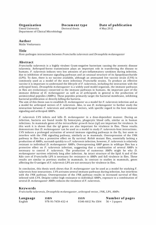
Table of Contents
Abstract………………………………………………………………………………….............................................iii
Sammanfattning på svenska……………………………………………................………..........................v
Papers in the thesis…………………………………..................……………………………….......................vii
Abbreviations……………………………………………………….…………………………………………………….viii
Introduction……………………………………………………………………………….……………………………….…1
Francisella tularensis............................................................................................................1
History.................................................................................................................1
Species and subspecies........................................................................................1
Geographical distribution...................................................................................2
Arthropod vectors...............................................................................................3
Ticks...........................................................................................................3
Mosquitoes.................................................................................................4
Disease manifestations.......................................................................................5
Virulence factors.................................................................................................5
Francisella pathogenicity island..............................................................5
Non-FPI virulence factors.........................................................................6
Lipopolysaccharide.............................................................................................7
Lipid A........................................................................................................7
Core polysaccharide..................................................................................8
O-antigen...................................................................................................8
Capsule................................................................................................................9
Intracellular growth............................................................................................9
Modulation of host signaling pathways............................................................10
Francisella – a passive pathogen?....................................................................11
Drosophila melanogaster....................................................................................................11
Model organism.................................................................................................11
Host defense......................................................................................................12
Immune signaling pathways.............................................................................13
Imd...........................................................................................................13
Toll............................................................................................................14
JNK...........................................................................................................15
JAK/STAT................................................................................................16
Antimicrobial peptides………………………………………………………………………………………..…16
Effectors of the innate immune system............................................................16
Drosophila antimicrobial peptides...................................................................17
Antimicrobial peptides in F. tularensis arthropod vectors..............................18
Antimicrobial peptides in humans...................................................................19
Aims of the thesis.......................................................................................................................20
Results and discussion..............................................................................................................21
ii
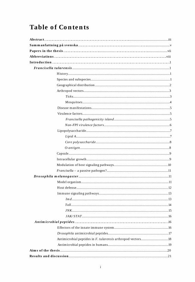
D. melanogaster as a model for F. tularensis (Paper I).............................................21
F. tularensis infects and kills D. melanogaster...............................................21
F. tularensis virulence factors are important in Drosophila………..................21
The Imd signaling pathway is involved in the defense against F. tularensis..22
The role of JNK signaling in F. tularensis infection in Drosophila
(Paper II)…………………………………………………………………………………………..……..………………22
Activation of immune signaling pathways…………………………………................23
Fat body JNK signaling is not important during LVS infection......................23
Activating JNK signaling in hemocytes prolongs fly survival after LVS
infection……………………………………………………………………………………………...24
Francisella is sensitive to insect antimicrobial peptides (Paper III)……………..…25
Francisella is sensitive to Attacin, Cecropin, Drosocin and Drosomycin.......25
The polysaccharide core structure is important for virulence in flies.............26
Modifications of the lipid A and outer membrane stability are important for
resistance to Drosophila AMPs........................................................................26
O-antigen, capsule and surface charge are not important for virulence in
Drosophila........................................................................................................27
Growth of LPS mutants in Drosophila cultured cells (Additional data)…..….27
Conclusions………………………………………………………………………………………………………………….29
Acknowledgements....................................................................................................................30
References....................................................................................................................................31
iii
Abstract
Francisella tularensis is a highly virulent Gram-negative bacterium causing the zoonotic disease tularemia. Arthropod-borne transmission plays an important role in transferring the disease to humans. F. tularensis induces very low amounts of pro-inflammatory cytokines during infection, due to inhibition of immune signaling pathways and an unusual structure of its lipopolysaccharide (LPS). To date, there is no vaccine available that is approved for public use, although an attenuated live vaccine strain (LVS) is commonly used as a model of the more infectious Francisella strains. To produce an effective vaccine it is important to understand the lifecycle of F. tularensis, including the interaction with the arthropod hosts. Drosophila melanogaster is a widely used model organism, which is increasingly being used in host-pathogen interaction studies as the immune pathways in flies are evolutionary conserved to the immune pathways in humans. An important part of the immune defense of D. melanogaster as well as of arthropods in general is the production of antimicrobial peptides. These peptides primarily target the bacterial membrane, inhibiting bacterial proliferation or directly killing the bacteria.
The aim of this thesis was to establish D. melanogaster as a model for F. tularensis infection and as a model for arthropod vectors of F. tularensis. Also, to use D. melanogaster to further study the interaction between F. tularensis and arthropod vectors, with specific regard to the host immune signaling and arthropod antimicrobial peptides.
F. tularensis LVS infects and kills D. melanogaster in a dose-dependent manner. During an infection, bacteria are found inside fly hemocytes, phagocytic blood cells, similar as in human infections. In mammals genes of the intracellular growth locus (igl) are important for virulence. In this work it is shown that the igl genes are also important for virulence in flies. These results demonstrate that D. melanogaster can be used as a model to study F. tularensis-host interactions.
LVS induces a prolonged activation of several immune signaling pathways in the fly, but seem to interfere with the JNK signaling pathway, similarly as in mammals. Overexpression of the JNK pathway in flies has a protective effect on fly survival.
Relish mutant flies, essentially lacking a production of antimicrobial peptides, succumb quickly to a F. tularensis infection, however, F. tularensis is relatively resistant to individual D. melanogaster antimicrobial peptides.

iv
Overexpressing antimicrobial peptide genes in wildtype flies has a protective effect on F. tularensis infection, suggesting that a combination of several antimicrobial peptides is necessary to control F. tularensis. The production of numerous antimicrobial peptides might be why D. melanogaster survives relatively long after infection. An intact structure of the lipid A and of the Kdo core of Francisella LPS is necessary for resistance to antimicrobial peptides and full virulence in flies. These results are similar to previous studies in mammals. In contrast to studies in mammals, genes affecting the O-antigen of F. tularensis LPS are not necessary for virulence in flies.
In conclusion, this thesis work shows that D. melanogaster can be used as a model for studying F. tularensis-host interactions. LVS activates several immune pathways during infection, but interfere with the JNK pathway. Overexpressing the JNK pathway results in increased survival of flies infected with LVS. Despite rather high resistance to individual antimicrobial peptides, exposure to a combination of several D. melanogaster antimicrobial peptides reduces the virulence of F. tularensis.
v
Sammanfattning på svenska
Francisella tularensis är en högvirulent gram-negativ bakterie som orsakar sjukdomen tularemi, även kallad harpest. En stor del av spridningen till människor sker genom bett av artropoder. F. tularensis inducerar väldigt låga nivåer av proinflammatoriska cytokiner under infektionen, på grund av inhibering av signalvägar i immunförsvaret och en ovanlig struktur på dess lipopolysackarid (LPS). Just nu finns det inget vaccin tillgängligt som är licencierat för allmän användning, även om en attenuerad levande vaccin-stam (LVS) används som en model för de mer virulenta Francisella stammarna. För att kunna tillverka ett effektivt vaccin är det viktigt att förstå F. tularensis livscykel och även interaktionerna med artropod vektorer. Drosophila melanogaster är en vanlig model organism som används mer och mer i värd-patogen interaktioner eftersom att signalvägar i immunförsvaret är evolutionärt liknande som hos människor. En viktig del av immunförsvaret i D. melanogaster och generellt hos artropoder är produktionen av antimikrobiella peptider. Dessa peptider söker sig till bakteriernas membran, där de inhiberar bakteriernas tillväxt eller direkt dödar dem.
Målet med denna avhandling var att etablera D. melanogaster som en model för F. tularensis infektioner och som en model för artropod vektorer av F. tularensis. Ett delmål var även att använda D. melanogaster för att studera interaktionerna mellan F. tularensis och artropod vektorer, med särskilt fokus på immunsignalering hos värden och antimikrobiella peptider.
F. tularensis LVS infekterar och dödar D. melanogaster på ett dosberoende sätt. Under en infektion återfinns bakterierna i hemocyter, fagocytiska blod celler, på likande sätt som i infektioner i människor. I däggdjur är igl gener viktiga för virulens. I detta arbete visas det att igl generna även är viktiga för virulens i flugor. Dessa resultat visar att D. melanogaster kan användas som en model för att studera F. tularensis-värd interaktioner.
En infektion med LVS orsakar en långvarig aktivering av flera immunsignalvägar, men verkar påverka JNK signaleringen negativt, liknande som i däggdjur. Överuttryck av JNK signalering i flugor har en positiv effekt på flugornas överlevnad.
Relish mutanta flugor, som i stort sett saknar en produktion av antimikrobiella peptider, dör snabbt på grund av en F. tulensis infektion, men F. tularensis är ganska resistent mot individuella D. melanogaster antimikrobiella peptider. Överuttryck av antimikrobiella peptider i vildtyps flugor har en positiv effekt på en F. tularensis infektion, vilket tyder på att en kombination av flera antimikrobiella peptider är nödvändigt för att kontrollera F. tularensis. Produktionen av många antimikrobiella peptider kan vara varför D. melanogaster överlever relativt länge efter infektion. En intakt struktur av lipid A och av Kdo kärnan av Francisella LPS är nödvändigt för resistens mot antimikrobiella peptider och för full virulens i

vi
flugor. Dessa resultat är liknande som tidigare studier i däggdjur. I motsats till studier i däggdjur är biosyntes gener för O-antigenen inte viktiga för virulens i flugor.
Detta avhandlingsarbete visar att D. melanogaster kan användas som en model för att studera F. tularensis-värd interaktioner. LVS aktiverar flera immunsignalvägar under infektionen, men påverkar JNK signaleringen negativt. Överuttryck av JNK signalering resulterar i ökad överlevnad av infekterade flugor. Trots resistens mot enskilda antimikrobiella peptider minskas virulensen av F. tularensis genom exponering av flera D. melanogaster antimikrobiella peptider.

vii
Papers in the thesis
I. Vonkavaara M#, Telepnev M#, Ryden P, Sjöstedt A and Stöven S Drosophila melanogaster as a model for elucidating the pathogenicity of Francisella tularensis. Cell. Microbiol. 2008; 10: 1327-1338
II. Vonkavaara M, Sjöstedt A and Stöven S
The role of JNK signaling in Francisella tularensis infection of Drosophila melanogaster. Manuscript
III. Vonkavaara M, Shaikh Terkis Islam P, Hölzl K, Nordfelth R, Sjöstedt A and Stöven S
Francisella is sensitive to insect antimicrobial peptides. Manuscript
#These authors contributed equally to the work.
Paper I was reprinted with permission from the publisher.

viii
Abbreviations
LVS Live vaccine strain
FPI Francisella pathogenicity island
T6SS Type VI secretion system
ROS Reactive oxygen species
LPS Lipopolysaccharide
TLR Toll-like receptors
NF-κB Nuclear factor-kappa B
MAPK Mitogen-activated protein kinase
AMP Antimicrobial peptide
Imd Immune deficiency
PGRP Peptidoglycan recognition protein
PGN Peptidoglycan
JNK Jun N-terminal kinase

1
Introduction Francisella tularensis History In the beginning of the 20th Century, McCoy and Chapin described a plague-like disease in ground squirrels in Tulare County, California. The disease was named tularemia. The causative agent, a small Gram-negative cocco-bacillus, was isolated and named Bacterium tularensis. Later, it was renamed Francisella tularensis to honor Edward Francis and his pioneering work on tularemia (reviewed in (Ellis et al., 2002; Foley and Nieto, 2010; Tärnvik and Berglund, 2003). F. tularensis is highly infectious; ten bacteria are sufficient to cause human disease. The high infectivity and mortality, together with the ease of aerosol dispersal, has lead the U.S. Centers for Disease Control and Prevention (CDC) to classify F. tularensis as a category A agent, that is, considered as a high risk biological threat agent. F. tularensis has been a subject of military research in both the United States and former Soviet Union for the same reasons. In the beginning and middle of the last century, the prevalence of tularemia was much higher than now. As an example, during the 1940’s, there were about 100,000 cases reported annually in the former Soviet Union, compared to a few hundred annual cases in the 1990s. This can be explained by improved sanitary conditions and reduced exposure to risk factors (such as rabbit hunting) (reviewed in (Sjöstedt, 2007). In Sweden, the number of tularemia cases has varied between 160-700 cases per year during the last decade (www.smittskyddsinstitutet.se/in-english/statistics/tularaemia). Species and subspecies The genus Francisella comprises two species that are well studied; F. tularensis and F. novicida. Two subspecies of F. tularensis are of clinical importance; subspecies tularensis (type A) and subspecies holarctica (type B), while a third subspecies, mediasiatica rarely causes human disease (reviewed in (Oyston, 2008). A live vaccine strain, LVS, derived from the holarctica subspecies was developed in the middle of the 20th Century, by repeated passage on solid media. Although used to vaccinate at-risk personnel, LVS is not licensed to be used as a public vaccine, since its mechanism of attenuation is not fully understood (reviewed in (Oyston, 2009). Since the LVS is still virulent in mice, it is often used as a model to study the more virulent F. tularensis strains. F. novicida only causes disease in immunocompromised individuals, but causes a tularemia-like disease in smaller mammals and is a widely used model for F. tularensis. There is an ongoing debate as to reclassify F. novicida as a subspecies of F. tularensis based on its DNA sequence similarity (97% identity) and biochemical characteristics (Huber et al., 2010).

2
Similar to F. novicida, F. philomiragia is less pathogenic than F. tularensis, and only causes disease in immunocompromised individuals (reviewed in (Keim et al., 2007). The genus Francisella continuously increases as more isolates are sequenced and identified. Recently, the fish-pathogen F. noatunensis was described as well as a mollusc pathogenic Francisella, F. halioticida (Brevik et al., 2011; Ottem et al., 2009). Geographical distribution Tularemia occurs all over the Northern Hemisphere, most frequently in Scandinavia, Northern America, Russia and Japan (Fig. 1). F. tularensis tularensis is limited to North America, while F. tularensis holarctica is widespread over the entire Northern hemisphere and often associated with water. F. tularensis mediasiatica is only known in central Asian regions of the former Soviet Union. F. novicida is thought to be spread over the northern hemisphere but is rarely isolated, F. novicida-like strains have also been isolated in Australia (Fig. 1) (reviewed in (Sjöstedt, 2007)) (Whipp et al., 2003).
Fig. 1 Geographical distribution of the three F. tularensis subspecies and F. novicida. The regions where Francisella have been isolated are shown in red. Adapted from (Oyston et al., 2004) and reprinted with permission from the publisher.
F. tularensis is thought to be maintained in nature by small mammals such as rodents and lagomorphs, as well as by ticks (Fig. 2). The bacteria have

3
been identified in as many as 250 different species of animals. In the United States, Sweden, Finland and Russia, arthropod bite is a common mode of transmission of tularemia. In contrast, in central Europe the disease is most commonly acquired by handling infected animals or by ingestion of contaminated food or water (reviewed in (Ellis et al., 2002; Foley and Nieto, 2010; Tärnvik and Berglund, 2003).
Fig. 2 Schematic picture showing the lifecycle of F. tularensis type A (subspecies tularensis) and type B (subspecies holarctica). Type A tularemia is transmitted by ticks (United States) and tabanid flies (western United States) and has a terrestrial lifecycle, the main reservoirs are cottontail rabbits and ticks. Type B tularemia is transmitted by blood-feeding mosquitoes (Sweden, Finland and Russia), ticks (United States) and tabanid flies (western United States and Russia) and has an aquatic lifecycle; in addition to residing in aquatic environments, reservoirs are semi-aquatic rodents, such as muskrats, beaver and ground voles.
Arthropod vectors Arthropods are an important part of the infectious lifecycle of Francisella (Fig. 2). Francisella infections have been documented in several different arthropods, including fleas, lice, midges, bedbugs, ticks, mosquitoes and flies. Only some of these (ticks, mosquitoes, deer flies and horse flies) have been identified as important for transmitting tularemia to humans (reviewed in (Petersen et al., 2009). Ticks In the United States, tick bite is one of the predominant modes of transmission of F. tularensis (Fig. 2). Ticks acquire F. tularensis while feeding on an infected animal. From the tick midgut the bacteria enter the hemolymph and migrate to the salivary glands, from where they are inoculated into the next host. Ticks are biological vectors of tularemia, they are able to carry the bacteria for long periods of time in addition to

4
transmitting the disease between hosts (reviewed in (Petersen et al., 2009). Ticks show different susceptibility to F. tularensis. Some tick species do not successfully molt when infected with F. tularensis, while infection in other species has no effect on survival (reviewed in (Foley and Nieto, 2010). It has been suggested that the geographical range of tick species contributes to the epidemiology of tick-borne tularemia. The tick species associated with human transmission in the United States, Dermacentor andersoni, D. variabilis and Amblyomma americanum, are not found outside North America. Although F. tularensis has been detected in European Dermacentor, these species feed on animals rather than humans (de Carvalho et al., 2007; Hubalek et al., 1998) and reviewed in (Petersen et al., 2009). Mosquitoes In contrast to ticks, which are biological vectors of tularemia, mosquitoes and biting flies are considered as mechanical vectors, carrying Francisella on their surface and not internally in hemolymph and salivary glands as in ticks. Mechanical transfer can be mediated by contaminated mouthparts, excrement deposited during feeding or crushing of the infected mosquito on the skin allowing for transfer, especially if followed by scratching (reviewed in (Petersen et al., 2009). Some of the largest epidemics of tularemia reported (>400 cases in Sweden) have been linked to mosquitoes (Christenson, 1984; Eliasson et al., 2002). In Sweden, it has been shown that a high prevalence of mosquitoes are a requirement for tularemia outbreaks in endemic areas (Ryden et al., 2012) (Fig. 2). F. tularensis holarctica can persist in aquatic environments during outbreaks, and in association with protozoa (Abd et al., 2003; Broman et al., 2011; Thelaus et al., 2009). LVS has also been shown to form biofilms in simulated natural waters (Mahajan et al., 2011). Studies suggest that mosquitoes come into contact with F. tularensis as water-living larvae, by feeding on infected protozoa or by directly feeding on F. tularensis residing in water (Lundström et al., 2011; Mahajan et al., 2011; Triebenbach et al., 2010) . F. tularensis can then be transstadially transmitted to host-seeking adult female mosquitoes (Lundström et al., 2011). In another study using F. novicida-fed larvae, bacteria could not be detected in pupae or adults and the survival of the larvae was not affected (Triebenbach et al., 2010). A third study found that larvae are sensitive to feeding on LVS, feeding leads to mortality and lengthened time to pupation (Mahajan et al., 2011). All three studies used different species of mosquitoes, suggesting a difference in susceptibility between mosquito species, similarly as in ticks. It is also possible that the infection methods and Francisella species affect the outcome. After feeding, LVS is localized intracellularly in the midgut and Malphigian tubule cells, suggesting translocation to the hemocoel of the larvae (Mahajan et al., 2011). One study has attempted to infect mice by allowing pre-infected mosquitoes to feed on them, but this was not successful (Triebenbach et al., 2010). More studies are needed though, to be able to draw accurate conclusions as how mosquitoes interact with Francisella and spread the bacteria to humans.

5
Disease manifestations F. tularensis is a facultative intracellular bacterium. The primary target cells are macrophages, but the bacteria can also infect and survive in neutrophils, dendritic cells, epithelial cells and hepatocytes (Ben Nasr et al., 2006; Conlan and North, 1992; Hall et al., 2007; McCaffrey and Allen, 2006), the intracellular lifestyle of Francisella will be described in more detail on page 9. The disease manifestations depend on the route of infection. Ulceroglandular tularemia is by far the most common form of the disease. It is often caused by vector borne-transmission, or by contact with an infected animal. An ulcer develops at the site of infection, later on, the lymph node draining the ulcer enlarges and becomes tender. From the lymph nodes the bacteria can disseminate to other tissues, such as the liver and spleen. Glandular tularemia is similar to ulceroglandular tularemia, but the local infection is not noticeable. Ingestion of contaminated food or water can lead to oropharyngeal tularemia, where the ulcer is located in the mouth. Respiratory tularemia is caused by inhalation of aerosolized F. tularensis. General symptoms of tularemia include high fever, headache, fatigue and general body ache. Ulceroglandular and glandular tularemia are rarely fatal, but it may require significant time to recover from the disease. The infection can also progress to septicemia, septicemic meningitis or pneumonia. F. tularensis tularensis causes the most severe form of tularemia with a mortality rate of up to 30% in humans if left untreated. With proper antibiotic treatment the mortality rate drops to 1-2%. The subspecies holarctica causes a milder form of the disease and is rarely lethal (reviewed in (Oyston, 2008; Oyston et al., 2004; Sjöstedt, 2007). Virulence factors Francisella pathogenicity island The Francisella pathogenicity island (FPI) is a gene cluster that is often detected in screens for attenuated virulence and/or defective intracellular growth. The FPI consists of 16-19 genes and its importance for virulence is well established (Nano et al., 2004) and (reviewed in (Bröms et al., 2010; Nano and Schmerk, 2007). The FPI is present in all Francisella genomes, the region is duplicated in all F. tularensis subspecies, but present as a single copy in F. novicida and F. philomiragia (Larsson et al., 2009; Nano et al., 2004). Several genes of the FPI show homology to Type VI secretion system (T6SS) components, important for virulence in other pathogenic bacteria (reviewed in (Jani and Cotter, 2010). However, the exact function of any of the genes in the FPI is not known.

6
Non-FPI virulence factors In addition to the genes of the FPI, several other genes important for virulence have been identified. During infection, Francisella resides inside macrophages, a hostile environment where reactive oxygen species (ROS) and reactive nitrogen species (RNS) are generated after phagocytosis of pathogens, causing oxidative damage to DNA, RNA, proteins and lipids. Francisella encodes two superoxide dismutases (SODs), sodB and sodC, that confer protection to host-derived oxidants and contribute to intramacrophage survival (Bakshi et al., 2006; Melillo et al., 2009). Francisella also encodes a catalase (katG), which detoxifies RNS. A katG mutant is attenuated in LVS, but virulent Francisella tularensis also have katG-independent mechanisms to detoxify RNS (Lindgren et al., 2007). Iron is essential for growth of essentially all bacteria, and Francisella has developed means for acquiring iron from the host during an infection. A siderophore locus (fsl) is necessary for synthesis, export and uptake of a Francisella siderophore, which sequesters iron from the host. Most genes involved in iron uptake, including fsl and feoB (iron transport protein), are under negative control of the ferric iron uptake regulator A (FurA) (Honn et al., 2012; Ramakrishnan et al., 2008; Sullivan et al., 2006). The role of type IV pili in virulence and transmission of tularemia is not clear. pilA mutants are attenuated in mice, they are unable to properly spread from the initial site of infection to the spleen, but they are not defective in macrophage survival (Forslund et al., 2006). In addition to these virulence factors, the Francisella lipopolysaccharide (LPS) and capsule are structures important for virulence, both of these will be described on pages 7-9. Several of the virulence mechanisms used by Francisella are also used by other pathogenic bacteria. T6SS, SODs, catalases, iron sequestering, pili, LPS and capsule are all known virulence mechanisms of other bacteria (reviewed in (Battistoni, 2003; Cornelis et al., 2011; Jani and Cotter, 2010; Livorsi et al., 2011). However, the effector molecules of the Francisella T6SS and the molecular mechanism of its escape from the phagosome are not known. Effector molecules used by other intracellular bacteria to escape the phagosome such as cytolysins, pore-forming toxins or hydrolytic enzymes have not been found in Francisella, suggesting a novel mechanism for escape (reviewed in (Ray et al., 2009). Importantly, the modulation of host immune signaling is not known either (discussed in more detail on pages 10-11).

7
Lipopolysaccharide LPS is the primary component of the outer membrane of Gram-negative bacteria. LPS contributes to the structural integrity of the membrane and also has a key role in immune stimulation. LPS is an endotoxin and is detected by host pattern recognition receptors such as Toll-like receptors (TLRs), inducing a strong immune response. The LPS structure includes lipid A, a polysaccharide core and the O-antigen.
Fig. 3 Schematic drawing of Francisella lipopolysaccharide (LPS). The core structure of F. tularensis lacks the glucose residue attached to N-acetyl galactosamine. The left drawing shows the free lipid A of F. novicida.
Lipid A Lipid A anchors the LPS structure in the membrane and is the biologically active component of LPS. The classical biphosphorylated, hexa-acylated lipid A of E. coli activates human TLR4 and induces the production of pro-inflammatory cytokines (reviewed in (Rietschel et al., 1994). Francisella lipid A differs from the lipid A found in most Gram-negative bacteria (Fig. 3). It is tetra-acylated and lacks the 4´-phosphate group, resulting in an unusually weak endotoxic activity of the LPS (Sandström et al., 1992). The phosphatase LpxF is responsible for removing the 4´-phosphate, a lpxF mutant also retains the 3´- acyl chain and is highly attenuated in mice (Wang et al., 2007). The lpxF mutant induces a higher proinflammatory response, however, no TNFα response is evoked, indicating that TLR4 is not activated. The lpxF mutant is hypersensitive to the antibiotic polymyxin,

8
suggesting that the mutant bacteria are more sensitive to host immune defenses than wildtype Francisella. Membrane damages might expose microbial ligands that are recognized by other pattern recognition receptors, such as bacterial lipoproteins or peptidoglycan by TLR2 or bacterial DNA by TLR9 (Wang et al., 2007). Francisella lipid A is further modified by the addition of two carbohydrate moieties, mannose and galactosamine. Two enzymes, FlmF1 and FlmF2 (Francisella lipid A modification) are involved in the transfer of mannose or galactosamine, respectively, to a carrier lipid required for the transport of water soluble precursors across a lipid membrane. A third protein, FlmK, transfers the individual carbohydrate moieties from the carrier lipid to lipid A (Kanistanon et al., 2008). Mutants in flmF2 and flmK are attenuated in mice, and the flmK mutant stimulates an increased immune response in macrophages, dependent on MyD88, suggesting that the flmK mutant is more readily recognized by the TLR than wildtype Francisella. All three flm genes are highly homologous in F. novicida and F. tularensis, suggesting a conserved function (Kanistanon et al., 2008) and reviewed in (Gunn and Ernst, 2007). Most of the lipid A in Francisella is present in “free” form, not covalently linked to LPS and thus lacking the usual Kdo, core and O-antigen (Fig. 3). Other well-characterized Gram-negative bacteria do not have any “free” lipid A, and the significance of this feature in Francisella is not known (Wang et al., 2006). Polysaccharide core The core region of Francisella LPS is linked to lipid A through the eight-carbon sugar, 3-deoxy-D-manno-octulosonic acid (Kdo). The core structures of F. tularensis and F. novicida LPS are almost identical, differing only in an additional glucose residue in F. novicida (Fig. 3). The core region lacks phosphate modifications and contains a single Kdo residue (reviewed in (Gunn and Ernst, 2007). F. novicida manB and manC mutants lack the O-antigen and have a defective core structure. This indicates that the ManB/C proteins are required in the mannose pathway. Without mannose in the core structure the O-antigen is not added to the LPS structure and the Kdo is the outermost polysaccharide. The F. novicida manB mutant is hypercytotoxic to macrophages and attenuated in mice, presumably because the mutant destroys its replicative niche (Lai et al., 2010). O-antigen The O-antigen is the outermost domain of non-encapsulated Gram-negative bacteria consisting of a number of repeats of an oligosaccharide, the O-unit. O-antigens are extremely variable, and the structure differs from strain to strain. The O-antigen repeat in Francisella consists of four sugar residues and is synthesized by the genes of the wbt locus (Fig. 3). The O-antigens of F. tularensis and F. novicida are structurally distinct and differ in the two

9
outermost sugar residues (Vinogradov et al., 2004). The differences in O-antigen structure are too great to confer cross-protection in immunized mice (Thomas et al., 2007). A wbtA mutant lacks the O-antigen and LVS wbtA mutants are extremely sensitive to human serum, in contrast to wildtype Francisella, which is serum resistant. Further, LVS wbtA mutants are attenuated in mice and fail to replicate intracellularly in macrophages (Raynaud et al., 2007; Sebastian et al., 2007). In contrast, a F novicida wbtA mutant grows normally intracellularly in macrophages and is only slightly serum sensitive (Cowley et al., 2000). Wildtype Francisella is resistant to complement-mediated lysis because of negative regulation of the complement activity on the surface of the bacteria, leading to opsonophagocytosis of bound Francisella instead of bacterial lysis. Mutant Francisella lacking the O-antigen are unable to negatively regulate the complement pathway, leading to initiation of the lytic pathway and killing of mutant bacteria (Clay et al., 2008). Capsule Francisella has long been considered encapsulated based on the appearance of rough and smooth colony phenotype, but until recently, no capsular preparation has been described. In 2010 , capsular material was isolated and structurally determined (Apicella et al., 2010). It consisted of tetrasaccharide repeats of identical structure and composition as the LPS O-antigen, but it is a separate entity. This O-antigen capsule was found in Francisella tularensis subspecies tularensis and subspecies holarctica. The LPS O-antigen wbt gene cluster plays a role in the biosynthesis of the capsule (Apicella et al., 2010). F. tularensis LVS adapts to the intracellular environment in host macrophages by expressing more capsule, thereby reducing the accessibility of the host immune response to bacterial outer membrane constituents. In addition to producing more O-antigen capsule, host-adapted bacteria also produce additional high molecular weight capsule-like material (Zarrella et al., 2011). Intracellular growth After phagocytosis by macrophages, the Francisella containing phagosome does not fuse with the lysosome, genes of the FPI are important for the modulation of the phagosome. This is likely mediated by type VI secretion of bacterial effector protein(s) (reviewed in (Bröms et al., 2010; Ray et al., 2009). Francisella rapidly escapes from the phagosome and replicates in the host cell cytoplasm (Bönquist et al., 2008; Clemens et al., 2004; Santic et al., 2005a; Santic et al., 2005b). Despite host cell immune responses, Francisella-infected macrophages eventually die by apoptosis, releasing the bacteria to infect new cells (Lai and Sjöstedt, 2003). Interestingly, the survival inside amoebae is also dependent on genes of the FPI (Lauriano et al., 2004). Thus, the strategy of Francisella to survive intracellularly seems to be an ancient mechanism.

10
Modulation of host signaling pathways F. tularensis has been called a “stealth pathogen”, due to the low inflammatory response during an infection (reviewed in (Sjöstedt, 2006). Proinflammatory cytokines such as TNF, IL-12 and IFN-γ are crucial for effective immunity to and survival of F. tularensis infection (Duckett et al., 2005; Leiby et al., 1992), however, Francisella actively suppresses the expression of the cytokines (Bosio et al., 2007; Mares et al., 2008; Telepnev et al., 2005). By targeting and suppressing the expression of proinflammatory cytokines early in infection, F. tularensis is able to replicate and disseminate within the host. LVS downregulates several TLR in monocytes (Telepnev et al., 2003) and induces a transient activation of NF-κB and the MAPK pathways components c-Jun and p38, which are important for expression of proinflammatory cytokines, but rapidly inhibits these signaling pathways (Fig. 4) (Moresco et al., 2011; Telepnev et al., 2005). F. novicida also impairs the PI3K-mediated and IFN-γ-mediated signaling (Parsa et al., 2008; Parsa et al., 2006). The modulation of host cell signaling and suppression of proinflammatory cytokines is dependent on phagosomal escape of Francisella, and is thus dependent on the FPI (Rajaram et al., 2006; Telepnev et al., 2005).
Fig. 4 Signals triggered by cytosolic Francisella. Francisella escapes the phagosome and replicates in the cytosol of the host cell, inhibiting host immune signaling. Infection eventually leads to apoptosis of the infected cell. For details see text.
The proinflammatory response to LVS in monocytic cells is TLR2-dependent, and LVS colocalizes with TLR2 and MyD88 inside macrophages
11
(Cole et al., 2007). F. novicida infection also activates SHIP early in infection, which negatively regulates activation of PI3K, Akt and subsequently NF-κB, leading to down-regulation of the proinflammatory cytokine production (Parsa et al., 2006; Rajaram et al., 2006). The activation of SHIP did not regulate F. novicida induced activation of MAPKs (Fig. 4) (Parsa et al., 2006). Somewhat contradictory, LVS activates PI3K, leading to expression of MKP-1, a negative regulator of MAPK, thereby LVS may suppress p38 MAPK activation in macrophages (Fig. 4) (Medina et al., 2010). These steps of modulation of host signaling pathways take place early in infection, within the first hour. LVS induce ERK1/2 MAPK activation for up to 6 hours, this together with the down-regulation of p38 MAPK, result in an imbalance in cell signaling leading to apoptosis of the host cell (Hrstka et al., 2005). LVS also induces apoptosis of host cells by activating caspase-3 (Fig. 4) (Lai and Sjöstedt, 2003). Even though caspase-3 is activated early in infection, Francisella delays cell death, thus the bacterium can use the cell for its growth and survival. F. novicida trigger rapid and temporal activation of Ras within 15 minutes after ingestion, resulting in down-regulation of caspase-3, this is important for successful cytosolic replication (Fig. 4) (Al-Khodor and Abu Kwaik, 2010). All of these results indicate that the modulation of host cell signaling by Francisella is complicated and acts on several pathways. In the end, the delicate balance between the different signals determines the outcome. The interaction of Francisella with immune signaling pathways of arthropod vectors has not been studied thus far, despite increased understanding of the importance of Francisella-vector interactions, exemplified in reviewes by (Akimana and Kwaik, 2011; Hazlett and Cirillo, 2009; Petersen et al., 2009).
Francisella – a passive pathogen? It is interesting that the virulence mechanisms of Francisella seem to be directed at avoiding the host immune response, rather than actively causing harm to the host. Francisella surface structures (LPS, capsule) are not recognized by host pathogen recognition receptors (reviewed in (Gunn and Ernst, 2007) and once inside the host cell Francisella down-regulates the host immune signaling, further avoiding recognition (Telepnev et al., 2003). Francisella does not secrete any known toxins (reviewed in (Ray et al., 2009), but is still able to successfully colonize a wide variety of hosts. A large number of close relatives to Francisella exists which are difficult to isolate and live as endosymbionts or parasites in ticks and amoeba. These strains have properties which are consistent with adaptation to an intrabiotic or intracellular lifestyle. The Francisella species which are mostly studied (F. tularensis and F. novicida) are difficult to isolate from nature, they have reduced genomes and fastidious growth requirements. Their many defective biochemical pathways are also consistent with adaptation to an intrabiotic environment (reviewed in (Keim et al., 2007). The ongoing genome reduction of F. tularensis suggests that it might be on the way of becoming an obligate intracellular symbiont or pathogen (Larsson et al., 2005).
12
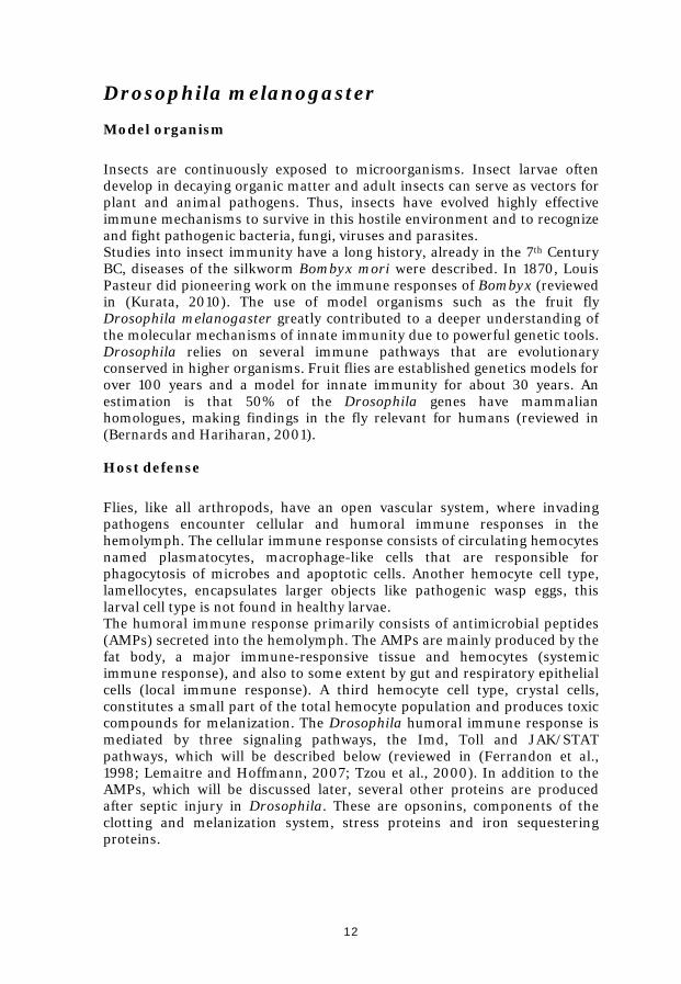
Drosophila melanogaster Model organism Insects are continuously exposed to microorganisms. Insect larvae often develop in decaying organic matter and adult insects can serve as vectors for plant and animal pathogens. Thus, insects have evolved highly effective immune mechanisms to survive in this hostile environment and to recognize and fight pathogenic bacteria, fungi, viruses and parasites. Studies into insect immunity have a long history, already in the 7th Century BC, diseases of the silkworm Bombyx mori were described. In 1870, Louis Pasteur did pioneering work on the immune responses of Bombyx (reviewed in (Kurata, 2010). The use of model organisms such as the fruit fly Drosophila melanogaster greatly contributed to a deeper understanding of the molecular mechanisms of innate immunity due to powerful genetic tools. Drosophila relies on several immune pathways that are evolutionary conserved in higher organisms. Fruit flies are established genetics models for over 100 years and a model for innate immunity for about 30 years. An estimation is that 50% of the Drosophila genes have mammalian homologues, making findings in the fly relevant for humans (reviewed in (Bernards and Hariharan, 2001). Host defense Flies, like all arthropods, have an open vascular system, where invading pathogens encounter cellular and humoral immune responses in the hemolymph. The cellular immune response consists of circulating hemocytes named plasmatocytes, macrophage-like cells that are responsible for phagocytosis of microbes and apoptotic cells. Another hemocyte cell type, lamellocytes, encapsulates larger objects like pathogenic wasp eggs, this larval cell type is not found in healthy larvae. The humoral immune response primarily consists of antimicrobial peptides (AMPs) secreted into the hemolymph. The AMPs are mainly produced by the fat body, a major immune-responsive tissue and hemocytes (systemic immune response), and also to some extent by gut and respiratory epithelial cells (local immune response). A third hemocyte cell type, crystal cells, constitutes a small part of the total hemocyte population and produces toxic compounds for melanization. The Drosophila humoral immune response is mediated by three signaling pathways, the Imd, Toll and JAK/STAT pathways, which will be described below (reviewed in (Ferrandon et al., 1998; Lemaitre and Hoffmann, 2007; Tzou et al., 2000). In addition to the AMPs, which will be discussed later, several other proteins are produced after septic injury in Drosophila. These are opsonins, components of the clotting and melanization system, stress proteins and iron sequestering proteins.

13
Immune signaling pathways Imd The Imd pathway was defined by a mutation named immune deficiency (imd) that impaired the expression of several AMPs in response to infection. It is homologous to the mammalian tumor necrosis factor receptor 1 (TNFR1) signaling pathway and is primarily activated by Gram-negative bacteria. The Imd pathway regulates both systemic (fat body) and local (gut epithelial cells) AMP expression. The transmembrane receptor peptidoglycan recognition protein-LC (PGRP-LC) is activated by bacterial meso-diaminopimelic (DAP)-type peptidoglycan (PGN) (mainly Gram-negative PGN), leading to recruitment of the adaptor Imd, a homolog to the human RIP (Fig. 5). Imd interacts with dFADD and the caspase Dredd (caspase-8 homolog). Dredd-dependent cleavage of Imd exposes a binding site for dIAP2. Imd-dIAP2 association leads to ubiquitination of Imd, producing a scaffold for the recruitment of dTAK,
Fig. 5 Drosophila Imd pathway with the JNK branch. Gram-negative peptidoglycan (PGN) are recognized by the transmembrane receptor PGRP-LC or the cytosolic PGRP-LE. Signaling proceeds through Imd, dFADD and the caspase Dredd. Imd is cleaved and binds dIAP2, resulting in activation of the dTAK1 kinase. dTAK1 activates both the IKK complex and the JNKK Hemipterous (Hep). Relish is phosphorylated by IKK and cleaved to release the active Rel-domain, which translocates into the nucleus and initiates transcription of AMP genes and other targets. The simultaneous activation of Basket (Bsk) leads to phosphorylation and nuclear translocation of AP-1. Mammalian homologues of pathway components are shown in blue. Negative regulators are not shown.

14
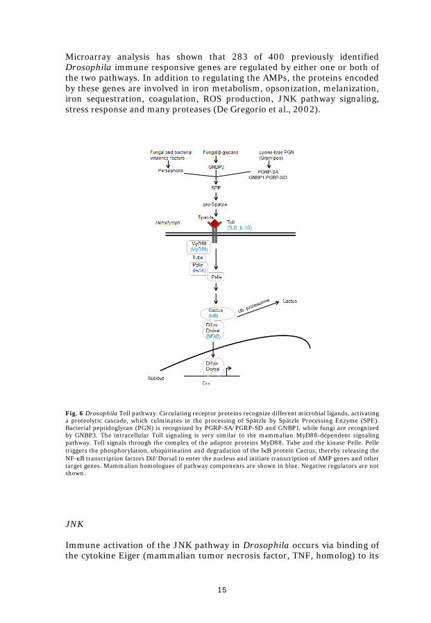
which becomes activated by TAB2. dTAK1 phosphorylates the ΙκΒ-kinase complex (IKKβ/Ird5 and IKKγ/Kenny), which in turn phosphorylates the NF-κB protein Relish. The phosphorylated Relish is cleaved, possibly by Dredd, separating the inhibitory ankyrin domain from the Rel-homology domain. The phosphorylated Rel domain translocates into the nucleus where it activates the transcription of Imd regulated genes. The Imd pathway can also be activated by the intracellular receptor PGRP-LE (reviewed in (Ganesan et al., 2011; Lemaitre and Hoffmann, 2007). Activated dTAK1 can also activate the JNK pathway, which will be discussed on pages 15-16. Toll The Toll pathway was originally discovered in screens for genes involved in embryonic dorsal/ventral polarity in Drosophila. Toll is a transmembrane receptor, which is activated in response to fungi, Gram-positive bacteria or yeast. The Toll pathway is similar to the signaling cascades downstream of mammalian IL-1R and TLR, pointing to a common ancestry of these immune pathways (Fig. 6) (reviewed in (Lemaitre and Hoffmann, 2007). Drosophila Toll is activated by the cytokine Spätzle in response to lysine-type PGN (mainly Gram-positive PGN), fungal β-glucans or virulence-associated proteases. Lys-type PGN is recognized by the circulating receptor proteins PGRP-SA and PGRP-SD, and the Gram-negative binding protein 1 (GNBP1). A proteolytic cascade is initiated, culminating in the activation of the Spätzle processing enzyme (SPE), which cleaves pro-Spätzle, releasing the active Toll ligand, Spätzle. Spätzle binds Toll, inducing receptor homodimerization and activates the intracellular signaling. The signal then goes through the MyD88 and Tube adaptors, the kinase Pelle (homolog to IRAK), leading to phosphorylation and degradation of Cactus, which is homologous to ΙκΒ. A transcription factor, homo- or heterodimer of the NF-κB proteins Dif and Dorsal, is no longer held in the cytoplasm but move into the nucleus and initiate the expression of downstream target genes, such as Drosomycin. It is unclear if Pelle directly phosphorylates Cactus, or if other intermediate kinase(s) are involved (reviewed in (Ganesan et al., 2011; Lemaitre and Hoffmann, 2007).
Drosophila NF-κB pathways show striking similarities, as well as differences, compared to the mammalian pathways. Mammalian cells sense microbes by TLRs whereas recognition of pathogens in flies is dependent on PGRPs. Human TLRs directly recognizes microbial ligands such as LPS, lipopeptides, flagellin and nucleic acids, while Drosophila Toll is a cytokine receptor, responding to microbial ligands indirectly. Drosophila and humans both use the conserved NF-κB pathway for similar responses in the rapid response to infection. However, Drosophila also uses the Toll signaling to establish the dorsal/ventral axis in the embryo, while humans use the NF-κB cascade in numerous situations, including regulation of apoptosis and oncogenesis (reviewed in (Ganesan et al., 2011).
15
Microarray analysis has shown that 283 of 400 previously identified Drosophila immune responsive genes are regulated by either one or both of the two pathways. In addition to regulating the AMPs, the proteins encoded by these genes are involved in iron metabolism, opsonization, melanization, iron sequestration, coagulation, ROS production, JNK pathway signaling, stress response and many proteases (De Gregorio et al., 2002).
Fig. 6 Drosophila Toll pathway. Circulating receptor proteins recognize different microbial ligands, activating a proteolytic cascade, which culminates in the processing of Spätzle by Spätzle Processing Enzyme (SPE). Bacterial peptidoglycan (PGN) is recognized by PGRP-SA/PGRP-SD and GNBP1, while fungi are recognized by GNBP3. The intracellular Toll signaling is very similar to the mammalian MyD88-dependent signaling pathway. Toll signals through the complex of the adaptor proteins MyD88, Tube and the kinase Pelle. Pelle triggers the phosphorylation, ubiquitination and degradation of the ΙκΒ protein Cactus, thereby releasing the NF-κB transcription factors Dif/Dorsal to enter the nucleus and initiate transcription of AMP genes and other target genes. Mammalian homologues of pathway components are shown in blue. Negative regulators are not shown.
JNK Immune activation of the JNK pathway in Drosophila occurs via binding of the cytokine Eiger (mammalian tumor necrosis factor, TNF, homolog) to its

16
receptor Wengen (mammalian TNF receptor homolog), or via the Imd pathway at the level of dTak1 (Fig. 5). The JNK pathway is involved in embryonic development, metamorphosis and mediates apoptosis, wound healing and immunity. The signaling downstream of Wengen involves the activation of one or more JNK kinase kinases (JNKKK), but the mechanisms are poorly understood. The JNKKK dTak1 activates the JNKK Hemipterous (mammalian MKK7 homolog), which in turn activates Basket (mammalian JNK homolog). Activated Basket can then activate transcription factors like AP-1 (Jun-related antigen, Jra, and Kayak, Kay) (reviewed in (Lemaitre and Hoffmann, 2007). JNK signaling in response to infection in Drosophila can be either beneficial or detrimental. Eiger null mutants are more susceptible than wildtype flies to killing by some pathogens, e.g. P. gingivalis and S. aureus, but are more resistant than wildtype flies to killing by some intracellular pathogens, e.g. S. typhimurium (reviewed in (Igboin et al., 2012). Eiger has been suggested to activate the cellular immune response to aid in the clearance of extracellular bacterial pathogens (Schneider et al., 2007). JAK/STAT The Drosophila JAK/STAT signaling pathway comprises of the same signaling components as the mammalian pathway. The contribution of the JAK/STAT pathway to immune regulation has not been studied as well as the Imd and Toll pathways. Similar to the Toll pathway, the JAK/STAT pathway is involved in several developmental events including hematopoiesis, and regulates the cellular immune response (Krzemien et al., 2007). A subset of the immune responsive genes are regulated by the JAK/STAT pathway, including the complement-like protein Tep2 (discussed more in results and discussion) and the Turandot stress genes. JAK/STAT deficient flies are as resistant to bacterial and fungal infection as wildtype flies, but are sensitive to infection with the Drosophila C virus. The JAK/STAT pathway is also involved in the immune response in the Drosophila gut, where it helps to maintain epithelial cell homeostasis, by regulating stem cell proliferation (reviewed in (Lemaitre and Hoffmann, 2007). Antimicrobial peptides Effectors of the innate immune system Antimicrobial peptides (AMPs) are key elements of the innate immunity, found throughout the animal and plant kingdom and are despite their ancient lineage, still effective weapons against bacteria and fungi. AMPs have also been shown to kill viruses, parasitic protozoa and even cancer cells (Barlow et al., 2011; Ceron et al., 2010; Howell et al., 2006; Hu and Aksoy, 2005). The diversity of sequences of AMPs is such that the same peptide sequence is rarely recovered from two different species of animals, even those closely related (reviewed in (Zasloff, 2002). The Antimicrobial Peptide

17
Database (http://aps.unmc.edu/AP/main.php) contains about 1900 sequences of peptides with proven antimicrobial activity. The AMPs primarily target the microbial membrane. The peptides electrostatically interact with negative charges on the membrane, thereafter they integrate into the membrane via various mechanisms, leading to destruction of the transmembrane potential and disruption of the permeability barrier (reviewed in (Devine and Hancock, 2002). AMPs can be classified into three major classes based on their structural features: i) linear α–helical peptides like insect Cecropins or human LL-37, ii) peptides with three or four disulphide bridges like various types of Defensins, and iii) peptides rich in proline and/or glycine like insect Attacins and Drosocins (reviewed in (Boman, 2003). In contrast to conventional antibiotics such as penicillin, acquisition of resistance to AMPs is surprisingly rare among microbes. Resistance may be acquired by altering the surface charge of the bacterium by modifying teichoic acids, lipid A or phospholipids to prevent peptide-binding (Salmonella, Morganella and Serratia) or by secreting digestive proteases that destroy the AMPs (Porphyromonas) (reviewed in (Koprivnjak and Peschel, 2011; Zasloff, 2002). AMPs are being investigated as novel therapeutic compounds, with increasing interest as multi-drug resistant pathogens continue to increase (Afacan et al., 2012; Reddy et al., 2004). Drosophila antimicrobial peptides In D. melanogaster AMPs are a key feature of the systemic immune response. The peptides are produced mainly by the fat body, and secreted into the hemolymph where they can reach a total concentration of 200 µM (reviewed in (Bulet et al., 1999). There are about 20 immune inducible AMPs that can be grouped into seven different types in D. melanogaster (reviewed in (Hetru et al., 2003; Lemaitre and Hoffmann, 2007; Naitza and Ligoxygakis, 2004), several of the peptides have been shown to have a synergistic function (Rabel et al., 2004; Tzou et al., 2002). The first AMP isolated was Cecropin (Hultmark et al., 1980). Active Cecropins are 35 to 40 amino acid long peptides belonging to the linear α –helical peptides, and they bind to the bacterial membrane, leading to membrane destruction (De Gregorio et al., 2002). Cecropins show a broad spectrum of activity against both Gram-negative and Gram-positive bacteria as well as fungi and viruses (Ekengren and Hultmark, 1999; Samakovlis et al., 1990; Wachinger et al., 1998). Defensin and Drosomycin belong to the second class of AMPs; peptides with several disulphide bridges. Insect Defensins are characterized by a six cysteine/three disulphide bridge pattern. The Defensins are 36 to 46 amino acids long and mostly active against Gram-positive bacteria. Defensins are the only class of AMPs that are conserved among all arthropods (Rodriguez de la Vega and Possani, 2005). Defensin forms channels in the bacterial membrane, leading to depolarization of the membrane and loss of cytoplasmic ATP and finally an inhibition of the respiration (Cociancich et al., 1993). Drosomycin is a 44-residue antifungal and antiparasitic peptide which shows high homology to

18
plant Defensins, the peptide acts on the cytoplasmic membrane causing lysis (Fehlbaum et al., 1994; Tian et al., 2008). To the third class of peptides belong Attacin, Drocosin and Diptericin. Some of these peptides have been shown to interfere with intracellular bacterial components in addition to the interaction with the bacterial membrane. Attacins, large glycine-rich peptides of approximately 200 amino acids, bind LPS via electrostatic interactions with the inner core and lipid A region of the LPS. This results in a partial integration of Attacin into the outer membrane and inhibition of the synthesis of outer membrane proteins, increased LPS synthesis, induction of stress proteins and finally death of the bacteria (Carlsson et al., 1991; Carlsson et al., 1998; Engström et al., 1984; Hultmark et al., 1983). The short (approximately 20 amino acids) proline-rich Drosocin enters the bacterial cytoplasm and binds to the bacterial protein DnaK (Otvos et al., 2000). Drosocin and other proline-rich peptides are mainly active against Gram-negative bacteria (Otvos, 2002). Diptericins are rather large proteins, approximately 180 to 190 amino acids, with sequence elements of both the proline-rich peptides and Attacin. Diptericin increases the permeability of the bacterial outer and inner membranes (Wicker et al., 1990; Winans et al., 1999). Antimicrobial peptides in Francisella arthropod vectors In mosquitoes, the AMPs Defensin, Cecropin, Attacin and Gambicin are produced. The Defensin and Cecropin families are more diverse in mosquitoes than in Drosophila. Anopheles gambiae and Aedes aegypti both have four Defensin genes compared to a single Defensin gene in D. melanogaster. An. gambiae has ten Cecropin genes and Ae. aegypti four, D. melanogaster also has four (Waterhouse et al., 2007). Gambicins have so far only been identified in mosquitoes. The 55 amino acid large mature peptide contains four disulfide bridges, and has bacteriocidal activity, as well as morphogenic effects on filamentous fungi (Vizioli et al., 2001). The AMPs in mosquitoes are regulated by both the Toll-Rel1 pathway and the Imd-Rel2 pathway (Luna et al., 2006). Mosquitoes are vectors of several human diseases, including malaria. There are suggestions to control mosquito-borne malaria using genetically modified mosquitoes, which ectopically express effector molecules such as AMPs (Alphey et al., 2002). There are several AMPs in ticks, including Defensins and novel AMPs found only in ticks. Ixosin is such a novel short AMP isolated from the salivary glands of Ixodes sinensis, with antimicrobial activity against both Gram-negative and Gram-positive bacteria as well as fungi (Yu et al., 2006). Microplusin was isolated from Boophilus microplus. Microplusin is a large histidine-rich α–helical anionic AMP, which has a bacteriostatic activity against Gram-positive bacteria and fungi. This anionic peptide does not affect the permeability barrier of microbial membranes, but chelates copper ions and thus induces metal starvation (Fogaca et al., 2004; Silva et al., 2009). Hebraein was isolated from Amblyomma hebraeum; sequence and structure similarities suggest that Hebraein and Microplusin belong to the

19
same family of AMPs. Hebraein was isolated from the hemolymph and is active against Gram-negative and Gram-positive bacteria and fungi (Lai et al., 2004). Defensins have been isolated from the hemolymph, midgut and hemocytes of several tick species, and expression has been detected in the fat body. The expression of Defensins is induced by blood-meals and microbial challenge, and they have antibacterial activity (Fogaca et al., 2004; Johns et al., 2001; Nakajima et al., 2001). The diversity of effector molecules such as AMPs even between closely related species probably reflecting adaptations to different environmental challenges the species encounter (Waterhouse et al., 2007). Antimicrobial peptides in humans In humans, AMPs can be found in epithelia such as the skin and mucosa. Also, some blood cells (such as neutrophils, eosinophils and platelets) contain large amounts of AMPs. The production of AMPs is constitutive or induced by injury/inflammation. The most prominent mammalian AMPs are the Defensins, characterized by a triple-stranded β–hairpin structure and six conserved disulphide-linked cysteine residues. Insect and mammalian Defensins have probably evolved independently (Dimarcq et al., 1994). Based on the pattern of cysteine pairing, two main subfamilies are distinguished; α– and β–Defensins. The Defensins are active against both Gram-negative and Gram-positive bacteria and have antifungal and antiviral properties. The Defensins are either constitutively produced or induced by injury or inflammation. α-Defensins are secreted from Paneth cells in the small intestine or by neutrophils, while β-Defensins are produced by mucosa and epithelial cells. In addition to their antimicrobial actions, Defensins also induce the production of proinflammatory cytokines, function as chemokines for neutrophils and enhance phagocytosis of macrophages (Cederlund et al., 2011; Hazlett and Wu, 2011; Selsted and Ouellette, 2005). While there are several Defensin genes in humans, only one Cathelicidin gene has been found (Larrick et al., 1996). The cathelicidin-gene product, LL-37 is an α–helical pore-forming AMP produced by neutrophils (Lee et al., 2011; Sorensen et al., 1997). In addition to its antibacterial function, LL-37 is also suggested to play a role in wound healing (reviewed in (Wiesner and Vilcinskas, 2010). LL-37 has also been shown to mediate killing of intracellular bacteria in macrophages (Sonawane et al., 2011). There are many more human AMPs, several of these have additional functions besides their antimicrobial activity. RNase7 has antimicrobial activity independent of its RNase activity, chemokines with antimicrobial properties are collectively called kinocidins, to mention a few (reviewed in (Wiesner and Vilcinskas, 2010).
20
Aims of the thesis
To investigate if F. tularensis infects Drosophila hemocytes and
causes a lethal infection in flies
To investigate if F. tularensis genes important for virulence in mammals are also important for virulence in the fly
To investigate the interaction of F. tularensis with Drosophila immune signaling pathways
To determine if Francisella is sensitive to insect antimicrobial
peptides, and to further study the importance of the LPS structure of Francisella in Drosophila infection

21
Results and discussion D. melanogaster as a model for F. tularensis (Paper I) When we started this project, the idea of using of Drosophila as a model host for human bacterial pathogens was quite new. Studies from several groups had recently established Drosophila as a model for pathogens such as Pseudomonas aeruginosa, Mycobacterium marinum and Listeria monocytogenes (D'Argenio et al., 2001; Dionne et al., 2003; Mansfield et al., 2003). Later, a paper studying the efficacy of antibacterial agents towards LVS in the greater wax moth Galleria mellonella was the first study using an invertebrate host model for Francisella (Aperis et al., 2007). However, this invertebrate model had limitations; the genome of Galleria was not sequenced resulting in limited genetic tractability compared to invertebrate models such as Drosophila or Caenorhabditis elegans. Thus, we aimed to establish Drosophila as a model for Francisella host-pathogen interactions, both to study virulence mechanisms important for human infections, and as a model for arthropod vectors of Francisella. F. tularensis infects and kills D. melanogaster We started by characterizing the infection of LVS in flies. LVS is able to infect and kill Drosophila in a dose-dependent manner. Injection of as few as 30 bacteria causes a lethal infection, but the flies survive several days with much higher doses, indicating that the Drosophila immune defense does limit the infection. LVS also multiplies in and kills Drosophila cultured macrophage-like cells. During an infection, LVS localizes in the phagocytic hemocytes, but also in the legs, wing veins, head and cardia, a valve-like structure that guards the midgut entrance. The bacteria multiply inside hemocytes, but the majority multiplies extracellularly in the open circulatory system of the fly. Hemocytes are important for the fly to fight the infection, if phagocytosis is blocked the flies succumb to a LVS infection significantly faster, suggesting that the hemocytes are able to limit or contain the infection. We were not able to establish an infection by allowing Drosophila larvae to feed on LVS. Bacteria were cleared from the larval gut within 24 hours and did not affect larval survival. Drosophila larvae normally live in and feed on decaying organic matter and are well adapted to resist microbial pathogens; local responses to infection in the gut includes the production of AMPs, lysosymes and generation of ROS (Ha et al., 2005; Tzou et al., 2000), to which Francisella is sensitive (Lindgren et al., 2004b). F. tularensis virulence factors are important in Drosophila We were also interested in determining if the infection process was similar in mammals and flies. We infected flies with attenuated LVS mutants to see if genes important for intracellular growth in mammals were also important for infection in flies. The iglC mutant is one of the genes in the Francisella

22
pathogenicity island (FPI, see introduction, page 5). It is unable to escape the phagosome and is attenuated in mammalian models (Lindgren et al., 2004a). A gene from the macrophage growth locus, mglA, is regulating the igl genes and is also important for the intracellular growth of Francisella (Lauriano et al., 2004). The iglB, iglC, iglD and mglA genes were important for virulence in flies. However, these mutants were not completely attenuated but still killed the flies, albeit at a lower rate than wildtype LVS. This is probably because LVS is able to survive and multiply extracellularly in flies. Our results show that Francisella uses similar virulence mechanisms in flies as in mammals, considering intracellular growth. The Imd signaling pathway is involved in the defense against F. tularensis Next, we wanted to know which immune pathway mediates the relatively long survival of LVS-infected flies. Of the two major immune pathways in flies, Imd and not Toll is important for the defense against LVS. Toll pathway mutants did not die significantly faster than wildtype flies, indicating that this pathway is not involved in the defense against F. tularensis. Imd pathway mutants are on the other hand extremely sensitive to LVS. LVS multiplies to high numbers within the first day of infection and the flies died shortly thereafter. These results suggest that the antimicrobial peptides (AMPs) secreted into the hemolymph under the control of the NF-κB transcription factor Relish are important in the defense against F. tularensis. This was the first paper published describing Drosophila as a model for Francisella. Since then, several papers have been published using Drosophila to screen for attenuated Francisella mutants or for host-factors important in Francisella infection (Akimana et al., 2010; Akimana and Kwaik, 2011; Asare et al., 2010; Moule et al., 2010; Santic et al., 2009; Åhlund et al., 2010). Drosophila offers a relatively cheap and straightforward in vivo method for screening for virulence genes as well as for host genes important for an effective immune response. In addition, the importance of arthropod vectors in the infectious lifecycle of Francisella is becoming clearer. The availability of a genetically tractable model host such as Drosophila will facilitate further studies on the interaction of Francisella with arthropod vectors.
The role of JNK signaling in F. tularensis infection in Drosophila (Paper II) To be able to survive and cause disease in insects, pathogens need to overcome the insect immune response. Two main strategies are generally used by pathogens to escape the insect systemic immune response: they avoid detection because they lack (or hide) immune elicitors on their cell surface, or they suppress the immune response. Essentially nothing is known about the interaction of Francisella with the insect (or arthropod) immune response, and how Francisella survives and multiplies during an infection.

23
In mammals, F. tularensis interferes with immune signaling pathways and thus avoids detection and elimination (Bosio et al., 2007; Mares et al., 2008; Telepnev et al., 2005). In the second paper, we were interested in the strategy used by Francisella during infection in insects; does Francisella avoid detection or suppress some part of the insect immune response? We know from our paper I that AMPs are induced after an infection with LVS in flies (Vonkavaara et al., 2008), thus a complete suppression of the immune response does not seem to be the strategy used by Francisella. Activation of immune signaling pathways First, we studied the activation of the immune signaling pathways in Drosophila after an infection with LVS by measuring pathway target gene expression. As we have shown before in paper I, infection with LVS caused a prolonged activation of the Imd and Toll pathways compared to the non-pathogenic Enterobacter cloacae (Vonkavaara et al., 2008). Infection with the iglC mutant also caused a prolonged activation of the Imd pathway, but not as strong as the LVS. The JAK/STAT pathway was also activated during the entire infection by LVS, but not by the iglC mutant. Interestingly, the JNK pathway was initially activated by both LVS and the iglC mutant, but 24 hours after infection, the iglC mutant induced significantly higher expression of the JNK target gene than LVS. Altogether, these results suggest that F. tularensis does not avoid the fly immune response by avoiding detection or repressing the immune response, except for a possible interference with the JNK pathway. Fat body JNK signaling is not important during LVS infection In mammalian macrophages, LVS initially activates c-Jun, a transcription factor in the JNK signaling pathway, but then down-regulates the pathway (Telepnev et al., 2005). The interaction of F. tularensis with MAPK signaling, and JNK signaling in particular, is not well understood (see chapter on modulation of host signaling pathways in introduction, pages 10-11). Drosophila is a genetically amendable model for JNK signaling in mammals, the pathway is evolutionary conserved with a sole JNK (Bsk) in flies compared to three JNK genes in mammals which can be spliced into ten isoforms with some redundancy in function (reviewed in (Stronach, 2005). This makes it possible to use Drosophila to elucidate the interactions between Francisella and JNK signaling in mammals as well as insects. If LVS inhibits JNK signaling, knock-down of the same pathway should not have any effect on fly survival. Since null mutants of the JNK pathway are embryonic lethal, we used the UAS/GAL4 system to investigate the hypothesis of JNK importance during LVS infection. We targeted the JNK signaling in the fly fat body, an important immune organ, and used different transgenic constructs to knock-down the pathway. Neither of the transgenic fly strains showed altered susceptibility to LVS when comparing life length of infected flies. These results indicate that the JNK pathway in the fat body is not involved in the fly immune response to F. tularensis or that it is inhibited by LVS and thus no effect is detected in our assay.
24
Activating JNK signaling in hemocytes prolongs fly survival after LVS infection If LVS actively suppresses the JNK signaling pathway in flies, overactivation of the same pathway might have an effect on fly survival. We again made use of the UAS/GAL4 system and expressed a constitutively active Hemipterous (JNKK) in hemocytes. This resulted in an increased survival of flies infected with LVS compared to control flies. This indicates that LVS interferes with the JNK signaling pathway upstream or at the JNKK Hep. In Drosophila, JNK signaling regulates genes involved in resistance to oxidative stress. Oxidative immune responses against infectious agents can also be damaging to the host. For example, in the Drosophila gut infection leads to production of ROS, which causes damage to the gut epithelium. The JNK pathway promotes local tissue reconstruction and is protective against a Pseudomonas entomophila infection (Buchon et al., 2009). It is possible that the prolonged activation of immune pathways during an LVS infection eventually induces infection-induced wasting or is otherwise harmful to the animal. Infection with F. tularensis in mammalian macrophages induces the expression of several host genes involved in counteracting oxidative stress (Andersson et al., 2006). In contrast to F. tularensis, Staphylococcus aureus induces JNK activation in infected macrophages, leading to inhibition of superoxide production and thus increases intracellular bacterial survival (Watanabe et al., 2007). This demonstrates the importance of JNK signaling in oxidative immune responses during bacterial infection.
Overexpression of the Drosophila JNK pathway receptor Wengen or its ligand Eiger results in apoptosis, possibly limiting infections by killing infected hemocytes early in infection. S. typhimurium inhibits JNK signaling to dampen apoptosis during infection in both mice and Drosophila models. The bacteria induce transient inflammation but do not destroy epithelial cells (Jones et al., 2008). However, we have shown in paper I that hemocytes are important for the survival of flies infected with LVS. Inhibiting phagocytosis of LVS-infected flies results in faster death of the flies (Vonkavaara et al., 2008). These results indicates that apoptosis of hemocytes is not the reason for the prolonged survival of flies with constitutively active JNK pathway. It is possible that by down-regulating JNK pathway signaling during infection, LVS inhibits cytokine production and apoptosis in phagocytic cells. In Drosophila, LVS multiplies both intra- and extracellularly, and the effect of inhibiting apoptosis of infected hemocytes might not be detected by studying fly survival. Our results demonstrate the importance of JNK signaling in LVS infection in Drosophila.
It would be interesting to further investigate the interaction with JNK signaling by looking specifically at the JNK activation in hemocytes and fat bodies of infected flies. It would also be interesting to inhibit the JNK

25
signaling pathway in hemocytes, and not only in the fat body. The hypothesis that LVS-infected flies are under an increased oxidative stress compared to iglC mutant-infected flies, could be tested by exposing them to an oxidizing agent like paraquat. The roles of ROS and oxidative stress in F. tularensis infection have so far only focused on the bacterial sensitivity. The effects of ROS and oxidative stress to host cells during a F. tularensis infection have not been studied before. Francisella is sensitive to insect antimicrobial peptides (Paper III) The third study built on our observation that Drosophila Imd mutants are very sensitive to LVS compared to wildtype flies. Thus, we hypothesized that the antimicrobial peptides (AMPs) that are largely regulated by the Imd pathway limit the infection. The production of AMPs is an important defense mechanism in insects, including mosquitoes (Lowenberger, 2001; Waterhouse et al., 2007). Francisella has an unusual structure of the LPS, making it not only unrecognizable by mammalian TLR, but also resistant to the peptide antibiotic polymyxin (reviewed in (Gunn and Ernst, 2007; Sandström et al., 1992). The sensitivity of Francisella to insect AMPs has not been studied before. Further, we were interested to know which part of the LPS structure that is important for resistance to AMPs. Francisella is sensitive to Attacin, Cecropin, Drosocin and Drosomycin By using ubiquitous expression of specific AMPs in transgenic flies in immune deficient genetic background, we found that Attacin A, Drosocin, and Drosomycin significantly prolonged fly survival after LVS infection. The effect of expressing individual peptides in flies completely lacking an immune defense was relatively small in our experiments, in contrast to other results showing that expression of individual AMPs can restore wildtype resistance to infection by some microorganisms (Tzou et al., 2002). However, the amounts of peptide mRNA expressed in the transgenic fly strains were only about 20% of the mRNA amounts produced by LVS-infected wildtype flies. Nevertheless, we were still able to detect a significant difference in survival after infection, which suggests that the AMPs are indeed effective against Francisella. We also overexpressed the broad spectrum AMP Cecropin in wildtype background. Overexpression of Cecropin A or Cecropin B also prolonged fly survival. It is interesting that we found Drosomycin to have an effect against Francisella, since this AMP is not considered to have antibacterial properties (Zhang and Zhu, 2009). However, Tzou and colleagues showed that expression of Drosomycin in combination with Drosocin in flies resulted in an increased resistance compared to expression of the individual peptides against the Gram-positive bacterium Micrococcus luteus (Tzou et al., 2002). Cecropin and Attacin can be found in mosquitoes as well, with some diversity between species

26
(Waterhouse et al., 2007). For example, there are four Cecropin genes in An. gambiae and ten in Ae. Aegypti. The diversity in number of genes and in sequences suggests a possible mechanism for the different susceptibility phenotypes towards Francisella displayed by different mosquito species (Lundström et al., 2011; Mahajan et al., 2011; Triebenbach et al., 2010). The polysaccharide core structure is important for virulence in flies Next, we determined which part of the LPS structure is important for the resistance to AMPs. Resistance to AMPs can be mediated by secretion of proteases that digest AMPs (Staphylococcus aureus, Serratia marcescens, Proteus sp., Porphyromonas gingivalis), alterations of net surface charge reducing interactions of outer membrane with AMPs (Salmonella sp, S. aureus, Klebsiella pneumonia, Acinetobacter baumannii), and by expressing efflux pumps transporting the AMPs out of the bacterial cells (Streptococcus pneumonia, Enterobacter cloacae, Pseudomonas aeruginosa). Despite the existence of several resistance mechanisms, acquisition of resistance to AMPs is relatively rare (reviewed in (Brogden, 2005; Zasloff, 2002). We made use of an existing transposon mutant library to study approximately one hundred F. novicida mutants somehow involved in cell surface structure. We found that an intact structure of the Kdo core of the LPS of F. novicida was important for virulence in Drosophila. ManB and KpsF are necessary for proper Kdo core structure (Lai et al., 2010; Tzeng et al., 2002), the mutants lack the O-antigen in addition to defect core structures. The manB and kpsF mutants were attenuated in Drosophila, this attenuated phenotype was rescued in Relish mutant flies, indicating that the bacterial mutants are sensitive to the Relish-mediated humoral immune response. We tested the manB mutant by in vitro growth inhibition assays for a direct assay of AMP sensitivity. The wildtype F. novicida was inhibited by Cecropin, Drosocin and Drosomycin, correlating well with the in vivo results for F. tularensis LVS. The manB mutant was inhibited by additional AMPs, suggesting that proper structure of the Kdo core is important for resistance to AMPs. Modifications of the lipid A and outer membrane stability are important for resistance to Drosophila AMPs A mutant in the lipid A phosphatase LpxF retains the 4´-phosphate and the 3´-acyl chain, and is attenuated in mouse (Wang et al., 2007). We found that it was also attenuated in Drosophila. Other modifications on the lipid made by the Flm proteins were not important for virulence in flies, in contrast to findings in mouse (Kanistanon et al., 2008). The lpxF mutant was sensitive to Relish-mediated immune responses and to all of the tested AMPs by in vitro growth inhibition assays. Francisella tolA and pal genes are also required for virulence in flies. In E. coli and several other Gram-negative bacteria they are part of a multiprotein complex stabilizing the outer membrane (reviewed in (Godlewska et al., 2009). Mutants in the tolA or pal genes are sensitive to detergents, several antibiotics and have leakage of periplasmic proteins (Cascales et al., 2002).
27
The tolA and pal mutants were sensitive to Relish-mediated immune responses, probably due to loss of membrane stability in the mutants. O-antigen, capsule and surface charge are not important for virulence in Drosophila O-antigen mutants were not attenuated in flies. In mammals, the O-antigen of Francisella is necessary for resistance to complement-mediated killing (Clay et al., 2008), but Drosophila lack a complement-system. Complement proteins belong to the thioester-containing protein (TEP) family. In Drosophila, there are six TEP genes, which seem to have a role in immunity, but the exact role in vivo is not known (Bou Aoun et al., 2011). In cultured Drosophila cells TEPs are involved for phagocytosis of E. coli and S. aureus (Stroschein-Stevenson et al., 2006). In mosquitoes, TEPs are also involved in phagocytosis of both Gram-positive and –negative bacteria (Moita et al., 2005). However, even though insect TEPs might have a role in opsonisation and phagocytosis of bacteria, they have not been shown to be involved in direct killing of bacteria, as are mammalian complements. The O-antigen biosynthesis genes (wbt) are also involved in capsular biosynthesis (Apicella et al., 2010). In addition, we did not see any attenuation of the F. novicida FTN1256 mutants in flies, the F. tularensis subspecies tularensis homolog of FTN1256 lack both O-antigen and capsule (Lindemann et al., 2011). These results suggest that the capsule is not involved in virulence in flies. AMPs are often cationic, binding to LPS via electrostatic interactions (Bulet et al., 1999). We measured the surface charge of the F. novicida mutants, but found that it was not connected to virulence in flies. Growth of LPS mutants in Drosophila cultured cells (Additional data) We also determined the intracellular growth of some of the LPS mutants in cultured Drosophila mbn-2 cells. We infected the cells for 2 hours with the different F. novicida strains, and then the cells were treated with gentamicin to remove extracellular bacteria. Similarly as in mammalian cells (Lai et al., 2010), the manB mutant is taken up at higher rates than the U112-strain (P<0.05) (Fig. 7). In addition to the higher internalization rate, at 48 hours the manB mutant had significantly lower bacterial numbers than the wildtype strain (P<0.05), demonstrating an intracellular growth defect. These results suggest that the altered mechanism of phagocytosis by the manB mutant in murine macrophages (Lai et al., 2010) also occurs in Drosophila macrophage-like cells. The lpxF mutant does not show a difference in uptake compared to the wildtype strain, however, at 24 hours and 48 hours the number of lpxF mutant bacteria are significantly lower than wildtype (P<0.05), suggesting a defect in intracellular growth (Fig. 7). If this is due to the mutants general growth defect or to sensitivity of the host intracellular immune responses is not clear. The flmK mutant is attenuate in mice, and the bacterial burden in flmK-infected mice is cleared by day 14 post-infection (Kanistanon et al., 2008). We could not detect a difference in intracellular growth in Drosophila macrophage-like cells (Fig. 7),

28
supporting our results that the flmK mutant is not attenuated in the Drosophila model.
Fig. 7 Intracellular growth of F. novicida U112 and U112-derived mutants. Drosophila mbn-2 cells were infected at an MOI of 100 for 2h with respective strain and thereafter further incubated up to 48h post infection. Data are represented as mean log10 CFU of duplicate wells in three independent experiments, error bars show standard error of mean. * Bacterial numbers are significant different from U112 wildtype (P<0.05).
Francisella does not secrete any known proteases that can digest AMPs (Ray et al., 2009), wildtype F. novicida have an almost neutral surface charge (compared with other bacterial pathogens which have clearly negative surface charge), alterations to the net surface charge is not connected to virulence in flies. Still, Francisella is quite resistant to AMPs. In Brucella abortus, manB mutants lack the O-polysaccharide and part of the core structure, and are more resistant to polymyxin (Monreal et al., 2003), suggesting that alterations to the outer membrane topology might hamper the self-promoted uptake mechanism used by some AMPs to penetrate cells. The LpxF phosphatase is important for virulence and human AMP resistance in Helicobacter pylori (Cullen et al., 2011) and lpxF mutants in Rhizobium etli are more sensitive to polymyxin (Ingram et al., 2010). These results are in line with ours showing that the dephosphorylation of lipid A is important for AMP resistance. In contrast to our results, PmrF in Klebsiella pneumoniae (homolog to Francisella FlmF1 and FlmF2) are involved in polymyxin resistance and bacterial survival inside macrophages (Cheng et al., 2010).

29
Conclusions
1) F. tularensis LVS infects and kills D. melanogaster in a dose-dependent manner. Similarly as in mammals, LVS infects phagocytic immune cells and require similar virulence genes in both mammals and flies.
2) The Drosophila Imd signaling pathway is important for the host response during a LVS infection.
3) Overexpression of the JNK signaling pathway in flies has a positive effect on fly survival after a LVS infection.
4) Francisella is sensitive to insect antimicrobial peptides, but is able to resist the combinations of peptides induces in Drosophila during an infection.
5) The structure of the LPS Kdo core and lipid A are important for resistance to insect antimicrobial peptides

30
Acknowledgements
First, I would like to thank my supervisor, Svenja Stöven. Your passion and enthusiasm for science, as well as life in general have been a true inspiration. Thank you for all your help during these years, I’ve learned so much from you. I would also like to thank my co-supervisor, Anders Sjöstedt. Your guidance and knowledge in the bacteriology field have been so important for me during the thesis work. The people in the lab, past and present members. Max Telepnev for introducing me into bacteriology lab work and the Francisella field. Marie H for your friendship and pleasant times, both at the work and outside. Mattias for your support, late lunches and nice talks. Marie L, Helena, Linda T, Lena L, Lena M, Kjell, Jeanette, Moa, Carl, Igor, Konstantin, Patrik, Henrik, Blanka, Cecilia, Monika Å, Kathrin, Roland, Pavel, Salah, thank you for all the practical help during the years, you have made the lab such a friendly place, I will deeply miss it. Other people at the “coffee corner”, Olena, Linda M, Mari N, Susanne, Anna, Alicia, Åsa, thank you for all the nice times. I will miss them. Maggan and Lena, thank you for all the administrative help and support, you make life so much easier. I am also grateful for the opportunity to work in Dan Hultmarks group. Your genuine interest in science creates a stimulating working environment. Of course I would also like to thank past and present people in the “fly lab”, without your help I could never have managed, Karin B, Magda-Lena, Jens-Ola, Jesper, Martin, Ines, Anna-Karin and Mazen. John, for your support and nice chats “upstairs”. All other people that are not mentioned here, but have helped me on the way. My mother and father for supporting me and believing in me, my sister for always being there for me and encouraging me. My friends for support and tolerance, I know I have been distracted lately. Finally, Robert, thanks for all the times you followed me to the lab at nights and weekends, and pushing me forward in every aspect of our lifes, and simply for being you! Freja och Tor, ni är stjärnorna i mitt liv!

31
References
Abd, H., Johansson, T., Golovliov, I., Sandström, G., and Forsman, M. (2003). Survival and growth of Francisella tularensis in Acanthamoeba castellanii. Appl Environ Microbiol 69, 600-606.
Afacan, N.J., Yeung, A.T., Pena, O.M., and Hancock, R.E. (2012). Therapeutic potential of host defense peptides in antibiotic-resistant infections. Curr Pharm Des.
Akimana, C., Al-Khodor, S., and Abu Kwaik, Y. (2010). Host factors required for modulation of phagosome biogenesis and proliferation of Francisella tularensis within the cytosol. PLoS One 5, e11025.
Akimana, C., and Kwaik, Y.A. (2011). Francisella-arthropod vector interaction and its role in patho-adaptation to infect mammals. Front Microbiol 2, 34.
Al-Khodor, S., and Abu Kwaik, Y. (2010). Triggering Ras signalling by intracellular Francisella tularensis through recruitment of PKCalpha and betaI to the SOS2/GrB2 complex is essential for bacterial proliferation in the cytosol. Cell Microbiol 12, 1604-1621.
Alphey, L., Beard, C.B., Billingsley, P., Coetzee, M., Crisanti, A., Curtis, C., Eggleston, P., Godfray, C., Hemingway, J., Jacobs-Lorena, M., et al. (2002). Malaria control with genetically manipulated insect vectors. Science 298, 119-121.
Andersson, H., Hartmanova, B., Ryden, P., Noppa, L., Näslund, L., and Sjöstedt, A. (2006). A microarray analysis of the murine macrophage response to infection with Francisella tularensis LVS. J Med Microbiol 55, 1023-1033.
Aperis, G., Fuchs, B.B., Anderson, C.A., Warner, J.E., Calderwood, S.B., and Mylonakis, E. (2007). Galleria mellonella as a model host to study infection by the Francisella tularensis live vaccine strain. Microbes Infect 9, 729-734.
Apicella, M.A., Post, D.M., Fowler, A.C., Jones, B.D., Rasmussen, J.A., Hunt, J.R., Imagawa, S., Choudhury, B., Inzana, T.J., Maier, T.M., et al. (2010). Identification, characterization and immunogenicity of an O-antigen capsular polysaccharide of Francisella tularensis. PLoS One 5, e11060.
Asare, R., Akimana, C., Jones, S., and Abu Kwaik, Y. (2010). Molecular bases of proliferation of Francisella tularensis in arthropod vectors. Environ Microbiol 12, 2587-2612.
Bakshi, C.S., Malik, M., Regan, K., Melendez, J.A., Metzger, D.W., Pavlov, V.M., and Sellati, T.J. (2006). Superoxide dismutase B gene (sodB)-deficient mutants of Francisella tularensis demonstrate hypersensitivity to oxidative stress and attenuated virulence. J Bacteriol 188, 6443-6448.

32
Barlow, P.G., Svoboda, P., Mackellar, A., Nash, A.A., York, I.A., Pohl, J., Davidson, D.J., and Donis, R.O. (2011). Antiviral activity and increased host defense against influenza infection elicited by the human cathelicidin LL-37. PLoS One 6, e25333.
Battistoni, A. (2003). Role of prokaryotic Cu,Zn superoxide dismutase in pathogenesis. Biochem Soc Trans 31, 1326-1329.
Ben Nasr, A., Haithcoat, J., Masterson, J.E., Gunn, J.S., Eaves-Pyles, T., and Klimpel, G.R. (2006). Critical role for serum opsonins and complement receptors CR3 (CD11b/CD18) and CR4 (CD11c/CD18) in phagocytosis of Francisella tularensis by human dendritic cells (DC): uptake of Francisella leads to activation of immature DC and intracellular survival of the bacteria. J Leukoc Biol 80, 774-786.
Bernards, A., and Hariharan, I.K. (2001). Of flies and men--studying human disease in Drosophila. Curr Opin Genet Dev 11, 274-278.
Boman, H.G. (2003). Antibacterial peptides: basic facts and emerging concepts. J Intern Med 254, 197-215.
Bosio, C.M., Bielefeldt-Ohmann, H., and Belisle, J.T. (2007). Active suppression of the pulmonary immune response by Francisella tularensis Schu4. J Immunol 178, 4538-4547.
Bou Aoun, R., Hetru, C., Troxler, L., Doucet, D., Ferrandon, D., and Matt, N. (2011). Analysis of thioester-containing proteins during the innate immune response of Drosophila melanogaster. J Innate Immun 3, 52-64.
Brevik, O.J., Ottem, K.F., Kamaishi, T., Watanabe, K., and Nylund, A. (2011). Francisella halioticida sp. nov., a pathogen of farmed giant abalone (Haliotis gigantea) in Japan. J Appl Microbiol 111, 1044-1056.
Brogden, K.A. (2005). Antimicrobial peptides: pore formers or metabolic inhibitors in bacteria? Nat Rev Microbiol 3, 238-250.
Broman, T., Thelaus, J., Andersson, A.C., Backman, S., Wikstrom, P., Larsson, E., Granberg, M., Karlsson, L., Back, E., Eliasson, H., et al. (2011). Molecular Detection of Persistent Francisella tularensis Subspecies holarctica in Natural Waters. Int J Microbiol 2011.
Bröms, J.E., Sjöstedt, A., and Lavander, M. (2010). The Role of the Francisella Tularensis Pathogenicity Island in Type VI Secretion, Intracellular Survival, and Modulation of Host Cell Signaling. Front Microbiol 1, 136.
Buchon, N., Broderick, N.A., Chakrabarti, S., and Lemaitre, B. (2009). Invasive and indigenous microbiota impact intestinal stem cell activity through multiple pathways in Drosophila. Genes Dev 23, 2333-2344.
Bulet, P., Hetru, C., Dimarcq, J.L., and Hoffmann, D. (1999). Antimicrobial peptides in insects; structure and function. Dev Comp Immunol 23, 329-344.

33
Bönquist, L., Lindgren, H., Golovliov, I., Guina, T., and Sjöstedt, A. (2008). MglA and Igl proteins contribute to the modulation of Francisella tularensis live vaccine strain-containing phagosomes in murine macrophages. Infect Immun 76, 3502-3510.
Carlsson, A., Engström, P., Palva, E.T., and Bennich, H. (1991). Attacin, an antibacterial protein from Hyalophora cecropia, inhibits synthesis of outer membrane proteins in Escherichia coli by interfering with omp gene transcription. Infect Immun 59, 3040-3045.
Carlsson, A., Nyström, T., de Cock, H., and Bennich, H. (1998). Attacin--an insect immune protein--binds LPS and triggers the specific inhibition of bacterial outer-membrane protein synthesis. Microbiology 144 ( Pt 8), 2179-2188.
Cascales, E., Bernadac, A., Gavioli, M., Lazzaroni, J.C., and Lloubes, R. (2002). Pal lipoprotein of Escherichia coli plays a major role in outer membrane integrity. J Bacteriol 184, 754-759.
Cederlund, A., Gudmundsson, G.H., and Agerberth, B. (2011). Antimicrobial peptides important in innate immunity. FEBS J 278, 3942-3951.
Ceron, J.M., Contreras-Moreno, J., Puertollano, E., de Cienfuegos, G.A., Puertollano, M.A., and de Pablo, M.A. (2010). The antimicrobial peptide cecropin A induces caspase-independent cell death in human promyelocytic leukemia cells. Peptides 31, 1494-1503.
Cheng, H.Y., Chen, Y.F., and Peng, H.L. (2010). Molecular characterization of the PhoPQ-PmrD-PmrAB mediated pathway regulating polymyxin B resistance in Klebsiella pneumoniae CG43. J Biomed Sci 17, 60.
Christenson, B. (1984). An outbreak of tularemia in the northern part of central Sweden. Scand J Infect Dis 16, 285-290.
Clay, C.D., Soni, S., Gunn, J.S., and Schlesinger, L.S. (2008). Evasion of complement-mediated lysis and complement C3 deposition are regulated by Francisella tularensis lipopolysaccharide O antigen. J Immunol 181, 5568-5578.
Clemens, D.L., Lee, B.Y., and Horwitz, M.A. (2004). Virulent and avirulent strains of Francisella tularensis prevent acidification and maturation of their phagosomes and escape into the cytoplasm in human macrophages. Infect Immun 72, 3204-3217.
Cociancich, S., Ghazi, A., Hetru, C., Hoffmann, J.A., and Letellier, L. (1993). Insect defensin, an inducible antibacterial peptide, forms voltage-dependent channels in Micrococcus luteus. J Biol Chem 268, 19239-19245.
Cole, L.E., Shirey, K.A., Barry, E., Santiago, A., Rallabhandi, P., Elkins, K.L., Puche, A.C., Michalek, S.M., and Vogel, S.N. (2007). Toll-like receptor 2-mediated signaling requirements for Francisella tularensis live vaccine strain infection of murine macrophages. Infect Immun 75, 4127-4137.
34
Conlan, J.W., and North, R.J. (1992). Early pathogenesis of infection in the liver with the facultative intracellular bacteria Listeria monocytogenes, Francisella tularensis, and Salmonella typhimurium involves lysis of infected hepatocytes by leukocytes. Infect Immun 60, 5164-5171.
Cornelis, P., Wei, Q., Andrews, S.C., and Vinckx, T. (2011). Iron homeostasis and management of oxidative stress response in bacteria. Metallomics 3, 540-549.
Cowley, S.C., Gray, C.J., and Nano, F.E. (2000). Isolation and characterization of Francisella novicida mutants defective in lipopolysaccharide biosynthesis. FEMS Microbiol Lett 182, 63-67.
Cullen, T.W., Giles, D.K., Wolf, L.N., Ecobichon, C., Boneca, I.G., and Trent, M.S. (2011). Helicobacter pylori versus the host: remodeling of the bacterial outer membrane is required for survival in the gastric mucosa. PLoS Pathog 7, e1002454.
D'Argenio, D.A., Gallagher, L.A., Berg, C.A., and Manoil, C. (2001). Drosophila as a model host for Pseudomonas aeruginosa infection. J Bacteriol 183, 1466-1471.
de Carvalho, I.L., Escudero, R., Garcia-Amil, C., Falcao, H., Anda, P., and Nuncio, M.S. (2007). Francisella tularensis, Portugal. Emerg Infect Dis 13, 666-667.
De Gregorio, E., Spellman, P.T., Tzou, P., Rubin, G.M., and Lemaitre, B. (2002). The Toll and Imd pathways are the major regulators of the immune response in Drosophila. EMBO J 21, 2568-2579.
Devine, D.A., and Hancock, R.E. (2002). Cationic peptides: distribution and mechanisms of resistance. Curr Pharm Des 8, 703-714.
Dimarcq, J.L., Hoffmann, D., Meister, M., Bulet, P., Lanot, R., Reichhart, J.M., and Hoffmann, J.A. (1994). Characterization and transcriptional profiles of a Drosophila gene encoding an insect defensin. A study in insect immunity. Eur J Biochem 221, 201-209.
Dionne, M.S., Ghori, N., and Schneider, D.S. (2003). Drosophila melanogaster is a genetically tractable model host for Mycobacterium marinum. Infect Immun 71, 3540-3550.
Duckett, N.S., Olmos, S., Durrant, D.M., and Metzger, D.W. (2005). Intranasal interleukin-12 treatment for protection against respiratory infection with the Francisella tularensis live vaccine strain. Infect Immun 73, 2306-2311.
Ekengren, S., and Hultmark, D. (1999). Drosophila cecropin as an antifungal agent. Insect Biochem Mol Biol 29, 965-972.
Eliasson, H., Lindback, J., Nuorti, J.P., Arneborn, M., Giesecke, J., and Tegnell, A. (2002). The 2000 tularemia outbreak: a case-control study of risk

35
factors in disease-endemic and emergent areas, Sweden. Emerg Infect Dis 8, 956-960.
Ellis, J., Oyston, P.C., Green, M., and Titball, R.W. (2002). Tularemia. Clin Microbiol Rev 15, 631-646.
Engström, P., Carlsson, A., Engström, A., Tao, Z.J., and Bennich, H. (1984). The antibacterial effect of attacins from the silk moth Hyalophora cecropia is directed against the outer membrane of Escherichia coli. EMBO J 3, 3347-3351.
Fehlbaum, P., Bulet, P., Michaut, L., Lagueux, M., Broekaert, W.F., Hetru, C., and Hoffmann, J.A. (1994). Insect immunity. Septic injury of Drosophila induces the synthesis of a potent antifungal peptide with sequence homology to plant antifungal peptides. J Biol Chem 269, 33159-33163.
Ferrandon, D., Jung, A.C., Criqui, M., Lemaitre, B., Uttenweiler-Joseph, S., Michaut, L., Reichhart, J., and Hoffmann, J.A. (1998). A drosomycin-GFP reporter transgene reveals a local immune response in Drosophila that is not dependent on the Toll pathway. EMBO J 17, 1217-1227.
Fogaca, A.C., Lorenzini, D.M., Kaku, L.M., Esteves, E., Bulet, P., and Daffre, S. (2004). Cysteine-rich antimicrobial peptides of the cattle tick Boophilus microplus: isolation, structural characterization and tissue expression profile. Dev Comp Immunol 28, 191-200.
Foley, J.E., and Nieto, N.C. (2010). Tularemia. Vet Microbiol 140, 332-338.
Forslund, A.L., Kuoppa, K., Svensson, K., Salomonsson, E., Johansson, A., Byström, M., Oyston, P.C., Michell, S.L., Titball, R.W., Noppa, L., et al. (2006). Direct repeat-mediated deletion of a type IV pilin gene results in major virulence attenuation of Francisella tularensis. Mol Microbiol 59, 1818-1830.
Ganesan, S., Aggarwal, K., Paquette, N., and Silverman, N. (2011). NF-kappaB/Rel proteins and the humoral immune responses of Drosophila melanogaster. Curr Top Microbiol Immunol 349, 25-60.
Godlewska, R., Wisniewska, K., Pietras, Z., and Jagusztyn-Krynicka, E.K. (2009). Peptidoglycan-associated lipoprotein (Pal) of Gram-negative bacteria: function, structure, role in pathogenesis and potential application in immunoprophylaxis. FEMS Microbiol Lett 298, 1-11.
Gunn, J.S., and Ernst, R.K. (2007). The structure and function of Francisella lipopolysaccharide. Ann N Y Acad Sci 1105, 202-218.
Ha, E.M., Oh, C.T., Bae, Y.S., and Lee, W.J. (2005). A direct role for dual oxidase in Drosophila gut immunity. Science 310, 847-850.
Hall, J.D., Craven, R.R., Fuller, J.R., Pickles, R.J., and Kawula, T.H. (2007). Francisella tularensis replicates within alveolar type II epithelial cells in vitro and in vivo following inhalation. Infect Immun 75, 1034-1039.

36
Hazlett, K.R., and Cirillo, K.A. (2009). Environmental adaptation of Francisella tularensis. Microbes Infect 11, 828-834.
Hazlett, L., and Wu, M. (2011). Defensins in innate immunity. Cell Tissue Res 343, 175-188.
Hetru, C., Troxler, L., and Hoffmann, J.A. (2003). Drosophila melanogaster antimicrobial defense. J Infect Dis 187 Suppl 2, S327-334.
Honn, M., Lindgren, H., and Sjöstedt, A. (2012). The role of MglA for adaptation to oxidative stress of Francisella tularensis LVS. BMC Microbiol 12, 14.
Howell, M.D., Wollenberg, A., Gallo, R.L., Flaig, M., Streib, J.E., Wong, C., Pavicic, T., Boguniewicz, M., and Leung, D.Y. (2006). Cathelicidin deficiency predisposes to eczema herpeticum. J Allergy Clin Immunol 117, 836-841.
Hrstka, R., Stulik, J., and Vojtesek, B. (2005). The role of MAPK signal pathways during Francisella tularensis LVS infection-induced apoptosis in murine macrophages. Microbes Infect 7, 619-625.
Hu, Y., and Aksoy, S. (2005). An antimicrobial peptide with trypanocidal activity characterized from Glossina morsitans morsitans. Insect Biochem Mol Biol 35, 105-115.
Hubalek, Z., Sixl, W., and Halouzka, J. (1998). Francisella tularensis in Dermacentor reticulatus ticks from the Czech Republic and Austria. Wien Klin Wochenschr 110, 909-910.
Huber, B., Escudero, R., Busse, H.J., Seibold, E., Scholz, H.C., Anda, P., Kampfer, P., and Splettstoesser, W.D. (2010). Description of Francisella hispaniensis sp. nov., isolated from human blood, reclassification of Francisella novicida (Larson et al. 1955) Olsufiev et al. 1959 as Francisella tularensis subsp. novicida comb. nov. and emended description of the genus Francisella. Int J Syst Evol Microbiol 60, 1887-1896.
Hultmark, D., Engström, A., Andersson, K., Steiner, H., Bennich, H., and Boman, H.G. (1983). Insect immunity. Attacins, a family of antibacterial proteins from Hyalophora cecropia. EMBO J 2, 571-576.
Hultmark, D., Steiner, H., Rasmuson, T., and Boman, H.G. (1980). Insect immunity. Purification and properties of three inducible bactericidal proteins from hemolymph of immunized pupae of Hyalophora cecropia. Eur J Biochem 106, 7-16.
Igboin, C.O., Griffen, A.L., and Leys, E.J. (2012). The Drosophila melanogaster host model. J Oral Microbiol 4.
Ingram, B.O., Sohlenkamp, C., Geiger, O., and Raetz, C.R. (2010). Altered lipid A structures and polymyxin hypersensitivity of Rhizobium etli mutants lacking the LpxE and LpxF phosphatases. Biochim Biophys Acta 1801, 593-604.

37
Jani, A.J., and Cotter, P.A. (2010). Type VI secretion: not just for pathogenesis anymore. Cell Host Microbe 8, 2-6.
Johns, R., Sonenshine, D.E., and Hynes, W.L. (2001). Identification of a defensin from the hemolymph of the American dog tick, Dermacentor variabilis. Insect Biochem Mol Biol 31, 857-865.
Jones, R.M., Wu, H., Wentworth, C., Luo, L., Collier-Hyams, L., and Neish, A.S. (2008). Salmonella AvrA Coordinates Suppression of Host Immune and Apoptotic Defenses via JNK Pathway Blockade. Cell Host Microbe 3, 233-244.
Kanistanon, D., Hajjar, A.M., Pelletier, M.R., Gallagher, L.A., Kalhorn, T., Shaffer, S.A., Goodlett, D.R., Rohmer, L., Brittnacher, M.J., Skerrett, S.J., and Ernst, R.K. (2008). A Francisella mutant in lipid A carbohydrate modification elicits protective immunity. PLoS Pathog 4, e24.
Keim, P., Johansson, A., and Wagner, D.M. (2007). Molecular epidemiology, evolution, and ecology of Francisella. Ann N Y Acad Sci 1105, 30-66.
Koprivnjak, T., and Peschel, A. (2011). Bacterial resistance mechanisms against host defense peptides. Cell Mol Life Sci 68, 2243-2254.
Krzemien, J., Dubois, L., Makki, R., Meister, M., Vincent, A., and Crozatier, M. (2007). Control of blood cell homeostasis in Drosophila larvae by the posterior signalling centre. Nature 446, 325-328.
Kurata, S. (2010). Fly immunity: recognition of pathogens and induction of immune responses. Adv Exp Med Biol 708, 205-217.
Lai, R., Takeuchi, H., Lomas, L.O., Jonczy, J., Rigden, D.J., Rees, H.H., and Turner, P.C. (2004). A new type of antimicrobial protein with multiple histidines from the hard tick, Amblyomma hebraeum. FASEB J 18, 1447-1449.
Lai, X.H., Shirley, R.L., Crosa, L., Kanistanon, D., Tempel, R., Ernst, R.K., Gallagher, L.A., Manoil, C., and Heffron, F. (2010). Mutations of Francisella novicida that alter the mechanism of its phagocytosis by murine macrophages. PLoS One 5, e11857.
Lai, X.H., and Sjöstedt, A. (2003). Delineation of the molecular mechanisms of Francisella tularensis-induced apoptosis in murine macrophages. Infect Immun 71, 4642-4646.
Larrick, J.W., Lee, J., Ma, S., Li, X., Francke, U., Wright, S.C., and Balint, R.F. (1996). Structural, functional analysis and localization of the human CAP18 gene. FEBS Lett 398, 74-80.
Larsson, P., Elfsmark, D., Svensson, K., Wikstrom, P., Forsman, M., Brettin, T., Keim, P., and Johansson, A. (2009). Molecular evolutionary consequences of niche restriction in Francisella tularensis, a facultative intracellular pathogen. PLoS Pathog 5, e1000472.
38
Larsson, P., Oyston, P.C., Chain, P., Chu, M.C., Duffield, M., Fuxelius, H.H., Garcia, E., Halltorp, G., Johansson, D., Isherwood, K.E., et al. (2005). The complete genome sequence of Francisella tularensis, the causative agent of tularemia. Nat Genet 37, 153-159.
Lauriano, C.M., Barker, J.R., Yoon, S.S., Nano, F.E., Arulanandam, B.P., Hassett, D.J., and Klose, K.E. (2004). MglA regulates transcription of virulence factors necessary for Francisella tularensis intraamoebae and intramacrophage survival. Proc Natl Acad Sci U S A 101, 4246-4249.
Lee, C.C., Sun, Y., Qian, S., and Huang, H.W. (2011). Transmembrane pores formed by human antimicrobial peptide LL-37. Biophys J 100, 1688-1696.
Leiby, D.A., Fortier, A.H., Crawford, R.M., Schreiber, R.D., and Nacy, C.A. (1992). In vivo modulation of the murine immune response to Francisella tularensis LVS by administration of anticytokine antibodies. Infect Immun 60, 84-89.
Lemaitre, B., and Hoffmann, J. (2007). The host defense of Drosophila melanogaster. Annu Rev Immunol 25, 697-743.
Lindemann, S.R., Peng, K., Long, M.E., Hunt, J.R., Apicella, M.A., Monack, D.M., Allen, L.A., and Jones, B.D. (2011). Francisella tularensis Schu S4 O-antigen and capsule biosynthesis gene mutants induce early cell death in human macrophages. Infect Immun 79, 581-594.
Lindgren, H., Golovliov, I., Baranov, V., Ernst, R.K., Telepnev, M., and Sjöstedt, A. (2004a). Factors affecting the escape of Francisella tularensis from the phagolysosome. J Med Microbiol 53, 953-958.
Lindgren, H., Shen, H., Zingmark, C., Golovliov, I., Conlan, W., and Sjöstedt, A. (2007). Resistance of Francisella tularensis strains against reactive nitrogen and oxygen species with special reference to the role of KatG. Infect Immun 75, 1303-1309.
Lindgren, H., Stenmark, S., Chen, W., Tärnvik, A., and Sjöstedt, A. (2004b). Distinct roles of reactive nitrogen and oxygen species to control infection with the facultative intracellular bacterium Francisella tularensis. Infect Immun 72, 7172-7182.
Livorsi, D.J., Stenehjem, E., and Stephens, D.S. (2011). Virulence factors of gram-negative bacteria in sepsis with a focus on Neisseria meningitidis. Contrib Microbiol 17, 31-47.
Lowenberger, C. (2001). Innate immune response of Aedes aegypti. Insect Biochem Mol Biol 31, 219-229.
Luna, C., Hoa, N.T., Lin, H., Zhang, L., Nguyen, H.L., Kanzok, S.M., and Zheng, L. (2006). Expression of immune responsive genes in cell lines from two different Anopheline species. Insect Mol Biol 15, 721-729.
39
Lundström, J.O., Andersson, A.C., Backman, S., Schafer, M.L., Forsman, M., and Thelaus, J. (2011). Transstadial transmission of Francisella tularensis holarctica in mosquitoes, Sweden. Emerg Infect Dis 17, 794-799.
Mahajan, U.V., Gravgaard, J., Turnbull, M., Jacobs, D.B., and McNealy, T.L. (2011). Larval exposure to Francisella tularensis LVS affects fitness of the mosquito Culex quinquefasciatus. FEMS Microbiol Ecol 78, 520-530.
Mansfield, B.E., Dionne, M.S., Schneider, D.S., and Freitag, N.E. (2003). Exploration of host-pathogen interactions using Listeria monocytogenes and Drosophila melanogaster. Cell Microbiol 5, 901-911.
Mares, C.A., Ojeda, S.S., Morris, E.G., Li, Q., and Teale, J.M. (2008). Initial delay in the immune response to Francisella tularensis is followed by hypercytokinemia characteristic of severe sepsis and correlating with upregulation and release of damage-associated molecular patterns. Infect Immun 76, 3001-3010.
McCaffrey, R.L., and Allen, L.A. (2006). Francisella tularensis LVS evades killing by human neutrophils via inhibition of the respiratory burst and phagosome escape. J Leukoc Biol 80, 1224-1230.
Medina, E.A., Morris, I.R., and Berton, M.T. (2010). Phosphatidylinositol 3-kinase activation attenuates the TLR2-mediated macrophage proinflammatory cytokine response to Francisella tularensis live vaccine strain. J Immunol 185, 7562-7572.
Melillo, A.A., Mahawar, M., Sellati, T.J., Malik, M., Metzger, D.W., Melendez, J.A., and Bakshi, C.S. (2009). Identification of Francisella tularensis live vaccine strain CuZn superoxide dismutase as critical for resistance to extracellularly generated reactive oxygen species. J Bacteriol 191, 6447-6456.
Moita, L.F., Wang-Sattler, R., Michel, K., Zimmermann, T., Blandin, S., Levashina, E.A., and Kafatos, F.C. (2005). In vivo identification of novel regulators and conserved pathways of phagocytosis in A. gambiae. Immunity 23, 65-73.
Monreal, D., Grillo, M.J., Gonzalez, D., Marin, C.M., De Miguel, M.J., Lopez-Goni, I., Blasco, J.M., Cloeckaert, A., and Moriyon, I. (2003). Characterization of Brucella abortus O-polysaccharide and core lipopolysaccharide mutants and demonstration that a complete core is required for rough vaccines to be efficient against Brucella abortus and Brucella ovis in the mouse model. Infect Immun 71, 3261-3271.
Moresco, E.M., LaVine, D., and Beutler, B. (2011). Toll-like receptors. Curr Biol 21, R488-493.
Moule, M.G., Monack, D.M., and Schneider, D.S. (2010). Reciprocal analysis of Francisella novicida infections of a Drosophila melanogaster model reveal

40
host-pathogen conflicts mediated by reactive oxygen and imd-regulated innate immune response. PLoS Pathog 6, e1001065.
Naitza, S., and Ligoxygakis, P. (2004). Antimicrobial defences in Drosophila: the story so far. Mol Immunol 40, 887-896.
Nakajima, Y., van der Goes van Naters-Yasui, A., Taylor, D., and Yamakawa, M. (2001). Two isoforms of a member of the arthropod defensin family from the soft tick, Ornithodoros moubata (Acari: Argasidae). Insect Biochem Mol Biol 31, 747-751.
Nano, F.E., and Schmerk, C. (2007). The Francisella pathogenicity island. Ann N Y Acad Sci 1105, 122-137.
Nano, F.E., Zhang, N., Cowley, S.C., Klose, K.E., Cheung, K.K., Roberts, M.J., Ludu, J.S., Letendre, G.W., Meierovics, A.I., Stephens, G., and Elkins, K.L. (2004). A Francisella tularensis pathogenicity island required for intramacrophage growth. J Bacteriol 186, 6430-6436.
Ottem, K.F., Nylund, A., Karlsbakk, E., Friis-Moller, A., and Kamaishi, T. (2009). Elevation of Francisella philomiragia subsp. noatunensis Mikalsen et al. (2007) to Francisella noatunensis comb. nov. [syn. Francisella piscicida Ottem et al. (2008) syn. nov.] and characterization of Francisella noatunensis subsp. orientalis subsp. nov., two important fish pathogens. J Appl Microbiol 106, 1231-1243.
Otvos, L., Jr. (2002). The short proline-rich antibacterial peptide family. Cell Mol Life Sci 59, 1138-1150.
Otvos, L., Jr., O, I., Rogers, M.E., Consolvo, P.J., Condie, B.A., Lovas, S., Bulet, P., and Blaszczyk-Thurin, M. (2000). Interaction between heat shock proteins and antimicrobial peptides. Biochemistry 39, 14150-14159.
Oyston, P.C. (2008). Francisella tularensis: unravelling the secrets of an intracellular pathogen. J Med Microbiol 57, 921-930.
Oyston, P.C. (2009). Francisella tularensis vaccines. Vaccine 27 Suppl 4, D48-51.
Oyston, P.C., Sjöstedt, A., and Titball, R.W. (2004). Tularaemia: bioterrorism defence renews interest in Francisella tularensis. Nat Rev Microbiol 2, 967-978.
Parsa, K.V., Butchar, J.P., Rajaram, M.V., Cremer, T.J., Gunn, J.S., Schlesinger, L.S., and Tridandapani, S. (2008). Francisella gains a survival advantage within mononuclear phagocytes by suppressing the host IFNgamma response. Mol Immunol 45, 3428-3437.
Parsa, K.V., Ganesan, L.P., Rajaram, M.V., Gavrilin, M.A., Balagopal, A., Mohapatra, N.P., Wewers, M.D., Schlesinger, L.S., Gunn, J.S., and Tridandapani, S. (2006). Macrophage pro-inflammatory response to Francisella novicida infection is regulated by SHIP. PLoS Pathog 2, e71.

41
Petersen, J.M., Mead, P.S., and Schriefer, M.E. (2009). Francisella tularensis: an arthropod-borne pathogen. Vet Res 40, 7.
Rabel, D., Charlet, M., Ehret-Sabatier, L., Cavicchioli, L., Cudic, M., Otvos, L., Jr., and Bulet, P. (2004). Primary structure and in vitro antibacterial properties of the Drosophila melanogaster attacin C Pro-domain. J Biol Chem 279, 14853-14859.
Rajaram, M.V., Ganesan, L.P., Parsa, K.V., Butchar, J.P., Gunn, J.S., and Tridandapani, S. (2006). Akt/Protein kinase B modulates macrophage inflammatory response to Francisella infection and confers a survival advantage in mice. J Immunol 177, 6317-6324.
Ramakrishnan, G., Meeker, A., and Dragulev, B. (2008). fslE is necessary for siderophore-mediated iron acquisition in Francisella tularensis Schu S4. J Bacteriol 190, 5353-5361.
Ray, K., Marteyn, B., Sansonetti, P.J., and Tang, C.M. (2009). Life on the inside: the intracellular lifestyle of cytosolic bacteria. Nat Rev Microbiol 7, 333-340.
Raynaud, C., Meibom, K.L., Lety, M.A., Dubail, I., Candela, T., Frapy, E., and Charbit, A. (2007). Role of the wbt locus of Francisella tularensis in lipopolysaccharide O-antigen biogenesis and pathogenicity. Infect Immun 75, 536-541.
Reddy, K.V., Yedery, R.D., and Aranha, C. (2004). Antimicrobial peptides: premises and promises. Int J Antimicrob Agents 24, 536-547.
Rietschel, E.T., Kirikae, T., Schade, F.U., Mamat, U., Schmidt, G., Loppnow, H., Ulmer, A.J., Zahringer, U., Seydel, U., Di Padova, F., and et al. (1994). Bacterial endotoxin: molecular relationships of structure to activity and function. FASEB J 8, 217-225.
Rodriguez de la Vega, R.C., and Possani, L.D. (2005). On the evolution of invertebrate defensins. Trends Genet 21, 330-332.
Ryden, P., Björk, R., Schafer, M.L., Lundström, J.O., Petersen, B., Lindblom, A., Forsman, M., Sjöstedt, A., and Johansson, A. (2012). Outbreaks of tularemia in a boreal forest region depends on mosquito prevalence. J Infect Dis 205, 297-304.
Samakovlis, C., Kimbrell, D.A., Kylsten, P., Engström, A., and Hultmark, D. (1990). The immune response in Drosophila: pattern of cecropin expression and biological activity. EMBO J 9, 2969-2976.
Sandström, G., Sjöstedt, A., Johansson, T., Kuoppa, K., and Williams, J.C. (1992). Immunogenicity and toxicity of lipopolysaccharide from Francisella tularensis LVS. FEMS Microbiol Immunol 5, 201-210.

42
Santic, M., Akimana, C., Asare, R., Kouokam, J.C., Atay, S., and Kwaik, Y.A. (2009). Intracellular fate of Francisella tularensis within arthropod-derived cells. Environ Microbiol 11, 1473-1481.
Santic, M., Molmeret, M., and Abu Kwaik, Y. (2005a). Modulation of biogenesis of the Francisella tularensis subsp. novicida-containing phagosome in quiescent human macrophages and its maturation into a phagolysosome upon activation by IFN-gamma. Cell Microbiol 7, 957-967.
Santic, M., Molmeret, M., Klose, K.E., Jones, S., and Kwaik, Y.A. (2005b). The Francisella tularensis pathogenicity island protein IglC and its regulator MglA are essential for modulating phagosome biogenesis and subsequent bacterial escape into the cytoplasm. Cell Microbiol 7, 969-979.
Schneider, D.S., Ayres, J.S., Brandt, S.M., Costa, A., Dionne, M.S., Gordon, M.D., Mabery, E.M., Moule, M.G., Pham, L.N., and Shirasu-Hiza, M.M. (2007). Drosophila eiger mutants are sensitive to extracellular pathogens. PLoS Pathog 3, e41.
Sebastian, S., Dillon, S.T., Lynch, J.G., Blalock, L.T., Balon, E., Lee, K.T., Comstock, L.E., Conlan, J.W., Rubin, E.J., Tzianabos, A.O., and Kasper, D.L. (2007). A defined O-antigen polysaccharide mutant of Francisella tularensis live vaccine strain has attenuated virulence while retaining its protective capacity. Infect Immun 75, 2591-2602.
Selsted, M.E., and Ouellette, A.J. (2005). Mammalian defensins in the antimicrobial immune response. Nat Immunol 6, 551-557.
Silva, F.D., Rezende, C.A., Rossi, D.C., Esteves, E., Dyszy, F.H., Schreier, S., Gueiros-Filho, F., Campos, C.B., Pires, J.R., and Daffre, S. (2009). Structure and mode of action of microplusin, a copper II-chelating antimicrobial peptide from the cattle tick Rhipicephalus (Boophilus) microplus. J Biol Chem 284, 34735-34746.
Sjöstedt, A. (2006). Intracellular survival mechanisms of Francisella tularensis, a stealth pathogen. Microbes Infect 8, 561-567.
Sjöstedt, A. (2007). Tularemia: history, epidemiology, pathogen physiology, and clinical manifestations. Ann N Y Acad Sci 1105, 1-29.
Sonawane, A., Santos, J.C., Mishra, B.B., Jena, P., Progida, C., Sorensen, O.E., Gallo, R., Appelberg, R., and Griffiths, G. (2011). Cathelicidin is involved in the intracellular killing of mycobacteria in macrophages. Cell Microbiol 13, 1601-1617.
Sorensen, O., Arnljots, K., Cowland, J.B., Bainton, D.F., and Borregaard, N. (1997). The human antibacterial cathelicidin, hCAP-18, is synthesized in myelocytes and metamyelocytes and localized to specific granules in neutrophils. Blood 90, 2796-2803.
Stronach, B. (2005). Dissecting JNK signaling, one KKKinase at a time. Dev Dyn 232, 575-584.

43
Stroschein-Stevenson, S.L., Foley, E., O'Farrell, P.H., and Johnson, A.D. (2006). Identification of Drosophila gene products required for phagocytosis of Candida albicans. PLoS Biol 4, e4.
Sullivan, J.T., Jeffery, E.F., Shannon, J.D., and Ramakrishnan, G. (2006). Characterization of the siderophore of Francisella tularensis and role of fslA in siderophore production. J Bacteriol 188, 3785-3795.
Telepnev, M., Golovliov, I., Grundström, T., Tärnvik, A., and Sjöstedt, A. (2003). Francisella tularensis inhibits Toll-like receptor-mediated activation of intracellular signalling and secretion of TNF-alpha and IL-1 from murine macrophages. Cell Microbiol 5, 41-51.
Telepnev, M., Golovliov, I., and Sjöstedt, A. (2005). Francisella tularensis LVS initially activates but subsequently down-regulates intracellular signaling and cytokine secretion in mouse monocytic and human peripheral blood mononuclear cells. Microb Pathog 38, 239-247.
Thelaus, J., Andersson, A., Mathisen, P., Forslund, A.L., Noppa, L., and Forsman, M. (2009). Influence of nutrient status and grazing pressure on the fate of Francisella tularensis in lake water. FEMS Microbiol Ecol 67, 69-80.
Thomas, R.M., Titball, R.W., Oyston, P.C., Griffin, K., Waters, E., Hitchen, P.G., Michell, S.L., Grice, I.D., Wilson, J.C., and Prior, J.L. (2007). The immunologically distinct O antigens from Francisella tularensis subspecies tularensis and Francisella novicida are both virulence determinants and protective antigens. Infect Immun 75, 371-378.
Tian, C., Gao, B., Rodriguez Mdel, C., Lanz-Mendoza, H., Ma, B., and Zhu, S. (2008). Gene expression, antiparasitic activity, and functional evolution of the drosomycin family. Mol Immunol 45, 3909-3916.
Triebenbach, A.N., Vogl, S.J., Lotspeich-Cole, L., Sikes, D.S., Happ, G.M., and Hueffer, K. (2010). Detection of Francisella tularensis in Alaskan mosquitoes (Diptera: Culicidae) and assessment of a laboratory model for transmission. J Med Entomol 47, 639-648.
Tzeng, Y.L., Datta, A., Strole, C., Kolli, V.S., Birck, M.R., Taylor, W.P., Carlson, R.W., Woodard, R.W., and Stephens, D.S. (2002). KpsF is the arabinose-5-phosphate isomerase required for 3-deoxy-D-manno-octulosonic acid biosynthesis and for both lipooligosaccharide assembly and capsular polysaccharide expression in Neisseria meningitidis. J Biol Chem 277, 24103-24113.
Tzou, P., Ohresser, S., Ferrandon, D., Capovilla, M., Reichhart, J.M., Lemaitre, B., Hoffmann, J.A., and Imler, J.L. (2000). Tissue-specific inducible expression of antimicrobial peptide genes in Drosophila surface epithelia. Immunity 13, 737-748.

44
Tzou, P., Reichhart, J.M., and Lemaitre, B. (2002). Constitutive expression of a single antimicrobial peptide can restore wild-type resistance to infection in immunodeficient Drosophila mutants. Proc Natl Acad Sci U S A 99, 2152-2157.
Tärnvik, A., and Berglund, L. (2003). Tularaemia. Eur Respir J 21, 361-373.
Wachinger, M., Kleinschmidt, A., Winder, D., von Pechmann, N., Ludvigsen, A., Neumann, M., Holle, R., Salmons, B., Erfle, V., and Brack-Werner, R. (1998). Antimicrobial peptides melittin and cecropin inhibit replication of human immunodeficiency virus 1 by suppressing viral gene expression. J Gen Virol 79 ( Pt 4), 731-740.
Wang, X., Ribeiro, A.A., Guan, Z., Abraham, S.N., and Raetz, C.R. (2007). Attenuated virulence of a Francisella mutant lacking the lipid A 4'-phosphatase. Proc Natl Acad Sci U S A 104, 4136-4141.
Wang, X., Ribeiro, A.A., Guan, Z., McGrath, S.C., Cotter, R.J., and Raetz, C.R. (2006). Structure and biosynthesis of free lipid A molecules that replace lipopolysaccharide in Francisella tularensis subsp. novicida. Biochemistry 45, 14427-14440.
Watanabe, I., Ichiki, M., Shiratsuchi, A., and Nakanishi, Y. (2007). TLR2-mediated survival of Staphylococcus aureus in macrophages: a novel bacterial strategy against host innate immunity. J Immunol 178, 4917-4925.
Waterhouse, R.M., Kriventseva, E.V., Meister, S., Xi, Z., Alvarez, K.S., Bartholomay, L.C., Barillas-Mury, C., Bian, G., Blandin, S., Christensen, B.M., et al. (2007). Evolutionary dynamics of immune-related genes and pathways in disease-vector mosquitoes. Science 316, 1738-1743.
Whipp, M.J., Davis, J.M., Lum, G., de Boer, J., Zhou, Y., Bearden, S.W., Petersen, J.M., Chu, M.C., and Hogg, G. (2003). Characterization of a novicida-like subspecies of Francisella tularensis isolated in Australia. J Med Microbiol 52, 839-842.
Wicker, C., Reichhart, J.M., Hoffmann, D., Hultmark, D., Samakovlis, C., and Hoffmann, J.A. (1990). Insect immunity. Characterization of a Drosophila cDNA encoding a novel member of the diptericin family of immune peptides. J Biol Chem 265, 22493-22498.
Wiesner, J., and Vilcinskas, A. (2010). Antimicrobial peptides: the ancient arm of the human immune system. Virulence 1, 440-464.
Winans, K.A., King, D.S., Rao, V.R., and Bertozzi, C.R. (1999). A chemically synthesized version of the insect antibacterial glycopeptide, diptericin, disrupts bacterial membrane integrity. Biochemistry 38, 11700-11710.
Vinogradov, E., Conlan, W.J., Gunn, J.S., and Perry, M.B. (2004). Characterization of the lipopolysaccharide O-antigen of Francisella novicida (U112). Carbohydr Res 339, 649-654.

45
Vizioli, J., Bulet, P., Hoffmann, J.A., Kafatos, F.C., Muller, H.M., and Dimopoulos, G. (2001). Gambicin: a novel immune responsive antimicrobial peptide from the malaria vector Anopheles gambiae. Proc Natl Acad Sci U S A 98, 12630-12635.
Vonkavaara, M., Telepnev, M.V., Ryden, P., Sjöstedt, A., and Stöven, S. (2008). Drosophila melanogaster as a model for elucidating the pathogenicity of Francisella tularensis. Cell Microbiol 10, 1327-1338.
Yu, D., Sheng, Z., Xu, X., Li, J., Yang, H., Liu, Z., Rees, H.H., and Lai, R. (2006). A novel antimicrobial peptide from salivary glands of the hard tick, Ixodes sinensis. Peptides 27, 31-35.
Zarrella, T.M., Singh, A., Bitsaktsis, C., Rahman, T., Sahay, B., Feustel, P.J., Gosselin, E.J., Sellati, T.J., and Hazlett, K.R. (2011). Host-adaptation of Francisella tularensis alters the bacterium's surface-carbohydrates to hinder effectors of innate and adaptive immunity. PLoS One 6, e22335.
Zasloff, M. (2002). Antimicrobial peptides of multicellular organisms. Nature 415, 389-395.
Zhang, Z.T., and Zhu, S.Y. (2009). Drosomycin, an essential component of antifungal defence in Drosophila. Insect Mol Biol 18, 549-556.
Åhlund, M.K., Ryden, P., Sjöstedt, A., and Stöven, S. (2010). Directed screen of Francisella novicida virulence determinants using Drosophila melanogaster. Infect Immun 78, 3118-3128.