Embed Size (px)
Citation preview

Foundations in
Microbiology Seventh Edition
Chapter 23
The Parasites of
Medical Importance
Lecture PowerPoint to accompany
Talaro
Copyright © The McGraw-Hill Companies, Inc. Permission required for reproduction or display.

2
23.1 Parasitology
• The study of eukaryotic parasites, protozoa,
and helminths
• Cause 20% of all infectious diseases
• Less prevalent in industrialized countries;
increasingly common in AIDS patients

3
23.2 Typical Protozoan Pathogens
• Single-celled, animal-like microbes, most having some form of motility
• Estimated 100,000 species, approximately 25 are important pathogens
• Life cycles vary
– Most propagate by simple asexual cell division of the active feeding cell (trophozoite)
– Many undergo formation of a cyst
– Others have a complex life cycle that includes asexual and sexual phases

4

5

6
Infective Amebas
7
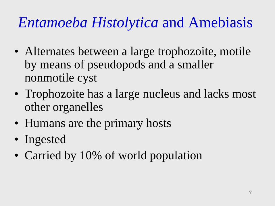
Entamoeba Histolytica and Amebiasis
• Alternates between a large trophozoite, motile by means of pseudopods and a smaller nonmotile cyst
• Trophozoite has a large nucleus and lacks most other organelles
• Humans are the primary hosts
• Ingested
• Carried by 10% of world population

Figure 23.1 Entamoeba histolytica
8

9
Entamoeba Histolytica
• Cysts are swallowed and arrive at the small intestine;
alkaline pH and digestive juices stimulate cysts to
release 4 trophozoites
• Trophozoites attach, multiply, actively move about and
feed
• Asymptomatic in 90% of patients
• Ameba may secrete enzymes that dissolve tissues and
penetrate deeper layers of the mucosa
• Causing dysentery, abdominal pain, fever, diarrhea,
and weight loss

Figure 23.2 Intestinal amebiasis
10

11
Entamoeba Histolytica
• Life-threatening manifestations are: hemorrhage, perforation, appendicitis, and tumorlike growths, amoebomas
• May invade liver and lung
• Severe forms of disease result in 10% fatality rate
• Effective drugs are iodoquinol, metronidazole, and chloroquine

Figure 23.3 Entamoeba histolytica in specimen
12

13
Amebic Infections of the Brain
• Caused by Naegleria fowleri and Acanthamoeba
• Ordinarily inhabit standing water
• Primary acute meningoencephalitis is acquired
through nasal contact with water or traumatic eye
damage
• Infiltration of brain is usually fatal

14
An Intestinal Ciliate: Balantidium Coli
• An occupant of the intestines of domestic animals such as pigs and cattle
• Acquired by ingesting cyst-containing food or water
• Trophozoite erodes intestine and elicits intestinal symptoms
• Healthy humans are resistant
• Rarely penetrates intestine or enters blood
• Treatment – tetracycline, iodoquinol, nitrimidazine or metronidazole
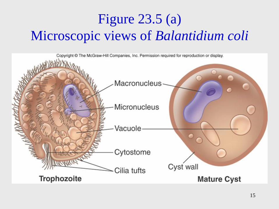
Figure 23.5 (a)
Microscopic views of Balantidium coli
15

16
The Flagellates

17
Trichomonads: Trichomonas Species
• Small, pear-shaped
• 4 anterior flagella and an undulating membrane
• Exist only in trophozoite form
• 3 infect humans:
– T. vaginalis
– T. tenax
– T. hominis

Figure 23.6 Trichomonads of humans
18

19
Trichomonas Vaginalis
• Causes an STD called trichomoniasis
• Reservoir is human urogenital tract
• 50% of infected are asymptomatic
• Strict parasite, cannot survive long outside of host
• 3 million cases yearly, a top STD
• Female symptoms – foul-smelling, green-to-yellow discharge; vulvitis; cervicitis; urinary frequency and pain
• Male symptoms – urethritis, thin, milky discharge, occasionally prostate infection
• Metronidazole

20
Giardia Lamblia and Giardiasis
• Pathogenic flagellate
• Unique symmetrical heart shape with concave ventral surface that acts like a suction cup
• Cysts are small, compact, and multinucleate
• Reservoirs include beavers, cattle, coyotes, cats, and humans
• Cysts can survive for 2 months in environment
• Usually ingested with water and food
• ID 10 to 100 cysts

Figure 23.7 (a)
Identification of trophozoites
21

22
• Cysts enter duodenum, germinate, travel to
jejunum to feed and multiply
• Causes giardiasis – diarrhea, abdominal pain
• Diagnosis is difficult because organism is shed
in feces intermittently
• Treatment: quinacrine or metronidazole
• Agent is killed by boiling, ozone, and iodine

23
Hemoflagellates: Vector-Borne Blood
Parasites
• Obligate parasites that live in blood and tissues of human host
• Cause life-threatening and debilitating zoonoses
• Spread in specific tropical regions by blood-sucking insects that serve as intermediate hosts
• Have complicated life cycles and undergo morphological changes
• Categorized according to cellular and infective stages

Hemoflagellates
• Amastigote: the form lacking a free
flagellum
• Promastigote: the stage bearing a single,
free, anterior flagellum
• Epimastigote: the flagellate stage
• Trypomastigote: large, fully formed stage
24

25

26
Trypanosoma Species and
Tropanosomiasis
• Distinguished by their infective stage; trypomastigote is an elongate, spindle-shaped cell with tapered ends, eel-like motility
• 2 types of trypanosomiasis:
– T. brucei – African sleeping sickness
– T. cruzi – Chagas disease – endemic to Central and South America

27
Trypanosoma Brucei and African
Sleeping Sickness
• Spread by tsetse flies
• Harbored by reservoir mammals
• Two variants of disease caused by 2 subspecies:
– T.b. gambiense – Gambian strain; West Africa
– T.b. rhodesiense – Rhodesian strain; East Africa
• Biting of fly inoculates skin with trypomastigotes, which multiplies in blood and damages spleen, lymph nodes, and brain
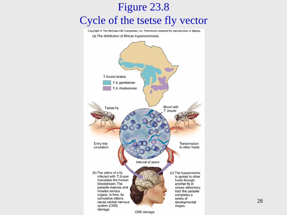
Figure 23.8
Cycle of the tsetse fly vector
28

Trypanosoma Brucei and African
Sleeping Sickness
29
• Chronic disease symptoms are sleep
disturbances, tremors, paralysis, and coma
• Trypanosomes are readily demonstrated in
blood, spinal fluid, or lymph nodes
• Treatment before neurological involvement
melarsoprol, eflornithine
• Control involves eliminating tsetse fly

30
Trypanosoma Cruzi
• Causes Chagas disease
• Reduviid bug (kissing bug) is the vector
• Infection occurs when bug feces is inoculated into a cutaneous portal
• Local lesion, fever, and swelling of lymph nodes, spleen, and liver
• Heart muscle and large intestine harbor masses of amastigotes
• Chronic inflammation occurs in the organs (especially heart and brain)
• Treatment nifurtimox and benzonidazole

Figure 23.9 American trypanosomiasis
(Chagas disease)
31

Figure 23.10 Conditions associated with
Chagas disease
32

33
Leishmania Species and Leishmaniasis
• Leishmaniasis – zoonosis transmitted among
mammalian hosts by female sand flies that require
a blood meal to produce eggs
• Endemic to equatorial regions
• Promastigotes are injected with sand fly bite,
convert to amastigote and multiply; if macrophage
does not migrate the infection is localized;
systemic if macrophage migrates

34
• Cutaneous-oriental sore, Baghdad boil –
localized ulcerated sore
• Espundia – skin and mucous membrane
infection of the head; chronic infection
• Systemic (visceral) – high intermittent
fever; weight loss, enlarged spleen, liver,
and lymph nodes
– Kala azar is the most severe and fatal form if
untreated
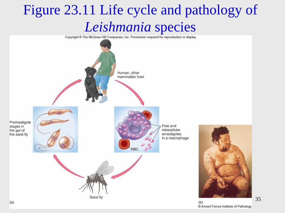
Figure 23.11 Life cycle and pathology of
Leishmania species
35

36
23.3 Apicomplexan Parasites
• Sporozoans
• Lack locomotor organelles in the trophozoite state
• Alternate between sexual and asexual phases and between different animal hosts
• Most form specialized infective bodies that are transmitted by arthropod vectors, food, water, or other means
– Plasmodium
– Toxoplasma
– Cryptosporidium

37
Plasmodium: The Agent of Malaria
• Dominant protozoan disease
• Obligate intracellular sporozoan
• 4 species: P. malariae, P. vivax,
P. falciparum, and P. ovale
• Female Anopheles mosquito is the primary vector;
blood transfusions, mother to fetus
• 300-500 million new cases each year
• 2 million deaths each year

Figure 23.12 The malaria belt
38

39
2 distinct phases of malarial parasite development:
• Asexual phase – human host – Infected female mosquito injects asexual sporozoite
which localizes in liver; it then undergoes
schizogony generating numerous merozoites which
enter circulation in 5-16 days depending on species
– Merozoites attach to and enter red blood cells,
convert to trophozoites and multiply; red cell bursts
releasing merozoites that differentiate into gametes

40
• Sexual phase – mosquito host
– Mosquito draws infected RBCs; gametes
fertilize forming diploid cell which forms
sporozoites in stomach
– Sporozoites lodge in salivary glands; available
to infect human host

41
Figure 23.13
Plasmodium

42
Plasmodium
• Symptoms include episodes of chills-fever-sweating,
anemia, and organ enlargement
• Symptoms occur at 48-72 hour intervals as RBCs
rupture; interval depends on species
• P. falciparum most malignant type; highest death rate
in children
• Diagnosis by presence of trophozoite in RBCs,
symptoms
• Increasing drug resistance
• Therapy is chloroquine, mefloquine

Figure 23.14 Malaria
43

44
Coccidian Parasites
• Zoonotic in domestic animals and birds

45
Toxoplasma Gondii and Toxoplasmosis
• Intracellular apicomplexan parasite with extensive distribution
• Lives naturally in cats that harbor oocysts in the GI tract
• Acquired by ingesting raw meats or substances contaminated by cat feces
• Most cases of toxoplasmosis go unnoticed except in fetus and AIDS patients who can suffer brain and heart damage
• Treatment: pyrimethamine and sulfadiazine

46
Figure 23.16
The life cycle
and
morphological
forms of
Toxoplasma
gondii

Figure 23.17 Toxoplasmosis in
the brain of an AIDS patient
47

48
Sarcocystis and Sarcocystosis
• Sarcocystis – parasites of cattle, swine, and sheep
• Domestic animals are intermediate hosts; they pick
up infective cysts while grazing on grass
contaminated with human feces
• Humans are infected when the meat is consumed
• Symptoms include diarrhea, nausea, and abdominal
pain
• No specific treatment

49
Cryptosporidium: A Newly
Recognized Intestinal Pathogen
• An intestinal pathogen
• Infects a variety of mammals, birds, and reptiles
• Exists in tissue and oocyst phases
• 1990s – 370,000 cases in Milwaukee, WI, due to contaminated water; filtration required for removal
• Ingestion of oocysts gives rise to sporozoites that penetrate intestinal cells
• Causes gastroenteritis, headache, sweating, vomiting, abdominal cramps, diarrhea
• AIDS patients may suffer chronic persistent diarrhea
• No effective drugs

Figure 23.18 Other apicomplexan parasites
50

51
Isospora Belli and Coccidiosis
• Intracellular intestinal parasite with oocyst stage
• Transmitted in fecally contaminated food or drink
• Infection usually asymptomatic or self-limited
• Symptoms include malaise, nausea and vomiting,
diarrhea, fatty stools, abdominal cramping, and
weight loss
• Treat with sulfadiazine and pyrimethamine, when
required

52
Cyclospora Cayetanensis and
Cyclosporiasis
• Emerging protozoan pathogen; causes cyclosporiasis
• Oral-fecal transmission; fresh produce and water
• Oocysts enter small intestine and invade the mucosa
• Symptoms of watery diarrhea, stomach cramps,
bloating, fever, muscles aches
• Diagnosis can be complicated
• Treatment: trimethoprim and sulfamethoxazole

Figure 23.19 Cyclospora in a human
fecal sample
53

54
Babesia Species and Babesiosis
• First protozoan found to cause a disease –
redwater fever of cattle
• First protozoan found to be associated with
a vector – tick
• Human babesiosis – relatively rare zoonosis
• Associated with infected rodents
• Infection resembles malaria

55
23.4 A Survey of Helminth Parasites
• Adults are large, multicellular animals with specialized tissues and organs
• Adult worms mate and produce fertilized eggs that hatch; larvae then mature in several stages to adults
• The sexes may separate or hermaphroditic
• Adulthood and mating occur in the definitive host
• Larval develop occurs in the intermediate host
• A transport host experiences no parasitic development
• Four basic patterns of life and transmission

Figure 23.20 Four helminth cycles
56

57

58
Helminths
• Pathology arises from worms feeding on and migrating through tissues, accumulation of worms, and worm products
• Diagnosis based on blood cell count (eosinophilia), serological tests; eggs, larvae, or adult worms in feces; sputum, urine, blood, or tissue biopsies
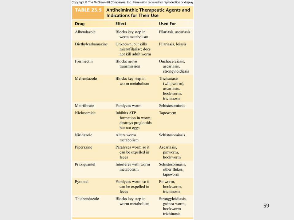
• Antihelminthic drugs suppress a helminthic metabolic process that differs from the human process, inhibit the worm’s movement, prevent it from holding position, and act locally in the intestine
59

60
Nematode (Roundworm) Infestations
• Most abundant animal groups; 50 species that affect humans
• Elongated, cylindrical worms with protective cuticles, circular muscles, a complete digestive tract, and separate sexes
• Divided into intestinal nematodes, and tissue nematodes
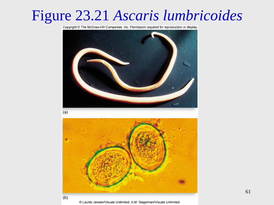
Figure 23.21 Ascaris lumbricoides
61

62
Ascaris Lumbricoides
• A large intestinal roundworm
• Most cases in the U.S. occur in the southeastern states
• Indigenous to humans
• Ascaris spends its larval and adult stages in humans;
release embryonic eggs in feces, and are spread to other
humans; food, drink, or contaminated objects
• Ingested eggs hatch into larvae and burrow through the
intestine into circulation and travel to the lungs and
pharynx and are swallowed
• Adult worms complete cycle in intestines and reproduce
– 200,000 eggs/day

63
Ascaris Lumbricoides
• Worms retain motility, do not attach
• Severe inflammatory reactions mark the
migratory route
• Allergic reactions can occur
• Heavy worm loads can retard physical and
mental development

64
Trichuris Trichiura and Whipworm
Infection
• Whipworm
• Humans sole host
• Trichuriasis has its highest incidence in the tropics
• Eggs hatch in intestines, larvae attach, penetrate the outer wall and develop into adults
• Females lay 3,000-5,000 eggs daily
• Worms can pierce capillaries, cause localized hemorrhage
• Heavy infestations can cause dysentery, rectal prolapse – can be fatal in children

65
Enterobius Vermicularis and
Pinworm Infection
• Pinworm or seatworm
• Enterobiasis most common worm disease of children in temperate zones
• Eggs are picked up from surroundings and swallowed
• After hatching in the small intestine, they develop into adults
• Anal itching occurs when mature females emerge from intestine to release eggs
• Self-inoculation is common
• Tape test

66
Hookworms
• Characteristic curved ends and hooked mouths
• Necator americanus and Ancylostoma duodenale
• Humans shed eggs in feces, which hatch into filariform larvae and burrow into the skin of bare feet
• Larvae travel from blood to lungs, proceed up bronchi and throat and are swallowed
• Worms mature and reproduce in small intestine and complete the cycle
• May cause pneumonia, nausea, vomiting, cramps, and bloody diarrhea
• Blood loss is significant – anemia

Figure 23.22 The hookworms
67

68
Strongyloides Stercoralis and
Strongyloidiasis
• Threadworm
• Tiny roundworms completes life cycle in humans or moist soil
• Larvae penetrate skin and migrate to lungs, are swallowed, and complete development in the intestine
• Can reinfect the same host without leaving the body
• Heavy worm loads can cause pneumonitis and eosinophilia, bloody diarrhea, liver enlargement, bowel obstruction, and malabsorption

69
Trichinella Spiralis and Trichinellosis
• Life cycle entirely within mammalian host
• Acquired from eating undercooked pork or bear
meat
• Larvae migrate from intestine to blood vessels,
muscle, heart, and brain, where it forms cysts
• First symptoms – flulike, diarrhea
• Second symptoms – muscle and joint pain, shortness
of breath, pronounced eosinophilia
• No cure after larva have encysted

Figure 23.24 The cycle of trichinellosis
70

71
Tissue Nematodes
• Complete their life cycle in human blood, lymphatics, or skin
• Filarial worms; elongate, filamentous bodies, spread by biting arthropods
• Cause chronic, deforming disease
• Wuchereria bancrofti – elephantiasis
• Onchocerca volvulus – river blindness
• Loa loa – eye worm

72
Wucherereia Bancrofti and
Bancroftian Filariasis
• Tropical infection spread by mosquitoes
• Vector deposits larvae which move into
lymphatics and develop into adults
• Chronic infection causes blockage of
lymphatic circulation and elephantiasis,
massive swelling in the extremities

Figure 23.25 Bancroftian filariasis
73

74
Onchocerca Volvulus and River
Blindness
• Transmitted by biting black flies
• Larvae develop into adults in subcutaneous tissues
• Adult females migrate via the blood to the eyes, provoking inflammatory reactions
• Coinfection with Wolbachia bacteria causes river blindness
• Treatment: tetracycline and ivermectin

75
Loa Loa: The African Eye Worm
• Spread by bite of small flies
• Temperature-sensitive worm migrates
around/under the skin and may enter the eye
• Treatment – pull worm from a small hole in
conjunctiva or diethylcarbamazine

76
Trematodes or Flukes
• Flatworms with ovoid leaflike bodies
• Have digestive, excretory, neuromuscular, and
reproductive systems
• Lack circulatory and respiratory systems
• Animals such as snails or fish are usually the
intermediate hosts and humans are the
definitive hosts

77
Blood Flukes: Schistosomes
• Schistosomiasis – prominent parasitic disease
• Schistosoma mansoni, S. japonicum,
S. haematobium
• Adult flukes live in humans who release eggs into
water; early larva (miracidium) develops in
freshwater snail into a 2nd larva (cercaria)
• This larva penetrates human skin and moves into the
liver to mature; adults migrate to intestine or bladder
and shed eggs, giving rise to chronic organ
enlargement

Figure 23.26 Stages in the life
cycle of Schistosoma
78

79
Liver and Lung Flukes
• Zoonotic
Liver flukes:
• Opisthorchis (Clonorchis) sinensis – cycles between mammals and snails and fish; humans are infected by eating inadequately cooked fish containing cercariae, larvae crawl into bile duct, mature, and shed eggs into feces; snail are infected
• Fasciola hepatica – cycles between herbivores, snails, and aquatic plants; humans are infected by eating raw aquatic plants; fluke lodges in liver

80
Lung fluke:
• Paragonimus westermani – cycles between
carnivorous animals, snails, and
crustaceans; humans infected by eating
undercooked crustaceans; intestinal worms
migrate to lungs

Figure 23.27 Fasciola hepatica
81

82
Cestode (Tapeworm) Infestations
• Flatworms
• Long, very thin, ribbonlike bodies composed of
sacs (proglottids) and a scolex that grips the
intestine
• Each proglottid is an independent unit adapted to
absorbing food and making and releasing eggs
• Taenia saginata
• Taenia solium

83
Taenia Saginata
• Beef tapeworm
• Very large, up to 2,000 proglottids
• Humans are the definitive host
• Animals are infected by grazing on land contaminated with human feces
• Infection occurs from eating raw beef in which the larval form has encysted
• In humans, larva attaches to the small intestine and becomes an adult
• Causes few symptoms; vague abdominal pain and nausea; proglottids in stool

Figure 23.28 Tapeworm infestation
in humans
84
85
Taenia Solium
• Pork tapeworm
• Infects humans through ingesting cysts or eggs
• Eggs hatch in intestine, releasing tapeworm larva that migrate to all tissues and encyst
• Most damaging if they lodge in heart muscle, eye, or brain
• May cause seizures, psychiatric disturbances

Figure 23.29 Cysticercosis
86

87
23.5 The Arthropod Vectors of
Infectious Disease
• Arthropods – exoskeleton and jointed legs;
includes arachnids and crustaceans; many
must feed on blood and tissue fluid of host
during life cycle; ectoparasites
• Those of medical importance transmit
infectious microbes in the process of
feeding – biological vectors

88

89
Insects
• Mosquitoes – require an aquatic habitat; females take blood meal transmitting disease: malaria, filariasis, Dengue fever
• Fleas – highly motile, flattened bodies; feed on warm-blooded animals; carry zoonotic diseases: plague, murine typhus
• Lice – small, soft; attach to head and body hair feeding inconspicuously on blood and tissue fluid; release feces that contaminate wound; epidemic typhus, relapsing fever

90
Arachnids
• Ticks – cling on vegetation and attach to host on contact; larvae, nymph, and adults get blood meal by piercing skin of host
– Hard or ixodid ticks – small compact, rigid bodies; transmit bacterial, rickettsial, and viral diseases
– Soft or argasid ticks – flexible outer bodies; transmit relapsing fever

Figure 23.30 Arthropod vectors
91