1
A case study from the Salt River Forensic Pathology Services Laboratory, Cape Town, South Africa INTRODUCTION Bronchiolitis is a respiratory tract infection of the bronchioles, the tiny terminal branches of the airways in the lungs. It is usually caused by a viral infection and mostly affects infants and young children because of their small airways 1,4 . Although it is generally a mild disease, it can develop into a more severe disease that requires hospitalization 2,3 . CASE PRESENTATION A 3-month old male infant was taken to the GP for fever and stomach cramps. The infant had been a full-term baby, delivered naturally, with a birth-weight of 2.3 kg. The GP diagnosed colic and the baby was sent home to be treated with Panado (paracetamol, an anti-pyretic) syrup and Scopex (a medication containing hyoscine, used to prevent intestinal cramping, and as a general sedative). A week later, the infant died. IMAGING AND DIAGNOSIS An external examination of the body showed no evidence of trauma to the body. A Lodox full- body X-ray was performed prior to autopsy. It showed bilateral pulmonary infiltrates (i) with a mottled reticular pattern and air bronchogram (b), suggestive of bronchiolitis. Since no foul play was suspected, and the X-ray image showed a pulmonary infection, an internal post-mortem examination was not performed. The cause of death was given as natural disease, most likely bronchiolitis. DISCUSSION Bronchiolitis typically occurs during the first two years of life, with peak occurrence at about three to six months of age 1 . It is more common in males, and in children who live in crowded conditions. The first symptoms may be confused with those of a common cold, and include a cough, a mild fever and a runny nose. The pathogens that cause bronchiolitis are spread in droplet form, either from fluid from an infected person’s nose or mouth, and can become airborne through sneezing or coughing. Therefore, children in close proximity to other infected children, such as in a crèche, are quite likely to contract the infection 4 . An external examination of the body showed no signs of trauma, and neither did a radiograph. However, the Lodox X-ray image revealed bilateral pulmonary infection with air entrapment (bronchogram) in the bronchioles. Considering the age of the infant, its medical history and socio-economic status, all indications were that this child died of natural causes, namely bronchiolitis. CONCLUSION Radiography and an external examination ruled out trauma as the cause of death: no obvious injuries, bruises or fractures were detected. The X-ray image, supported by the medical history, was sufficient to rule out foul play and to confirm a suspected case of bronchiolitis. Thus, an internal post-mortem examination was not deemed necessary. This saved time in the mortuary and freed up a forensic pathologist to attend to another case. It is difficult to quantify the value to the next-of-kin in this case of not having an autopsy. In many instances, often for religious reasons, family members do not want an autopsy to be performed. In the case of an infant, there may also be some comfort for the parent knowing that the body was not cut. FULL-BODY IMAGING TO OBVIATE AUTOPSY IN A CASE OF DEATH FROM BRONCHIOLITIS References 1. Fraire AE, Barrios RJ. Diseases of Small Airways. In: Pulmonary Pathology, Zander DS, Farver CF, editors. Churchill Livingstone, Elsevier, Philadelphia, Pennsylvania, 2008 2. Greenough A, Cox S, Alexander J, Lenney W, Turnbull F, Burgess S, Chetcuti PAJ, Shaw NJ, Woods A, Boorman J, Coles S, Turner J. Health care utilisation of infants with chronic lung disease, related to hospitalisation for RSV infection. Archives of Disease in Childhood, 2001;85:463-468 3. Hasegawa K, Tsugawa Y, Brown DFM, Mansbach JM, Carlos A, Camargo CA Jr. Trends in Bronchiolitis Hospitalizations in the United States, 2000–2009. Pediatrics, 2013; 132(1):28-36 4. Schwarz MI, King TE. Infectious Bronchiolitis in Children. In: Interstitial Lung Disease, 2nd Edition. Mosby Year Book, Missouri, 1993 www.lodox.com CS-14-0011

Forensics Case Study - LOK Corporation

  • Upload
    others

  • View
    1

  • Download
    0

Embed Size (px)

Citation preview

A case study from the Salt River Forensic Pathology Services Laboratory, Cape Town, South Africa

INTRODUCTIONBronchiolitis is a respiratory tract infection of the bronchioles, the tiny terminal branches of the airways in the lungs. It is usually caused by a viral infection and mostly affects infants and young children because of their small airways1,4. Although it is generally a mild disease, it can develop into a more severe disease that requires hospitalization2,3.

CASE PRESENTATIONA 3-month old male infant was taken to the GP for fever and stomach cramps. The infant had been a full-term baby, delivered naturally, with a birth-weight of 2.3 kg. The GP diagnosed colic and the baby was sent home to be treated with Panado (paracetamol, an anti-pyretic) syrup and Scopex (a medication containing hyoscine, used to prevent intestinal cramping, and as a general sedative). A week later, the infant died.

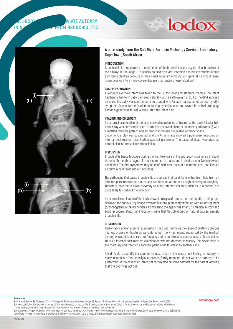
IMAGING AND DIAGNOSISAn external examination of the body showed no evidence of trauma to the body. A Lodox full-body X-ray was performed prior to autopsy. It showed bilateral pulmonary infiltrates (i) with a mottled reticular pattern and air bronchogram (b), suggestive of bronchiolitis.Since no foul play was suspected, and the X-ray image showed a pulmonary infection, an internal post-mortem examination was not performed. The cause of death was given as natural disease, most likely bronchiolitis.

DISCUSSIONBronchiolitis typically occurs during the first two years of life, with peak occurrence at about three to six months of age1. It is more common in males, and in children who live in crowded conditions. The first symptoms may be confused with those of a common cold, and include a cough, a mild fever and a runny nose.

The pathogens that cause bronchiolitis are spread in droplet form, either from fluid from an infected person’s nose or mouth, and can become airborne through sneezing or coughing. Therefore, children in close proximity to other infected children, such as in a crèche, are quite likely to contract the infection4.

An external examination of the body showed no signs of trauma, and neither did a radiograph. However, the Lodox X-ray image revealed bilateral pulmonary infection with air entrapment (bronchogram) in the bronchioles. Considering the age of the infant, its medical history and socio-economic status, all indications were that this child died of natural causes, namely bronchiolitis.

CONCLUSIONRadiography and an external examination ruled out trauma as the cause of death: no obvious injuries, bruises or fractures were detected. The X-ray image, supported by the medical history, was sufficient to rule out foul play and to confirm a suspected case of bronchiolitis. Thus, an internal post-mortem examination was not deemed necessary. This saved time in the mortuary and freed up a forensic pathologist to attend to another case.

It is difficult to quantify the value to the next-of-kin in this case of not having an autopsy. In many instances, often for religious reasons, family members do not want an autopsy to be performed. In the case of an infant, there may also be some comfort for the parent knowing that the body was not cut.

FULL-BODY IMAGING TO OBVIATE AUTOPSYIN A CASE OF DEATH FROM BRONCHIOLITIS

References1. Fraire AE, Barrios RJ. Diseases of Small Airways. In: Pulmonary Pathology, Zander DS, Farver CF, editors. Churchill Livingstone, Elsevier, Philadelphia, Pennsylvania, 20082. Greenough A, Cox S, Alexander J, Lenney W, Turnbull F, Burgess S, Chetcuti PAJ, Shaw NJ, Woods A, Boorman J, Coles S, Turner J. Health care utilisation of infants with chronic lung disease, related to hospitalisation for RSV infection. Archives of Disease in Childhood, 2001;85:463-4683. Hasegawa K, Tsugawa Y, Brown DFM, Mansbach JM, Carlos A, Camargo CA Jr. Trends in Bronchiolitis Hospitalizations in the United States, 2000–2009. Pediatrics, 2013; 132(1):28-364. Schwarz MI, King TE. Infectious Bronchiolitis in Children. In: Interstitial Lung Disease, 2nd Edition. Mosby Year Book, Missouri, 1993

www.lodox.com

CS-14-0011