Embed Size (px)
Citation preview
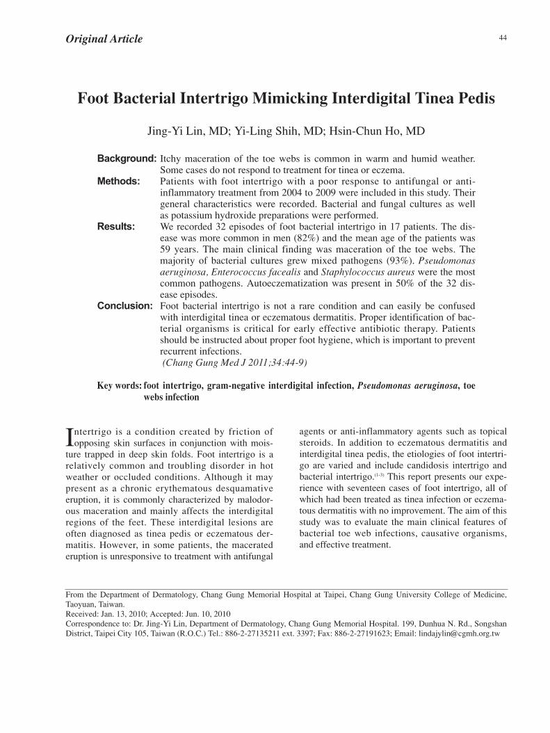
44Original Article
Foot Bacterial Intertrigo Mimicking Interdigital Tinea Pedis
Jing-Yi Lin, MD; Yi-Ling Shih, MD; Hsin-Chun Ho, MD
Background: Itchy maceration of the toe webs is common in warm and humid weather.Some cases do not respond to treatment for tinea or eczema.
Methods: Patients with foot intertrigo with a poor response to antifungal or anti-inflammatory treatment from 2004 to 2009 were included in this study. Theirgeneral characteristics were recorded. Bacterial and fungal cultures as wellas potassium hydroxide preparations were performed.
Results: We recorded 32 episodes of foot bacterial intertrigo in 17 patients. The dis-ease was more common in men (82%) and the mean age of the patients was59 years. The main clinical finding was maceration of the toe webs. Themajority of bacterial cultures grew mixed pathogens (93%). Pseudomonasaeruginosa, Enterococcus facealis and Staphylococcus aureus were the mostcommon pathogens. Autoeczematization was present in 50% of the 32 dis-ease episodes.
Conclusion: Foot bacterial intertrigo is not a rare condition and can easily be confusedwith interdigital tinea or eczematous dermatitis. Proper identification of bac-terial organisms is critical for early effective antibiotic therapy. Patientsshould be instructed about proper foot hygiene, which is important to preventrecurrent infections.(Chang Gung Med J 2011;34:44-9)
Key words: foot intertrigo, gram-negative interdigital infection, Pseudomonas aeruginosa, toewebs infection
From the Department of Dermatology, Chang Gung Memorial Hospital at Taipei, Chang Gung University College of Medicine,Taoyuan, Taiwan.Received: Jan. 13, 2010; Accepted: Jun. 10, 2010Correspondence to: Dr. Jing-Yi Lin, Department of Dermatology, Chang Gung Memorial Hospital. 199, Dunhua N. Rd., SongshanDistrict, Taipei City 105, Taiwan (R.O.C.) Tel.: 886-2-27135211 ext. 3397; Fax: 886-2-27191623; Email: [email protected]
Intertrigo is a condition created by friction ofopposing skin surfaces in conjunction with mois-
ture trapped in deep skin folds. Foot intertrigo is arelatively common and troubling disorder in hotweather or occluded conditions. Although it maypresent as a chronic erythematous desquamativeeruption, it is commonly characterized by malodor-ous maceration and mainly affects the interdigitalregions of the feet. These interdigital lesions areoften diagnosed as tinea pedis or eczematous der-matitis. However, in some patients, the maceratederuption is unresponsive to treatment with antifungal
agents or anti-inflammatory agents such as topicalsteroids. In addition to eczematous dermatitis andinterdigital tinea pedis, the etiologies of foot intertri-go are varied and include candidosis intertrigo andbacterial intertrigo.(1-3) This report presents our expe-rience with seventeen cases of foot intertrigo, all ofwhich had been treated as tinea infection or eczema-tous dermatitis with no improvement. The aim of thisstudy was to evaluate the main clinical features ofbacterial toe web infections, causative organisms,and effective treatment.

Chang Gung Med J Vol. 34 No. 1January-February 2011
Jing-Yi Lin, et alFoot bacterial intertrigo
45
METHODS
Between 2004 and 2009 in one outpatient clinic,we collected 17 cases of foot intertrigo that had apoor response to therapy for fungus or eczematousdermatitis. The duration of therapy failure prior tovisiting our clinic ranged from 11 days to 6 months.We performed bacterial cultures and sensitivity on allpatients. All patients were treated with systemicand/or topical antibiotics on the basis of an antibi-ogram. Potassium hydroxide preparations and fungalcultures were performed thereafter if there was a sus-pected fungal infection component.
RESULTS
Seventeen patients affected by foot intertrigowere studied. The mean age of the patients was 59years (range, 36-81 years). Fourteen (82%) of themwere men. Fourteen had initially been treated withtopical or systemic antifungal medication and 5 withanti-inflammatory agents, such as topical or systemicsteroids.
The main clinical features were erythema,vesiculopustules, erosion, maceration, and malodor-ous discharge. The lesions affected the interdigitalspaces of the feet, and some extended toward thesole or the dorsal area of the feet (Fig. 1, 2). Thesepatients frequently reported a burning, painful, pru-ritic sensation.
Nine patients (53%) had recurrent toe web
infections during this study; six patients had 2 dis-ease episodes, one patient had 3, one patient had 4,and another patient had 5. Therefore, a total of 32disease episodes were recorded.
Twenty-nine cultures were isolated from the 32disease episodes. Twenty-seven bacterial cultures(93%) grew more than one organism. Eighty-six per-cent of our cultures grew gram-negative bacteria.Pseudomonas aeruginosa (16/29, 55%),Enterococcus facealis (12/29, 41%), andStaphylococcus aureus (12/29, 41%) were the mostfrequently isolated pathogens. Coagulase-negativestaphylococci were isolated from 6 of the 29 samples(21%). The other pathogens isolated are shown in theTable 1. The two single-pathogen cultures grewEnterococcus facealis and Acinetobacter baumannii.
After one to two weeks of treatment with sys-temic antibiotics and local application of antisepticagents, all patients experienced significant reductionin pruritus and pain. The infection in all patientsimproved markedly with rapid resolution of macera-tion. The topical therapy included aluminum chloridesolution, potassium permanganate solution, gen-tamycin cream, and povidone iodine ointment orsolution. Systemic antibiotics were used in twenty-eight of 32 episodes. The antibiotics used includedpenicillin, oxacillin, ampicilin, sulfamethoxazole-trimethoprim, cephalosporine, ciprofloxacin, andgentamycin. Pseudomonas isolated from thesepatients was sensitive to ciprofloxacin, gentamycin,ceftazidime, and cefepime but was resistant tooxacillin.
Fig. 1 Maceration of the second, third and fourth interdigitalspaces. Fig. 2 Maceration of the toe web.

Chang Gung Med J Vol. 34 No. 1January-February 2011
Jing-Yi Lin, et alFoot bacterial intertrigo
46
Enterococcus faecalis and Staphylococcusaureus were usually found to be associated withPseudomonas aeruginosa or other gram-negativebacteria. Systemic antibiotic treatment based on theantibiogram also appeared to be successful.
Ten patients had both tinea pedis and ony-chomycosis of the toes. Two patients had tinea pediswithout toenail infection. Potassium hydroxidepreparations (KOH) and fungal cultures were per-formed in the eight patients who did not have com-plete improvement after systemic antibiotic therapy.Five patients had both KOH and fungus culture. Twopatients had fungus culture alone and the other onehad KOH alone. One of the six KOH studiesrevealed positive results, and six of the seven cul-tures grew fungus. The types of fungi isolated wereCandida albicans in two patients, Candida parap-silosis in two, Trichophyton terrestre in one, andTrichosporon sp. in one. These ten patients acceptedantifungal treatment.
In addition to foot intertrigo, some patients haditchy red papules, papulovesicles and plaques ontheir extremities and/or the trunk, so- called autosesi-tization dermatitis. (Fig. 3) Itchy lesions developedin 16 of the 32 recorded disease episodes (50%).
DISCUSSION
Gram-negative bacterial toe web infections werefirst described as a distinct disorder by Amonette andRosenburg in 1973.(4) They reported twelve patientswith maceration of the toe webs. The maceration wasinduced by gram-negative bacteria and was moresevere than that induced by Candida albicans. In theliterature, gram-negative bacterial toe web infectionsare relatively common, troublesome disorders.(4-10)
The infection involves the toe web space and extendsto the adjacent plantar surface. The clinical featuresinclude vesiculopustules, macerations, malodorousdischarge, and marked edema and erythema of thesurrounding tissues. Patients usually feel a burningsensation or pruritus. In some severe cases, patientsare unable to walk. Men appear to be more frequent-ly affected than women, as in our study.(5,7) Promotingfactors include hot weather, closed-toe or tight-fittingshoes, hyperhidrotic toe webs, athletic or recreationalactivities, and use of germicidal soaps, as well asprevious prolonged antibiotic or antifungal thera-py.(5,6,9)
In the 1973 study of gram-negative toe webinfection by Amonette and Rosenburg, Pseudomonasaeruginosa and Proteus mirabilis were the mostcommonly isolated organisms.(4) Those two organ-isms, along with enterococcus species, were the mostcommonly isolated in a study by Eaglstein et al.(11) Ina study of foot bacterial intertrigo by Aste et al,pseudomonas aeruginosa, often together with other
Table 1. Main Pathogens Isolated in Foot Intertrigo
Number of cultures with Isolated pathogens positive pathogens
(total number of cultures: 29)
Pseudomonas aeruginosa 16
Enterococcus faecalis 12
Staphylococcus aureus 12
Coagulase-negative staphylococci 6
Escherichia coli 4
Group A β-hemolytic streptococci 3
Group B β-hemolytic streptococci 3
Acinetobacter baumannii 3
Proteus mirabilis 3
Corynebacterium sp. 2
Staphylococcus saprophyticus 1
Acinetobacter lowffii 1
Viridans streptococcus 1
Stenotrophomonas maltophilia 1
Klebsiella pneumonia 1
Peptostrepto. magnus 1
Morganella morganii 1
Fig. 3 Autoeczematization: pruritic red papules and vesicleson the dorsal foot.
Chang Gung Med J Vol. 34 No. 1January-February 2011
Jing-Yi Lin, et alFoot bacterial intertrigo
47
gram-negative bacteria, was the most common etio-logic agent.(5) In the study by Karaca et al, the mostcommon pathogen was coagulase-negative staphylo-cocci, followed by Pseudomonas aeruginosa.(7) Inour study, we found a frequency of 55% forPseudomonas aeruginosa, 41% for Enterococcusfacealis, 41% for Staphylococcus aureus, and 29%for coagulase-negative staphylococci. The mixedinfection rate is around 22.6% to 75% in the litera-ture and was 93% in our series.(4,7,11,12) The most com-mon concomitant pathogens were dermatophytes andcoagulase-negative staphylococci in the Karaca et alstudy.(7) There was a higher mixed infection rate inour study, and this might be related to the diseaseduration and severity. Pseudomonas aeruginosacombined with other gram-negative bacteria orgram-positive bacteria was the most common con-comitant pathogen.
The interdigital space is typically colonized bypolymicrobial flora. Dermatophytes may damage thestratum corneum and produce substances with antibi-otic properties. Gram-negative bacteria may resistantibiotic-like substances and proliferate. Thisprocess may progress to gram-negative foot intertri-go.
Several pathogens and factors might play a rolein toe web infections. Maceration is seen in gram-negative, gram-positive, and Candida albicans infec-tions, severe tinea pedis, and eczematous dermatitis.Although this symptom is frequently seen in bacteri-al foot infections, especially in gram-negative infec-tions, the clinical appearance is not helpful in diag-nosing the nature of the causative organism.However, physicians should be reminded of bacterialfoot intertrigo, especially if the foot maceration issevere or combined with cellulitis.
In 1973, Amonette and Rosenburg reported dif-ficulty in the treatment of foot intertrigo. The sys-temic antibiotics available had significant sideeffects and topical therapeutic modalities failed toprovide satisfactory improvement.(4) In two series, athird generation cephalosporin and ciprofloxacinwere much more effective and provided excellentresults in gram-negative bacterial toe web infec-tion.(5,11)
In our study, topical therapy alone was foundinadequate for treatment in some cases. Systemicantibiotics should be considered in these patients iftopical treatment fails or there is extensive disease.
Oral ciprofloxacin 250-500 mg twice daily for 2weeks was effective against Pseudomonas aerugi-nosa in our study. Westmoreland et al. presented apatient with presumed tinea pedis, whose culturegrew Pseudomonas. The infection resolved with oralciprofloxacin.(13)
As polymicrobial infections are common, it isadvisable to combine topical antibiotics that act ongram-positive and gram-negative microorganisms.Topical antimicrobial therapy should be broad-spec-trum, because dermatophytes select bacteria by pro-ducing penicillin and streptomycin- like sub-stances.(14) Antiseptic and astringent agents, such asaluminum chloride and Castellani’s paint, are helpfulin severely macerated, bacterially infected inter-spaces.(15) Local application of aluminum chlorideand gentamycin cream or povidone-iodine twicedaily was an effective option in our study.
In addition to the pharmacological approach,debridement may be helpful.(16) Superficial debride-ment is performed with application of moistened 1%povidone-iodine dressings (10% povidone-iodine:saline = 1:9). Debridement may remove the necrotictissue and allow topical agents to reach the infectedarea faster. Other important measures include goodhygiene, keeping the toe webs dry, avoidance ofocclusive footwear, and avoidance of water-relatedactivities.(10,17)
In our study, there was a higher recurrence rateof foot bacterial intertrigo than that in the literature(53% vs. 7%).(5) There was no significant differencein seasons, occupation, or incidence of diabetes mel-litus in our study. This might be explained by under-lying dermatophyte infection of the soles or toe nails,or eczema with disruption of the cutaneous barrier.Patients who have fungal infection of the soles andtoenails have reservoirs of spores that can spread tothe interdigital area. These patients require prolongedtherapy to eradicate fungi from toenails and soles.The antifungal agents econazole nitrate cream andciclopirox olamine both exhibit broad- spectrumactivity against many gram-negative organisms.(18,19)
Econazole nitrate has been demonstrated effectivefor the treatment of severe interdigital bacterialinfections.
Patients with uncontrolled or flaring foot bacter-ial intertrigo can have autoeczematization on thetrunk and extremities.(4) However, there is little datarelated to foot intertrigo with autoeczematization in

Chang Gung Med J Vol. 34 No. 1January-February 2011
Jing-Yi Lin, et alFoot bacterial intertrigo
48
the literature. We observed a high frequency (50%)of autoeczematization in disease episodes in thisstudy. The autoeczematization progressed when toeweb infections persisted and resolved rapidly whenthe infection was under control. In this study, sys-temic steroids were given to patients with severeautoeczematization (75%). Autoeczematization islikely due to a hyperirritability of the skin inducedby either immunologic or nonimmunologic stimuli.Infection and wounding have been reported torelease a variety of epidermal cytokines. Thesecytokines can heighten the sensitivity of the skin tostimuli and cause autoeczematization.(20)
The course of disease of bacterial intertrigo isvery favorable if there is an early, accurate diagnosisand appropriate treatment. It is also important toinstruct patients in appropriate hygiene measures toavoid heat and moisture in their feet.
REFERENCES
1. Kates SG, Nordstrom KM, McGinley KJ, Leyden JJ.Microbial ecology of interdigital infections of toe webspaces. J Am Acad Dermatol 1990;22:578-82.
2. Neubert U, Braun-Falco O. Maceration of the interdigitalspaces and gram-negative infection of feet. Hautarzt1976;27:538-43.
3. Hope YM, Clayton YM, Hay RJ, Noble WC, Elder-SmithJG. Foot infection in coal miners: a reassessment. Br JDermatol 1985;112:405-13.
4. Amonette RA, Rosenberg EW. Infection of toe webs bygram-negative bacteria. Arch Dermatol 1973;107:71-3.
5. Aste N, Atzori L, Zucca M, Pau M, Biggio P. Gram-nega-tive bacterial toe web infection: a survey of 123 casesfrom the district of Cagliari, Italy. J Am Acad Dermatol2001;45:537-41.
6. Janniger CK, Schwartz RA, Szepietowski JC, Reich A.Intertrigo and common secondary skin infections. AmFam Physician 2005;72:833-8.
7. Karaca S, Kulac M, Cetinkaya Z, Demirel R. Etiology offoot intertrigo in the District of Afyonkarahisar, Turkey: abacteriologic and mycologic study. J Am Podiatr MedAssoc 2008;98:42-4.
8. Silvestre JF, Betlloch MI. Cutaneous manifestations dueto Pseudomonas infection. Int J Dermatol 1999;38:419-31.
9. Abramson C, Steinmetz R. Antifungal activity ofPseudomonas aeruginosa in gram-negative athlete’s foot.J Am Podiatry Assoc 1983;73:227-34.
10. Leyden JJ, Kligman AM. Interdigital athlete’s foot. Theinteraction of dermatophytes and resident bacteria. ArchDermatol 1978;114:1466-72.
11. Eaglstein NF, Marley WM, Marley NF, Rosenberg EW,Hernandez AD. Gram-negative bacterial toe web infec-tion: successful treatment with a new third generationcephalosporin. J Am Acad Dermatol 1983;8:225-8.
12. Abramson C. Athlete’s foot caused by pseudomonasaeruginosa. Clin Dermatol 1983;1:14-24.
13. Westmoreland TA, Ross EV, Yeager JK. Pseudomonas toeweb infections. Cutis 1992;49:185-6.
14. Youssef N, Wyborn CH, Holt G. Antibiotic production bydermatophyte fungi. J Gen Microbiol 1978;105:105-11.
15. Leyden JJ, Kligman AM. Aluminum chloride in the treat-ment of symptomatic athelete’s foot. Arch Dermatol1975;111:1004-10.
16. King DF, King LA. Importance of debridement in thetreatment of gram-negative bacterial toe web infection. JAm Acad Dermatol 1986;14:278-9.
17. Leyden JJ. Progression of interdigital infections from sim-plex to complex. J Am Acad Dermatol 1993;28:S7-S11.
18. Kates SG, Myung KB, McGinley KJ, Leyden JJ. Theantibacterial efficacy of econazole nitrate in interdigitaltoe web infections. J Am Acad Dermatol 1990;22:583-6.
19. Gupta AK, Skinner AR, Cooper EA. Interdigital tineapedis (dermatophytosis simplex and complex) and treat-ment with ciclopirox 0.77% gel. Int J Dermatol 2003;42Suppl 1:23-7.
20. Williams IR, Kupper TS. Immunity at the surface: homeo-static mechanisms of the skin immune system. Life Sci1996;58:1485-507.
49
2004 2009 KOH
17 32 (82%) 59
Pseudomonas aeruginosa, Enterococcus facealis,Staphylococcus aureus 32 50%
( 2011;34:44-9)
99 1 13 99 6 10105 199
Tel.: (02)27135211 3397; Fax: (02)27191623; Email: [email protected]