Embed Size (px)
Citation preview
Food and Chemical Toxicology 50 (2012) 4412–4420
Contents lists available at SciVerse ScienceDirect
Food and Chemical Toxicology
journal homepage: www.elsevier .com/locate/ foodchemtox
The protective effect of Canova homeopathic medicinein cyclophosphamide-treated non-human primates
Mariana Ferreira Leal a,⇑, Lusânia Maria Greggi Antunes b, Maria Fernanda Vita Lamarão c,Carla Elvira Araújo da Silva c, Ismael Dale Cotrim Guerreiro da Silva d, Paulo Pimentel Assumpção e,Edilson Ferreira Andrade c, Alexandre Pingarilho Rezende c, Aline Amaral Imbeloni f,José Augusto Pereira Carneiro Muniz f, Giovanny Rebouças Pinto g, Marília de Arruda Cardoso Smith a,Rommel Rodríguez Burbano c
a Disciplina de Genética, Departamento de Morfologia e Genética, Universidade Federal de São Paulo, São Paulo 04023-900, SP, Brazilb Departamento de Análises Clínicas, Toxicológicas e Bromatológicas, Faculdade de Ciências Farmacêuticas de Ribeirão Preto, Universidade de São Paulo,Ribeirão Preto 14040-903, SP, Brazilc Laboratório de Citogenética Humana, Instituto de Ciências Biológicas, Universidade Federal do Pará, Belém, PA, Brazild Disciplina de Ginecologia, Departamento de Tocoginecologia, Universidade Federal de São Paulo, São Paulo 04039-032, SP, Brazile Serviço de Cirurgia, Hospital Universitário João de Barros Barreto, Universidade Federal do Pará, Belém 66073-000, PA, Brazilf Centro Nacional de Primatas, Ministério da Saúde, Ananindeua 77030-000, PA, Brazilg Laboratório de Genética e Biologia Molecular, Universidade Federal do Piauí, Parnaíba 64202-020, PI, Brazil
a r t i c l e i n f o a b s t r a c t
Article history:Received 23 November 2011Accepted 4 September 2012Available online 12 September 2012
Keywords:CanovaHomeopathic medicineCyclophosphamideNon-human primatesCebus apella
0278-6915/$ - see front matter � 2012 Elsevier Ltd. Ahttp://dx.doi.org/10.1016/j.fct.2012.09.002
⇑ Corresponding author. Address: Disciplina deMorfologia e Genética, Universidade Federal de São PPaulo CEP 04023-900, SP, Brazil. Tel.: +55 11 557642
E-mail addresses: [email protected] (M.F. LealAntunes), [email protected] (M.F.V. Lamacom.br (C.E.A. da Silva), [email protected] (I.D.C.G d(P.P. Assumpção), [email protected] (E.F. Andhotmail.com (Alexandre Pingarilho Rezende), alineiImbeloni), [email protected] (J.A.P.C. MunizPinto), [email protected] (M.A.C. Smith), romm
Background: Canova activates macrophages and indirectly induces lymphocyte proliferation. Here weevaluated the effects of Canova in cyclophosphamide-treated non-human primates.
Methods: Twelve Cebus apella were evaluated. Four animals were treated with Canova only. Eight ani-mals were treated with two doses of cyclophosphamide (50 mg/kg) and four of these animals receivedCanova. Body weight, biochemistry and hematologic analyses were performed for 40 days. Micronucleusand comet assays were performed for the evaluation of DNA damage.
Results: We observed that cyclophosphamide induced abnormal WBC count in all animals. However,the group treated with cyclophosphamide plus Canova presented a higher leukocyte count than thatwhich received only cyclophosphamide. Cyclophosphamide induced micronucleus and DNA damage inall animals. The frequency of these alterations was significantly lower in the Canova group than in thegroup without this medicine.
Conclusions: Our results demonstrated that Canova treatment minimizes cyclophosphamide myelotox-icity in C. apella.
� 2012 Elsevier Ltd. All rights reserved.
1. Introduction
Cancer is a leading cause of death worldwide (WHO, 2010). Sev-eral protocols using chemotherapic and radiotherapic approacheshave been developed for anticancer treatments. However,
ll rights reserved.
Genética, Departamento deaulo, Rua Botucatu 740, São
60; fax: +55 91 55764260.), [email protected] (L.M.G.rão), [email protected] Silva), [email protected]), alexandrepingarilho@[email protected] (A.A.), [email protected] ([email protected] (R.R. Burbano).
protecting patients from adverse effects of these intensive thera-pies has been a goal of clinical oncologists (Bukowski, 1999).
For over 40 years, cyclophosphamide (CP) has been in clinicaluse for the treatment of malignant and nonmalignant disorders(Hosseinimehr and Karami, 2005). However, CP is an alkylatingagent that produces gene mutations, chromosome aberrations,micronuclei (MN) and sister chromatid exchanges (Andersonet al., 1995). CP causes adverse effects especially in the hematopoi-etic system (primarily represented by leukopenia), and the gastro-intestinal tract and also can produce secondary tumors due to itsgenotoxicity (Schuurman et al., 2005).
New adjuvant therapeutic approaches to known therapies andchemoprotectants, for example glutathione, amifostine and quer-cetin (Jena et al., 2010), have been developed as methods to protectnormal tissues from the toxicity of antineoplastic agents without

M.F. Leal et al. / Food and Chemical Toxicology 50 (2012) 4412–4420 4413
compromising antitumor efficacy and, thus, providing a betterquality of life during anticancer treatment (Stolarska et al., 2006).
Canova (CA) is a complex homeopathic medicine indicated forpatients whose immune system is depressed. CA treatment seemsto enhance an individual’s ability to trigger a specific immunologicresponse against several pathological conditions (Cesar et al.,2008).
It was previously demonstrated that CA treatment induces mac-rophage activation and indirectly induces lymphocyte proliferationin vivo and in vitro (Abud et al., 2006; Burbano et al., 2009; Cesaret al., 2008; Da Rocha Piemonte and De Freitas Buchi, 2002; deOliveira et al., 2006; Lopes et al., 2006; Pereira et al., 2005). More-over, our research group has previously demonstrated that CAtreatment does not induce cytotoxic or genotoxic effects at thechromosomal level in vitro (Seligmann et al., 2003).
Due to their phylogenetic proximity to humans, non-humanprimates offer a useful model for basic research into genetic andimmunopathogenesis mechanisms as well as for the developmentand validation of new therapies for several diseases. Non-humanprimates are also of particular relevance in evaluating the potentialtoxicity of drugs and environmental agents. Cebus apella is an inter-esting model for drug studies, since they can be easily housed inPrimate Research Centers due to their flexibility, opportunism,adaptability, small size, and with annual reproduction of these ani-mals being possible in primatology centers. Currently, Cebus spe-cies are frequently used in neuroscience, dentistry, reproduction,and behavioral research (Torres et al., 2010).
Since CA treatment leads to lymphocyte differentiation and pro-liferation (Cesar et al., 2008), the aim of the present study was toevaluate whether this drug has a protective effect against the tox-icity caused by chemotherapy. We analyzed the effects of CA med-icine in CP-treated non-human primates, C. apella.
2. Materials and methods
2.1. Canova
‘Canova do Brazil’, a Brazilian company, holds the international patent of thismedicine (CANOVA, 2012). Experiments were performed with commercial CA do-nated by ‘Canova do Brazil’.
CA is an aqueous, colorless and odorless solution. The Hahnemannian homeo-pathic method used to prepare the Canova medicine is described in the Brazilianhomeopathic pharmacopoeia (CPRFB, 1997). dH units (decimal Hahnemannian)were used. The number before the dH is the number of times the decimal dilutionis made; 10 dH would be 1 � 10�10. The final product contains Aconitum napellusdH20, Apis mellifica dH19, Arsenicum album dH17, Asa foetida dH20, Baryta carbonicadH20, Bryonia alba dH14, Calcarea carbonica dH20, Conium maculatum dH16, Ipeca-cuanha dH13, Lachesis muta dH18, Lycopodium clavatum dH20, Pulsatilla nigricansdH13, Rhus toxicodendrum dH17, Ricinus communis dH14, Silicea dH18, Thuya occi-dentalis dH16, Veratrum album dH20 and less than 1% ethanol in distilled water.In natural medicine, each of these components is used separately in the treatmentof different diseases, including facial paralysis, joint pain, gout, uterine and colorec-tal polyps, and cancer.
Fig. 1. Time line of drug treatments and blood collections. �, Animals from CP-
2.2. Animals and treatments
Twelve male adult C. apella (6–8 years old) were evaluated (2.8–3.4 kg). All ani-mals were born and bred in captivity in Centro Nacional de Primatas (CENP), ParáState, Brazil. The C. apella is easily available in CENP.
In this study, the details of animal welfare and steps taken to ameliorate suffer-ing were in accordance with the recommendations of the Weatherall report, ‘‘Theuse of non-human primates in research’’ (Sir David Weatherall FRS FMedSci’sworking group, 2006). This study received the approval of the Ethics Committeeof Universidade Federal do Pará (PARECER MED002/2007).
Animals were identified with microchips and were individually housed in theCENP. All animals were maintained in aluminum squeeze cages(80 � 90 � 80 cm) under a natural photoperiod. The animals were fed with freshfruit, vegetables and commercial food pellets (FOXY Junior Supreme 28% crude pro-tein; PROVIMI, Brazil). Water was given ad libitum.
Before the study, animals were confined in individual cages during 40–50 days.Blood, urine and stool tests were performed and the body weight was recorded. Theanimals received appropriated veterinary care if any abnormality was detected.Animals without standard body weight get specific diet. After 40–50 days, new testswere performed and the animals which presented some health problem were re-moved from the study (3 of 15 animals). Among the three animals excluded fromentry in the study, two animals presented the weight below the average and theother animal presented some hematology parameters suggestive of anemia.
According to a basic veterinary examination, all animals were consideredhealthy at the time of the first blood sampling. This was confirmed by the animals’behavior as judged by the veterinary check.
The animals were randomly divided into three groups of four C. apella each. Onegroup received only CP (CP group), the second group was treated with CA only (CAgroup) and the third group was treated with both CP and CA (CP + CA group).
The CP and CP + CA groups were treated with 50 mg/kg B.W. CP (Cytoxan�,Bristol–Myers Oncology, USA) dissolved in sterile physiological saline solution onthe day of dosing. The CP dose level was determined based on previous studies withnonhuman primates which evaluated different applications of CP (da Costa et al.,2011; Schuurman et al., 2005). All eight animals received CP twice (day 1 and20). The interval between CP treatments was determined considering the recoveryof leukocyte count.
The CA and CP + CA groups received injections of 1.67 ll/g commercial CA(intravenous form) on day 1 and 20. Those in the CP + CA group were administeredthe CP at the same time. The CA dose level was determined according the study ofMoreira et al. (2012) in C. apella which evaluated different applications of CA.
The animals were weighed daily and the doses calculated at that time. CP andCA were injected by slow infusion in the right femoral vein of C. apella in a singledose (Sharma et al., 1999).
2.3. Animal evaluation
Two days before the infusions (day 1 and 0), blood samples of all animals werecollected for the determination of hematologic parameters and for the evaluation ofhepatic and renal functions (Fig. 1). These samples were used as base values forthese analyses. For genotoxicity analyses, the blood sample of day 0 was used asbase values.
During treatment periods, animals were inspected daily and clinical symptomsrecorded. For minor manipulation or close observation, physical restraint (squeezecages) and chemical restraint were used. Daily, the animal were sedated with ket-amine hydrochloride (Ketaral, 15 mg/kg), and, then, body weight was determinedand about 3 ml of peripheral blood was collected for serum biochemistry and hema-tologic analyses. The blood samples were obtained from femoral veins and the vol-ume of blood collected was based on each animal’s weight in accordance with the‘‘Guide for the care and use of laboratory animals’’ (National Research Council,1996).
group that were euthanized. CP, cyclophosphamide. CA, Canova medicine.
4414 M.F. Leal et al. / Food and Chemical Toxicology 50 (2012) 4412–4420
Serum chemistry analysis was performed using Vitros DT60 II Dry ChemistrySystem (Johnson & Johnson, USA) and included glucose, urea nitrogen, creatinine,total protein, albumin, globulin, total bilirubin, cholesterol, triglyceride, alanineaminotransferase, aspartate aminotransferase, c-glutamyl transpeptidase, lactatedehydrogenase, creatine kinase, amylase, calcium, inorganic phosphorus, sodium,potassium, and chloride. Clinical hematology included red blood cell count, hemo-globin, hematocrit, mean corpuscular volume, mean corpuscular hemoglobin,mean corpuscular hemoglobin concentration, reticulocyte count, platelet count,white blood cell (WBC) and differential (segmented neutrophil, lymphocyte,monocyte, eosinophil and basophile) counts. Hematology analyses were per-formed using an automated blood cell counter (CC-530, Celm, Brazil). ManualWBC differential was obtained by counting 100 leukocytes in Wright-stainedblood smear. Reference values for male adult animals were previously describedby Riviello and Wirz (2001). Most of the analyses were performed for 40 days(Fig. 1).
Alterations in the hematology parameters, especially WBC and differential, rep-resented major CP adverse effects observed in the laboratory parameters analyzed,as also previously observed (Schuurman et al., 2005). Thus, our results focus mainlyon these parameters.
All animals were carefully monitored to avoid pain signals, discomfort, stress orinfections before, during, and after drug injections. Allergic and toxic chemo-relatedreactions were also evaluated, including the analysis of chromosomal aberrationand DNA damage due to CP effects, through micronucleus and comet assays.
2.4. Animal’s euthanasia
Animals suffering and with presumed terminal illness due to serious adverseside effects were euthanized. Animals were euthanized by intravenous administra-tion of Ketaral� (Cetamine chloride, 50 mg/kg), Dormonid� (Midazolam, 50 mg/kg)and Methotrimeprazine� (Levomepromazine, 50 mg/kg). After euthanasia, the ani-mals were submitted for necropsy and organs and body fluids were collected forlaboratory analysis. Before euthanasia, a blood sample was also collected for cometassay.
2.5. Genotoxicity analyses
To evaluate the in vivo genotoxicity of CA medicine, we performed in vivo cometand MN assays. These two tests have been successfully combined to determine druggenotoxicity in vivo since they presented differences in sensitivity, endpoints mea-sured and the type of data generated (Vasquez, 2010). While DNA damage, mea-sured by the comet assay, cause structural chromosome aberrations followingerrors of DNA replication, micronucleus arises from acentric fragments, one of thestructural aberrations, when the fragment its failed to be included in a daughter nu-clei and is left in the cytoplasm (Igarashi et al., 2010). Combining these two assaysallow the reduction of animal use in safety testing (Vasquez, 2010).
2.5.1. Lymphocyte-MN assayWe previously observed that the highest CA effect in lymphocyte culture occurs
after 72 h. Peripheral blood samples were collected from all C. apella 24 h after CP,CA, or CP + CA treatments (day 2 and 21, respectively; Fig. 1). Short-term lympho-cyte cultures were initiated according to standard protocol (Preston et al., 1987).Lymphocytes were cultured in HAM-F10 medium (Sigma–Aldrich, USA) supple-mented with 19.2% heat-inactivated fetal calf serum, 1% streptomycin, 1% penicillinand 2% (v/v) phytohemagglutinin (PHA, M Form, #10576015, Life Technologies,USA). Lymphocytes were cultured for 72 h (37 �C, humidified atmosphere with 5%CO2). Forty-four hours after PHA stimulation, 3 lg/ml of cytochalasin B (Sigma–Aldrich, USA) was applied to all cultures. Cytochalasin B is an inhibitor of actin poly-merization, which prevents the separation of daughter cells after mitosis and, then,leads to binucleated cells (Carter, 1967). Cultures were harvested 28 h after cyto-chalasin B addition.
Cellular suspension was centrifuged at 1000 rpm for 5 min. Then, cells wereresuspended in a mild hypotonic treatment (KCl 0.075 M) maintained at 4 �C for3 min. Subsequently, the cells were centrifuged and a methanol/acetic acid (5:1)solution was gently added. The fixation step was repeated twice in methanol/aceticacid (3:1) and finally, cells were deposited onto clean slides. The slides were stainedwith 10% Giemsa (pH 6.8) for 3–4 min. Slides were mounted and coded prior to themicroscopic analysis.
The frequency and the distribution per cell of MN were determined by analyz-ing 2000 binucleated cells with well-preserved cytoplasm obtained from each ani-mal. The identification of MN was carried out as previously described (Fenech,1993). MN were identified as structures that appeared separated from the nucleus,were round or oval, showed staining characteristics similar to those of the nuclei,and were smaller in size than one-third of the area of the original nucleus. All anal-yses were performed in a blind test.
2.5.2. Alkaline comet assayConsidering our previous observation that the highest CA effect in lymphocyte
culture occurs after 3 days, peripheral blood samples were collected from C. apellafor alkaline comet assay (single-cell gel electrophoresis) analysis 72 h after CP, CAor CP + CA treatments (Fig. 1). A blood sample was collected 48 h after CP treatmentfrom one animal of the CP group, due to the process of euthanasia (day 22).
Peripheral blood lymphocytes were isolated by Ficoll density gradient (Hyst-opaque 1077, Sigma–Aldrich, USA). Before the comet assay, the cell viability wasanalyzed by trypan blue exclusion. Lymphocytes were stained with 0.25% (w/v) try-pan blue (T8154, Sigma–Aldrich, USA). The unstained (viable) cells were countedunder light microscopy. One hundred cells were counted to determine the percent-age of viable cells excluding trypan blue. All samples presented cell viability greaterthan 75% and were used for the comet assay.
Alkaline comet assay was performed as described by Singh et al. (1988), withminor modifications. Lymphocytes were mixed with low-melting-point agarose.Slides were prepared in duplicate and 100 nucleoids were screened per sample(50 cells from each duplicate slide) with a fluorescence microscope (Olympus,USA). Undamaged cells appeared as intact nuclei without tails, whereas damagedcells had the appearance of a comet. Comets were classified visually as belongingto one of five classes according to tail size into five classes: 0, undamaged; 1, lowdamage; 2, medium damage; 3, high damage; and 4, almost all DNA in the tail(maximally damaged). The total number of comets for each class was multipliedby the value of its class, thus creating an arbitrary unit (score). Therefore, the totalscore for 100 comets can range from 0 (all undamaged) to 400 (all maximally dam-aged) (Speit and Hartmann, 1999). The DNA damage index (DI) is based on thelength of migration and on the amount of DNA in the tail and is considered a sen-sitive measure of DNA damage.
2.6. Statistical analysis
In the present study, only non-parametric tests were applied. Kruskal–Wallisfollowed by the Games-Howell post hoc test were performed to compare the bodyweight, serum biochemistry values, hematological parameters, MN frequencies andDNA DI among CP, CA and CP + CA groups. Friedman was performed to compare MNfrequency, number of comets and DNA DI among treatments. The Wilcoxon’s testwith Bonferroni correction were applied as a post hoc analysis for the Friedman’stest. The correlations between WBC or lymphocyte count (using as base value onlythe analyses of day 0), MN frequency and DNA DI were analyzed by the Spearman’stest, in which a Rho (q) value below 0.30 was determined as a weak correlation;0.30–0.70 as a medium correlation; and above 0.70 as a strong correlation. In allanalyses, p values less than 0.05 were considered significant.
3. Results
3.1. CP serious adverse side effects in C. apella
In baseline (days 1 and 0), none of hematological and biochem-istry parameters, as well as the body weight, differed amonggroups. During the study, two animals from the CP group were sac-rificed on days 22 and 24 due to the presence of serious adverseside effect (Fig. 1).
The two euthanized animals presented reduced WBC count of2.64 and 1.75 � 103/lL, sleepiness, giddiness, loss of balance, lowfood consumption, nonspecific gastrointestinal symptoms (diar-rhea and vomiting), cutaneous eruptions and caustic and ulcerativeoral lesions. Before the sacrifice of these animals, they also pre-sented renal and respiratory failure, hypokalemia, and elevationof alanine aminotransferase, bilirubin, creatinine, and phosphoruslevels.
The necropsy showed renal damage with degeneration of adi-pose tissue, hepatic venoocclusive disease, inflammatory lesionsof the gastrointestinal system, atrophy of villi and sterile hemor-rhagic cystitis (noninfectious). No sign of pneumonia was observedin the two euthanized animals. These findings suggested that thesetwo animals presented CP intoxication.
In addition, the two euthanized animals presented small her-pesvirus lesions on the tongue, lips and/or skin. These two animalspresented more lesions than the other two CP-treated animals. In asmall number, the herpesvirus lesions were also detected in theanimals of CP + CA group. However, after the end of the presentstudy, no animal presented herpesvirus lesions.

M.F. Leal et al. / Food and Chemical Toxicology 50 (2012) 4412–4420 4415
Moreover, the surviving animals did not present serious adverseside effects. They were clinically monitored for one year after theend of the experiment and they did not show complications result-ing from the CP or CA treatments.
3.2. CA effects in CP-treated C. apella
During the study, the body weight differed among groups ondays 7–29 and 32–36 (p < 0.05 for all comparisons, Kruskal–Wallistest). The Games-Howell post hoc analyses showed that the bodyweight was significantly lower in the CP group than in the CAgroup on all these days (p < 0.05). The CP group also presented low-er weight than CP + CA group on days 18 and 20 (p < 0.05). More-over, the CP + CA group presented lower weight than the CAgroup on days 23–29 and 35 (p < 0.05) (Fig. 2, Supplementary Table1).
The CA induced a slight increase of WBC count in the CA group.The main effect of CA on WBC count of C. apella was on the 5th day(Fig. 3A, Supplementary Table 2). The WBC count differed amonggroups on days 4–9, 22–30, and 40 (p < 0.05 for all comparisons,Kruskal–Wallis test). The CP group presented a lower WBC countthan the CA group on days 4–9, 22–24, 26, 28–30 and 40, and lowerthan the CP + CA group on days 4, 23–24, 26, 28–29 (p < 0.05 for allcomparisons, Games-Howell post hoc analysis). Moreover, theCP + CA group presented a lower WBC count than CA group on days4–7 and 22–30 (p < 0.05 for all comparisons). Considering the nor-mal limit of WBC count of 4.3–12.15 � 103/lL (Riviello and Wirz,2001), no animals from the CA and CP + CA groups presentedabnormal WBC count. In the CP group, two animals presented aWBC count below the normal limit on days 4 and 5. The WBC countof these two animals returned to the normal limit before the sec-ond CP dose. However, animals from the CP group returned topresent abnormal WBC count after the second treatment and twoanimals were sacrificed due to serious adverse side effects (days22 and 24; Fig. 3A, Supplementary Table 2).
In the CP and CP + CA groups, the WBC count reduction was dueto a significant reduction in the number of lymphocytes and ofneutrophils (Fig. 3B and C, Supplementary Table 2). CA was ableto induce an increase of lymphocyte count in CP-treated animalssince it was possible to observe that lymphocytes were signifi-cantly lower in the CP than in the CP + CA group on days 3–22,24–29 and 32 (p < 0.05 for all comparison, Games-Howell posthoc analysis for Kruskal–Wallis test). Moreover, all animals in the
Fig. 2. Median and range of body weight of Cebus apella by day. CP group: animals thareceived only Canova medicine. CP + CA group: animals that received Canova medicine wCP and CA groups (p < 0.05) by Games-Howell post hoc analysis for Kruskal–Wallis test;post hoc analysis for Kruskal–Wallis test; #, significant difference between CA and CP + CAline: highest and lowest value observed in the baseline.
CP group and no animals from the CA and CP + CA groups pre-sented abnormal count of lymphocytes.
Neutrophils were also significantly lower in the CP than theCP + CA group on days 9–15, 18–20, 25, 27–30, 33, 35–37, and 39(p < 0.05 for all comparisons, Games-Howell post hoc analysis forKruskal–Wallis test). The eosinophil counts did not differ betweengroups. No group presented monocytes and basophils in bloodsamples.
The erythrocyte count differed among groups on days 2–14, 20–21, 23–39 (p < 0.05 for all comparisons, Kruskal–Wallis test). Theerythrocyte count was also reduced to abnormal values with CPtreatment in CP and CP + CA groups. However, the number oferythrocytes was significantly lower in the CP than the CP + CAgroup on days 2, 4–9, 34, and 38–39 (p < 0.05 for all comparisons,Games-Howell post hoc analysis for Kruskal–Wallis test; Fig. 3D,Supplementary Table 2).
The urea nitrogen level was higher in the CP and CP + CA groupsthan in the CA group during most of the studied days (Fig. 4A, Sup-plementary Table 3). The Games-Howell post hoc analysis for Krus-kal–Wallis test showed that urea nitrogen level was significantlyhigher in the CP group than CP + CA group on days 3–6, 8–9, 25–26 (p < 0.05 for all comparisons), demonstrating a CA effect onCP-treated animals. Moreover, all animals of the CP group and noanimal from the CA presented increased values of urea nitrogen.
The creatinine level differed among groups on several days(Fig. 4B). Creatinine level was significantly higher in the CP groupthan the CP + CA group on days 25–26 and 33–34 (p < 0.05 for allcomparisons, Games-Howell post hoc analysis for Kruskal–Wallistest). According to the normal levels of C. apella serum chemistryvalues, only two animals from the CP group presented abnormalvalues of creatinine. The animals showing elevated values werethose that were sacrificed after the second CP treatment.
The phosphorus (Fig. 4C), alanine aminotransferase (Fig. 4D),and total bilirubin (Fig. 4E) levels were significantly higher in theCP and CP + CA groups than in the CA group during several days(Supplementary Table 3). However, no significant effect of CAwas observed in CP-treated animals since any difference betweenCP and CP + CA groups was detected. According to the normal lev-els of total bilirubin in C. apella, only two animals from the CPgroup presented abnormal values of total bilirubin, as well as cre-atinine, and were those that were sacrificed after the second CPtreatment.
The albumin level was reduced due to CP treatment in CP andCP + CA group compared to CA group on days 30–40 (p < 0.05 for
t received 50 mg/kg cyclophosphamide on days 1 and 20. CA group: animals thatith 50 mg/kg cyclophosphamide on days 1 and 20. �, Significant difference between+, significant difference between CP and CP + CA groups (p < 0.05) by Games-Howell
groups (p < 0.05) by Games-Howell post hoc analysis for Kruskal–Wallis test. Black
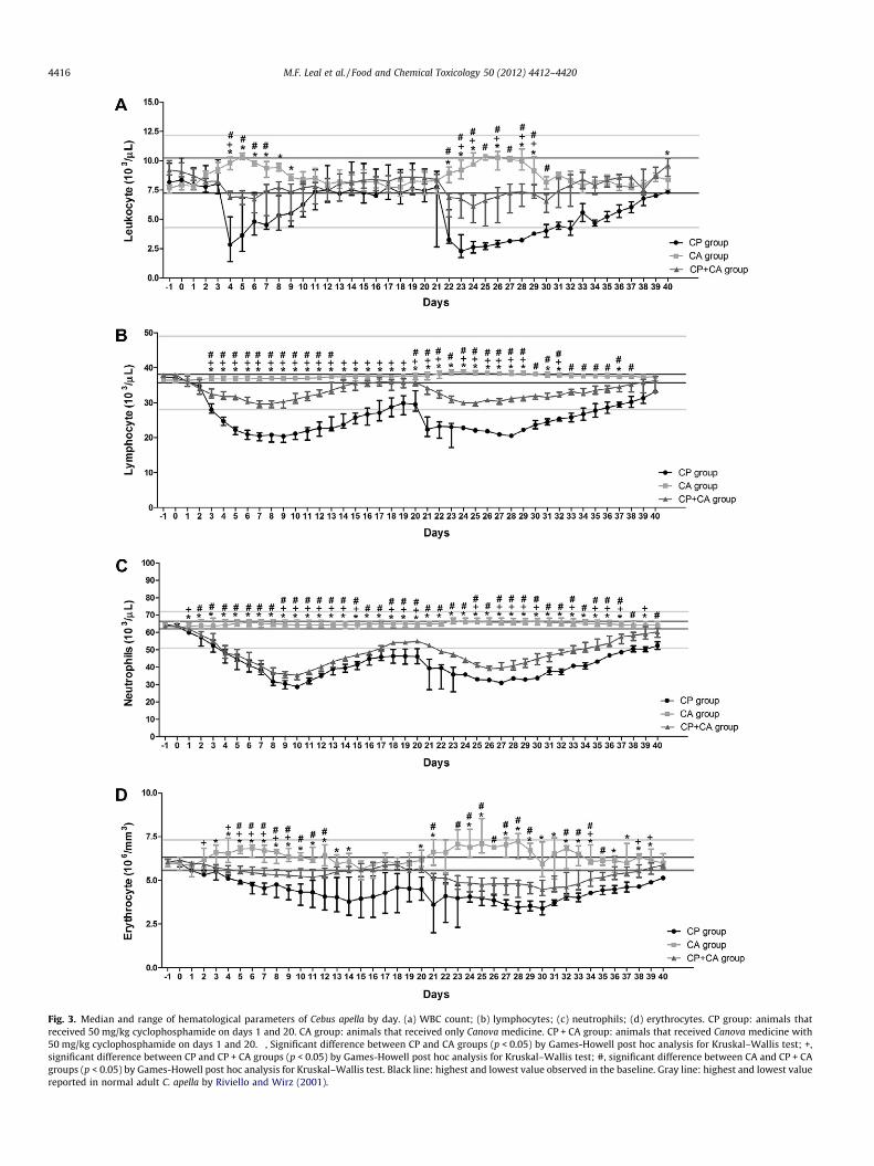
Fig. 3. Median and range of hematological parameters of Cebus apella by day. (a) WBC count; (b) lymphocytes; (c) neutrophils; (d) erythrocytes. CP group: animals thatreceived 50 mg/kg cyclophosphamide on days 1 and 20. CA group: animals that received only Canova medicine. CP + CA group: animals that received Canova medicine with50 mg/kg cyclophosphamide on days 1 and 20. �, Significant difference between CP and CA groups (p < 0.05) by Games-Howell post hoc analysis for Kruskal–Wallis test; +,significant difference between CP and CP + CA groups (p < 0.05) by Games-Howell post hoc analysis for Kruskal–Wallis test; #, significant difference between CA and CP + CAgroups (p < 0.05) by Games-Howell post hoc analysis for Kruskal–Wallis test. Black line: highest and lowest value observed in the baseline. Gray line: highest and lowest valuereported in normal adult C. apella by Riviello and Wirz (2001).
4416 M.F. Leal et al. / Food and Chemical Toxicology 50 (2012) 4412–4420
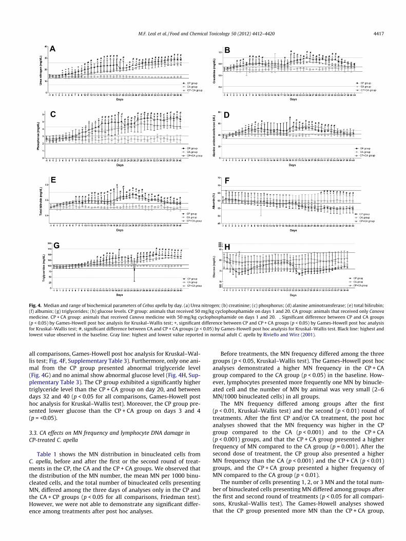
Fig. 4. Median and range of biochemical parameters of Cebus apella by day. (a) Urea nitrogen; (b) creatinine; (c) phosphorus; (d) alanine aminotransferase; (e) total bilirubin;(f) albumin; (g) triglycerides; (h) glucose levels. CP group: animals that received 50 mg/kg cyclophosphamide on days 1 and 20. CA group: animals that received only Canovamedicine. CP + CA group: animals that received Canova medicine with 50 mg/kg cyclophosphamide on days 1 and 20. �, Significant difference between CP and CA groups(p < 0.05) by Games-Howell post hoc analysis for Kruskal–Wallis test; +, significant difference between CP and CP + CA groups (p < 0.05) by Games-Howell post hoc analysisfor Kruskal–Wallis test; #, significant difference between CA and CP + CA groups (p < 0.05) by Games-Howell post hoc analysis for Kruskal–Wallis test. Black line: highest andlowest value observed in the baseline. Gray line: highest and lowest value reported in normal adult C. apella by Riviello and Wirz (2001).
M.F. Leal et al. / Food and Chemical Toxicology 50 (2012) 4412–4420 4417
all comparisons, Games-Howell post hoc analysis for Kruskal–Wal-lis test; Fig. 4F, Supplementary Table 3). Furthermore, only one ani-mal from the CP group presented abnormal triglyceride level(Fig. 4G) and no animal show abnormal glucose level (Fig. 4H, Sup-plementary Table 3). The CP group exhibited a significantly highertriglyceride level than the CP + CA group on day 20, and betweendays 32 and 40 (p < 0.05 for all comparisons, Games-Howell posthoc analysis for Kruskal–Wallis test). Moreover, the CP group pre-sented lower glucose than the CP + CA group on days 3 and 4(p = <0.05).
3.3. CA effects on MN frequency and lymphocyte DNA damage inCP-treated C. apella
Table 1 shows the MN distribution in binucleated cells fromC. apella, before and after the first or the second round of treat-ments in the CP, the CA and the CP + CA groups. We observed thatthe distribution of the MN number, the mean MN per 1000 binu-cleated cells, and the total number of binucleated cells presentingMN, differed among the three days of analyses only in the CP andthe CA + CP groups (p < 0.05 for all comparisons, Friedman test).However, we were not able to demonstrate any significant differ-ence among treatments after post hoc analyses.
Before treatments, the MN frequency differed among the threegroups (p < 0.05, Kruskal–Wallis test). The Games-Howell post hocanalyses demonstrated a higher MN frequency in the CP + CAgroup compared to the CA group (p < 0.05) in the baseline. How-ever, lymphocytes presented more frequently one MN by binucle-ated cell and the number of MN by animal was very small (2–6MN/1000 binucleated cells) in all groups.
The MN frequency differed among groups after the first(p < 0.01, Kruskal–Wallis test) and the second (p < 0.01) round oftreatments. After the first CP and/or CA treatment, the post hocanalyses showed that the MN frequency was higher in the CPgroup compared to the CA (p < 0.001) and to the CP + CA(p < 0.001) groups, and that the CP + CA group presented a higherfrequency of MN compared to the CA group (p = 0.001). After thesecond dose of treatment, the CP group also presented a higherMN frequency than the CA (p < 0.001) and the CP + CA (p < 0.01)groups, and the CP + CA group presented a higher frequency ofMN compared to the CA group (p < 0.01).
The number of cells presenting 1, 2, or 3 MN and the total num-ber of binucleated cells presenting MN differed among groups afterthe first and second round of treatments (p < 0.05 for all compari-sons, Kruskal–Wallis test). The Games-Howell analyses showedthat the CP group presented more MN than the CP + CA group,

Table 1Micronuclei distribution in binucleated cells from Cebus apella treated with cyclophosphamide and/or Canova.
Treatment Distribution of MN in BN [median (IR)] Total of MN MN/1000 BN [median (IR)]
1 MN 2 MN 3 MN
CP groupBefore treatmenta 2.00 (0.25) 0.50 (0.75) 0.00 (0.00) 3.50 (3.75) 1.75 (0.88)First treatment 172.50 (9.75) 60.00 (6.00) 25.00 (6.75) 367.00 (13.50) 183.75 (6.75)Second treatment 181.50 (15.00)b 67.50 (6.00)b 30.50 (7.00)b 408.00 (7.50)b 204.00 (3.75)b
CA groupBefore treatmenta 2.00 (0.50) 0.50 (1.00) 0.00 (0.00) 2.00 (0.50) 1.00 (0.25)First treatment 3.00 (0.5)d 0.50 (1.0)d 0.00 (0.00)d 4.00 (2.50)d 2.00 (1.25)d
Second treatment 2.50 (1.5)f 0.00 (0.5)f 0.00 (0.25)f 3.50 (2.25)f 1.75 (1.13)f
CP + CA groupBefore treatmenta 3.00 (0.25) 1.00 (0.24) 0.00 (0.00) 5.00 (0.25) 2.50 (0.13)c
First treatment 67.00 (5.75)d,e 42.50 (5.00)d,e 9.50 (4.75)d 179.00 (29.50)d,e 89.75 (14.75)d,e
Second treatment 77.50 (11.75)b,f,g 47.50 (7.50)b,f,g 11.00 (7.50)b,f 204.50 (50.25)b,f,g 102.25 (25.13)b,f,g
MN, micronuclei according Fenech (1993); BN, binucleated cells. IR: interquartile range. 8000 cells were analyzed per treatment for MN score, in which 1 MN represents thebinucleated cells presenting one MN, and so on. CP group: animals that received 50 mg/kg cyclophosphamide on days 1 and 20. CA group: animals that received only Canovamedicine. CP + CA group: animals that received Canova medicine with 50 mg/kg cyclophosphamide on days 1 and 20. Peripheral blood samples for lymphocyte-MN assaywere collected 24 h after CP, CA, or CP + CA treatments.
a Day 0b Significant difference among treatments (p < 0.05), intragroup analysis by Friedman test.c Significant difference compared to the CA group in the baseline (p < 0.05), by Games-Howell post hoc analysis for Kruskal–Wallis test.d Significant difference compared to the CP group after the first treatment (p < 0.05), by Games-Howell post hoc analysis for Kruskal–Wallis test.e Significant difference compared to the CA group after the first treatment (p < 0.05), by Games-Howell post hoc analysis. for Kruskal–Wallis testf Significant difference compared to the CP group after the second treatment (p < 0.05), by Games-Howell post hoc analysis for Kruskal–Wallis test.g Significant difference compared to the CA group after the second treatment (p < 0.05), by Games-Howell post hoc analysis for Kruskal–Wallis test.
4418 M.F. Leal et al. / Food and Chemical Toxicology 50 (2012) 4412–4420
which presented more MN than the CA group after the first andsecond treatments (p < 0.05 for all comparisons). For the compari-son of the number of cells presenting 3 MN between the CA andCP + CA group after the second round of treatment, we did not ob-serve a significant difference.
In addition, the frequency of MN was inversely correlated toWBC and to lymphocyte count in the CP (p < 0.01, q = �0.746;p < 0.01, q = �0.818, respectively; Spearman correlation) andCP + CA (p < 0.05, q = �0.669; p < 0.001, q = �0.944, respectively)groups.
Table 2 shows the DNA damage scores and index by the cometassay in C. apella from the CP, CA, and CP + CA groups. We also ob-served that comet classes 1, 3 and 4 and DNA DI differed amongbaseline, as well as the first and the second doses of CP in CP and
Table 2DNA damage scores and index by comet assay in Cebus apella treated with cyclophospham
Treatment DNA damage score [median (IR)]
0 1 2
CP groupBefore treatmenta 78.25 (6.25) 12.50 (1.63) 9.00 (3.00)First treatment 67.00 (2.75) 15.75 (1.13) 14.50 (1.38Second treatment 56.50 (3.00)b 21.00 (2.38)b 14.75 (0.75
CA groupBefore treatmenta 79.25 (1.00) 12.25 (2.88) 8.25 (2.88)First treatment 81.25 (3.88) 11.50 (2.50) 5.75 (2.38)Second treatment 80.75 (2.00) 12.00 (6.00) 7.75 (2.13)
CP + CA groupBefore treatmenta 78.00 (1.00) 11.50 (0.38) 9.25 (1.00)First treatment 71.50 (0.63) 17.00 (1.50) 11.00 (0.63Second treatment 65.75 (0.88)b 18.75 (1.00)b 12.50 (1.90
IR, interquartile range; DI, damage index. 400 cells were analyzed per treatment for DNA don days 1 and 20. CA group: animals that received only Canova medicine. CP + CA group:and 20. Peripheral blood samples for comet assay were collected 72 h after CP, CA, or C
a Day 0.b Significant difference among treatments (p < 0.05), intragroup analysis by Friedmanc Significant difference compared to the CP group after the first treatment (p < 0.05),d Significant difference compared to the CA group after the first treatment (p < 0.05),e Significant difference compared to the CP group after the second treatment (p < 0.05f Significant difference compared to the CA group after the second treatment (p < 0.0
CA + CP groups (p < 0.05 for all comparisons, Friedman test). Inaddition, comet class 2 also differed among the analyzed days inthe CP group (p < 0.05). However, we were not able to demonstrateany significant difference among treatments after post hocanalyses.
The DNA DI did not differ significantly among groups in thebaseline and differed significantly among groups after the first(p < 0.01, Kruskal–Wallis test) and second treatments (p < 0.01).The Games-Howell post hoc analyses showed that DNA DI was sig-nificantly higher in the CP group than the CA group after the first(p = 0.001) and second (p = 0.001) treatments. Moreover, the CPgroup presented higher DNA DI than the CP + CA group after thesecond round of treatment, but this was not statistically signifi-cant. Only a tendency to present a higher DNA DI was observed
ide and/or Canova.
DNA DI [Median (IR)]
3 4
0.50 (0.38) 0.00 (0.00) 33.50 (8.75)) 1.75 (0.88) 0.50 (1.13) 53.00 (6.00))b 4.00 (1.13)b 2.50 (1.13)b 74.25 (5.75)b
0.5 (0.5) 0.00 (0.00) 29.50 (4.63)c 0.75 (0.63) 0.00 (0.13) 26.25 (5.63)c
e 0.50 (0.38)e 0.00 (0.00)e 28.25 (4.63)e
0.75 (0.63) 0.00 (0.00) 33.75 (2.00))d 1.00 (0.25) 0.00 (0.00) 41.00 (1.50)d
) 1.75 (0.63)b,e 0.75 (0.63)b,e,f 52.75 (2.13)b,e,f
amage index analysis. CP group: animals that received 50 mg/kg cyclophosphamideanimals that received Canova medicine with 50 mg/kg cyclophosphamide on days 1P + CA treatments.
test.by Games-Howell post hoc analysis for Kruskal–Wallis test.by Games-Howell post hoc analysis for Kruskal–Wallis test.), by Games-Howell post hoc analysis for Kruskal–Wallis test.
5), by Games-Howell post hoc analysis for Kruskal–Wallis test.
M.F. Leal et al. / Food and Chemical Toxicology 50 (2012) 4412–4420 4419
in the CP group compared to the CP + CA group after the first roundof treatment (not statistically significant). In addition, the CP + CAgroup presented a significantly higher DNA DI than the CA groupafter the first (p < 0.01) and the second (p < 0.001) CP treatment.
A higher frequency of comet class 2 was also observed in the CP(p < 0.05, Games-Howell post hoc analysis) and CP + CA (p < 0.05)groups compared to the CA group after the first treatment. Afterthe second CP treatment, the distribution of comets classes 2, 3and 4 also increased in the CP group compared to CA group(p < 0.05, p = 0.001, p < 0.05, respectively). The CP group also pre-sented a higher frequency of comets class 3 and 4 compared tothe CP + CA group after the second treatment (p = 0.009, p < 0.05,respectively). After the second CP treatment, the CP + CA groupalso presented a higher frequency of comet class 4 compared tothe CA group (p < 0.05).
In addition, a direct correlation was observed between the fre-quency of MN and DNA DI in the CP (p < 0.001, q = 0.951; Spear-man correlation) and CP + CA (p < 0.01, q = 0.761) groups.Furthermore, the DNA DI was inversely correlated to WBC andlymphocyte count in the CP (p < 0.01, q = �0.8; p = 0.001,q = �0.864, respectively) and CP + CA (p < 0.05, q = �0.783;p < 0.01, q = �0.797, respectively) groups.
4. Discussion
In the present study, the CP + CA group presented body weightand hematology data between CP and CA groups. On the otherhand, the serum chemistry values observed in the CP + CA groupshowed an outcome more close to the CP group and different fromthe CA group. Thus, at the dose levels given, CA seems to present areduced effect regarding liver and kidney toxicity when comparedto myelotocicity and body weight effect. Since the CP + CA groupreceived CA and CP drugs at the same time and via the same route,we cannot exclude that CA components may interact with CP be-fore the CP exerts its toxic action, reducing the exposure of the ani-mal to CP.
In this study, animals of CP + CA group presented higher num-ber of WBC, lymphocytes, erythrocytes and neutrophils comparedto animals treated only with CP. This finding allows some hypoth-esis. Firstly, CA may up-regulate these cells in non-human prima-tes treated with CP. It was previously reported that CA canincrease directly or indirectly the number of lymphocytes (CoelhoMoreira et al., 2012; Sato et al., 2005). In addition, Abud et al.(2006) suggested that CA medicine also facilitates the productionof erythroid differentiation regulator, which would justify the in-crease of erythrocytes in the peripheral blood of C. apella treatedwith CP + CA compared to the CP group in some periods of thisstudy. However, further analyses are necessary to understandhow CA may act in neutrophils count.
Secondly, CA may have a cytoprotective effect on cells vulnera-ble to CP effects. This cyprotective effect might also lead to thereduction of DNA damage in the CP + CA group compared to theCP group. CP induces toxicity through its enzymatic conversionto active alkylating intermediates by cytochrome P-450 (CYP)monooxygenases. The CYP2B and 2C subfamily enzymes aremainly responsible for the convertion of CP into its active metabo-lites acrolein and phosphoramide mustard (Yu et al., 1999), whileCYP3A enzymes metabolize CP to dechloroethylcyclophosphamideand chloroacetaldehyde and seem to play a key role in the detox-ification of this akylating agent (Yu et al., 1999).
CA is a complex homeopathic medicine and the effect of itscompound is not completely understood. It was previously re-ported that Asa foetida, a CA compound, is able to reduce the levelsof cytochrome P450 and b5 and restore the antioxidant system inMNU-induced mammary carcinogenesis in rats (Mallikarjuna et al.,
2003). On the other hand, Lycopodium clavatum, another CA com-pound, may have potential to inactivate CYP3A4 (Tam et al.,2011). Since some plant extracts are able to modify the expressionand activity of CYP enzymes and, thus, may alter the activation ordetoxification of CP (Bagchi et al., 2002; Kumarappan et al., 2011;Ray et al., 2001), further studies are necessary to evaluate whetherCA has a cytoprotector effect in vivo and in its target cells and, then,determine if CA may be used as a chemopreventive agent in thetreatment of solid tumors.
Thirdly, CA may inhibit CP-mediated leukocytopenia and lym-phocytopenia. Thus, our findings suggest that CA treatment mini-mizes CP myelotoxicity in C. apella. The CA effects on WBC count,especially lymphocytes, may have human therapeutic applicationssuch as restoring the hematopoietic system after chemotherapy forsolid tumors.
Concerning serum chemistry values, the highest difference be-tween groups was observed in urea nitrogen and creatinine levels,indicators of glomerular function of the kidney. CP is nephrotoxicbesides being urotoxic thereby limiting its clinical utility (Hamsaand Kuttan, 2011). Here, we observed that CP induced an increasein urea nitrogen and creatinine level, as well as other parametersrelated to renal damage, compared to the pretreatment serum levelof C. apella. However, the CP group presented a significant increaseof this parameter compared to the CP + CA group. Thus, this studyshows that CA treatment may have a role in the reduction of CP-induced renal damage. However, additional investigations are nec-essary to evaluate if CA may act in renal cells.
5. Conclusions
CP induced abnormal WBC count, MN and DNA damage in allanimals. However, CA treated animals presented a higher leuko-cyte count than the animals without this treatment. Moreover,CA-treated animals presented lower frequency of MN and DNAdamage than animals without this medicine. Thus, CA treatmentminimizes CP myelotoxicity in C. apella.
Conflict of Interest
The authors declare that they have no conflicts of interest.
Acknowledgments
This study was supported by the Conselho Nacional de Desen-volvimento Científico e Tecnológico [550885/2007-2, R.R.B,M.A.C.S and I.D.C.G.S] and the Fundação de Amparo à Pesquisa doEstado de São Paulo [M.F.L].
Appendix A. Supplementary data
Supplementary data associated with this article can be found, inthe online version, at http://dx.doi.org/10.1016/j.fct.2012.09.002.
References
National Research Council, 1996. Guide for the Care and Use of Laboratory Animals,seventh ed. National Academy Press, Washington DC.
Sir David Weatherall FRS FMedSci’s working group, 2006. The use of non-humanprimates in research.
Abud, A.P., Cesar, B., Cavazzani, L.F., de Oliveira, C.C., Gabardo, J., Buchi Dde, F., 2006.Activation of bone marrow cells treated with Canova in vitro. Cell Biol. Int. 30,808–816.
Anderson, D., Bishop, J.B., Garner, R.C., Ostrosky-Wegman, P., Selby, P.B., 1995.Cyclophosphamide: review of its mutagenicity for an assessment of potentialgerm cell risks. Mutat. Res. 330, 115–181.
Bagchi, D., Bagchi, M., Stohs, S., Ray, S.D., Sen, C.K., Preuss, H.G., 2002. Cellularprotection with proanthocyanidins derived from grape seeds. Ann. NY. Acad.Sci. 957, 260–270.
4420 M.F. Leal et al. / Food and Chemical Toxicology 50 (2012) 4412–4420
Bukowski, R., 1999. Cytoprotection in the treatment of pediatric cancer: review ofcurrent strategies in adults and their application to children. Med. Pediatr.Oncol. 32, 124–134.
Burbano, R.R., Leal, M.F., da Costa, J.B., Bahia Mde, O., de Lima, P.D., Khayat, A.S.,Seligman, I.C., de Assumpcao, P.P., Buchi Dde, F., Smith Mde, A., 2009.Lymphocyte proliferation stimulated by activated human macrophagestreated with Canova. Homeopathy 98, 45–48.
CANOVA, Canova do Brazil web site. http://www.canovadobrasil.com.br. AccessedJune 26, 2012.
Carter, S.B., 1967. Effects of cytochalasins on mammalian cells. Nature 213, 261–264.
Cesar, B., Abud, A.P., de Oliveira, C.C., Cardoso, F., Gremski, W., Gabardo, J., BuchiDde, F., 2008. Activation of mononuclear bone marrow cells treated in vitrowith a complex homeopathic medication. Micron 39, 461–470.
Coelho Moreira, C.O., de Fatima Ferreira Borgesda Costa, J., Leal, M.F., FerreiradeAndrade, E., Rezende, A.P., Imbeloni, A.A., Pereira Carneiro Muniz, J.A., de ArrudaCardoso Smith, M., Burbano, R.R., de Assumpcao, P.P., 2012. Lymphocyteproliferation stimulated by activated Cebus apella macrophages treated with acomplex homeopathic immune response modifiers. Homeopathy 101, 74–79.
CPRFB, 1997. Farmacopéia homeopática brasileira. Atheneu, São Paulo.da Costa, J.F., Leal, M.F., Silva, T.C., Andrade Jr., E.F., Rezende, A.P., Muniz, J.A., Lacreta
Jr., A.C., Assumpcao, P.P., Calcagno, D.Q., Demachki, S., Rabenhorst, S.H., SmithMde, A., Burbano, R.R., 2011. Experimental gastric carcinogenesis in Cebus apellanonhuman primates. PLoS One 6, e21988.
Da Rocha Piemonte, M., De Freitas Buchi, D., 2002. Analysis of IL-2, IFN-gamma andTNF-alpha production, alpha5 beta1 integrins and actin filaments distributionin peritoneal mouse macrophages treated with homeopathic medicament. J.Submicrosc. Cytol. Pathol. 34, 255–263.
de Oliveira, C.C., de Oliveira, S.M., Godoy, L.M., Gabardo, J., Buchi Dde, F., 2006.Canova, a Brazilian medical formulation, alters oxidative metabolism of micemacrophages. J. Infect. 52, 420–432.
Fenech, M., 1993. The cytokinesis-block micronucleus technique: a detaileddescription of the method and its application to genotoxicity studies inhuman populations. Mutat. Res. 285, 35–44.
Hamsa, T.P., Kuttan, G., 2011. Protective role of Ipomoea obscura (L.) on cyclo-phosphamide-induced uro- and nephrotoxicities by modulating antioxidantstatus and pro-inflammatory cytokine levels. Inflammopharmacology 19, 155–167.
Hosseinimehr, S.J., Karami, M., 2005. Chemoprotective effects of captopril againstcyclophosphamide-induced genotoxicity in mouse bone marrow cells. Arch.Toxicol. 79, 482–486.
Igarashi, M., Nagata, M., Itoh, S., Yamoto, T., Tsuda, S., 2010. Relationship betweenDNA damage and micronucleus in mouse liver. J. Toxicol. Sci. 35, 881–889.
Jena, G., Vikram, A., Tripathi, D.N., Ramarao, P., 2010. Use of chemoprotectants inchemotherapy and radiation therapy: the challenges of selecting an appropriateagent. Integr. Cancer Ther. 9, 253–258.
Kumarappan, C., Vijayakumar, M., Thilagam, E., Balamurugan, M., Thiagarajan, M.,Senthil, S., Das, S.C., Mandal, S.C., 2011. Protective and curative effects ofpolyphenolic extracts from Ichnocarpus frutescense leaves on experimentalhepatotoxicity by carbon tretrachloride and tamoxifen. Ann. Hepatol. 10, 63–72.
Lopes, L., Godoy, L.M., de Oliveira, C.C., Gabardo, J., Schadeck, R.J., de Freitas Buchi,D., 2006. Phagocytosis, endosomal/lysosomal system and other cellularaspectsof macrophage activation by Canova medication. Micron 37, 277–287.
Mallikarjuna, G.U., Dhanalakshmi, S., Raisuddin, S., Rao, A.R., 2003.Chemomodulatory influence of Ferula asafoetida on mammary epithelialdifferentiation, hepatic drug metabolizing enzymes, antioxidant profiles andN-methyl-N-nitrosourea-induced mammary carcinogenesis in rats. BreastCancer Res. Treat. 81, 1–10.
Moreira, C.O.C., da Costa Jde, F., Leal, M.F., Andrade, E.F., Rezende, A.P., Imbeloni,A.A., Muniz, J.A.P.C., Smith, M.A.C., Burbano, R.R., Assumpcao, P.P., 2012.Lymphocyte proliferation stimulated by activated Cebus apella macrophagestreated with a complex homeopathic immune response modifiers. Homeopathy101, 74–79.
Pereira, W.K., Lonardoni, M.V., Grespan, R., Caparroz-Assef, S.M., Cuman, R.K.,Bersani-Amado, C.A., 2005. Immunomodulatory effect of Canova medication onexperimental Leishmania amazonensis infection. J. Infect. 51, 157–164.
Preston, R.J., San Sebastian, J.R., McFee, A.F., 1987. The in vitro human lymphocyteassay for assessing the clastogenicity of chemical agents. Mutat. Res. 189, 175–183.
Ray, S.D., Parikh, H., Hickey, E., Bagchi, M., Bagchi, D., 2001. Differential effects ofIH636 grape seed proanthocyanidin extract and a DNA repair modulator 4-aminobenzamide on liver microsomal cytochrome 4502E1-dependent anilinehydroxylation. Mol. Cell Biochem. 218, 27–33.
Riviello, M.C., Wirz, A., 2001. Haematology and blood chemistry of Cebus apella inrelation to sex and age. J. Med. Primatol. 30, 308–312.
Sato, D.Y., Wal, R., de Oliveira, C.C., Cattaneo, R.I., Malvezzi, M., Gabardo, J., BuchiDde, F., 2005. Histopathological and immunophenotyping studies on normaland sarcoma 180-bearing mice treated with a complex homeopathicmedication. Homeopathy 94, 26–32.
Schuurman, H.J., Smith, H.T., Cozzi, E., 2005. Tolerability of cyclosphosphamide andmethotrexate induction immunosuppression in nonhuman primates.Toxicology 213, 1–12.
Seligmann, I.C., Lima, P.D., Cardoso, P.C., Khayat, A.S., Bahia, M.O., Buchi Dde, F.,Cabral, I.R., Burbano, R.R., 2003. The anticancer homeopathic composite‘‘Canova method’’ is not genotoxic for human lymphocytes in vitro. Genet.Mol. Res. 2, 223–228.
Sharma, N., Trikha, P., Athar, M., Raisuddin, S., 1999. Protective effect of Cassiaoccidentalis extract on chemical-induced chromosomal aberrations in mice.Drug Chem. Toxicol. 22, 643–653.
Singh, N.P., McCoy, M.T., Tice, R.R., Schneider, E.L., 1988. A simple technique forquantitation of low levels of DNA damage in individual cells. Exp. Cell Res. 175,184–191.
Speit, G., Hartmann, A., 1999. The comet assay (single-cell gel test). A sensitivegenotoxicity test for the detection of DNA damage and repair. Methods Mol.Biol. 113, 203–212.
Stolarska, M., Mlynarski, W., Zalewska-Szewczyk, B., Bodalski, J., 2006.Cytoprotective effect of amifostine in the treatment of childhood neoplasticdiseases–a clinical study including the pharmacoeconomic analysis. Pharmacol.Rep. 58, 30–34.
Tam, T.W., Liu, R., Arnason, J.T., Krantis, A., Staines, W.A., Haddad, P.S., Foster, B.C.,2011. Cree antidiabetic plant extracts display mechanism-based inactivation ofCYP3A4. Can. J. Physiol. Pharmacol. 89, 13–23.
Torres, L.B., Silva Araujo, B.H., Gomes de Castro, P.H., Romero Cabral, F., SargesMarruaz, K., Silva Araujo, M., Gomes da Silva, S., Muniz, J.A., Cavalheiro, J.A.,2010. The use of new world primates for biomedical research: an overview ofthe last four decades. Am. J. Primatol. 72, 1055–1061.
Vasquez, M.Z., 2010. Combining the in vivo comet and micronucleus assays: apractical approach to genotoxicity testing and data interpretation. Mutagenesis25, 187–199.
WHO, World Health Organization. WHO web site. http://www.who.int/cancer/en/.Accessed January 10, 2010.
Yu, L.J., Drewes, P., Gustafsson, K., Brain, E.G., Hecht, J.E., Waxman, D.J., 1999. In vivomodulation of alternative pathways of P-450-catalyzed cyclophosphamidemetabolism: impact on pharmacokinetics and antitumor activity. J. Pharmacol.Exp. Ther. 288, 928–937.