Embed Size (px)
Citation preview

1. IntroductionLow pressure plasma based processes are a versa-tile tool for surface modification of polymers.Depending on the plasma parameters, plasma etch-ing or material deposition can predominate [1]. Inthe intermediate case, a surface layer with a thick-ness of few tens of nanometers is modified [2]. Thistype of plasma treatment is relevant for a wide vari-ety of applications. With a simple one-step proce-dure the surface properties of the polymer can be
changed significantly while the bulk propertiesremain unchanged. On the other hand, there arewell-known drawbacks of this technique, as theheterogeneity of the obtained surface chemistry [3]and the lack of longterm stability (hydrophobicrecovery) [4]. However, low pressure plasma treat-ment of polymers is an attractive approach, at leastfrom a technological point of view, and manystrategies were proposed to avoid or minimize theunfavourable mentioned side-effects. These
70
*Corresponding author, e-mail: [email protected]© BME-PT and GTE
Fluorination of poly(dimethylsiloxane) surfaces bylow pressure CF4 plasma – physicochemical and antifoulingproperties
A. L. Cordeiro1*, M. Nitschke1, A. Janke1, R. Helbig1, F. D’Souza2, G. T. Donnelly2,P. R. Willemsen2, C. Werner1
1Leibniz Institute of Polymer Research Dresden, Max Bergmann Center of Biomaterials Dresden; Hohe Str. 6; Dresden,Germany
2TNO Science and Industry, Bevesierweg MML (Harssens), Den Helder, The Netherlands
Received 8 November 2008; accepted in revised form 22 December 2008
Abstract. Fluorinated surface groups were introduced into poly(dimethylsiloxane) (PDMS) coatings by plasma treatmentusing a low pressure radio frequency discharge operated with tetrafluoromethane. Substrates were placed in a remote posi-tion downstream the discharge. Discharge power and treatment time were tuned to alter the chemical composition of theplasma treated PDMS surface. The physicochemical properties and stability of the fluorine containing PDMS were charac-terized by X-ray photoelectron spectroscopy (XPS), atomic force microscopy (AFM) and contact angle measurements.Smooth PDMS coatings with a fluorine content up to 47% were attainable. The CF4 plasma treatment generated a harder,non-brittle layer at the top-most surface of the PDMS. No changes of surface morphology were observed upon one weekincubation in aqueous media. Surprisingly, the PDMS surface was more hydrophilic after the introduction of fluorine. Thismay be explained by an increased exposure of oxygen containing moieties towards the surface upon re-orientation of fluo-rinated groups towards the bulk, and/or be a consequence of oxidation effects associated with the plasma treatment. Exper-iments with strains of marine bacteria with different surface energies, Cobetia marina and Marinobacterhydrocarbonoclasticus, showed a significant decrease of bacteria attachment upon fluorination of the PDMS surface. Alto-gether, the CF4 plasma treatments successfully introduced fluorinated groups into the PDMS, being a robust and versatilesurface modification technology that may find application where a minimization of bacterial adhesion is required.
Keywords: coatings, plasma treatment, poly(dimethylsiloxane), fluorination, marine bacteria
eXPRESS Polymer Letters Vol.3, No.2 (2009) 70–83Available online at www.expresspolymlett.comDOI: 10.3144/expresspolymlett.2009.11

include the covalent grafting of appropriate mole-cules onto plasma activated surfaces or the durableadsorption of polyelectrolytes on charged surfaces(as obtained for example after ammonia plasmatreatment) [5]. In addition to modification of sur-face chemical composition, plasma techniqueshave also been used to modify the surface morphol-ogy (roughening) of polymeric surfaces aiming forexample at changes in wetting behaviour [6–8].Poly(dimethylsiloxane) (PDMS) is a commerciallyavailable type of silicone rubber widely used in thefabrication of medical [9] and microfluidic devices[10], and as foul-release coating to control marinebiofouling [11, 12]. Foul-release coatings are cur-rently a successful non-toxic, environmentallyfriendly alternative to classical biocide-containingcoatings [12]. Commercially available foul-releasecoatings are based on silicone elastomers sincethese possess a combination of properties that min-imize the chemical and mechanical locking of thefouling species [13].In this paper we report on the fluorination of PDMS(Sylgard 184) by application of tetrafluoromethane(CF4) plasma to combine the bulk elasticity ofPDMS with a desirable surface chemistry, withoutintroducing changes in surface roughness. Towardsthis goal, a customized low pressure plasma set-upwith a sample position far away from the excitationvolume of the discharge was used to fabricate sur-faces with a particularly high fluorine content con-sisting mainly of CF2 and CF3 moieties. Thisapproach based on remote plasma configurationensures an especially smooth but efficient treat-ment. The properties and stability of the fluorinecontaining PDMS were characterized in detail con-cerning chemical composition by X-ray photoelec-tron spectroscopy (XPS), morphology and elasticityby atomic force microscopy (AFM) and wetting bycontact angle measurements. The antifoulingpotentialities of the CF4 plasma treated PDMStowards microbes were evaluated by testing theattachment and adhesion strength of two species ofbacteria (Marinobacter hydrocarbonoclasticus andCobetia marina). These bacteria form biofilms thatcoat surfaces when in contact with sea water –either when surfaces are immersed in the sea (e.g.ship hulls) or are exposed to sea water (as in cool-ing systems) [14].
2. Experimental
2.1. Materials
The silicone elastomer Sylgard 184 and the Primer1200 OS were purchased from Dow Corning (Ger-many). Tetrafluoromethane (99.99%) was obtainedfrom Messer Griesheim (Germany). For stabilitytests and surface characterization experiments anartificial sea water (ASW*) solution was preparedusing the five main components of natural seawater as described in ASTM D 1141–98 (24.53 g/lNaCl; 5.20 g/l MgCl2; 4.09 g/l Na2SO4; 0.695 g/lKCl; 1.16 g/l CaCl2) [15]. The ASW* solution wasprepared using high purity salts and autoclavedbefore use. For the assays with marine bacteria,SSP growth medium (containing sea salt and pep-tone) was purchased from Sigma Aldrich (Ger-many), and natural sea water (SW) was collected inDen Helder, The Netherlands.
2.2. Sample preparation
Poly(dimethylsiloxane) (PDMS) coatings were pre-pared on inorganic carriers of variable dimensionsfor further modification by low pressure CF4
plasma treatment. The PDMS coatings were fabri-cated using the two-part elastomer Sylgard 184.The elastomer was prepared using a ratio base tocuring agent 10:1 (weight), following the sugges-tion of the manufacturer. The base and curing agentwere mixed using a homogeneizer (Kontes; USA)for 3 min and then degassed under vacuum until allair bubbles were removed (ca. 30 min). The poly-mer was then deposited onto either silicon wafersof different sizes for physicochemical analysis, oronto standard microscope glass slides (26×76 mm2)for biological testing.Silicon wafers were coated by pouring definedamounts of pre-polymer (depending on the waferdimensions) onto the cleaned surface. The pre-polymer was air-spread for complete, homoge-neous coverage of the surface. The coatings werecured at 65°C for 5 h 45 min.For preparation of PDMS coatings onto standardmicroscopy slides used to test bacteria biofilm for-mation and release, an in-house film preparationsystem was developed (Figure 1). This consists of ametallic base onto which the glass slides were
71
Cordeiro et al. – eXPRESS Polymer Letters Vol.3, No.2 (2009) 70–83
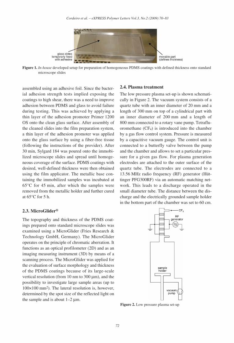
assembled using an adhesive foil. Since the bacter-ial adhesion strength tests implied exposing thecoatings to high shear, there was a need to improveadhesion between PDMS and glass to avoid failureduring testing. This was achieved by applying athin layer of the adhesion promoter Primer 1200OS onto the clean glass surface. After assembly ofthe cleaned slides into the film preparation system,a thin layer of the adhesion promoter was appliedonto the glass surface by using a fiber-free tissue(following the instructions of the provider). After30 min, Sylgard 184 was poured onto the immobi-lized microscope slides and spread until homoge-neous coverage of the surface. PDMS coatings withdesired, well-defined thickness were then obtainedusing the film applicator. The metallic base con-taining the immobilized samples was incubated at65°C for 45 min, after which the samples wereremoved from the metallic holder and further curedat 65°C for 5 h.
2.3. MicroGlider®
The topography and thickness of the PDMS coat-ings prepared onto standard microscope slides wasexamined using a MicroGlider (Fries Research &Technology GmbH, Germany). The MicroGlideroperates on the principle of chromatic aberration. Itfunctions as an optical profilometer (2D) and as animaging measuring instrument (3D) by means of ascanning process. The MicroGlider was applied forthe evaluation of surface morphology and thicknessof the PDMS coatings because of its large-scalevertical resolution (from 10 nm to 300 μm), and thepossibility to investigate large sample areas (up to100×100 mm2). The lateral resolution is, however,determined by the spot size of the reflected light onthe sample and is about 1–2 μm.
2.4. Plasma treatment
The low pressure plasma set-up is shown schemati-cally in Figure 2. The vacuum system consists of aquartz tube with an inner diameter of 20 mm and alength of 300 mm on top of a cylindrical part withan inner diameter of 200 mm and a length of800 mm connected to a rotary vane pump. Tetraflu-oromethane (CF4) is introduced into the chamberby a gas flow control system. Pressure is measuredby a capacitive vacuum gauge. The control unit isconnected to a butterfly valve between the pumpand the chamber and allows to set a particular pres-sure for a given gas flow. For plasma generationelectrodes are attached to the outer surface of thequartz tube. The electrodes are connected to a13.56 MHz radio frequency (RF) generator (Hüt-tinger PFG300RF) via an automatic matching net-work. This leads to a discharge operated in thesmall diameter tube. The distance between the dis-charge and the electrically grounded sample holderin the bottom part of the chamber was set to 60 cm.
72
Cordeiro et al. – eXPRESS Polymer Letters Vol.3, No.2 (2009) 70–83
Figure 1. In-house developed setup for preparation of homogeneous PDMS coatings with defined thickness onto standardmicroscope slides
Figure 2. Low pressure plasma set-up
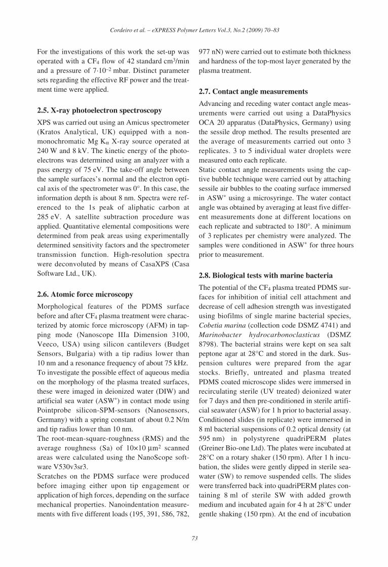
For the investigations of this work the set-up wasoperated with a CF4 flow of 42 standard cm3/minand a pressure of 7·10–2 mbar. Distinct parametersets regarding the effective RF power and the treat-ment time were applied.
2.5. X-ray photoelectron spectroscopy
XPS was carried out using an Amicus spectrometer(Kratos Analytical, UK) equipped with a non-monochromatic Mg Kα X-ray source operated at240 W and 8 kV. The kinetic energy of the photo-electrons was determined using an analyzer with apass energy of 75 eV. The take-off angle betweenthe sample surfaces’s normal and the electron opti-cal axis of the spectrometer was 0°. In this case, theinformation depth is about 8 nm. Spectra were ref-erenced to the 1s peak of aliphatic carbon at285 eV. A satellite subtraction procedure wasapplied. Quantitative elemental compositions weredetermined from peak areas using experimentallydetermined sensitivity factors and the spectrometertransmission function. High-resolution spectrawere deconvoluted by means of CasaXPS (CasaSoftware Ltd., UK).
2.6. Atomic force microscopy
Morphological features of the PDMS surfacebefore and after CF4 plasma treatment were charac-terized by atomic force microscopy (AFM) in tap-ping mode (Nanoscope IIIa Dimension 3100,Veeco, USA) using silicon cantilevers (BudgetSensors, Bulgaria) with a tip radius lower than10 nm and a resonance frequency of about 75 kHz.To investigate the possible effect of aqueous mediaon the morphology of the plasma treated surfaces,these were imaged in deionized water (DIW) andartificial sea water (ASW*) in contact mode usingPointprobe silicon-SPM-sensors (Nanosensors,Germany) with a spring constant of about 0.2 N/mand tip radius lower than 10 nm.The root-mean-square-roughness (RMS) and theaverage roughness (Sa) of 10×10 μm2 scannedareas were calculated using the NanoScope soft-ware V530v3sr3.Scratches on the PDMS surface were producedbefore imaging either upon tip engagement orapplication of high forces, depending on the surfacemechanical properties. Nanoindentation measure-ments with five different loads (195, 391, 586, 782,
977 nN) were carried out to estimate both thicknessand hardness of the top-most layer generated by theplasma treatment.
2.7. Contact angle measurements
Advancing and receding water contact angle meas-urements were carried out using a DataPhysicsOCA 20 apparatus (DataPhysics, Germany) usingthe sessile drop method. The results presented arethe average of measurements carried out onto 3replicates. 3 to 5 individual water droplets weremeasured onto each replicate.Static contact angle measurements using the cap-tive bubble technique were carried out by attachingsessile air bubbles to the coating surface immersedin ASW* using a microsyringe. The water contactangle was obtained by averaging at least five differ-ent measurements done at different locations oneach replicate and subtracted to 180°. A minimumof 3 replicates per chemistry were analyzed. Thesamples were conditioned in ASW* for three hoursprior to measurement.
2.8. Biological tests with marine bacteria
The potential of the CF4 plasma treated PDMS sur-faces for inhibition of initial cell attachment anddecrease of cell adhesion strength was investigatedusing biofilms of single marine bacterial species,Cobetia marina (collection code DSMZ 4741) andMarinobacter hydrocarbonoclasticus (DSMZ8798). The bacterial strains were kept on sea saltpeptone agar at 28°C and stored in the dark. Sus-pension cultures were prepared from the agarstocks. Briefly, untreated and plasma treatedPDMS coated microscope slides were immersed inrecirculating sterile (UV treated) deionized waterfor 7 days and then pre-conditioned in sterile artifi-cial seawater (ASW) for 1 h prior to bacterial assay.Conditioned slides (in replicate) were immersed in8 ml bacterial suspensions of 0.2 optical density (at595 nm) in polystyrene quadriPERM plates(Greiner Bio-one Ltd). The plates were incubated at28°C on a rotary shaker (150 rpm). After 1 h incu-bation, the slides were gently dipped in sterile sea-water (SW) to remove suspended cells. The slideswere transferred back into quadriPERM plates con-taining 8 ml of sterile SW with added growthmedium and incubated again for 4 h at 28°C undergentle shaking (150 rpm). At the end of incubation
73
Cordeiro et al. – eXPRESS Polymer Letters Vol.3, No.2 (2009) 70–83
the slides were again gently rinsed, then placed intoslide holders and partially air-dried. The attachedbiomass was quantified by staining the attachedcells with 1.5 μM of the fluorochrome SYTO13(Invitrogen) and measuring absorption with aTecan plate reader (GENios, Magellan software)[16].The adhesion strength of adhered bacteria wasquantified using a rotating drum test that was origi-nally designed for the determination of antifoulingperformance of marine antifouling coatings asdescribed in ASTM D 4939 [17]. After the growthstep, replicate slides were mounted on the surfaceof a custom-made high-speed rotating drum [18].The drum was then rotated with the surface speedof ~340 m/min for 10 min in SW. This rotationalspeed of the drum exposes the bacteria to shearstress (turbulent flow), removing bacteria cellsfrom the surfaces. The remaining bacteria werequantified using the stain SYTO13 as describedabove. The results were expressed as the percent-age of bacteria removed or detached by shear stress[(RFU of attached bacteria before release – RFU ofbacteria remaining after release)/RFU of attachedbacteria before release ×100]. Confidence limits of95% were calculated.
3. Results and discussion
3.1. Coating preparation
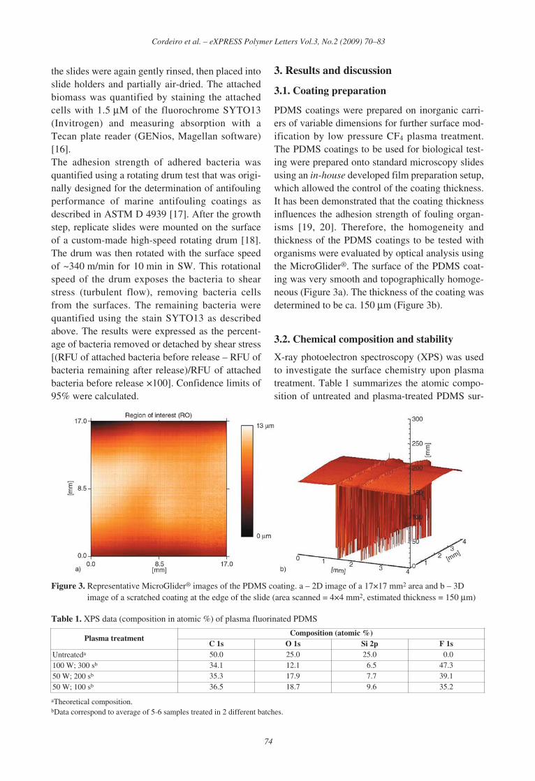
PDMS coatings were prepared on inorganic carri-ers of variable dimensions for further surface mod-ification by low pressure CF4 plasma treatment.The PDMS coatings to be used for biological test-ing were prepared onto standard microscopy slidesusing an in-house developed film preparation setup,which allowed the control of the coating thickness.It has been demonstrated that the coating thicknessinfluences the adhesion strength of fouling organ-isms [19, 20]. Therefore, the homogeneity andthickness of the PDMS coatings to be tested withorganisms were evaluated by optical analysis usingthe MicroGlider®. The surface of the PDMS coat-ing was very smooth and topographically homoge-neous (Figure 3a). The thickness of the coating wasdetermined to be ca. 150 μm (Figure 3b).
3.2. Chemical composition and stability
X-ray photoelectron spectroscopy (XPS) was usedto investigate the surface chemistry upon plasmatreatment. Table 1 summarizes the atomic compo-sition of untreated and plasma-treated PDMS sur-
74
Cordeiro et al. – eXPRESS Polymer Letters Vol.3, No.2 (2009) 70–83
Figure 3. Representative MicroGlider® images of the PDMS coating. a – 2D image of a 17×17 mm2 area and b – 3Dimage of a scratched coating at the edge of the slide (area scanned = 4×4 mm2, estimated thickness = 150 μm)
Table 1. XPS data (composition in atomic %) of plasma fluorinated PDMS
aTheoretical composition.bData correspond to average of 5-6 samples treated in 2 different batches.
Plasma treatmentComposition (atomic %)
C 1s O 1s Si 2p F 1sUntreateda 50.0 25.0 25.0 00.0100 W; 300 sb 34.1 12.1 06.5 47.350 W; 200 sb 35.3 17.9 07.7 39.150 W; 100 sb 36.5 18.7 09.6 35.2

faces for different treatment conditions. The fluo-rine content increased with power and treatmenttime up to 47%. For comparison, poly(tetrafluo-roethylene) (PTFE), the most common fully fluori-nated polymer, has a fluorine content of 66.6%.To further characterize the chemical structure ofthe plasma treated PDMS surfaces, high resolutionC1s spectra were recorded. It was assumed, that thespectra consist of five components correspondingto aliphatic carbon (285.0 eV), –C–CF– (287.6 eV),–CF– (290.4 eV), –CF2– (292.0 eV) and –CF3
(294.0 eV) [21]. To evaluate the C1s spectra of thefluorinated PDMS surfaces obtained under differ-ent plasma treatment parameters, a peak deconvo-lution procedure was applied. Five componentpeaks with a given shape (Gaussian-Lorentzianratio 50:50) were set to the energy values men-tioned above. The fit procedure was allowed tovary the component energies except 285 eV withina range of ±0.5 eV, the component intensities and acommon value for the full width at half maximum.Deconvoluted C1s spectra of fluorinated PDMSprepared using different plasma treatment condi-tions are presented in Figure 4. The assumptions ofthe peak deconvolution model were adequate todescribe the shape of the C1s spectra, agreeing with
other studies where complex fluorinated polymersystems, as obtained by plasma-based techniques,were investigated [22–24]. All deconvoluted spec-tra show the full range of differently fluorinatedcarbon species suggesting a significantly branchedand/or cross-linked structure. For the investigatedparameter range the relative amount of highly fluo-rinated species increases continuously with increas-ing power and treatment time ([CF3]:[CxHy]increased from 0.13 to 0.45, and [CF2]:[CxHy]increased from 0.62 to 1.71 (Figure 4)).Considering potential underwater applications, andthe evidence of chemical re-structuring of siliconesupon contacting water [25], we have evaluated thechemical stability of the plasma treated PDMS inaqueous media. Additionally, to assess the influ-ence of ionic strength on the chemical stability ofthe plasma treated samples, and envisaging the pos-sibility to use these surfaces in marine environ-ment, the fluorine content was determined by XPSimmediately after plasma treatment, after storage inambient conditions, after incubation in recirculat-ing deionized water (DIW), and in artificial seawater (ASW*) for up to one month (Figure 5).The fluorine content decreased by 6% when sam-ples were simply stored in ambient conditions for
75
Cordeiro et al. – eXPRESS Polymer Letters Vol.3, No.2 (2009) 70–83
Figure 4. Representative XPS C1s spectra of CF4 plasma treated PDMS using the parameters: 50 W, 100 s (left), and100 W, 300 s (right). Peak deconvolution (1) CxHy, (2) –C–CF–, (3) –CF–, (4) –CF2–, and (5) –CF3.

30 days pointing towards a re-orientation/migrationof the fluorine groups away from the surface duringaging. The introduction of functional groups ontothe surface of silicone has been recognized to lackstability due to the reorganization of silicone elas-tomers over time [26, 27, 29, 31, 32]. The phenom-ena of hydrophobic recovery upon PDMS plasmaoxidation has been extensively investigated, andexplained namely by the reorientation of surfacehydrophilic groups towards the bulk and/or reorien-tation of non-polar groups in the bulk towards thesurface, by the diffusion of pre-existing low molec-ular weight (LMW) species through the bulk to thesurface, or migration of created LMW species dur-ing treatment to the surface [26]. These sameeffects are believed to be responsible for theobserved decrease of fluorine content on the sur-face plasma treated PDMS during aging.When samples were incubated in aqueous media,the fluorine content suffered a steep initial decrease(Figure 5). The fluorine content further decreasedafter 1 week of incubation, but no additional signif-icant differences could be observed after 4 weeks.This decrease can be explained by re-arrangementsof fluorinated groups when the coatings areimmersed in water, and by the removal of lowmolecular weight compounds generated by plasmatreatment. The loss of fluorine was significantlyhigher when the surfaces were incubated in ASW*
(loss of 45% of initial content) as compared withDIW (loss of 29% of initial content), revealing the
deleterious effect of the presence of salt on the sta-bility of the plasma treated surfaces.
3.3. Morphology
The plasma treatment of polymer surfaces maygreatly alter their morphology [28, 29]. For thisreason, we have investigated the morphologicalfeatures of the PDMS surface before and afterplasma treatment by atomic force microscopy(AFM) in tapping mode. The surface of the plasmatreated PDMS, even using the ‘harshest’ plasmatreatment parameters tested (Figure 6b), was verysmooth and comparable to the non-plasma treatedsurface (Figure 6a). Possible effects of plasmatreatment time on surface morphology were inves-tigated for different treatment times (100, 200, and300 s). The surfaces were very smooth (RMS =0.5–0.6 nm), no significant morphological differ-ences between treatments could be detected (resultsnot shown).Additionally, as the exposure of silicones to aque-ous media (sea water, phosphate buffered saline)can alter surface morphology [29, 30], the morpho-logical features of the plasma treated PDMS whenincubated in DIW and ASW* for 7 days were inves-tigated. Typical height images of the PDMSexposed to the ‘harshest’ plasma conditions testedwhen incubated in aqueous media are presented inFigure 6c (DIW) and Figure 6d (ASW*). No mor-phological changes were observed upon incubationin either of the media tested.
3.4. Mechanical properties
It has been reported that the plasma treatment ofPDMS, using a variety of different gases, generatesa brittle silica-like layer at the PDMS surface [29,31–35]. The formation of this hard layer can beexplained by the surface oxidation and the etchingeffects associated with the plasma treatment,exposing some of the silica filler to the surface ofthe PDMS [29]. In order to investigate the extent ofthe impact of the CF4 plasma treatment on themechanical properties of the PDMS, AFM imagingwas performed after indentation measurements andafter surface scratching with the AFM tip underhigh load (Figure 7).The results showed that the CF4 plasma treatmentresulted in the formation of a harder layer on the
76
Cordeiro et al. – eXPRESS Polymer Letters Vol.3, No.2 (2009) 70–83
Figure 5. Fluorine content (atomic %) of CF4 plasmatreated PDMS. After plasma treatment (time = 0),after storage in ambient conditions (•), afterincubation in recirculating DIW ( ), and afterincubation in recirculating ASW* ( ).

top-most surface of the PDMS, as higher force(>195 nN) was needed to cut through the surface,as compared with the untreated PDMS (data notshown). An apparent wrinkling of the surface wasobserved (Figure 7) as compared with the charac-teristic cracking of brittle surfaces [31–33, 35]. Thetop-most layer seems to elongate under stress,resulting in local thinning of the layer, beingreflected on an apparent wrinkling.The variety and extent of plasma-based surfacemodification effects, in particular the alteration ofelastic properties of PDMS, depend on a multitudeof interaction mechanisms, each of them acting ona characteristic depth scale. In low pressure plasma
reactors an effect on the scale of several tens ofnanometers is most probably attributed to (vacuum)ultraviolet irradiation. As the ultraviolet irradianceof plasma sources with different process gases anddifferent geometries can be different by orders ofmagnitude [36], the mechanical properties of thetreated surface may vary over a certain extend. Thispossibly explains the difference between our exper-imental findings (non-brittle surface) and the obser-vations reported in the literature (brittle surface)[31–33, 35].The differences in mechanical properties ofuntreated and CF4 plasma treated PDMS surfaceswere further investigated by performing nanoin-
77
Cordeiro et al. – eXPRESS Polymer Letters Vol.3, No.2 (2009) 70–83
Figure 6. AFM height images of a – PDMS (RMS = 1.15 nm), and b – plasma fluorinated PDMS (plasma treatment con-ditions: 100 W; 300 s) after preparation (RMS = 1.34 nm); c – after 7 days incubation in recirculating DIW(RMS = 1.33 nm), and d – after 7 days incubation in recirculating ASW* (RMS = 1.58 nm). a) and b) imagingin air using tapping mode, c) imaging in DIW using contact mode, and d) imagining in ASW* using contactmode.

dentation measurements with five different loads.The analysis of the obtained curves allowed to con-clude on the presence of a possible double-layerstructure, estimate its thickness, and evaluate thehardness of the top-most surface of the PDMS. Theforce-separation curves of unmodified PDMSclearly differed from the curves obtained forplasma treated PDMS (results not shown). In thecase of the plasma treated surfaces, a break in theforce-separation curve was observed, indicating thepresence of a double layer coating with themechanical properties of the top-most layer differ-ing from the underlying one. The measured thick-
ness of the top-most layer generated by the plasmatreatment and selected indentation results foruntreated and plasma treated samples (extractedfrom load-indentation curves) are presented inTable 2.The results showed that the thickness of the top-most layer increased with increasing plasma treat-ment time. The mechanical properties of thetop-most layer generated by plasma treatment canbe compared for the different plasma conditionstested by comparing the indentation at constant lowload. It was observed that when applying a load of195 nN, the indentation of all plasma treated sam-
78
Cordeiro et al. – eXPRESS Polymer Letters Vol.3, No.2 (2009) 70–83
Figure 7. AFM height (left) and amplitude (right) images of plasma fluorinated PDMS (plasma conditions: 50 W; 200 s).Imaging after a – indentation measurement at high force (440 nN); and b – scratching the surface with the AFMtip using high force (440 nN). Imaging in air using tapping mode.
ples was below 180 nm (i.e. within the thickness ofthe top-most layer) allowing the characterization ofthe top-most surface of the plasma treated PDMS.A significantly higher indentation was measuredfor the untreated PDMS as compared with theplasma treated PDMS, indicating that the top-mostlayer generated by the plasma treatment was char-acterized by a higher Young modulus. No signifi-cant differences could be detected between thedifferent plasma conditions tested. This suggeststhat the plasma conditions tested only had a signifi-cant effect on the thickness of the generated top-most layer but not on its mechanical properties.When applying loads higher than 195 nN, theindentation was deeper than the thickness of thetop-most layer, resulting in scattered indentationdata due to the contribution of the bulk PDMS.
3.5. Wettability
The extent of changes in the wetting properties ofthe PDMS surface generated by CF4 plasma treat-ment was evaluated by measuring advancing andreceding water contact angles using the sessile droptechnique (Table 3). Contrary to what would beexpected, the CF4 plasma treatment resulted in amore hydrophilic surface as compared with theuntreated surface. This can be explained if we con-sider that the effect of introduction of fluorine isovercompensated by side effects associated withplasma treatment as surface oxidation. Further-more, the fluorine groups originally at the surfacewill re-orient towards the bulk exposing moietiesrich in oxygen to the surface, resulting in a decreaseof the measured water contact angle. The contact
angle hysteresis increased after plasma treatment,possibly due to chemical heterogeneities [37] intro-duced by the plasma treatment and/or due to sur-face reorganization [38].Considering the potential use of these surfaces inthe marine environment, and the effect of sea wateron the surface properties of silicones [39, 40], wehave measured static water contact angles onuntreated and plasma treated surfaces using captiveair bubbles in ASW* (Table 4). The captive bubbleresults in ASW* were in good agreement with thedata obtained using the sessile drop technique. ThePDMS surface was found to be more hydrophilicafter CF4 plasma treatment.
3.6. Marine bacteria: attachment andadhesion strength
The effect of the introduction of fluorine into thePDMS surface on the inhibition of initial attach-ment and on the adhesion strength of the marinebacteria Cobetia marina and Marinobacter hydro-carbonoclasticus was investigated. These twospecies were selected as they are characterized bydistinct surface energies: Cobetia marina ishydrophilic, and Marinobacter hydrocarbonoclas-ticus is hydrophobic [41]. The results of the assayson glass control slides, untreated PDMS coatings,and CF4 plasma treated PDMS with a fluorine con-tent of 47% (atomic %) are presented in Figure 8.The attachment of both Marinobacter hydrocar-bonoclasticus and of Cobetia marina were dramati-
79
Cordeiro et al. – eXPRESS Polymer Letters Vol.3, No.2 (2009) 70–83
Table 2. Thickness of the top-most layer generated by plasma treatment and indentation at low load (195 nN), determinedby analysis of AFM load-indentation curves
a data obtained for 2 independent samplesb data corresponds to the average of results extracted from 10 independent curves (± standard deviation)
Plasma treatmentUntreated 50 W; 100 s 50 W; 200 s 50 W; 300 s
Top-most layer thickness [nm]a n.a. 180–200 250–280 280–300Indentation [nm] at load = 195 nNb 300 ± 11 112 ± 3 108 ± 3 111 ± 1
Table 3. Advancing and receding water contact anglesmeasured by the sessile drop technique
Plasma treatmentAdvancing watercontact angle [°]
Receding watercontact angle [°]
Untreated 118.7 ± 1.4 85.6 ± 2.8100 W; 300 s 100.9 ± 1.1 43.9 ± 5.3
Table 4. Static water contact angle measured using thecaptive bubble technique
Plasma treatmentUntreated 100 W; 300 s
Static water contact angle [°] 95.2 ± 0.9 124.7 ± 1.3

cally reduced when introducing fluorine into thePDMS coating. Bacterial adhesion to surfaces hasbeen attributed to many factors, including surfacechemical composition [42, 43], surface hydro-philicity [44], surface roughness [45], and surfacemechanical properties [46]. As no significant dif-ferences in surface roughness were detectedbetween untreated and plasma treated samples(Figure 6), the differences observed in bacteriaattachment cannot be attributed to differences insurface morphology. The plasma treatment hasaltered the mechanical properties of the top-mostsurface of the PDMS (Table 2). A positive correla-tion between substrate stiffness and the initialattachment of the bacteria Staphylococcus epider-mis has been reported [46]. However this correla-tion does not apply in the case of the present studyas lower bacteria attachment was observed for theplasma treated (harder) surface. Recent bacteriastudies using silicon oxide coatings deposited byplasma-assisted chemical vapour deposition(PACVD) methods showed that the attachment ofPseudomonas fluorescens decreased with increas-ing the surface water contact angle, the same trendbeing observed for Cobetia marina [41]. An oppo-site effect was observed in our experiments. Con-cerning the attachment of Marinobacter hydrocar-bonoclasticus, it was observed to increase withincreasing the water contact angle of the siliconoxide surfaces [41], being in good agreement withour observations. The mechanism of bacteria adhe-sion is very complex, depending not only on thesurface physicochemical properties but also on theproperties of the bacteria. Although hydrophobicity
has been generally considered to play a significantrole in bacteria adhesion, our results indicate thatthe nature of the surface functional groups is criti-cal in defining the interaction between substrateand bacteria. The mechanism of antimicrobialaction provided by the presence of fluorinatedspecies at the plasma treated PDMS surface is how-ever unknown and deserves further investigations.The adhesion strength of Marinobacter hydrocar-bonoclasticus decreased upon introduction of fluo-rine into the PDMS surface (% removal increasesfrom 65 to 76%), while the adhesion strength ofCobetia marina increased after plasma treatment(% removal decrease from 87 to 64%). The easierdetachment of hydrophobic bacteria (Marinobac-ter) from more hydrophilic surface (plasma treated)and of hydrophilic bacteria (Cobetia) from morehydrophobic surface (untreated) support that boththe native properties of the individual strain of bac-teria and the chemical composition of the surfacedetermine bacteria adhesion strength [47].
4. Conclusions
Low pressure CF4 plasma can be used to success-fully introduce fluorinated groups into PDMS sur-faces. The plasma treatment generated a smooth,non-brittle layer at the top-most surface of thePDMS, with very high fluorine content and possi-bly a significantly branched and/or cross-linkedstructure. Stability tests revealed a loss of fluorinein the plasma treated surface upon incubation inaqueous media, possibly due to the removal of lowmolecular weight (LMW) compounds generated by
80
Cordeiro et al. – eXPRESS Polymer Letters Vol.3, No.2 (2009) 70–83
Figure 8. Marine bacterial biofilms of a – Marinobacter hydrocarbonoclasticus and b – Cobetia marina on glass,untreated PDMS, and CF4 plasma treated PDMS. Bacterial biofilm formation (black bars) and remainingmarine bacteria biofilm after rotation at 340 m/min for 10 min (white bars). The release percentages (Ο) repre-sent the percentages of biofilm removal upon exposure to shear. N = 45; error bars = 2×standard error derivedfrom arcsine-transformed data.
plasma treatment, and/or to the re-orientation of thefluorine moieties away from the surface. As a con-sequence of oxidation effects associated with theplasma treatment, and the re-orientation of fluori-nated groups, the PDMS surface was more hydro-philic after plasma treatment. The antifoulingpotentialities of PDMS towards two species ofmarine bacteria was improved after CF4 plasmatreatment suggesting that the approach describedfor the fluorination of PDMS surfaces may beapplied to minimize microbial adhesion.
AcknowledgementsThe authors are grateful to C. Arnhold and C. Teichert forsample preparation, to M. Grimmer for assistance with XPSmeasurements, to Dr. S. Zschoche for discussion and to V.Körber for the support with the design and fabrication ofthe PDMS coatings preparation setup. This work was sup-ported by the AMBIO project (NMP4-CT-2005-011827)funded by the 6th framework program of the EuropeanCommunity.
References[1] Nitschke M.: Plasma modification of polymer sur-
faces and plasma polymerization. in ‘Polymer sur-faces and interfaces: Characterization, modificationand applications’ (ed.: Stamm M.) Springer, Berlin,203–214 (2008).DOI: 10.1007/978-3-540-73865-7
[2] Hollander A., Wilken R., Behnisch J.: Subsurfacechemistry in the plasma treatment of polymers. Sur-face and Coatings Technology, 116–119, 788–791(1999).DOI: 10.1016/S0257-8972(99)00297-2
[3] Meyer-Plath A. A., Schröder K., Finke B., Ohl A.:Current trends in biomaterial surface functionaliza-tion- nitrogen-containing plasma assisted processeswith enhanced selectivity. Vacuum, 71, 391–406(2003).DOI: 10.1016/S0042-207X(02)00766-2
[4] Rangel E. C., Gadioli G. Z., Cruz N. C.: Investigationson the stability of plasma modified silicone surfaces.Plasmas and Polymers, 9, 35–48 (2004).DOI: 10.1023/B:PAPO.0000039815.07810.bd
[5] Nitschke M., König U., Lappan U., Minko S., SimonF., Zschoche S., Werner C.: Low pressure plasmabased approaches to fluorocarbon polymer surfacemodification. Journal of Applied Polymer Science,103, 100–109 (2007).DOI: 10.1002/app.24717
[6] Vlachopolou M. E., Petrou P. S., Kakabakos S. E.,Tserepi A., Gogolides E.: High-aspect-ratio plasma-induced nanotextured poly(dimethylsiloxane) surfaceswith enhanced protein adsorption capacity. Journal ofVacuum Science and Technology B, 26, 2543–2548(2008).
[7] Tserepi A. D., Vlachopoulou M. E., Gogolides E.:Nanotexturing of poly(dimethylsiloxane) in plasmasfor creating robust super-hydrophobic surfaces. Nano-technology, 17, 3977–3983 (2006).DOI: 10.1088/0957-4484/17/15/062
[8] Minko S., Müller M., Motornov M., Nitschke M.,Grundke K., Stamm M.: Two-level structured self-adaptive surfaces with reversibly tunable properties.Journal of American Chemical Society, 125,3896–3900 (2003).DOI: 10.1021/ja0279693
[9] Abbasi F., Mirzadeh H., Katbab A-A.: Modification ofpolysiloxane polymers for biomedical applications: Areview. Polymer International, 50, 1279–1287 (2001).DOI: 10.1002/pi.783
[10] McDonald J. C., Whitesides G. M.: Poly(dimethyl-siloxane) as a material for fabricating microfluidicdevices. Accounts of Chemical Research, 35, 491–499(2002).DOI: 10.1021/ar010110q
[11] Anderson C., Atlar M., Callow M. E., Candries M.,Milne A., Townsin R.: The development of foul-release coatings for seagoing vessels. Journal ofMarine Design and Operations, 84, 11–23 (2003).
[12] Yebra D. M., Kiil S., Dam-Johansen K.: Antifoulingtechnology- Past, present and future steps towardsefficient and environmentally friendly antifoulingcoatings. Progress in Organic Coatings, 50, 75–104(2004).DOI: 10.1016/j.porgcoat.2003.06.001
[13] Brady Jr. R. F.: Properties which influence marinefouling resistance in polymers containing silicon andfluorine. Progress in Organic Coatings, 35, 31–35(1999).DOI: 10.1016/S0300-9440(99)00005-3
[14] Cooksey K. E., Wigglesworth-Cooksey B.: Adhesionof bacteria and diatoms to surfaces in the sea: Areview. Aquatic Microbial Ecology, 9, 87–96 (1995).
[15] ASTM D 1141: Standard practice for the preparationof substitute ocean water 1998 (2003).
[16] Bers A. V., D’Souza F., Klijnstra J. W., Willemsen P.R., Wahl M.: Chemical defence in mussels: Antifoul-ing effect of crude extracts of the periostracum of theblue mussel Mytilus edulis. Biofouling, 22, 251–259(2006).DOI: 10.1080/08927010600901112
[17] ASTM D 4939: Standard test method for subjectingmarine antifouling coating to biofouling and fluidshear forces in natural seawater 1989 (2003).
81
Cordeiro et al. – eXPRESS Polymer Letters Vol.3, No.2 (2009) 70–83

[18] D’Souza F., Bruin A., Biersteker R., Donnelly G. T.,Klijnstra J. W., Rentrop C. H. A., Willemsen P. R.:Bacterial assay for rapid assessment of antifoulingproperties of coatings and materials. in preparation(2009).
[19] Kim J., Chisholm B. J., Bahr J.: Adhesion study of sil-icone coatings: The interaction of thickness, modulusand shear rate on adhesion force. Biofouling, 23,113–120 (2007).DOI: 10.1080/08927010701189708
[20] Chaudhury M. K., Finlay J. A., Chung J. Y., CallowM. E., Callow J. A.: The inluence of elastic modulusand thickness on the release of the soft-fouling greenalga Ulva linza (syn. Enteromorpha linza) frompoly(dimethylsiloxane) (PDMS) model networks.Biofouling, 21, 41–48 (2005).DOI: 10.1080/08927010500044377
[21] Beamson G., Briggs D.: High resolution XPS oforganic polymers, The Sienta ESCA 300 Database.Wiley and Sons, Chichester (1992).
[22] Béche B., Papet P., Debarnot D., Gaviot E., Zyss J.,Poncin-Epaillard F.: Fluorine plasma treatment onSU-8 polymer for integrated optics. Optics Communi-cations, 246, 25–28 (2005).DOI: 10.1016/j.optcom.2004.10.081
[23] Sawada Y., Kogoma M.: Plasma-polymerized tetra-fluoroethylene coatings on silica particles by atmos-pheric-pressure glow discharge. Powder Technology,90, 245–250 (1997).DOI: 10.1016/S0032-5910(96)03223-8
[24] Martin I. T., Dressen B., Boggs M., Liu Y., Henry C.S., Fisher E. R.: Plasma modification of PDMSmicrofluidic devices for control of electroosmoticflow. Plasma Processes and Polymers, 4, 414–424(2007).DOI: 10.1002/ppap.200600197
[25] Chen C., Wang J., Chen Z.: Surface restructuringbehaviour of various types of poly(dimethylsiloxane)in water detected by SFG. Langmuir, 20, 10186–10193(2004).DOI: 10.1021/la049327u
[26] Kim J., Chaudhury M. K., Owen M. J., Orbeck T.: Themechanisms of hydrophobic recovery of polydi-methylsiloxane elastomers exposed to partial electri-cal discharges. Journal of Colloid and InterfaceScience, 244, 200–207 (2001).DOI: 10.1006/jcis.2001.7909
[27] Murakami T., Kuroda S-I., Osawa Z.: Dynamics ofpolymeric solid surfaces treated with oxygen plasma:Effect of aging media after plasma treatment. Journalof Colloid and Interface Science, 202, 37–44 (1998).DOI: 10.1006/jcis.1997.5386
[28] Coen M. C., Dietler G., Groening P., Kasas S.: AFMmeasurements of the topography and the roughness ofECR plasma treated polypropylene. Applied SurfaceScience, 103, 27–34 (1996).DOI: 10.1016/0169-4332(96)00461-8
[29] Williams R. L., Wilson D. J., Rhodes N. P.: Stabilityof plasma-treated silicone rubber and its influence onthe interfacial aspects of blood compatibility. Bioma-terials, 25, 4659–4673 (2004).DOI: 10.1016/j.biomaterials.2003.12.010
[30] Arce F. T., Avci R., Beech I. B., Cooksey K. E., Wig-glesworth-Cooksey B.: Modification of surface prop-erties of a poly(dimethylsiloxane)-based elastomer,RTV11, upon exposure to seawater. Langmuir, 22,7217–7225 (2006).DOI: 10.1021/la060809a
[31] Fritz J. L., Owen M. J.: Hydrophobic recovery ofplasma-treated polydimethylsiloxane. Journal ofAdhesion, 54, 33–45 (1995).
[32] Everaert E. P., van der Mei H. V., de Vries J., Buss-cher H. J.: Hydrophobic recovery of repeatedlyplasma-treated silicone rubber. Part 1. Storage in air.Journal Adhesion Science Technology, 9, 1263–1278(1995).DOI: 10.1163/156856195X01030
[33] Owen M. J., Smith P. J.: Plasma treatment of polydi-methylsiloxane. Journal of Adhesion Science andTechnology, 8, 1063–1075 (1994).DOI: 10.1163/156856194X00942
[34] Hillborg H., Ankner J. F., Gedde U. W., Smith G. D.,Yasuda H. K., Wikström K.: Crosslinked polydi-methylsiloxane exposed to oxygen plasma studied byneutron reflectometry and other surface specific tech-niques. Polymer, 41, 6851–6863 (2000).DOI: 10.1016/S0032-3861(00)00039-2
[35] Cordeiro A. L., Zschoche S., Janke A., Nitschke M.,Werner C.: Functionalization of poly(dimethysilox-ane) surfaces with maleic anhydride copolymer films.Langmuir, in press (2009).DOI: 10.1021/la803054s
[36] Holländer A., Klemberc-Sapieha J. E., Werthelmer M.R.: Vacuum-ultraviolet induced oxidation of the poly-mers polyethylene and polypropylene. Journal ofPolymer Science Part A: Polymer Chemistry, 33,2013–2025 (1995).DOI: 10.1002/pola.1995.080331208
[37] Li D., Neumann A. W.: Surface heterogeneity andcontact angle hysteresis. Colloid and Polymer Sci-ence, 270, 498–504 (1992).DOI: 10.1007/BF00665995
[38] Yasuda H., Sharma A. K., Yasuda T.: Effect of orien-tation and mobility of polymer molecules at surfaceson contact angle and its hysteresis. Journal of PolymerScience: Polymer Physics Edition, 19, 1285–1291(1981).DOI: 10.1002/pol.1981.180190901
82
Cordeiro et al. – eXPRESS Polymer Letters Vol.3, No.2 (2009) 70–83

[39] Estarlich F. F., Lewey S. A., Nevell T. G., Thorpe A.A., Tsibouklis J., Upton A. C.: The surface propertiesof some silicone and fluorosilicone coating materialsimmersed in sea water. Biofouling, 16, 2–4 (2000).
[40] Nevell T. G., Edwards D. P., Davis A. J., Pullin R. A.:The surface properties of silicone elastomers exposedto seawater. Biofouling, 10, 199–212 (1996).
[41] Akesso L., Pettitt M. E., Callow J. A., Callow M. E.,Stallard J., Teer D., Liu C., Wang S., Zhao Q.,D’Souza F., Willemsen P. R., Donnelly G. T., DonikC., Kocijan A., Jenko M., Jones L. A., Guinaldo P. C.:The potential of nanostructured silicon oxide typecoatings deposited by PACVD for control of aquaticbiofouling. Biofouling, 25, 55–67 (2009).DOI: 10.1080/08927010802444275
[42] Tang H., Cao T., Wang A., Liang X., Salley S. O.,McAllister II J. P.: Effect of surface modification ofsiliconeon Staphylococcus epidermis adhesion andcolonization. Journal Biomedical Materials ResearchPart A, 80, 885–894 (2007).DOI: 10.1002/jbm.a.30952
[43] Cao T., Tang H., Liang X., Wang A., Auner G. W.,Salley S. O., Ng K. Y. S.: Nanoscale investigation onadhesion of E. Coli to surface modified silicone usingatomic force microscopy. Biotechnology and Bioengi-neering, 94, 167–176 (2006).DOI: 10.1002/bit.20841
[44] Dexter S. C., Sullivan Jr. J. D., Williams III J., WatsonS. W.: Influence of substrate wettability on the attach-ment of marine bacteria to various surfaces. AppliedMicrobiology, 30, 298–308 (1975).
[45] Taylor R. L., Verran J., Less G. C., Ward A. J. P.: Theinfluence of substratum topography on bacterial adhe-sion to polymethyl methacrylate. Journal of MaterialsScience: Materials in Medicine, 9, 17–22 (1998).
[46] Lichter J. A., Thompson M. T., Delgadilho M.,Nishikawa T., Rubner M. F., van Vliet K. J.: Substratamechanical stiffness can regulate adhesion of viablebacteria. Biomacromolecules, 9, 1571–1578 (2008).DOI: 10.1021/bm701430y
[47] Liu Y., Zhao Q.: Influence of surface energy on modi-fied surfaces on bacterial adhesion. BiophysicalChemistry, 117, 39–45 (2005).DOI: 10.1016/j.bpc.2005.04.015
83
Cordeiro et al. – eXPRESS Polymer Letters Vol.3, No.2 (2009) 70–83


![Surface treatments to modulate bioadhesion: A critical reviewchesterrep.openrepository.com/cdr/bitstream/10034/6036… · Web view[69] who also showed that micro-patterning of poly(dimethylsiloxane)](https://img.dokumen.tips/doc/110x75/5ac26ad97f8b9ae45b8e8586/surface-treatments-to-modulate-bioadhesion-a-critical-web-view69-who-also-showed.jpg)

![Poly(dimethylsiloxane) - University Of Marylandpdms).pdf · Poly(dimethylsiloxane) ALEX C. M. KUO ACRONYM, ALTERNATE NAMES, TRADE NAMES PDMS; poly[oxy(dimethylsilylene)]; dimethicone;](https://img.dokumen.tips/doc/110x75/5a6fad9c7f8b9ab6538b4f50/polydimethylsiloxane-university-of-marylandwwwrubloffgroupumdedupublicationsetcpdh-735pdmspdfpdf.jpg)

![Preparation and characterization of poly(dimethylsiloxane ... · and structure stability over time [13]. PDMS is commonly used as VOCs-permselective membrane; its hydrophobic and](https://img.dokumen.tips/doc/110x75/60c9a1c7e4b9eb293f076566/preparation-and-characterization-of-polydimethylsiloxane-and-structure-stability.jpg)


![Impact of the poly(propylene oxide)-b-poly(dimethylsiloxane ......[15]. Byczyński et al. reported PURs coatings, based on different soft segments. With the use of α,ω-dihydroxy-](https://img.dokumen.tips/doc/110x75/60ffb46eeaacf422803d8ca3/impact-of-the-polypropylene-oxide-b-polydimethylsiloxane-15-byczyski.jpg)


