Embed Size (px)
Citation preview

HÖGSKOLAN I HALMSTAD • Box 823 • 301 18 Halmstad • www.hh.se
EXAMENSARBETE | BACHELOR’S THESIS
A NEW TRAINING DEVICE TO OPTIMIZE
MUSCLE ACTIVATION OF THE GLUTEUS
MEDIUS DURING PROGRESSIVE HIP
FLEXION
Niklas Andersson & Johan Herö
Biomedicin med inriktning fysisk träning
Högskolan i Halmstad
Sofia Brorsson
Halmstad den 1 maj 2011

2
Abstract
Background: The Gluteus Medius (GM) muscle has an important role in stabilizing the
pelvis and controlling the knees during athletic activities. Weakness in the GM can affect
performance negatively and increase the risk of lower extremity (LE) injuries. During
functional activities different parts of the muscle becomes activated depending on the degree
of hip flexion. However, many GM strength exercises only train the GM in one fixed degree
of hip flexion. Purpose: The purpose of the present study was to develop and validate a new
training device designed to increase the muscle activation of the GM during progressive hip
flexion in squats. Methods: The new device was developed to offer resistance training against
hip abduction during squats. To be able to validate the new device in activating the GM, 32
female athletes (mean age 20 ± 3) with various athletic backgrounds was included in the
study. All subjects performed squats on and off the device while surface electromyographical
(SEMG) activity was recorded from GM on both sides of the body.
Results: All test subjects were able to perform the squat and to activate the GM. When the
squats were performed on the new device the muscle activation in GM was significantly
higher compared to bodyweight squats (Z=-4.9, p < 0.001). Correlation tests between a
complete sequence of five squats and one selected repetition revealed that activation was
consistent throughout the exercise, (right GM: rs = 0.93, p < 0.001, left GM: rp = 0.92, p < 0.001) .
No differences in activation were found between the right and left GM when squatting on the
device. Conclusion: This study showed that the newly developed training device increased
the muscle activity in GM during squats. Moreover, the results showed that squatting on the
device activates the left and right side of the body equally and that the GM was activated
during the whole exercise, under ongoing hip flexion. This information could be used to
develop new training methods with the aim to improve stabilization of the pelvis and lower
extremities during functional activities.
Sammanfattning Bakgrund: Gluteus medius (GM) fyller en viktig funktion vid idrottsliga aktiviteter genom
att den stabiliserar bäckenet och kontrollerar knäna. Svaghet i GM kan påverka prestationen
negativt samt öka risken för skador i de lägre extremiteterna (LE). Vid funktionella aktiviteter
aktiveras olika delar av GM beroende på graden av höftflexion. Många styrkeövningar för
GM tränar emellertid muskeln i endast en fixerad grad av höft flexion. Syfte: Syftet med den
här studien har varit att utveckla samt validera ett nytt träningsredskap, designat för att
optimera muskelaktiveringen av GM under höftflexion. Metod: Träningsredskapet
utvecklades för att erbjuda motstånd mot abduktion vid knäböj. För att validera redskapets
förmåga att aktivera GM inkluderades 32 kvinnliga idrottare (medelålder, 20 ± 3 år) med
varierande idrottslig bakgrund. Alla försökspersoner utförde knäböjningar med och utan
träningsredskapet samtidigt som elektromyografisk aktivitet mättes i höger och vänster GM.
Resultat: Alla försökspersoner kunde utföra knäböjningar och lyckades aktivera GM.
Knäböjningar som utfördes på träningsredskapet resulterade i signifikant högre aktivering av
GM jämfört med knäböjningar utan redskapet (Z=-4.9, p < 0.001). Korrelations test mellan
kompletta sekvenser om fem repetitioner och enstaka repetitioner visade att aktiveringen var
konstant under hela övningen, (höger GM: rs = 0.93, p < 0.001, vänster GM: rp = 0.92, p < 0.001).
Inga skillnader i aktivering hittades mellan höger och vänster GM vid knäböjningar på
redskapet. Slutsats: Studien visade att det utvecklade träningsredskapet ökade aktiveringen av
GM vid knäböjningar. Resultaten visade också att denna aktivitet var jämnt fördelad mellan
höger och vänster GM samt att aktiveringen var konstant under hela övningen. Resultaten i
denna studien kan användas för att utveckla nya träningsmetoder med syfte att förbättra
stabiliseringen av bäckenet och de lägre extremiteterna vid funktionella aktiviteter.

3
Table of contents
1. Introduction.............................................................................................................................4
1.1 Background.....................................................................................................4
1.2 Purpose............................................................................................................7
2. Method....................................................................................................................................7
2.1 Function of the device......................................................................................7
2.2 Subjects............................................................................................................7
2.3 Testing procedures...........................................................................................7
2.4 Data collection.................................................................................................8
2.5 Data treatment..................................................................................................8
2.6 Statistical analysis............................................................................................8
3. Results.....................................................................................................................................9
4. Discussion.............................................................................................................................10
4.1 Conclusions...................................................................................................12
5. References.............................................................................................................................12
6. Appendix ..............................................................................................................................16

4
1. Introduction
The Gluteus Medius (GM) muscle has an important role in stabilizing the pelvis and
controlling the knees during athletic activities (Krause et al., 2009). As a stabilizer of the
pelvis in the coronal plane the GM helps to create a stable base for force development of the
mobilizing muscles (Blazevich, 2000). For instance, during running activities the GM
prevents tilting and lateral displacement of the pelvis which otherwise would pull the athlete’s
centre of gravity sideways from the desired path of direction and decrease the propulsive
force forward (Pettitt and Dolski, 2000). Electromyography studies of joggers have shown
that to control coronal plane motion during the stance phase, the GM must exert a continuous
hip abductor moment (Fredericsson and Guillet, 2000). During vertical jumps the GM
stabilizes the pelvis and the knee so that the force can be directed upwards (Nagano et al.,
2005). The GM also has a role in controlling axial rotation of the pelvis in throwing and
punching motions (McGill et al., 2009, Oliver and Keeley, 2010).
Weakness in the GM will cause the loaded leg to adduct, the femur to rotate internally and the
tibia to rotate externally, placing the knee in a valgus position (Krause et al., 2009, Presswood
et al., 2008, Ireland, 1999). A weak GM may be a result of inactivity or improper training.
Other factors that can contribute are injuries like hip rotator cuff tears or dislocations, poor
posture that weakens the GM muscles (Presswood et al., 2008, Leetun et al., 2004) and body
structure - females with their wider hips have an increased Q-angle compared to men, which
increases the femoral lever arm acting on GM, thus reducing its force producing capabilities
(Jacobs et al., 2007). Additionally, untrained or injured individuals often have decreased
somatosensory awareness. This does not necessarily imply weakness but entails unawareness
of the limbs – because of poor feedback from sensors in the muscles and joints - and inability
to control the muscles that control the limbs, i.e. poor neuromuscular control (Swanik et al.,
1997).
Ireland (Ireland, 1999) refers to the valgus position as “the position of no return", because the
extensors and abductors of the hip are put in a mechanical disadvantage making recovery
difficult. Aside from affecting force development negatively this is also a high-risk position
for injuries (Hewett et al., 2005 ,Leetun et al., 2004, Ireland, 1999). The increased valgus
angle together with the weight of the body creates a moment around the knee unbalanced by
the disengaged muscles. Athletic activities like running and jumping also involve high ground
reaction forces that will multiply this moment several times, putting great stress on the passive
structures of the knee joints medial side (Hewett et al., 2005 ).
Many studies have linked lower extremity (LE) injuries to decreased hip abductor strength
and a subsequent valgus movement pattern: In a prospective study by Hewett et al., (2005 )
adolescent female soccer, volleyball and basketball players were prescreened and followed
over two seasons. The prescreening consisted of kinematic and kinetic analysis of the LE
during drop jumps. By the end of the study it was found that athletes who sustained knee
injury, in the form of anterior cruciate ligament (ACL) injury, were athletes who exhibited
increased dynamic valgus loading and decreased coronal plane knee stability during drop
jumps. It was concluded that knee valgus angles and moment were the primary predictors of
ACL injury and it was suggested that hip abductor strength should be increased to improve
knee stability. Another prospective study was performed by Leetun et al.(2004) in which 80
female and 60 male basketball players were monitored during one season. Before the season

5
began core stability (hip abduction and external rotation strength, abdominal muscle function,
and back extensor and quadratus lumborum endurance) was measured for each athlete. After
the season core stability measures of athletes who had sustained LE injury were compared to
non injured athletes and it was found that injured athletes had displayed significantly lower
hip abduction and external rotation strength. This agrees with earlier findings of Ireland et al.
(2003) where 15 female subjects (12-13 years old) with patellofemoral pain syndrome (PFPS,
undefined knee pain, also known as anterior knee pain) exhibited significantly lower hip
abduction and external rotation strength compared to healthy subjects. The injured subjects
also exhibited an increased knee valgus movement pattern. Brindle et al. (2003) conducted a
electromyographical (EMG) study of the GM, the vastus medialis oblique and the vastus
lateralis in 16 subjects (12 females, 4 males; age, 18-35) with anterior knee pain (AKP). EMG
activation was measured during stair ascent/descent and compared to an aged match control
group of 12 subjects (7 females, 5 males).While no difference was found in the VMO and VL
between AKP group and control group the EMG activation of the GM was delayed and
diminished in the AKP group compared to the control group. Fredericsson et al. (2000)
performed an intervention study on 24 distance runners (14 females and 10 males, 18-41 years
of age) with Iliotibial band (ITB) syndrome - impingement of the ITB against the lateral
epicondyle of femur with subsequent inflammation. It was postulated in the study that the
cause of ITB syndrome may be weak GM muscles and a subsequent valgus movement pattern
that increased tension of the ITB. Before the intervention hip abductor strength was measured
and it was found that the injured side of the body was significantly weaker than the non
injured side and compared to athletes in a control group consisting of 30 healthy distance
runners (14 female and 16 male). The injured runners were then enrolled in a six week
intervention program aiming to increase GM strength. The exercises that were used to
strengthen the GM consisted of side-lying hip abduction and a single leg pelvic drop exercise.
After the intervention female athletes had increased hip abductor strength with an average of
34, 9 % in the injured limb. For males the average increase was 51, 4 %. Of the 24 athletes 22
became pain free and could return to running. At the six month follow up there were no
reoccurrence of injury among these athletes. Petrofsky (2001) conducted a study where
electromyogram biofeedback was used to decrease Trendelenburg gait - an abnormal gait
pattern primarily caused by weakness of the GM. Ten male subjects (22-26 years old) with
Trendelenburg gait participated in the study. All subjects performed the intervention workout
five days a week for eight weeks. The workout consisted of resistance training for quadriceps,
gluteus maximus and gluteus medius. The subjects also performed stair stepping exercises for
15 minutes and treadmill walking for 30 minutes each day. During the treadmill walking
EMG electrodes were attached to the GM to give feedback about the gait pattern. If too little
GM activity was recorded an audio cue alerted the subjects to correct the gait pattern.
Additionally, half of the group wore an EMG device at home for continuous biofeedback. By
the end of the study subjects that only performed the intervention workout had increased their
strength and decreased hip drop by 50 % while the group with continuous feedback had a
significantly greater strength increase compared to the first group and an almost normal gait
pattern.
Strength exercises have an important role in decreasing GM weakness. The most frequently
used exercises in GM strengthening interventions are variations of hip abduction, balance
exercises on devices like wobble boards or bosu balls and single leg balance /squats
(Presswood et al., 2008). While hip abduction exercises may strengthen the GM they only do
so in one fixed degree of hip flexion. These exercises also work the muscle dynamically.
Conversely, during functional activities like running or jumping the GM has to stabilise the
hip and knee isometrically over a range of hip flexion. Herman et al. (2008) found that a nine

6
week intervention program consisting of hip abduction-, hip extension-, knee flexion- and
knee extension exercises, performed 3 days a week, did not improve LE kinetics and
kinematics in 66 female athletes during jump tasks. Herman et al. suggested that the exercises
may not have been functional enough to induce changes in kinetics/kinematics during
functional tasks. Balance devices like wobble boards and bosu balls may help to increase
balance but may not be effective in strengthening the GM. Krause et al. (2009) compared
EMG activity in the GM during single limb stance and single limb squats, performed on the
floor versus on an unstable surface in the form of an Airex cushion. 20 recreational athletes
(14 female, 6 male, 21-30 years old) participated as subjects. It was found that performing the
exercises on an Airex cushion produced no significantly greater EMG values in the GM
compared to performing them on a firm surface. This finding is in accordance with earlier
findings by Paterno et al. (2004) who conducted a study on postural stability in 41 female
high school athletes. An extensive program of balance exercises on balls was used to
strengthen the hip, pelvis and trunk. The subjects significantly improved anterior-posterior
stability but not medial-lateral stability. Single leg exercises have been shown to increase
activation of the GM but require a foundation of strength, balance and coordination to be
performed correctly (Zeller et al., 2003).
According to principles of training specificity a muscle should be trained in a way that is
specific to how it will function (Behm and Anderson, 2006). Functional anatomy of the GM
reveals that different parts of the muscle become primarily activated depending on the degree
of hip flexion (Delp et al., 1999, Dostal et al., 1986). Situated under musculus gluteus
maximus and the gluteal aponeurosis the GM is a broad and fan shaped muscle. It originates
from the outer surface of the ilium between the iliac crest and the posterior and anterior
gluteal lines. The muscle fibres converge and inserts on the posterior and lateral surface of
trochanter major (Putz and Pabst, 2006). From a functional perspective the muscle is divided
into three parts with three distinct muscle fibre directions (Gottschalk et al., 1987). At zero
degrees of hip flexion the anterior part of the GM is in an advantageous position to abduct the
hip and stabilize the pelvis laterally (since this is accomplished through isometric hip
abduction). With an increasing hip flexion the moment arm vector for abduction decreases for
the anterior part of the GM while it increases for the intermediate part, making this part the
primary abductor. Sequentially, as hip flexion continues to increase the moment arm vector
for the intermediate part decreases while it increases for the posterior part. At angles beyond
40 degrees of hip flexion the GM no longer abducts the hip (Dostal et al., 1986). The GM also
externally and internally rotates the femur and as with abduction of the hip the function
changes depending on the hip flexion angle. At zero degrees of flexion the most anterior part
of the muscle works as an internal rotator while the rest of the muscle functions as an external
rotator. As the hip flexion angle increases the external rotational moment arms decreases for
the rest of the muscle. At between 10-20 degrees of hip flexion the intermediate part of GM
becomes an internal rotator as well. The posterior part of the muscle remains an external
rotator up until 45 degrees of hip flexion. At greater hip flexion angles the whole muscle
functions as an internal rotator with the internal rotational moment arms increasing with an
increasing angle (Delp et al., 1999). Since the function of the GM changes depending on the
hip flexion angle and because this angle varies during functional activities the exercises that
are used to strengthen the muscle should activate it over a range of hip flexion. Additionally,
because the primary function of the GM during functional activities is isometric stabilisation
the muscle should be trained isometrically. In this study we have developed a new device that
is intended to activate the GM during hip flexion in squats.

7
1.2 Purpose The overall purpose of the present study was to develop and validate a new training device
designed to increase the muscle activation of the GM during progressive hip flexion in squats.
The specific aims were:
(i) To evaluate the muscle activation in m. Gluteus Medius when performing a squat
on the new training device compared to performing a squat on the floor.
(ii) To analyse if the muscle activation in m. Gluteus Medius is consistent throughout
the exercise (one squat repetition compared to five squat repetitions).
(iii) To investigate the muscle activity in both left and right side of m. Gluteus Medius.
2. Method
2.1 Function of the device A new device was developed to offer resistance against abduction during a complete squatting
movement to strengthen all parts of the GM. Figure 1a shows the assembled device,
consisting of a top and a bottom circular plate. The top plate was centred in the bottom plate
through a centre axis, allowing rotation of the plates in relation to each other. Rubber chords
inside the device offer resistance to rotation. On top of the top plate a rail was attached, acting
as foot support. The device is used in pairs and placed on the floor, approximately a shoulder
width apart. The subject stands with one foot on each device, with the outside of her feet
against the foot supports and the heels as far back as possible while still on the device (figure
1b). By pressing the feet apart, out against the foot supports, the top plate will rotate internally
(figure 1c). It should be emphasized that the subject will not actively rotate her feet; this will
merely be a consequence of pushing them apart. When the feet are pointing straight forward
or slightly out to the sides the subject is in position to squat. The outward force of the feet
should be continuous throughout the squatting movement. To enforce this, a feedback
mechanism was installed in the device that alerted when the feet were rotated beyond a certain
point, reminding the subjects to apply more force.
Figure 1. (a) Side view of the device, Ø 0.32 m, height 0.065 m. (b) Starting position with the feet against the
supports, (c) By pushing the feet apart the top plates will rotate internally. The subject is in position to squat.
2.2 Subjects 32 female athletes (mean age, 20 ± 3 years; height, 167 ± 7 cm; weight, 63 ± 9 kg) were
recruited as subjects. A sample size of 32 subjects was deemed sufficient based on pre-tests
that allowed estimation of an effect-size above 0.8. Females were chosen as subjects based on
documented findings of decreased GM strength in females compared to males(Zeller et al.,
2003, Jacobs et al., 2007). The group consisted of 15 volleyball players, 5 soccer players, 3
equestrians, 3 golfers, 3 table tennis players and 3 dancers all active on an amateur level.
Subjects with a recent or present knee injury were excluded from the study.

8
All participants read a description of the study that included purpose, procedures and
information that participation was voluntary and that they could stop to participate at any time
before signing a written informed consent (app. 2).
2.3 Testing procedures Before testing began subjects were instructed on the exercises and practiced them for a few
minutes (Krause et al., 2009). The first test exercise was bodyweight squats performed with a
90 degree knee flexion angle, which was determined using a set square. This knee flexion
angle was selected for standardization purposes and has previously been used by Isear et al.
(1997) measuring EMG activation in the lower extremities during an unloaded squat. Three
trials of five repetitions (Andersen et al., 2006) each were performed. The repetition pace in
the squat was set as follows: One second at the top (standing up), two seconds down
(eccentrically), zero seconds at the bottom and one second up (concentrically), after which a
new repetition began. Isear et al. (1997) used similar time intervals except that the eccentric
phase lasted one second instead of two seconds. A metronome was used to help subjects hold
the determined pace (Farina et al., 2002). The second exercise was squats on the training
device. The execution of this squat was identical to the previous one, except that subjects
were informed to push outwards against the resistance of the plates during the whole squat.
Two cues were used to enforce this: “push the plates apart” and “try to perform a split”.
Subjects were also told to not let the feet rotate externally. If this occurred the feedback
mechanism would signal and the subjects were told to respond to this by applying more force.
During all squatting exercises, on and off the device, subjects were encouraged to maintain
good squat form by aligning knees with their feet.
Finally, all subjects performed an isometric maximal voluntary isometric contraction (MVIC)
of the gluteus medius, enabling SEMG data to be normalized (Krause et al., 2009). The MVIC
was performed with the subject on a table, in a side prone position as described by Kendall et
al. (1993) The hip was abducted 30 degrees before MVIC was performed against the hand of
one of the test leader. This contraction was then held for 6 seconds (Ebersole et al., 1998).
Three trials were performed on each side, with adequate rest in between sets (Krause et al.,
2009). Test leaders verbally encouraged the subjects to perform maximally during all trials.
2.4 Data collection For SEMG measurements the Biomonitor ME6000 8-channels system (Mega Electronics
Ltd., Kuopio, Finland) was being used. The preamplifier cables were connected to its
specified cable sockets on the Biomonitor ME6000. The amplifiers combined permitted gain
was 100-1000 with a bandwidth 8-500 Hz. The common mode rejection was 110 dB. Data
were collected at a sampling frequency of 1000Hz.
Electrical activity of the gluteus medius on both sides of the body was measured during all
exercises. The skin was cleaned with ethanol to minimize impedance before two disposable
and pre-filled Ag/AgCl ambu blue sensor surface electrodes (Ambu A/S. Ballerup, Denmark)
were attached over the muscle belly of the gluteus medius, in line with the direction of the
fibres. According to recommendations of the research project Surface ElectroMyoGraphy for
the Non-Invasive Assessment of Muscles (SENIAM) the precise attachment sites were at 50
% of the distance from trochanter major to crista iliaca and with a 20 mm inter electrode
distance. A third reference electrode was attached on the iliac crest, perpendicular to the two
other electrodes (Hermens et al., 2000). A set up with three electrodes allows for differential
amplification, increasing the possibility of rejecting external noise.

9
2.5 Data treatment Raw EMG was processed with the Megawin software (Mega Electronics Ltd., Kuopio,
Finland). The data was corrected to the root mean square (RMS) average. Values from the
third MVIC trial of each subject were obtained and inserted in the reference control table. The
complete sequences of squats, on the device and without it, and the second repetition of each
sequence was normalized and expressed as a percentage of the MVIC, enabling inter-
muscular and inter-subject comparisons to be made (Krause et al., 2009).
2.6 Statistical analysis Descriptive data included means and standard deviation (SD) and range (min-max values).
For group comparisons of independent samples the Wilcoxon Signed-rank test (Z) was used.
Spearman´s rank (rs) correlation test was applied to assess correlation between complete
sequences of squats and single repetitions in the right GM, since these results were non-
parametric. Correlations for the left side, that showed parametric results, were assessed with
the Pearson`s correlation test (rp). A p-value of less than 0.05 (two-tailed test) was considered
to be significant. SPSS version 18.0 for Windows XP was used in the statistical analysis.
3. Results
All subjects exhibited increased EMG activation when performing squats on the device. Table 1
presents the mean ± SD (min-max) values for activation during complete sequences of squats and one
repetition only, on and off the device.
The Wilcoxon Signed-rank test showed a significant difference between complete sequences, right and
left GM (Z= -4.9, p<0.001, r= 0.9) and between single repetitions, right and left GM (Z= -4.9,
p<0.001, r= 0.9). Significant correlations were found between single repetitions and full sequences of
squats for the right (rs = 0.93, p < 0.001) and left side GM (rp = 0.92, p < 0.001) (Table 4). Figure 5
depicts differences in activation between the right and left GM during squats on the device. No
significant differences were found between sides, neither during complete sequences (Z= -1.6, p
=0.12, r= 0.3) nor between the selected single repetitions (Z = -1.9, p=0.06, r= 0.3).
Table 1. Descriptive statistics of EMG (% MVIC) activation in the GM during squats on and off the device (N=32) Z values for
the Wilcoxon signed-rank test and significant differences between squats on and off the device are shown in the table.
Squats On device mean values ± SD (min-max)
Off device mean values ± SD (min-max)
Z-values
p Complete Sequences Right GM Left GM
39.1 ± 13.4 (22-82) 42.6 ± 14.5 (18-74)
10.8 ± 3.8 (5-21) 11.5 ± 4.3 (5-21)
-4.9 <0.001
-4.9 <0.001 Single Repetition Right GM Left GM
39.9 ± 14.9 (20-94) 44.5 ± 17.9 (17-84)
10.8 ± 4.1 (5-22) 11.8 ± 5.2 (5-26)
-4.9 <0.001
-4.9 <0.001

10
Figure 4. Scatter plots of EMG activation during complete sequences of squats against single repetition of
squats on the device. (A) EMG activation in the right GM; (B) EMG activation in the left GM.
Figure 5. Mean EMG during squats on the device, expressed as a percentage of maximum voluntary isometric
contraction. Activation during complete sequences of squats for the right GM (CSRG) and left GM (CSLG) can
be seen as well as activation during single repetitions on the right (SRRG) and left (SRLG) side.
4. Discussion
Our results show that squatting on the new device activates the GM to a significantly higher
degree compared to squatting without it. No significant differences in activation were found
between the left and right GM. Correlation tests between the selected repetition and the
complete sequences of squats on the device showed an almost perfectly linear relationship for
both sides.
In this study our aim was to develop and validate an exercise that strengthens the GM in a
way that is specific to its role in functional activities. The squat is a fundamental movement
pattern that, in one form or another, is included in most sports and many daily activities.
Squats also challenges the stabilising functions of the GM over a range of hip flexion, much
like in functional activities like running, walking and jumping (Kritz et al., 2009, Abelbeck,
2002). Hip abductor exercises do not regard this aspect of GM strengthening and may be a
0
20
40
60
80
100
0 10 20 30 40 50 60 70 80 90
Sin
gle
re
p r
igh
t G
M
A Complete sequence
right GM
0
20
40
60
80
100
0 10 20 30 40 50 60 70 80
Sin
gle
re
p le
ft G
M
B Complete sequence
left GM
0
10
20
30
40
50
60
CSRG CSLG SRRG SRLG
Mean EMG (% MVIC)

11
contributing factor to why increased hip abductor strength as a result of hip abductor exercises
is not paralleled by an improvement in kinetics or kinematics during functional activities
(Herman et al., 2008). In this study we found that squatting with the new device resulted in
significantly higher GM activation compared to regular bodyweight squats, indicating that the
former is a more challenging exercise for the GM. Previous studies focusing on GM
activation during squatting exercises have found that unilateral exercises produce higher EMG
values than bilateral exercises, with greater values the smaller the support surface becomes.
Boudreau et al. (2009) compared SEMG activity in the GM during single-leg squats, lunges
and a step-up-and-over exercise. SEMG activation was the greatest during the single-leg squat
with a RMS SEMG of 30.1± 9.1, normalized activity. Mean activation during the lunge and
step-up-and-over exercise was 17.7 ± 8.8 and 15.2 ± 6.9 respectively. Ekstrom et al. (2007)
analysed SEMG activity in core, hip and thigh muscles during 9 rehabilitation exercises,
among those a lateral step-up and a lunge. Normalized RMS SEMG in the GM during the
lateral step-up was recorded as 43 ± 18 and during the lunge as 29 ± 12. However, only
isometric holds were performed in this study. Zeller et al. (2003) looked at differences in
kinematics and maximum SEMG activity during single-legged squats between men and
women. It was found that men exhibited a mean maximum activity of 77.3 ± 64.3 in the GM.
Women exhibited a mean maximum activity of 41 ± 21.5. Kinematic analysis also showed
that females had trouble performing the exercise correctly, suggesting that the single-leg squat
may not be an appropriate exercise for everyone. An obvious advantage with the device that
we have presented in this study is that it does not require the same balance and coordination
skills as unilateral leg exercises. Squatting on our device also resulted in higher mean EMG
values of the GM compared to what have been recorded in dynamic unilateral leg exercises.
This indicates that squatting with the device may be a better choice of exercise if the intent is
to provide greater challenge for the GM. It may also be an appropriate exercise for individuals
who have trouble performing unilateral leg exercises, e.g. because of injury. Another
advantage with the device is that the resistance can be adjusted allowing progressive
resistance exercise. To progressively strengthen the GM in a way that is specific to functional
activities may possibly help to decrease LE movement patterns associated with poor
performance and injuries. However, this remains to be explored and is something that future
research should focus on.
The high correlation that was found between single repetitions and complete sequences of
squats indicates that GM activation is consistent throughout the exercise. Minimum values
reveal that there was a consistent activation throughout each repetition, which is important
because different parts of the GM become activated depending on the hip flexion angle. No
significant differences in activation were found between right and left GM, indicating that
both sides were loaded equally. However, some individual subjects exhibited great
discrepancies. A plausible explanation for this might be that these subjects had strength
imbalances between the right and left side GM. Another possible use of the device, in
conjunction with a dynamometer, could be to measure symmetry of strength and reveal
imbalances in the GM during a weight bearing activity.
When working with SEMG it is important to have an understanding of the method and to
realize its possibilities and limitations. The SEMG measures the electrical impulse of the
motor unit action potentials (MUAP) as they transfer from the motor end plates to the muscle
fibres in a working muscle. As the force demands of a muscle increases two things happen:
First, the number of motor units that are being recruited increases (Milner-Brown et al.,
1973b) and secondly, the motor unit firing frequency increases (Milner-Brown et al., 1973a).
The SEMG registers the total impulse, i.e. the sum of all action potentials, yielding a higher

12
result with increasing force (Roeleveld and Stegeman, 2002). However, despite a relationship
between force and SEMG values force cannot be estimated through SEMG alone. For
instance, exerting the same amount of force may require different amounts of muscle
activation from different subjects due to variability in strength levels (Cram and Kasman,
1998). Body mass index (BMI) may also influence the SEMG signal in that increased BMI
increases impedance, thereby decreasing the strength of the signal (Nordander et al., 2003).
Additionally, the relationship between force and SEMG amplitude may vary between
muscles, because of differences in recruitment properties and firing rates (Cram and Kasman,
1998). Because of this muscular and individual variability SEMG data cannot be compared
between different subjects or between different muscles in a single subject without first being
normalized, i.e. expressed as a percentage of the MVC (Disselhorst-Klug et al., 2008).
Another factor that may influence the SEMG value is the speed of muscle contraction, where
higher speed of contraction yields greater SEMG values (Krause et al., 2009). We tried to
minimize this influence by having the subjects perform the exercises in a pre-determined
pace.
In this study we chose to correct the raw SEMG data to the root mean square (RMS).
Basmajian and DeLuca (2008) has recommended this method over the integral average
because the latter is a measure of the area under the rectified SEMG and has no specific
physical meaning, while the RMS is a measure of the power of the signal.
One limitation of this study is that all subjects performed squats on the device with the same
amount of resistance, disregarding individual strength levels. This may have affected mean
activation and may be a contributing factor to why standard deviations were relatively high.
Another limitation is that only female subjects were included in the study. It is uncertain if
male subjects would have exhibited similar values. The feedback mechanism that was
installed in the device was used in the study but not evaluated. For this reason no conclusions
can be drawn about its effectiveness, or lack thereof.
4.1 Conclusion The results of this study demonstrate that squatting on the new device resulted in significantly
higher SEMG activation in the GM compared to performing regular bodyweight squats. The
activation was consistent throughout the exercise and equally divided between the right and
left GM. This suggests that using the device may be effective in increasing GM activation
during progressive hip flexion in squats. This information could be used to develop new
training methods with the aim to improve stabilization of the pelvis and lower extremities
during functional activities.
5. References
ABELBECK, K. 2002. Biomechanical Model and Evaluation of a Linear Motion Squat Type
Exercise. Journal of Strength & Conditioning Research, 16, 516-524.
ANDERSEN, L. L., MAGNUSSON, S. P., NIELSEN, M., HALEEM, J., POULSEN, K. &
AAGAARD, P. 2006. Neuromuscular Activation in Conventional Therapeutic
Exercises and Heavy Resistance Exercises: Implications for Rehabilitation. Physical
Therapy, 86, 683-697.

13
BASMAJIAN, J. & DE LUCA, C. 2008. Muscles Alive, Williams & Wilkins.
BEHM, D. G. & ANDERSON, K. G. 2006. The Role of Instability With Resistance Training.
Journal of Strength and Conditioning Research, 20, 716-722.
BLAZEVICH, A. J. 2000. Optimizing Hip Musculature For Greater Sprint Running Speed.
National Strength & Conditioning Association, 22, 22-27.
BOUDREAU, S., DWYER, M. K., MATTACOLA, C., LATTERMANN, C., UHL, T. &
MCKEON, J. M. 2009. Hip-Muscle Activation During the Lunge, Single-Leg Squat,
and Step-Up-and-Over Exercises. Journal of Sport Rehabilitation, 18, 91-103.
BRINDLE, T. J., MATTACOLA, C. & MCCRORY, J. 2003. Electromyographic changes in
the gluteus medius during stair ascent and descent in subjects with anterior knee pain.
Knee Surg Sports Traumatol Arthrosc, 11, 244-251.
CRAM, J. & KASMAN, G. 1998. Introduction to surface electromyography, Aspen
Publishers.
DELP, S. L., HESS, W. E., HUNGERFORD, D. S. & JONES, L. E. 1999. Variation of
rotation moment arms with hip flexion. Journal of Biomechanics, 32, 493-501.
DISSELHORST-KLUG, C., SCHMITZ-RODE, T. & RAU, G. 2008. Surface
electromyography and muscle force: Limits in sEMG–force relationshipand new
approaches for applications. Clinical biomechanics, 24, 225-235.
DOSTAL, W. F., SODERBERG, G. L. & ANDREWS, J. G. 1986. Actions of Hip Muscles.
Physical Therapy, 66, 351-359.
EBERSOLE, K. T., HOUSH, T. J., JOHNSON, G. O., EVETOVICH, T. K., SMITH, D. B. &
PERRY, S. R. 1998. MMG and EMG responses of the superficial quadriceps femoris
muscles. Journal of Electromyography and Kinesiology, 9, 219-227.
EKSTROM, R., DONATELLI, R. & CARP, K. 2007. Electromyographic Analysis of Core
Trunk, Hip, and Thigh Muscles During 9 Rehabilitation Exercises. journal of
orthopaedic & sports physical therapy, 37, 754-762.
FARINA, D., FOSCI, M. & MERLETTI, R. 2002. Motor unit recruitment strategies
investigated by surface EMG variables. Journal of applied physiology, 92, 235-247.
FREDERICSSON, M., COOKINGHAM, C. L., CHAUDHARI, A. M., DOWDELL, B. C.,
OESTREICHER, N. & SAHRMANN, S. A. 2000. Hip Abductor Weakness in
Distance Runners with Iliotibial Band Syndrome. Clinical Journal of Sport Medicine,
10, 169-175.
FREDERICSSON, M. & GUILLET, M. 2000. Quick Solutions for Iliotibial Band Syndrome.
The Physician and Sportsmedicine, 28, 52-61.
GOTTSCHALK, F., KOUROSH, S. & LEVEAU, B. 1987. The functional anatomy of tensor
fasciae latae and gluteus medius and minimus. Journal of Anatomy, 166, 179-189.
HERMAN, C. D., WEINHOLD, S. P., GUSKIEWICZ, K. M., GARRETT, W. E., YU, B. &
PADUA, D. A. 2008. The Effects of Strength Training on the Lower Extremity
Biomechanics of Female Recreational Athletes During a Stop-Jump Task. American
journal of sports medicine, 36, 733-740.
HERMENS, H., FRERIKS, B., DISSELHORST-KLUG, B. & RAU, G. 2000. Development
of recommendations for SEMG sensors and sensor placement procedures. Journal of
Electromyography and Kinesiology, 10, 361-706.
HEWETT, T., MEYER, G., FORD, K., HEIDT, R., COLOSIMO, A., MCLEAN, S., VAN
DEN BOGERT, A., PATERNO, M. & SUCCOP, P. 2005 The Effect of
Biomechanical Measures of Neuromuscular Control and Valgus Loading of the Knee
Predict Anterior Cruciate Ligament Injury Risk in Female Athletes A Prospective
Study. American journal of sports medicine, 27, 699-706.
IRELAND, M. L. 1999. Anterior Cruciate Ligament Injury in Female Athletes:
Epidemiology. Journal of Athletic Training, 34, 150–154.

14
IRELAND, M. L., WILSON, J. D., BALLANTYNE, B. T. & DAVIS, I. M. 2003. Hip
strength in females with and without patellofemoral pain. Journal of Orthopaedic &
Sports Physical Therap, 33, 671-676.
ISEAR, J. A., ERICKSON, J. C. & WORREL, T. W. 1997. EMG analysis of lower extremity
muscle recruitment patterns during an unloaded squat. Medicine & Science in Sports
& Exercise, 29, 532-539.
JACOBS, C. A., UHL, T. L., MATTACOLA, C. G., SHAPIRO, R. & RAYENS, W. S. 2007.
Hip Abductor Function and Lower Extremity Landing Kinematics: Sex Differences.
Journal of Athletic Training, 42, 76-83.
KENDALL, F., MCGREARY, E. & PROVANCE, P. 1993. Muscles: Testing and Function,
4th ed, Baltimore: Williams & Wilkins.
KRAUSE, D. A., JACOBS, R. S., PILGER, K. E., SATHER, B. R., SIBUNKA, S. B. &
HOLLMAN, J. H. 2009. Electromyographic Analysis of the Glutues Medius in Five
Weight Bearing Exercises. Journal of Strength & Conditioning Research, 23, 2689-
2694.
KRITZ, M., CRONIN, J. & HUME, P. 2009. The Bodyweight Squat: A Movement Screen for
the Squat Pattern. Strength & Conditioning Journal, 31, 76-85.
LEETUN, D., IRELAND, M. L., WILLSON, J. D., BALLANTYNE, B. T. & DAVIS
MCCLAY, I. 2004. Core Stability Measures as Risk Factors for Lower Extremity
Injury in Athletes. Medicine & Science in Sports & Exercise, 36, 926-934.
MCGILL, S., KARPOWICZ, A. & FENWICH, C. M. J. 2009. Ballistic Abdominal Exercises:
Muscle Activation Patterns during Three Activities along the Stability/Mobility
Continuum. Journal of Strength and Conditioning Research, 23, 898-905.
MILNER-BROWN, H. S., STEIN, R. B. & YEMM, R. 1973a. Changes in firing rate in
human motor units during linearly changing voluntary contractions. Journal of
Physiology, 230, 371-390.
MILNER-BROWN, H. S., STEIN, R. B. & YEMM, R. 1973b. The orderly recruitment of
human motor units during voluntary isometric contractions. Journal of Physiology,
230, 359-370.
NAGANO, A., KOMURA, T., FUKASHIRO, S. & HIMENO, R. 2005. Force, work and
power output of lower limb muscles during human maximal-effort countermovement
jumping. Journal of Electromyography and Kinesiology, 15, 367-376.
NORDANDER, C., WILLNER, J. H., GA., LARSSON, B., UNGE, J., GRANQUIST, L. &
SKERFVING, S. 2003. Influence of the subcutaneous fat layer, as measured by
ultrasound, skinfold calipers and BMI, on the EMG amplitude. European Journal of
Applied Physiology, 89, 514–519.
OLIVER, G. D. & KEELEY, D. W. 2010. Gluteal Muscle Group Activation and its
Relationship with Pelvis and Torso Kinematics in High School Baseball Pitchers.
Journal of Strength and Conditioning Research, 24, 3015-3022.
PATERNO, M. V., MYER, G. D., FORD, K. R. & HEWETT, T. E. 2004. Neuromuscular
Training Improves Single-Limb Stability in Young Female Athletes. Journal of
Orthopaedic & Sports Physical Therapy, 34, 305-314.
PETROSKY, J. S. 2001. The use of electromyogram feedback to reduce Trendelenburg gait.
European Journal of Applied Physiology, 85, 491-495.
PETTITT, R. & DOLSKI, A. 2000. Corrective Neuromuscular Approach to the Treatment of
Iliotibial Band Friction Syndrome: A Case Report. Journal of Athletic Training, 35,
96-99.
PRESSWOOD, L., CRONIN, J., KEOGH, J. & WHATMAN, C. 2008. Gluteus Medius:
Applied Anatomy, Dysfunction, Assessment, and Progressive Strengthening. Strength
& Conditioning Journal, 30, 41-53.

15
PUTZ, R. & PABST, R. 2006. Sobotta Atlas of Human Anatomy: Trunk, Viscera, Lower
limb.11th ed.
ROELEVELD, K. & STEGEMAN, D. F. 2002. What do we learn from motor unit action
potentials in surface electromyography? Muscle & Nerve, 25, 92-97.
SWANIK, B. C., LEPHART, S. M., GIANNANTONIO, F. P. & FU, F. H. 1997.
Reestablishing proprioception and neuromuscular control in the ACL-injured athlete.
Journal of Sport Rehabilitation, 6, 182-206.
ZELLER, B., MCCRORY, J., KIBLER, B. & UHL, T. 2003. Differences in Kinematics and
Electromyographic Activity Between Men
and Women during the Single-Legged Squat*. The American journal of sports medicine, 31,
449-456.

16
6. Appendix
Figure 1. Knee angle was measured with a set
square before the testing began, to determine squat depth.
Figure 2. Starting position for the bodyweight squat.

17
Figure 3. The subjects were instructed to
push the knees and feet out to keep correct squat form during the whole exercise.
Figure 4. Correct squat form. Knees aligned with the
feet.
18
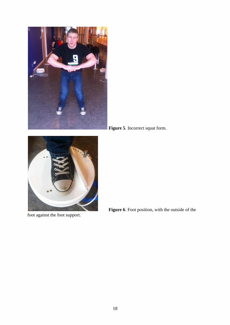
Figure 5. Incorrect squat form.
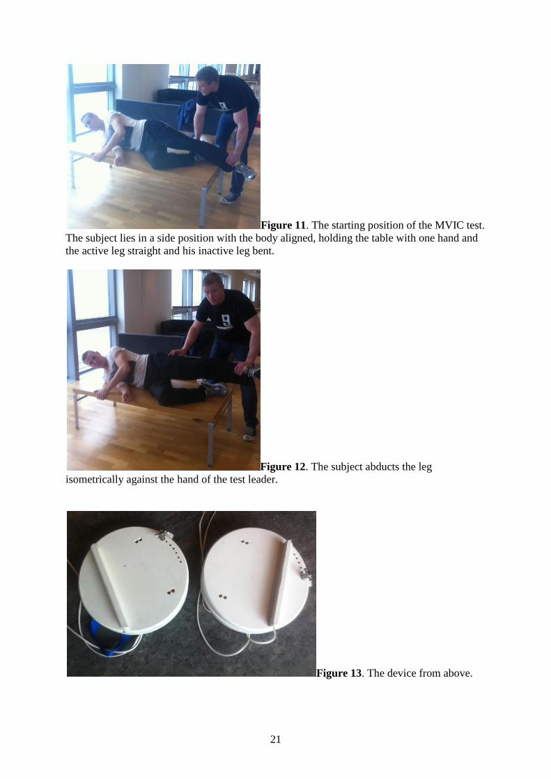
Figure 6. Foot position, with the outside of the
foot against the foot support.

19
Figure 7. Starting position on the device.
Figure 8. The subjects were instructed to push the
knees and feet out and to not rotate the feet internally to keep a correct squat form during the
whole exercise.

20
Figure 9. Correct squat form on the device. Knees
aligned with the feet.
Figure 10. Incorrect squat form on the device.
Knees not aligned with the feet.
21
Figure 11. The starting position of the MVIC test.
The subject lies in a side position with the body aligned, holding the table with one hand and
the active leg straight and his inactive leg bent.
Figure 12. The subject abducts the leg
isometrically against the hand of the test leader.
Figure 13. The device from above.

22
Figure 14. Feedback system.
Figure 15. Feedback system.


![^Z^JTT*?'WG Sti a99?ssive|y and ' Whwe quite successfully ......^Z^JTT*?'WG Sti" a99?ssive|y and ' Whwe quite successfully pSS^SSK^ to Snhfi ,h ^T06^1 af lhf,CaSes we br0U9ht recent]y](https://img.dokumen.tips/doc/110x75/6034f0f89372b042924de56f/zjttwg-sti-a99ssivey-and-whwe-quite-successfully-zjttwg-sti.jpg)