Embed Size (px)
Citation preview

TdbospwsiflWiattla
tfim
ST1i
6
First Web-Space Reconstruction by theAnterolateral Thigh Flap
Roberto Adani, MD, Luigi Tarallo, MD, Ignazio Marcoccio, MD,Umberto Fregni, MD
From the Department of Orthopaedic Surgery, University of Modena and Reggio Emilia, Modena, Italy.
Four patients with severe contracture of the first web space were treated with an anterolateralthigh perforator flap. The flap size ranged from 10 to 13 cm in length and from 7 to 8 cm inwidth. The donor site was closed directly and thinning of the flap was performed in all cases.All flaps survived and there were no re-explorations. Web space opening was maintainedover the follow-up period. There was an average postoperative increase of the angle of thefirst web space of 61°. The thinned anterolateral thigh flap provides a pliable vascularizedtissue for resurfacing the skin after release of severe contracture of the first web space andrepresents a reliable alternative to other flaps. (J Hand Surg 2006;31A:640–646. Copyright ©2006 by the American Society for Surgery of the Hand.)Key words: First web space, anterolateral thigh flap, microsurgery, perforator flaps.
TfbdbbbmbTtsltajtpsdasi
stt
he presence of an adequate first web space isessential for thumb and prehensile hand func-tion. The web contracture may be the result of
amage to the skin, muscles, tendons, nerves, orones of the first 2 rays, generally as a consequencef trauma, burn, infection, and neoplasia. If there is aevere adduction of the thumb it is necessary toerform a wide release of all contracted deep first-eb structures, which results in a skin defect on both
ides of the hand. In this situation a local flap isnadequate1 and it is necessary to perform distantap,2–5 island flap,6–13 or a free-tissue transfer.14–17
ith the recent development of perforator flaps somenvestigators have described the application of thenterolateral thigh (ALT) flap for reconstruction ofhe hand and upper extremity.18–22 In these studieshe ALT flap often was used in its entire thickness,eaving bulkiness that was unsuitable for particularreas of the hand such as the first web space.
This article describes the advantages of using thehin ALT flap for treating severe contracture of therst web space and compares this flap with com-only used techniques.
urgical Techniquehe ALT flap first was described by Song et al23 in984 as a septocutaneous flap based on the descend-
ng branch of the lateral femoral circumflex artery. b40 The Journal of Hand Surgery
he lateral circumflex femoral system originatesrom the deep femoral vessels, passing transverselyetween the rectus femoris and the vastus interme-ius muscles. The system is composed of 3 mainranches: the ascending, transverse, and descendingranches. The descending branch passes downwardetween the rectus femoris and the vastus lateralisuscles and sends several muscular and cutaneous
ranches to the anterolateral skin of the thigh.24–26
hese cutaneous perforators can be classified in sep-ocutaneous vessels, which run in the intermuscularpace between the rectus femoris and the vastusateralis muscles or the musculocutaneous perfora-ors that penetrate the vastus lateralis muscle afterrising from the descending branch.27,28 In the ma-ority of cases the flap was supplied by musculocu-aneous perforators; septocutaneous perforators sup-lied the flap in a small percentage of cases.28–30 Auitable skin vessel usually is present; during 672issections Wei et al28 reported only 6 cases of anbsence of adequate musculocutaneous perforators oreptocutaneous vessels, most of which presented dur-ng the early part of the learning curve.
A line is drawn between the anterior superior iliacpine and the superolateral border of the patella onhe donor thigh with the patient in the supine posi-ion.24,26 This line represents the muscular septum
etween the rectus femoris and the vastus lateralis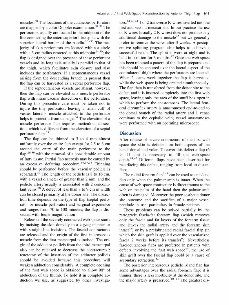
maplsjwflvtiat
tflDivhmtp
uafloasswptctras
bwamgatswoad
tfioapeshhtcWwTdswotcw
DAsh�drfl
flcwosp
roaiwffdss
st
Adani et al / First Web-Space Reconstruction by Anterior Thigh Flap 641
uscles.30 The locations of the cutaneous perforatorsre mapped by a color Doppler examination.31,32 Theerforators usually are located in the midpoint of theine connecting the anterosuperior iliac spine with theuperior lateral border of the patella.30–33 The ma-ority of skin perforators are located within a circleith a 3-cm radius centered at this midpoint24,30; theap is designed over the presence of these perforatoressels and its long axis usually is parallel to that ofhe thigh, which facilitates skin closure and alsoncludes the perforators. If a septocutaneous vesselrising from the descending branch is present thenhe flap can be harvested as a septal perforator flap.
If the septocutaneous vessels are absent, however,hen the flap can be elevated as a muscle perforatorap with intramuscular dissection of the perforators.uring this procedure care must be taken not to
njure the tiny perforator; leaving a small cuff ofastus lateralis muscle attached to the perforatorelps to protect it from damage.34 The elevation of auscle perforator flap requires meticulous dissec-
ion, which is different from the elevation of a septalerforator flap.35
The flap can be thinned to 3 to 4 mm almostniformly over the entire flap except for 2.5 to 3 cmround the entry of the main perforator to theap,26,36 with the removal of a considerable amountf fatty tissue. Partial flap necrosis may be caused byn excessive defatting procedure.34,37,38 Thinninghould be performed before the vascular pedicle iseparated.39 The length of the pedicle is 8 to 16 cm,ith a vessel diameter of greater than 2 mm, and theedicle artery usually is associated with 2 concomi-ant veins.28 A defect of less than 8 to 9 cm in widthan be closed primarily at the donor site. The dissec-ion time depends on the type of flap (septal perfo-ator or muscle perforator) and surgical experiencend ranges from 70 to 100 minutes; the flap is dis-ected with loupe magnification
Release of the severely contracted web space startsy incising the skin dorsally in a zigzag manner orith straight-line incisions. The fascial contractures
re released and the origin of the first interosseoususcle from the first metacarpal is incised. The ori-
in of the adductor pollicis from the third metacarpallso can be released to decrease the contractures1;enotomy of the insertion of the adductor pollicishould be avoided because this procedure willeaken adduction considerably. A complete openingf the first web space is obtained to allow 90° ofbduction of the thumb. To hold it in complete ab-
uction we use, as suggested by other investiga- tors,14,40,41 1 or 2 transverse K-wires inserted into therst and second metacarpals. In our practice the usef K-wires (usually 2 K-wires) does not produce anydditional damage to the muscle42 but we generallyrefer to remove the wires after 3 weeks. A postop-rative splinting program also helps to achieve auccessful result. The splint is worn at night and iseld in position for 3 months.42 Once the web spaceas been released a pattern of the flap is prepared andhis should be centered over the lateral aspect of theontralateral thigh where the perforators are located.hen 2 teams work together the flap is harvestedhile the web space is being created simultaneously.he flap then is transferred from the donor site to theefect and it is inserted completely into the first webpace, leaving only the area of the snuff box open inhich to perform the anastomoses. The lateral fem-ral circumflex artery is anastomosed end-to-end tohe dorsal branch of the radial artery and 1 venaeomitans to the cephalic vein; vessel anastomosesere performed with an operating microscope.
iscussionfter release of severe contracture of the first web
pace the skin is deficient on both aspects of theand: dorsal and volar. To cover this defect a flap (6
11 cm) is necessary to fill the web-spaceepth.14,42 Different flaps have been described foresurfacing this defect, ranging from local to distantaps.The radial forearm flap6–8 can be used as an island
ap only when the palmar arch is intact. When theause of web space contracture is direct trauma to theeb or the palm of the hand then the palmar archften is damaged. Moreover the poor aesthetic donorite outcome and the sacrifice of a major vesselreclude its use, particulary in female patients.These problems can be solved partially by the
etrograde fascia-fat forearm flap (which removesnly the fascia and fat layers of the forearm tissuend leaves the radial artery and the forearm skinntact43) or by a prefabricated radial fascial flap (inhich the skin graft is applied over the vascularized
ascia 2 weeks before its transfer9). Neverthelessasciocutaneous flaps are preferred in patients withefects involving the first web space44; the use ofkin graft over the fascial flap could be a cause ofecondary retraction.42
The posterior interosseous pedicle island flap hasome advantages over the radial forearm flap: it ishinner, there is less morbidity at the donor site, and
he major artery is preserved.10–13 The greatest dis-
atopa
aappbt9el
eflgs
fsriossfaoa
setstupt
w1snRttiftstpi
rmawowdfiwa
spelslaerr
ab
642 The Journal of Hand Surgery / Vol. 31A No. 4 April 2006
dvantages reside in the limited sizes available forhe flap: closure of the donor site can be achievednly if it is less than 3 to 4 cm wide10,11,45; otherwiseoor aesthetic results are observed when the donorrea is skin grafted.46
When the defect is large it is necessary to performdistant flap or a free-tissue transfer. The abdomen2
nd the groin3 have been used in the past for thisurpose with poor cosmetic results. The lateral armedicle flap4,5 is a cross-arm flap. The skin over theiceps muscle is soft, hairless, and thin. Furthermorehe released web space can be maintained easily in0° abduction while the flap remains attached; how-ver, this technique requires at least 2 surgical stages,eaving the hand in an unnatural position.
A variety of free flaps have been used with differ-nt functional and cosmetic results: the parascapularap47 is too bulky and the dorsalis pedis providesood skin quality but creates unsatisfactory donor-ite defects.17,48
The lateral arm free flap has been used in the pastor releasing severe scar contractures of the first webpace.14 It ensures a good cosmetic and functionaleconstruction of the first web space without produc-ng severe damage to the donor site. Disadvantagesf this technique include the bulkiness of the flap, thehortness of the vascular pedicle, the need to use akin graft if a flap wider than 6 cm is taken, and theact that harvesting the lateral arm flap results in anrea of numbness below the elbow.14,49,50 The designf the flap over the condylar and the proximal fore-rm area offers a partial solution to these problems.51
The ALT flap has become the standard flap foroft-tissue reconstruction of the upper and lowerxtremities and the trunk28,31,33,35,37 but currentlyhe ALT flap has received little attention among handurgeons18 and only a few reports have focused onhis procedure in hand reconstruction.19–22 We havesed the ALT flap in 4 cases characterized by com-lete adduction contracture of the first web space of
Table 1. Patient Characteristics
Patient Age, y Site Cause
PreoperativFirst Web
Space Ang
1 35 R Burn 20°2 25 L Burn 15°3 34 R Explosives 18°4 38 L Crush 20°
SPF, septal perforator flap; MPF, muscle perforator flap.
he hand, with a mean preoperative angle of the first a
eb space measured at 18° (range, 15°–20°) (Table). In these circumstances the release of the first webtructure creates a defect so wide and deep that it isot possible to cover it with the common local flaps.econstruction requires well-vascularized, supple,
hin tissue that will not contract after the postopera-ive period and donor site morbidity should be min-mized, especially in patients with burn defects, whorequently have multiple sites of injury22 (eg, pa-ients 1, 2) (Fig. 1). The ALT flap permits recon-truction of a normal web space that is correct ana-omically in depth and width and this flap inarticular matches the skin of the dorsum of the handn texture and thickness.
All flaps in our patients survived and no re-explo-ations were needed; the web space opening wasaintained over the follow-up period. The preoper-
tive and postoperative angle of the first web spaceas measured with the patient’s hand positioned flatn a table with the thumb in maximum abductionithout any passive thumb stretch and then the in-ex–thumb angle was followed, outlining it with ane pen.52 The mean postoperative angle of the firsteb space measured 79° (range, 75°–85°), with an
verage postoperative increase of 61° (Table 1).Compared with other flaps the ALT flap offers
ome advantages: simultaneous flap elevation andreparation are possible, total surgical time is short-ned, and a vascular pedicle about 10 cm long and aarge skin paddle can be obtained even when only aingle cutaneous perforator is available.24,33,34 Theength and size of the pedicle are adapted to the sizend location of the recipient vessels; this allows annd-to-end anastomosis to the dorsal branch of theadial artery or an end-to-side anastomosis to theadial artery to be performed.
The reconstruction of the first web space requiresflap that allows for good function and early reha-
ilitation of the underlying structures.The ALT flap can be thinned to a thickness of
Flap Size,cm
Type ofFlap
Follow-UpPeriod,
mo
PostoperativeFirst Web
Space Angle
10 � 7 SPF 54 85°13 � 7 SPF 42 80°12 � 8 MPF 27 75°12 � 7 MPF 9 78°
e
le
pproximately 3 to 4 mm with removal of a con-

Fp(
Adani et al / First Web-Space Reconstruction by Anterior Thigh Flap 643
igure 1. Patient 1. (A) Preoperative view of a patient with severe adduction contracture caused by a burn. (B) A free ALT septalerforator flap by the descending branch of the lateral circumflex femoral vessel was elevated to reconstruct the first web space.
C) The ALT flap in place. (D) Appearance of the reconstructed first web space.
sfltWobdimtftgfhaDapamponav3mptvltor
T
D
aa
tm
R
1
1
1
1
Fc ver the
644 The Journal of Hand Surgery / Vol. 31A No. 4 April 2006
iderable amount of fatty tissue. Thinning of theap was performed in all 4 cases before dividing
he pedicle without signs of marginal necrosis.ith this procedure it is possible to obtain an
ptimal match between donor tissue and the area toe reconstructed with a pliable flap (Fig. 2). Theonor site can be closed primarily when the defects smaller than 8 cm with almost no donor-siteorbidity and with its scar being less noticeable
han that of other flaps, especially those obtainedrom the arm and forearm.53 Despite its versatilityhe ALT flap has some potential drawbacks: thereatest concerns have been variability of the per-orators and flap reliability.54 Large clinical seriesave shown that almost all patients will have andequate vessel for supplying the flap28,35,55 andoppler examination can identify the perforators
ccurately before surgery,32 allowing accurate flaplanning. If compared with the other flaps (radialrtery flap, lateral arm flap) the ALT flap is de-anding surgically,22 but after an initial learning
eriod the time consumption is equivalent to thatf the posterior interosseous flap. If a septocuta-eous vessel is found the flap can be harvested asseptal perforator flap (patients 1, 2); if the skin
essel is a musculocutaneous perforator (patients, 4) then the procedure is more difficult. Intra-uscular dissection of the perforators must be
erformed carefully by using meticulous surgicalechniques to avoid injury to the vessel in theastus lateralis muscle. A sensory flap using theateral femoral cutaneous nerve also can be ob-ained18,29,33,54; however, no attempt was made inur patients to suture the nerve of the flap to aadial sensory nerve in the recipient hand.
he authors thank Mr. Silvio Tocco for his English and critical review.Received for publication April 7, 2005; accepted in revised form
ecember 7, 2005.
igure 2. Patient 3. (A) Preoperative view of a patient witosmetic restoration of the first web space. (C) Linear scar o
No benefits in any form have been received or will be received from
commercial party related directly or indirectly to the subject of thisrticle.
Corresponding author: Roberto Adani, Clinica Ortopedica e Trauma-ologica—Policlinico, Largo del Pozzo n.71, 41100 Modena, Italy; e-ail: [email protected] © 2006 by the American Society for Surgery of the Hand0363-5023/06/31A04-0020$32.00/0doi:10.1016/j.jhsa.2005.12.009
eferences1. Caroli A, Adani R, Scagni R, Marcialis M. Skin flap in first
web retraction. Tech Hand Upper Extrem Surg 1999;3:197–202.
2. Miura T. Use of paired abdominal flaps for release of ad-duction contractures of the thumb. Plast Reconstr Surg 1979;63:242–244.
3. McGregor IA, Jackson IT. The groin flap. Br J Plast Surg1972;25:3–16.
4. Mutz SB. Thumb web contracture. Hand 1972;4:236–246.5. Bonola A, Fiocchi R. Cross-arm double flap in the repair of
severe adduction contracture of the thumb. Hand 1975;7:287–290.
6. Foucher G, Van Genechten F, Merle M, Michon J. A com-pound radial artery forearm flap in hand surgery: an originalmodification of the Chinese forearm flap. Br J Plast Surg1984;37:139–148.
7. Braun FM, Hoang P, Merle M, Van Genechten F, FoucherG. Technique and indications of the forearm flap in handsurgery: report of thirty-three cases. Ann Chir Main 1985;4:85–97.
8. Akyurek M, Safak T, Kecik A. Coverage of thumb woundand correction of first web space contracture using a longi-tudinally split reverse radial forearm flap. Ann Plast Surg2001;47:453–457.
9. Prakash V, Mishra A. Management of adduction contractureof the thumb with a prefabricated radial fascial flap. PlastReconstr Surg 2004;114:1681–1682.
0. Zancolli EA, Angrigiani C. Posterior interosseous islandforearm flap. J Hand Surg 1988;13B:130–135.
1. Costa H, Soutar DS. The distally based island posteriorinterosseous flap. Br J Plast Surg 1988;41:221–227.
2. Buchler U, Frey HP. Retrograde posterior interosseous flap.J Hand Surg 1991;16A:283–292.
3. Lu LJ, Gong X, Liu ZG, Zhang ZX. Antebrachial reverseisland flap with pedicle of posterior interosseous artery: a
re adduction contracture caused by explosives. (B) GoodALT donor site.
h seve
report of 90 cases. Br J Plast Surg 2004;57:645–652.

1
1
1
1
1
1
2
2
2
2
2
2
2
2
2
2
3
3
3
3
3
3
3
3
3
3
4
4
4
4
4
4
4
4
4
Adani et al / First Web-Space Reconstruction by Anterior Thigh Flap 645
4. Scheker LR, Lister GD, Wolff TW. The lateral arm free flapin releasing severe contracture of the first web space. J HandSurg 1988;13B:146–150.
5. Schoofs M, Leps P, Millot F. [The value of the free externalbrachial flap in surgery of the hand.] Ann Chir Main 1989;8:217–222.
6. Tonkin MA, Stern H. The posterior interosseous artery freeflap. J Hand Surg 1989;14B:215–217.
7. Woo SH, Choi BC, Oh SJ, Seul JH. Classification of the firstweb space free flap of the foot and its applications inreconstruction of the hand. Plast Reconstr Surg 1999;103:508–517.
8. Javaid M, Cormack GC. Anterolateral thigh free flap forcomplex soft tissue hand reconstructions. J Hand Surg 2003;28B:21–27.
9. Koshima I, Nanba Y, Tsutsui T, Takahashi Y. New antero-lateral thigh perforator flap with a short pedicle for recon-struction of defects in the upper extremities. Ann Plast Surg2003;51:30–36.
0. Rui Y, Shou K, Zhang Q, Xu Y, Sun Z, Xu L. Combinedfree-tissue transfer for primary reconstruction of radial partof the hand. Microsurgery 2004;24:59–62.
1. Chen HC, Tang YB, Mardini S, Tsai BW. Reconstruction ofthe hand and upper limb with free flaps based on musculo-cutaneous perforators. Microsurgery 2004;24:270–280.
2. Tsai FC, Yang JY, Mardini S, Chuang SS, Wei FC. Freesplit-cutaneous perforator flaps procured using a three-di-mensional harvest technique for the reconstruction of post-burn contracture defects. Plast Reconstr Surg 2004;113:185–193.
3. Song YG, Chen GZ, Song YL. The free thigh flap: a newfree flap concept based on the septocutaneous artery. Br JPlast Surg 1984;37:149–159.
4. Xu DC, Zhong SZ, Kong JM, Wang GY, Liu MZ, Luo LS,Gao JH. Applied anatomy of the anterolateral femoral flap.Plast Reconstr Surg 1988;82:305–310.
5. Koshima I, Yamamoto H, Hosoda M, Moriguchi T, Orita Y,Nagayama H. Free combined composite flaps using thelateral circumflex femoral system for repair of massive de-fects of the head and neck regions: an introduction to thechimeric flap principle. Plast Reconstr Surg 1993;92:411–420.
6. Koshima I, Fukuda H, Yamamoto H, Moriguchi T, Soeda S,Ohta S. Free anterolateral thigh flaps for reconstruction ofhead and neck defects. Plast Reconstr Surg 1993;92:421–428.
7. Wei FC, Jain V, Suominen S, Chen HC. Confusion amongperforator flaps: what is a true perforator. Plast ReconstrSurg 2001;107:874–876.
8. Wei FC, Jain V, Celik N, Chen HC, Chuang DC, Lin CH.Have we found an ideal soft-tissue flap? An experience with672 anterolateral thigh flaps. Plast Reconstr Surg 2002;109:2219–2230.
9. Kimata Y, Uchiyama K, Ebihara S, Nakatsuka T, Harii K.Anatomic variations and technical problems of the antero-lateral thigh flap: a report of 74 cases. Plast Reconstr Surg1998;102:1517–1523.
0. Chana JS, Wei FC. A review of the advantages of theanterolateral thigh flap in head and neck reconstruction. Br JPlast Surg 2004;57:603–609.
1. Kuo YR, Jeng SF, Kuo MH, Huang MN, Liu YT, Chang
YC, et al. Free anterolateral thigh flap for extremityreconstruction: clinical experience and functionalassessment of donor site. Plast Reconstr Surg 2001;107:1766 –1771.
2. Tsukino A, Kurachi K, Inamiya T, Tanigaki T. Preoperativecolor Doppler assessment in planning of anterolateral thighflaps. Plast Reconstr Surg 2004;113:241–246.
3. Kuo YR, Jeng SF, Kuo FM, Liu YT, Lai PW. Versatility ofthe free anterolateral thigh flap for reconstruction of soft-tissue defects: review of 140 cases. Ann Plast Surg 2002;48:161–166.
4. Kimata Y, Uchiyama K, Ebihara S, Yoshizumi T, Asai M,Saikawa M, et al. Versatility of the free anterolateral thighflap for reconstruction of head and neck defects. Arch Oto-laryngol Head Neck Surg 1997;123:1325–1331.
5. Celik N, Wei FC, Lin CH, Cheng MH, Chen HC, Jeng SF,Kuo YR. Technique and strategy in anterolateral thigh per-forator flap surgery, based on an analysis of 15 complete andpartial failures in 439 cases. Plast Reconstr Surg 2002;109:2211–2218.
6. Kimura N, Satoh K. Consideration of a thin flap as an entityand clinical applications of the thin anterolateral thigh flap.Plast Reconstr Surg 1996;97:985–992.
7. Alkureishi LWT, Shaw-Dunn J, Ross GL. Effect of thinningthe anterolateral thigh flap on the blood supply to the skin.Br J Plast Surg 2003;56:401–408.
8. Ross GL, Dunn R, Kirkpatrick J, Koshy CE, AlkureishiLWT, Bennett N, et al. To thin or not to thin: the use of theanterolateral thigh flap in the reconstruction of intraoraldefects. Br J Plast Surg 2003;56:409–413.
9. Rajacic N, Gang RK, Krishnan J, Lal Bang R. Thin antero-lateral thigh free flap. Ann Plast Surg 2002;48:252–257.
0. Caroli A, Zanasi S. First web-space reconstruction by Caro-li’s technique in congenital hand deformities with severethumb ray adduction. Br J Plast Surg 1989;42:653–659.
1. Upton J, Havlik RJ, Coombs CJ. Use of forearm flaps for theseverely contracted first web space in children with congen-ital malformation. J Hand Surg 1996;21A:470–477.
2. Del Pinal F, Garcia-Bernal FJ, Delgado J. Is posttraumaticfirst web contracture avoidable? Prophylactic guidelines andtreatment-oriented classification. Plast Reconstr Surg 2004;113:1855–1860.
3. Weinzweig N, Chen L, Chen ZW. The distally based radialforearm fasciosubcutaneous flap with preservation of theradial artery: an anatomic and clinical approach. Plast Re-constr Surg 1994;94:675–684.
4. Kostakoglu N, Kecik A. Upper limb reconstruction withreverse flaps: a review of 52 patients with emphasis on flapselection. Ann Plast Surg 1997;39:381–389.
5. Brunelli F, Valenti P, Dumontier C, Panciera P, Gilbert A.The posterior interosseous reverse flap: experience with 113flaps. Ann Plast Surg 2001;47:25–30.
6. Angrigiani C, Grilli D, Dominikow D, Zancolli EA. Poste-rior interosseous reverse forearm flap: experience with 80consecutive cases. Plast Reconstr Surg 1993;92:285–293.
7. Nassif TM, Vidal L, Bovet JL, Baudet J. The parascapularflap: a new cutaneous microsurgical free flap. Plast ReconstrSurg 1982;69:591–600.
8. Ohmori K, Harii K. Free dorsalis pedis sensory flap to thehand, with microneurovascular anastomoses. Plast Reconstr
Surg 1976;58:546–554.
4
5
5
5
5
5
5
646 The Journal of Hand Surgery / Vol. 31A No. 4 April 2006
9. Graham B, Adkins P, Scheker LR. Complications and mor-bidity of the donor and recipient sites in 123 lateral armflaps. J Hand Surg 1992;17B:189–192.
0. Moffett TR, Madison SA, Derr JW Jr, Acland RD. Anextended approach for the vascular pedicle of the lateral armfree flap. Plast Reconstr Surg 1992;89:259–267.
1. Hamdi M, Coessens BC. Evaluation of the donor site mor-bidity after lateral arm flap with skin paddle extending overthe elbow joint. Br J Plast Surg 2000;53:215–219.
2. Jensen CB, Rayan GM, Davidson R. First web space con-
tracture and hand function. J Hand Surg 1993;18A:516–520.3. Huang CH, Chen HC, Huang YL, Mardini S, Feng GM.Comparison of the radial forearm flap and the thinned an-terolateral thigh cutaneous flap for reconstruction of tonguedefects: an evaluation of donor-site morbidity. Plast Recon-str Surg 2004;114:1704–1710.
4. Wang HT, Erdmann D, Fletcher JW, Levin LS. Anterolateralthigh flap technique in hand and upper extremity reconstruc-tion. Tech Hand Upper Extrem Surg 2004;8:257–261.
5. Luo S, Raffoul W, Luo J, Luo L, Gao J, Chen L, Egloff DV.Anterolateral thigh flap: a review of 168 cases. Microsurgery
1999;19:232–238.